Review

Oxygen and ROS in Photosynthesis

Department of Biochemistry/Molecular Plant Biology, University of Turku, FI-20014 Turku, Finland
*
Author to whom correspondence should be addressed.
Plants 2020, 9(1), 91; https://doi.org/10.3390/plants9010091
Submission received: 27 November 2019 / Revised: 29 December 2019 / Accepted: 2 January 2020 / Published: 10 January 2020
(This article belongs to the Special Issue ROS Responses in Plants)

Abstract

:
Oxygen is a natural acceptor of electrons in the respiratory pathway of aerobic organisms and in many other biochemical reactions. Aerobic metabolism is always associated with the formation of reactive oxygen species (ROS). ROS may damage biomolecules but are also involved in regulatory functions of photosynthetic organisms. This review presents the main properties of ROS, the formation of ROS in the photosynthetic electron transport chain and in the stroma of chloroplasts, and ROS scavenging systems of thylakoid membrane and stroma. Effects of ROS on the photosynthetic apparatus and their roles in redox signaling are discussed.

1. Introduction

Molecular oxygen (O2) is a natural acceptor of electrons in the respiratory pathway of aerobic organisms and in many other biochemical reactions. In its ground state, O2 has two unpaired electrons with parallel spins in two separate π antibonding orbitals. Thus, ground-state O2 is a triplet diradical. According to Pauli’s principle, O2 reacts slowly with many biomolecules because spin restriction causes a kinetic barrier, as almost all biomolecules are in the singlet state, having paired electrons with opposite spins. The kinetic barrier of O2 is removed either by spin inversion of one unpaired electron or by a partial reduction in O2. Spin inversion requires the absorption of energy and converts the triplet state of O2 to the singlet state of molecular oxygen (1O2). All other forms of active oxygen are produced via an electron transfer mechanism. 1O2 and partially reduced forms of oxygen have a higher reactivity towards many organic molecules than the ground state of oxygen and they are collectively called reactive oxygen species (ROS).
ROS can be classified as radical and non-radical species. In addition, ROS can be roughly divided into free ROS, small molecules composed of oxygen and hydrogen only, and incorporated ROS, in which oxygen is bound to other molecules to form reactive oxygen derivatives. Furthermore, a family of reactive species containing nitrogen moieties associated with oxygen are classified as reactive nitrogen species, and reactive oxygen derivatives like lipid peroxyl radicals (LOO) can be classified as both ROS and reactive lipid species. The free ROS are 1O2, superoxide anion radical (O2•−), hydroperoxyl radical (HO2), hydrogen peroxide (H2O2), hydroxyl radical (HO) and ozone (O3). Other molecules containing active oxygen are ROS derivatives. Table 1 presents the most important reactive species containing active oxygen according to this classification.
Chloroplasts convert light energy to energy for chemical bonds. Light absorption by the chlorophyll (Chl) molecules of Photosystems I and II (PSI and PSII) triggers a sequence of redox reactions along the thylakoid membrane. These reactions result in the oxidation of water, reduction of NADP+ to NADPH and formation of a proton gradient across the thylakoid membrane. In chloroplasts, O2 appears due to light-dependent water-splitting catalyzed by PSII. The steady-state concentration of O2 inside intact chloroplasts in the light depends on the external concentration of O2. At low external O2 concentrations (30 µM), the ratio of the internal to the external is about five, whereas at concentrations corresponding to those in air-saturated water (≈258 µM), the O2 concentration of isolated chloroplasts is similar to that of the medium [1].
1O2 is mainly formed via the interaction of O2 with the triplet excited state of chlorophyll (3Chl). The reduction in O2 by reduced forms of electron carriers in the photosynthetic electron transfer chain (PETC) can produce O2•− and H2O2. The reaction of O2 with PETC is considered as the main source of ROS in chloroplasts, and light is essential for this ROS production. ROS can be interconverted by interaction with redox active compounds of the chloroplast, and ROS are produced both via O2 mediated primary ROS generation mechanisms and ROS-mediated interconversion reactions.
ROS are unfavorable for chloroplast functions because ROS cause oxidative damage by reacting with biomolecules. Chloroplasts have ROS scavenging systems to prevent damage [2,3,4,5]. In addition to ROS-scavenging systems, chloroplasts have pathways, like non-photochemical quenching of excitation energy (NPQ), cyclic electron flow and plastid terminal oxidase (PTOX)-mediated chlororespiration, that diminish the appearance of long-lived redox active compounds [6,7,8]. All aerobic organisms have ROS-scavenging mechanisms to prevent ROS damage. Environmental stressors (high light, high or low temperature and others) enhance ROS production in chloroplasts and change the balance between ROS scavenging and ROS production. Imbalances between ROS production and scavenging cause changes in the redox state of the cell through a change in the levels of reduced and oxidized forms of antioxidants like ascorbate (AscH2), glutathione and thiol-containing compounds. Changes in the redox levels of the antioxidants trigger reactions where antioxidants act as ROS processing and signaling mediators that cause changes in gene expression [9,10]. In particular, the formation of ROS in Mehler’s reaction initiates light-signaling that depends on AscH2 and reduced glutathione (GSH) [11].
ROS production quenches excitation energy photochemically via the so-called water–water cycle that consists of the production of O2 in PSII, reduction in O2 to O2•− and H2O2, scavenging of ROS, and regeneration of the scavenger [12]. The primary function of the water–water cycle is to scavenge ROS. Besides ROS scavenging and the photochemical quenching of excitation energy, the water–water cycle generates a proton gradient across the thylakoid membrane for both ATP production and the enhancement of NPQ [13].
Aerobic metabolism inevitably leads to the formation of ROS, and chloroplasts have to spend metabolic energy to build and maintain the ROS metabolism. The chloroplast provides light sensor functions for the whole plant cell, and the chloroplast ROS network participate in this signaling.
In the present review, we will mainly discuss the roles of 1O2, O2•−, HO2, H2O2, HO and lipid peroxides in chloroplast metabolism. O3, although it is biologically important, is only shortly discussed because this ROS is not found within cells. The review presents the main properties of ROS and their typical reactions, the formation of ROS in the photosynthetic electron transport chain and in the stroma of chloroplasts, and the ROS-scavenging systems of thylakoid membrane and stroma.

2. ROS Properties and Basic Reactions

2.1. Singlet Oxygen, 1O2

Ground-state O2 is a triplet (3Σ+gO2) and can be converted to the singlet form via the absorption of energy that leads to spin inversion of one unpaired electron. Molecular oxygen has two singlet forms because two electrons with antiparallel spins may reside either in two different orbitals (1Σ+gO2) or both in one orbital (1∆gO2). The energy above the ground-state of O2 is 155 and 92 kJ/mol for 1Σ+gO2 and 1∆gO2, respectively [14]. 1Σ+gO2 is rapidly converted to 1∆gO2 or 3Σ+gO2 and, in the liquid phase, the lifetime of 1Σ+gO2 is only 10−11 s [14], which is too short for 1Σ+gO2 to take part in biochemical reactions. Therefore, 1O2 will be used here to designate 1∆gO2. O2 will be used to designate 3Σ+gO2.

2.1.1. Formation of 1O2

The most common mechanism of 1O2 generation is photosensitization, i.e., the reaction of O2 with a photoexcited sensitizer dye (S*). Both forms of 1O2, 1Σ+gO2 and 1∆gO2, can be produced via the spin-conserved Reactions (1) and (2).
1S* + O23S + 1O2
3S + O21S + 1O2
The second reaction is more common because singlet excited states (1S*) are usually short-lived and because only a few dye molecules have a large enough energy gap between the 1S* and triplet states (3S) to convert O2 to 1O2 [15]. 3Chl reacts rapidly with O2 with a second-order rate constant close to 109 M−1 s−1, and the relative quantum yield of 1O2 generation by chlorophyll a (Chl a) was around 80% when meso-tetraphenylporphyrin and tetra(p-sulfophenyl) porphyrin were used as standards [16]. The spin transition O21Σ+gO2 is associated with the absorption band of gaseous O2 at 762 nm. Absorption at 1268 nm, in turn, was found for the transition O21∆gO2 in liquids and in the atmosphere [17].
In addition to photosensitized generation, 1O2 can be produced by several chemical reactions that usually involve reduced forms of oxygen like H2O2, O2•− and reactive oxygen derivatives like organic peroxides (ROOH) and peroxyl radicals (ROO) [14,18].
1O2 is produced by decomposition of H2O2 via the Haber–Weiss mechanism (Reactions (3) and (4)) [19,20].
H2O2 + O2•− → HO + HO + (O2 or 1O2)
H2O2 + HO2 → HO + H2O + (O2 or 1O2)
The rate constants of Reactions (3) and (4) in aqueous medium are 0.13 to 0.23 M−1 s−1 and 0.5 M−1 s−1, respectively [21,22,23]. It has been suggested that the biologically important oxidant produced by Reaction (3) is HO rather than 1O2 [24]. 1O2 can also be produced by O2•− dismutation (Reaction (5)) or by electron transfer from O2•− to radical (A) or non-radical (A) electron acceptors (Reactions (6) and (7)) [25,26,27].
O2•− + O2•− + 2H+ → H2O2 + (O2 or 1O2)
O2•− + A+ → (O2 or 1O2) + A or O2•− + A•+ → (O2 or 1O2) + A
O2•− + HO → HO + 1O2
Efficient production of 1O2 was found in a reaction of O2•− with benzoyl peroxide or lauroyl peroxide (Reaction (8)) [28].
2O2•− + RCOOCR → 2RCO2 +21O2
1O2 can be generated via the Russel mechanism (Reaction (9)), in which two ROO radicals react to form a linear tetraoxide (ROOOOR) intermediate that rapidly decomposes to the corresponding ketone (R=O), alcohol (R–OH) and 1O2 [29]. In fact, ROOOOR decomposition releases either 1O2 or excited triplet carbonyl (R=3O*). However, the relative yield of 1O2 is 10%, while the relative yield of R=3O* is only 0.01% [30,31,32].
ROO + ROO ∆ ROOOOR → R–OH + R=O + 1O2
1O2 can be produced in the reaction of O2 with an R=3O* (Reaction (10)), which can be formed by the decomposition of ROOOOR (Reaction (9)) [33].
R=3O* + O2 → R=O + 1O2
Other specific ions, like the hypohalite ion OCl and the molybdate ion MoO42−, can react with H2O2, forming 1O2 (Reactions (11)–(13)) [34,35,36]; these reactions are probably not of biological importance.
H2O2 + H+ + OCl → H2O + HCl + 1O2
MoO42− + H2O2 ∆ MoO62− + 2H2O
MoO62− → MoO42− + 1O2

2.1.2. Physical Deactivation of 1O2

Both excited singlet states of oxygen are metastable and can lose excitation energy via radiative and non-radiative pathways. The latter is physical quenching of 1O2. The radiative deactivation is the transition of 1∆gO2 to O2 associated with light emission (hν; Reaction (14)).
1O2 → O2 + hν
The phosphorescence spectrum has a major maximum at 1268 nm [37]. The phosphorescence is extremely weak, as the deactivation of 1O2 mostly proceeds non-radiatively due to the collision of 1O2 with another molecule. The quantum yield of luminescence is from 10−6 to 10−3 [32]. Non-radiative deactivation mechanisms include electronical-to-vibrational energy transfer, charge-transfer-induced quenching and electronic energy transfer. In the deactivation of 1Σ+gO2 and 1∆gO2 by an electronic-to-vibrational process, the excitation energy of 1O2 is converted into vibration of the O2 molecule and a quencher molecule Qr (Reaction (15)).
1O2 (v = 0) + Qr (v = 0)→ O2 (v = m) + Qr (v = n) + Emn
where Emn is the energy difference between the reactants and the products and v is the vibrational energy level of a molecule; m and n are vibrational modes.
The deactivation of 1O2 by collisions of 1O2 with other molecules limits the lifetime of 1O2 in many solvents. The lifetime of 1O2 for many organic solvents is within 8–100 µs. The substitution of hydrogen with deuterium in the solvent molecule leads to a significant increase in the lifetime of 1O2, usually by a factor of ten or more [17,38,39,40]. The second order rate constant for the deactivation of 1O2 via an electronic-to-vibrational process varies widely, from 10−2 to 106 M−1 s−1 [17].
In addition to the electronic–vibrational non-radiative deactivation, 1O2 can be deactivated via charge-transfer-induced quenching (Reaction (16)) and an electronic energy transfer mechanism (Reaction (17)).
1O2 + 1A ∆ 1(O2 A) ∆ 3(O2 A) → O2 + 1A
1O2 + A → 3(O2 A) → O2 + 3A,
where A is an acceptor.
Molecules with high triplet energies (more than 94 kJ mol−1) and low oxidation potential (midpoint redox potential (Em) around 1.9 V vs. Normal Hydrogen Electrode (NHE)) can deactivate 1O2 with the charge-transfer mechanism. Second-order rate constants for deactivation of 1O2 via the charge-transfer mechanism are within 103 to 109 M−1 s−1 [17]. In the charge-transfer mechanism, the 1(O2 A) complex finally dissociates to A and O2 without charge separation. Electronic energy-transfer quenching of 1O2 occurs via the interaction of molecules with a lower triplet state energy than the energy of 1O2. The deactivation of 1O2 via the electronic energy-transfer mechanism is very efficient and its second-order rate constant is close to the diffusion-controlled limit. Carotenoids including β-carotene and lutein are the most efficient quenchers of 1O2, and the second order rate constant for many carotenoids is about 1010 M−1 s−1 [17,41].

2.1.3. Chemical Reactions of 1O2

The term “chemical deactivation” of 1O2 can be applied to reactions in which the products have less reactivity and toxicity towards cell metabolism than 1O2. The redox potential relative to NHE for the pair 1O2/O2•− is 0.65 V [42]. 1O2 is an electrophilic agent and reacts with electron-rich organic molecules via three well-known mechanisms.
The ene reaction (Alder-ene reaction) is associated with the formation of a hydroperoxide (Reaction (18)).
Plants 09 00091 i001
Cycloaddition is associated with dioxetane formation (Reaction (19)).
Plants 09 00091 i002
Cycloaddition is associated with aromatic compounds and formation of an endoperoxide via the Diels–Alder mechanism (Reaction (20)).
Plants 09 00091 i003
1O2 can react with the unsaturated fatty acids of membrane lipids to form both conjugated and non-conjugated diene hydroperoxides (Reaction (21)) with a second order rate constant of about 104 M−1 s−1 [4,43].
Plants 09 00091 i004
1O2 can react with amino acids that have double bonds or an electron-rich sulfur atom, such as tryptophan, histidine, tyrosine, methionine and cysteine, to form corresponding peroxides. The second-order rate constant for reaction of 1O2 with those amino acids is around 107 M−1 s−1 [44]
1O2 can efficiently oxidize amines to imines with the formation of HO2 (Reaction (22)) [45].
Plants 09 00091 i005
1O2 can oxidize electron-rich compounds like phenols to benzoquinones by an electron transfer mechanism (Reaction (23)) [46].
Plants 09 00091 i006
The oxidation of AscH2 (Reaction (24)) and plastoquinol (PQH2) (Reaction (25)) by 1O2 can proceed as a two-electron reduction of 1O2 to H2O2 [47,48].
AscH2 + 1O2 → DHA + H2O2
PQH2 + 1O2 → PQ + H2O2
where DHA and PQ are dehydroascorbate and plastoquinone, respectively.
The second-order rate constant for the reaction of 1O2 with AscH2 depends on pH and varies from 105 M−1 s−1 to 108 M−1 s−1 [49]. Prenyllipids like PQH2-9 and α-tocopherol react with 1O2 with second-order rate constants of about 108 M−1 s−1 [50].

2.1.4. Lifetime and Diffusion Distance of 1O2

Solvents and other deactivating compounds play significant roles in controlling the lifetime of 1O2 and the lifetime, in turn, determines both the diffusion distance and the ability of 1O2 to react with other substances. The lifetimes of 1O2 in a pure lipid membrane and in the thylakoid membrane have been estimated to be 7 µs and 70 ns, respectively [51]. Thus, the respective diffusion distances, approximated using the diffusion coefficient of O2, are 220 and 5.5 nm. The very short lifetime in the thylakoid membrane may be caused by a high concentration of compounds deactivating 1O2. In a nerve cell, the lifetime of 1O2 is about 200 ns, which leads to a diffusion distance of about 270 nm [52]. The lifetimes and diffusion distances of 1O2 in different tissues have been recently reviewed [23].

2.2. Superoxide Anion Radical, O2•−

The O2•− is the one-electron reduced form of molecular oxygen. Detailed analyses of the properties and biological roles of O2•− are available in several comprehensive reviews [53,54,55,56]. O2•− is a deprotonation agent and can be protonated to HO2 (Reaction (26)).
O2•− + H+ → HO2
The deprotonation ability of O2•− depends strongly on the solvent. In aqueous solutions, O2•− is a weak deprotonation agent with pKa of 4.8 due to strong solvation of O2•−. The free energy of hydration for O2•− was estimated to be around 355 kJ mol−1 [57]. Therefore, only 0.25% of O2•− is protonated at physiological pH values. In non-aqueous solutions like organic solvents, O2•− is a strong deprotonation agent. For example, in dimethylformamide (DMF), the pKa value of HO2 is around 12 [54]. However, in reality the protonation of O2•− in the presence of a protonated compound AH can accompany the reduction of HO2 to hydroperoxyl anion (HO2) and occur via a two-step mechanism (Reactions (27) and (28)), which makes O2•− a much stronger deprotonation agent than would follow from its basicity.
O2•− + AH ∆ HO2 + A
O2•− + HO2 → O2 + HO2
which sum up to
2O2•− + AH ∆ O2 + HO2 + A.
The equilibrium constant of Reaction (29) is about 109 [54]. Therefore, in the deprotonation process the pKa value of O2•− should be considered equivalent to a base with pKa 24 [53,54]. The Em of the pair O2/O2•− is pH-dependent, due to protonation of O2•− and formation of HO2. In an aqueous solution at pH 7, the Em of the pair O2/O2•− is −160–−180 mV vs. NHE, and the Em value becomes more positive under a low pH, around 100 mV [12,58]. In aprotic media offering only a weak solvation of O2•−, O2•− acts as a strong reductant and the redox potential of the pair O2/O2•− is estimated to range between −550 and −600 mV vs. NHE [54] in DMF and around −640 mV in acetonitrile [59,60].

2.2.1. Formation of O2•−

O2•− is mainly formed via the interaction of O2 with reduced compounds having a low redox potential (A) (Reaction (30)).
A + O2 ∆ A + O2•−
O2•− can be formed in a potentially important equilibrium reaction with semiquinone anion radicals (Q•−) with the formation of the respective quinone Q (Reaction (31)).
Q•− + O2 ∆ Q + O2•−
The equilibrium constant of Reaction (31) can be determined from the redox potentials of Q/Q•− and O2/O2•−. In aqueous solutions at pH 7, the equilibrium constant for Reaction (31) is estimated as 2 × 10⁻⁵ for benzosemiquinone with a redox potential around 100 mV, and 26 for durosemiquinone with redox potential around −260 mV [61]. The formation of O2•− via Reaction (31) is favorable for Q•−, with the redox potential of the Q/Q•− pair lower than 160–−180 mV because, with this redox potential, the forward rate constant (kforward) of Reaction (31) is larger than the reverse rate constant (kreverse). Reaction (31) when Q•− is benzosemiquinone proceeds with kforward of 5 × 10⁵ M−1 s−1 and with kreverse of 108 M−1 s−1. For durosemiquinone, kforward and kreverse were estimated to be 2.2 × 10⁸ M−1 s−1 and 107 M−1 s−1, respectively [61]. However, if O2•− is efficiently removed after Reaction (31), then the rate of formation depends only on kforward.

2.2.2. Reactions of O2•−

Reactions of O2•− with organic and inorganic molecules can proceed in five ways: deprotonation reaction (protonation of O2•− by H+ or the attraction of a proton from a proton donor), attraction of hydrogen, electron transfer reaction, nucleophilic substitution or addition, and addition to a metal or metal complex.
In electron transfer reactions, O2•− can act as both an oxidant and a reductant. In many cases, the electron transfer reaction involves a deprotonation reaction (deprotonation–oxidation mechanism). O2•− can reduce or oxidize organic and inorganic molecules like quinones and cytochromes (cyt) or transition metal ions in equilibrium reactions via a one-electron transfer mechanism, Reactions (31)–(33).
O2•− + cyt (Fe3+) ∆ O2 + cyt (Fe2+)
O2•− + Fe3+ ∆ O2 + Fe2+
In many cases, O2•− oxidizes organic and inorganic molecules by hydrogen attraction via the deprotonation–oxidation mechanism. Oxidation of AscH2 by O2•− in aqueous solution (Reaction (34)) proceeds as a two-step reaction (Reactions (35) and (36)) with a second-order rate constant of 3.3 × 10⁵ M−1 s−1 at pH 7.8 [62,63,64].
O2•− + AscH2 → H2O2 + Asc
O2•− + AscH2 ∆ HO2 + AscH
HO2 + AscH → H2O2 + Asc•−
The same mechanism is suggested for oxidation by O2•− of thiols as in reduced GSH and lipophilic compounds as in α-tocopherol, Reactions (37)–(40), respectively [64,65].
O2•− + GSH ∆ + HO2 + GS
HO2 + GS ∆ + HO2 + GS
O2•− + α-Tocopherol-H ∆ HO2 + α-Tocopherol
HO2 + α-Tocopherol ∆ HO2 + α-Tocopherol
In Reactions (37) and (38), GS and GS stand for deprotonated reduced glutathione and oxidized glutathione, respectively.
The second-order rate constant of the reaction of O2•− with α-tocopherol incorporated into soybean or dimyristoyl phosphatidylcholine liposomal membranes was estimated to be 4.9 × 10³ M−1 s−1 [62]. The second-order rate constant of the reaction of O2•− with GSH was estimated to be 103 M−1 s−1 [66,67].
The main mechanism of deactivation of O2•− is spontaneous or enzymatic dismutation (Reaction (5)). Non-enzymatic dismutation of O2•− usually proceeds in aqueous solution and very strongly depends on pH because the protonation of O2•− determines the rate. Dismutation can be considered as a two-step reaction: protonation of O2•−, Reaction (41) and a radical–radical reaction between O2•− and HO2 or between two molecules of HO2—Reactions (42) and (43), respectively.
O2•− + H+ ∆ HO2, pKa = 4.8
O2•− + HO2 + H+→ H2O2 + O2
HO2 + HO2 → H2O2 + O2
The second-order rate constant of the dismutation of O2•− has a maximum (108 M−1 s−1) at pH 4.8, equal to the pKa value of HO2. The rate constant decreases with increasing pH and becomes very low, around 0.3 M−1 s−1, at alkaline pH. At physiological pH, the rate constant is about 105 M−1 s−1 [68]. The enzymatic dismutation of O2•− is catalyzed by superoxide dismutase (SOD, EC 1.15.1.1). The SOD-catalyzed reaction proceeds as a sequence of oxidation and reduction of O2•− by a metal ion (M) of the SOD enzyme, Reactions (44) and (45).
M(n+1)+-SOD + O2•− → Mn+-SOD + O2
Mn+-SOD + O2•− + 2H+ → M(n+1)+-SOD + H2O2
The rate constant of O2•− dismutation catalyzed by SOD is about 6.4 × 10⁹ M−1 s−1 [69].
O2•− is a powerful nucleophile in aprotic medium and can be involved in nucleophilic reactions with various organic compounds. O2•− reacts with alkyl halides (RX), acyl halides and acyl anhydrides to form ROO intermediates through nucleophilic substitution reactions [54]. O2•− can add to positively charged carbon–carbon double bonds [70] and carbon–nitrogen double bonds [71]. For example, Reactions (46)–(48) illustrate the reaction of O2•− with RX, acyl halide and anhydride, respectively. The peroxy and alkoxy radicals are more reactive than O2•− itself.
O2•− + RX → ROO + X
Plants 09 00091 i007
Plants 09 00091 i008
In addition to nucleophilic reactions with organic molecules, O2•− can bind to both transition metals and to metal complexes. For example, in PSII, the interaction of O2•− with a ferrous heme iron leads to the formation of a ferric–peroxo ((Fe3+)-OO) complex which can be protonated to a ferric–hydroperoxo ((Fe3+)-OOH) complex, Reaction (49) and (50) [72].
O2•− + L-(Fe2+) → L-(Fe3+)-OO + H+
L-(Fe3+)-OO + H+ → L-(Fe3+)-OOH
where L is a ligand.

2.2.3. Lifetime and Diffusion Distance of O2•−

The lifetime of O2•− is controlled by dismutation (Reaction (5)). Thus, in the absence of SOD, the lifetime of O2•− depends on pH in aqueous solutions and on the presence of a proton donor in aprotic media. O2•− is more stable in alkaline aqueous solutions (t1/2 = 50 s at pH 14), and the lifetime of O2•− decreases with decreasing pH (t1/2 = 0.2 s at pH 10). The lifetime of O2•− prepared in a two-electrode cell in DMF was found to be 76 min at 0 °C for 0.1 M O2•−, and around 35 h for the O2•− concentrations from 0.001 to 0.01 M [54]. In cells, the lifetime of O2•− is efficiently controlled by SOD, and the lifetime will depend on SOD activity. In the periplasm of Escherichia coli, the lifetime of O2•− was estimated to be longer than 0.6 s using the rate of O2•− formation and the rate constant of its dismutation. The diffusion distance was calculated as 35 µm, assuming a general diffusion coefficient of small molecules of about 105 cm2 s−1 [73].

2.3. Hydrogen Peroxide, H2O2

H2O2 is the result of two-electron reduction of O2 and considered a major biological ROS. In cells, H2O2 is mostly present in the neutral form because its pKa is 11.8. H2O2 is a strong, two-electron oxidant with a standard redox potential (E0) of 1.32 V at pH 7.0. However, H2O2 reacts slowly or does not react with most biological molecules, including low-molecular-weight antioxidants, due to a high activation energy barrier [74]. Even if a reaction with H2O2 is thermodynamically favorable, it may be very slow.
Low-potential compounds reduce H2O2 with one electron, as the redox potential of H2O2/HO is 0.3 V [12]. H2O2 can also act as an electrophile due to the polarizability of the O–O bond. H2O2 has a permanent dipole moment of 2.26 Debye. The O–O bond is relatively weak and susceptible to homolysis. H2O2 is decomposed by heating, radiolysis, photolysis, or by reaction with redox active transition metals [74].

2.3.1. Formation of H2O2.

Reduction of O2 to O2•− followed by its dismutation (Reaction (5)) is the main pathway for the formation of H2O2 in cells.
H2O2 can be formed via oxidation of a quinol by O2. For example, hydroanthraquinone is widely used for the commercial synthesis of H2O2, Reaction (51) [75].
Plants 09 00091 i009
H2O2 can be produced by a reaction of 1O2 or O2•− with an electron donor, like AscH2, Reactions (24) and (34), respectively [47,64].
The main reaction of 1O2 with PQH2 in methanol was found to result in the formation of PQ and H2O2, Reaction (25) and the amount of H2O2 produced was essentially the same as the amount of oxidized PQH2 [48].
The firect formation of H2O2 in a reaction of O2 and H2 can occur over catalysts containing palladium (PdAu, Pd-SiO2, PdZn and others), Reaction (52) [76].
O 2 + H 2   Pd-catalyst   H 2 O 2
No direct formation of H2O2 from H2 and O2 is expected in aerobic cells because the production of hydrogen requires anaerobic conditions [77]. H2 is consumed by the bidirectional hydrogenase in green algae [78], but an enzyme-catalyzing Reaction (52) has not been found.

2.3.2. Reactions of H2O2

Most biological molecules that do not bind transition metal ions do not react directly with H2O2. However, thiol and cysteine residues of proteins, as well as low-molecular-weight thiols, can directly react with H2O2 [67]. The reaction of H2O2 with thiols (RS) depends strongly on the pKa value of the thiol, because the reaction exclusively proceeds via the thiolate anion to form sulfenic acid (RSOH), Reaction (53).
RS + H+ + H2O2 → RSOH + H2O
The rate constants of Reaction (53) range from 0.16 to 107 M−1 s−1. Sulfenic acids have a lower pKa than the corresponding thiols [79]. Sulfenic acid can react with another thiol or H2O2 to give the corresponding disulfide (RSSR) or sulfinic acid (RSO2), Reactions (54) and (55), respectively. However, the second-order rate constant of Reaction (55) is about 103 times lower than that of Reaction (54) [74,80].
RSOH + RSH → RSSR +H2O
RSO + H+ + H2O2 → RSO2 + H2O
The second-order rate constants of Reaction (53) for free GSH, cysteine and thioredoxin (TRX) are 0.89 M−1 s−1, 2.9 M−1 s−1 and 1.05 M−1 s−1, respectively [74,81]. However, H2O2 can react efficiently with peroxiredoxins (PRX); the second order rate constant is 107–108 M−1 s−1 [74]. H2O2 reacts with pyruvate to form acetate and CO2 with a rate constant of 2.2 M−1 s−1. The reaction of H2O2 proceeds via a two-electron pathway: (i) reversible formation of a tetrahedral intermediate; (ii) irreversible decomposition of the intermediate to CO2, acetate, and water, Reaction (56) [82].
Plants 09 00091 i010
H2O2 reacts with carbon dioxide to form peroxymonocarbonate (HCO4) with a second-order rate constant for the forward reaction of 2 × 10⁻² M−1 s−1, Reaction (57) [74].
H2O2 + CO2 ∆ HCO4 + H+
Carbonic anhydrase significantly accelerates Reaction (57) [83]. Transition metals (M) like iron and copper react with H2O2 via the Fenton mechanism, in which the transition metal cleaves the O–O bond to form HO and HO, Reaction (58).
M+(n−1) + H2O2 → M+n + OH + HO
Rate constants of the Fenton reaction depend on the metal or metal complex and are in the 5–20 × 103 M−1 s−1 range [84,85,86,87]. In addition to Reaction (58), the interaction of H2O2 with transition metals or metal complexes leads to the formation of a higher oxidation state of the metal as L-M(H2O2)n+, L-M(n+2)+ or L-MO(n+2)+ Reactions (59)–(61) respectively, where L is a ligand of the metal [74,88,89].
L-Mn+ + H2O2 → L-M-(H2O2)n+
L-M-(H2O2)n+ → L-M(n+2)+ + 2HO
L-M-(H2O2)n+ → L-MO(n+2)+ + H2O
H2O2 reacts rapidly with heme peroxidases, for example, myeloperoxidase and lactoperoxidase, with a second order rate constant in the range of 107–108 M−1 s−1 [90]. The rate constant of the reaction of H2O2 with ascorbate peroxidase (APX) was estimated to be 107 M−1 s−1 with Km for H2O2 of 80 µM [91,92].
The scavenging of H2O2 by peroxidases (PX) proceeds via the peroxidase mechanism (Reactions (62)–(64)) [93].
PX-Fe(III)-porphyrin + H2O2 → PX-Fe(IV)=O-porphyrin+ + H2O
PX-Fe(IV)=O- porphyrin+ + AH → PX-Fe(IV)=O- porphyrin + A +H+
PX-Fe(IV)=O- porphyrin + AH → PX-Fe(III)- porphyrin + A + OH,
where A is a reductant, e.g., AscH2.
Catalase (CAT)-dependent scavenging of H2O2 occurs via a ping-pong mechanism, Reaction (65) and (66), where one H2O2 molecule is used as an electron donor.
H2O2 + Fe(III)-CAT → H2O + Fe(IV)=O-CAT+
H2O2 + Fe(IV)=O-CAT+→ H2O + Fe(III)-CAT + O2

2.3.3. Lifetime and Diffusion Distance of H2O2

H2O2 is a small and neutral molecule that can readily diffuse from the site of its production. However, the diffusion of H2O2 through membranes is hindered [94,95,96] and aquaporins may regulate the diffusion of H2O2 across membranes. H2O2 is rather stable and its lifetime in cells is limited by scavenging enzymes and other substances reacting with H2O2. The lifetime of H2O2 is 1–3 min in mammalian cells [97] and around 10 s in Arabidopsis guard cells [98].

2.4. Hydroxyl Radical, HO

HO has one unpaired electron and is one of the most powerful oxidizing agents. HO is able to react unselectively with surrounding organic molecules due to the very high positive redox potential, of the pair HO/H2O, E0′ of 2.3 V [99]. The rate constants of reactions of HO with many molecules are estimated to be larger than 109 M−1 s−1 [100,101,102].

2.4.1. Formation of HO

The best-known reaction producing HO is the Fenton reaction, in which the O–O bond of H2O2 is cleaved by reaction with a transition metal ion (Reaction (58)). HO is produced via the same mechanism in the reaction of H2O2 with O2•− (metal-catalyzed Haber–Weiss reaction), Reaction (3).
Another well-known means of HO generation is through the photolysis of oxygen-containing species. In aqueous solution, the nitrate anion (NO3) can absorb UV radiation and produce HO, Reactions ((67) and (69). The formation of HO is also observed upon photolysis of the nitrite ion (NO2), Reactions (68) and (69) [102,103]. Due to the requirement of short-wavelength UV radiation, this process does not occur in biological systems.
NO3 + hν ⇆ + [O•−NO2] → O•− + NO2
NO2 + hν → O•− + NO2
O•− + H+ ⇆ HO
The photolysis of a H2O2 molecule gives two HO with a quantum yield of approximately 0.5 in aqueous solutions, Reaction (70). H2O2 photolysis requires UV-C radiation because the molar absorption coefficient of H2O2 is very low above 300 nm. H2O2 photolysis is an effective way of generating HO in aqueous solutions [102,104].
H2O2 ∆ [HO–HO] → 2HO
Another potential source of HO is O3. The addition of an electron to an O3 molecule leads to the decomposition of O3 to HO and O2 via the formation of an ozonide anion radical [105]. O3 can also be decomposed to O2 and HO via reduction by exited chlorophyll (Chl*), Reaction (71) [102].
O3 + Chl* + H+ → O2 + HO + Chl+
However, O3 has not been found inside plant cells.
O3 reacts with O2•− to form HO and O2 as final products, Reaction (72)–(74)
O3 + O2•− → O3•− + O2
O3•− + H+ → HO3
HO3 → HO + O2
HO can be also produced in a radical–radical reaction of HO2 with RO2, Reaction (75) [102].
RO2 + HO2 → RO + HO + O2

2.4.2. Reactions of HO

HO participates in several typical reactions:
abstraction of hydrogen atom from an organic molecule (RH) with the formation of H2O and radical (R) of substrate (76);
HO + RH → H2O + R
addition to double bonds with the formation of a hydroxylated radical (77);
Plants 09 00091 i011
electron transfer reactions leading to the formation of a neutral radical (78) or a cation radical (79) [106]; SCN is the thiocyanate ion.
HO + SCN + OH + SCN
Plants 09 00091 i012
Formation of an aromatic-OH adduct due to a reaction of an aromatic compound with HO is one of the methods for HO detection with high-performance liquid chromatography–mass spectrometry. For example, HO can react with phenylalanine to form isomers of tyrosine, Reaction (80) [107]. Isomers of tyrosine are rather stable and not normally present in proteins, and can serve as HO traps in biological samples [108].
Plants 09 00091 i013
HO interacts with many metal (M) cations via an electron transfer Reaction (81), with a rate constant of ~108 M−1 s−1 [100].
HO + Mn+ → M(n+1)+ + OH
HO initiates lipid peroxidation, resulting in hydrogen abstraction from a pentyl group of an unsaturated fatty acid, and the formation of a radical that interacts with O2 to form an ROO with a rate constant of ~108 M−1 s−1 [109], Reaction (82).
Plants 09 00091 i014

2.4.3. Lifetime and Diffusion Distance of HO

The lifetime of HO in aqueous solution has been estimated to range from picoseconds to nanoseconds. The self-diffusion coefficient of HO in water has been estimated to be 2.8 × 10⁻⁵ cm2 s−1, and consequently the diffusion distance of HO is a few molecular diameters from the site of origin [110,111].

3. Production of ROS in Chloroplasts

Chloroplasts have a high metabolic activity accompanied with intensive formation of redox active compounds, which are able to react with oxygen to produce ROS. Most ROS production in the chloroplast occurs by the components of the light reactions. Photorespiration is responsible for 70% of total H2O2 production in the leaves of C3 plants [112,113], but this reaction runs in peroxisomes outside of the chloroplast.

3.1. ROS Production in Chloroplast Stroma

3.1.1. Formation of 1O2 in the Stroma

The chloroplast stroma is not considered as a significant source of 1O2, although disintegration of the antenna complexes under stress conditions and disturbances in Chl synthesis and the accumulation of its precursors may lead to 1O2 production in the stroma [114]. The lack of FLU, a nuclear-encoded chloroplast protein that plays a key role during the negative feedback control of Chl biosynthesis, leads to the accumulation of protochlorophyllide in plastids and, consequently, to photosensitized generation of 1O2 [115]. It has been recently shown that lipoxygenase localized in the chloroplast is responsible for 1O2 formation [116]. Lipoxygenase initiates lipid oxidation to corresponding lipid peroxides, which decompose to lipid peroxyl radicals (LOO). LOO can react with each other, forming a cyclic endoperoxide (dioxetane) intermediate. Dioxetane, in turn, can decompose via the Russel mechanism to form 1O2, Reaction (9).
Theoretically, the Haber-Weiss mechanism (Reactions (3) and (4)) can cause the formation of 1O2 in the stroma, but the rate of this reaction is expected to be low due to very efficient scavenging of O2•− by chloroplasts [12] and the low rate constant of Reactions (3) and (4) [21].

3.1.2. Formation of Reduced Forms of Oxygen, O2•−, H2O2, HO, by Fd in the Stroma

Ferredoxin (Fd) and free flavins (FL) and flavoenzymes are considered as the main stromal components involved in O2 reduction and ROS formation. Fd is involved in electron transfer from the acceptor side of PSI to NADP+ in a reaction catalyzed by Fd-NADP+ reductase (FNR) (EC 1.18.1.2) [117]. Fd is a soluble 10 kDa protein [118] containing a 2Fe-2S center [119]. The leaf-type Fd from higher plants has an Em vs. NHE (at pH 8.0) from −390 to −425 mV [120]. The redox potential of Fdox/Fdred is much more negative than the redox potential of O2/O2•− (−160–−180 mV) in aqueous solutions, and the reduction in O2 by Fd is thermodynamically very favorable. It was suggested that the oxidation of Fd can occur via 2-step reaction with the formation of the Fd-O2•− complex [121], Reactions (83) and (84).
Fd(II) + O2 ∆ Fd(II)-O2
Fd(II)-O2 ∆ Fd(III)-O2•−
The subsequent dissociation of the Fd(III)-O2•− yields O2•−. The involvement of Fd in O2 reduction has been studied by the addition of Fd to a suspension of thylakoid membranes in the light. In this case, Fd is reduced by PSI. Fd increased the rate of O2 consumption in response to an increase in Fd concentration. The rate of O2 consumption was saturated at 60 μM Fd and became sevenfold higher than in the absence of Fd [122,123]. On the other hand, Fd did not stimulate the formation of O2•− measured using O2•− traps such as epinephrine or cytochrome c [124]. This suggests that the formation of O2•− by exogenous Fd is very slow in comparison to the formation of O2•− by thylakoid-bound photoreductants. This correlates with the finding that the rate of direct oxidation of Fd by O2 was rather low, with a second-order rate constant of 103 M−1 s−1 for chemically (dithionite as reductant) reduced Fd [125]. Recent studies show that O2 reduction in a thylakoid suspension in the presence of Fd is a result of O2 reduction by both a membrane-bound reductant and Fd. The distribution of electron flow from Fd and membrane-bound reductant to O2 is sensitive to light intensity and NADP+ but not to Fd concentration. Furthermore, Fd stimulates the reduction in O2 by membrane-bound reductants [126]. Interestingly, NADP+ very strongly inhibits O2 reduction by Fd but stimulates O2 reduction by a thylakoid-membrane-bound reductant [126]. These results suggest that Fd has a minor role in the direct reduction of O2 in vivo.
Catalase, added to a suspension of illuminated thylakoid membranes, almost completely suppressed Fd-dependent O2 consumption, suggesting that H2O2 is the final product of O2 reduction by Fd. This is clear from the well-known stoichiometry between O2 consumption and O2 evolution in isolated thylakoids when a reduction in O2 occurs by electrons originally arising from water-splitting in PSII without any electron acceptors or ROS traps, Reactions (85)–(89). In this case, H2O2 is produced via dismutation of O2•− [127,128].
2H2O → O2 + 4H+ + 4e  water splitting in PSII
4Fd(III) + 4e → 4Fd(II)  Fd reduction in PSI,
4Fd(II) + O2 → 4Fd(III) + 4O2•−  O2•− formation,
4O2•−+ 4H+ → 2H2O2 + 2O2   O2•− dismutation,
2H2O2 → 2H2O + O2  H2O2 decomposition,
Reaction (84) is included only to describe an experiment with isolated thylakoids. Chloroplasts do not contain CAT and, in chloroplasts, H2O2 is scavenged by APX (Reactions (62)–(64)) [12]. Fd-mediated photosynthetic O2 consumption is inhibited by SOD, suggesting that autoxidation of Fd is involved in both the reduction of O2 to O2•− and of O2•− to H2O2, Reactions (90) and (91) [129].
Fd(II) +O2 → Fd(III) + O2•−
Fd(II) + O2•− + 2H+ → Fd(III) + H2O2
The reduction in O2•− by Fd can produce H2O2 in the chloroplast stroma. The very low stimulation of O2•− production by Fd in comparison to O2 consumption [124,127] could result from Reaction (91). It is also possible that Fd stimulates O2 reduction in chloroplasts through a more complicated process than the direct reduction in O2.

3.1.3. Formation of Reduced Forms of Oxygen, O2•−, H2O2, HO, by Flavins in the Stroma

The reduction in O2 by FLs can also produce ROS in the stroma, as FLs react with O2 to form O2•− and H2O2. Free FLs are oxidized by O2 via the formation of an intermediate complex containing a radical pair that can decompose with the formation of a flavin semiquinone radical (FLH) and O2•−, Reaction (92). The radical pair may also be transformed to a flavin hydroperoxide (FLHOOH) that can decompose to a FL and H2O2, Reaction (93) [130,131,132,133,134].
FLH + O2 → [FLH-OO•−] → FLH + O2•−
[FLH-OO•−] + H+ → FLHOOH → FL + H2O2
The second-order rate constant of Reaction (92) is estimated to be only 2.5 × 10² M−1 s−1 [133,134]. The formation of FLH (Reaction (92)) can also initiate complex autocatalytic FL oxidation. In solution, some amount of the FLH is formed in a mixture of oxidized and reduced FL (FLH2) via an equilibrium reaction, Reaction (94).
FLH2 + FL ∆ 2FLH
With free FLs, the second-order rate constants for forward and backward Reaction (94) are 106 M−1 s−1 and 5 × 10⁸ M−1 s−1, respectively [133]. Thus, the equilibrium constant of Reaction (94) is 2 × 10⁻³. Only 1–5% of the radical is stabilized in an equimolar mixture of oxidized and reduced FL [134,135]. The flavin semiquinone radical can exist in neutral (FLH) or anionic form (FL•−), with a pKa of ≈8.5 (Reaction (95)).
FLH ∆ FL•− + H+
The next steps of the autocatalytic process are described by Reactions (96)–(101), in which the oxidation of FLH or FL•− by O2 produces O2•−, Reaction (96) and (97), and then O2•− or HO2 can react with FLH2 to form FLH or FL•− and H2O2, Reaction (98) and (99). O2•− and HO2 can also react with FLH to form FL and HO2 or H2O2, Reaction (100) and (101).
FLH + O2 → FL + O2•− + H+ → FL + HO2
FL•− + O2 → FL + O2•−
FLH2 + O2•− → FL•− + H2O2
FLH2 + HO2 → FLH + H2O2
FLH + O2•− → FL + HO2
FLH + HO2 → FL + H2O2
The second-order rate constant for the reaction of O2 with FLH (Reaction (96)) is around 104 M−1 s−1 and that of the reaction of O2 with FL•− (Reaction (97)) is much larger, around 108 M−1 s−1 [133]. The Em at pH 7 for FL/FLH of FL mononucleotide is estimated to be −313 mV vs. NHE, which is more negative than the redox potential of O2/O2•− (−160 mV) in aqueous solution [134,136]. As predicted from the redox potentials, the reaction of FL•− with O2 is thermodynamically favorable and the rate of oxidation of FLs by O2 via autocatalytic mechanisms can strongly depend on both the stability and pKa value of FLH. Free flavins can therefore be involved in the formation of ROS in the chloroplast stroma.
Flavoenzymes can also be involved in the production of O2•− and H2O2 in chloroplasts. The reactivity of FLs in flavoenzymes is modulated by the protein environment of reduced FLs and the second-order rate constant of O2 reduction by flavoenzymes varies from 2 M−1 s−1 to 2 × 10⁶ M−1 s−1 [134]. The redox potential of flavoenzymes vs. NHE varies from −16 to −263 mV and −60 to −231 mV for FL/FLH and for FLH/FLH2, respectively [134]. The reactivity of flavoenzymes towards O2 may differ by several orders of magnitude between flavoenzymes having similar redox potentials [134]. Such very high differences are due to the protein environment, which affects both O2 movement and the binding of O2 to the active site. In addition, the polarity of the protein environment in the active site can change the redox potential of O2/O2•− because the redox potential of O2/O2•− becomes very negative (≈−600 mV vs. NHE) in a non-polar solvent [55]. The reduction in O2 by a FL in a non-polar active site is thus unlikely [134]. The reactivity of flavoenzymes towards O2 depends on the stabilization of the semiquinone in the active site because semiquinones show higher reactivity with O2 than fully reduced FLs [133]. The reactivities of flavoenzymes with O2 can be limited by a substrate that acts as a specific electron acceptor of the flavoenzyme. Some flavoenzymes, like glucose oxidase and xanthine oxidase, employ O2 as a natural acceptor, forming both H2O2 and O2•− with high efficiency [137,138]. The mechanism of O2 reduction by flavoenzymes has recently been reviewed in detail [139].
Some stromal flavoenzymes, such as FNR, monodehydroascorbate reductase (MDAR), glutathione reductase (GR) and glycolate oxidase, can efficiency reduce O2 to O2•− in the absence of the specific substrate [12,140]. The flavoenzymes are reduced and oxidized by their specific electron donors and acceptors with high rates. For example, MDAR is reduced by NAD(P)H with a second-order rate constant of 1.8 × 10⁸ M−1 s−1 [141], and the reduced MDAR can be oxidized by monodehydroascorbate radical (AscH and its anionic form (Asc•−), abbreviated as MDA) with a second-order rate constant of 2.6 × 10⁸ M−1 s−1 [142]. The flavoenzymes MDAR, GR, FNR and glycolate oxidase can, after reduction by Fd or NAD(P)H, efficiently reduce O2 to form O2•− as a primary product with a maximum rate in isolated thylakoids of 300 μmol of O2•− (mg Chl)−1 h−1. The Km value for O2 in the reaction with MDAR was 100 μM, while the Km for O2 reduction by thylakoid membranes was 10 μM [140]. In the work of Goetze and Carpentier [143] the addition of FNR to a thylakoid suspension increased H2O2 formation by 33%. The rates of O2 consumption in the absence and presence of FNR were 28 and 37 μmol (mg Chl)−1 h−1, respectively, which imply (in a system that produces O2 at PSII) O2•− production rates of 112 μmol (mg Chl)−1 h−1 and 148 μmol (mg Chl)−1 h−1, respectively [143]. However, the autoxidation rates of the MDAR, glutathione reductase and FNR that can be reduced by NAD(P)H are extremely slow [141,144], suggesting that the high rates of O2 reduction by photoreduced flavoenzymes result from the formation of a stable semiquinone. Photoreduction of the flavoenzymes, added to thylakoid membranes, can occur at the FA/FB (4Fe-4S clusters of PSI) [140]. The prosthetic group of MDAR can be reduced by FA/FB to the semiquinone form, as the Em of FA/FA and FB/FB pairs are −479 and −539 mV, respectively [145]. The stable semiquinones of flavoenzymes can be oxidized by O2 with a high rate [133]. In leaves, the photoreduction rate of O2 was estimated to be 18–26 μmol O2 (m−2 of leaf area) s−1 [146] which gives a rate of O2•− production of 240–350 μmol (mg Chl)−1 h−1 assuming 0.6 mmol Chl (m leaf area)−2 [147]. Thus, flavoenzymes may contribute to the high rates of O2 photoreduction in chloroplasts.
In cyanobacteria [148,149], flavodiiron proteins reduce oxygen to water without ROS production. A substantial fraction of the total photosynthetic electron flow may be directed to this route [150]. Flavodiiron proteins have later been found from all oxygenic phototrophs except for angiosperms and some non-green algae (for review, see [151]).

3.1.4. Formation of H2O2 in the Stroma

In most cases, the reduction in O2 by Fd or by flavoenzymes yields O2•− as the primary product [127,140] and H2O2 is usually formed via the dismutation of O2•− [152]. The dismutation of O2•− in Reaction (5) is a major mechanism of H2O2 formation in the stroma. At physiological pH, the rate constant for O2•− dismutation is about 105 M−1 s−1 [68]. SOD catalyzes the disproportionation of O2•− at a diffusion-controlled rate, as the second-order rate constant was estimated to be 2.2 × 10⁸ M−1 s−1 for stromal conditions [152]. H2O2 can also be formed via a reaction of O2•− with a reduced stromal compound like AscH2, GSH or Fd [127,152]. The yields of these reactions are very small in the presence of SOD. The second-order rate constants of reduction of O2•− with AscH2 and GSH were estimated to be 3.3 × 10⁵ M−1 s−1 and 103 M−1 s−1, respectively [62,66].
In chloroplasts, H2O2 is efficiently scavenged by an enzyme-catalyzed reduction in H2O2 by AscH2 to form H2O and DHA (see Section 5.1). The reaction is catalyzed by both stromal and thylakoid-bound APXs [5,152]. Accumulation of H2O2 can lead to the generation of HO via the Fenton reaction (Reaction (58)) if the scavenging of H2O2 by the antioxidant enzymes is not fast enough for the efficient removal of H2O2. The Fenton reaction is possible in the chloroplast because up to 80% of cellular Fe in leaf cells is found in chloroplasts [153]. The involvement of free Fe in Fenton reaction is limited, since the Fe is stored in a redox inactive form as ferritin [154,155]. However, Fe can be activated and released from ferritin via interaction of ferritin with O2•− [156]. In addition to free transition metals, Fd can be involved in the production of HO [157,158]. The second-order rate constant for the reaction of reduced Fd with H2O2 was found to be 5 × 10³ M−1 s−1 [121], which is two orders of magnitude higher than the second-order rate constant of HO production in the Fenton reaction, 84 M−1 s−1 [84].

3.2. Formation of ROS in Thylakoid Membranes

The PETC employs three membrane protein complexes: PSI, PSII, their respective light-harvesting complexes (LHCI and LHCII), and the cytochrome b6/f complex (Cyt b6f, a plastoquinol-plastocyanin-oxidoreductase). Electron transfer between the complexes involves two mobile electron carriers, PQ and plastocyanin (PC). The liposoluble PQ mediates electron flow from PSII to Cyt b6f complex, and the water-soluble lumenal protein PC mediates electron flow from Cyt b6f to PSI. ROS are formed in several sites of the PETC, including PSII, PSI, the PQ pool and the light-harvesting complexes (LHC).

3.2.1. Formation of 1O2 in Thylakoids

The production of 1O2 in plants occurs mainly by the interaction of O2 with excited states of Chls (where 1Chl* and 3Chl are the excited singlet and triplet states of Chl, respectively) via spin-conserved reactions, (Reactions (102) and (103)).
1Chl* + O23Chl + 1O2
3Chl + O21Chl + 1O2
Reaction (102) is negligible because the lifetime of 1Chl* is very short (~10−8 s) [137]. The lifetime of 3Chl is around 10−3 s under anaerobic conditions [16,45]. In solution, the quenching of 3Chl by O2 mostly occurs via 1O2 generation with a second-order rate constant of 2 × 10⁹ M−1 s−1 [16]. 3Chl is formed both in the LHCs and in the reaction centers (RC) of PSII and PSI. In the LHCs, 3Chl is formed by intersystem crossing (ISC) from 1Chl* [114,159] and in the RCs by charge recombination. In PSII, the charge recombination between P680+ (the primary donor) and QA (bound quinone) produces 3P680 [114,160]. 3P680 is formed through a time-dependent “virtual triplet state” of the primary radical pair P680+ Pheophytin (Pheo) [161]. A triplet state of P700, the primary donor of PSI, can also be formed via charge recombination [162].
Chls are mostly bound to LHCII and CP47 and CP43 proteins of PSII and the PSA A/B proteins of PSI. According to the high concentration of Chl in chloroplasts, around 60 mM [163], a significant formation of 3Chl via ISC in LHCs should be observed. However, there is no experimental evidence for the production of 1O2 by the formation of 3Chl in LHCs in vivo [114]. The formation of both 3Chl and 1O2 has been observed in isolated LHCs. The formation of 1O2 was found in isolated LHCII with an electron paramagnetic resonance (EPR) measurement of 2,2,6,6-tetramethylpiperidine as a spin trap of 1O2 [164,165]. The appearance of long-lived 3Chl in LHCs has been suggested to result from a small population of Chls that are substantially uncoupled from the matrix of LHC [166,167]. In a reconstructed Chl-protein complex, light-dependent 1O2 formation is lower by a factor of four compared to free Chl [168]. As the isolated protein does not contain pigments that would effectively quench 3Chl or 1O2, it was suggested that the low 1O2 formation is caused by the tight packing of Chl molecules inside the hydrophobic zone of the pigment–protein complex where the interaction of 3Chl with O2 is limited [168]. The same situation can probably be realized in LHCs where Chls are tightly packed. Furthermore, LHCs contain carotenoids that efficiently quench 3Chl. In addition, the highly efficient transfer of excitation energy to the RC lowers the probability of ISC. Thus, 1O2 can only be formed in LHCs in sites where Chl is weakly bound to the protein matrix and 3Chl cannot be efficiently quenched by carotenoids.
The main source of 1O2 appears to be O2 reacting with 3P680. As in the case of antenna complexes, the formation of 1O2 in the RC also depends on two factors: the lifetime of 3P680 and the probability that O2 reacts with 3P680. Assuming that the accessibility of O2 to 3P680 is not changed significantly in different conditions; the yield of 1O2 generation in the RC of PSII is mainly limited by the rate of 3P680 formation. The formation of 3P680 proceeds via charge separation and charge recombination. The formation of the excited singlet state 1P680* is followed by the formation of the radical pair [P680+Pheo] [169]. In the next step, an electron from Pheo is transferred to QA and the pair [P680+PheoQA] is formed. P680+Pheo can recombine to yield 3P680, and the formation of 1O2 with a high yield was observed in an isolated PSII RC lacking QA and a functional donor side [114,170,171]. In thylakoid membranes where both the donor and acceptor side are functional, electron transfer to QA and then to QB prevents the formation of a long-lived primary radical pair [172]. However, pairs [P680+Pheo] and [P680+PheoQA] recombine when forward electron transfer is impossible. A fraction of the recombination of the pair [P680+PheoQA] produces [P680+PheoQA] [173]. Originally, [P680+Pheo] is formed from [1P680*Pheo] in a virtual singlet state 1[P680+Pheo] that recombines to 1P680*. However, the long lifetime of the state [P680+PheoQA] destroys spin correlation, and therefore the recombination [P680+PheoQA] to [P680+PheoQA] often produces a virtual triplet state of the primary radical pair 3[P680+PheoQA] that has such a spin configuration that its recombination to an excited state of the primary donor produces a triplet, 3P680 [174]. At 40 K, the triplet state is mainly localized on the monomeric chlorophyll ChlD1 [175], while, at 25 °C, about 30% of this state is associated with the chlorophyll PD1 [176]. Thus, the site of 1O2 production is mainly localized in the D1 protein of PSII. The formation of 3P680 has a high probability because the two β-carotene molecules of the PSII RC are located far from ChlD1 and PD1, at 19.9 Å and around 30 Å, respectively [174,177]. Such a long distance does not allow for the direct quenching of 3P680 [178].
1O2 generation by photosynthetic samples has been measured using several methods, including the spin traps 2,2,6,6-tetramethylpiperidine [179,180,181,182,183,184,185], 2,2,6,6-tetramethyl-4-piperidone [183,184,186], 3-[N-(β-diethylaminoethyl)-N-dansyl]aminomethyl-2,2,5,5-tetramethyl-2,5-dihydro-1H-pyrrole [187,188,189], trans-1-(2′-methoxyvinyl)pyrene [190], histidine [191,192,193] and Singlet Oxygen Sensor Green [190,194,195]. The 1270 nm luminescence has been used to measure 1O2 generation by isolated RC complexes [171,196]. The methods of 1O2 measurement were recently reviewed [23].
The absolute rate of 1O2 production can be estimated by comparing the signal strength (e.g., 1270 nm luminescence intensity, fluorescence yield, the yield of 2,2,6,6-tetramethylpiperidine-1yl) oxyl or EPR signal amplitude) with the signal obtained from a sensitizer chemical with a known 1O2 yield. The main limitation of such an estimation is that, in photosynthetic material, 1O2 may be effectively quenched before reacting with the sensor, and therefore all estimates of 1O2 yield of photosynthetic material represent a lower limit. At the PPFD (photosynthetic photon flux density) of 2000 µmol m−2s−1, histidine-dependent O2 uptake measurements showed that isolated PSII RCs (6 Chl/RC, [197]) produce 1O2 at a rate of 4000 µmol 1O2 (mg Chl)−1 h−1, with a quantum yield 0.16 [191]. The yield of 1O2 per 3P680 is very high, as the quantum yield of 3P680 formation in the same preparations was 0.3 [191,198]. 2,2,6,6-tetramethylpiperidine measurements at the same PPFD showed that isolated thylakoid membranes produced 3.73 × 10⁻⁷ 1O2 molecules per Chl molecule s−1, and the quantum yield of 1O2 formation was 2.59 × 10⁻⁴ [185]. Assuming 490 and 173 Chls per PSII and PSI, respectively [197], and a PSII:PSI ratio of 1 [199], the ratio of Chl to RC of PSII is 663 for a plant thylakoid membrane. Thus, isolated RCs and thylakoids produce, at PPFD 2000 µmol m−2s−1, 21,600 and 0.89 1O2 per RC per h, respectively. The large difference probably reflects differences in both the actual 1O2 production rate and in the experimental method. In cyanobacteria illuminated at PPFD 2300 µmol m−2 s−1 in deuterium oxide, a decrease in O2 concentration in the presence of histidine showed 1O2 production of approximately 27 µmol (mg Chl)−1 h−1 [192], suggesting that 1O2 production in vivo may actually be of the same order of magnitude as the maximum rate of O2 evolution. A similar conclusion was drawn from measurements with isolated RCs [191].
Inactivation of the oxygen-evolving complex (OEC) of PSII leads to an increase in the redox potential of the QA/QA pair, so that 3P680 is no longer formed, and therefore virtually no 1O2 can be produced through recombination reactions. However, even in this situation, some 1O2 production can be expected because inactivation of the Mn-cluster leads to the oxidation of organic molecules, presumably by P680+ or TyrZ•+ (the redox active tyrosine residue in the D1 protein), and the formation of organic hydroperoxides [200]. Recently, 1O2 formation has been detected in Mn-depleted PSII membranes and correlated with R formation on the donor side of PSII. It was proposed that 1O2 is formed via the Russell mechanism, Reactions (9), (104) and (105) [201,202].
P680•+ (or TyrZ) + RH → P680 (TyrZ) + R
R + O2 → ROO
Thus, the formation of 1O2 associated with PETC can proceed both via the interaction of O2 with 3Chl and decomposition of ROOOOR (Reaction (9)). The formation of 1O2 can occur in at least three sites of PETC: (1) LHCs; (2) RC of PSII; and (3) the donor side of the PSII (Figure 1).
PSI is not considered as a site of 1O2 production, although theoretically the formation of 3Chl can occur through charge recombination between P700+ and its electron acceptors. In isolated PSI membrane fragments, the recombination of [P700+A0] in the presence of dithionite was found to lead to the formation of the triplet state in PSI RCs with a quantum yield of approximately 30% [203]. In PSI particles, the flash-induced absorption changes at 820 nm are attributed to the formation of 3P700 via conversion of the cation–anion biradical pair [P700+A0], with a yield approaching approximately 50% for 10 ns [204]. It was found that an increase in absorption at 820 nm is immediately followed by a multiphasic decay, including a major fast phase within 5–10 µs and an intermediate phase (about 10–15% of the signal) within 2 ms [204]. Interestingly, O2 does not affect the decay. It can be speculated that this indicates that O2 is unable to efficiently quench 3P700 in isolated PSI complexes and thereby produce 1O2. It seems that, even if 3P700 is formed in PSI, its quenching by O2 is minimized. It has also been suggested that charge recombination mainly occurs between P700+ and phylloquinone A1, which minimizes the formation of 3P700 triplet [205]. 1O2 could be detected in PSII membrane fragments and PSII core complexes but not in PSI particles under the same conditions [181]. However, isolated PSI-LHCI supercomplexes of Arabidopsis produced 1O2 at a rate of approximately one tenth of that measured in PSII-LHCII supercomplexes, and the rate of 1O2 production by PSI-LHCI supercomplexes of the low-carotene szl1 mutant was approximately one fourth of that measured in PSII-LHCII supercomplexes [206]. There is also some evidence that the Fe-S centers of PSI produce 1O2 [207].

3.2.2. Oxygen Reduction in PETC

The first evidence that O2 can accept electrons from PETC was observed by Mehler who found that O2 was consumed and H2O2 evolved under the illumination of broken chloroplasts [208]. Light-dependent O2 consumption as an indicator of Mehler’s reaction has been reported also in vivo in algae and cyanobacteria [209,210], and in isolated intact chloroplasts with the capacity for CO2 fixation [211,212]. Later studies have shown that O2 reduction occurs in different sites of the PETC (including both PSII and PSI), and illumination of thylakoids triggers the appearance of several forms of reduced O2, including O2•−, HO2, H2O2 and HO [213]. Because the acceptor side of PSI is the major site of O2 reduction in thylakoid membranes (see reviews [213,214,215]), the term “Mehler’s reaction” has become synonymous with O2 reduction at the acceptor side of PSI, with H2O2 as the final product.

3.2.3. Formation of Reduced Forms of Oxygen, O2•−, H2O2, HO, in PSII

Although 1O2 is the main ROS produced in PSII, O2•−, H2O2 and HO have also been found to be formed [160,174]. The contribution of PSII to the generation of O2•− in the intact chloroplast is generally small [216]. The O2 consumption associated with O2 reduction by PSII membranes capable of water-splitting is about 1 µmol O2 (mg Chl)−1 h−1 or 4 µmol O2•− (mg Chl)−1 h−1 when O2 is the only electron acceptor [128]. The rate of O2 reduction is higher in disintegrated PSII complexes, which might suggest that, during stress conditions in vivo, when the structure and functional activity of PSII are disturbed, more O2•−, H2O2, or HO is produced in PSII [217,218].
Of the redox active components of PSII, the Pheos and QA and QB may be able to reduce O2. Formation of 21 µmol O2•− (mg Chl)−1 h−1, measured as an SOD-dependent cytochrome c reduction, was observed in D1/D2/cytochrome b559 (cyt b559) complexes illuminated at 200 W m−2 in the presence of an artificial electron donor [217]. D1/D2/cyt b559 complexes lack QA and the Mn-cluster, and therefore the result suggests that PheoD1 (Pheo bound to the D1 protein) can be involved in the generation of O2•−. However, the reaction of O2 with PheoD1 is expected to be negligible in native RCs because of the very short lifetime of PheoD1 (Table 2). The redox potential of PheoD2/PheoD2 (Pheo bound to the D2 protein) is 80–210 mV more negative than that of PheoD1/PheoD1 [219] and therefore, if PheoD2 is formed, it would have a low enough redox potential to reduce O2.
It is difficult to tell whether QA can reduce O2, as the Em of the pair O2/O2•− depends on the hydrophobicity of the environment [12,54,58,59,60]. If the environment of O2 is equivalent to an aqueous solution, QA would have a low enough potential to reduce O2, whereas the redox potential of O2/O2•− in a hydrophobic environment is so low that QA would be a poor reductant, although its lifetime is long enough for chemical reactions (Table 2). However, the participation of QA in O2 reduction has been suggested [160,234,235,236]. O2•− production was found to increase in the presence of an inhibitor of electron transfer at the QB site of PSII, DCMU (3-(3,4-di-chlorophenyl)-1,1-dimethyl urea), which was explained by the fact that DCMU increases the lifetime of QA [236].
The production of O2•− by both PSII with a functional Mn-cluster and by Ca2+ and Cl-depleted PSII was detected using 5-diethoxyphosphoryl-5-methyl-1-pyrroline-N-oxide as a spin trap [235]. The generation of O2•− at QA may occur due to the flexibility of the redox potential of QA/QA, that has been reported to range from −80 to −200 mV (Table 2) and to depend on the structural and functional state of PSII. A shift in the redox potential of the QA/QA to a negative direction may cause the enhanced production of O2•− in a PsbS (a chloroplast-localized protein required for NPQ) knock-out mutant [237].
Double-reduced QA has a very negative redox potential (Table 2) and can reduce O2. However, QA2− can only be formed by chemical treatment or by strong illumination in the absence of O2 [238]. Thus, the involvement of QA2− in O2 reduction seems unlikely.
Reduced QB is less likely to reduce O2 than QA because the redox potential of QB/QB is around −45 mV (Table 2). Although QB has a much longer lifetime than QA when electron donation from QA does not occur (Table 2), the quinone in the QB site is involved in proton-coupled electron transfer, and the redox potential of QB becomes positive when the quinone is protonated (Table 2). The possible generation of O2•− by quinones at QA and QB pockets is illustrated in Figure 2A.
In untreated chloroplasts, cyt b559 is found in high potential (HP), intermediate potential (IP) and low potential (LP) forms [239]. A very low potential (VLP) form was observed in isolated RCs of PSII [240] and seems to be an isolation artefact. The Em values of the three forms of cyt b559 are (see review [239]):
  • HP form: 350–450 mV;
  • IP form: 150–260 mV;
  • LP form: −50–110 mV.
The ratio between the forms is variable and depends on isolation procedure. For example, modification of the donor side of PSII by removal of the Mn-cluster leads to the conversion of the HP form to the LP form [241]. In intact chloroplasts, the ratio of HP to LP forms was found to be 58 to 31, with respective redox potentials of 383 and 77 mV [242]. In untreated PSII membranes, the ratio HP:IP:LP was estimated as 44:31:25, with redox potentials of 375, 228 and 57 mV, respectively [243,244]. In isolated thylakoid membranes, 85% of cyt b559 was in the HP form [245]. The values measured from intact chloroplasts may best reflect the situation in vivo.
Cyt b559 has been suggested to be involved in cyclic electron transfer around PSII, where PQ bound in the specific binding pockets QD and QC acts as both an electron donor and an electron acceptor [239]. This idea is supported by the finding that PQH2 can reduce cyt b559 in both intact chloroplasts [246] and in PSII RC preparates [247]. The photoreduction of cyt b559 was found in isolated thylakoids and was inhibited by DCMU [248]. In Triton X-100-solubilized PSII particles, which mostly have the LP form of cyt b559, short-chain PQs stimulated both photoreduction and dark oxidation of cyt b559 [248].
The involvement of cyt b559 in electron transfer reactions of PSII indicates that cyt b559 is a redox active component that can potentially reduce O2. It has been shown that fast, dark reoxidation of the PQ pool in thylakoid membranes is not caused by direct oxidation of PQH2 by O2, and it was suggested that the LP form of cyt b559 can transfer an electron to O2 and thereby act as a PQH2:O2 oxidoreductase [248]. In isolated PSII membranes, O2 has been shown to compete with prenylquinones for oxidation of the LP form of cyt b559, suggesting that LP cyt b559 can form O2•− [249]. Exogenously added short-chain quinones significantly enhance O2•− production by PSII [245]. This finding was interpreted to indicate that these quinones reduce LP cyt b559, which then undergoes spontaneous autoxidation, resulting in O2•− formation.
However, the reduction in O2 by LP cyt b559 is thermodynamically unfavorable, taking into account that the redox potential of the LP form is usually within 20–110 mV in untreated membranes, although sometimes an LP form with a negative potential is observed [239]. To explain O2 reduction via an apparently thermodynamically unlikely reaction, it has been suggested that the E0 of O2/O2•− should be calculated by the Nernst equation, due to differences in concentrations of O2 and O2•− [160]. According to the Nernst equation, and assuming concentrations of 250 µM and 500 nM for O2 and O2•−, respectively, the redox potential becomes close to 0 mV. Thus, the electron transfer from LP cyt 559 to O2 becomes feasible. However, it seems that the comparison of standard middle point potentials is more correct, as cyt b559 is bound within a protein matrix, and a considerable difference in the local concentrations of O2 and O2•− is questionable.
A possible alternative solution is that cyt b559 mediates the formation of semiquinones at QD and QC sites. Experimental evidence of the reduction in O2 by a loosely bound plastosemiquinone anion radical (PQ•−) at the QC site was provided by Yadav et al. [250]. The authors showed that PQ•− can be formed by a one electron reduction in PQ at the QB site and one electron oxidation of PQH2 by cyt b559 at the QC site. Because a PQ molecule has been crystallographically detected in the QC site [251], PQ might be tightly bound within the QC pocket and act as an electron carrier from cyt b559 to P680. The environment of the QD pocket is probably flexible and lipophilic and can facilitate a PQ/PQH2 exchange. In this case, the PQ pool can serve as an electron donor for cyt b559. It might be proposed that the formation of O2•− in a cyt b559-dependent pathway couples cyt b559 and quinones and depends on the redox state of the PQ pool (Figure 2A). The rate constant of cyt b559-mediated reduction of O2 was estimated to be about 10−6 M−1 s−1 inside the thylakoid membrane, assuming that the reaction proceeds as a second-order chemical reaction [252].
The formation of O2•− in PSII causes the formation of H2O2 via spontaneous dismutation (Reaction (5)) [234] or via a cyt b559-dependent catalytic reaction [253]. In isolated RCs of PSII, cyt b559 was found to exhibit SOD activity [217]. As proposed by Pospíšil [160], the catalytic formation of H2O2 by cyt b559 proceeds as a two-step reduction–oxidation reaction involving two molecules of O2•−. The first step is reduction of cyt b559 (Fe3+) to cyt b559 (Fe2+), Reaction (106). The second step is the oxidation of cyt b559 (Fe2+) by HO2, the protonated form of O2•−, with formation of cyt b559 (Fe3+) and H2O2, Reaction (107).
O2•− + cyt b559 (Fe3+) → O2 + cyt b559 (Fe2+)
HO2 + cyt b559 (Fe2+) → H2O2 + cyt b559 (Fe3+)
According to this mechanism, the catalytic disproportionation of O2•− should be pH-dependent because the protonated form of O2•− is needed.
In addition to its formation by the dismutation of O2•−, H2O2 might also appear during incomplete oxidation of H2O by the Mn-cluster. It has been suggested that incomplete oxidation of H2O can occur during the two-electron oxidation of water by the Mn-cluster at the transition from the S2 state to the S0 state [234,254]. However, the two-electron oxidation of water during the transition of the Mn-cluster from S3 to S1 does not result in the formation of H2O2 [255].
In PSII, the Fenton mechanism involving a metal (M) cation, Reaction (58), can also function, leading to the formation of HO.
In PSII, HO can be formed both in the dark and in the light. HO formation was shown when PSII membrane particles were heated in the dark [256]. The authors suggested that this process is associated with heat-induced changes of the PSII donor side and proceeds via the Fenton mechanism. The formation of HO was suppressed by CAT and metal chelators, indicating that the appearance of HO is related to the decomposition of H2O2. However, a high concentration of CAT, around 5000 U/mL, was required to suppress the appearance of HO. Exogenous calcium and chloride prevented the appearance of HO. Furthermore, no HO-related EPR signal was observed after removal of the Mn-cluster by Tris-treatment of PSII membranes [257]. These data confirm that the Mn-cluster is likely involved in HO formation in PSII under heat stress in the dark.
The light-dependent formation of HO occurs in untreated PSII membranes [235,258,259,260]. Experimental results suggest that HO can be produced in the light by two pathways: firstly, by the well-known Fenton mechanism and secondly, by the reduction of a peroxide bound to the non-heme iron on the acceptor side of PSII [72]. The formation of HO at the non-heme iron is initiated by the binding of O2•− and formation of an O2•−-iron complex that can be protonated to a ferric–hydroperoxo complex, Reactions (49) and (50). The ferric–hydroperoxo complex can be decomposed via reduction by QA with the formation of HO and a ferric–oxo ((Fe3+)-O) complex, Reaction (108).
L-(Fe3+)-OOH + QA → L-(Fe3+)-O + HO + QA,
where L is a ligand. Reaction (108) can be considered as a Fenton reaction proceeding with a bound hydroperoxide. The possible sites of formation in PSII are shown in Figure 2B.
The formation of bound hydroperoxides has been found to occur on the donor side of the PSII (Figure 2C). The mechanism is associated with the formation of a long-lived species, having a high positive redox potential in PSII. In PSII membranes holding an intact Mn-cluster, the O2 consumption rate is very low, around 1 μmol O2 (mg of Chl)−1 h−1 [128], but the rate becomes 6-fold higher in alkaline-treated and Mn-depleted PSII membranes [261]. O2 consumption was found to be associated, at least partially, with the generation of a component with positive charge(s) on the donor side of PSII, as the electron donors diphenylcarbazide and ferrocyanide suppressed the rate of O2 consumption caused by disruption of the donor side of PSII. A further study revealed that the removal of Mn from the OEC of PSII leads to O2 photoconsumption with a maximum at the first flash, with a yield comparable to the yield of O2 evolution on the third flash measured in the PSII samples before Mn removal [262]. Inactivation of the OEC can lead to the formation of both P680•+ and TyrZ. In the absence of electron donation from the OEC, both will have a long lifetime, and will therefore be able to interact with surrounding molecules such as Chls, carotenoids and amino acids. Based on these results, it has been proposed that the formation of peroxides on the donor side of PSII proceeds via a radical chain mechanism, starting with P680•+ (or TyrZ), Reactions (104), (105) and (109).
ROO + RH → ROOH + R
The evidence of ROOH production on the donor side of PSII was obtained using the specific fluorescence probe SPY-HP [200]. In this work, highly lipophilic peroxides (LOOH) and relatively hydrophilic ones (ROOH), were distinguished by the rate of reaction with Spy-HP. The formation rates of both LOOH and ROOH were estimated to be 0.022 µmol LOOH (µmol RC)−1 s−1 and 1.11 µmol ROOH (µmol RC)−1 s−1, respectively [200]. The formation of carbon centred radicals, in turn, was found in PSII membranes with EPR spin-trapping technique when PSII membranes were treated by high light and heating. It has recently been found that exposure of Mn-depleted PSII membranes to high light results in the formation of protein radicals located mainly in the D1, D2, CP43 and CP47 proteins [202]. The formation of protein radicals is suppressed by diphenylcarbazide, indicating that protein radicals were formed by the oxidation of proteins by P680•+ or TyrZ. The formation of protein radicals was correlated with the formation of hydroperoxides measured with the SPY-HP probe. The formation of R can initiate chain propagation reactions, and thereby lead to accumulation of ROOH (Reactions (105) and (109)).

3.2.4. Formation of Reduced Forms of Oxygen, O2•−, H2O2, HO, in PSI

The acceptor side of PSI is believed to be the predominant site of O2 reduction in thylakoid membranes, as O2 reduction depends on the PSI activity (see reviews [12,213,214,215]). It has been shown that both the photoreduction of cytochrome c and photooxidation of epinephrine, which have been used as traps for O2•−, were inhibited by SOD. This indicates that the reduction in O2 proceeds via univalent reduction, and O2•− was identified as the primary product in illuminated thylakoids [124,263]. The predominant role of PSI in O2 reduction was shown in experiments with specific inhibitors that block PETC at different sites, and using a PSI-deficient mutant. The photoproduction of O2•− in thylakoids is inhibited by DCMU and can be restored by the addition of AscH2 and N,N,N′,N′-tetramethyl-p-phenylenediamine, to provide electron donation to PC and P700, respectively [124,263,264]. This indicates that the contribution of PSII to the photoproduction of O2 in thylakoids is small. A slight influence of O2 on the steady-state level of Chl fluorescence in a PSI-deficient mutant of Oenothera sp. was attributed to insignificant leakage of electrons from PETC to O2, due to the suppression of Mehler’s reaction [265]. On the other hand, a significant rate of O2 reduction by thylakoids was observed in the presence of dibromothymoquinone (DBMIB) and dinitrophenylether of 2-iodo-4-nitrothymol (DNP-INT), inhibitors of PQH2 oxidation by Cyt b6f, [128,266]. It was found that the contribution of other sites of PETC, besides PSI, to O2 reduction increased with an increase in light intensity, and at high intensities achieved 60% of total O2 reduction in PETC. These data suggest that PSI is not the only site of O2 reduction in thylakoid membranes, but other sites of PETC can contribute to O2 reduction [128]. Thus, experiments with isolated PSI membranes can provide more correct measurements of activity of PSI in the photoproduction of O2•−.
The electron transport chain within PSI (Figure 3) consists of two quasisymmetrical branches (A and B) containing six Chl, two phylloquinones (A1), and three 4Fe-4S clusters (FX, FA, and FB). Two Chl a molecules have been assigned to the spectroscopically characterized primary acceptor A0. Another pair of Chl a molecules is located between P700 and A0 and assigned as accessory Chls that may participate in excitation and/or electron transfer (for more details, see review [267]).
The mechanism of O2 reduction in PSI is still under debate. It was suggested that O2•− production within the thylakoid membranes most likely occurs via autooxidation of the membrane-bound primary electron acceptors in PSI, possibly 4Fe-4S clusters (FX, FA, and FB) [152]. The Em of FA/FA and FB/FB and FX/FX vs. NHE were estimated to be −479, −539, and −650 mV, respectively (Figure 3) [145]. The reduction in O2 by FX is thermodynamically favorable but kinetically less likely than a reduction in O2 by FA or FB, as the lifetime of FX is less than 50 ns (Figure 3). When FA and FB clusters are reduced, the lifetime of FX is limited by charge recombination [P700+FX] and estimated to be ~250 µs [276].
Electron transfer from FB to Fd occurs within 1 µs, and therefore the oxidation of FA and FB by O2 in an aqueous region is not kinetically favorable in the presence of oxidized Fd (Figure 3). The lifetimes of FA and FB become much longer if Fd is mostly reduced or its diffusion to the FA and FB sites is limited. The charge recombination of [P700+FA] and [P700+FB] has a lifetime of about 50 ms when no extrinsic electron acceptors and donors are present [277]. The rate of O2•− production by PSI in both isolated thylakoids and isolated PSI complexes ranges from 15 to 30 µmol O2•− (mg Chl)−1 h−1, corresponding to 2.5–4.5 O2•− per P700 s−1 if the ratio of P700 to total amount of Chl is 1 to 600 for isolated thylakoid membranes [124,264]. The rate of O2•− production is at least one order of magnitude higher in PSI subchloroplast fragments in the presence of the surfactant Triton X-100 than in its absence [278]. The Km value for O2 in photoreduction by PSI was estimated to be 2–3 µM in both thylakoid membranes and PSI subchloroplast fragments and the second order rate constant for O2 reduction by the electron acceptors of PSI was calculated to be 1.5 × 10⁷ M−1 s−1 [278]. In another work, the Km value for O2 was estimated to equal to ~8 and ~3 µM for thylakoids, in the absence and in the presence of Triton X-100, respectively [279].
Experiments with O2•−-dependent protein iodination showed that O2•− can also be produced in the aprotic interior of the thylakoid membrane close to the RC of PSI [272]. Thus, not only FA and FB, but also FX and A1, might be involved in O2•− production within the thylakoid membrane. It has been suggested that O2•− mediates cyclic electron transfer by donating electrons to Cyt b6f or to P700+, and this cycle would explain why the observed rate of O2•− production is low in intact PSI [12]. The increase in O2•− production in PSI subchloroplast fragments in the presence of Triton X-100 could result from the prevention of the putative O2•− mediated cyclic electron flow around PSI, due to the disintegration of the supermolecular structure of PSI by Triton X-100 [12]. The increase in O2•− production in the presence of ammonium ions, and amines is considered as evidence of an O2•−-mediated cyclic electron flow in PSI [272], as these substances supply protons to the membranes and accelerate the dismutation of O2•−. The dismutation of O2•− prevents the O2•−-mediated cyclic electron flow, thereby increasing the detectable production of O2•−. Thus, dismutation of O2•− in thylakoid membranes and cyclic electron flow in isolated PSI complexes would explain why the rate of O2•− production is similar in these two preparations.
It has recently been suggested that the reduction in O2•− by FA, FB and FX occurs in a lipophilic region [273]. As the dielectric constant in the immediate environment of the FA and FB centers is 5.4 [280], the redox potential of O2/O2•− in aprotic medium (−550 and −600 mV vs. NHE [54] in DMF) should be used in comparisons of the redox potentials of PSI cofactors and O2/O2•−. Thus, the difference in the redox potentials of O2/O2•− and PSI redox cofactors FA/FA and FB/FB would make the reduction of O2 by FA and FB thermodynamically less favorable. If we assume that O2•− is produced via O2 reduction by FA and FB in a lipophilic environment, then the differences in redox potentials would easily explain why the rate of O2•− production is low in PSI subchloroplast fragments. In this case, the effect of Triton X-100 would be to make the immediate area of FA and FB less lipophilic, which would shift the redox potential of O2/O2•− toward positive values.
According to the Em, Fx and A1 would be favorable reductants of O2, even in an aprotic environment, as the Em values of the pairs A1/A1 located on the A- and B-branches of PSI electron transfer chain are −0.7 and −0.81 mV, respectively (Figure 3). Indeed, phylloquinone A1 stimulated the flash-induced photoconsumption of O2 when added to thylakoid membranes from which A1 had been partially removed [266]. It was suggested that A1 could be the main reductant in O2•− production in PSI. However, results regarding the importance of the phylloquinone in O2•− production vary. Firstly, the stimulation of O2 photoconsumption by addition of A1 was observed only on the first flash [266]. The appearance of O2•− on the outside and inside of thylakoid membranes was tested with hydrophilic and lipophilic cyclic hydroxylamines that react with O2•−, forming nitroxide radicals with a specific EPR spectrum [274]. In this work, a significant effect of SOD on the formation of both hydrophilic and lipophilic nitroxide radicals suggested that 90% of O2•− is formed at the membrane surface or outside of the membrane. On the other hand, evidence of the participation of A1 in O2•− formation was obtained with PSI complexes isolated from menB mutant, a phylloquinone-less knockout strain of the gene encoding 1,4-hydroxynaphthoyl-CoA-synthase of the cyanobacterium Synechocystis sp. PCC 6803. The mutant contains PQ at the phylloquinone-binding site A1 [275]. In the mutant, the redox potential of PQ bound to the A1 site was −553–−693 mV, close to the redox potential of FX/FX and about 100 mV more positive than that of A1/A1 [281]. O2 photoconsumption in isolated PSI complexes of the mutant was found to be slower than in the wild type [275]. The low rate of O2 photoconsumption in the mutant was explained by the difference in the redox potentials of PQ and A1, and the results suggest that A1 is the main site of O2 reduction in PSI. N,N,N′,N′-Tetramethyl-p-phenylenediamine and AscH2 were used as electron donors.
A1, located in the B-branch of PSI, decays within 20 ns by electron transfer to FX, whereas electron transfer from the A-branch A1 takes 170 ns (Figure 3). A1 can accumulate in high light when electron transfer from A1 to FX is limited. In this case, the electron flow from FA and FB to the MDA radical can prevent the accumulation of A1, which minimizes the interaction of O2 with A1. MDA is formed mainly by a reaction between APX and AscH2 (Reactions (62)–(64)). MDA can also be formed by a reaction between AscH2 and O2•−, and MDA is an effective electron acceptor of PSI, effectively competing with methyl viologen [282]. The reduction in MDA by PSI occurs via reduced Fd [283]. In summary, a number of AscH2-related reactions can influence the photoreduction of O2 by PSI (Reactions (34), (62)–(64) and (110)–(113)), and the large number of reactions and reactants makes it difficult to estimate the importance of AscH2/MDA/DHA in O2 reduction in PSI.
PSIred + O2 → PSIox + O2•−
O2•− + AscH2 → H2O2 + MDA
PSIred + MDA + H+ → PSIox + AscH
MDA + MDA + 2H+ → AscH2 + DHA
From data presented by Kozuleva et al. [275], the rate of O2•− production by PSI can be estimated to be 2.5 O2•− per P700 s−1 according to the O2 consumption rate (250 µmol O2 (mg Chl)−1 h−1) assuming 40 molecules of Chl per P700 in isolated PSI complexes [278]. If the production of O2•− by PSI proceeds as an elementary second-order reaction, then the second-order rate constant is about 104 M−1 s−1 for a saturated concentration of O2 in aqueous solution. 4Fe-4S clusters of PSI can have a low efficiency toward O2 reduction, and the second-order rate constant of the reaction of O2 with Fe-S proteins like Fd is about 103 M−1 s−1 [121]. However, the rate of O2 reduction by PSI was saturated to above 20 µM of O2, with the second-order rate constant 1.5 × 10⁷ M−1 s−1 at a high light intensity [278]. The reaction of O2 with semiquinones having low redox potential proceeds with rate constants in the range of 108–109 M−1 s−1 [61]. These data may suggest that cooperation between 4Fe-4S clusters and phylloquinones A1 can provide flexibility for the O2•− formation inside and outside of the thylakoid membrane. In high light, O2•− formation by A1 becomes more important, which leads to the accumulation of O2•− within the membrane.
In the aqueous phase, the dismutation of O2•− is catalyzed by SOD (Reaction (5)). Intramembranous formation of O2•−, in turn, can lead to the formation of H2O2 within the thylakoid membrane due to the reaction of O2•− with PQH2, Reaction (114) [128,264,266].
PQH2 + O2•− → PQ•− + H2O2
The second-order rate constant for the reaction of O2•− with PQH2 was estimated to be 4 × 10⁴ M−1 s−1 in acetonitrile [252]. The accumulation of H2O2 in a thylakoid suspension in the presence of cytochrome c, that reacts with O2•− in aqueous phase (Reaction (31)) preventing the dismutation of O2•−, was attributed to the formation of H2O2 within the thylakoid membrane via Reaction (114) [284].
The formation of H2O2 may contribute to the production of HO near PSI via the Fenton and Haber-Weiss reactions (Reactions (3) and (58)).
It was shown that the formation of HO in broken chloroplasts was suppressed by DCMU and it was suggested that HO is predominantly produced in PSI via the reduction in H2O2 by protein-bound iron in PSI, as the metal chelator Desferal did not suppress HO production [157]. The formation of HO via the reaction of O2•− with the terminal acceptors FX, FA, and FB of PSI was recently suggested [285]. However, this route of HO formation requires the dismutation of O2•− to form H2O2 as an intermediate. In the presence of PQH2, the production of O2•− would lead to the accumulation of H2O2 within the thylakoid membrane. The redox potential of H2O2/(HO, OH) is 400–600 mV in organic solvents [286], and therefore the presence of H2O2 in the vicinity of the A1 site, with a much more negative redox potential (Figure 3), would lead to the formation of HO in a Fenton-type reaction of H2O2 with phylloquinone A1•− (Reaction (115)) [252].
A1•− + H2O2 → A1+ HO + OH

3.2.5. Formation of Reduced Forms of Oxygen, O2•−, H2O2, HO, in the PQ Pool and by Cyt b6f

PQ is a prenyllipid consisting of 2,3-dimethyl-1,4-benzoquinone and a side chain of nine isoprenyl units attached to Position 5. The total amount of PQ in leaves is in the range 25–40 molecules per P700 [248,287,288,289]. PQ has been found in thylakoid membranes, the envelope of the chloroplast and plastoglobules. [290,291,292,293,294]. The ratio of PQ in the envelope and PQ in the thylakoid membrane was found to be 2:5 [293]. The PQ involved in electron transfer in the thylakoid membrane is called the photoactive PQ and its amount is in the range 6–15 PQ per P700, assuming that the ratio of P700 and Chl is 1/600 [248,288,295,296,297,298]. PQ can be present in three forms: PQ, PQ•−, and PQH2. Both reduced forms can exist in protonated and deprotonated forms: PQHor PQ•−, and PQH2, PQH or PQ2−. The pK1 and pK2 values of PQH2 in aqueous solutions are 10.8 and 12.9; the pKa value of PQH is 5.9 [299]. The above data were measured for plastoquinone-1, which has only one prenyl group attached to Position 5 of 2,3-dimethyl-1,4-benzoquinone.
Significant PSI-independent O2 reduction was observed in a thylakoid suspension in the presence of the DNP-INT, that prevents the oxidation of PQH2 by Cyt b6f [128,236,266]. In the work of Kruk et al. [266], significant O2 reduction was also demonstrated in the presence of DBMIB, another inhibitor of oxidation of PQH2 by the Cyt b6f. DBMIB was found to strongly inhibit O2•− production, whereas the formation of H2O2 was only partially inhibited. Furthermore, the rate of H2O2 production increased with the concentration of DBMIB [300]. On other hand, the removal, by a repeated freeze-thaw procedure, of PC, suppressed O2 reduction by thylakoid membranes. In addition, the PC-inhibitor HgCl2 significantly suppressed O2 reduction [266]. These data may suggest that the suppression of PSI-independent O2 reduction requires a strong inhibition procedure that may cause unspecific damage to the photosynthetic apparatus. In the work of Cleland and Grace [236], the production of O2•− in the presence of DNP-INT was attributed to O2 reduction by QA. However, Khorobrykh and Ivanov [128] showed that PSI-independent O2 consumption in thylakoids was suppressed by DCMU, and O2 consumption by isolated PSII membranes was low. Thus, PSI-independent O2 consumption in thylakoid membranes in the presence of DNP-INT was interpreted as O2 reduction occurring in the PQ pool. The amount of detectable O2•−, measured using cytochrome c as a trap of O2•−, was found to be about 25% of the amount of O2•− estimated from the O2 consumption rate [128]. This indicates that O2 reduction occurs mainly inside the thylakoid membrane, where O2•− can be consumed in concomitant reactions. It has been proposed that O2 reduction in the PQ pool develops as a two-stage autocatalytic process that starts by the production of PQH via dismutation (Reaction (116)) and is followed by the deprotonation of PQH and subsequent oxidation of PQ•− by O2 with the formation of O2•− and PQ. Furthermore, PQH2 can be oxidized by O2•− with the formation of PQ•− that would again react with O2 to produce O2•− and PQ (Figure 4) [128].
Thylakoid membranes have also been shown to accumulate H2O2 in the presence of cytochrome c that reacts with O2•− and prevents the formation of H2O2 via superoxide dismutation (Reaction (31)) [284]. These data suggest that a considerable amount of H2O2 is generated inside the thylakoid membrane in the reaction of O2•− with PQH2 ([284], Figure 4), as earlier suggested by Khorobrykh and Ivanov [128]. These results contradict with the results of Asada et al. [124], where cytochrome c completely inhibited H2O2 formation by thylakoids. However, in a later work of Takahashi and Asada [272], the formation of H2O2 in the presence of cytochrome c was shown. It is possible that different light intensities caused the contradiction, as H2O2 formation appears to increase with light intensity [284].
The mechanism and efficiency of O2 reduction in the PQ pool are under debate. Autooxidation of PQH2 is one possible mechanism (Figure 4) but is it biologically significant? According to the redox potential, the reduction in O2 by both PQ•− and PQ2− is thermodynamically favorable in aqueous solution since the redox potentials of PQ/PQ•− and PQ•−/PQ2− are −165 and −274 mV, respectively [299]. The deprotonation of PQH2 or PQH is essential for O2 reduction. Since PQ2− is mostly protonated under physiological pH, PQ•− was considered the main form of reduced PQ that could be involved in O2 reduction in thylakoids. The reactions of semiquinones with O2 with formation of O2•− are equilibrium reactions where the quinone can be reduced by O2•− (Reaction (31)).
The equilibrium constant for the reaction of O2 with PQ•−, as determined by the equation (RT/F)lnK = E(O2/O2•−) − E(Q/Q•−), where R is the gas constant, T is temperature and K is the equilibrium constant, and F is the Faraday constant, is 1.56 if the redox potentials of PQ/PQ•− and O2/O2•− are −165 and −160 mV, respectively [61]. The forward and reverse second-order rate constants for the formation of O2•− by PQ•− (Reaction (31)) are kforward ~ 108 M−1 s−1 and kreverse ~ 7 × 10⁷ M−1 s−1 [61]. The equilibrium constant for Reaction (116) was estimated to be 10−9.2 [301], which shows that the formation of PQH via Reaction (116) is negligible. Thus, the apparent rate of O2•− production in the PQ pool is determined by the rate of PQ•− appearance, rate of O2•− production via reaction of O2 with PQ•− (Reaction (31)) and the rate of O2•− removal from the equilibrium Reaction (31). In solvents with pure of PQH2 and PQ, the apparent rate of O2•− production obviously results from the following reactions:
PQH2 + PQ ∆ 2PQH
PQ•− + H+ ∆ PQH
Q•− + O2 ∆ Q + O2•− Reaction (31)
PQH2 + O2•− → PQ•− + H2O2 Reaction (114)
O2•− + O2•− + 2H+ → H2O2 + (O2 or 1O2) Reaction (5)
In thylakoid membranes, PQ is reduced by PSII to PQH2 in the light. This lowers the concentration of PQ, thereby preventing reaction (31) and leading to an increase in PQH2 oxidation by O2•−.
In organic solvents, the redox potentials of both PQ/PQ•− and O2/O2•− become more negative. The redox potentials of PQ/PQ•− and O2/O2•− were estimated to be −400 [302,303] and −600 mV vs. NHE in DMF [54] and around −640 mV in acetonitrile [59,60], respectively. According to the redox potentials, the equilibrium constant is 10−7.8, and therefore a reaction of PQ•− with O2 is thermodynamically unfavorable in an aprotic medium. Thus, efficient O2•− production via Reaction (31) can be observed only in an aqueous solution or at the membrane–water interface, or in a protein pocket where the redox potentials of O2/O2•− and PQ/PQ•− can be equal. The second-order rate constants for the autoxidation of PQH2 in different solvents, estimated from the initial rates, were found to range from 10−2 to 10−3 M−1 s−1 for both aqueous and aprotic solvents [248,252]. However, fast PQH2 oxidation in organic solvent was observed after the addition of KOH [252]. This reaction likely results from the formation of PQ2− with a very negative redox potential, −1.1 V for PQ/PQ2− [301]. The rate constant of PQH2 oxidation associated with O2 reduction by the PQ pool in thylakoids was estimated to be ~103 M−1 s−1 if the reaction occurs inside the thylakoid membrane [128], and a later work calculated this rate constant to be 1.21 × 10⁻³ M s−1 while the rate of PQH2 autoxidation was ~10−8 M s−1 [252]. These rates were calculated assuming that PQH2 oxidation by O2 is a second-order chemical reaction and the oxidation of PQH2 occurs in the volume of thylakoid membrane. The steady-state concentration of PQ•− inside the thylakoid membrane produced via reaction (116) can be estimated to be about 10−8 M. The following values were used in the calculations: amount of photoactive PQ, 14 × 10−3 mol PQ (mol Chl)−1 [248]; volume of thylakoid membrane, 4.6 × 10⁻⁶ L (mg Chl)−1 [304]; molar mass of Chl, 894 g mol−1 [304]; the equilibrium constant for reaction (116) was taken as 10−10; and the ratio of PQ and PQH2 was taken as 1/9. If O2•− is very rapidly removed and therefore Reaction (31) can be considered an irreversible reaction with a second order rate constant of about 108 M−1 s−1, then the rate of PQH2 oxidation by O2 inside the thylakoid membrane can be estimated to be 2.4 × 10⁻³ M s−1. This estimated rate is close to the rate of PQH2 oxidation by O2 inside the thylakoid membrane, calculated from the O2 consumption rate [252]. Thus, O2 reduction by the PQ pool via an autoxidation mechanism (Figure 4) occurs when O2•− is efficiently consumed and the second-order rate constant is about 108 M−1 s−1. The consumption of O2•− can occur via the reaction of O2•− with PQH2, Reaction (114). The second-order rate constant for the reaction of O2•− with PQH2 has recently been estimated to be 4 × 10⁴ M−1 s−1 [252]. The second-order rate constant for the oxidation of PQH2 by O2 in illuminated thylakoids is within 102–103 M−1 s−1. Thus, the autoxidation of PQH2 by O2 can explain O2 consumption in the light in the presence of an inhibitor of Cyt b6f only with some assumptions. In addition, the rate of PQH2 oxidation in thylakoids in the light is over 20 times as fast as the rate of oxidation of PQH2 in the dark after photoreduction [248], suggesting that light-dependent reaction(s) dominate in the O2-dependent oxidation of the PQ pool.
The PQ pool has also been found to scavenge 1O2 in thylakoid membranes [305,306,307], with the second-order rate constant of the reaction of 1O2 with PQH2, 0.97 × 10⁸ M−1 s−1 in acetonitrile [50]. The reaction of 1O2 with PQH2 in methanol was found to lead to the formation of H2O2 (Reaction (119)) [48]. It was suggested that the reaction of 1O2 with PQH2 is initiated by the formation of 1O2 in PSII and can also proceed inside the thylakoid membrane [48]. The formation of H2O2 via oxidation of PQH2 by 1O2 may occur in two ways. In the first one, 1O2 reacts with PQH2 to form an unstable hydroperoxide adduct of the quinone ring (PQH2-OO), which directly decomposes to form H2O2 and PQ (Reaction (119)). In the second way, the hydroperoxide adduct decomposes to form HO2 and PQH (Reaction (120)). This indirect mechanism would be similar to that proposed for the oxidation of AscH2 by 1O2 [47]. In the indirect mechanism, H2O2 is produced by the oxidation of PQH2 to PQH by HO2 (Reaction (121)).
3P680 + O2 → P680 + 1O2
1O2 + PQH2 → [PQH2-OO] → PQ + H2O2
1O2 + PQH2 → [PQH2-OO] → PQH + HO2
PQH2 + HO2 → PQH + H2O2
Thus, the PSI-independent O2 reduction in the PQ pool may depend on 1O2 production in PSII, and this reaction can cause the formation of H2O2 inside the thylakoid membrane.
O2 reduction, associated with the PQ pool, also occurs without any inhibitors [264]. The ratio of the rate of O2 reduction in PSI and the rate of O2 reduction in the PQ pool, in the absence of any inhibitors, reaches 1:1 at a high light intensity [264]. However, the rate of O2 reduction by the PQ pool in the presence of DNP-INT is saturated at a low light intensity [128,264,308]. These data confirm that O2 reduction in a PQ pool in thylakoids without any inhibitors can occur parallel to O2 reduction in PSI. Interestingly, efficient O2 reduction by the PQ pool is observed at pH 5.0 in the absence of inhibitors, but not in the presence of DNP-INT [128,264]. The simplest explanation for differences in O2 reduction by the PQ pool in the absence and presence of DNP-INT, is to assume that the formation of O2•− occurs in PSI. O2•− can react with PQH2 to form H2O2 via Reaction (114) [264].
The autoxidation of PQH2 does not imply the participation of any enzymes. However, the oxidation of the PQ pool with PTOX is widely discussed [309]. PTOX is a non-heme diiron quinol oxidase that oxidizes PQH2 and reduces O2 to H2O. PTOX is localized in the non-appressed regions of the thylakoid membrane [310]. It has been suggested that PTOX provides an alternative electron flow from the PQ pool to O2 to prevent photoinhibition of PSII [311]. However, in higher plants, PTOX-mediated electron flow to O2 is negligible [312] or its contribution is less than one percent of the total electron flow through PETC [313,314]. However, PTOX may depend on conditions, as high PTOX content and high PTOX activity were induced in the alpine species Ranunculus glacialis L. during growth in strong light [315]. The rate of PTOX-mediated electron flow is approximately 0.3 e s−1 (P680)−1 [313]. This makes the rate of PQH2 oxidation equal to 0.15 PQH2 (P680)−1 s−1, or 0.35 μmol PQH2 (mg Chl)−1 h−1, assuming that the ratio of PSII to Chl is 1:420. Thus, the rate of PQH2 oxidation can be estimated to be 8.68 × 10⁻⁵ M s−1 inside the thylakoid membrane [252]. The second-order rate constant of PTOX-mediated oxidation of PQH2 inside the thylakoid membrane is 10.6 M−1 s−1. In the light, the oxidation of PQH2 by PTOX, associated with the reduction of O2 to H2O, would not lead to consumption of O2 because of its matching stoichiometry with O2 production by PSII, Reactions (122) and (123).
2PQH2 + O2 → 2PQ + 2H2O
PSII + 2H2O + 2PQ → 2PQH2 + O2
PTOX-mediated electron flow to O2 is assumed to produce no ROS. However, isolated PTOX can oxidize decylPQH2 with the formation of O2•− or H2O2 at pH 8.0 or in substrate-limiting concentrations [316]. The efficiency of ROS production by PTOX was estimated to be around 17% of the total O2-reduction activity of PTOX [316]. The rate of PTOX-mediated PQH2 oxidation associated with the formation of H2O2 was estimated to be 1.47 × 10⁻⁵ M s−1, with the second-order rate constant of 1.8 M−1 s−1 inside the thylakoid membrane [252]. Thus, the estimated rate of PTOX-mediated O2 reduction is 100 times less than the rate of O2 reduction by the PQ pool in illuminated thylakoids. Furthermore, if O2•− is formed by PTOX, PQ•− might also be formed.
PQ•− is also considered a source of O2•− production by Cyt b6f via the reaction of O2 with PQ•−. It has been suggested that PQ•−, generated via one-electron oxidation of PQH2 at the QO site by the 2Fe-2S cluster of the high-potential, Rieske iron–sulfur protein of the Cyt b6f (Reaction (124)), can be oxidized by the conversion of O2 to O2•− [317].
PQH2 + 2Fe-2Sox → PQ•− + 2Fe-2Sred + 2H+
Isolated Cyt b6f complexes have been shown to produce H2O2 when decylPQH2 and PC were used as an electron donor and electron acceptor, respectively [317]. It was suggested that H2O2 appeared via O2•− dismutation. No detectable O2•− formation is observed in the presence of DBMIB, which has been shown to bind to an iron-sulfur binding site and at a position distal to the iron–sulfur binding site in Cyt b6f. This indicates that the mechanism of H2O2 production is related to the oxidation of PQH2 at the QO site of Cyt b6f. The production of O2•− in Cyt b6f was also shown with EPR spectroscopy [317]. O2•− can be formed via the interaction of O2 with PQ•− in the QO pocket or/and with the interaction of O2 with the reduced form of p-side heme bp [317]. The rate of O2•− production by the isolated Cyt b6f is 4.5 (Cyt b6f)−1 s−1. This gives a rate of O2•− production inside thylakoid membrane of about 2.6 × 10⁻³ M s−1, assuming that the ratio of Cyt b6f and Chl is 1:420 and the volume of the thylakoid membrane is 4.6 × 10⁻⁶ L (mg Chl)−1 [304]. This rate is close to the rate of O2•− production by the PQ pool in the presence of DNP-INT. As DNP-INT blocks the oxidation of PQH2 at the QO site, the oxidation of PQH2 in the QO site cannot be responsible for O2 reduction in the PQ pool in the presence of DNP-INT. The formation of PQ•− at the Qi site of Cyt b6f appears to cause O2 reduction in the PQ pool in the presence of DNP-INT (S. Khorobrykh and E. Tyystjärvi, unpublished data). The possible means of the reduction in O2 in Cyt b6f are shown in Figure 5.
The formation of HO has never been detected in the PQ pool, although it is supposed to happen. In the chloroplast stroma, H2O2 is efficiently scavenged, which would limit HO formation. However, H2O2 formed inside membranes by the PQ pool is not efficiently scavenged, and may therefore react with PQ•− to form HO via the Fenton mechanism (Reaction (125)).
PQ•− + H2O2 → PQ + HO + OH

4. Damage Caused by ROS in the Chloroplast

4.1. Damage to PSII

PSII is the main producer of 1O2 in the chloroplast and a minor producer of other ROS (see Section 3.2), and therefore it is of great interest whether PSII is damaged by 1O2. In isolated PSII core complexes, electron transfer activity is lost and pigments are bleached only in the presence of O2, suggesting an effect of ROS [318]. Furthermore, PSII is sensitive to damage caused by externally applied 1O2, as shown by a decrease in the quantum yield of PSII in lincomycin-treated tobacco leaves illuminated with the 1O2 sensitizer Rose Bengal [189].
The above results indicate that PSII can be damaged by 1O2 but do not prove that 1O2 produced by PSII is the agent of damage in the photoinhibition of PSII (for reviews, see [319,320]). The photoinhibition of thylakoid membranes does not depend on O2 and has a similar action spectrum under aerobic and anaerobic conditions [321], and photoinhibition in lincomycin-treated spinach leaf disks is only slightly slower in CO2 doped N2 than in air [322]. The effects of both deuterium oxide and ROS scavengers (reviewed by [319]) are variable and may depend on the type of complex. Similarly, effects of intrinsic 1O2 quenchers and scavengers vary, as overproduction of the xanthophyll zeaxanthin protects against photoinhibition in vivo in the green alga Chlamydomonas reinhardtii [323] and the carotenoid-rich mutant ΔSigCDE of the cyanobacterium Synechocystis sp. PCC 6803 show protection against the damaging reaction of photoinhibition [193], whereas the same reaction is not more rapid in α-tocopherol-deficient mutants of Arabidopsis [324] and Synechocystis [325]. Further indirect evidence on the participation of 1O2 in photoinhibition of PSII also varies, as the modification of the recombination reactions of PSII toward non-1O2-producing direction provides protection against photoinhibition [326], whereas the protection offered by NPQ is very limited [327], suggesting that the photoinhibition of PSII may not depend only on the excitation of Chl [320,328]. The photoinhibition-tolerant green alga Chlorella ohadii exhibits a recombination reaction model that is expected to lead to low 1O2 production [329].
Apart from their direct effect on PSII electron transfer activity, ROS have been shown to cause loss [318] and fragmentation [330,331,332,333] of the D1 protein in isolated PSII core complexes [330] and PSII membranes [333]. Both 1O2 [330] and H2O2 [332] cause fragmentation of the D1 protein. Miyao [333] concluded, on the basis of the protective effects of ROS scavengers, that several ROS, including O2•−, H2O2, 1O2 and HO, participate in protein damage in PSII. The connection of these results to what happens in vivo is unclear, as the proteases responsible for the degradation of the D1 protein in vivo (for review, see [334]) have not been shown to be present in isolated PSII preparations. Kale et al. [335] detected the formation of O2•− and HO in illuminated PSII membranes. Furthermore, the oxidative modifications of several amino acid residues of the D1 and D2 proteins were found to be associated with the formation of the radicals. Interestingly, both radicals were formed by PSII membranes throughout the illumination period, suggesting that they could contribute to the O2-dependent part of photoinhibition of PSII.

4.2. Damage to PSI

PSI has long been known to become inhibited, at least in certain plants, at chilling temperatures [336], in a reaction that depends on electron transfer from PSII to PSI [337]. The damage targets the iron–sulfur centers of PSI, and the remaining inactive PSI still functions as an excitation energy quencher [338]. The dependence of the photoinhibition of PSI on electron transfer, and the ability of PSI to reduce O2 to O2•−, strongly suggest that O2•−, H2O2 or HO participate in the damage [339]. However, neither the identity of the inhibitory ROS nor the exact site and the mechanism of production are known. Damage to PSI can be specifically induced by the application of fluctuating light, either in the form of short (10–300 ms) strong flashes [340], or in the form of few-seconds-long, saturating but not very strong flashes fired on top of short-term exposure of the plant to weak, PSII-specific light [341].

4.3. Oxidation of Membrane Lipids by ROS

Unsaturated fatty acids of membrane lipids can become peroxidated in a reaction with 1O2 (Reaction (20)) or HO (reaction (77)). Peroxidation by 1O2 dominates the non-enzymatic formation of lipid peroxides in leaves, whereas radical-induced peroxidation is more common in non-photosynthetic tissues [4].
Fatty acid peroxides, in turn, decompose either spontaneously or enzymatically to oxylipin carbonyls [342]. Tri-unsaturated fatty acids especially fragment to malondialdehyde that is highly reactive in its protonated dialdehyde form (O=CH-CH2-CH=O) [343]. Both malondealdehyde and acrolein, another highly reactive fragmentation product, are produced under non-stressed conditions but their concentrations increase during stress [344,345]. Lipid–peroxide-derived aldehydes and ketones like malondialdehyde function both as agents of damage and signaling molecules in Arabidopsis [344,346]. Due to their reactivity towards ROS, tri-unsaturated fatty acids may function as ROS sinks [347], and signaling by products of lipid oxidation may be essential for plant cells’ ability to survive oxidative stress [346].

4.4. Damage to Stromal Proteins

The production of ROS in the chloroplast is expected to damage proteins of the compartment of origin. In thylakoid membranes, light-induced damage primarily targets the photosystems. In the stroma, several proteins are known to be targets of ROS damage. The inhibitory effects are often ascribed to the oxidation of cysteine residues.
ROS have a strong inhibitory effect on translation in cyanobacteria [348]. The mechanism of the inhibition by H2O2 is the oxidation of cysteine residues and the subsequent formation of an intramolecular disulfide bond in translation elongation factor G [348], and the formation of a sulfenic acid and an intermolecular disulfide bond in elongation factor Tu [349,350]. The inhibition of translational elongation exerts its effect on the activity of PSII by inhibiting or slowing down the turnover of the D1 protein [351]. Similar ROS effects are expected in chloroplasts.
The Calvin–Benson cycle is inhibited by H2O2 [352] with the ribulose-1,5-bisphosphate carboxylase oxygenase (rubisco) as the most important target of oxidation [353]. Analysis of the proteome of H2O2-treated chloroplasts revealed modified cysteine residues in both subunits of rubisco, Fd-dependent glutamate synthase, ferredoxin-NADP+ oxidoreductase 1 (FNR1) and glyceraldehyde 3-phosphate dehydrogenase subunit B, and a similar analysis after methyl viologen treatment revealed oxidative changes in 24 chloroplast proteins and modified cysteines in rubisco large subunit, FNR1, myrosinase and NAD(P)-binding Rossman-fold-containing protein [353]. The authors suggested that, due to its large amount, rubisco functions as a redox buffer in the chloroplast.

4.5. Damage to Chloroplast DNA

ROS are known to react with DNA [2], and chloroplast DNA is not an exception. A comparison of the integrity of DNA of the chloroplasts of mesophyll and bundle sheath cells of maize, a C4 plant, offers insight into ROS damage within the chloroplasts [354]. In C4 plants, mesophyll cells carry out the photosynthetic electron transfer reactions that produce NADPH and ATP, but also ROS, whereas the bundle sheath chloroplasts are almost devoid of PSII that produces O2. A drastically larger amount of DNA damage, analyzed with a long-sequence-specific variant of polymerase chain reaction, was found in the chloroplast DNA of light-grown maize plants in mesophyll cells than in bundle sheath cells [354]. Interestingly, mitochondrial DNA showed a similar difference between mesophyll and bundle sheath mitochondria. Doping soil with Cr(VI) that causes ROS production in leaves also caused damage, visualized by staining with 4′,6-diamidino-2- phenylindole, in the chloroplast DNA [355].

5. Detoxification of ROS in Plant Chloroplasts

5.1. Detoxification of O2•− and H2O2

Plants have evolved a multitude of enzymatic and non-enzymatic ROS-scavenging and quenching mechanisms. ROS-mediated signaling and ROS detoxification are coupled, as signaling is generally initiated by the oxidation of target molecules, that therefore also act as antioxidants (see reviews [10,356,357]). Here, we discuss the main scavenging mechanisms and antioxidant molecules controlling ROS in the chloroplasts, with emphasis on ROS detoxification in the thylakoid membrane, or stromal-scavenging mechanisms in its immediate vicinity.
O2•−, produced in chloroplasts, is scavenged efficiently by copper/zinc SODs residing on the stromal face of the thylakoid membrane [12,358]. The dismutation reaction, catalyzed by CuZnSODs, is described in Reactions (5), (44) and (45). While SOD is the main catalyst, the dismutation reaction can also be catalyzed by redox reactive metals such as manganese [359,360], or it can occur non-catalytically [68]. O2•− can also oxidize two highly important chloroplast antioxidants, AscH2 [62,64] (Reaction (34)) and GSH [66,67] (Reaction (37)).
H2O2 is reduced by AscH2 in a reaction catalyzed by APXs (Reactions (62)–(64)) [93]. The net reaction of H2O2 scavenging by AscH2 can be summarized as (Reaction (126)).
H2O2 + 2AscH2 → 2H2O + 2MDA + 2H+
The reaction produces water and MDA. Different APX isoenzymes are found in different chloroplast compartments. Stromal APXs and thylakoid APXs have specific roles in, e.g., plant development, but exhibit functional redundancy in ROS detoxification in mature leaves [361,362,363]. The rest of the ascorbate–glutathione cycle regenerates AscH2 [12,364]. The first step is the reduction of MDA to AscH2 by Fdred (Reaction (127)),
MDA + Fdred + 2H+ → AscH2 + Fd
or by NADPH in a reaction catalyzed by MDA reductase (Reaction (128)).
2MDA + NADPH + 3H+ → 2AscH2 +NADP+
The complete description of the catalytic cycle of reduction of MDA to AscH2 is described in [12]. The MDA molecules that are not immediately reduced dismutate non-catalytically, forming AscH2 and DHA (Reaction (113)).
GSH donates electrons to DHA either non-catalytically or through catalytic oxidation mediated by DHA reductase, forming AscH2 and glutathione disulfide (GSSG) (Reaction (129)).
DHA + 2GSH → AscH2 + GSSG
NADPH, formed by the PETC, is then used by glutathione reductase to reduce GSSG back to GSH (Reaction (130))
GSSG + NADPH + H+ → NADP+ + 2GSH,
thereby completing the ascorbate–glutathione cycle. The functions of APXs in plants have been reviewed by [365]. Because the electrons utilized in the reduction of O2 to O2•− by PSI originate from water molecules broken down by PSII, and the end product of the production and scavenging of H2O2 is water, the whole scavenging system is often referred to as the water–water cycle [12].
PRXs, particularly two-cysteine peroxiredoxins (2-Cys PRX), have been shown to function in conjunction with thylakoid APXs in downplaying H2O2 accumulation during conditions causing oxidative stress in plants [366,367,368]. 2-Cys PRXs facilitate a peroxidative reduction of H2O2, utilizing electrons from NADPH in a reaction catalyzed by thioredoxin reductase C or, less efficiently, from reduced TRXs [366,367,368]. The catalytic cycle of peroxide detoxification by 2-Cys PRXs and their subsequent regeneration is described in detail in [369]. NADPH is produced both in the light, by PETC, and in the dark, by the oxidative pentose phosphate pathway [370], whereas TRXs are recycled to their reduced form by Fd produced in the light by PSI; the reduction in TRXs is catalyzed by thioredoxin reductases [371]. 2-Cys PRXs are not the only enzymes that can facilitate TRX-dependent H2O2 detoxification, as glutathione peroxidases also utilize TRXs as substrates instead of reduced glutathiones in plant chloroplasts, and can likely initiate a similar cycle to 2-Cys PRXs [372,373,374,375]. Many other components have been suggested to take part in the recycling of the PRX/glutathione peroxidases-initiated H2O2 detoxification cycle, such as glutaredoxin, cyclophilins and AscH2 [369].

5.2. Detoxification of 1O2

Carotenoids and tocopherols are the main antioxidants against 1O2 in chloroplasts [43]. Carotenoids function in the NPQ of singlet excited Chl (reviewed in [376,377]), quench 3Chl and quench and scavenge 1O2. Each LHCII subunit contains two luteins, a neoxanthin and a violaxanthin/zeaxanthin [378]. All eight Chl a molecules of an LHCII subunit are positioned within close proximity to either of the two luteins or neoxanthin, which facilitates efficient 3Chl-quenching especially by lutein, and lowers the probability of 3Chl interaction with O2, quenching 95% of 3Chl in LHCII [43,369,370,371,372,373,374,375,376,377,378,379,380,381]. Violaxanthin and zeaxanthin are not likely to be involved in 3Chl quenching in LHCII, as they are bound far from the Chl molecules [382,383]. However, zeaxanthin can quench 3Chls in the monomeric Lhcb antenna subunits of PSII (Lhcb4–6) and in the dimeric Lhca subunits of PSI antennae [384]. In LHCII, zeaxanthin is specifically involved in NPQ. In high light, zeaxanthin is produced by violaxanthin de-epoxidase from violaxanthin through the intermediate antheraxanthin, and the newly formed zeaxanthin replaces violaxanthin in LHCII. A switch back to moderate light or darkness induces the epoxidation of zeaxanthin back to violaxanthin and the subsequent replacement of zeaxanthin with violaxanthin in LHCII [385].
The PSII core, consisting of the proximal antennae CP43 and CP47, the Mn-cluster and the RC (D1/D2/Cyt b559) [386,387], binds 11 β-carotenes, two of which are located in the RC [386]. The distance between these two β-carotenes and the RC Chl P680 is too long to allow the participation of the β-carotenes in quenching of 3P680 [170,191,386,388]. However, there are indications that β-carotenes in other parts of the isolated PSII core are likely to quench 3Chls [386].
The detoxification of 1O2 itself [192] by carotenoids occurs mainly through physical quenching via electronic energy transfer mechanism (Reaction (17), where A is a carotenoid). The resulting triplet state of the carotenoid (3Car) dissipates its excitation energy via a nonradiative transition to its ground state [23,41,43,389]. Carotenoids can also take part in the chemical scavenging of 1O2 [43,390]. Oxidation products of β-carotene found in plants in high light suggest that 1O2 can oxidize the β-carotenes of PSII reaction centre [301,390]. β-cyclocitral (β-CC), a volatile product of oxidation of β-carotene by 1O2, has been shown to be involved in cell signaling [391] (see Section 6.1). In LHCII, 1O2 produced by the interaction between O2 and the residual 3Chl that is not quenched by carotenoids, is rapidly inactivated, due to the abundance of carotenoids in LHCII and free carotenoids such as zeaxanthin in the surrounding lipid matrix [388,392].
Other antioxidants in the thylakoid membrane are not bound to LHCs or, in stroma, offer an even greater capacity for the physical or chemical quenching of 1O2. Tocopherols, or specifically α-tocopherol, are considered as important antioxidants against 1O2 [393]. The rate constants of the physical quenching of 1O2 by tocopherols in organic solvents are significantly higher than those of chemical scavenging [389], suggesting that, similarly to carotenoids, the main quenching mechanism by α-tocopherol is physical quenching (Reaction (17)) [43]. However, the oxidation of α-tocopherol by 1O2 produces 8-hydroperoxy-tocopherone that can be re-reduced to α-tocopherol by AscH2 [394,395], which lends the recyclability of the stromal ascorbate-glutathione cycle to 1O2 detoxification of the lipid phase. AscH2 also has the capacity to scavenge 1O2 (reaction 24) that reaches the stroma [47]. Chloroplasts contain flavonoids in the envelope membrane, and they have the potential to quench 1O2 both physically and chemically [396,397]. Even though the relatively remote location from the most prominent 1O2 production sites does put their role as 1O2 antioxidants in question, flavonoids have been shown to be involved in lowering the amount of 1O2 in high light in vivo [397]. Other potential 1O2 antioxidants include polyunsaturated fatty acids [4], PQH2 [48,305,306,307,398] and isoprene [399,400].

6. ROS Produced by Plant Chloroplasts Function as Signaling Molecules

ROS are known to participate in retrograde signaling, acclimation to biotic or abiotic stresses, programmed cell death (PCD) and many other processes (for recent reviews, see [9,10,11,401,402,403,404,405,406,407]). Here, we aim to briefly summarize what is known (and what is not) about how chloroplast-derived ROS are sensed and how the signaling cascades are initiated. Signaling by ROS produced by enzymes like NADPH-oxidase (reviewed in [408]) will not be discussed here.

6.1. Signaling by 1O2

The lifetime of 1O2 in plant cells has not been measured, but is generally assumed to be too short (for review, see [23]; Section 2.1.4) to enable diffusion out of chloroplasts and, consequently, 1O2 itself is unlikely to function as a messenger molecule. Instead, the accumulation of β-CC (a reaction product of β-carotene and 1O2) has been shown to induce gene expression, leading to stress (e.g., high light) acclimation [391,409,410]. In theory, β-CC could directly travel to the nucleus and activate 1O2 responsive genes (for discussion, see [411]), however, direct evidence is lacking. Methylene Blue Sensitivity 1 protein might participate in transferring the signal from cytosol to the nucleus [412,413]. In addition, β-CC can be converted to water-soluble β-cyclocitric acid, which also could function as a signaling molecule [410]. Other oxidation products of 1O2 might have signaling functions, too [346,414].
Another 1O2-induced pathway involves the Executer1 (EX1) (and possibly Executer2) proteins [415,416]. The oxidation of a tryptophan residue of EX1, presumably by 1O2 [417], leads to the degradation of EX1 by FtsH, a protease that is also important to the repair cycle of PSII [418]. Afterwards, a signaling cascade leading to PCD is activated [419]. EX1 is not simply a repressor of the PCD pathway [407], however, it is not understood how the degradation of EX1 leads to the induction of PCD. In addition to cell death, EX1 is important in systemic acquired acclimation [420].
The β-CC and EX1 pathways are thought to operate independently [391]. A possible explanation of the need for two pathways is that small amounts of 1O2 lead to acclimation responses while larger amounts initiate PCD (and still higher amounts cause damage and unregulated cell death [403]). Accordingly, under severe stress, the β-CC pathway, through Oxidative Signal-Inducible 1 kinase, may also lead to PCD, but even this route is EX1-independent [421,422]. Most β-CC is produced from the β-carotene located in the RC of PSII [390,423], and therefore in the grana core [424], whereas EX1 is located in grana margins [425]. As 1O2 is not expected to diffuse far, the site of production rather than the amount may determine which signaling pathway is activated.
Why do plants need to react differently to 1O2 produced in different sites? PSII repair occurs mainly in grana margins (for a review, see [426]), and it has been speculated that EX1 would activate PCD if PSII repair is impaired, possibly under adverse environmental conditions when loose Chls might produce 1O2 [425]. Chl turnover was shown to associate with the repair of PSII [427], implying 1O2 generation, even though Chl synthesis and degradation are tightly regulated and loose Chls are thought to be bound to specific proteins that prevent 1O2 production [428]. During a low light to high light transition, the FtsH-protease may get transiently inactivated (possibly indirectly by H2O2; [429]), thus preventing activation of the EX1-induced PCD. This is in agreement with the view that the EX1 pathway does not respond to high light stress, but it is the β-CC pathway that initiates high light acclimation. Alternatively, the EX1 pathway might be important in plant defense against pathogens [402,430]. 1O2 produced by Chl catabolites has been proposed to be involved in the hypersensitive response [431], and similarly to the flu-mutant [115], 1O2 produced by Chl catabolites has been suggested to initiate the EX1 pathway also in the wild type [432]. Interestingly, NADPH-protochlorophyllide oxidoreductases were shown to associate with EX1 and FtsH [425], though the interactions may be weak or transient, as they are not always observed [433]. PSII is a target of many pathogens [434] and a non-functional PSII repair cycle might also be involved in plant immunity [435]. However, the physiological role of the EX1 pathway is still unclear.

6.2. Signaling by H2O2

In contrast to 1O2, the long lifetime of H2O2 enables its function as a messenger molecule. Exposito-Rodriguez et al. [436] observed that photosynthesis-derived H2O2 rapidly accumulated in the nuclei, and the addition of cytosolic H2O2 scavengers did not prevent this. The authors proposed that H2O2 originated from chloroplasts closely associated with the nucleus. The diffusion of H2O2 through membranes is not extremely rapid [95], but the transport may be facilitated by (specialized?) aquaporins [95,437,438]. The formation of stromules has been observed under stress [439], and they have been suggested to allow for direct contact between chloroplasts and the nucleus [440]. Another hurdle that chloroplast-originated H2O2 needs to overcome is that the powerful antioxidant systems of stroma (see Section 5.1) are believed to efficiently scavenge H2O2. Accordingly, it has been proposed that H2O2 produced inside the thylakoid membranes (see Section 3.2) might have a great importance in signaling [441]. On the contrary, a meta-analysis of 79 transcriptomic studies concluded that ROS responses are determined by timing rather than the site of origin [442]. Therefore, H2O2 may participate in multiple pathways, some of which are sensitive to the site of H2O2 production [443].
H2O2 is involved in many signaling pathways. For example, photosynthesis-derived ROS, probably H2O2, may induce enzymatic O2 production by cytosolic NADPH-oxidases 408]. In addition, a reduced PQ pool was proposed to cause stomatal closure via H2O2 accumulation [444]. Borisova-Mubarakshina et al. [445] showed evidence that H2O2 regulates PSII antenna size in barley during long-term acclimation to high light.
It is not clear what senses H2O2 in plant cells. SAL1 (an inositol polyphosphate 1-phosphatase) degrades phosphoadenosine phosphate (PAP) in chloroplasts. The oxidation of cysteine residues of SAL1, e.g., under high light, probably by H2O2, leads to the inactivation of SAL1 and accumulation of PAP [446]. PAP can be transported into the nucleus and activate genes protecting plants from oxidative stress [447,448]. In addition, it has been proposed that a glutathione peroxidase [449], heat shock transcription factors [450], APX [362] and protein phosphatases (reviewed in [451]) might function as H2O2 sensors.
In general, genes responding to 1O2 were found to differ from those known to be regulated by H2O2 [391]. The available data suggest that H2O2 actually antagonizes EX1-mediated 1O2 signaling [452,453]. On the other hand, H2O2 and the β-CC-mediated 1O2 signaling pathways may converge at Oxidative Signal-Inducible 1 kinase [407,454]. β-CC also down-regulates SAL1 and up-regulates genes generating PAP [391,423], supporting the view that both H2O2- and β-CC-signaling pathways induce stress acclimation.

6.3. Signaling by O2•−

In plant cells, SOD rapidly converts O2•− to H2O2. The reactivity of O2•− may also limit its specificity in signaling. However, the literature suggests that a set of genes is specifically induced by O2•− [455,456,457]. For example, Zinc-Finger Protein 12 was shown to be induced more strongly by O2•− than by H2O2 [458].

Author Contributions

Conceptualization, S.K., E.T., V.H. and H.M.; writing—original draft preparation, S.K., V.H., H.M. and E.T.; writing—review and editing, E.T., S.K., H.M. and V.H.; visualization, S.K.; supervision, E.T.; project administration, E.T.; funding acquisition, E.T. All authors have read and agreed to the published version of the manuscript.

Funding

This study was supported by the Academy of Finland (grant 307335, to ET), by Vilho, Yrjö and Kalle Väisälä Foundation (to HM), University of Turku Graduate School and Finnish Cultural Foundation (to VH).

Conflicts of Interest

The authors declare no conflict of interest. The funders had no role in the design of the study; in the collection, analyses, or interpretation of data; in the writing of the manuscript, or in the decision to publish the results.

Abbreviations

β-CCβ-cyclocitral
1Chl, 1Chl* and 3Chlrespectively, singlet state, singlet excited state and triplet excited state of chlorophyll
1O2singlet oxygen (1∆gO2)
2-Cys PRXtwo-cysteine peroxiredoxin
Aacceptor
A1phylloquinone of PSI
APXascorbate peroxidase
AscH2ascorbate, ascorbic acid
Car and 3Carrespectively, singlet and triplet state of carotenoid
CATcatalase
Chl and Chl*respectively, chlorophyll and excited chlorophyll
Chl achlorophyll a
cytcytochrome
cyt b559cytochrome b559
Cyt b6fcytochrome b6/f complex
DBMIB2,5-dibromo-6-isopropyl-3-methyl-1,4-benzoquinone
DCMU3-(3,4-di-chlorophenyl)-1,1-dimethyl urea
DHAdehydroascorbate; DMF, dimethylformamide
DNP-INT2-(2,4-dinitrophenoxy)-3-iodo-4-methyl-1-(1-methylethyl)-5-nitro-benzene
Emmidpoint redox potential
E0standard redox potential
EPRelectron paramagnetic resonance
EX1Executer1
Fd, Fdox and Fdredferredoxin, and oxidized and reduced ferredoxin, respectively
FLflavin
FL•−anion form of flavin semiquinone
FLHflavin semiquinone radical
FLHOOHflavin hydroperoxide
FLUa protein important in control of chlorophyll biosynthesis
FNRferredoxin-NADP+ reductase
FtsHa protease involved in PSII repair; FX, FA, and FB, 4Fe-4S clusters of PSI
GSHreduced glutathione
GSSGglutathione disulfide
H2O2hydrogen peroxide
HOhydroxyl radical
HO2hydroperoxyl radical
HO2hydroperoxyl anion
HP, IP, LP and VLPrespectively, high, intermediate, low and very low potential forms of cytochrome b559
energy of a photon
ISCintersystem crossing
kforward and kreverseforward and reverse rate constant, respectively
LHClight harvesting complex; LHCI and LHCII, respectively, light harvesting complex of PSI and PSII
LOOlipid peroxyl radical; M, metal
MDAmonodehydroascorbate
MDARmonodehydroascorbate reductase
MenB1,4-hydroxynaphthoyl-coenzyme A synthase
NHENormal Hydrogen Electrode
NPQnon-photochemical quenching of excitation energy
O2molecular oxygen (3Σ+gO2); O3, ozone
O2•−superoxide anion radical
OECoxygen-evolving complex
P680primary donor of PSII
P700primary donor of PSI
PAPphosphoadenosine phosphate
PCplastocyanin
PCDprogrammed cell death
PETCphotosynthetic electron transport chain
Pheopheophytin
PheoD1 and PheoD2respectively, pheophytins bound to D1 and D2 proteins of PSII
PPFDphotosynthetic photon flux density
PQplastoquinone
PQ•−plastosemiquinone anion radical
PQH2plastoquinol
PRXperoxiredoxin; PSI and PSII, Photosystems I and II, respectively
PsbSa chloroplast-localized protein required for NPQ
PTOXplastid terminal oxidase
PXperoxidase
Qquinone
Q•−semiquinone anion radical
Rorganic radical
RCreaction center
ROOperoxyl radical
ROOHorganic peroxide
ROOOORlinear tetraoxide
ROSreactive oxygen species
RSthiol
rubiscoribulose-1,5-bisphosphate carboxylase oxygenase
Ssensitizer
SAL1an inositol polyphosphate phosphatase
SODsuperoxide dismutase
TRXthioredoxin
TyrZthe redox active tyrosine of PSII

References

  1. Steiger, H.M.; Beck, E.; Beck, R. Oxygen concentration in isolated chloroplasts during photosynthesis. Plant Physiol. 1977, 60, 903–906. [Google Scholar] [CrossRef] [Green Version]
  2. Halliwell, B.; Gutteridge, J.M.C. Free Radicals in Biology and Medicine, 5th ed.; Oxford University Press: New York, NY, USA, 2015. [Google Scholar]
  3. Tyystjärvi, E. Phototoxicity. In Plant Cell Death Processes; Noodén, L.D., Ed.; Academic Press: San Diego, CA, USA, 2004; pp. 271–283. [Google Scholar]
  4. Triantaphylidès, C.; Krischke, M.; Hoeberichts, F.A.; Ksas, B.; Gresser, G.; Havaux, M.; Van Breusegem, F.; Mueller, M.J. Singlet oxygen is the major reactive oxygen species involved in photooxidative damage to plants. Plant Physiol. 2008, 148, 960–968. [Google Scholar] [CrossRef] [Green Version]
  5. Asada, K. Production and scavenging of reactive oxygen species in chloroplasts and their functions. Plant Physiol. 2006, 141, 391–396. [Google Scholar] [CrossRef] [Green Version]
  6. Miyake, C.; Yonekura, K.; Kobayashi, Y.; Yokota, A. Cyclic electron flow within PSII functions in intact chloroplasts from spinach leaves. Plant Cell Physiol. 2002, 43, 951–957. [Google Scholar] [CrossRef] [Green Version]
  7. Heber, U. Irrungen, Wirrungen? The Mehler reaction in relation to cyclic electron transport in C3 plants. Photosynth. Res. 2002, 73, 223–231. [Google Scholar] [CrossRef]
  8. Peltier, G.; Tolleter, D.; Billon, E.; Cournac, L. Auxiliary electron transport pathways in chloroplasts of microalgae. Photosynth. Res. 2010, 106, 19–31. [Google Scholar] [CrossRef]
  9. Noctor, G.; Reichheld, J.P.; Foyer, C.H. ROS-related redox regulation and signaling in plants. Semin. Cell Dev. Biol. 2018, 80, 3–12. [Google Scholar] [CrossRef] [PubMed] [Green Version]
  10. Foyer, C.H. Reactive oxygen species, oxidative signaling and the regulation of photosynthesis. Environ. Exp. Bot. 2018, 154, 134–142. [Google Scholar] [CrossRef] [PubMed]
  11. Foyer, C.H.; Noctor, G. Ascorbate and glutathione: The heart of the redox hub. Plant Physiol. 2011, 155, 2–18. [Google Scholar] [CrossRef] [PubMed] [Green Version]
  12. Asada, K. The water–water cycle in chloroplasts: Scavenging of active oxygens and dissipation of excess photons. Annu. Rev. Plant Physiol. Plant Mol. Biol. 1999, 50, 601–639. [Google Scholar] [CrossRef] [PubMed]
  13. Myake, C. Alternative electron flows (water-water cycle and cyclic electron flow around PSI) in photosynthesis: Molecular mechanisms and physiological functions. Plant Cell Physiol. 2010, 51, 1951–1963. [Google Scholar] [CrossRef] [PubMed] [Green Version]
  14. Krinsky, N.I. Singlet oxygen in biological systems. Trends Biochem. Sci. 1977, 2, 35–38. [Google Scholar] [CrossRef]
  15. Krasnovsky, A.A., Jr. Singlet oxygen and primary mechanisms of photodynamic therapy and photodynamic diseases. In Photodynamic Therapy at the Cellular Level; Uzdensky, A.B., Ed.; Research Signpost: Kerala, India, 2007; pp. 17–62. [Google Scholar]
  16. Krasnovsky, A.A., Jr. Singlet molecular oxygen and primary mechanisms of photo-oxidative damage of chloroplasts. Studies based on detection of oxygen and pigment phosphorescence. Proc. R. Soc. Edinb. 1994, 102, 219–235. [Google Scholar] [CrossRef]
  17. Schweitzer, C.; Schmidt, R. Physical mechanisms of generation and deactivation of singlet oxygen. Chem. Rev. 2003, 5, 1685–1757. [Google Scholar] [CrossRef] [PubMed]
  18. You, Y. Chemical tools for the generation and detection of singlet oxygen. Org. Biomol. Chem. 2018, 16, 4044–4060. [Google Scholar] [CrossRef]
  19. Haber, F.; Weiss, J. The catalytic decomposition of hydrogen peroxide by iron salts. Proc. R. Soc. Lond. A 1934, 147, 332–351. [Google Scholar]
  20. Kellogg, E.W., III; Fridovich, I. Superoxide, hydrogen peroxide, and singlet oxygen in lipid peroxidation by a xanthine oxidase system. J. Biol. Chem. 1975, 250, 8812–8817. [Google Scholar]
  21. Weinstein, J.; Bielski, B.H.J. Kinetics of the interaction of HO2 and O2 radicals with hydrogen peroxide; The Haber–Weiss reaction. J. Am. Chem. Soc. 1979, 101, 58–62. [Google Scholar] [CrossRef]
  22. Melhuish, W.H.; Sutton, H.C. Study of the Haber–Weiss reaction using a sensitive method for detection of OH radicals. J. Chem. Soc. Chem. Commun. 1978, 22, 970–971. [Google Scholar] [CrossRef]
  23. Mattila, H.; Khorobrykh, S.; Havurinne, V.; Tyystjärvi, E. Reactive oxygen species: Reactions and detection from photosynthetic tissues. J. Photochem. Photobiol. B Biol. 2015, 152, 176–214. [Google Scholar] [CrossRef]
  24. MacManus-Spencer, L.A.; Edhlund, B.L.; McNeill, K. Singlet oxygen production in the reaction of superoxide with organic peroxides. J. Org. Chem. 2006, 71, 796–799. [Google Scholar] [CrossRef] [PubMed]
  25. Mayeda, E.A.; Bard, A.J. The production of singlet oxygen in electrogenerated radical ion electron transfer reactions. J. Am. Chem. Soc. 1973, 95, 6223–6226. [Google Scholar] [CrossRef]
  26. Takahama, U.; Nishimura, M. Formation of singlet molecular oxygen in illuminated chloroplasts. Effects on photoinactivation and lipid peroxidation. Plant Cell Physiol. 1975, 16, 737–748. [Google Scholar]
  27. Khan, A.U.; Kasha, M. Singlet molecular oxygen in the Haber-Weiss reaction. Proc. Natl. Acad. Sci. USA 1994, 91, 12362–12367. [Google Scholar] [CrossRef] [PubMed] [Green Version]
  28. Danen, W.C.; Arudi, R.L. Generation of singlet oxygen in the reaction of superoxide anion radical with diacyl peroxides. J. Am. Chem. Soc. 1978, 100, 3944–3945. [Google Scholar] [CrossRef]
  29. Russell, G.A. Deuterium-isotope effects in the autoxidation of aralkylhydrocarbons-mechanism of the interaction of peroxy radicals. J. Am. Chem. Soc. 1957, 79, 3871–3877. [Google Scholar] [CrossRef]
  30. Mendenhall, G.D.; Sheng, X.C.; Wilson, T. Yields of excited carbonyl species from alkoxyl and from alkylperoxyl radical dismutations. J. Am. Chem. Soc. 1991, 113, 8976–8977. [Google Scholar] [CrossRef]
  31. Niu, Q.J.; Mendenhall, G.D. Yields of singlet molecular oxygen from peroxyl radical termination. J. Am. Chem. Soc. 1992, 114, 165–172. [Google Scholar] [CrossRef]
  32. Miyamoto, S.; Martinez, G.R.; Medeiros, M.H.; Di Mascio, P. Singlet molecular oxygen generated by biological hydroperoxides. J. Photochem. Photobiol. B. 2014, 139, 24–33. [Google Scholar] [CrossRef]
  33. Kanofsky, J.R. Singlet oxygen production from the reactions of alkylperoxy radicals. Evidence from 1268-nm chemiluminescence. J. Org. Chem. 1986, 51, 3386–3388. [Google Scholar] [CrossRef]
  34. Khan, A.U. The discovery of the chemical evolution of singlet oxygen. Some current chemical, photochemical, and biological applications. Int. J. Quantum Chem. 1991, 39, 251–267. [Google Scholar] [CrossRef]
  35. Adam, W.; Kazakov, D.V.; Kazakov, V.P. Singlet-oxygen chemiluminescence in peroxide reactions. Chem. Rev. 2005, 105, 3371–3387. [Google Scholar] [CrossRef] [PubMed]
  36. Aubry, J.M.; Cazin, B. Chemical sources of singlet oxygen. 2. Quantitative generation of singlet oxygen from hydrogen peroxide disproportionation catalyzed by molybdate ions. Inorg. Chem. 1988, 27, 2013–2014. [Google Scholar] [CrossRef]
  37. Khan, A.U. Singlet molecular oxygen spectroscopy: Chemical and photosensitized. In Singlet O2; Frimer, A.A., Ed.; CRC Press: Boca Raton, FL, USA, 1985; pp. 39–79. [Google Scholar]
  38. Ogilby, P.R.; Foote, C.S. Chemistry of singlet oxygen. 42. Effect of solvent, solvent isotopic substitution and temperature on the lifetime of singlet molecular oxygen (1∆gO2). J. Am. Chem. Soc. 1983, 105, 3423–3430. [Google Scholar] [CrossRef]
  39. Arellano, J.B.; Yousef, Y.A.; Melø, T.B.; Mohamad, S.B.; Cogdell, R.J.; Naqvi, K.R. Formation and geminate quenching of singlet oxygen in purple bacterial reaction center. J. Photochem. Photobiol. B Biol. 2007, 87, 105–112. [Google Scholar] [CrossRef] [Green Version]
  40. Bregnhøj, M.; Westberg, M.; Jensen, F.; Ogilby, P.R. Solvent-dependent singlet oxygen lifetimes: Temperature effects implicate tunneling and charge-transfer interactions. Phys. Chem. Chem. Phys. 2016, 18, 22946–22961. [Google Scholar] [CrossRef]
  41. Conn, P.F.; Schalch, W.; Truscott, T.G. The singlet oxygen and carotenoid interaction. J. Photochem. Photobiol. B Biol. 1991, 11, 41–47. [Google Scholar] [CrossRef]
  42. Koppenol, W.H. Reactions involving singlet oxygen and the superoxide anion. Nature 1976, 262, 420–421. [Google Scholar] [CrossRef]
  43. Triantaphylidès, C.; Havaux, M. Singlet oxygen in plants: Production, detoxification and signaling. Trends Plant. Sci. 2009, 14, 219–228. [Google Scholar] [CrossRef]
  44. Davies, M.J. Reactive species formed on proteins exposed to singlet oxygen. Photochem. Photobiol. Sci. 2004, 3, 17–25. [Google Scholar] [CrossRef]
  45. Jiang, G.; Chen, J.; Huang, J.S.; Che, C.M. Highly efficient oxidation of amines to imines by singlet oxygen and its application in Ugi-type reactions. Org. Lett. 2009, 11, 4568–4571. [Google Scholar] [CrossRef] [PubMed]
  46. Li, C.; Hoffman, M.Z. Oxidation of phenol by singlet oxygen photosensitized by the Tris(2,2′-bipyridine)ruthenium(II) ion. J. Phys. Chem. A 2000, 104, 5998–6002. [Google Scholar] [CrossRef]
  47. Kramarenko, G.G.; Hummel, S.G.; Martin, S.M.; Buettner, G.R. Ascorbate reacts with singlet oxygen to produce hydrogen peroxide. Photochem. Photobiol. 2006, 82, 1634–1637. [Google Scholar] [CrossRef]
  48. Khorobrykh, S.A.; Karonen, M.; Tyystjärvi, E. Experimental evidence suggesting that H2O2 is produced within the thylakoid membrane in a reaction between plastoquinol and singlet oxygen. FEBS Lett. 2015, 589, 779–786. [Google Scholar] [CrossRef] [PubMed] [Green Version]
  49. Bisby, R.H.; Morgan, C.G.; Hamblett, I.; Gormahn, A.A. Quenching of singlet oxygen by trolox C, ascorbate and amino acids: Effects of pH and temperature. J. Chem. Phys. A 1999, 103, 7454–7459. [Google Scholar] [CrossRef]
  50. Gruszka, J.; Pawlak, A.; Kruk, J. Tocochromanols, plastoquinol, and other biological prenyllipids as singlet oxygen quenchers–determination of singlet oxygen quenching rate constants and oxidation products. Free Radic. Biol. Med. 2008, 45, 920–928. [Google Scholar] [CrossRef] [PubMed]
  51. Krasnovsky, A.A., Jr. Singlet molecular oxygen in photobiochemical systems: IR phosphorescence studies. Membr. Cell Biol. 1998, 12, 665–690. [Google Scholar]
  52. Skovsen, E.; Snyder, J.W.; Lambert, J.D.; Ogilby, P.R. Lifetime and diffusion of singlet oxygen in a cell. J. Phys. Chem. B 2005, 109, 8570–8573. [Google Scholar] [CrossRef]
  53. Sawyer, D.T.; Gibian, M.J. The chemistry of superoxide ion. Tetrahedron 1979, 35, 1471–1481. [Google Scholar] [CrossRef]
  54. Afanas’ev, I.B. Superoxide Ion: Chemistry and Biological Implications; CRC Press: Boca Raton, FL, USA, 1989; p. 296. [Google Scholar]
  55. Frimer, A.A. Superoxide chemistry in non-aqueous media. In Oxygen Radicals in Biology and Medicine. Basic Life Sciences; Simic, M.G., Taylor, K.A., Ward, J.F., von Sonntag, C., Eds.; Springer: Boston, MA, USA, 1988; Volume 49, pp. 29–38. [Google Scholar]
  56. Todres, Z.V. Ion-Radical Organic Chemistry: Principles and Applications, 2nd ed.; CRC Press: Boca Raton, FL, USA, 2008; p. 496. [Google Scholar]
  57. Koppenol, W.H. Solvation of the superoxide anion. In Oxy Radicals and Their Scavenger System; Gohen, G., Greenwald, R.A., Eds.; Elsevier Biomedical: New York, NY, USA, 1983; pp. 274–277. [Google Scholar]
  58. Armstrong, D.A.; Huie, R.E.; Koppenol, W.H.; Lymar, S.V.; Merenyi, G.; Neta, P.; Ruscic, B.; Stanbury, D.M.; Steenken, S.; Wardman, P. Standard electrode potentials involving radicals in aqueous solution: Inorganic radicals (IUPAC Technical Report). Pure Appl. Chem. 2015, 87, 1139–1150. [Google Scholar] [CrossRef]
  59. Sawyer, D.T.; Roberts, J.L., Jr. Hydroxide ion: An effective one-electron reducing agent? Acc. Chem. Res. 1988, 21, 469–476. [Google Scholar] [CrossRef]
  60. Peover, M.E.; White, B.S. Electrolytic reduction of oxygen in aprotic solvents: The superoxide ion. Electrochim. Acta 1966, 11, 1061–1067. [Google Scholar] [CrossRef]
  61. Song, Y.; Buettner, G.R. Thermodynamic and kinetic considerations for the reaction of semiquinone radicals to form superoxide and hydrogen peroxide. Free Radic. Biol. Med. 2010, 49, 919–962. [Google Scholar] [CrossRef] [PubMed] [Green Version]
  62. Gotoh, N.; Niki, E. Rates of interactions of superoxide with vitamin E, vitamin C and related compounds as measured by chemiluminescence. Biochim. Biophys. Acta 1992, 1115, 201–207. [Google Scholar] [CrossRef]
  63. Cabelli, D.E.; Bielski, B.H.J. Kinetics and mechanism for the oxidation of ascorbic acid/ascorbate by HO2/O2 (hydroperoxyl/superoxide) radicals. A pulse radiolysis and stopped-flow photolysis study. J. Phys. Chem. 1983, 87, 1809–1812. [Google Scholar] [CrossRef]
  64. Afanas’ev, I.B.; Grabovetskii, V.V.; Kuprianova, N.S. Kinetics and mechanism of the reactions of superoxide ion in solution. Part 5. Kinetics and mechanism of the interaction of superoxide ion with vitamin E and ascorbic acid. J. Chem. Soc. Perkin Trans. 1987, 2, 281–285. [Google Scholar] [CrossRef]
  65. Wefers, H.; Sies, H. Oxidation of glutathione by the superoxide radical to the disulfide and the sulfonate yielding singlet oxygen. Eur. J. Biochem. 1983, 137, 29–36. [Google Scholar] [CrossRef]
  66. Winterbourn, C.C.; Metodiewa, D. The reaction of superoxide with reduced glutathione. Arch. Biochem. Biophys. 1994, 314, 284–290. [Google Scholar] [CrossRef]
  67. Winterbourn, C.C.; Metodiewa, D. Reactivity of biologically important thiol compounds with superoxide and hydrogen peroxide. Free Radic. Biol. Med. 1999, 27, 322–328. [Google Scholar] [CrossRef]
  68. Bielski, B.H.J.; Cabelli, D.E.; Arudi, R.L.; Ross, A.B. Reactivity of HO2/O2 radicals in aqueous solution. J. Phys. Chem. Ref. Data 1985, 14, 1041–1100. [Google Scholar] [CrossRef]
  69. Frimer, A.A. The organic chemistry of superoxide anion radical. In The Chemistry of Functional Groups: Peroxides; Patai, S., Ed.; Wiley: Chichester, UK, 1983; pp. 429–461. [Google Scholar]
  70. Kobayashi, S.; Tezuka, T.; Ando, W. Nucleophilic and electron transfer oxidations of troponoid compounds by superoxide ion. J. Chem. Soc. Chem. Commun. 1979, 11, 508–510. [Google Scholar] [CrossRef]
  71. Shibata, K.; Saito, Y.; Urano, K.; Matsui, M. Reaction of Schiff bases with superoxide ion in acetonitrile. Bull. Chem. Soc. Jpn. 1986, 59, 3323–3325. [Google Scholar] [CrossRef]
  72. Pospíšil, P. Production of reactive oxygen species by photosystem II. Biochim. Biophys. Acta 2009, 1787, 1151–1160. [Google Scholar] [CrossRef] [PubMed] [Green Version]
  73. Korshunov, S.; Imlay, J.A. Detection and quantification of superoxide formed within the periplasm of Escherichia coli. J. Bacteriol. 2006, 188, 6326–6334. [Google Scholar] [CrossRef] [PubMed] [Green Version]
  74. Winterbourn, C.C. The biological chemistry of hydrogen peroxide. Methods Enzymol. 2013, 528, 3–25. [Google Scholar]
  75. Campos-Martin, J.M.; Blanco-Brieva, G.; Fierro, J.L.G. Hydrogen Peroxide Synthesis: An Outlook beyond the Anthraquinone Process. Angew. Chem. Int. Ed. 2006, 45, 6962–6984. [Google Scholar] [CrossRef]
  76. Flaherty, D.W. Direct Synthesis of H2O2 from H2 and O2 on Pd Catalysts: Current Understanding, Outstanding Questions, and Research Needs. ACS Catal. 2018, 82, 1520–1527. [Google Scholar] [CrossRef] [Green Version]
  77. Melis, A.; Zhang, L.; Forestier, M.; Ghirardi, M.L.; Seibert, M. Sustained photobiological hydrogen gas production upon reversible inactivation of oxygen evolution in the green alga Chlamydomonas reinhardtii. Plant Physiol. 2000, 122, 127–136. [Google Scholar] [CrossRef] [Green Version]
  78. Kosourov, S.; Jokel, M.; Aro, E.-M.; Allahverdiyeva, Y. A new approach for sustained and efficient H2 photoproduction by Chlamydomonas reinhardtii. Energy Environ. Sci. 2018, 11, 1431–1436. [Google Scholar] [CrossRef] [Green Version]
  79. Claiborne, A.; Miller, H.; Parsonage, D.; Ross, R.P. Protein-sulfenic acid stabilization and function in enzyme catalysis and gene regulation. FASEB J. 1993, 7, 1483–1490. [Google Scholar] [CrossRef] [Green Version]
  80. Salmeen, A.; Andersen, J.N.; Myers, M.P.; Meng, T.C.; Hinks, J.A.; Tonks, N.K.; Barford, D. Redox regulation of protein tyrosine phosphatase 1B involves a sulphenyl-amide intermediate. Nature 2003, 423, 769–773. [Google Scholar] [CrossRef] [PubMed]
  81. Stone, J.R. An assessment of proposed mechanisms for sensing hydrogen peroxide in mammalian systems. Arch. Biochem. Biophys. 2004, 422, 119–124. [Google Scholar] [CrossRef] [PubMed]
  82. Vasquez-Vivar, J.; Denicola, A.; Radi, R.; Augusto, O. Peroxynitrite-mediated decarboxylation of pyruvate to both carbon dioxide and carbon dioxide radical anion. Chem. Res. Toxicol. 1997, 10, 786–794. [Google Scholar] [CrossRef] [PubMed]
  83. Bakhmutova-Albert, E.V.; Yao, H.; Denevan, D.E.; Richardson, D.E. Kinetics and mechanism of peroxymonocarbonate formation. Inorg. Chem. 2010, 49, 11287–11296. [Google Scholar] [CrossRef] [PubMed]
  84. Mizuta, Y.; Masumizu, T.; Kohno, M.; Mori, A.; Packer, L. Kinetic analysis of the Fenton reaction by ESR-spin trapping. Biochem. Mol. Biol. Int. 1997, 43, 1107–1120. [Google Scholar] [CrossRef]
  85. Masarwa, M.; Cohen, H.; Meyerstein, D.; Hickman, D.L.; Bakac, A.; Espenson, J.H. Reactions of low valent transition metal complexes with hydrogen peroxide. Are they ‘‘Fenton-like” or not? 1. The case of Cu+aq and Cr2+aq. J. Am. Chem. Soc. 1988, 110, 4293–4297. [Google Scholar] [CrossRef]
  86. Moffett, J.W.; Zika, R.G. Reaction kinetics of hydrogen peroxide with copper and iron in seawater. Environ. Sci. Technol. 1987, 21, 804–810. [Google Scholar] [CrossRef]
  87. Salgado, P.; Melin, V.; Contreras, D.; Moreno, Y.; Mansilla, H.D. Fenton reaction driven by iron ligands. J. Chil. Chem. Soc. 2013, 58, 2096–2101. [Google Scholar] [CrossRef] [Green Version]
  88. Winterbourn, C.C. Toxicity of iron and hydrogen peroxide: The Fenton reaction. Toxicol. Lett. 1995, 82/83, 969–974. [Google Scholar] [CrossRef]
  89. Goldstein, S.; Meyerstein, D.; Czapski, G. The Fenton reagents. Free Radic. Biol. Med. 1993, 15, 435–445. [Google Scholar] [CrossRef]
  90. Davies, M.J.; Hawkins, C.L.; Pattison, D.I.; Rees, M.D. Mammalian heme peroxidases: From molecular mechanisms to health implications. Antioxid. Redox Signal. 2008, 10, 1199–1234. [Google Scholar] [CrossRef] [PubMed]
  91. Miayke, C.; Michihata, F.; Asada, K. Scavenging of hydrogen peroxide in prokaryotic and eukaryotic algae: Acquisition of ascorbate peroxidase during the evolution of cyanobacteria. Plant Cell Physiol. 1991, 32, 33–43. [Google Scholar]
  92. Asada, K. The water–water cycle as alternative photon and electron sinks. Philos. Trans. R. Soc. Lond. B Biol. Sci. 2000, 355, 1419–1431. [Google Scholar] [CrossRef] [Green Version]
  93. Deyhimi, F.; Nami, F. Peroxidase-catalyzed electrochemical assay of hydrogen peroxide: A ping-pong mechanism. Int. J. Chem. Kinet. 2012, 44, 699–704. [Google Scholar] [CrossRef]
  94. Antunes, F.; Cadenas, E. Estimation of H2O2 gradients across biomembranes. FEBS Lett. 2000, 475, 121–126. [Google Scholar] [CrossRef] [Green Version]
  95. Bienert, G.P.; Møller, A.L.; Kristiansen, K.A.; Schulz, A.; Møller, I.M.; Schjoerring, J.K.; Jahn, T.P. Specific aquaporins facilitate the diffusion of hydrogen peroxide across membranes. J. Biol. Chem. 2007, 282, 1183–1192. [Google Scholar] [CrossRef] [Green Version]
  96. Bienert, G.P.; Schjoerring, J.K.; Jahn, T.P. Membrane transport of hydrogen peroxide. Biochim. Biophys. Acta 2006, 1758, 994–1003. [Google Scholar] [CrossRef] [Green Version]
  97. Weller, J.; Kizina, K.M.; Can, K.; Bao, G.; Müller, M. Response properties of the genetically encoded optical H2O2 sensor HyPer. Free Radic. Biol. Med. 2014, 76, 227–241. [Google Scholar] [CrossRef]
  98. Costa, A.; Drago, I.; Behera, S.; Zottini, M.; Pizzo, P.; Schroeder, J.I.; Pozzan, T.; Lo Schiavo, F. H2O2 in plant peroxisomes: An In Vivo analysis uncovers a Ca2+- dependent scavenging system. Plant J. 2010, 62, 760–772. [Google Scholar] [CrossRef] [Green Version]
  99. Buettner, G.R. The pecking order of free radicals and antioxidants: Lipid peroxidation, α-tocopherol, and ascorbate. Arch. Biochem. Biophys. 1993, 300, 535–543. [Google Scholar] [CrossRef]
  100. Buxton, G.V.; Greenstock, C.L.; Helman, W.P.; Ross, A.B. Critical review of rate constants for reactions of hydrated electrons, hydrogen atoms and hydroxyl radicals (OH/O) in aqueous solution. J. Phys. Chem. Ref. Data. 1988, 17, 513–886. [Google Scholar] [CrossRef] [Green Version]
  101. Haag, W.R.; Yao, C.C.D. Rate constants for reaction of hydroxyl radicals with several drinking water contaminants. Environ. Sci. Technol. 1992, 26, 1005–1013. [Google Scholar] [CrossRef]
  102. Gligorovski, S.; Strekowski, R.; Barbati, S.; Vione, D. Environmental implications of hydroxyl radicals (OH). Chem. Rev. 2015, 115, 13051–13092. [Google Scholar] [CrossRef] [PubMed]
  103. Herrmann, H. On the Photolysis of simple anions and neutral molecules as sources of O/OH, SOx− and Cl in Aqueous Solution. Phys. Chem. Chem. Phys. 2007, 9, 3935–3964. [Google Scholar] [CrossRef]
  104. Kwon, B.G.; Kwon, J.-H. Measurement of the hydroxyl radical formation from H2O2, NO3, and Fe(III) using a continuous flow Injection analysis. J. Ind. Eng. Chem. 2010, 16, 193–199. [Google Scholar] [CrossRef]
  105. Peyton, G.R.; Glaze, W.H. Destruction of pollutants in water with ozone in combination with ultraviolet radiation. 3. Photolysis of aqueous ozone. Environ. Sci. Technol. 1988, 22, 761–767. [Google Scholar] [CrossRef]
  106. Tripathi, G.N.R. Electron-transfer component in hydroxyl radical reactions observed by time resolved resonance Raman spectroscopy. J. Am. Chem. Soc. 1998, 120, 4161–4166. [Google Scholar] [CrossRef]
  107. Fisher, S.C.; Schoonen, M.A.; Brownawell, B.J. Phenylalanine as a hydroxyl radical-specific probe in pyrite slurries. Geochem. Trans. 2012, 13, 3. [Google Scholar] [CrossRef] [Green Version]
  108. Leeuwenburgh, C.; Hansen, P.; Shaish, A.; Holloszy, J.O.; Heinecke, J.W. Markers of protein oxidation by hydroxyl radical and reactive nitrogen species in tissues of aging rats. Am. J. Physiol. 1998, 274, 453–461. [Google Scholar] [CrossRef]
  109. Koppenol, W.H.; Butler, J. Energetics of interconversion reactions of oxyradicals. Adv. Free Radic. Biol. Med. 1985, 1, 91–131. [Google Scholar] [CrossRef]
  110. Pryor, W.A. Oxy-radicals and related species: Their formation, lifetimes, and reactions. Annu. Rev. Physiol. 1986, 48, 657–667. [Google Scholar] [CrossRef] [PubMed]
  111. Codorniu-Hernández, E.; Kusalik, P.G. Mobility mechanism of hydroxyl radicals in aqueous solution via hydrogen transfer. J. Am. Chem. Soc. 2012, 134, 532–538. [Google Scholar] [CrossRef] [PubMed]
  112. Peterhansel, C.; Horst, I.; Niessen, M.; Blume, C.; Kebeish, R.; Kürkcüoglu, S.; Kreuzaler, F. Photorespiration. Arab. Book 2010, 8, e0130. [Google Scholar] [CrossRef]
  113. Noctor, G.; Veljovic-Jovanovic, S.; Driscoll, S.; Novitskaya, L.; Foyer, C.H. Drought and oxidative load in the leaves of C3 plants: A predominant role for photorespiration? Ann. Bot. 2002, 89, 841–850. [Google Scholar] [CrossRef] [PubMed]
  114. Krieger-Liszkay, A. Singlet oxygen production in photosynthesis. J. Exp. Bot. 2005, 56, 337–346. [Google Scholar] [CrossRef] [PubMed] [Green Version]
  115. op den Camp, R.G.L.; Przybyla, D.; Ochsenbein, C.; Laloi, C.; Kim, C.; Danon, A.; Wagner, D.; Hideg, E.; Göbel, C.; Feussner, I.; et al. Rapid induction of distinct stress responses after the release of singlet oxygen in Arabidopsis. Plant Cell 2003, 15, 2320–2332. [Google Scholar] [CrossRef] [Green Version]
  116. Prasad, A.; Sedlářová, M.; Kale, R.S.; Pospíšil, P. Lipoxygenase in singlet oxygen generation as a response to wounding: In Vivo imaging in Arabidopsis thaliana. Sci. Rep. 2017, 7, 9831. [Google Scholar] [CrossRef]
  117. Carrillo, N.; Ceccarelli, E.A. Open questions in ferredoxin-NADP+ reductase catalytic mechanism. Eur. J. Biochem. 2003, 270, 1900–1915. [Google Scholar] [CrossRef]
  118. Zanetti, G.; Curti, B. Interactions between ferredoxin-NADP+ reductase and ferredoxin at different reduction levels of the two proteins. FEBS Lett. 1981, 129, 201–204. [Google Scholar] [CrossRef] [Green Version]
  119. Pagani, S.; Vecchio, G.; Iametti, S.; Bianchi, R.; Bonomi, F. On the role of the 2Fe-2S cluster in the formation of the structure of spinach ferredoxin. Biochim. Biophys. Acta 1986, 870, 538–544. [Google Scholar] [CrossRef]
  120. Cammack, R.; Rao, K.K.; Bargeron, C.P.; Hutson, K.G.; Andrew, P.W.; Rogers, L. Midpoint redox potentials of plant and algal ferredoxins. Biochem. J. 1977, 168, 205–209. [Google Scholar] [CrossRef] [Green Version]
  121. Hosein, B.; Palmer, G. The kinetics and mechanism of oxidation of reduced spinach ferredoxin by molecular oxygen and its reduced products. Biochim. Biophys. Acta 1983, 723, 383–390. [Google Scholar] [CrossRef] [Green Version]
  122. Allen, J.F. Oxygen reduction and optimum production of ATP in photosynthesis. Nature 1975, 256, 599–600. [Google Scholar] [CrossRef]
  123. Furbank, R.; Badger, M. Oxygen exchange associated with electron transport and photophosphorilation in spinach thylakoids. Biochim. Biophys. Acta 1983, 723, 400–409. [Google Scholar] [CrossRef]
  124. Asada, K.; Kiso, K.; Yoshikawa, K. Univalent reduction of molecular oxygen by spinach chloroplasts on illumination. J. Biol. Chem. 1974, 249, 2175–2181. [Google Scholar]
  125. Golbeck, J.H.; Radmer, R. Is the rate of oxygen uptake by reduced ferredoxin sufficient to account for photosystem I-mediated O2 reduction? In Advances in Photosynthesis Research; Sybesma, C., Ed.; Martinus Nijhoff/Dr W Junk Publishers: The Hague, The Netherlands, 1984; pp. 561–564. [Google Scholar]
  126. Kozuleva, M.A.; Ivanov, B.N. Evaluation of the participation of ferredoxin in oxygen reduction in the photosynthetic electron transport chain of isolated pea thylakoids. Photosynth. Res. 2010, 105, 51–61. [Google Scholar] [CrossRef] [PubMed]
  127. Allen, J.F.; Hall, D.O. The relationship of oxygen uptake to electron transport in photosystem I of isolated chloroplasts: The role of superoxide and ascorbate. Biochem. Biophys. Res. Commun. 1974, 58, 579–585. [Google Scholar] [CrossRef]
  128. Khorobrykh, S.A.; Ivanov, B.N. Oxygen reduction in a plastoquinone pool of isolated pea thylakoids. Photosynth. Res. 2002, 71, 209–219. [Google Scholar] [CrossRef] [PubMed]
  129. Allen, J.F. A two-step mechanism for photosynthetic reduction of oxygen by ferredoxin. Biochem. Biophys. Res. Commun. 1975, 66, 36–43. [Google Scholar] [CrossRef]
  130. Gibson, Q.H.; Hastings, J.W. The oxidation of reduced flavin mononucleotide by molecular oxygen. Biochem. J. 1962, 83, 368–377. [Google Scholar] [CrossRef]
  131. Kemal, C.; Chan, T.W.; Bruice, T.C. Reaction of 3O2 with dihydroflavins. 1. N3,5-Dimethyl-1,5-dihydrolumiflavin and 1,5-dihydroisoalloxazines. J. Am. Chem. Soc. 1977, 99, 7272–7286. [Google Scholar] [CrossRef] [PubMed]
  132. Kemal, C.; Bruice, T.C. The chemistry of an N5-methyl-1,5-dihydroflavin and its aminium cation radical. J. Am. Chem. Soc. 1976, 98, 3955–3964. [Google Scholar] [CrossRef] [PubMed]
  133. Massey, V. Activation of molecular oxygen by flavins and flavoproteins. J. Biol. Chem. 1994, 269, 22459–22462. [Google Scholar] [PubMed]
  134. Massey, V. The reactivity of oxygen with flavoproteins. Int. Congr. Ser. 2002, 1233, 3–11. [Google Scholar] [CrossRef]
  135. Massey, V. The chemical and biological versatility of riboflavin. Biochem. Soc. Trans. 2000, 28, 283–296. [Google Scholar] [CrossRef] [PubMed]
  136. Mayhew, S.G. The effects of pH and semiquinone formation on the oxidation-reduction potentials of flavin mononucleotide. A reappraisal. Eur. J. Biochem. 1999, 265, 698–702. [Google Scholar] [CrossRef]
  137. Hille, R.; Nishino, T. Flavoprotein structure and mechanism. 4. Xanthine oxidase and xanthine dehydrogenase. FASEB J. 1995, 9, 995–1003. [Google Scholar] [CrossRef]
  138. Wohlfahrt, G.; Trivić, S.; Zeremski, J.; Peričin, D.; Leskovac, V. The chemical mechanism of action of glucose oxidase from Aspergillus niger. Mol. Cell Biochem. 2004, 260, 69–83. [Google Scholar] [CrossRef]
  139. Romero, E.; Gómez Castellanos, J.R.; Gadda, G.; Fraaije, M.W.; Mattevi, A. Same substrate, many reactions: Oxygen activation in flavoenzymes. Chem. Rev. 2018, 118, 1742–1769. [Google Scholar] [CrossRef] [Green Version]
  140. Miyake, C.; Schreiber, U.; Hormann, H.; Sano, S.; Asada, K. The FAD-enzyme monodehydroascorbate radical reductase mediates photoproduction of superoxide radicals in spinach thylakoid membranes. Plant Cell Physiol. 1998, 39, 821–829. [Google Scholar] [CrossRef] [Green Version]
  141. Sano, S.; Miyake, C.; Mikami, B.; Asada, K. Molecular characterization of monodehydroascorbate radical reductase from cucumber highly expressed in Eschericia coli. J. Biol. Chem. 1995, 270, 21354–21361. [Google Scholar] [CrossRef] [PubMed] [Green Version]
  142. Kobayashi, K.; Tagawa, S.; Sano, S.; Asada, K. A direct demonstration of the catalytic action of monodehydroascorbate reductase by pulse radiolysis. J. Biol. Chem. 1995, 270, 27551–27554. [Google Scholar] [CrossRef] [PubMed] [Green Version]
  143. Goetze, D.C.; Carpentier, R. Ferredoxin-NADP+ reductase is the site of oxygen reduction in pseudocyclic electron transport. Can. J. Bot. 1994, 72, 256–260. [Google Scholar] [CrossRef]
  144. Hossain, M.A.; Asada, K. Monodehydroascorbate reductase from cucumber is a flavin adenine dinucleotide enzyme. J. Biol. Chem. 1985, 260, 12920–12926. [Google Scholar]
  145. Ptushenko, V.V.; Cherepanov, D.A.; Krishtalik, L.I.; Semenov, A.Y. Semi-continuum electrostatic calculations of redox potentials in photosystem I. Photosynth. Res. 2008, 97, 55–74. [Google Scholar] [CrossRef]
  146. Osmond, C.B.; Grace, S.C. Perspectives on photoinhibition and photorespiration in the field: Quintessential inefficiencies of the light and dark reactions of photosynthesis? J. Exp. Bot. 1995, 48, 1351–1362. [Google Scholar] [CrossRef]
  147. Badger, M.; Sharkey, T.D.; Von Caemmerer, S. The relationship between steady-state gas-exchange of bean leaves and the levels of carbon-reducing-cycle intermediates. Planta 1984, 160, 305–313. [Google Scholar] [CrossRef]
  148. Helman, Y.; Tchernov, D.; Reinhold, L.; Shibata, M.; Ogawa, T.; Schwarz, R.; Ohad, I.; Kakplan, A. Genes encoding A-type flavoproteins are essetial for photoreduction of O2 in cyanobacteria. Curr. Biol. 2003, 13, 230–235. [Google Scholar] [CrossRef] [Green Version]
  149. Zhang, P.; Allahverdiyeva, Y.; Eisenhut, M.; Aro, E.-M. Flavodiiron proteins in oxygenic photosynthetic organisms: Photoprotection of Photosystem II by Flv2 and Flv4 in Synechocystis sp. PCC 6803. PLoS ONE 2009, 4, e5331. [Google Scholar] [CrossRef] [PubMed] [Green Version]
  150. Santana-Sanchez, A.; Solymosi, D.; Mustila, M.; Bersanini, L.; Aro, E.-M.; Allahverdiyeva, Y. Flavodiiron proteins 1–to-4 function in versatile combinations in O2 photoreduction in cyanobacteria. eLife 2019, 8, e45766. [Google Scholar] [CrossRef]
  151. Shimakawa, G.; Miyake, C. Oxidation of P700 ensures robust photosynthesis. Front. Plant. Sci. 2018, 9, 1617. [Google Scholar] [CrossRef] [Green Version]
  152. Asada, K. Radical production and scavenging in the chloroplasts. In Photosynthesis and the Environment; Baker, N.R., Ed.; Kluwer Academic Publisher: Dordrecht, The Netherlands, 1996; pp. 128–150. [Google Scholar]
  153. Yruela, I. Transition metals in plant photosynthesis. Metallomics 2013, 5, 1090–1109. [Google Scholar] [CrossRef] [PubMed] [Green Version]
  154. Waldo, G.S.; Wright, E.; Whang, Z.H.; Briat, J.F.; Theil, E.C.; Sayers, D.E. Formation of the ferritin iron mineral occurs in plastids. Plant Physiol. 1995, 109, 797–802. [Google Scholar] [CrossRef] [PubMed] [Green Version]
  155. Theil, E.C. Iron, ferritin, and nutrition. Annu. Rev. Nutr. 2004, 24, 327–343. [Google Scholar] [CrossRef] [PubMed] [Green Version]
  156. Thomas, C.E.; Morehouse, L.A.; Aust, S.D. Ferritin and superoxide-dependent lipid peroxidation. J. Biol. Chem. 1985, 260, 3275–3280. [Google Scholar] [PubMed]
  157. Šnyrychová, I.; Pospíšil, P.; Nauša, J. Reaction pathways involved in the production ofhydroxyl radicals in thylakoid membrane: EPR spin-trapping study. Photochem. Photobiol. Sci. 2006, 5, 472–476. [Google Scholar] [CrossRef]
  158. Jakob, B.; Heber, U. Photoproduction and detoxification of hydroxyl radicals in chloroplasts and leaves and relation to photoinactivation of photosystems I and II. Plant Cell Physiol. 1996, 37, 629–635. [Google Scholar] [CrossRef] [Green Version]
  159. Santabarbara, S.; Agostini, G.; Casazza, A.P.; Syme, C.D.; Heathcote, P.; Böhles, F.; Evans, M.C.; Jennings, R.C.; Carbonera, D. Chlorophyll triplet states associated with photosystem I and photosystem II in thylakoids of the green alga Chlamydomonas reinhardtii. Biochim. Biophys. Acta 2007, 1767, 88–105. [Google Scholar] [CrossRef]
  160. Pospíšil, P. Molecular mechanisms of production and scavenging of reactive oxygen species by photosystem II. Biochim. Biophys. Acta 2012, 1817, 218–231. [Google Scholar] [CrossRef] [Green Version]
  161. Hoff, A.J.; Rademaker, H.; van Grondelle, R.; Duysens, L.N.M. On the magnetic field dependence of the yield of the triplet state in reaction centers of photosynthetic bacteria. Biochim. Biophys. Acta 1977, 460, 547–554. [Google Scholar] [CrossRef]
  162. Sétif, P.; Brettel, K. Photosystem I photochemistry under highly reducing conditions: Study of the P700 triplet state formation from the secondary radical pair (P700+-A1). Biochim. Biophys. Acta 1990, 1020, 232–238. [Google Scholar] [CrossRef]
  163. Antal, T.K.; Kovalenko, I.B.; Rubin, A.B.; Tyystjärvi, E. Photosynthesis-related quantities for education and modeling. Photosynth. Res. 2013, 117, 1–30. [Google Scholar] [CrossRef] [PubMed]
  164. Rinalducci, S.; Pedersen, J.Z.; Zolla, L. Formation of radicals from singlet oxygen produced during photoinhibition of isolated light-harvesting proteins of photosystem II. Biochim. Biophys. Acta 2004, 1608, 63–73. [Google Scholar] [CrossRef] [PubMed]
  165. Zolla, L.; Rinalducci, S. Involvement of active oxygen species in degradation of light-harvesting proteins under light stresses. Biochemistry 2002, 41, 14391–14402. [Google Scholar] [CrossRef] [PubMed]
  166. Santabarbara, S.; Cazzalini, I.; Rivadossi, A.; Garlaschi, F.M.; Zucchelli, G.; Jennings, R.C. Photoinhibition In Vivo and In Vitro involves weakly coupled chlorophyll-protein complexes. Photochem. Photobiol. 2002, 6, 613–618. [Google Scholar] [CrossRef]
  167. Santabarbara, S.; Neverov, K.; Garlaschi, F.M.; Zucchelli, G.; Jennings, R.C. Involvement of uncoupled antenna chlorophylls in photoinhibition in thylakoids. FEBS Lett. 2001, 491, 109–113. [Google Scholar] [CrossRef] [Green Version]
  168. Schmidt, K.; Fufezan, C.; Krieger-Liszkay, A.; Satoh, H.; Paulsen, H. Recombinant watersoluble chlorophyll protein from Brassica oleracea var. botrys binds various chlorophyll derivatives. Biochemistry 2003, 42, 7427–7433. [Google Scholar] [CrossRef]
  169. Cardona, T.; Sedoud, A.; Cox, N.; Rutherford, A.W. Charge separation in Photosystem II: A comparative and evolutionary overview. Biochim. Biophys. Acta 2012, 1817, 26–43. [Google Scholar] [CrossRef] [Green Version]
  170. Durrant, J.R.; Giorgi, L.B.; Barber, J.; Klug, D.R.; Porter, G. Characterization of triplet-states in isolated photosystem II reaction centres—Oxygen quenching as a mechanism for photodamage. Biochim. Biophys. Acta 1990, 1017, 167–175. [Google Scholar]
  171. Macpherson, A.N.; Telfer, A.; Truscott, T.G.; Barber, J. Direct detection of singlet oxygen from isolated photosystem II reaction centers. Biochim. Biophys. Acta 1993, 1143, 301–309. [Google Scholar] [CrossRef]
  172. Krieger-Liszkay, A.; Fufezan, C.; Trebst, A. Singlet oxygen production in photosystem II and related protection mechanism. Photosynth. Res. 2008, 98, 551–564. [Google Scholar] [CrossRef] [PubMed]
  173. Rappaport, F.; Lavergne, J. Thermoluminescence: Theory. Photosynth. Res. 2009, 101, 205–216. [Google Scholar] [CrossRef] [PubMed]
  174. Pospíšil, P. Production of reactive oxygen species by photosystem II as a response to light and temperature stress. Front. Plant Sci. 2016, 7, 1950. [Google Scholar] [CrossRef] [PubMed]
  175. Diner, B.A.; Schlodder, E.; Nixon, P.J.; Coleman, W.J.; Rappaport, F.; Lavergne, J.; Vermaas, W.F.J.; Chisholm, D.A. Site-directed mutations at D1-His198 and D2-His197 of photosystem II in Synechocystis PCC 6803: Sites of primary charge separation and cation and triplet stabilization. Biochemistry 2001, 40, 9265–9281. [Google Scholar] [CrossRef] [Green Version]
  176. Noguchi, T.; Tomo, T.; Inoue, Y. Triplet formation on a monomeric chlorophyll in the photosystem II reaction center as studied by time-resolved infrared spectroscopy. Biochemistry 2001, 40, 2176–2185. [Google Scholar] [CrossRef]
  177. Loll, B.; Kern, J.; Saenger, W.; Zouni, A.; Biesiadka, J. Towards complete cofactor arrangement in the 3.0 Å resolution structure of photosystem II. Nature 2005, 438, 1040–1044. [Google Scholar] [CrossRef]
  178. Telfer, A.; Barber, J. Role of carotenoid bound to the photosystem II reaction centre. In Photosynthesis: From Light to Biosphere; Mathis, P., Ed.; Kluwer: Dordrecht, The Netherlands, 1995; Volume 5, pp. 15–20. [Google Scholar]
  179. Hideg, E.; Spetea, C.; Vass, I. Singlet oxygen production in thylakoid membranes during photoinhibition as detected by EPR spectroscopy. Photosynth. Res. 1994, 39, 191–199. [Google Scholar] [CrossRef]
  180. Hideg, E.; Spetea, C.; Vass, I. Singlet oxygen and free radical production during acceptor- and donor-side-induced photoinhibition. Studies with spin trapping EPR spectroscopy. Biochim. Biophys. Acta 1994, 1186, 143–152. [Google Scholar] [CrossRef]
  181. Hideg, E.; Vass, I. Singlet oxygen is not produced by photosystem I under photoinhibitory conditions. Photochem. Photobiol. 1995, 62, 949–952. [Google Scholar] [CrossRef]
  182. Fischer, B.B.; Krieger-Liszkay, A.; Eggen, R.L. Photosensitizers neutral red (type I) and rose bengal (type II) cause light-dependent toxicity in Chlamydomonas reinhardtii and induce the Gpxh gene via increased singlet oxygen formation. Environ. Sci. Technol. 2004, 38, 6307–6313. [Google Scholar] [CrossRef]
  183. Hakala-Yatkin, M.; Tyystjärvi, E. Inhibition of Photosystem II by the singlet oxygen sensor compounds TEMP and TEMPD. Biochim. Biophys. Acta 2011, 1807, 243–250. [Google Scholar] [CrossRef] [PubMed] [Green Version]
  184. Hideg, E.; Déak, Z.; Hakala-Yatkin, M.; Karonen, M.; Rutherford, A.W.; Tyystjärvi, E.; Vass, I.; Krieger-Liszkay, A. Pure forms of the singlet oxygen sensors TEMP and TEMPD do not inhibit Photosystem II. Biochim. Biophys. Acta 2011, 1807, 1658–1661. [Google Scholar] [CrossRef] [PubMed] [Green Version]
  185. Karonen, M.; Mattila, H.; Huang, P.; Mamedov, F.; Styring, S.; Tyystjärvi, E. A tandem mass spectrometric method for singlet oxygen measurement. Photochem. Photobiol. 2014, 90, 965–971. [Google Scholar] [CrossRef] [PubMed]
  186. Fischer, B.B.; Eggen, R.I.; Trebst, A.; Krieger-Liszkay, A. The glutathione peroxidase homologous gene Gpxh in Chlamydomonas reinhardtii is upregulated by singlet oxygen produced in photosystem II. Planta 2006, 223, 583–590. [Google Scholar] [CrossRef] [PubMed]
  187. Hideg, E.; Kálai, T.; Hideg, K.; Vass, I. Photoinhibition of photosynthesis in vivo results in singlet oxygen production detection via nitroxide-induced fluorescence quenching in broad bean leaves. Biochemistry 1998, 37, 11405–11411. [Google Scholar] [CrossRef]
  188. Hideg, E.; Ogawa, K.; Kálai, T.; Hideg, K. Singlet oxygen imaging in Arabidopsis thaliana leaves under photoinhibition by excess photosynthetically active radiation. Physiol. Plant. 2001, 112, 10–14. [Google Scholar] [CrossRef]
  189. Hideg, E.; Kós, P.B.; Vass, I. Photosystem II damage induced by chemically generated singlet oxygen in tobacco leaves. Physiol. Plant. 2007, 131, 33–40. [Google Scholar] [CrossRef]
  190. Hideg, E. A comparative study of fluorescent singlet oxygen probes in plant leaves. Cent. Eur. J. Biol. 2008, 330, 273–284. [Google Scholar] [CrossRef]
  191. Telfer, A.; Bishop, S.M.; Phillips, D.; Barber, J. Isolated photosynthetic reaction center of photosystem II as a sensitizer for the formation of singlet oxygen. Detection and quantum yield determination using a chemical trapping technique. J. Biol. Chem. 1994, 269, 13244–13253. [Google Scholar]
  192. Rehman, A.U.; Czer, K.; Sass, L.; Vass, I. Characterization of singlet oxygen production and its involvement in photodamage of Photosystem II in the cyanobacterium Synechocystis PCC 6803 by histidine-mediated chemical trapping. Biochim. Biophys. Acta 2013, 1827, 689–698. [Google Scholar] [CrossRef] [Green Version]
  193. Hakkila, K.; Antal, T.; Rehman, A.U.; Kurkela, J.; Wada, H.; Vass, I.; Tyystjärvi, E.; Tyystjärvi, T. Oxidative stress and photoinhibition can be separated in the cyanobacterium Synechocystis sp. PCC 6803. Biochim. Biophys. Acta 2014, 1837, 217–225. [Google Scholar] [CrossRef] [PubMed] [Green Version]
  194. Davis, G.A.; Kanazawa, A.; Schöttler, M.A.; Kohzuma, K.; Froelich, J.E.; Rutherford, A.W.; Satoh-Cruz, M.; Minhas, D.; Tietz, S.; Dhingra, A.; et al. Limitations to photosynthesis by proton motive force-induced photosystem II photodamage. eLife 2016, 5, e16921. [Google Scholar] [CrossRef] [PubMed] [Green Version]
  195. Prasad, A.; Sdelářová, M.; Pospíšil, P. Singlet oxygen imaging using fluorescent probe Singlet Oxygen Sensor Green in photosynthetic organisms. Sci. Rep. 2018, 8, 13685. [Google Scholar] [CrossRef] [PubMed]
  196. Telfer, A.; Dhami, S.; Bishop, S.M.; Phillips, D.; Barber, J. β-Carotene quenches singlet oxygen formed by isolated photosystem II reaction centers. Biochemistry 1994, 33, 14469–14474. [Google Scholar] [CrossRef]
  197. Caffarri, S.; Tibiletti, T.; Jennings, R.C.; Santabarbara, S. A comparison between plant photosystem I and photosystem II architecture and functioning. Curr. Protein Pept. Sci. 2014, 15, 296–331. [Google Scholar] [CrossRef]
  198. Barber, J.; Chapman, D.J.; Telfer, A. Characterisation of a PS II reaction centre isolated from the chloroplasts of Pisum sativum. FEBS Lett. 1987, 220, 67–73. [Google Scholar] [CrossRef] [Green Version]
  199. Yin, L.; Fristedt, R.; Herdean, A.; Solymosi, K.; Bertrand, M.; Andersson, M.X.; Mamedov, F.; Vener, A.V.; Schoefs, B.; Spetea, C. Photosystem II function and dynamics in three widely used Arabidopsis thaliana accessions. PLoS ONE 2012, 7, e46206. [Google Scholar] [CrossRef]
  200. Khorobrykh, S.A.; Khorobrykh, A.A.; Yanykin, D.V.; Ivanov, B.N.; Klimov, V.V.; Mano, J. Photoproduction of catalase-insensitive peroxides on the donor side of manganese-depleted photosystem II: Evidence with a specific fluorescent probe. Biochemistry 2011, 50, 10658–10665. [Google Scholar] [CrossRef]
  201. Yadav, D.K.; Pospíšil, P. Evidence on the formation of singlet oxygen in the donor side photoinhibition of photosystem II: EPR spin-trapping study. PLoS ONE 2012, 7, e45883. [Google Scholar] [CrossRef] [Green Version]
  202. Pathak, V.; Prasad, A.; Pospíšil, P. Formation of singlet oxygen by decomposition of protein hydroperoxide in photosystem II. PLoS ONE 2017, 12, e0181732. [Google Scholar] [CrossRef] [Green Version]
  203. Shuvalov, V.A.; Nuijs, A.M.; van Gorkom, H.J.; Smit, H.W.J.; Duysens, L.N.M. Picosecond absorbance changes upon selective excitation of the primary electron donor P-700 in photosystem I. Biochim. Biophys. Acta 1986, 850, 319–323. [Google Scholar] [CrossRef]
  204. Sétif, P.; Hervo, G.; Mathis, P. Flash-induced absorption changes in Photosystem I, Radical pair or triplet state formation? Biochim. Biophys. Acta 1981, 638, 257–267. [Google Scholar] [CrossRef]
  205. Rutherford, A.W.; Osyczka, A.; Rappaport, F. Back-reactions, short-circuits, leaks and other energy wasteful reactions in biological electron transfer: Redox tuning to survive life in O(2). FEBS Lett. 2012, 586, 603–616. [Google Scholar] [CrossRef] [PubMed] [Green Version]
  206. Cazzaniga, S.; Li, Z.; Niyogi, K.K.; Bassi, R.; Dall’Osto, L. The Arabidopsis szl1 mutant reveals a critical role of β-carotene in photosystem I photoprotection. Plant Physiol. 2012, 159, 1745–1758. [Google Scholar] [CrossRef] [Green Version]
  207. Jung, J.; Kim, H.-S. The chromophores as endogenous sensitizers involved in the photogeneration of singlet oxygen in spinach thylakoids. Photochem. Photobiol. 1990, 52, 1003–1009. [Google Scholar] [CrossRef]
  208. Mehler, A.H. Studies on reactivity of illuminated chloroplasts. Mechanism of the reduction of oxygen and other Hill reagents. Arch. Biochem. Biophys. 1951, 33, 65–77. [Google Scholar] [CrossRef]
  209. Glidewell, S.M.; Raven, J.A. Measurement of simultaneous oxygen evolution and uptake in Hydrodictyon africanum. J. Exp. Bot. 1975, 26, 479–488. [Google Scholar] [CrossRef]
  210. Patterson, C.O.P.; Myers, J. Photosynthetic production of hydrogen peroxide by Anacystis nidulans. Plant Physiol. 1973, 51, 104–109. [Google Scholar] [CrossRef] [Green Version]
  211. Egneus, H.; Heber, U.; Matthiesen, U.; Kirk, M. Reduction of oxygen by the electron transport chain of chloroplasts during assimilation of carbon dioxide. Biochim. Biophys. Acta 1975, 408, 252–268. [Google Scholar] [CrossRef]
  212. Radmer, R.; Kok, B. Photoreduction of O2 primes and replaces CO2 assimilation. Plant Physiol. 1976, 58, 336–340. [Google Scholar] [CrossRef] [Green Version]
  213. Elstner, E.F. Oxygen activation and oxygen toxicity. Annu. Rev. Plant Physiol. 1982, 33, 73–96. [Google Scholar] [CrossRef]
  214. Badger, M.R. Photosynthetic oxygen exchange. Annu. Rev. Plant Physiol. 1985, 36, 27–53. [Google Scholar] [CrossRef]
  215. Robinson, J.M. Does O2 photoreduction occur within chloroplasts In Vivo? Physiol. Plant. 1988, 72, 666–680. [Google Scholar] [CrossRef]
  216. Asada, K. Production and action of active oxygen species in photosynthesis tissues. In Causes of Photooxidative Stress and Amelioration of Defense Systems in Plants; Foyer, C.H., Mullineaux, P.M., Eds.; CRC Press: Boca Raton, FL, USA, 1994; pp. 77–104. [Google Scholar]
  217. Ananyev, G.M.; Renger, G.; Wacker, U.; Klimov, V. The photoproduction of superoxide radicals and the superoxide-dismutase activity of photosystem II. The possible involvement of cytochrome b559. Photosynth. Res. 1994, 41, 327–338. [Google Scholar] [CrossRef] [PubMed]
  218. Nugent, J.H.A. Photoreducible high spin iron electron paramagnetic resonance signals in dark-adapted Photosystem II: Are they oxidised non-haem iron formed from interaction of oxygen with PSII electron acceptors? Biochim. Biophys. Acta 2001, 1504, 288–298. [Google Scholar] [CrossRef] [Green Version]
  219. Ishikita, H.; Biesiadka, J.; Loll, B.; Saenger, W.; Knapp, E.W. Cationic state of accessory chlorophyll and electron transfer through pheophytin to plastoquinone in photosystem II. Angew. Chem. Int. Ed. 2006, 45, 1964–1965. [Google Scholar] [CrossRef]
  220. Rutherford, A.W.; Mullet, J.E.; Crofts, A.R. Measurement of the midpoint potential of the pheophytin acceptor of photosystem II. FEBS Lett. 1981, 123, 235–237. [Google Scholar] [CrossRef] [Green Version]
  221. Klimov, V.V.; Allakhverdiev, S.I.; Demeter, S.; Krasnovskii, A.A. Photoreduction of pheophytin in the photosystem 2 of chloroplasts with respect to the redox potential of the medium. Dokl. Akad. Nauk SSSR 1979, 249, 227–230. [Google Scholar]
  222. Mamedov, M.D.; Kurashov, V.N.; Cherepanov, D.A.; Semenov, A.Y. Photosysem II: Where does the light-induced voltage come from? Front. Biosci. 2010, 15, 1007–1017. [Google Scholar] [CrossRef] [Green Version]
  223. Kato, Y.; Sugiura, M.; Oda, A.; Watanabe, T. Spectroelectrochemical determination of the redox potential of pheophytin a, the primary electron acceptor in photosystem II. Proc. Natl. Acad. Sci. USA 2009, 106, 17365–17370. [Google Scholar] [CrossRef] [Green Version]
  224. Crofts, A.R.; Wraight, C.A. The electrochemical domain of photosynthesis. Biochim. Biophys. Acta 1983, 726, 149–185. [Google Scholar] [CrossRef]
  225. Allakhverdiev, S.I.; Tsuchiya, T.; Watabe, K.; Kojima, A.; Los, D.A.; Tomo, T.; Klimov, V.V.; Mimuro, M. Redox potentials of primary electron acceptor quinone molecule (QA)− and conserved energetics of photosystem II in cyanobacteria with chlorophyll a and chlorophyll d. Proc. Natl. Acad. Sci. USA 2011, 108, 8054–8058. [Google Scholar] [CrossRef] [PubMed] [Green Version]
  226. Tyystjärvi, E.; Vass, I. Light emission as a probe of charge separation and recombination in the photosynthetic apparatus: Relation of prompt fluorescence to delayed light emission and thermoluminescence. In Chlorophyll a Fluorescence: A Signature of Photosynthesis; Papageorgiou, G.C., Ed.; Kluwer Academic Publishers: Dordrecht, The Netherlands, 2004; pp. 363–388. [Google Scholar]
  227. Brinkert, K.; De Causmaecker, S.; Krieger-Liszkay, A.; Fantuzzi, A.; Rutherford, A.W. Bicarbonate-induced redox tuning in Photosystem II for regulation and protection. Proc. Natl. Acad. Sci. USA 2016, 113, 12144–12149. [Google Scholar] [CrossRef] [Green Version]
  228. Renger, G.; Eckert, H.J.; Bergmann, A.; Bernarding, J.; Liu, B.; Napiwotzki, A.; Reifarth, F.; Eichler, H.J. Fluorescence and spectroscopic studies of exciton trapping and electron transfer in photosystem II of higher plants. Aust. J. Plant Physiol. 1995, 22, 167–181. [Google Scholar] [CrossRef]
  229. de Wijn, R.; van Gorkom, H.J. Kinetics of electron transfer from QA to QB in photosystem II. Biochemistry 2001, 40, 11912–11922. [Google Scholar] [CrossRef]
  230. Rich, P.R.; Moss, D.A. The reaction of quinones in higher plant photosynthesis. In The Light Reaction; Barber, J., Ed.; Elsevier: Amsterdam, The Netherlands, 1987; pp. 421–445. [Google Scholar]
  231. Crofts, A.R.; Robinson, H.H.; Snozzi, M. Reactions of quinols at catalytic sites: A diffusionrole in H-transfer. In Advances in Photosynthesis Research; Sybesma, C., Ed.; M. Nijhoff/Dr. W. Junk Publisher: The Hague, The Netherlands, 1984; pp. 1461–1468. [Google Scholar]
  232. Kato, M.; Zhang, J.Z.; Paul, N.; Reisner, E. Protein film photoelectrochemistry of the water oxidation enzyme photosystem II. Chem. Soc. Rev. 2014, 43, 6485–6497. [Google Scholar] [CrossRef] [Green Version]
  233. Zhu, Z.; Gunner, M.R. Energetics of quinone-dependent electron and proton transfers in Rhodobacter sphaeroides photosynthetic reaction centers. Biochemistry 2005, 44, 82–96. [Google Scholar] [CrossRef]
  234. Klimov, V.V.; Ananyev, G.M.; Zastrizhnaya, O.M.; Wydrzynski, T.; Renger, G. Photoproduction of hydrogen peroxide in Photosystem II membrane fragments: A comparison of four signals. Photosynth. Res. 1993, 38, 409–416. [Google Scholar] [CrossRef]
  235. Arato’, A.; Bondarava, N.; Krieger-Liszkay, A. Production of reactive oxygen species in chloride- and calcium-depleted photosystem II and their involvement in photoinhibition. Biochim. Biophys. Acta 2004, 1608, 171–180. [Google Scholar] [CrossRef] [Green Version]
  236. Cleland, R.E.; Grace, S.C. Voltammetric detection of superoxide production by photosystem II. FEBS Lett. 1999, 457, 348–352. [Google Scholar] [CrossRef]
  237. Zulfugarov, I.S.; Tovuu, A.; Eu, Y.J.; Dogsom, B.; Poudyal, R.S.; Nath, K.; Hall, M.; Banerjee, M.; Yoon, U.C.; Moon, Y.H.; et al. Production of superoxide from Photosystem II in a rice (Oryza sativa L.) mutant lacking PsbS. BMC Plant Biol. 2014, 14, 242. [Google Scholar] [CrossRef] [PubMed] [Green Version]
  238. Vass, I.; Gatzen, G.; Holzwarth, A.R. Picosecond time-resolved fluorescence studies on photoinhibition and double reduction of QA in Photosystem II. Biochim. Biophys. Acta 1993, 1183, 388–396. [Google Scholar] [CrossRef]
  239. Müh, F.; Zouni, A. Cytochrome b 559 in photosystem II. In Cytochrome Complexes: Evolution, Structures, Energy Transduction, and Signaling; Cramer, W.A., Kallas, T., Eds.; Springer: Dordrecht, The Netherlands, 2015; pp. 147–175. [Google Scholar]
  240. Shuvalov, V.A. Composition and function of cytochrome b559 in reaction centers of Photosystem II of green plants. J. Bioenerg. Biomembr. 1994, 26, 619–626. [Google Scholar] [CrossRef] [PubMed]
  241. Yamashita, T. Modification of oxygen evolving center by Tris-washing. Photosynth. Res. 1986, 10, 473–481. [Google Scholar] [CrossRef] [PubMed]
  242. Horton, P.; Croze, E. The relationship between activity of chloroplast Photosystem II and the midpoint oxidation-reduction potential of cytochrome b-559. Biochim. Biophys. Acta 1977, 462, 86–101. [Google Scholar] [CrossRef]
  243. Berthold, D.A.; Babcock, G.T.; Yocum, C.F. A highly resolved, oxygen-evolving photosystem II preparation from spinach thylakoid membranes: EPR and electron-transport properties. FEBS Lett. 1981, 134, 231–234. [Google Scholar] [CrossRef] [Green Version]
  244. Babcock, G.T.; Widger, W.R.; Cramer, W.A.; Oertling, W.A.; Metz, J.G. Axial ligands of chloroplast cytochrome b-559: Identification and requirement for a heme-cross-linked polypeptide structure. Biochemistry 1985, 24, 3638–3645. [Google Scholar] [CrossRef]
  245. Pospíšil, P.; Šnyrychová, I.; Kruk, J.; Strzałka, K.; Nauš, J. Evidence that cytochrome b559 is involved in superoxide production in Photosystem II: Effect of synthetic short-chain plastoquinones in a cytochrome b559 tobacco mutant. Biochem. J. 2006, 397, 321–327. [Google Scholar] [CrossRef] [Green Version]
  246. Whitmarsh, J.; Cramer, W.A. A pathway for the reduction of cytochrome b-559 by Photosystem II in chloroplasts. Biochim. Biophys. Acta 1978, 501, 83–93. [Google Scholar] [CrossRef]
  247. Gounaris, K.; Chapman, D.J.; Barber, J. Reconstitution of plastoquinone in the D1/D2/cytochrome b-559 Photosystem II reaction centre complex. FEBS Lett. 1988, 240, 143–147. [Google Scholar] [CrossRef] [Green Version]
  248. Kruk, J.; Strzałka, K. Dark reoxidation of the plastoquinone-pool is mediated by the low-potential form of cytochrome b-559 in spinach thylakoids. Photosynth. Res. 1999, 62, 273–279. [Google Scholar] [CrossRef]
  249. Kruk, J.; Strzałka, K. Redox changes of cytochrome b559 in the presence of plastoquinones. J. Biol. Chem. 2001, 276, 86–91. [Google Scholar] [CrossRef] [Green Version]
  250. Yadav, D.K.; Prasad, A.; Kruk, J.; Pospíšil, P. Evidence for the involvement of loosely bound plastosemiquinones in superoxide anion radical production in photosystem II. PLoS ONE 2014, 9, e115466. [Google Scholar] [CrossRef]
  251. Guskov, A.; Kern, J.; Gabdulkhakov, A.; Broser, M.; Zouni, A.; Saenger, W. Cyanobacterial Photosystem II at 2.9 Å resolution: Role of quinones, lipids, channels and chloride. Nat. Struct. Mol. Biol. 2009, 16, 334–342. [Google Scholar] [CrossRef]
  252. Khorobrykh, S.; Tyystjärvi, E. Plastoquinol generates and scavenges reactive oxygen species in organic solvent: Potential relevance for thylakoids. Biochim. Biophys. Acta 2018, 1859, 1119–1131. [Google Scholar] [CrossRef]
  253. Tiwari, A.; Pospíšil, P. Superoxide oxidase and reductase activity of cytochrome b559 in photosystem II. Biochim. Biophys. Acta 2009, 1787, 985–994. [Google Scholar] [CrossRef] [Green Version]
  254. Fine, P.L.; Frasch, W.D. The oxygen-evolving complex requires chloride to prevent hydrogen-peroxide formation. Biochemistry 1992, 31, 12204–12210. [Google Scholar] [CrossRef]
  255. Antal, T.K.; Sarvikas, P.; Tyystjärvi, E. Two-Electron Reactions S2QB → S0QB and S3QB → S1QB are involved in deactivation of higher S states of the oxygen-evolving complex of photosystem II. Biophys. J. 2009, 96, 1–9. [Google Scholar] [CrossRef] [Green Version]
  256. Pospíšil, P.; Šnyrychová, I.; Nauš, J. Dark production of reactive oxygen species in photosystem II membrane particles at elevated temperature: EPR spin-trapping study. Biochim. Biophys. Acta 2007, 1767, 854–859. [Google Scholar] [CrossRef] [Green Version]
  257. Yamashita, A.; Nijo, N.; Pospísil, P.; Morita, N.; Takenaka, D.; Aminaka, R.; Yamamoto, Y.; Yamamoto, Y. Quality control of photosystem II: Reactive oxygen species are responsible for the damage to photosystem II under moderate heat stress. J. Biol. Chem. 2008, 283, 28380–28391. [Google Scholar] [CrossRef] [Green Version]
  258. Navari-Izzo, F.; Pinzino, C.; Quartacci, M.F.; Sgherri, C.L. Superoxide and hydroxyl radical generation, and superoxide dismutase in PSII membrane fragments from wheat. Free Radic. Res. 1999, 33, 3–9. [Google Scholar] [CrossRef]
  259. Zhang, S.; Weng, J.; Pan, J.; Tu, T.; Yao, S.; Xu, C. Study on the photo-generation of superoxide radicals in Photosystem II with EPR spin trapping techniques. Photosynth. Res. 2003, 75, 41–48. [Google Scholar] [CrossRef]
  260. Pospísil, P.; Arató, A.; Krieger-Liszkay, A.; Rutherford, A.W. Hydroxyl radical generation by photosystem II. Biochemistry 2004, 43, 6783–6792. [Google Scholar] [CrossRef]
  261. Khorobrykh, S.A.; Khorobrykh, A.A.; Klimov, V.V.; Ivanov, B.N. Photoconsumption of oxygen in photosystem II preparations under impairment of the water-oxidizing complex. Biochemistry 2002, 67, 683–688. [Google Scholar]
  262. Yanykin, D.V.; Khorobrykh, A.A.; Khorobrykh, S.A.; Klimov, V.V. Photoconsumption of molecular oxygen on both donor and acceptor sides of photosystem II in Mn-depleted subchloroplast membrane fragments. Biochim. Biophys. Acta 2010, 1797, 516–523. [Google Scholar] [CrossRef] [Green Version]
  263. Asada, K.; Kiso, K. The photooxidation of epinephrine by spinach chloroplasts and its inhibition by superoxide dismutase: Evidence for the formation of superoxide radicals in chloroplasts. Agric. Biol. Chem. 1973, 37, 453–454. [Google Scholar] [CrossRef]
  264. Khorobrykh, S.; Mubarakshina, M.; Ivanov, B. Photosystem I is not solely responsible for oxygen reduction in isolated thylakoids. Biochim. Biophys. Acta 2004, 1657, 164–167. [Google Scholar] [CrossRef] [Green Version]
  265. Fork, D.C.; Heber, U.W. Studies on electron-transport reactions of photosynthesis in plastome mutants of Oenothera. Plant Physiol. 1968, 43, 606–612. [Google Scholar] [CrossRef] [Green Version]
  266. Kruk, J.; Jemiola-Rzeminska, M.; Burda, K.; Schmid, G.; Strzalka, K. Scavenging of superoxide generated in photosystem I by plastoquinol and other prenyllipids in thylakoid membranes. Biochemistry 2003, 42, 8501–8505. [Google Scholar] [CrossRef]
  267. Nelson, N.; Yocum, C.F. Structure and function of photosystems I and II. Annu. Rev. Plant Biol. 2006, 57, 521–565. [Google Scholar] [CrossRef] [Green Version]
  268. Brettel, K. Electron transfer and arrangement of the redox cofactors in photosystem I. Biochim. Biophys. Acta 1997, 1318, 322–373. [Google Scholar] [CrossRef] [Green Version]
  269. Brettel, K.; Leibl, W. Electron transfer in photosystem I. Biochim. Biophys. Acta 2001, 1507, 100–114. [Google Scholar] [CrossRef] [Green Version]
  270. Kirchhoff, H.; Schöttler, M.A.; Maurer, J.; Weis, E. Plastocyanin redox kinetics in spinach chloroplasts: Evidence for disequilibrium in the high potential chain. Biochim. Biophys. Acta 2004, 1659, 63–72. [Google Scholar] [CrossRef] [PubMed] [Green Version]
  271. Nakamura, A.; Suzawa, T.; Kato, Y.; Watanabe, T. Species dependence of the redox potential of the primary electron donor p700 in photosystem I of oxygenic photosynthetic organisms revealed by spectroelectrochemistry. Plant Cell Physiol. 2011, 52, 815–823. [Google Scholar] [CrossRef] [Green Version]
  272. Takahashi, M.; Asada, K. Superoxide production in aprotic interior of chloroplast thylakoids. Arch. Biochem. Biophys. 1988, 267, 714–722. [Google Scholar] [CrossRef]
  273. Kozuleva, M.A.; Ivanov, B.N. The mechanisms of oxygen reduction in the terminal reducing segment of the chloroplast photosynthetic electron transport chain. Plant Cell Physiol. 2016, 57, 1397–1404. [Google Scholar] [CrossRef] [Green Version]
  274. Kozuleva, M.; Klenina, I.; Proskuryakov, I.; Kirilyuk, I.; Ivanov, B. Production of superoxide in chloroplast thylakoid membranes: ESR study with cyclic hydroxylamines of different lipophilicity. FEBS Lett. 2011, 585, 1067–1071. [Google Scholar] [CrossRef] [Green Version]
  275. Kozuleva, M.A.; Petrova, A.A.; Mamedov, M.D.; Semenov, A.Y.; Ivanov, B.N. O2 reduction by photosystem I involves phylloquinone under steady-state illumination. FEBS Lett. 2014, 588, 4364–4368. [Google Scholar] [CrossRef] [Green Version]
  276. Sauer, K.; Mathis, P.; Acker, S.; van Best, J.A. Electron acceptors associated with P-700 in Triton solubilized photosystem I particles from spinach chloroplasts. Biochim. Biophys. Acta 1978, 503, 120–134. [Google Scholar] [CrossRef] [Green Version]
  277. Hiyama, T.; Ke, B. A further study of P430: A possible primary electron acceptor of photosystem I. Arch. Biochem. Biophys. 1971, 147, 99–108. [Google Scholar] [CrossRef]
  278. Asada, K.; Nakano, Y. Affinity for oxygen in photoreduction of molecular oxygen and scavenging of hydrogen peroxide in chloroplasts. Photochem. Photobiol. 1978, 28, 917–920. [Google Scholar] [CrossRef]
  279. Takahashi, M.; Asada, K. Dependence of oxygen affinity for Mehler reaction on photochemical activity of chloroplast thylakoids. Plant Cell Physiol. 1982, 23, 1457–1461. [Google Scholar]
  280. Semenov, A.Y.; Mamedov, M.D.; Chamorovsky, S.K. Photoelectric studies of the transmembrane charge transfer reactions in photosystem I pigment-protein complexes. FEBS Lett. 2003, 553, 223–228. [Google Scholar] [CrossRef] [Green Version]
  281. Semenov, A.Y.; Vassiliev, I.R.; van der Est, A.; Mamedov, M.D.; Zybailov, B.; Shen, G.; Stehlik, D.; Diner, B.A.; Chitnis, P.R.; Golbeck, J.H. Recruitment of a foreign quinone into the A1 site of photosystem I. Altered kinetics of electron transfer in phylloquinone biosynthetic pathway mutants studied by time-resolved optical, EPR, and electrometric techniques. J. Biol. Chem. 2000, 275, 23429–23438. [Google Scholar] [CrossRef] [Green Version]
  282. Ivanov, B.N.; Ignatova, L.K.; Ovchinnikova, V.I.; Khorobrykh, S.A. Photoreduction of acceptor generated in an ascorbate peroxidase reaction in pea thylakoids. Biochemistry 1997, 62, 1082–1088. [Google Scholar]
  283. Miyake, C.; Asada, K. Ferredoxin-dependent photoreduction of the monodehydroascorbate radical in spinach thylakoids. Plant Cell Physiol. 1994, 35, 539–549. [Google Scholar] [CrossRef]
  284. Mubarakshina, M.; Khorobrykh, S.; Ivanov, B. Oxygen reduction in chloroplast thylakoids results in production of hydrogen peroxide inside the membrane. Biochim. Biophys. Acta 2006, 1757, 1496–1503. [Google Scholar] [CrossRef] [Green Version]
  285. Takag, D.; Takumi, S.; Hashiguchi, M.; Sejima, T.; Miyake, C. Superoxide and singlet oxygen produced within the thylakoid membranes both cause photosystem I photoinhibition. Plant Physiol. 2016, 171, 1626–1634. [Google Scholar] [CrossRef] [Green Version]
  286. Curci, R.; Edwards, J.O. Activation of hydrogen peroxide by organic compounds. In Catalytic Oxidations with Hydrogen Peroxide as Oxidant. Catalysis by Metal Complexes; Strukul, G., Ed.; Springer: Dordrecht, The Netherlands, 1992; Volume 9, pp. 45–95. [Google Scholar]
  287. Kruk, J.; Strzałka, K. Identification of plastoquinone-C in spinach and maple leaves by reverse-phase high-performance liquid chromatography. Phytochemistry 1998, 49, 2267–2271. [Google Scholar] [CrossRef]
  288. Kruk, J.; Karpinski, S. An HPLC-based method of estimation of the total redox state of plastoquinone in chloroplasts, the size of the photochemically active plastoquinone-pool and its redox state in thylakoids of Arabidopsis. Biochim. Biophys. Acta 2006, 1757, 1669–1675. [Google Scholar] [CrossRef] [Green Version]
  289. Kruk, J.; Karpinski, S. Redox state analysis of the plastoquinone-pool in Arabidopsis thaliana reveals unexpected changes under different light conditions. In Photosynthesis: Fundamental Aspects to Global Perspectives; van der Est, A., Bruce, D., Eds.; International Society of Photosynthesis Research: Montreal, QC, Canada, 2005; pp. 568–570. [Google Scholar]
  290. Lichtenthaler, H.K. Localization and functional concentrations of lipoquinones in chloroplasts. In Progress in Photosynthesis Research; Metzner, H., Ed.; Laupp: Tübingen, Germany, 1969; Volume 1, pp. 304–314. [Google Scholar]
  291. Lichtenthaler, H.K.; Prenzel, U.; Douce, R.; Joyard, J. Localization of prenylquinones in the envelope of spinach chloroplasts. Biochim. Biophys. Acta 1981, 641, 99–105. [Google Scholar] [CrossRef]
  292. Lichtenthaler, H.K. Biosynthesis, accumulation and emission of carotenoids, α-tocopherol, plastoquinone, and isoprene in leaves under high photosynthetic irradiance. Photosynth. Res. 2007, 92, 163–179. [Google Scholar] [CrossRef] [PubMed]
  293. Block, M.A.; Douce, R.; Joyard, J.; Rolland, N. Chloroplast envelope membranes: A dynamic interface between plastids and the cytosol. Photosynth. Res. 2007, 92, 225–244. [Google Scholar] [CrossRef] [PubMed] [Green Version]
  294. Austin, J.R., 2nd; Frost, E.; Vidi, P.A.; Kessler, F.; Staehelin, L.A. Plastoglobules are lipoprotein subcompartments of the chloroplast that are permanently coupled to thylakoid membranes and contain biosynthetic enzymes. Plant Cell 2006, 18, 1693–1703. [Google Scholar] [CrossRef] [PubMed] [Green Version]
  295. McCauley, S.W.; Melis, A. Quantitation of plastoquinone photoreduction in spinach chloroplasts. Photosynth. Res. 1986, 8, 3–16. [Google Scholar] [CrossRef] [PubMed]
  296. Graan, T.; Ort, D.R. Quantitation of the rapid electron donors to P700, the functional plastoquinone pool, and the ratio of the photosystems in spinach chloroplasts. J. Biol. Chem. 1984, 259, 14003–14010. [Google Scholar]
  297. Joliot, P.; Lavergne, J.; Beal, D. Plastoquinone compartmentation in chloroplasts. I. Evidence for domains with different rates of photo-reduction. Biochim. Biophys. Acta 1992, 1101, 1–12. [Google Scholar] [CrossRef]
  298. Chapman, D.J.; Barber, J. Analysis of plastoquinone-9 levels in appressed and non-appressed thylakoid membrane regions. Biochim. Biophys. Acta 1986, 850, 170–172. [Google Scholar] [CrossRef]
  299. Rich, P.R.; Bendall, D.S. The kinetics and thermodynamics of the reduction of cytochrome c by substituted p-benzoquinols in solution. Biochim. Biophys. Acta 1980, 592, 506–518. [Google Scholar] [CrossRef]
  300. Elstner, E.F.; Frommeyer, D. Production of hydrogen peroxide by Photosystem II of spinach chloroplast lamellae. FEBS Lett. 1978, 86, 143–147. [Google Scholar] [CrossRef] [Green Version]
  301. Hauska, G.; Hurt, E.; Gabellini, N.; Lockau, W. Comparative aspects of quinol-cytochrome c/plastocyanin oxidoreductases. Biochim. Biophys. Acta 1983, 726, 97–133. [Google Scholar] [CrossRef]
  302. Prince, R.C.; Dutton, P.L.; Bruce, J.M. Electrochemistry of ubiquinones: Menaquinones and plastoquinones in aprotic solvents. FEBS Lett. 1983, 160, 273–276. [Google Scholar] [CrossRef] [Green Version]
  303. Wardman, P. Bioreductive activation of quinones: Redox properties and thiol reactivity. Free Radic. Res. Commun. 1990, 8, 219–229. [Google Scholar] [CrossRef] [PubMed]
  304. Lawlor, D.W. Photosynthesis: Metabolism, Control and Physiology, 1st ed.; Longman Scientific & Technica: Essex, UK, 1987; p. 262. [Google Scholar]
  305. Kruk, J.; Trebst, A. Plastoquinol as a singlet oxygen scavenger in photosystem II. Biochim. Biophys. Acta 2008, 1777, 154–162. [Google Scholar] [CrossRef] [PubMed] [Green Version]
  306. Yadav, D.K.; Kruk, J.; Sinha, R.K.; Pospíšil, P. Singlet oxygen scavenging activity of plastoquinol in photosystem II of higher plants: Electron paramagnetic resonance spin-trapping study. Biochim. Biophys. Acta 2010, 1797, 1807–1811. [Google Scholar] [CrossRef] [Green Version]
  307. Nowicka, B.; Kruk, J. Plastoquinol is more active than α-tocopherol in singlet oxygen scavenging during high light stress of Chlamydomonas reinhardtii. Biochim. Biophys. Acta 2012, 1817, 389–394. [Google Scholar] [CrossRef] [Green Version]
  308. Vetoshkina, D.V.; Ivanov, B.N.; Khorobrykh, S.A.; Proskuryakov, I.I.; Borisova-Mubarakshina, M.M. Involvement of the chloroplast plastoquinone pool in the Mehler reaction. Physiol. Plant. 2017, 161, 45–55. [Google Scholar] [CrossRef]
  309. Cournac, L.; Josse, E.-M.; Joet, T.; Rumeu, D.; Redding, K.; Kuntz, M.; Peltier, G. Flexibility in photosynthetic electron transport: A newly identified chloroplasts oxidase involved in chlororespiration. Philos. Trans. R. Soc. Lond. B Biol. Sci. 2000, 355, 1447–1454. [Google Scholar] [CrossRef]
  310. Lennon, A.M.; Prommeenate, P.; Nixon, P.J. Location, expression and orientation of the putative chlororespiratory enzymes, Ndh and IMMUTANS, in higher-plant plastids. Planta 2003, 218, 254–260. [Google Scholar] [CrossRef]
  311. McDonald, A.E.; Ivanov, A.G.; Bode, R.; Maxwell, D.P.; Rodermel, S.R.; Huner, N.P. Flexibility in photosynthetic electron transport: The physiological role of plastoquinol terminal oxidase (PTOX). Biochim. Biophys. Acta 2011, 1807, 954–967. [Google Scholar] [CrossRef] [Green Version]
  312. Nixon, P.J.; Rich, P.R. Chlororespiratory pathways and their physiological significance. In The Structure and Function of Plastids. Advances in Photosynthesis and Respiration; Wise, R.R., Hoober, J.K., Eds.; Springer: Dordrecht, The Netherlands, 2006; Volume 23, pp. 237–251. [Google Scholar]
  313. Trouillard, M.; Shahbazi, M.; Moyet, L.; Rappaport, F.; Joliot, P.; Kuntz, M.; Finazzi, G. Kinetic properties and physiological role of the plastoquinone terminal oxidase (PTOX) in a vascular plant. Biochim. Biophys. Acta 2012, 1817, 2140–2148. [Google Scholar] [CrossRef] [PubMed]
  314. Krieger-Liszkay, A.; Feilke, K. The dual role of the plastid terminal oxidase PTOX: Between a protective and a pro-oxidant function. Front. Plant Sci. 2016, 6, 1147. [Google Scholar] [CrossRef] [PubMed] [Green Version]
  315. Yu, Q.; Feilke, K.; Krieger-Liszkay, A.; Beyer, P. Functional and molecular characterization of plastid terminal oxidase from rice (Oryza sativa). Biochim. Biophys. Acta 2014, 1837, 1284–1292. [Google Scholar] [CrossRef] [PubMed] [Green Version]
  316. Baniulis, D.; Hasan, S.S.; Stofleth, J.T.; Cramer, W.A. Mechanism of enhanced superoxide production in the cytochrome b6f complex of oxygenic photosynthesis. Biochemistry 2013, 52, 8975–8983. [Google Scholar] [CrossRef] [PubMed] [Green Version]
  317. Sang, M.; Xie, J.; Qin, X.C.; Wang, W.D.; Chen, X.B.; Wang, K.B.; Zhang, J.P.; Li, L.B.; Kuang, T.Y. High-light induced superoxide radical formation in cytochrome b₆f complex from Bryopsis corticulans as detected by EPR spectroscopy. J. Photochem. Photobiol. B 2011, 102, 177–181. [Google Scholar] [CrossRef] [PubMed]
  318. Mishra, N.P.; Francke, C.; van Gorkom, H.J.; Ghanotakis, D.F. Destructive role of singlet oxygen during aerobic illumination of the Photosystem II core complex. Biochim. Biophys. Acta 1994, 1186, 81–90. [Google Scholar] [CrossRef]
  319. Tyystjärvi, E. Photoinhibition of Photosystem II. Int. Rev. Cell Mol. Biol. 2013, 300, 243–303. [Google Scholar]
  320. Vass, I. Role of charge recombination processes in photodamage and photoprotection of the photosystem II complex. Physiol. Plant. 2011, 142, 6–16. [Google Scholar] [CrossRef]
  321. Hakala, M.; Tuominen, I.; Keränen, M.; Tyystjärvi, T.; Tyystjärvi, E. Evidence for the role of the oxygen-evolving manganese complex in photoinhibition of Photosystem II. Biochim. Biophys. Acta 2005, 1706, 68–80. [Google Scholar] [CrossRef] [Green Version]
  322. Fan, D.Y.; Ye, Z.P.; Wang, S.C.; Chow, W. Multiple roles of oxygen in the photoinactivation and dynamic repair of Photosystem II in spinach leaves. Photosynth. Res. 2016, 127, 307–319. [Google Scholar] [CrossRef]
  323. Jahns, P.; Depka, B.; Trebst, A. Xanthophyll cycle mutants from Chlamydomonas reinhardtii indicate a role for zeaxanthin in the D1 protein turnover. Plant Physiol. Biochem. 2000, 38, 371–376. [Google Scholar] [CrossRef]
  324. Hakala-Yatkin, M.; Sarvikas, P.; Paturi, P.; Mäntysaari, M.; Mattila, H.; Tyystjärvi, T.; Nedbal, L.; Tyystjärvi, E. Magnetic field protects plants against high light by slowing down production of singlet oxygen. Physiol. Plant. 2011, 142, 26–34. [Google Scholar] [CrossRef] [PubMed]
  325. Inoue, S.; Ejima, K.; Iwai, E.; Hayashi, H.; Appel, J.; Tyystjärvi, E.; Murata, N.; Nishiyama, Y. Protection by α-tocopherol of the repair of photosystem II during photoinhibition in Synechocystis sp. PCC 6803. Biochim. Biophys. Acta 2011, 1807, 236–241. [Google Scholar] [CrossRef] [PubMed] [Green Version]
  326. Fufezan, C.; Gross, C.M.; Sjödin, M.; Rutherford, A.W.; Krieger-Liszkay, A.; Kirilovsky, D. Influence of the redox potential of the primary quinone electron acceptor on photoinhibition of photosystem II. J. Biol. Chem. 2007, 282, 12492–12502. [Google Scholar] [CrossRef] [Green Version]
  327. Sarvikas, P.; Hakala, M.; Pätsikkä, E.; Tyystjärvi, T.; Tyystjärvi, E. Action spectrum of photoinhibition in leaves of wild type and npq1-2 and npq4-1 mutants of Arabidopsis thaliana. Plant Cell Physiol. 2006, 47, 391–400. [Google Scholar] [CrossRef] [Green Version]
  328. Ohnishi, N.; Allakhverdiev, S.I.; Takahashi, S.; Higashi, S.; Watanabe, M.; Nishiyama, Y.; Murata, N. Two-step mechanism of photodamage to photosystem II: Step I occurs at the oxygen-evolving complex and step 2 occurs at the photochemical reaction center. Biochemistry 2005, 44, 8494–8499. [Google Scholar] [CrossRef] [PubMed]
  329. Treves, H.; Raanan, H.; Kedem, I.; Murik, O.; Keren, N.; Zer, H.; Berkowicz, S.M.; Giordano, M.; Norici, A.; Shotland, Y.; et al. The mechanisms whereby the green alga Chlorella ohadii, isolated from desert soil crust, exhibits unparalleled photodamage resistance. New Phytol. 2016, 210, 1229–1243. [Google Scholar] [CrossRef] [Green Version]
  330. Mishra, N.P.; Ghanotakis, D.F. Exposure of Photosystem II complex to chemically generated singlet oxygen results in D1 fragments similar to the ones observed during aerobic photoinhibition. Biochim. Biophys. Acta 1994, 1187, 296–300. [Google Scholar] [CrossRef]
  331. Okada, K.; Ikeuchi, M.; Yamamoto, N.; Ono, T.; Miyao, M. Selective and specific cleavage of the D1 and D2 proteins of Photosystem II by exposure to singlet oxygen: Factors responsible for the susceptibility to cleavage of the proteins. Biochim. Biophys. Acta 1996, 1274, 73–79. [Google Scholar] [CrossRef] [Green Version]
  332. Miyao, M.; Ikeuchi, M.; Yamamoto, N.; Ono, T. Specific degradation of the D1 protein of photosystem II by treatment with hydrogen peroxide in darkness: Implications for the mechanism of degradation of the D1 protein under illumination. Biochemistry 1995, 34, 10019–10026. [Google Scholar] [CrossRef]
  333. Miyao, M. Involvement of active oxygen species in degradation of the D1 protein under strong illumination in isolated subcomplexes of photosystem II. Biochemistry 1994, 33, 9722–9730. [Google Scholar] [CrossRef] [PubMed]
  334. Nixon, P.J.; Michoux, F.; Yu, J.; Boehm, M.; Komenda, J. Recent advances in understanding the assembly and repair of photosystem II. Ann. Bot. 2010, 106, 1–16. [Google Scholar] [CrossRef] [PubMed] [Green Version]
  335. Kale, R.; Hebert, A.E.; Frankel, L.K.; Sallans, L.; Bricker, T.M.; Pospíšil, P. Amino acid oxidation of the D1 and D2 proteins by oxygen radicals during photoinhibition of Photosystem II. Proc. Natl. Acad. Sci. USA 2017, 114, 2988–2993. [Google Scholar] [CrossRef] [PubMed] [Green Version]
  336. Terashima, I.; Funayama, S.; Sonoike, K. The site of photoinhibition in leaves of Cucumis sativus L. at low temperatures is photosystem I, not photosystem II. Planta 1994, 193, 300–306. [Google Scholar] [CrossRef]
  337. Sonoike, K. Degradation of psaB gene product, the reaction center subunit of photosystem I, is caused during photoinhibition of photosystem I: Possible involvement of active oxygen species. Plant Sci. 1996, 115, 157–164. [Google Scholar] [CrossRef]
  338. Tiwari, A.; Mamedov, F.; Grieco, M.; Suorsa, M.; Jajoo, A.; Styring, S.; Tikkanen, M.; Aro, E.-M. Photodamage of iron-sulphur clusters in photosystem I induces non-photochemical energy dissipation. Nat. Plants 2016, 2, 16035. [Google Scholar] [CrossRef] [PubMed]
  339. Sonoike, K. Photoinhibition of photosystem I. Physiol. Plant. 2011, 142, 56–64. [Google Scholar] [CrossRef]
  340. Sejima, T.; Takagi, D.; Fukayama, H.; Makino, A.; Miyake, C. Repetitive short-pulse light mainly inactivates photosystem I in sunflower leaves. Plant Cell Physiol. 2014, 55, 1184–1193. [Google Scholar] [CrossRef]
  341. Tikkanen, M.; Grebe, S. Switching off photoprotection of photosystem I–a novel tool for gradual PSI photoinhibition. Physiol. Plant. 2018, 162, 156–161. [Google Scholar] [CrossRef]
  342. Blée, E. Phytooxylipins and plant defense reactions. Prog. Lipid Res. 1998, 37, 33–72. [Google Scholar] [CrossRef]
  343. Farmer, E.E.; Davoine, C. Reactive electrofile species. Curr. Opin. Plant Biol. 2007, 10, 380–386. [Google Scholar] [CrossRef]
  344. Mêne-Saffraneé, L.; Davoine, C.; Stolz, S.; Majcherczyk, P.; Farmer, E.E. Genetic removal of tri-unsaturated fatty acids suppresses developmental and molecular phenotypes of an Arabidopsis tocopherol-deficient mutant. Whole-body mapping of malondialdehyde pools in a complex eukaryote. J. Biol. Chem. 2007, 49, 35749–35756. [Google Scholar] [CrossRef] [PubMed] [Green Version]
  345. Mano, J.; Khorobrykh, S.; Matsui, K.; Iijima, Y.; Sakurai, N.; Suzuki, H.; Shibata, D. Acrolein is formed from trienoic fatty acids in chloroplast: A targeted metabolimics approach. Plant Biotechnol. 2014, 31, 535–543. [Google Scholar] [CrossRef] [Green Version]
  346. Farmer, E.E.; Mueller, M.J. ROS-mediated lipid peroxidation and RES-activated signaling. Annu. Rev. Plant. Biol. 2013, 64, 429–450. [Google Scholar] [CrossRef] [PubMed]
  347. Mêne-Saffraneé, L.; Dubugnon, L.; Chételat, A.; Stolz, S.; Gouhier-Darimont, C.; Farmer, E.E. Nonenzymatic oxidation of trienoic fatty acids contributes to reactive oxygen species management in Arabidopsis. J. Biol. Chem. 2009, 284, 1702–1708. [Google Scholar] [CrossRef] [Green Version]
  348. Kojima, K.; Oshita, M.; Nanjo, Y.; Kasai, K.; Tozawa, Y.; Hayashi, H.; Nishiyama, Y. Oxidation of elongation factor G inhibits the synthesis of the D1 protein of photosystem II. Mol. Microbiol. 2007, 65, 936–947. [Google Scholar] [CrossRef]
  349. Yutthanasirikul, R.; Nagano, T.; Jimbo, H.; Hihara, Y.; Kanamori, T.; Ueda, T.; Haruyama, T.; Konno, H.; Yoshida, K.; Hisabori, T.; et al. Oxidation of a cysteine residue in elongation factor EF-Tu reversibly inhibits translation in the cyanobacterium Synechocystis sp. PCC 6803. J. Biol. Chem. 2016, 291, 5860–5870. [Google Scholar] [CrossRef] [Green Version]
  350. Jimbo, H.; Yutthanasirikul, R.; Nagano, T.; Hisabori, T.; Hiharo, Y.; Nishiyama, Y. Oxidation of translation factor EF-Tu inhibits the repair of photosystem II. Plant Physiol. 2018, 176, 2691–2699. [Google Scholar] [CrossRef] [Green Version]
  351. Nishiyama, Y.; Allakhverdiev, S.I.; Yamamamoto, H.; Hayashi, H.; Murata, N. Singlet oxygen inhibits the repair of photosystem II by suppressing the translation elongation of the D1 protein in Synechocystis sp. PCC 6803. Biochemistry 2004, 43, 11321–11330. [Google Scholar] [CrossRef]
  352. Kaiser, W. The effect of hydrogen peroxide on CO2 fixation of isolated intact chloroplasts. Biochim. Biophys. Acta 1976, 440, 476–482. [Google Scholar] [CrossRef]
  353. Muthuramalingam, M.; Matros, A.; Scheibe, R.; Mock, H.-P.; Dietz, K.-J. The hydrogen peroxide-sensitive proteome of the chloroplast in vitro and in vivo. Front. Plant Sci. 2013, 4, 54. [Google Scholar] [CrossRef] [PubMed] [Green Version]
  354. Kumar, R.; Oldenburg, D.J.; Bendich, A.J. Molecular integrity of chloroplast DNA and mitochondrial DNA in mesophyll and bundle sheath cells of maize. Planta 2015, 241, 1221–1230. [Google Scholar] [CrossRef] [PubMed]
  355. Balasaraswathi, K.; Jayaveni, S.; Sridevi, J.; Sujatha, D.; Aaron, K.P.; Rose, C. Cr-induced cellular injury and necrosis in Glycine max L.: Biochemical mechanism of oxidative damage in chloroplast. Plant Physiol. Biochem. 2017, 118, 653–666. [Google Scholar] [CrossRef] [PubMed]
  356. Noctor, G.; Foyer, C.H. Intracellular redox compartmentation and ROS-related communication in regulation and signaling. Plant Physiol. 2016, 171, 1581–1592. [Google Scholar] [CrossRef] [PubMed] [Green Version]
  357. Foyer, C.H.; Ruban, A.V.; Noctor, G. Viewing oxidative stress through the lens of oxidative signalling rather than damage. Biochem. J. 2017, 474, 877–883. [Google Scholar] [CrossRef] [PubMed] [Green Version]
  358. Ogawa, K.; Kanematsu, S.; Takabe, K.; Asada, K. Attachment of CuZn-superoxide dismutase to thylakoid membranes at the site of superoxide generation (PSI) in spinach chloroplasts: Detection by immuno-gold labeling after rapid freezing and substitution method. Plant Cell Physiol. 1995, 36, 565–573. [Google Scholar]
  359. Gray, B.; Carmichael, A.J. Kinetics of superoxide scavenging by dismutase enzymes and manganese mimics determined by electron spin resonance. Biochem. J. 1992, 281, 795–802. [Google Scholar] [CrossRef]
  360. Barnese, K.; Gralla, E.B.; Valentine, J.S.; Cabelli, D.E. Biologically relevant mechanism for catalytic superoxide removal by simple manganese compounds. Proc. Natl. Acad. Sci. USA 2012, 109, 6892–6897. [Google Scholar] [CrossRef] [Green Version]
  361. Giacomelli, L.; Masi, A.; Ripoll, D.R.; Lee, M.J.; van Wijk, K.J. Arabidopsis thaliana deficient in two chloroplast ascorbate peroxidases shows accelerated light-induced necrosis when levels of cellular ascorbate are low. Plant Mol. Biol. 2007, 65, 627–644. [Google Scholar] [CrossRef]
  362. Kangasjärvi, S.; Lepistö, A.; Hännikäinen, K.; Piippo, M.; Luomala, E.M.; Aro, E.M.; Rintamäki, E. Diverse roles for chloroplast stromal and thylakoid-bound ascorbate peroxidases in plant stress responses. Biochem. J. 2008, 412, 275–285. [Google Scholar] [CrossRef] [Green Version]
  363. Maruta, T.; Tanouchi, A.; Tamoi, M.; Yabuta, Y.; Yoshimura, K.; Ishikawa, T.; Shigeoka, S. Arabidopsis chloroplastic ascorbate peroxidase isoenzymes play a dual role in photoprotection and gene regulation under photooxidative stress. Plant Cell Physiol. 2010, 51, 190–200. [Google Scholar] [CrossRef] [PubMed]
  364. Shigeoka, S.; Ishikawa, T.; Tamoi, M.; Miyagawa, Y.; Takeda, T.; Yabuta, Y.; Yoshimura, K. Regulation and function of ascorbate peroxidase isoenzymes. J. Exp. Bot. 2002, 53, 1305–1319. [Google Scholar] [CrossRef]
  365. Pandey, P.; Singh, J.; Achary, V.M.M.; Reddy, M.K. Redox homeostasis via gene families of ascorbate-glutathione pathway. Front. Environ. Sci. 2015, 3, 25. [Google Scholar] [CrossRef] [Green Version]
  366. Awad, J.; Stotz, H.U.; Fekete, A.; Krischke, M.; Engert, C.; Havaux, M.; Berger, S.; Mueller, M.J. 2-Cysteine peroxiredoxins and thylakoid ascorbate peroxidase create a water-water cycle that is essential to protect the photosynthetic apparatus under high light stress conditions. Plant Physiol. 2015, 167, 1592–1603. [Google Scholar] [CrossRef] [PubMed] [Green Version]
  367. Pérez-Ruiz, J.M.; Spínola, M.C.; Kirchsteiger, K.; Moreno, J.; Sahrawy, M.; Cejudo, F.J. Rice NTRC is a high-efficiency redox system for chloroplast protection against oxidative damage. Plant Cell 2006, 18, 2356–2368. [Google Scholar] [CrossRef] [Green Version]
  368. Bernal-Bayard, P.; Ojeda, V.; Hervás, M.; Cejudo, F.J.; Navarro, J.A.; Velázquez-Campoy, A.; Pérez-Ruiz, J.M. Molecular recognition in the interaction of chloroplast 2-Cys peroxiredoxin with NADPH-thioredoxin reductase C (NTRC) and thioredoxin x. FEBS Lett. 2014, 588, 4342–4347. [Google Scholar] [CrossRef] [Green Version]
  369. Pulido, P.; Spínola, M.C.; Kirchsteiger, K.; Guinea, M.; Pascual, M.B.; Sahrawy, M.; Sandalio, L.M.; Dietz, K.J.; González, M.; Cejudo, F.J. Functional analysis of the pathways for 2-Cys peroxiredoxin reduction in Arabidopsis thaliana chloroplasts. J. Exp. Bot. 2010, 61, 4043–4054. [Google Scholar] [CrossRef]
  370. Dietz, K.J. Peroxiredoxins in plants and cyanobacteria. Antioxid. Redox Signal. 2011, 15, 1129–1159. [Google Scholar] [CrossRef] [Green Version]
  371. Spínola, M.C.; Pérez-Ruiz, J.M.; Pulido, P.; Kirchsteiger, K.; Guinea, M.; González, M.; Cejudo, F.J. NTRC new ways of using NADPH in the chloroplast. Physiol. Plant. 2008, 133, 516–524. [Google Scholar] [CrossRef]
  372. Crawford, N.A.; Droux, M.; Kosower, N.S.; Buchanan, B.B. Evidence for function of the ferredoxin/thioredoxin system in the reductive activation of target enzymes of isolated intact chloroplasts. Arch. Biochem. Biophys. 1989, 271, 223–239. [Google Scholar] [CrossRef]
  373. Herbette, S.; Lenne, C.; Leblanc, N.; Julien, J.L.; Drevet, J.R.; Roeckel-Drevet, P. Two GPX-like proteins from Lycopersicon esculentum and Helianthus annuus are antioxidant enzymes with phospholipid hydroperoxide glutathione peroxidase and thioredoxin peroxidase activities. Eur. J. Biochem. 2002, 269, 2414–2420. [Google Scholar] [CrossRef] [PubMed]
  374. Jung, B.G.; Lee, K.O.; Lee, S.S.; Chi, Y.H.; Jang, H.H.; Kang, S.S.; Lee, K.; Lim, D.; Yoon, S.C.; Yun, D.J.; et al. A Chinese cabbage cDNA with high sequence identity to phospholipid hydroperoxide glutathione peroxidases encodes a novel isoform of thioredoxin-dependent peroxidase. J. Biol. Chem. 2002, 277, 12572–12578. [Google Scholar] [CrossRef] [PubMed] [Green Version]
  375. Chang, C.C.C.; Ślesak, I.; Jordá, L.; Sotnikov, A.; Melzer, M.; Miszalski, Z.; Mullineaux, P.M.; Parker, J.E.; Karpińska, B.; Karpiński, S. Arabidopsis chloroplastic glutathione peroxidases play a role in cross talk between photooxidative stress and immune responses. Plant Physiol. 2009, 150, 670–683. [Google Scholar] [CrossRef] [Green Version]
  376. Ruban, A.V. Nonphotochemical chlorophyll fluorescence quenching: Mechanism and effectiveness in protecting plants from photodamage. Plant Phys. 2016, 170, 1903–1916. [Google Scholar] [CrossRef] [PubMed] [Green Version]
  377. Malnoë, A. Photoinhibition or photoprotection of photosynthesis? Update on the (newly termed) sustained quenching component qH. Environ. Exp. Bot. 2018, 154, 123–133. [Google Scholar] [CrossRef]
  378. Liu, Z.; Yan, H.; Wang, K.; Kuang, T.; Zhang, J.; Gui, L.; An, X.; Chang, W. Crystal structure of spinach major light-harvesting complex at 2.72 Å resolution. Nature 2004, 428, 287–292. [Google Scholar] [CrossRef]
  379. Peterman, E.J.; Dukker, F.M.; van Grondelle, R.; van Amerongen, H. Chlorophyll a and carotenoid triplet states in light-harvesting complex II of higher plants. Biophys. J. 1995, 69, 2670–2678. [Google Scholar] [CrossRef] [Green Version]
  380. Barzda, V.; Peterman, E.J.; van Grondelle, R.; van Amerongen, H. The influence of aggregation on triplet formation in light-harvesting chlorophyll a/b pigment-protein complex II of green plants. Biochemistry 1998, 37, 546–551. [Google Scholar] [CrossRef]
  381. Mozzo, M.; Dall’Osto, L.; Hienerwadel, R.; Bassi, R.; Croce, R. Photoprotection in the antenna complexes of photosystem II: Role of individual xanthophylls in chlorophyll triplet quenching. J. Biol. Chem. 2008, 283, 6184–6192. [Google Scholar] [CrossRef] [Green Version]
  382. Croce, R.; Remelli, R.; Varotto, C.; Breton, J.; Bassi, R. The neoxanthin binding site of the major light harvesting complex (LHCII) from higher plants. FEBS Lett. 1999, 456, 1–6. [Google Scholar] [CrossRef] [Green Version]
  383. Caffarri, S.; Croce, R.; Breton, J.; Bassi, R. The major antenna complex of photosystem II has a xanthophyll binding site not involved in light harvesting. J. Biol. Chem. 2001, 276, 35924–35933. [Google Scholar] [CrossRef] [PubMed] [Green Version]
  384. Dall’Osto, L.; Holt, N.E.; Kaligotla, S.; Fuciman, M.; Cazzaniga, S.; Carbonera, D.; Frank, H.A.; Alric, J.; Bassi, R. Zeaxanthin protects plant photosynthesis by modulating chlorophyll triplet yield in specific light-harvesting antenna subunits. J. Biol. Chem. 2012, 287, 41820–41834. [Google Scholar] [CrossRef] [PubMed] [Green Version]
  385. Niyogi, K.K.; Grossman, A.R.; Björkman, O. Arabidopsis mutants define a central role for the xanthophyll cycle in the regulation of photosynthetic energy conversion. Plant Cell 1998, 10, 1121–1134. [Google Scholar] [CrossRef] [PubMed] [Green Version]
  386. Umena, Y.; Kawakami, K.; Shen, J.R.; Kamiya, N. Crystal structure of oxygen-evolving photosystem II at a resolution of 1.9 Å. Nature 2011, 473, 55–60. [Google Scholar] [CrossRef]
  387. Santabarbara, S.; Agostini, A.; Casazza, A.P.; Zucchelli, G.; Carbonera, D. Carotenoid triplet states in photosystem II: Coupling with low-energy states of the core complex. Biochim. Biophys. Acta 2015, 1847, 262–275. [Google Scholar] [CrossRef]
  388. Fischer, B.B.; Hideg, É.; Krieger-Liszkay, A. Production, detection, and signaling of singlet oxygen in photosynthetic organisms. Antioxid. Redox Signal. 2013, 18, 2145–2162. [Google Scholar] [CrossRef]
  389. Di Mascio, P.; Devasagayam, T.P.; Kaiser, S.; Sies, H. Carotenoids, tocopherols and thiols as biological singlet molecular oxygen quenchers. Biochem. Soc. Trans. 1990, 18, 1054–1056. [Google Scholar] [CrossRef] [Green Version]
  390. Ramel, F.; Birtic, S.; Cuiné, S.; Triantaphylidès, C.; Ravanat, J.L.; Havaux, M. Chemical quenching of singlet oxygen by carotenoids in plants. Plant Physiol. 2012, 158, 1267–1278. [Google Scholar] [CrossRef] [Green Version]
  391. Ramel, F.; Birtic, S.; Ginies, C.; Soubigou-Taconnat, L.; Triantaphylidès, C.; Havaux, M. Carotenoid oxidation products are stress signals that mediate gene responses to singlet oxygen in plants. Proc. Natl. Acad. Sci. USA 2012, 109, 5535–5540. [Google Scholar] [CrossRef] [Green Version]
  392. Johnson, M.P.; Havaux, M.; Triantaphylidès, C.; Ksas, B.; Pascal, A.A.; Robert, B.; Davison, P.A.; Ruban, A.V.; Horton, P. Elevated zeaxanthin bound to oligomeric LHCII enhances the resistance of Arabidopsis to photooxidative stress by a lipid-protective, antioxidant mechanism. J. Biol. Chem. 2007, 282, 22605–22618. [Google Scholar] [CrossRef] [Green Version]
  393. Foyer, C.H.; Trebst, A.; Noctor, G. Signaling and integration of defense functions of tocopherol, ascorbate and glutathione. In Photoprotection, Photoinhibition, Gene Regulation, and Environment. Advances in Photosynthesis and Respiration; Demmig-Adams, B., Adams, W.W., Mattoo, A.K., Eds.; Springer: Dordrecht, The Netherlands, 2008; Volume 21, pp. 241–268. [Google Scholar]
  394. Neely, W.C.; Martin, J.M.; Barker, S.A. Products and relative reaction rates of the oxidation of tocopherols with singlet molecular oxygen. Photochem. Photobiol. 1988, 48, 423–428. [Google Scholar] [CrossRef] [PubMed]
  395. Krieger-Liszkay, A.; Trebst, A. Tocopherol is the scavenger of singlet oxygen produced by the triplet states of chlorophyll in the PSII reaction centre. J. Exp. Bot. 2006, 57, 1677–1684. [Google Scholar] [CrossRef] [PubMed]
  396. Nagai, S.; Ohara, K.; Mukai, K. Kinetic study of the quenching reaction of singlet oxygen by flavonoids in ethanol solution. J. Phys. Chem. B 2005, 109, 4234–4240. [Google Scholar] [CrossRef] [PubMed]
  397. Agati, G.; Matteini, P.; Goti, A.; Tattini, M. Chloroplast-located flavonoids can scavenge singlet oxygen. New Phytol. 2007, 174, 77–89. [Google Scholar] [CrossRef] [PubMed]
  398. Ferretti, U.; Ciura, J.; Ksas, B.; Rác, M.; Sedlářová, M.; Kruk, J.; Havaux, M.; Pospíšil, P. Chemical quenching of singlet oxygen by plastoquinols and their oxidation products in Arabidopsis. Plant J. 2018, 95, 848–861. [Google Scholar] [CrossRef]
  399. Affek, H.P.; Yakir, D. Protection by isoprene against singlet oxygen in leaves. Plant Physiol. 2002, 129, 269–277. [Google Scholar] [CrossRef] [Green Version]
  400. Velikova, V.; Edreva, A.; Loreto, F. Endogenous isoprene protects Phragmites australis leaves against singlet oxygen. Physiol. Plant. 2004, 122, 219–225. [Google Scholar] [CrossRef]
  401. Mignolet-Spruyt, L.; Xu, E.; Idänheimo, N.; Hoeberichts, F.; Mühlenbock, P.; Brosché, M.; Van Breusegem, F.; Kangasjärvi, J. Spreading the news: Subcellular and organellar reactive oxygen species production and signalling. J. Exp. Bot. 2016, 67, 3831–3844. [Google Scholar] [CrossRef] [Green Version]
  402. Crawford, T.; Lehotai, N.; Strand, Å. The role of retrograde signals during plant stress responses. J. Exp. Bot. 2018, 69, 2783–2795. [Google Scholar] [CrossRef]
  403. Dogra, V.; Rochaix, J.D.; Kim, C. Singlet oxygen-triggered chloroplast-to-nucleus retrograde signalling pathways: An emerging perspective. Plant Cell Environ. 2018, 41, 1727–1738. [Google Scholar] [CrossRef]
  404. Mullineaux, P.M.; Exposito-Rodriguez, M.; Laissue, P.P.; Smirnoff, N. ROS-dependent signalling pathways in plants and algae exposed to high light: Comparisons with other eukaryotes. Free Radic. Biol. Med. 2018, 122, 52–64. [Google Scholar] [CrossRef] [PubMed]
  405. Borisova-Mubarakshina, M.M.; Vetoshkina, D.V.; Ivanov, B.N. Antioxidant and signaling functions of the plastoquinone pool in higher plants. Physiol. Plant. 2019, 166, 181–198. [Google Scholar] [CrossRef] [PubMed]
  406. Dietz, K.J.; Wesemann, C.; Wegener, M.; Seidel, T. Toward an integrated understanding of retrograde control of photosynthesis. Antioxid. Redox Signal. 2019, 30, 1186–1205. [Google Scholar] [CrossRef] [PubMed]
  407. Wang, L.; Apel, K. Dose-dependent effects of 1O2 in chloroplasts are determined by its timing and localization of production. J. Exp. Bot. 2019, 70, 29–40. [Google Scholar] [CrossRef] [PubMed]
  408. Černý, M.; Habánová, H.; Berka, M.; Luklová, M.; Brzobohatý, B. Hydrogen peroxide: Its role in plant biology and crosstalk with signalling networks. Int. J. Mol. Sci. 2018, 19, 2812. [Google Scholar] [CrossRef] [PubMed] [Green Version]
  409. Lv, F.; Zhou, J.; Zeng, L.; Xing, D. β-cyclocitral upregulates salicylic acid signalling to enhance excess light acclimation in Arabidopsis. J. Exp. Bot. 2015, 66, 4719–4732. [Google Scholar] [CrossRef] [Green Version]
  410. D’Alessandro, S.; Mizokami, Y.; Légeret, B.; Havaux, M. The apocarotenoid β-cyclocitric acid elicits drought tolerance in plants. iScience 2019, 19, 461–473. [Google Scholar] [CrossRef] [Green Version]
  411. Havaux, M. Small molecules: From structural diversity to signaling and regulatory roles. Carotenoid oxidation products as stress signals in plants. Plant J. 2013, 79, 597–606. [Google Scholar] [CrossRef]
  412. Shao, N.; Duan, G.Y.; Bock, R. A mediator of singlet oxygen responses in Chlamydomonas reinhardtii and Arabidopsis identified by a luciferase-based genetic screen in algal cells. Plant Cell 2013, 25, 4209–4226. [Google Scholar] [CrossRef] [Green Version]
  413. Shumbe, L.; D’alessandro, S.; Shao, N.; Chevalier, A.; Ksas, B.; Block, R.; Havaux, M. METHYLENE BLUE SENSITIVITY 1 (MBS1) is required for acclimation of Arabidopsis to singlet oxygen and acts downstream of β-cyclocitral. Plant Cell Environ. 2017, 40, 216–226. [Google Scholar] [CrossRef] [Green Version]
  414. Shumbe, L.; Bott, R.; Havaux, M. Dihydroactinidiolide, a high light-induced β-carotene derivative that can regulate gene expression and photoacclimation in Arabidopsis. Mol. Plant 2014, 7, 1248–1251. [Google Scholar] [CrossRef] [PubMed] [Green Version]
  415. Wagner, D.; Przybyla, D.; Op den Camp, R.; Kim, C.; Landgraf, F.; Lee, K.P.; Würsch, M.; Laloi, C.; Nater, M.; Hideg, E.; et al. The genetic basis of singlet oxygen-induced stress responses of Arabidopsis thaliana. Science 2004, 306, 1183–1185. [Google Scholar] [CrossRef] [PubMed]
  416. Lee, K.P.; Kim, C.; Landgraf, F.; Apel, K. EXECUTER1- and EXECUTER2-dependent transfer of stress-related signals from the plastid to the nucleus of Arabidopsis thaliana. Proc. Natl. Acad. Sci. USA 2007, 104, 10270–10275. [Google Scholar] [CrossRef] [PubMed] [Green Version]
  417. Dogra, V.; Li, M.; Singh, S.; Li, M.; Kim, C. Oxidative post-translational modification of EXECUTER1 is required for singlet oxygen sensing in plastids. Nat. Commun. 2019, 10, 2834. [Google Scholar] [CrossRef] [PubMed] [Green Version]
  418. Dogra, V.; Duan, J.; Lee, K.P.; Lv, S.; Liu, R.; Kim, C. FtsH2-dependent proteolysis of EXECUTER1 is essential in mediating singlet oxygen-triggered retrograde signaling in Arabidopsis thaliana. Front. Plant Sci. 2017, 8, 1145. [Google Scholar] [CrossRef] [PubMed] [Green Version]
  419. Danon, A.; Coll, N.S.; Apel, K. Cryptochrome-1-dependent execution of programmed cell death induced by singlet oxygen in Arabidopsis thaliana. Proc. Natl. Acad. Sci. USA 2006, 103, 17036–17041. [Google Scholar] [CrossRef] [Green Version]
  420. Carmody, M.; Crisp, P.A.; d’Alessandro, S.; Ganguly, D.; Gordon, M.; Havaux, M.; Albrecht-Borth, V.; Pogson, B.J. Uncoupling high light responses from singlet oxygen retrograde signaling and spatial-temporal systemic acquired acclimation. Plant Physiol. 2016, 171, 1734–1749. [Google Scholar] [CrossRef] [Green Version]
  421. Ramel, F.; Ksas, B.; Havaux, M. Jasmonate: A decision maker between cell death and acclimation in the response of plants to singlet oxygen. Plant Signal. Behav. 2013, 8, e26655. [Google Scholar] [CrossRef] [Green Version]
  422. Shumbe, L.; Chevalier, A.; Legeret, B.; Taconnat, L.; Monnet, F.; Havaux, M. Singlet oxygen-induced cell death in Arabidopsis under high-light stress is controlled by OXI1 kinase. Plant Physiol. 2016, 170, 1757–1771. [Google Scholar] [CrossRef] [Green Version]
  423. D’Alessandro, S.; Havaux, M. Sensing β-carotene oxidation in photosystem II to master plant stress tolerance. New Phytol. 2019, 223, 1776–1783. [Google Scholar] [CrossRef]
  424. Armond, P.A.; Arntzen, C.J. Localization and characterization of photosystem II in grana and stroma lamellae. Plant Physiol. 1977, 59, 398–404. [Google Scholar] [CrossRef] [PubMed] [Green Version]
  425. Wang, L.; Kim, C.; Xu, X.; Piskurewicz, U.; Dogra, V.; Singh, S.; Mahler, H.; Apel, K. Singlet oxygen- and EXECUTER1-mediated signaling is initiated in grana margins and depends on the protease FtsH2. Proc. Natl. Acad. Sci. USA 2016, 113, E3792–E3800. [Google Scholar] [CrossRef] [PubMed] [Green Version]
  426. Järvi, S.; Suorsa, M.; Aro, E.M. Photosystem II repair in plant chloroplasts–Regulation, assisting proteins and shared components with photosystem II biogenesis. Biochim. Biophys. Acta 2015, 1847, 900–909. [Google Scholar] [CrossRef] [PubMed] [Green Version]
  427. Feierabend, J.; Dahne, S. Fate of the porphyrin cofactors during the light-dependent turnover of catalase and of the photosystem II reaction center protein D1 in mature rye leaves. Planta 1996, 198, 413–422. [Google Scholar] [CrossRef]
  428. Hernandez-Prieto, M.A.; Tibiletti, T.; Abasova, L.; Kirilovsky, D.; Vass, I.; Funk, C. The small CAB-like proteins of the cyanobacterium Synechocystis sp. PCC 6803: Their involvement in chlorophyll biogenesis for Photosystem II. Biochim. Biophys. Acta 2011, 1807, 1143–1151. [Google Scholar] [CrossRef] [Green Version]
  429. Zaltsman, A.; Feder, A.; Adam, Z. Developmental and light effects on the accumulation of FtsH protease in Arabidopsis chloroplasts: Implications for thylakoid formation and photosystem II maintenance. Plant J. 2005, 42, 609–617. [Google Scholar] [CrossRef]
  430. Stael, S.; Kmiecik, P.; Willems, P.; Van Der Kelen, K.; Coll, N.S.; Teige, M.; Van Breusegem, F. Plant innate immunity–sunny side up? Trends Plant Sci. 2015, 20, 3–11. [Google Scholar] [CrossRef] [Green Version]
  431. Mur, L.A.; Aubry, S.; Mondhe, M.; Kingston-Smith, A.; Gallagher, J.; Timms-Taravella, E.; James, C.; Papp, I.; Hörtensteiner, S.; Thomas, H.; et al. Accumulation of chlorophyll catabolites photosensitizes the hypersensitive response elicited by Pseudomonas syringae in Arabidopsis. New Phytol. 2010, 188, 161–174. [Google Scholar] [CrossRef]
  432. Tarahi Tabrizi, S.; Sawicki, A.; Zhou, S.; Luo, M.; Willows, R.D. GUN4-Protoporphyrin IX is a singlet oxygen generator with consequences for plastid retrograde signaling. J. Biol. Chem. 2016, 291, 8978–8984. [Google Scholar] [CrossRef] [Green Version]
  433. Kato, Y.; Hyodo, K.; Sakamoto, W. The photosystem II repair cycle requires FtsH turnover through the EngA GTPase. Plant Physiol. 2018, 178, 596–611. [Google Scholar] [CrossRef] [Green Version]
  434. de Torres Zabala, M.; Littlejohn, G.; Jayaraman, S.; Studholme, D.; Bailey, T.; Lawson, T.; Tillich, M.; Licht, D.; Bölter, B.; Delfino, L.; et al. Chloroplasts play a central role in plant defence and are targeted by pathogen effectors. Nat. Plants 2015, 1, 15074. [Google Scholar] [CrossRef] [PubMed]
  435. Järvi, S.; Isojärvi, J.; Kangasjärvi, S.; Salojärvi, J.; Mamedov, F.; Suorsa, M.; Aro, E.M. Photosystem II repair and plant immunity: Lessons learned from Arabidopsis mutant lacking the THYLAKOID LUMEN PROTEIN 18.3. Front. Plant Sci. 2016, 7, 405. [Google Scholar] [CrossRef] [PubMed] [Green Version]
  436. Exposito-Rodriguez, M.; Laissue, P.P.; Yvon-Durocher, G.; Smirnoff, N.; Mullineaux, P.M. Photosynthesis dependent H2O2 transfer from chloroplasts to nuclei provides a high-light signalling mechanism. Nat. Commun. 2017, 8, 49. [Google Scholar] [CrossRef] [PubMed] [Green Version]
  437. Mubarakshina-Borisova, M.M.; Kozuleva, M.A.; Rudenko, N.N.; Naydov, I.A.; Klenina, I.B.; Ivanov, B.N. Photosynthetic electron flow to oxygen and diffusion of hydrogen peroxide through the chloroplast envelope via aquaporins. Biochim. Biophys. Acta 2012, 1817, 1314–1321. [Google Scholar] [CrossRef] [PubMed] [Green Version]
  438. Sadhukhan, A.; Kobayashi, Y.; Nakano, Y.; Iuchi, S.; Kobayashi, M.; Sahoo, L.; Koyama, H. Genomewide association study reveals that the aquaporin NIP1;1 contributes to variation in hydrogen peroxide sensitivity in Arabidopsis thaliana. Mol. Plant 2017, 10, 1082–1094. [Google Scholar] [CrossRef]
  439. Brunkard, J.O.; Runkel, A.M.; Zambryski, P.C. Chloroplasts extend stromules independently and in response to internal redox signals. Proc. Natl. Acad. Sci. USA 2015, 112, 10044–10049. [Google Scholar] [CrossRef] [Green Version]
  440. Caplan, J.L.; Kumar, A.S.; Park, E.; Padmanabhan, M.S.; Hoban, K.; Modla, S.; Czymmek, K.; Dinesh-Kumar, S.P. Chloroplast stromules function during innate immunity. Dev. Cell 2015, 34, 45–57. [Google Scholar] [CrossRef] [Green Version]
  441. Ivanov, B.N.; Borisova-Mubarakshina, M.M.; Kozuleva, M.A. Formation mechanisms of superoxide radical and hydrogen peroxide in chloroplasts, and factors determining the signalling by hydrogen peroxide. Funct. Plant Biol. 2017, 45, 102–110. [Google Scholar] [CrossRef]
  442. Willems, P.; Mhamdi, A.; Stael, S.; Storme, V.; Kerchev, P.; Noctor, G.; Gevaert, K.; Van Breusegem, F. The ROS wheel: Refining ROS transcriptional footprints. Plant Physiol. 2016, 171, 1720–1733. [Google Scholar] [CrossRef] [Green Version]
  443. Sewelam, N.; Jaspert, N.; Van Der Kelen, K.; Tognetti, N.B.; Schmitz, J.; Frerigmann, H.; Stahl, E.; Zeier, J.; Van Breusegem, F.; Maurino, V.G. Spatial H2O2 signaling specificity: H2O2 from chloroplasts and peroxisomes modulates the plant transcriptome differentially. Mol. Plant 2014, 7, 1191–1210. [Google Scholar] [CrossRef] [Green Version]
  444. Wang, W.H.; He, E.M.; Chen, J.; Guo, Y.; Chen, J.; Liu, X.; Zheng, H.L. The reduced state of the plastoquinone pool is required for chloroplast-mediated stomatal closure in response to calcium stimulation. Plant J. 2016, 86, 132–144. [Google Scholar] [CrossRef] [PubMed] [Green Version]
  445. Borisova-Mubarakshina, M.M.; Ivanov, B.N.; Vetoshkina, D.V.; Lubimov, V.Y.; Fedorchuk, T.P.; Naydov, I.A.; Kozuleva, M.A.; Rudenko, N.N.; Dall’Osto, L.; Cazzaniga, S.; et al. Long-term acclimatory response to exess excitation energy: Evidence for a role of hydrogen peroxide in the regulation of photosystem II antenna size. J. Exp. Bot. 2015, 66, 7151–7164. [Google Scholar] [CrossRef] [PubMed] [Green Version]
  446. Chan, K.X.; Mabbitt, P.D.; Phua, S.Y.; Mueller, J.W.; Nisar, N.; Gigolashvili, T.; Stroeher, E.; Grassl, J.; Arlt, W.; Estavillo, G.M.; et al. Sensing and signaling of oxidative stress in chloroplasts by inactivation of the SAL1 phosphoadenosine phosphatase. Proc. Natl. Acad. Sci. USA 2016, 113, E4567–E4576. [Google Scholar] [CrossRef] [PubMed] [Green Version]
  447. Estavillo, G.M.; Crisp, P.A.; Pornsiriwong, W.; Wirtz, M.; Collinge, D.; Carrie, C.; Giraud, E.; Whelan, J.; David, P.; Javot, H.; et al. Evidence for a SAL1-PAP chloroplast retrograde pathway that functions in drought and high light signaling in Arabidopsis. Plant Cell 2011, 23, 3992–4012. [Google Scholar] [CrossRef] [PubMed] [Green Version]
  448. Gigolashvili, T.; Geier, M.; Ashykhmina, N.; Frerigmann, H.; Wulfert, S.; Krueger, S.; Mugford, S.G.; Kopriva, S.; Haferkamp, I.; Flügge, U.-I. The arabidopsis thylakoid ADP/ATP carrier TAAC has an additional role in supplying plastidic phosphoadenosine 5′-phosposulfate to the cytosol. Plant Cell 2012, 24, 4187–4204. [Google Scholar] [CrossRef] [PubMed] [Green Version]
  449. Miao, Y.; Lv, D.; Wang, P.; Wang, X.C.; Chen, J.; Miao, C.; Song, C.P. An Arabidopsis glutathione peroxidase functions as both a redox transducer and a scavenger in abscisic acid and drought stress responses. Plant Cell 2006, 18, 2749–2766. [Google Scholar] [CrossRef] [Green Version]
  450. Miller, G.; Mittler, R. Could heat shock transcription factors function as hydrogen peroxide sensors in plants? Ann. Bot. 2006, 98, 279–288. [Google Scholar] [CrossRef] [Green Version]
  451. Bheri, M.; Pandey, G.K. Protein phosphatases meet reactive oxygen species in plant signaling networks. Environ. Exp. Bot. 2019, 161, 26–40. [Google Scholar] [CrossRef]
  452. Laloi, C.; Stachowiak, M.; Pers-Kamczyc, E.; Warzych, E.; Murgia, I.; Apel, K. Cross-talk between singlet oxygen- and hydrogen peroxide-dependent signaling of stress responses in Arabidopsis thaliana. Proc. Natl. Acad. Sci. USA 2007, 104, 672–677. [Google Scholar] [CrossRef] [Green Version]
  453. Šimková, K.; Moreau, F.; Pawlak, P.; Vriet, C.; Baruah, A.; Alexandre, C.; Hennig, L.; Apel, K.; Laloi, C. Integration of stress-related and reactive oxygen species-mediated signals by Topoisomerase VI in Arabidopsis thaliana. Proc. Natl. Acad. Sci. USA 2012, 109, 16360–16365. [Google Scholar] [CrossRef] [Green Version]
  454. Rentel, M.C.; Lecourieux, D.; Ouaked, F.; Usher, S.L.; Petersen, L.; Okamoto, H.; Knight, H.; Peck, S.C.; Grierson, C.S.; Hirt, H.; et al. OXI1 kinase is necessary for oxidative burst-mediated signalling in Arabidopsis. Nature 2004, 427, 858–861. [Google Scholar] [CrossRef] [PubMed]
  455. Gadjev, I.; Vanderauwera, S.; Gechev, T.S.; Laloi, C.; Minkov, I.N.; Shulaev, V.; Apel, K.; Inzé, D.; Mittler, R.; Van Breusegem, F. Transcriptomic footprints disclose specificity of reactive oxygen species signaling in Arabidopsis. Plant Physiol. 2006, 141, 436–445. [Google Scholar] [CrossRef] [Green Version]
  456. Scarpeci, T.E.; Zanor, M.I.; Carrillo, N.; Mueller-Roeber, B.; Valle, E.M. Generation of superoxide anion in chloroplasts of Arabidopsis thaliana during active photosynthesis: A focus on rapidly induced genes. Plant Mol. Biol. 2008, 66, 361–378. [Google Scholar] [CrossRef] [PubMed] [Green Version]
  457. Lee, D.J.; Choi, H.J.; Moon, M.E.; Chi, Y.T.; Ji, K.Y.; Choi, D. Superoxide serves as a putative signal molecule for plant cell division: Overexpression of CaRLK1 promotes the plant cell cycle via accumulation of O2 and decrease in H2O2. Physiol. Plant. 2017, 159, 228–243. [Google Scholar] [CrossRef] [PubMed]
  458. Xu, J.; Tran, T.; Padilla Marcia, C.S.; Braun, D.M.; Goggin, F.L. Superoxide-responsive gene expression in Arabidopsis thaliana and Zea mays. Plant Physiol. Biochem. 2017, 117, 51–60. [Google Scholar] [CrossRef] [PubMed]
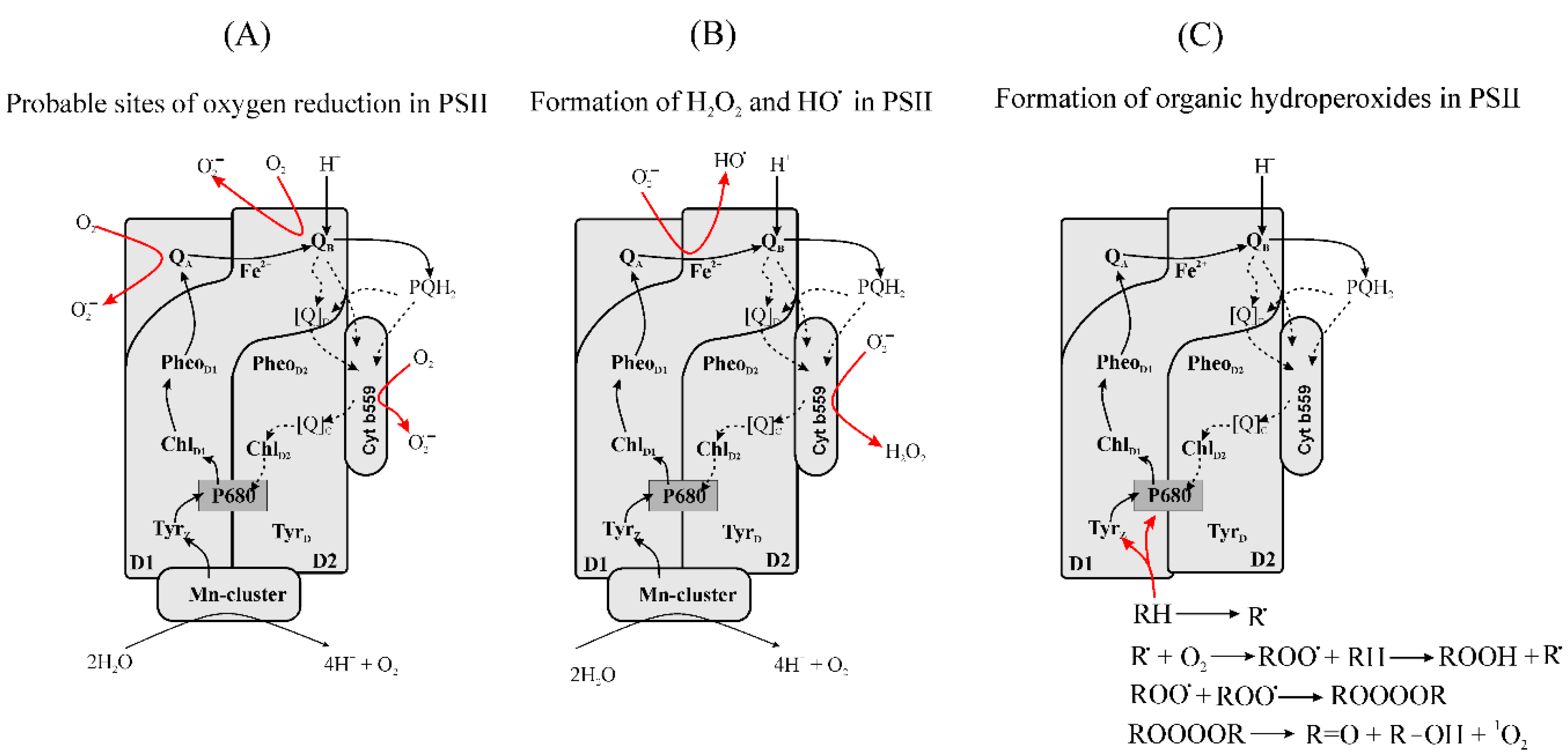
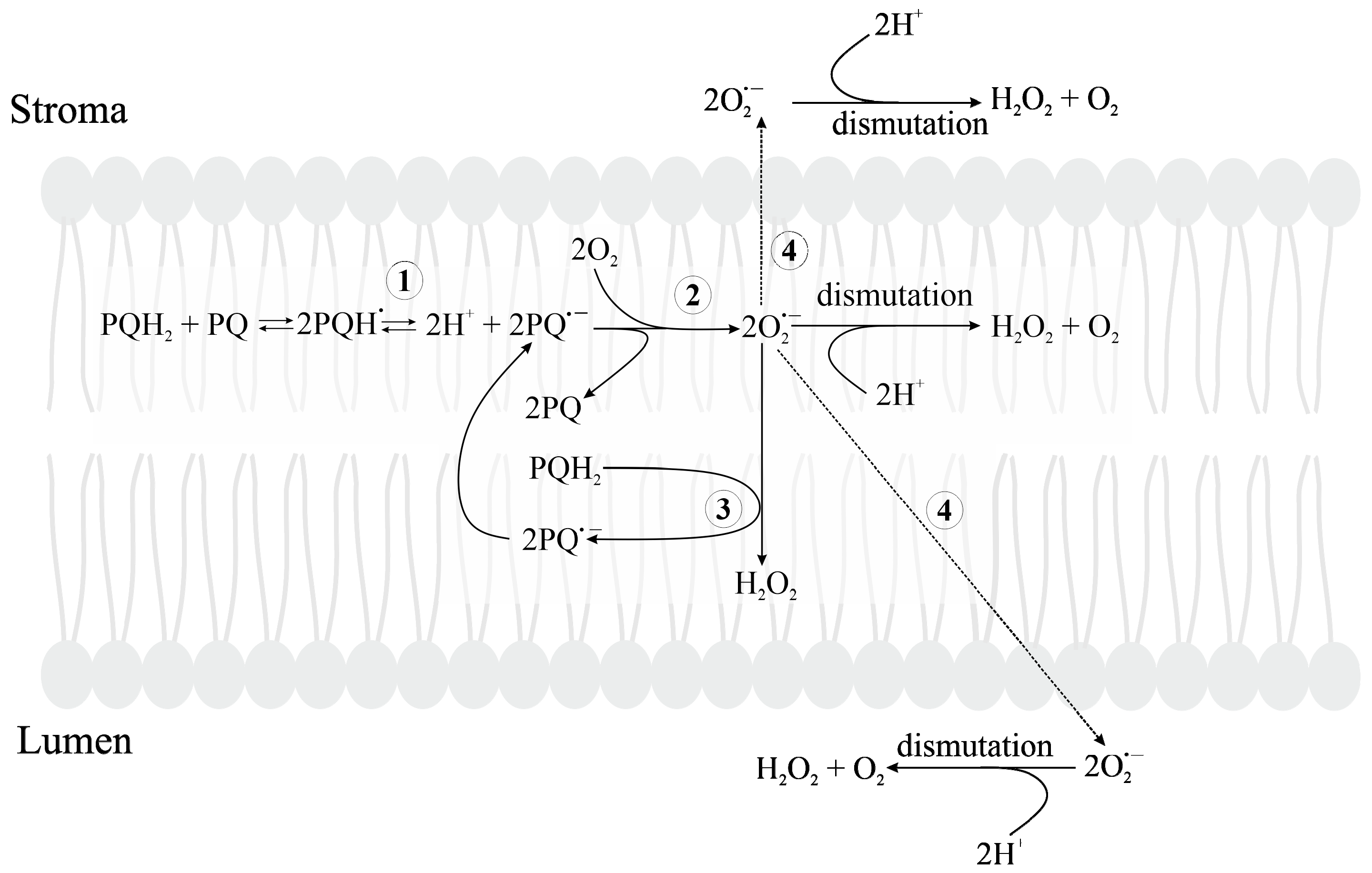
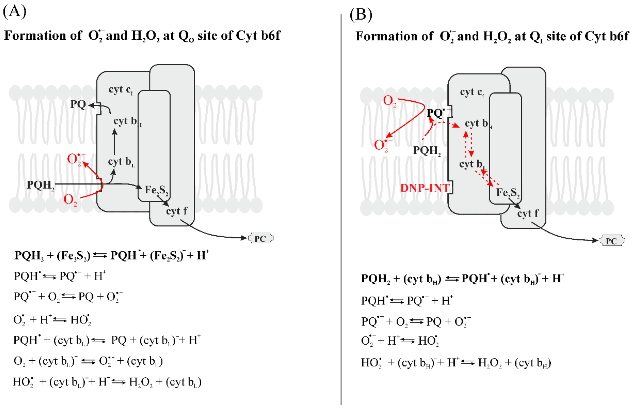
Figure 1. Singlet oxygen (1O2) generation in the antenna complex (A) and in the reaction center of PSII via charge recombination reactions (B). Proposed mechanism for the formation of 1O2 on the donor side of PSII via formation of organic peroxyl radicals (C). (A) In the antenna complex, the absorption of a photon by a molecule of Chl leads to the formation of a singlet excited state, 1Chl*, that can transform to the triplet state 3Chl via intersystem crossing. The formation of 1O2 via intersystem crossing has been demonstrated to occur in isolated LHCII [164,165]. (B) In the reaction centre of PSII, [P680+Pheo] is originally formed via electron transfer from 1P680* to Pheo in a virtual singlet state 1[P680+Pheo] that recombines to 1P680*. Charge recombination of P680+PheoQA] causes the formation of 1[P680+Pheo]. The long lifetime of the state [P680+PheoQA] destroys spin correlation, and therefore the recombination [P680+PheoQA] to 1[P680+PheoQA] often produces a “virtual triplet state” of the primary radical pair, i.e., a radical pair with such a spin configuration that, in its recombination to an excited state of the primary donor, produces a triplet, 3P680 [172,173,174]. (C) On the donor side of PSII, carbon centered radicals (R) can be formed via the oxidation of lipids and proteins by P680+ if electron donation from Mn-cluster to P680+ does not function. R are able to react with O2 to form peroxyl radical (ROO). Two peroxyl radicals react with each other to form linear tetraoxide (ROOOOR) that decomposes to 1O2, carbonyl (R=O) and alcohols (R–OH) via the Russell mechanism [200,201,202].
Plants 09 00091 g001
Figure 2. Formation of reactive oxygen species (ROS) in PSII. (A) Formation of superoxide (O2•−) can occur with the interaction of O2 with a semiquinone anion radicals at the QA and QB sites, when the electron flow from QB to the PQ pool is limited. The low potential form of cyt b559 can reduce O2 to O2•− inside the thylakoid membrane [234,235,236]. (B) Formation of H2O2 and HO. Cyt b559 can catalyze the formation of H2O2 inside the thylakoid membrane by a O2•− dismutation mechanism [72,160]. O2•− can reduce cyt b559 (Fe3+) to cyt b559 (Fe2+). O2•− + cyt b559 (Fe3+) ∆ O2 + cyt b559 (Fe2+). The following interaction of HO2 with Cyt b559 (Fe2+) leads to the formation of a ferric–hydroperoxo intermediate of cyt b559 (Fe3+–OOH) which can spontaneously decompose to cyt b559 (Fe3+) and H2O2. HO2 + cyt b559 (Fe2+) → cyt b559 (Fe3+–OOH) + H+→ cyt b559 (Fe3+) + H2O2. The formation of H2O2 in a cyt b559-dependent way requires the protonation of O2•− to form HO2. The interaction of O2•− with Fe2+ on the acceptor side of PSII can result in the formation of a ferric–peroxo intermediate [Fe3+–OO] that can be protonated to a ferric–hydroperoxo intermediate [Fe3+–OOH]. O2•− + [Fe2+] → [Fe3+–OO] + H+ → [Fe3+–OOH]. [Fe3+–OOH] can be reduced by an electron from QA, which causes its decomposition to a ferric–oxo intermediate [Fe3+–O] and HO. QA + [Fe3+–OOH] → QA + [Fe3+–O] + HO. (C) Formation of organic hydroperoxides on the donor side of PSII. Charge separation when the OEC is inactive leads to the formation of P680•+ and TyrZ which have a long lifetime and are therefore able to interact with surrounding molecules such as Chls, carotenoids and amino acids. The interaction of P680•+ or TyrZ with an organic molecule (RH) can proceed via a radical chain mechanism [200,201,202].
Plants 09 00091 g002
Figure 3. (A) Forward electron transfer chain, lifetimes and midpoint redox potentials of the cofactors of PSI; (B) charge recombination reactions and recombination lifetimes of the cofactors of PSI; the values were taken from [145,268,269,270,271]; (C) possible means of ROS formation in PSI [152,266,272,273,274,275]. PC is plastocyanin; P700 is a dimer of Chl a molecules, the primary electron donor; A0A and A0B are Chl a molecules located in branches A and B, respectively, both act as primary electron acceptors; A1A and A1B are phylloquinone molecules located in branch A and B, respectively, both acting as electron acceptors; FX, a 4Fe-4S cluster, a secondary electron acceptor; FA and FB, 4Fe-4S clusters, terminal electron acceptors.
Plants 09 00091 g003
Figure 4. Autocatalytic oxidation of reduced plastoquinone (PQH2) by O2 in the thylakoid membrane. 1—formation of a plastosemiquinone radical (PQH) by a dismutation reaction of PQH2 with PQ; 2—reduction in O2 by a plastosemiquinone anion radical (PQ•−) with formation of superoxide anion radical (O2•−); 3—oxidation of PQH2 by O2•− with formation of hydrogen peroxide (H2O2) and PQ; 4—diffusion of O2•− from thylakoid membrane to stroma and to lumen. In the autocatalytic oxidation of PQH2, the reaction of O2•− with PQH2 provides excess PQ•− that can be involved in the formation of O2•− and, in turn, accelerates the oxidation of PQH2 [128].
Plants 09 00091 g004
Figure 5. The proposed generation of O2•− in Cyt b6f. (A) In the QO site of Cyt b6f, PQH is generated (Table 2) by the 2Fe-2S cluster of the high-potential Rieske iron−sulfur protein. PQ•− that has a long residence time within the QO pocket, and cytochrome bL can also serve as a reductant for the generation of O2•− in Cyt b6f. In addition, HO2, can be reduced by cytochrome bL to form H2O2 [317]. (B) The proposed generation of O2•− in Cyt b6f in the presence of DNP-INT, an inhibitor of PQH2 oxidation by Cyt b6f. The oxidation of PQH2 does not occur in QO site, formation of PQH and its deprotonation can occur in the Qi site. PQH can be oxidized in subsequent reactions with O2 or with hemes bH or bL. H2O2 can be formed via the reaction of O2•− with PQH2 or via the reaction of HO2 with cytochrome bH or cytochrome bL.
Plants 09 00091 g005
Table 1. Most significant reactive oxygen species (ROS) and ROS derivatives. R is a residual of an organic molecule.
RadicalsNon-Radicals
Reactive oxygen species
Superoxide anion radical, O2•−Singlet oxygen, 1O2
Hydroperoxyl radical, HO2Hydrogen peroxide, H2O2
Hydroxyl radical, HOOzone, O3
ROS derivatives and reactive nitrogen species
Peroxyl radical, ROOOrganic peroxides, ROOH
Alkoxyl radical, ROPeroxynitrite ion, ONOO
Nitric Oxide, NOAlkyl peroxynitrite, ROONO
Nitrogen dioxide, NO2
Table 2. Redox midpoint potentials and lifetimes of primary and secondary electron acceptors in PSII. The values were taken from [220,221,222,223,224,225,226,227,228,229,230,231,232,233].
Redox Active CofactorsMidpoint Redox Potential vs. Normal Hydrogen Electrode (NHE), mVLifetime, sRemarks
Pheo/Pheo≈−610 [220,221](2–5) × 10−10 [222]Reoxidation of Pheo via forward electron transfer to QA
−588 [219]
−505 [223](4–30) × 10−8 [222]Reoxidation of Pheo via recombination of [P680+Pheo]
QA/QA−80–−200 [224,225](0.1–0.2) × 10−3 [224,226]Reoxidation of QA via forward electron transfer to QB
Shift from −145 to −70 [227](0.3–0.5) × 10−3 [224,226,228]Removal of HCO3 bound to acceptor side of PSII.
(2–4.6) × 10−3 [226,228,229]Reoxidation of QA via forward electron transfer to QB and protonation.
(0.2–2) × 10−1 [222]Reoxidation of QA by PQ that binds to an empty QB site.
1–2 [226]Reoxidation of QA via charge recombination with oxidized TyrZ.
Reoxidation of QA via charge recombination with S2
QA/QA2−−500 [230]Double reduction achieved either by chemical treatment or by strong illumination in anaerobic conditions. No doubly reduced QA accumulates during aerobic light treatment.
QB/QB−45–−60 [220,231,232](0.3–0.5) × 10−3 [224,226,228]Reduction of QB via electron transfer from QA
>0.4 [222]Reoxidation of QB via charge recombination with oxidized TyrZ.
30 [222]Reoxidation of QB via charge recombination with S2
QB/QBH100 [220](0.3–0.5) × 10−3 [224,226,228]Assuming the same time as for QB
>0.4 [222]
30 [222]
QB/QB2−−200–−464 [233]
QBH/QBH290–373 [233]

