
Contrasting Dihydronaphthoquinone Patterns in Closely Related Drosera (Sundew) Species Enable Taxonomic Distinction and Identification

 , and
, and 
Abstract

1. Introduction
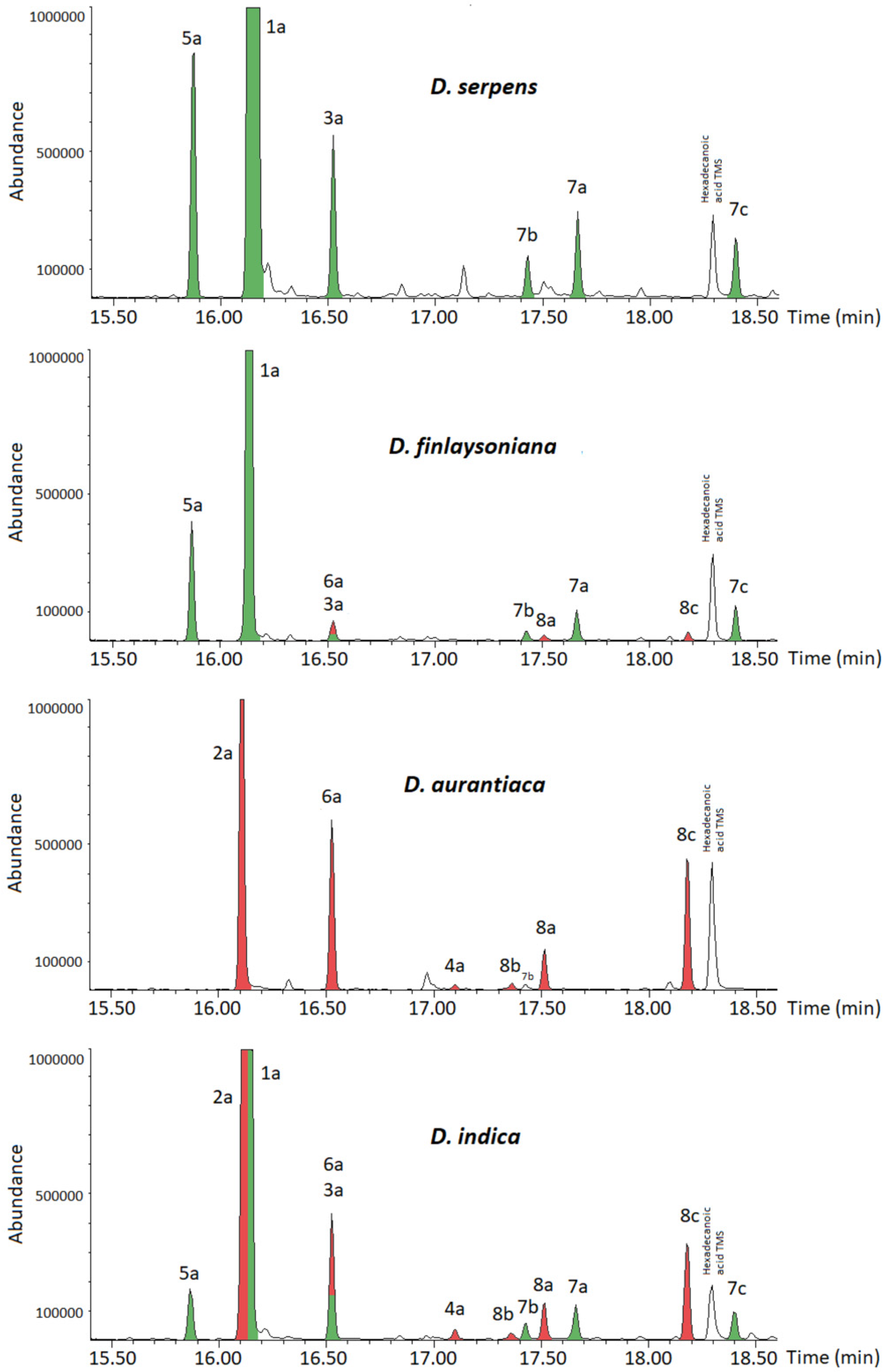
2. Results
3. Discussion
4. Materials and Methods
4.1. General Experimental Procedures
4.2. Plant Material
4.3. Extraction and Isolation
- Plumbagin (1): C11H8O3, EIMS m/z 188 [M]+ (100), 173 [M-CH3]+ (30), 160 [M-CO]+ (25), 132 (30), 131 [M-C3H5O]+ (50), 121 [M-C4H3O]+ (15), 120 [M-C4H4O]+ (25), 103 [M-C4H5O2]+ (10), 92 [M-C5H4O2]+ (40), 77 (15), 63 (40).
- Ramentaceone (2): C11H8O3, EIMS m/z 188 [M]+ (100), 187 [M − H]+ (30), 173 [M − CH3]+ (10), 160 [M − CO]+ (15), 134 [M − C3H2O]+ (20), 132 (30), 131 [M − C3H5O]+ (30), 106 [M − C4H2O2]+ (20), 104 (10), 103 [M − C4H5O2]+ (10), 78 (15), 77 (25), 63 (15), 62 (10), 51 (25).
- Isoshinanolone (3): C11H12O3, EIMS m/z 192 [M]+ (70), 177 [M − CH3]+ (20), 174 [M − H2O]+ (10), 150 [M − C3H6]+ (40), 149 [M − C2H3O]+ (25), 131 [M − C2H5O2]+ (20), 122 [M − C3H2O2]+ (45), 121 [M − C4H7O]+ (100), 115 (10), 93 (25), 77 (20), 65 (30), 51 (20).
- Shinanolone (4): C11H12O3, EIMS m/z 192 [M]+ (70), 177 [M − CH3]+ (10), 174 [M − H2O]+ (10), 164 [M − C2H4]+ (20), 149 [M − C2H3O]+ (15), 135 [M − C3H5O]+ (100), 107 [M − C4H5O2]+ (20).
- Dihydroplumbagin (5): C11H10O3, EIMS m/z 190 [M]+ (85), 175 [M − CH3]+ (100), 162 [M − CO]+ (20), 147 [M − C2H3O]+ (20), 120 [M − C4H6O]+ (60), 92 [M − C5H6O2]+ (40), 63 (15) (Figure S1).
- Dihydroramentaceone (6): C11H10O3, EIMS m/z 190 [M]+ (100), 175 [M − CH3]+ (10), 162 [M − CO]+ (15), 134 [M − C3H4O]+ (55), 106 [M − C4H4O2]+ (20) (Figure S2).
- 5-O-Trimethylsilyl-plumbagin (1a): C14H16O3Si, EIMS m/z 245 [M − CH3]+ (100), 217 [M − CH3 − CO]+ (30), 186 (10), 115 (10) (Figure S3).
- 5-O-Trimethylsilyl-ramentaceone (2a): C14H16O3Si, EIMS m/z 245 [M − CH3]+ (100), 217 [M − CH3 − CO]+ (20), 187 (10), 115 (10) (Figure S4).
- 4,8-Di-(O-trimethylsilyl)-isoshinanolone (3a): C17H28O3Si2, EIMS m/z 321 [M − CH3]+ (5), 231 [M − CH3 − C3H10OSi]+ (100), 216 [M − 2CH3 − C3H10OSi]+ (20), 201 [M − 3CH3 − C3H10OSi]+ (10), 186 [M − 4CH3 − C3H10OSi]+ (5) (Figure S5).
- 4,8-Di-(O-trimethylsilyl)-shinanolone (4a): C17H28O3Si2, EIMS m/z 321 [M − CH3]+ (5), 231 [M − CH3 − C3H10OSi]+ (100), 216 [M − 2CH3 − C3H10OSi]+ (20), 201 [M − 3CH3 − C3H10OSi]+ (10), 186 [M − 4CH3 − C3H10OSi]+ (5) (Figure S6).
- 5-O-Trimethylsilyl-dihydroplumbagin (5a): C14H18O3Si, EIMS m/z 247 [M − CH3]+ (100), 219 [M − CH3 − CO]+ (10) (Figure S7).
- 5-O-Trimethylsilyl-dihydroramentaceone (6a): C14H18O3Si, EIMS m/z 247 [M − CH3]+ (100), 219 [M − CH3 − CO]+ (10) (Figure S8).
- 1,5-Di-(O-trimethylsilyl)-2-methyl-naphtho-1,4,5-triol (7a): C17H26O3Si2, EIMS m/z 318 [M − CH4]+ (100), 288 [M − CH4 − 2CH3]+ (20), 273 [M − CH4 − 3CH3]+ (10) (Figure S9).
- 4,5-Di-(O-trimethylsilyl)-2-methyl-naphtho-1,4,5-triol (7b): C17H26O3Si2, EIMS m/z 319 [M − CH3]+ (100), 245 (15), 217 (10).
- 2-Methyl-1,4,5-tri-(O-trimethylsilyl)-naphtho-1,4,5-triol (7c): C20H34O3Si3, EIMS m/z 406 [M]+ (100).
- 1,5-Di-(O-trimethylsilyl)-7-methyl-naphtho-1,4,5-triol (8a): C17H26O3Si2, EIMS m/z 318 [M − CH4]+ (100), 288 [M − CH4 − 2CH3]+ (20), 273 [M − CH4 − 3CH3]+ (10) (Figure S10).
- 4,5-Di-(O-trimethylsilyl)-7-methyl-naphtho-1,4,5-triol (8b): C17H26O3Si2, EIMS m/z 319 [M − CH3]+ (100), 245 (15), 217 (10).
- 7-Methyl-1,4,5-tri-(O-trimethylsilyl)-naphtho-1,4,5-triol (8c): C20H34O3Si3, EIMS m/z 406 [M]+ (100).
Supplementary Materials
Author Contributions
Funding
Institutional Review Board Statement
Informed Consent Statement
Data Availability Statement
Acknowledgments
Conflicts of Interest
References
- Fleischmann, A.; Gonella, P.M. Species of carnivorous plants. In Carnivorous Plants; Ellison, A., Adamec, L., Eds.; University Press: Oxford, UK, 2017; p. 415. [Google Scholar]
- Culham, A.; Gornall, R.J. The taxonomic significance of naphthoquinones in the Droseraceae. Biochem. Syst. Ecol. 1994, 22, 507–515. [Google Scholar] [CrossRef]
- Schlauer, J.; Nerz, J.; Rischer, H. Carnivorous plant chemistry. Acta Bot. Gall. 2005, 152, 187–195. [Google Scholar] [CrossRef][Green Version]
- Schlauer, J.; Fleischmann, A. Chemical evidence for hybridity in Drosera (Droseraceae). Biochem. Syst. Ecol. 2016, 66, 33–36. [Google Scholar] [CrossRef]
- Schlauer, J.; Hartmeyer, S.R.H.; Hartmeyer, I. Unexpected Discovery of 7-Methyljuglone (Ramentaceone) in Several Australian Sundews; Carnival Corporation & plc: Miami, FL, USA, 2017; Volume 46, pp. 20–22. [Google Scholar]
- Schlauer, J.; Hartmeyer, S.R.H.; Hartmeyer, I.; Hennern, H.; Hennern, A. Sundew Chemistry and Emergence Updates; Carnival Corporation & plc: Miami, FL, USA, 2018; Volume 47, pp. 10–17. [Google Scholar]
- Schlauer, J.; Hartmeyer, S.R.H.; Hartmeyer, I.; Hennern, H.; Hennern, A. New Sundew Quinone and Emergence Data; Carnival Corporation & plc: Miami, FL, USA, 2019; Volume 48, pp. 6–12. [Google Scholar]
- Schlauer, J.; Hartmeyer, S.R.H.; Hartmeyer, I. Quinone Patterns and Identification of Japanese Spider Leg Sundews (Drosera Sect. Arachnopus); Carnival Corporation & plc: Miami, FL, USA, 2019; Volume 48, pp. 161–163. [Google Scholar]
- Budzianowski, J.; Budzianowska, A.; Kromer, K. Naphthalene glucoside and other phenolics from the shoot and callus cultures of Drosophyllum lusitanicum. Phytochemistry 2002, 61, 421–425. [Google Scholar] [CrossRef]
- Bringmann, G.; Hamm, A.; Günther, C.; Michel, M.; Brun, R.; Mudogo, V. Ancistroealaines A and B, two new bioactive naphthylisoquinolines, and related naphthoic acids from Ancistrocladus ealaensis. J. Nat. Prod. 2000, 63, 1465–1470. [Google Scholar] [CrossRef] [PubMed]
- Binder, R.G.; Benson, M.E.; Flath, R.A. Eight 1,4-naphthoquinones from Juglans. Phytochemistry 1989, 28, 2799–2801. [Google Scholar] [CrossRef]
- Higa, M.; Takashima, Y.; Yokaryo, H.; Harie, Y.; Suzuka, T.; Ogihara, K. Naphthoquinone derivatives from Diospyros maritima. Chem. Pharm. Bull. 2017, 65, 739–745. [Google Scholar] [CrossRef] [PubMed][Green Version]
- Revirego, F.; Alkorta, I.; Elguero, J. Desmotropy in reduced plumbagins: α- and β-dihydroplumbagins. J. Mol. Struct. 2008, 891, 325–328. [Google Scholar] [CrossRef]
- Budzianowski, J. Naphthohydroquinone glucosides of Drosera rotundifolia and D. intermedia from in vitro cultures. Phytochemistry 1996, 42, 1145–1147. [Google Scholar] [CrossRef]
- Schlauer, J.; Hartmeyer, S.R.H.; Hartmeyer, I. Chemistry and Surface Micromorphology of the Queensland Sundews (Drosera Section Prolifera); Carnival Corporation & plc: Miami, FL, USA, 2019; Volume 48, pp. 111–116. [Google Scholar]
- Schlauer, J.; Carow, T.; Fleischmann, A. Quinones from “Gondwanan” Sundews; Carnival Corporation & plc: Miami, FL, USA, 2019; Volume 48, pp. 13–17. [Google Scholar]


| Species (Provenance, Accession No.) | 2-Methyl Series | 7-Methyl Series | |||||
|---|---|---|---|---|---|---|---|
| Plumbagin (1) | Dihydroplumbagin (5)/(7) | Isoshinanolone (3) | Ramentaceone (2) | Dihydroramentaceone (6)/(8) | Shinanolone (4) | ||
| D. serpens (Australia, 2020_101) | + | + | + | - | - | - | |
| D. finlaysoniana (Tropical Asia, 2020_102) | + | + | + | - | + * | - | |
| D. hartmeyerorum (Australia, 2020_103) | - | - | - | + | + | + | |
| D. aurantiaca (Australia, 2020_104) | - | - | - | + | + | + | |
| D. indica (Tropical Asia, 2020_105) | + | + | + | + | + | + | |
| Dionaea muscipula (USA, 2020_106) | + | + | + | - | - | - | |
| Drosophyllum lusitanicum (SW Europe, 2020_107) | + | + | + | - | + * | - | |
| Rt (GC) [min] | 14.49 | 14.47 | 15.46 | 14.70 | 15.56 | 15.97? | |
| M+ [m/z] | 188.0 | 190.1 | 192.1 | 188.0 | 190.1 | 192.1 | |
| Rt (GC) [min]; characteristic MS signal [m/z] of TMS-Derivatives | TMS | 16.15; 245.1 (1a) | 15.88; 247.1 (5a) | - | 16.13; 245.1 (2a) | 16.54; 247.1 (6a) | |
| TMS2 | - | 17.65; 318.2 (7a) 17.43; 319.1 (7b) | 16.52; 231.1 (3a) | - | 17.52; 318.2 (8a) 17.37; 319.1 (8b) | 17.10; 231.1 (4a) | |
| TMS3 | - | 18.41; 406.3 (7c) | - | - | 18.18; 406.3 (8c) | - | |
Publisher’s Note: MDPI stays neutral with regard to jurisdictional claims in published maps and institutional affiliations. |
© 2021 by the authors. Licensee MDPI, Basel, Switzerland. This article is an open access article distributed under the terms and conditions of the Creative Commons Attribution (CC BY) license (https://creativecommons.org/licenses/by/4.0/).
Share and Cite
Schlauer, J.; Hartmeyer, S.R.H.; Hartmeyer, I.; Seppänen-Laakso, T.; Rischer, H. Contrasting Dihydronaphthoquinone Patterns in Closely Related Drosera (Sundew) Species Enable Taxonomic Distinction and Identification. Plants 2021, 10, 1601. https://doi.org/10.3390/plants10081601
Schlauer J, Hartmeyer SRH, Hartmeyer I, Seppänen-Laakso T, Rischer H. Contrasting Dihydronaphthoquinone Patterns in Closely Related Drosera (Sundew) Species Enable Taxonomic Distinction and Identification. Plants. 2021; 10(8):1601. https://doi.org/10.3390/plants10081601
Chicago/Turabian StyleSchlauer, Jan, Siegfried R. H. Hartmeyer, Irmgard Hartmeyer, Tuulikki Seppänen-Laakso, and Heiko Rischer. 2021. "Contrasting Dihydronaphthoquinone Patterns in Closely Related Drosera (Sundew) Species Enable Taxonomic Distinction and Identification" Plants 10, no. 8: 1601. https://doi.org/10.3390/plants10081601
APA StyleSchlauer, J., Hartmeyer, S. R. H., Hartmeyer, I., Seppänen-Laakso, T., & Rischer, H. (2021). Contrasting Dihydronaphthoquinone Patterns in Closely Related Drosera (Sundew) Species Enable Taxonomic Distinction and Identification. Plants, 10(8), 1601. https://doi.org/10.3390/plants10081601