Review

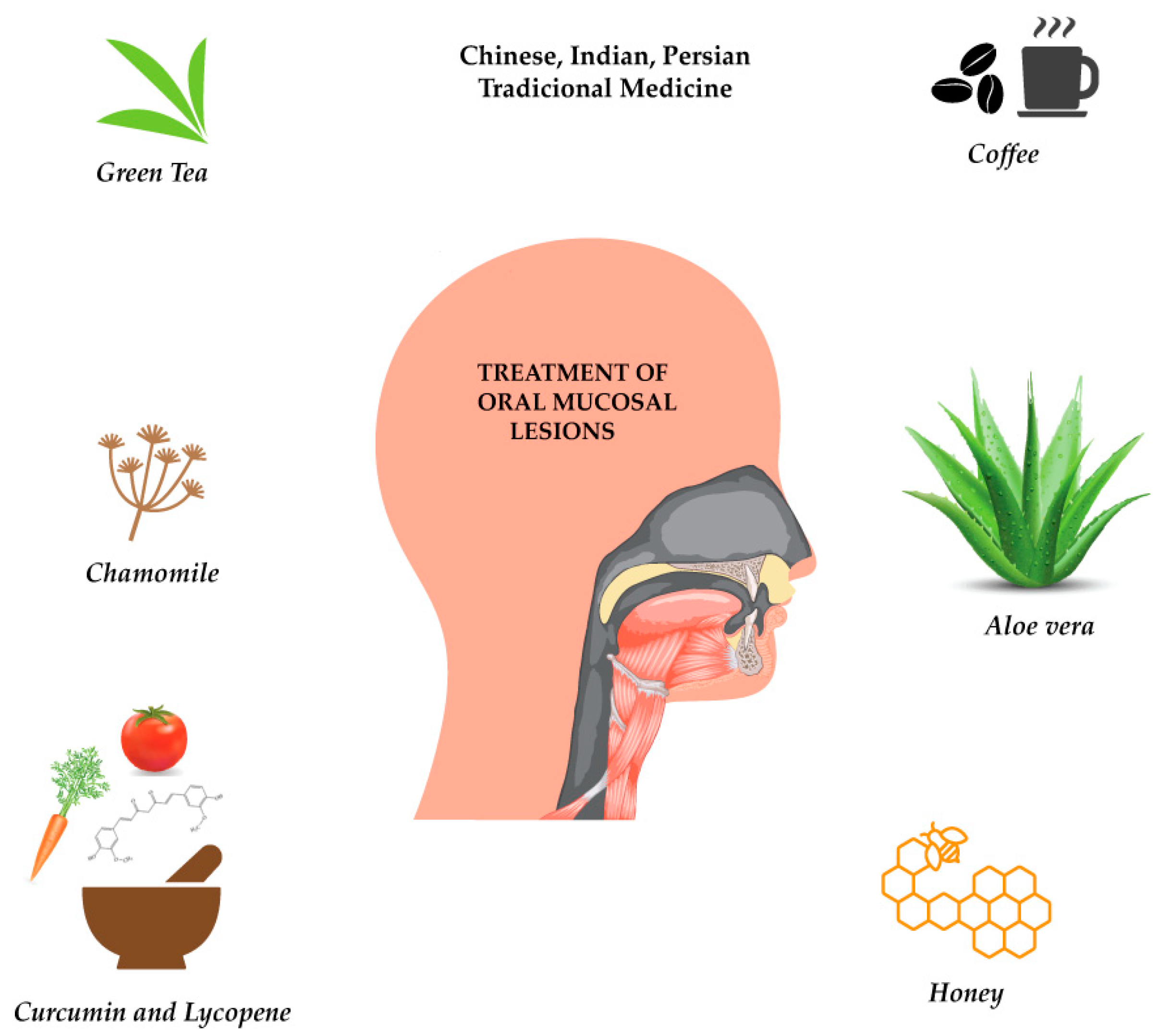
Plant-Derived Bioactives in Oral Mucosal Lesions: A Key Emphasis to Curcumin, Lycopene, Chamomile, Aloe vera, Green Tea and Coffee Properties

by
Bahare Salehi
1,
Pia Lopez-Jornet
2,
Eduardo Pons-Fuster López
3,
Daniela Calina
4,
Mehdi Sharifi-Rad
5,*,
Karina Ramírez-Alarcón
6,
Katherine Forman
6,
Marcos Fernández
7,
Miquel Martorell
6,*,
William N. Setzer
8,
Natália Martins
9,10,*,
Célia F. Rodrigues
11 and
Javad Sharifi-Rad
12,*
1
Student Research Committee, School of Medicine, Bam University of Medical Sciences, Bam 44340847, Iran
2
Instituto Murciano de InvestigaciónBiosanitaria (IMIB-Arrixaca-UMU), Clínica Odontológica Universitaria Hospital Morales Meseguer Adv. Marques de los velez s/n, 30008 Murcia, Spain
3
University of Murciaand, Clínica Odontológica Universitaria Hospital Morales Meseguer, Adv. Marques de los velez s/n, 30008 Murcia, Spain
4
Department of Clinical Pharmacy, University of Medicine and Pharmacy of Craiova, 200349 Craiova, Romania
5
Department of Medical Parasitology, Zabol University of Medical Sciences, Zabol 61663-335, Iran
6
Department of Nutrition and Dietetics, Faculty of Pharmacy, University of Concepcion, Concepcion 4070386, Chile
7
Department of Pharmacy, Faculty of Pharmacy, University of Concepcion, Concepcion 4070386, Chile
8
Department of Chemistry, University of Alabama in Huntsville, Huntsville, AL 35899, USA
9
Faculty of Medicine, University of Porto, Alameda Prof. Hernâni Monteiro, 4200-319 Porto, Portugal
10
Institute for Research and Innovation in Health (i3S), University of Porto, 4200-135 Porto, Portugal
11
LEPABE, Department of Chemical Engineering, Faculty of Engineering, University of Porto, Rua Dr. Roberto Frias, s/n, 4200-465 Porto, Portugal
12
Food Safety Research Center (Salt), Semnan University of Medical Sciences, Semnan 35198-99951, Iran
*
Authors to whom correspondence should be addressed.
Biomolecules 2019, 9(3), 106; https://doi.org/10.3390/biom9030106
Submission received: 27 January 2019 / Revised: 6 March 2019 / Accepted: 13 March 2019 / Published: 17 March 2019

Abstract

:
Oral mucosal lesions have many etiologies, including viral or bacterial infections, local trauma or irritation, systemic disorders, and even excessive alcohol and tobacco consumption. Folk knowledge on medicinal plants and phytochemicals in the treatment of oral mucosal lesions has gained special attention among the scientific community. Thus, this review aims to provide a brief overview on the traditional knowledge of plants in the treatment of oral mucosal lesions. This review was carried out consulting reports between 2008 and 2018 of PubMed (Medline), Web of Science, Embase, Scopus, Cochrane Database, Science Direct, and Google Scholar. The chosen keywords were plant, phytochemical, oral mucosa, leukoplakia, oral lichen planus and oral health. A special emphasis was given to certain plants (e.g., chamomile, Aloe vera, green tea, and coffea) and plant-derived bioactives (e.g., curcumin, lycopene) with anti-oral mucosal lesion activity. Finally, preclinical (in vitro and in vivo) and clinical studies examining both the safety and efficacy of medicinal plants and their derived phytochemicals were also carefully addressed.

1. Introduction

Oral mucosal lesions (OML) have been defined as any abnormal change in oral mucosa surface, appearing as pigmented, ulcerative, red and white features or any swelling or developmental defect variants [1]. Oral mucosal lesions have many predisposing factors, including infection (viruses, bacteria, parasites, fungi), thermal and/or physical, immune system deficiencies, neoplasia, systemic disease, trauma, as well as aging and chronic behaviors (e.g., betel nut, alcohol, and tobacco use) [2]. In 2003, OML was considered one of the main public health problems worldwide by the World Health Organization (WHO) [3]. In fact, it has been suggested that oral mucosa can reflect the patient’s general health [4].
More than 200 mucosal conditions have been identified [5]. The most important ones include leukoplakia, erythroplakia, oral lichen planus and lichenoid reactions, focal epithelial hyperplasia, exophytic neoplasia (oral fibroma and squamous papilloma/oral wart), herpes and aphthous lesions, tobacco-related lesions (white lesions associated with smokeless tobacco and nicotine stomatitis), non-specific ulcerations (defined as epithelium loss), candidiasis, traumatic ulcers and geographic tongue [6]. Oral mucosa is a highly responsive, diversified and dynamic environment that, although highly accessible, present true challenges for oral drug delivery [7]. Since steroids and other drugs that have been commonly used to treat different oral diseases present several side effects, scientists are searching for other methods with equivalent potency and little or no side effects [8].
Traditional knowledge on medicinal plants used for different health purposes has attracted much attention among the scientific community because of their effectiveness in the treatment of many diseases [9,10,11,12,13,14,15], including oral diseases [16]. There are many reports on the use of natural products and their derived bioactives, often called phytochemicals, in oral disease treatment [16,17,18]. It has been demonstrated that medicinal plants rich in several chemical constituents are highly effective in OML treatment. In this sense, based on the above-highlighted aspects, the present review aims to provide a close relationship between plant-derived bioactives and OML treatment efficacy. A special emphasis was also given to plant-derived bioactives with anti-oral mucosal lesion activity, with a special emphasis to curcumin, lycopene, chamomile, Aloe vera, green tea, and Coffea. Finally, preclinical (in vitro and in vivo) and clinical studies examining the safety and efficacy of these medicinal plants and their derived bioactives were also carefully examined.

2. Research Methodology

Given the main focus of this work: o validate the traditional use of medicinal plants and their derived bioactives in OML treatment, the present review was carried out by consulting PubMed (Medline), Web of Science, Embase, Scopus, Cochrane Database, Science Direct, and Google Scholar (as a search engine) databases to retrieve the most updated articles on this topic. The following keywords were considered: plant, phytochemical, oral mucosa, leukoplakia, oral lichen planus and oral health. All articles were carefully analyzed by the authors to assess their strengths and weaknesses, and to select the more useful ones for the purpose of review, prioritizing articles published from 2008 to 2018.

3. Traditional Knowledge on Plants’ Use against Oral Mucosal Lesions

Medicinal plants have been used as folk remedies since ancient times and in many parts of the world. Specifically, their therapeutic applications in oral diseases include wound healing, anti-inflammatory, analgesic, antioxidant and antimicrobial benefits. Numerous reports have shown the effective potential of plant-derived bioactives in the treatment of OML, including recurrent aphthous stomatitis, mucositis caused by radiotherapy and chemotherapy, erosive leukoplakia and oral lichen planus. These phytopharmacological attributes have been documented in distinct traditional systems, such as Chinese, Ayurveda and Persian Medicine.

3.1. Traditional Chinese Medicine

Chinese herbal medicine has been used for a very long time in the treatment of oral diseases. In general, each herbal medicine is a combination of several herbs. Thus, in the treatment of OML, it is possible to find different preparations of medicinal herb mixtures. Liuwei Dihuang consists of six ingredients extracted from Rehmannia glutinosa, Cornus officinalis, Dioscorea opposita, Alisma orientalis, Poria cocos, and Paeonia suffruticosa. The stomatitis-healing granule is a herbal mixture of Asparagus cochinchinensis, Ophiopogon japonicus, Scrophularia ningpoensis, Lonicera japonica and Glycyrrhiza uralensis, indicated for stomatitis [19]. Another preparation is Xianhuayin, a collutory (mouthwash), effective to treat oral, buccal mucosal premalignant lesions. Xianhuayin is a decoction consisting of Phellodendrona murense, Amomum villosum, Sclerotium poriae, Helianthus, and Glycyrrhiza glabra, where the main component in G. glabra is licorice zinc, which facilitates epithelial cell regeneration [20]. Nevertheless, it is also possible to find in Chinese medicine herbal preparations consisting of a single herb in the treatment of oral lesions. For instance, Salvia miltiorrhiza is a Chinese herbal medicine that has been used against tumor cell growth, inflammation and oxidation [21]. Salvia miltiorrhiza has been used in the treatment of scleroderma (an uncommon disorder characterized by thickening or hardening of the skin and fibrosis of the involved tissues) [22]. Achyrantes bidentata and Achyrantes aspera alcoholic extracts are often used in the treatment of aphtha (gargled) since they have demonstrated significant wound healing effects [23]. Salvia officinalis, Matricaria chamomilla, A. vera, and Gentiana lutea have also been used in traditional Chinese medicine as herbal formulae in the treatment of oral ulceration. They are often selected to be used against chemotherapy-induced mucositis, through the form of single infusions for gargling or topical application [24].

3.2. Ayurveda, Indian Traditional System of Medicine

Indian herbal medicine also has a thousand years of rich traditional knowledge in wound treatment and management. Among the wide variety of plants used in wounds management, it is possible to differentiate those intended for specific oral lesions treatment. Aloe vera is an indigenous medicinal plant found throughout India [25]. This plant is effective in the treatment of oral aphthous ulceration [26], but has been reported as having many other properties, such as antiulcer, antiseptic, antibacterial, anti-inflammatory, antioxidant, and wound healing properties [8,27]. Centella asiatica is distributed throughout the plains of India [28]. This plant is effective in the treatment of mouth ulcers. It has a remarkable effect on wounds healing and promotes connective tissue growth [29]. The wound healing effect has been attributed to several mechanisms, including antioxidant activity, collagen synthesis, and angiogenesis promotion [30]. Curcuma longa is another very useful and old spice used in Ayurvedic medicine. C. longa extracts can be used in the treatment of oral cavity lesions [31]. One of the most important components of C. longa is curcumin, a potent antioxidant. Also, C. longa has prominent anti-inflammatory, antibacterial and wound-healing effects [32]. Emblica officinalis has an interesting antioxidant and astringent property, and it has been demonstrated to be effective in the treatment of aphthous stomatitis and other types of mouth ulcers [33]. Tinospora cordifolia has anti-inflammatory, antioxidant and immunomodulatory effects. This plant can reduce mucositis severity in radiotherapy patients [34]. Jasminum grandiflorum leaves are widely used in the treatment of ulcerative stomatitis and oral wounds, closely related to its antioxidant properties [32].

3.3. Traditional Persian Medicine

Wounds treatment is also an important feature in traditional Persian Medicine, where some plants can be effectively used to treat diseases of the oral cavity. Lawsonia alba is used as a rinsing decoction in aphtha treatment. Its wound healing effect is mediated through its anti-inflammatory, antioxidant and antibacterial activities [35]. Punica granatum is also used as a rinsing decoction with vinegar to treat aphtha through its anti-inflammatory, antioxidant and antibacterial activities [36,37]. Finally, Vitis vinifera seed extract also displays anti-inflammatory and antioxidant effects [38].

4. Curcumin, Lycopene, Chamomile, Aloe vera, Green Tea and Coffee in Oral Mucosal Lesions: A Brief Overview

The mouth is considered the human organism gateway, with many functions including nutrition, communication (speech) and facial expression [39]. In recent decades, there has been increasing interest in assessing the therapeutic effects of plants for the treatment of oral mucosal lesions [40,41]. Plant-derived preparations interact through different biochemical pathways, which culminates in distinct biological potentialities, including antioxidant, analgesic, anti-inflammatory, antifungal, antiseptic and anticarcinogenic effects (Figure 1).
Curcumin is a polyphenol commonly known as turmeric. Its main active components include flavonoids and volatile compounds such as tumerone, atlantone, and zingiberone. Recent scientific evidence has shown that turmeric, and particularly curcumin, exhibit powerful anti-inflammatory effects in a wide variety of target systems [42,43,44]. Also, remarkable antioxidant, anti-tumor, analgesic, immunostimulant, antiviral, antibacterial and antifungal effects have been attributed to this molecule, as well as a great capacity to fight against diseases, such as diabetes, asthma, allergies, neurodegenerative, arthritis, atherosclerosis, OML, and even cancer.
Lycopene is a carotenoid and natural liposoluble pigment responsible for the red and orange color of some fruits and vegetables. An important factor affecting lycopene bioavailability is its synergy with other antioxidants, as occurs, for example, with vitamins C and E. Carotenoids’ toxicity stated in some observational and interventional studies is closely related to the doses used and its corresponding interactions. It should be noted that most of these studies have used rodents, which absorb carotenoids less efficiently than humans. Also, high concentrations of one carotenoid may interfere with the bioavailability of others, leading to imbalance, as occurs between β-carotene and lycopene [45].
Chamomile (Matricaria recutita L., syn. Matricaria chamomilla L.) is a medicinal plant belonging to the Asteraceae family and contains flavonoids, coumarins and essential oils with antiseptic, carminative, sedative and protector effect against mucosal ulcers [46,47,48,49,50].
The Aloe vera species contains over 200 biologically active substances, such us anthraquinones (barbaloin, isobarbaloin, anthranols, aloetic acid), hydrosoluble and liposoluble vitamins, minerals, enzymes, polysaccharides, phenolic compounds, and organic acids, with antibacterial, antimicrobial, anti-inflammatory and immuno-modulatory effects [51,52,53,54,55,56]. Lignin is very abundant in A. Vera pulp and easily penetrates into epithelial tissues, carrying other compounds such as saponins (glucosides with a cleansing and antiseptic activity), chrysophanic acid and emodin derivatives used in the treatment of psoriasis outbreaks and skin mycoses [57].
Tea is a product made from Camellia sinensis leaf and bud, and it is the most widely consumed drink in the world after water, being second only to water in popularity as a beverage [58]. This is an important source of polyphenols with renowned antioxidant, anti-inflammatory and bactericidal effects [59].

5. In Vitro and In Vivo Anti-Oral Mucosal Lesions Effects

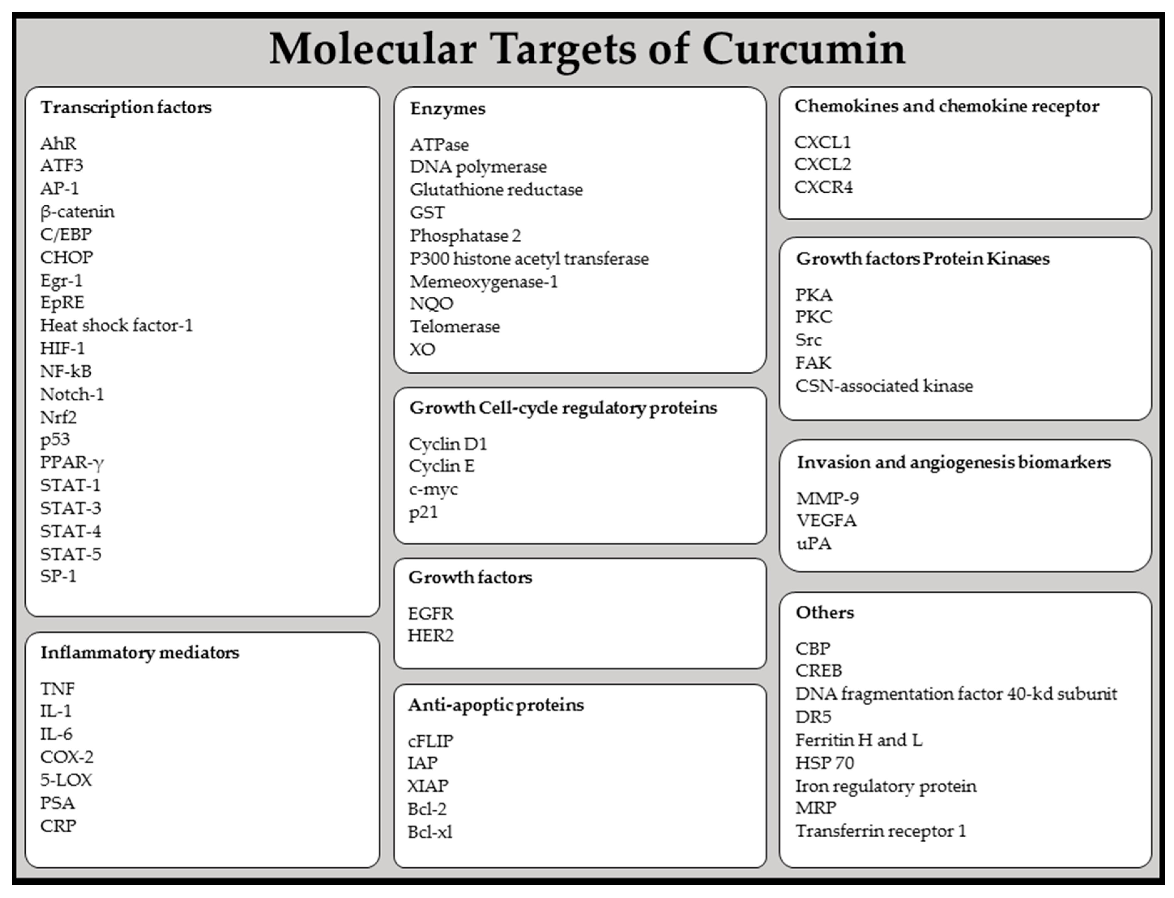
5.1. Curcumin: Effects on Oral Mucosal Carcinogenesis and Anti-Inflammatory Properties

5.1.1. Effects on Oral Mucosal Carcinogenesis

Curcumin modulates key pathways involved in inflammation and carcinogenesis [42,60]. In vivo studies in rats fed with curcumin at 0.5 g/kg during initiation and post-initiation phases showed a 91% reduction in 4-nitroquinoline-1-oxide-induced tongue carcinoma frequency [61]. Furthermore, the incidence of potentially malignant N-nitrosomethylbenzylamine-induced oral lesions decreased as a result of curcumin administration during the initiation and post-initiation phases [62]. In another study, where curcumin was administered alone and in combination with green tea (C. sinensis), it was found that it exerts inhibitory effects against oral carcinogenesis in hamsters, suppressing cell proliferation, inducing apoptosis and inhibiting angiogenesis [63,64]. Manoharan et al. [65] induced oral squamous cell carcinoma (OSCC) in hamsters through using 7,12-dimethylbenz[a]anthracene (DMBA). Curcumin and piperine administered orally in hamsters with DMBA-induced OSCC hampered oral tumor formation, probably due to their antioxidant effects. Lin et al. [66] observed that curcumin significantly inhibited human OSCC cell (SAS cell line) proliferation and growth in mice subcutaneously inoculated. Indeed, curcumin cytotoxic effect targeted the G2/M phase of the cell cycle. On the other hand, Lee et al. [67] also found that copper supplements significantly improved curcumin inhibitory effect against oral cancer cell migration and viability. Recent studies have also investigated the effects of curcumin radiosensitization in OSCC cell lines and in head and neck squamous carcinoma cells (HNSCC) [68,69,70]. It was demonstrated that the combination of curcumin and irradiation had additive effects [60]. In another study, using SCC-1 cell lines, curcumin decreased cyclooxygenase-2 (COX-2) expression and inhibited epidermal growth factor receptor (EGFR) phosphorylation [71]. Lopez-Jornet et al. [68] found that curcumin administration in OSCC cells (PE/CA-PJ15), exposed to different irradiation doses (1.0, 2.5 and 5.0 Gy), led to an increase in cytotoxic activity, acting in OSCC cells. Tuttle et al. [70] examined curcumin-induced radiosensitization in HNSCC cell lines at different stages of human papillomavirus (HPV) infection, finding that curcumin increased the radiation response in HPV-resistant (-) cell lines, but had no effects in HPV (+) cells. In general, curcumin biological safety, combined with its low cost and efficacy, as well as thousands of years of experimentation, justifies it’s being known as the Indian solid gold.
One of the criticisms of curcumin research is that most evidence on its therapeutic potential is based on in vitro studies. Therefore, the ideal dose to treat any disease is still unclear. In addition, there are some additional components in turmeric that may have beneficial effects, alone or in combination with curcumin, and some studies suggest that agents such as piperine may increase the bioavailability of curcumin. Well-managed clinical trials are thus required to determine curcumin potential in the prevention and treatment of both diseases [70,72,73,74]. In light of its potential properties, curcumin can be very useful in dentistry and play an important role in the treatment of periodontal disease, OML, and even oral cancer.

5.1.2. Anti-Inflammatory Properties

Curcumin’s pharmacological safety, combined with its anti-inflammatory action makes it an effective therapeutic and preventive agent for treating inflammatory disorders [75]. Among the existing data, it has been found that curcumin modulates cell activity of various growth factors, cytokines, and transcription factors, often involved in inflammatory processes [42,60]. The in vitro inhibitory effects of curcumin on pro-inflammatory cytokines production in human monocytes and macrophages has been studied with lipopolysaccharide (LPS). Curcumin showed the ability to inhibit interleukin 8 (IL-8) production, monocyte chemoattractant protein-1 (MCP-1), interleukin 1 beta (IL-1β) and tumor necrosis factor-alpha (TNF-α). Also, it has been suggested that curcumin’s inhibition of cytokine production could be due to the inhibition of different signaling pathways activation, such as the protein kinase C (PKC) pathway (Figure 2 and Table 1).
Curcumin anti-inflammatory effects have also been associated with its chemical structure, as it possesses two phenyl and methoxy groups in the ortho position, which have been shown to have a great ability to inhibit nuclear factor kappa B (NF-κB) and so halt TNF-α and IL-6 production [42,60]. It is likely that curcumin anti-inflammatory effect is mediated through its ability to inhibit COX-2, lipoxygenase (LOX) and inducible nitric oxide synthase (iNOS), important enzymes that mediate inflammatory processes [44]. IInadequate positive regulation of both COX-2 and/or iNOS has been linked with the physiopathology of certain types of cancer in humans, as well as with inflammatory disorders [85]. The fact that inflammation is closely related to tumor promotion indicates that curcumin, with its powerful anti-inflammatory effects, may exert chemopreventive effects on carcinogenesis [86]. Indeed, curcumin modulates key pathways involved in inflammation and carcinogenesis [42,60]. It has been shown that curcumin protects against the skin, oral, intestinal and colon cancer, and also suppresses angiogenesis and metastasis in a wide variety of models [87]. It also inhibits cancerous cell proliferation, detaining cells in different phases of the cell cycle, and inducing apoptosis [88]. Furthermore, curcumin has a promising capacity to inhibit carcinogenic activation using specific cytochrome P450 isoenzymes suppression, as well as by inducing the activity or expression of carcinogen-detoxifying phase II enzymes [89]. Further pharmacodynamic studies are needed to supply sufficient data in support of clinical assessment of curcumin’s chemopreventive potential.

5.2. Lycopene: Antioxidant and Chemopreventive Properties

5.2.1. Antioxidant Properties

Lycopene has shown beneficial effects in the treatment of OML based on its antioxidant properties, inhibition of cancer cell proliferation and interference with growth factor stimulation, phase II enzyme induction, transcription regulation and gap junction restoration [90,91,92,93,94,95,96,97]. The mechanisms underlying lycopene’s inhibitory effects on carcinogenesis seems to involve reactive oxygen species (ROS) elimination, positive regulation of detoxification systems, interference in cell proliferation, induction of Gap-junction communication, or inhibition of cell cycle progression. Also, it has been reported that lycopene increases p53 protein levels in cancer cells [98]. Lycopene appears to be a promising antioxidant as a treatment modality for oral leukoplakia; it can protect cells against damage and plays a protective role against oral dysplasia progression through inhibiting tumor cell proliferation. Indeed, initial reports regarding lycopene efficacy on human oral cancer cells describe significant therapeutic effects [99,100,101,102]. In addition, and given its antioxidant potential, it has been hypothesized that carotenoid ingestion can reduce the risk of head and neck cancer (HNC). This agrees with the information provided by several major studies that analyzed the overall carotenoids impact in HNC. Participants with low carotenoid intake, who smoke heavily, or who drink alcohol appeared to present higher HNC risk [103,104].

5.2.2. Chemopreventive Properties

Bhuvaneswari et al. [105] studied the chemopreventive properties of lycopene and tomato paste in an oral carcinogenesis hamster model. Both significantly reduced the incidence of oral mucosa tumors modulated lipid peroxidation and enhanced reduced glutathione (GSH) and GSH-dependent enzymes in the cheek pouch, liver, and erythrocytes [106,107]. These results suggest that lycopene exerts its anticarcinogenic effects through different mechanisms [102]. Cheng et al. [108] investigated the chemopreventive effect of lycopene and other carotenoids in a betel quid extract-induced hamster oral cancer model. Lycopene and mixed carotenoids prevented carcinoma apparition, while in dysplastic lesions, which appeared in all groups, the expression of proliferating cell nuclear antigen was less in the lycopene group when compared with controls [108].

5.3. Chamomile: Antioxidant, Antimicrobial, Oral Mucosal Protector and Anti-Carcinogenic Effects

5.3.1. Antioxidant Properties

Bisabolol and chamazulene possess potent antioxidant effects [96]. One study conducted in animals assessed the protective effects of chamomile extracts against ROS. Chamomile plant extract was able to prevent reactive chemical species production and to block lipid peroxidation through different chemical processes. Furthermore, chamomile-extracted chamazulene inhibited Fe2+/ascorbate-induced lipid peroxidation and dimethyl sulfoxide (DMSO) autoxidation [46,47,109,110,111].

5.3.2. Antimicrobial Properties

Chamomile essential oils have also exhibited antimicrobial effects against certain bacteria, fungi, and viruses [112]. The antibacterial effect of chamomile extract fractions was assessed against two Gram-negative bacteria. Results obtained confirmed its antibacterial effect, conferred by its main components, coumarin, flavonoids, phenolic acids and fatty acids [113]. Chamomile has moderate in vitro antioxidant and significant antiplatelet activity [48,49,50,109,114,115,116]. Studies in animal models indicate that it has a potent anti-inflammatory activity, some antimutagenic and hypolipidemic effects.

5.3.3. Oral Mucosal Protector

Chamomile has been investigated as an oral mucosa protector and can also be effective for treating or preventing mouth ulcers triggered by chemo or radiotherapy. However, topical chamomile application through a form of a mouthwash is contradictory, although there are possible reasons to explain the distinct results obtained such as the composition and implementation characteristics of the drug used in each study, the type of chamomile and the way through which the plant was handled or processed [117,118,119,120]. Also, the clinical efficacy of topical administration depends on factors inherent to medication (chemical structure, concentration, vehicle used, presence of other substances), implementation (applications number andway, frequency and duration) and of the individual subject (nature, location and lesion degree, individuals age and gender) [118]. Moreover, the type of chamomile used and the way through which it was handled can influence the results obtained. For example, it was shown that qualitative and quantitative differences in chamomile essential oil are not affected by cultivation conditions (i.e., fertilization, irrigation, pesticides use), but can significantly vary between the distinct regions where chamomile grows, between cultivated and wild chamomile, and under different processing conditions [112].

5.3.4. Anti-Carcinogenic Properties

There is a growing interest in the use of apigenin, the main chemical compound of Chamomilla as a health-promoting agent because it has recently been recognized as a chemoprotector, due to its powerful antioxidant and anti-inflammatory effects. For instance, it has been shown that apigenin exerts antimutagenic and antiviral effects [46,47,109,110,121,122,123]. Apigenin impairs OSCC cells due to its cytotoxic effects and its ability to act as cell cycle modulator [124]. Further it can inhibit hypoxia-induced stem cell marker expression [125]. Apigenin’s antitumor effects are due to the fact that apigenin regulates all carcinogenesis stages. In the initiation phase, apigenin protects the DNA of different cells from damage induced by genotoxic compounds, avoiding mutations that could provoke tumor development. In this way, it can promote metal chelation, regulate enzyme activities related to carcinogens metabolism, such as cytochrome P450, eliminate free radicals, and stimulate phase II detoxifying enzymes [122]. In the promotion phase, apigenin inhibits or slows cell division through mediating the cell-cycle regulation and inducing apoptosis. It acts as a cell-cycle regulator in many cell lines. It also reduces the levels of various cyclins (A, D1, 2 and B1), deactivates cyclin-dependent kinase (CDK), and positively regulates CDK inhibitors. Moreover, it has the ability to stabilize p53 protein, responsible for activating p21/WAF1 protein and for inducing Rb protein dephosphorylation, which in turn avoids cyclin D and E expression related to cell cycle. Its pro-apoptotic activity is due to the fact that it increases suppressant p53 protein expression, which frequently evolves cancer, reduces Bcl-2 and Bcl-xL protein expression (anti-apoptotic effect), increases Bax and Bak expression (pro-apoptotic effect), increases the exit flow of cytochrome c with the consequent caspase-9 and caspase-3 activation, activates caspase cascade, and inhibits type II DNA topoisomerase [122]. Finally, in the last phase of carcinogenesis, progression phase, it has been shown that apigenin inhibits angiogenesis and metastasis processes. The effects of apigenin would appear to be mediated mainly through hypoxia-inducible factor 1-alpha expression, COX-2 expression and nitric oxide-2 synthesis suppression, and vascular endothelial growth factor (VEGF) and lipoxygenase reduction [124].

5.4. Aloe vera: Oral Lesions Healing and Tissue Regeneration, Immunomodulatory, Anti-Inflammatory, Antioxidant, Antibacterial, Antifungal, Antiviral, and Anti-Carcinogenic Effects

Aloe vera’s complex and heterogeneous composition gives rise to numerous pharmacological actions that have been confirmed in diverse research [52,53,54,55,56,126,127,128]. These include healing and tissue regeneration, anti-inflammatory, analgesic, bacteriostatic and bactericide, antioxidant and anti-diabetic effects. Below is presented a brief outline of these effects, also summarized in Table 2.

5.4.1. Oral Lesions Healing and Tissue Regeneration

Aloe vera has a great capacity to increase cell proliferation and help in the healing process of oral lesions, rapid and effectively; the same applies to all kinds of skin lesions [133]. These effects have been confirmed in animal studies [126,127,128,129,134,135,136]. This direct effect on healing process manifests as an increase in the rate of lesion area contraction and is directly attributed to the presence of mannose-6-phosphate [126]. In fibroblasts, Aloe polysaccharides promote both fibroblast proliferation and hyaluronic acid and hydroxyproline production, which play an important role in extracellular matrix remodeling during lesions healing [136]. In vitro studies have found that the A. vera glycoprotein fraction accelerates healing and cell migration in human keratinocytes [129]. The encapsulation of A. Vera liposomes helps in collagen proliferation and synthesis in human skin cell lines [137]. Some of the polysaccharides isolated from A. vera include acemanans, which have powerful cell regenerating properties and are able to promote gingival fibroblast proliferation, collagen type I production, and increases VEGF and keratinocyte growth factor I (KGF-I), effects that have been related to oral lesion healing in rats [135]. It acts on periodontal ligament cell proliferation, positive growth factor regulation/differentiation, and collagen type I and alkaline phosphate activity in primary human periodontal ligament cells [134].

5.4.2. Immunomodulatory Potential

Aloe vera gel has strong immunomodulatory effects using macrophage cell activation, that generates nitric oxide, secretes cytokines (like TNF-α, IL-1, IL-6, and interferon alpha (INF-α)), and presents cell surface markers [52,53,54,55,56].

5.4.3. Anti-Inflammatory Potential

Recent studies in A. vera’s anti-inflammatory mechanisms of action suggest that they are probably produced by an inhibitory effect on arachidonic acid pathway production, by means of cyclooxygenase [53,54,55,56].

5.4.4. Antioxidant Potential

It has been shown that A. vera has a pronounced antioxidant content regardless of whether it is administered orally or locally applied to wounds. Main antioxidants that are able to protect against oxidative stress and trigger cell death [51,52,53,54,55,56,126], include phenols, such as aloin, barbaloin and isobarbaloin, among others, of which the most significant are vitamins A, C, and E (with protective activity on skin, mucosa, and lipid components), and minerals, such as selenium, zinc or copper (that help to form hydrosoluble complexes or that participate as enzymatic cofactors) [138].

5.4.5. Antibacterial, Antifungal, and Antiviral Potential

In vitro studies have shown that A. vera display antimicrobial effects against isolated gram-negative and gram-positive bacteria [55,139]. Other studies have stated its powerful antifungal activity against different Candida species, especially Candida parapsilosis, Candida krusei and Candida albicans [139,140]. Research has also suggested that A. vera has antiviral action preventing virus absorption, fixation and entry into host cells. It has also been shown that A. vera gel has an antiviral effect against herpes simplex virus type 2 strains [141].

5.4.6. Anticarcinogenic Potential

Aloe vera, mainly due to its anthraquinone compound, aloe-emodin, is a good agent in cancer therapy, since it induces cell apoptosis, among other mechanisms [52,53,54]. Aloin treatment, another anthraquinone present in A. vera, has shown to inhibit angiogenesis and tumor growth. In vitro studies have found that aloe-emodin halts the cell cycle of nasopharyngeal carcinoma through metalloprotein expression and p38 pathway and NF-κB induction. This also induces apoptosis through caspase activation [142,143]. In an in vitro study using human oral cancer cells, aloe-emodin increased alkaline phosphate activity and induced G2/M cell cycle arrest, which indicates that it could help in oral cancer treatment [144]. Xiao et al. [145] investigating the oral cancer cell line found that aloe-emodin compound reduced cell proliferation and migration in human oral cancer through protein kinase C inhibition. Thus, given its beneficial effects, in particular the mechanism of action by which Aloe exerts its protective and/or preventive effects, this plant, it’s extracts and even compounds may be conceived as having a great potential to be used for pharmaceutical, cosmetic and nutritional purposes, helping to reduce the risks and secondary effects of many diseases.

6. Clinical Effects of Phytochemicals in Oral Mucosal Lesions in Humans

6.1. Curcumin

Curcumin exerts beneficial effects in patients with oral lichen planus and leukoplakia [146,147]. Curcumin caplets (900 mg + 80 mg desmethoxycurcumin + 20 mg bisdesmethoxycurcumin) increased antioxidant levels, such as vitamins C and E, in saliva and serum, and prevented lipid peroxidation and DNA damage [146]. This study was conducted in 100 patients with oral leukoplakia, lichen planus, oral submucous fibrosis and in healthy subjects rankging from 17 to 50 years. The diverse parameters, such as pain, measured by a visual analog scale, and lesion size in all illnesses studied were significantly improved with treatment. The mouth opening in oral submucous fibrosis patients was also remedied (or relieved) (Table 3).
Curcuminoid’s efficacy, assessed using high doses (about 6 g/day, divided into three doses) for 12 days in a randomized, double-blind, placebo-controlled study, has been related to a decrease in oral lichen planus signs and symptoms in participants older than 21 years [147]. Moreover, at high doses, a greater reduction in symptoms was measured, using the numerical rating scale (NRS) and total modified oral mucositis Index (MOMI) score; both quantification scales have been validated for the measurement of oral lichen planus-associated symptoms [148]. Also, a significant reduction in signs was stated as measured by percentage change in erythema and in ulceration level as compared with the placebo group [148]. On the other hand, studies using lower doses (2 g/day of a standardized turmeric extract containing 95% curcuminoids, divided into two doses) for seven weeks revealed to be ineffective [147].
In young people, the use of curcumin to treat oral lichen planus generated positive results [149]. At doses tested (1 g for two weeks, and, then, tapered to 500 mg for two weeks), clinical signs and symptoms disappeared with no adverse effects [149]. On the other hand, different formulations containing curcumin together with other bioactive matrices, such as Bidens pilosa extract, have been developed for the prevention and treatment of chemoradiotherapy-induced OML, also testing the safety of these compounds [150]. In this study, 20 healthy adults were randomized into two groups, who received different formulations, 10 mg/mL or 20 mg/mL of curcuminoids extract plus 20% or 40% v/v of B. pilosa extract, for 10 days, three times a day. None of these formulations increased pro-inflammatory cytokines levels, such as IL-1, IL-6 or TNF-α, nitric oxide concentration or induced micronucleus frequencies changes, a marker of genotoxicity in oral mucosa cells [151].

6.2. Lycopene

The efficacy of lycopene in the treatment of oral diseases has also been investigated in various studies [76,152,153,154]. In a study conducted in 20 patients with moderate periodontitis or moderate gingivitis, two treatment groups were designed, one received 4 mg lycopene/day for two weeks with oral prophylaxis, and controls received only oral prophylaxis [152].
Lycopene exerted it’s effect as an adjunct to oral prophylaxis, based in full-mouth scaling and root planing, in patients with moderate periodontal disease; thus, the authors concluded that the inhibition of ROS production was the mechanism involved [152]. In another study, conducted in 20 patients with gingivitis, 10 ingested 8 mg lycopene for two weeks, and the control group consumed a placebo preparation [76].
Lycopene significantly reduced gingivitis, bleeding index and non-invasive measures of plaque [76]. Also, a case-control study performed in Japan, including 9536 subjects over 40 years, found that among males with leukoplakia, mean serum lycopene and β-carotene levels were significantly lower than those of controls [153]. Moreover, lower lycopene levels were found in patients with atrophic/erosive oral lichen planus [154].

6.3. Green Tea and Coffee

Green tea is a rich source of flavonoids, such as catechins that evidenced bactericidal effects in a clinical study of periodontal disease [155]. In fact, periodontal status was improved using a combination of mechanical treatment and local application of green tea, rich in catechins, for eight weeks, through slow-release hydroxyl-ethyl-cellulose oral strips [155]. Another clinical study, performed in 30 patients with chronic periodontitis and ranging in age from 18 to 60, demonstrated the beneficial effects of green tea in periodontitis severity indicators [156]. The clinical measurement of periodontal disease included probing depth (PD), gingival index (GI) and clinical attachment level (CAL). In this study, patients were advised to brush twice daily (2–5 min) using a green tea dentifrice containing 60% to 90% epigallocatechin, which was able to improve PD, GI, and CAL in these patients [156]. Moreover, green tea treatment significantly increased antioxidant activity through increasing glutathione-S-transferase [156].
On the other hand, caffeine is the most routinely ingested bioactive substance throughout the world. It is a natural alkaloid present in coffee, tea and cocoa, and with different properties. Caffeine has antioxidant and anti-inflammatory effects [165], and its protective effects against oxidative DNA damage in rats has also been reported [166]. An analysis of pooled case-control studies from nine HNC studies found an inverse association between coffee drinking and the risk of cancer of the oral cavity and pharynx [157]. Information was collected as cups of caffeinated or decaffeinated coffee, and cups of tea per day [157]. Also, a double-blinded randomized clinical trial of a total of 75 participants with oral mucositis investigated the therapeutic effects of coffee, honey and the combination of both on mucositis lesions [158]. The tested doses were 300 g of honey and 20 g of instant coffee every 3 h for one week. In this study, every treatment reduced lesion severity, which suggests that oral mucositis can be successfully treated, particularly using a combination of honey and coffee [158].
Daneshyar et al. showed that the application of green tea varnish could inhibits root caries, which is a common and debilitating condition, mainly in the elderly population, that can lead to tooth loss. Indeed, the authors found that following green tea application, anti-cariostatic effects were prominent, with remarkable differences in carious lesions, when applied every 24 or 48 h over 21 days [167]. In a systematic review related to leukoplakia and oral cancer prevention, it was reported that some authors demonstrated benefits in the treatments with green tea, when compared with placebo [168]. On the other hand, Ghorbani et al. treated 22 patients with Candida species-induced denture stomatitis using green tea mouthwash 0.5% and nystatin suspension 100,000 U/mL. The authors found that green tea displays a comparable anti-Candida species activity regarding nystatin, making it a possible alternative treatment [169]. Other tactics have also been studied. The combination of EGCG with miconazole, fluconazole or amphotericin B has been revealed to have a synergistic effect against planktonic and biofilm cells of Candida species. As in the main findings, it was stated that this combination could lower the dosages of antifungal drugs needed to treat fungal infections, inhibiting adverse effects and the emergence of resistant strains [170,171]. Another report described an evaluation of the effects of tea and coffee intakes on oral OSCC stratified by milk intake. Yan et al. explained that tea intake was significantly correlated with a decreased OSCC risk. Also, there was a link between starting to drink tea at age 25 or later, drinking green tea and oolong tea, moderate tea concentration, and water temperature and a decrease in OSCC risk. A multiplicative, but not additive interaction, was found amongst tea drinking and milk intake, but no connection was detected in the case of coffee intake and OSCC (with or without milk intake) [172].
Regarding, specifically, coffee, Song et al. [173] evaluated the link between coffee intake and tooth loss in Korean adults. The results showed that the prevalence of having less than 20 remaining teeth was 69% higher in groups with daily coffee intake than those with coffee intake of less than once a month. These findings demonstrate that daily coffee consumption is closely associated with tooth loss in Korean adults. However, this outcome may be possibly related to sugar intake (carbonated additives) added to beverages, as the same authors have clearly demonstrated [174].
There are conflicting results on the influence of coffee in oral and pharyngeal cancer risk. Miranda et al. performed a systemic review and meta-analysis on this matter and concluded that the results present an inverse association between high coffee consumption and the risk of oral and pharyngeal cancers, indicating that coffee may have a protective role against these cancers. However, the authors indicate that further larger prospective observational cohort studies are needed to address any effect of other possible co-factors [175]. Other authors reached similar conclusions [176].

6.4. Chamomile

As already mentioned, chamomile has several bioactive effects, including upon oral health. Chamomile has antiseptic and anti-inflammatory properties, which together with its moderate antioxidant and antimicrobial activities confer protection against ulcers [109,112]. Some studies have been evaluated the chamomile potential as a mucosal protective agent, but the obtained results are controversial [177]. In a clinical case of methotrexate-induced oral mucositis, conducted in an elderly patient with rheumatoid arthritis, the beneficial effect of wild chamomile mouthwashes 4 times daily was demonstrated [159]. Chamomile was administered adding 8 g of the dried flowers into 1000 mL of boiling water, covering and infusing for 15 min. The suggested amount and duration of each mouthwash was 20 mL of the preparation for 1–2 min [159].
A randomized pilot study was performed in patients with cancer receiving 5-fluorouracil and leucovorin to investigate the chamomile infusion (2.5%) effect in oral mucositis prevention and reduction of intensity at day 8, 15, and 22 after the first day of chemotherapy [160]. The parameters analyzed were mouth pain, by a numerical 10-point scale [178], mucositis level, measured by the WHO assessment scale reference [179], and the presence of ulceration. Chamomile group showed lower mouth pain score and fewer ulcerations than the control group, besides never having developed high levels of mucositis.

6.5. Aloe Vera

The role of A. vera in oral illness has also been widely studied in the context of oral lichen planus, radiation-induced mucositis, burning mouth syndrome, xerostomia, recurrent aphthous ulcers, among others [8]. A. vera has recently gained a huge importance in oral mucositis prevention in patients receiving systemic anticancer therapies [180]. But, although the current A. vera benefits in reducing disease severity, the evidence is not conclusive. A double-blind, prospective, randomized trial was developed to study the efficacy of A. vera oral gel on radiation-induced mucositis in HNC patients [161]. This study assessed mucositis severity, head-and-neck radiotherapy tolerance, pain level, quality-of-life perception, analgesic therapy, oral infections, among other parameters. No significant differences were observed [161]. On the other hand, it has been suggested that A. vera mouthwash may prevent radiation-induced mucositis by promoting wound healing and reducing inflammation. Furthermore, A. vera antifungal and immunomodulatory effects may reduce oral candidiasis severity in patients with head and neck radiotherapy [162].
Topical A. vera application has been associated with the treatment of oral lichen planus [163]. In 46 patients with oral lichen planus, A. vera mouthwash reduced the visual analogue score developed by Thongprasom, a scale that measured pain as well as the presence and extent of the white stretch, erythema, and atrophy after treatment. Also, A. vera proved to be as useful as triamcinolone acetonide 0.1% [163]. In a randomized, double-blind, placebo-controlled trial conducted in 54 patients with oral lichen planus for eight weeks, the efficacy of A. vera gel (70% A. vera mucilage, sorbitol, potassium sorbate, sodium metabisulphite, and hydroxyethylcellulose) was compared to placebo in the topical management of oral lichen planus [164]. A. vera gel treated group demonstrated clinical and symptoms improvement [164].

7. Conclusions

This review brings knowledge on the traditional use of medicinal plants, its corresponding extracts, and even phytochemicals, in the treatment of OML, and attempts to be useful to scholars, scientists and health professionals working on drug discovery to develop effective anti-OML drugs. Numerous reports have assessed the efficacy of plant-derived bioactive molecules in the treatment of OML, commonly used in traditional Chinese, Ayurveda, Indian and Persian Medicine. Due its traditional uses, the efficacy of certain herbal preparations has been substantially investigated. Oral mucosal lesions have many triggering factors and plant-based treatments intervene through displaying diverse mechanisms of action, such as antioxidant, analgesic, anti-inflammatory, antifungal, antiseptic and anticarcinogenic effects. The in vitro and in vivo efficacy of certain herbal remedies and phytochemicals, including curcumin, lycopene, chamomile and A. vera, have been exhaustively described in malignant, or potentially malignant, oral diseases. In this sense, the studies reviewed here are the basis to discover new, effective and safer anti-oral mucosal lesion drugs, from a natural origin, which would be of great promise on OML management. More efficient clinical studies are therefore required for further validation, and further efforts are needed to characterize the active principles of medicinal plants in the treatment of OML.

Author Contributions

All authors contributed equally to this work. B.S., M.S.-R., M.M., W.N.S., N.M., and J.S.-R., critically reviewed the manuscript. All the authors read and approved the final manuscript.

Funding

This research received no external funding.

Acknowledgments

N.M. would like to thank the Portuguese Foundation for Science and Technology (FCT–Portugal) for the Strategic project ref. UID/BIM/04293/2013 and “NORTE2020—Programa Operacional Regional do Norte” (NORTE-01-0145-FEDER-000012) and C.F.R. for the project UID/EQU/00511/2019—Laboratory for Process Engineering, Environment, Biotechnology and Energy—LEPABE funded by national funds through FCT/MCTES (PIDDAC).

Conflicts of Interest

The authors declare no conflict of interest.

References

  1. Espinoza, I.; Rojas, R.; Aranda, W.; Gamonal, J. Prevalence of oral mucosal lesions in elderly people in Santiago, Chile. J. Oral Pathol. Med. 2003, 32, 571–575. [Google Scholar] [CrossRef]
  2. Reichart, P.A. Oral mucosal lesions in a representative cross-sectional study of aging germans. Community Dent. Oral Epidemiol. 2000, 28, 390–398. [Google Scholar] [CrossRef] [PubMed]
  3. Petersen, P.E. The world oral health report 2003: Continuous improvement of oral health in the 21st century—The approach of the who global oral health programme. Community Dent. Oral Epidemiol. 2003, 31 (Suppl. 1), 3–23. [Google Scholar] [CrossRef] [PubMed]
  4. Bork, K. Diseases of the lips and mouth. In Braun-Falcoґs Dermatology, 3rd ed.; Burgdorf, W.H.C., Plewig, G., Wolff, H.H., Landthaler, M., Eds.; Springer: Heidelberg, Germany, 2009. [Google Scholar]
  5. Pindborg, J.J. Atlas of Diseases of the Oral Mucosa, 5th ed.; Munksgaard: Copenhagen, Denmark, 1992; 258p. [Google Scholar]
  6. Al-Maskari, A.Y.; Al-Maskari, M.Y.; Al-Sudairy, S. Oral manifestations and complications of diabetes mellitus: A review. Sultan Qaboos Univ. Med. J. 2011, 11, 179–186. [Google Scholar] [PubMed]
  7. Yakubov, G.E.; Gibbins, H.; Proctor, G.B.; Carpenter, G.H. Oral Mucosa: Physiological and Physicochemical Aspects. In Mucoadhesive Materials and Drug Delivery Systems; Khutoryanskiy, V.V., Ed.; John Wiley & Sons: West Sussex, UK, 2014. [Google Scholar]
  8. Nair, G.R.; Naidu, G.S.; Jain, S.; Nagi, R.; Makkad, R.S.; Jha, A. Clinical effectiveness of aloe vera in the management of oral mucosal diseases—A systematic review. J. Clin. Diagn. Res. 2016, 10, ZE01–ZE07. [Google Scholar] [CrossRef]
  9. Sharifi-Rad, M.; Fokou, P.; Sharopov, F.; Martorell, M.; Ademiluyi, A.; Rajkovic, J.; Salehi, B.; Martins, N.; Iriti, M.; Sharifi-Rad, J. Antiulcer agents: From plant extracts to phytochemicals in healing promotion. Molecules 2018, 23, 1751. [Google Scholar] [CrossRef]
  10. Salehi, B.; Valussi, M.; Jugran, A.K.; Martorell, M.; Ramírez-Alarcón, K.; Stojanović-Radić, Z.Z.; Antolak, H.; Kręgiel, D.; Mileski, K.S.; Sharifi-Rad, M. Nepeta species: From farm to food applications and phytotherapy. Trends Food Sci. Technol. 2018, 80, 104–122. [Google Scholar] [CrossRef]
  11. Sharifi-Rad, M.; Ozcelik, B.; Altın, G.; Daşkaya-Dikmen, C.; Martorell, M.; Ramírez-Alarcón, K.; Alarcón-Zapata, P.; Morais-Braga, M.F.B.; Carneiro, J.N.P.; Leal, A.L.A.B.; et al. Salvia spp. Plants-from farm to food applications and phytopharmacotherapy. Trends Food Sci. Technol. 2018, 80, 242–263. [Google Scholar] [CrossRef]
  12. Zahidin, N.S.; Saidin, S.; Zulkifli, R.M.; Muhamad, I.I.; Ya’akob, H.; Nur, H. A review of Acalypha indica L. (Euphorbiaceae) as traditional medicinal plant and its therapeutic potential. J. Ethnopharmacol. 2017, 207, 146–173. [Google Scholar] [CrossRef]
  13. Abd Rani, N.Z.; Husain, K.; Kumolosasi, E. Moringa genus: A review of phytochemistry and pharmacology. Front. Pharmacol. 2018, 9, 108. [Google Scholar] [CrossRef]
  14. Prakash, M.A.; Sharifi-Rad, M.; Shariati, M.; Mabkhot, Y.; Al-Showiman, S.; Rauf, A.; Salehi, B.; Župunski, M.; Sharifi-Rad, M.; Gusain, P. Bioactive compounds and health benefits of edible Rumex species—A review. Cell. Mol. Biol. 2018, 64, 27. [Google Scholar] [CrossRef]
  15. Mishra, A.; Saklani, S.; Salehi, B.; Parcha, V.; Sharifi-Rad, M.; Milella, L.; Iriti, M.; Sharifi-Rad, J.; Srivastava, M. Satyrium nepalense, a high altitude medicinal orchid of Indian Himalayan region: Chemical profile and biological activities of tuber extracts. Cell. Mol. Biol. 2018, 64, 35–43. [Google Scholar] [CrossRef]
  16. Palombo, E.A. Traditional medicinal plant extracts and natural products with activity against oral bacteria: Potential application in the prevention and treatment of oral diseases. Evid.-Based Complement. Altern. Med. 2011, 2011, 680354. [Google Scholar] [CrossRef] [PubMed]
  17. Teanpaisan, R.; Kawsud, P.; Pahumunto, N.; Puripattanavong, J. Screening for antibacterial and antibiofilm activity in thai medicinal plant extracts against oral microorganisms. J. Tradit. Complement. Med. 2017, 7, 172–177. [Google Scholar] [CrossRef]
  18. Akhalwaya, S.; van Vuuren, S.; Patel, M. An in vitro investigation of indigenous south african medicinal plants used to treat oral infections. J. Ethnopharmacol. 2018, 210, 359–371. [Google Scholar] [CrossRef] [PubMed]
  19. Zheng, L.W.; Hua, H.; Cheung, L.K. Traditional chinese medicine and oral diseases: Today and tomorrow. Oral Dis. 2011, 17, 7–12. [Google Scholar] [CrossRef]
  20. Xu, Y.Z.; Qiu, Y.L.; An, Z.G.; Yang, F.Y. Role of the chinese herbal medicine xianhuayin on the reversal of premalignant mucosal lesions in the golden hamster buccal pouch. Int. J. Oral Sci. 2010, 2, 53–58. [Google Scholar] [CrossRef]
  21. Xing, L.; Tan, Z.R.; Cheng, J.L.; Huang, W.H.; Zhang, W.; Deng, W.; Yuan, C.S.; Zhou, H.H. Bioavailability and pharmacokinetic comparison of tanshinones between two formulations of Salvia miltiorrhiza in healthy volunteers. Sci. Rep. 2017, 7, 4709. [Google Scholar] [CrossRef]
  22. Liu, X.S.; Gao, Y.; Zheng, L.W.; Hua, H. New alternative therapy for orofacial localized scleroderma. Oral Surg. Oral Med. Oral Pathol. Oral Radiol. Endodontol. 2010, 110, E15–E19. [Google Scholar] [CrossRef]
  23. He, X.R.; Wang, X.X.; Fang, J.C.; Chang, Y.; Ning, N.; Guo, H.; Huang, L.H.; Huang, X.Q. The genus achyranthes: A review on traditional uses, phytochemistry, and pharmacological activities. J. Ethnopharmacol. 2017, 203, 260–278. [Google Scholar] [CrossRef]
  24. Meyer-Hamme, G.; Beckmann, K.; Radtke, J.; Efferth, T.; Greten, H.J.; Rostock, M.; Schroder, S. A survey of chinese medicinal herbal treatment for chemotherapy-induced oral mucositis. Evid.-Based Complement. Altern. Med. 2013, 2013, 284959. [Google Scholar] [CrossRef]
  25. Saini, S.; Dhiman, A.; Nanda, S. Traditional indian medicinal plants with potential wound healing activity: A review. Int. J. Pharm. Sci. Res. 2016, 7, 1809–1819. [Google Scholar]
  26. Bhalang, K.; Thunyakitpisal, P.; Rungsirisatean, N. Acemannan, a polysaccharide extracted from Aloe vera, is effective in the treatment of oral aphthous ulceration. J. Altern. Complement. Med. 2013, 19, 429–434. [Google Scholar] [CrossRef] [PubMed]
  27. Chatterjee, P.; Chakraborty, B.; Nandy, S. Aloe vera plant: Review with significant pharmacological activities. Mintage J. Pharm. Med. Sci. 2013, 1, 21–24. [Google Scholar]
  28. Jamil, S.; Nizami, Q.; Salam, M. Centella asiatica (linn.) urban—A review. Nat. Prod. Radiance 2007, 6, 158–170. [Google Scholar]
  29. Maquart, F.X.; Bellon, G.; Gillery, P.; Wegrowski, Y.; Borel, J.P. Stimulation of collagen-synthesis in fibroblast-cultures by a triterpene extracted from centella-asiatica. Connect. Tissue Res. 1990, 24, 107–120. [Google Scholar] [CrossRef] [PubMed]
  30. Liu, M.; Dai, Y.; Li, Y.; Luo, Y.B.; Huang, F.; Gong, Z.N.; Meng, Q.Y. Madecassoside isolated from centello asiatica herbs facilitates burn wound healing in mice. Planta Med. 2008, 74, 809–815. [Google Scholar] [CrossRef]
  31. Pistorius, A.; Willershausen, B.; Steinmeier, E.M.; Kreisler, M. Efficacy of subgingival irrigation using herbal extracts on gingival inflammation. J. Periodontol. 2003, 74, 616–622. [Google Scholar] [CrossRef]
  32. Naz, S.; Jabeen, S.; Ilyas, S.; Manzoor, F.; Aslam, F.; Ali, A. Antibacterial activity of curcuma longa varieties against different strains of bacteria. Pak. J. Bot. 2010, 42, 455–462. [Google Scholar]
  33. Nadkarni, K. Indian Materia Medica with Ayurvedic, Unani-Tibbi, Siddha, Allopathic, Homeopathic, Naturopathic and Home Remedies, 3rd ed.; Popular Prakashan: Bombay, India, 1999; Volume 1. [Google Scholar]
  34. Amruthesh, S.; Pramod, K.; Venkatesh, B.; Ramesh, C. Evaluation of radio protective effects of tinospor acordifolia in patients on radiotherapy for squamous cell carcinoma of head and neck-pilot study. Int. J. Contemp. Dent. 2010, 1, 24–30. [Google Scholar]
  35. Mandawgade, S.; Patil, K. Wound healing potential of some active principles of Lawsonia alba lam. Leaves. Ind. J. Pharm. Sci. 2003, 65, 390–394. [Google Scholar]
  36. Hosein Farzaei, M.; Abbasabadi, Z.; Reza Shams-Ardekani, M.; Abdollahi, M.; Rahimi, R. A comprehensive review of plants and their active constituents with wound healing activity in traditional iranian medicine. Wounds 2014, 26, 197–206. [Google Scholar]
  37. Vasconcelos, L.C.; Sampaio, M.C.; Sampaio, F.C.; Higino, J.S. Use of punica granatum as an antifungal agent against candidosis associated with denture stomatitis. Mycoses 2003, 46, 192–196. [Google Scholar] [CrossRef]
  38. Hemmati, A.A.; Aghel, N.; Rashidi, I.; Gholampur-Aghdami, A. Topical grape (Vitis vinifera) seed extract promotes repair of full thickness wound in rabbit. Int. Wound. J. 2011, 8, 514–520. [Google Scholar] [CrossRef] [PubMed]
  39. Radulescu, M. The pharmacologic management of common lesions of the oral cavity. Dent. Clin. N. Am. 2016, 60, 407–420. [Google Scholar] [CrossRef] [PubMed]
  40. Nagi, R.; Patil, D.J.; Rakesh, N.; Jain, S.; Sahu, S. Natural agents in the management of oral mucositis in cancer patients-systematic review. J. Oral Biol. Craniofac. Res. 2018, 8, 245–254. [Google Scholar] [CrossRef] [PubMed]
  41. Salehi, B.; Varoni, E.M.; Sharifi-Rad, M.; Rajabi, S.; Zucca, P.; Iriti, M.; Sharifi-Rad, J. Epithelial-mesenchymal transition as a target for botanicals in cancer metastasis. Phytomedicine 2019, 55, 125–136. [Google Scholar] [CrossRef] [PubMed]
  42. Rahmani, A.H.; Al Zohairy, M.A.; Aly, S.M.; Khan, M.A. Curcumin: A potential candidate in prevention of cancer via modulation of molecular pathways. BioMed Res. Int. 2014, 2014, 761608. [Google Scholar] [CrossRef]
  43. Nelson, K.M.; Dahlin, J.L.; Bisson, J.; Graham, J.; Pauli, G.F.; Walters, M.A. The essential medicinal chemistry of curcumin. J. Med. Chem. 2017, 60, 1620–1637. [Google Scholar] [CrossRef]
  44. Salehi, B.; Stojanović-Radić, Z.; Matejić, J.; Sharifi-Rad, M.; Kumar, N.V.A.; Martins, N.; Sharifi-Rad, J. The therapeutic potential of curcumin: A review of clinical trials. Eur. J. Med. Chem. 2019, 163, 527–545. [Google Scholar] [CrossRef]
  45. Yin, Y.; Zheng, Z.; Jiang, Z. Effects of lycopene on metabolism of glycolipid in type 2 diabetic rats. Biomed. Pharmacother. 2019, 109, 2070–2077. [Google Scholar] [CrossRef] [PubMed]
  46. Srivastava, J.K.; Shankar, E.; Gupta, S. Chamomile: A herbal medicine of the past with a bright future (review). Mol. Med. Rep. 2010, 3, 895–901. [Google Scholar] [PubMed]
  47. McKay, D.L.; Blumberg, J.B. A review of the bioactivity and potential health benefits of chamomile tea (Matricaria recutita L.). Phytother. Res. 2006, 20, 519–530. [Google Scholar] [CrossRef] [PubMed]
  48. Avallone, R.; Zanoli, P.; Puia, G.; Kleinschnitz, M.; Schreier, P.; Baraldi, M. Pharmacological profile of apigenin, a flavonoid isolated from matricaria chamomilla. Biochem. Pharmacol. 2000, 59, 1387–1394. [Google Scholar] [CrossRef]
  49. Pino, J.A.; Bayat, F.; Marbot, R.; Aguero, J. Essential oil of chamomile Chamomilla recutita (L.) rausch from Iran. J. Essent. Oil Res. 2002, 14, 407–408. [Google Scholar] [CrossRef]
  50. Pirzad, A.; Alyari, H.; Shakiba, M.; Zehtab-Salmasi, S.; Mohammadi, A. Essential oil content and composition of german chamomile (Matricaria chamomilla L.) at different irrigation regimes. J. Agron. 2006, 5, 451–455. [Google Scholar]
  51. Hamman, J. Composition and applications of Aloe vera leaf gel. Molecules 2008, 13, 1599–1616. [Google Scholar] [CrossRef] [PubMed]
  52. Ahlawat, K.; Khatkar, B. Processing, food applications and safety of Aloe vera products: A review. J. Food Sci. Technol. 2011, 48, 525–533. [Google Scholar] [CrossRef]
  53. Bawankar, R.; Deepti, V.; Singh, P.; Subashkumar, R.; Vivekanandhan, G.; Babu, S. Evaluation of bioactive potential of an Aloe vera sterol extract. Phytother. Res. 2013, 27, 864–868. [Google Scholar] [CrossRef]
  54. Boudreau, M.D.; Beland, F.A. An evaluation of the biological and toxicological properties of Aloe barbadensis (miller), Aloe vera. J. Environ. Sci. Health C Environ. Carcinog. Ecotoxicol. Rev. 2006, 24, 103–154. [Google Scholar] [CrossRef]
  55. Radha, M.H.; Laxmipriya, N.P. Evaluation of biological properties and clinical effectiveness of aloe vera: A systematic review. J. Tradit. Complement. Med. 2015, 5, 21–26. [Google Scholar] [CrossRef] [PubMed]
  56. Bozzi, A.; Perrin, C.; Austin, S.; Vera, F.A. Quality and authenticity of commercial Aloe vera gel powders. Food Chem. 2007, 103, 22–30. [Google Scholar] [CrossRef]
  57. Misir, J.; Brishti, F.H.; Hoque, M. Aloe vera gel as a novel edible coating for fresh fruits: A review. Am. J. Food Sci. Technol. 2014, 2, 93–97. [Google Scholar] [CrossRef]
  58. Khan, N.; Mukhtar, H. Tea and health: Studies in humans. Curr. Pharm. Des. 2013, 19, 6141–6147. [Google Scholar] [CrossRef]
  59. Basu, A.; Masek, E.; Ebersole, J.L. Dietary polyphenols and periodontitis—A mini-review of literature. Molecules 2018, 23. [Google Scholar] [CrossRef]
  60. Zlotogorski, A.; Dayan, A.; Dayan, D.; Chaushu, G.; Salo, T.; Vered, M. Nutraceuticals as new treatment approaches for oral cancer—I: Curcumin. Oral Oncol. 2013, 49, 187–191. [Google Scholar] [CrossRef] [PubMed]
  61. Tanaka, T.; Makita, H.; Ohnishi, M.; Hirose, Y.; Wang, A.J.; Mori, H.; Satoh, K.; Hara, A.; Ogawa, H. Chemoprevention of 4-nitroquinoline 1-oxide-induced oral carcinogenesis by dietary curcumin and hesperidin—Comparison with the protective effect of β-carotene. Cancer Res. 1994, 54, 4653–4659. [Google Scholar] [PubMed]
  62. Ushida, J.; Sugie, S.; Kawabata, K.; Pham, Q.V.; Tanaka, T.; Fujii, K.; Takeuchi, H.; Ito, Y.; Mori, H. Chemopreventive effect of curcumin on N-nitrosomethylbenzylamine-induced esophageal carcinogenesis in rats. Jpn. J. Cancer Res. 2000, 91, 893–898. [Google Scholar] [CrossRef] [PubMed]
  63. Azuine, M.A.; Bhide, S.V. Adjuvant chemoprevention of experimental cancer—Catechin and dietary turmeric in forestomach and oral-cancer models. J. Ethnopharmacol. 1994, 44, 211–217. [Google Scholar] [CrossRef]
  64. Li, N.; Chen, X.X.; Liao, J.; Yang, G.Y.; Wang, S.; Josephson, Y.; Han, C.; Chen, J.S.; Huang, M.T.; Yang, C.S. Inhibition of 7,12-dimethylbenz a anthracene (DMBA)-induced oral carcinogenesis in hamsters by tea and curcumin. Carcinogenesis 2002, 23, 1307–1313. [Google Scholar] [CrossRef]
  65. Manoharan, S.; Balakrishnan, S.; Menon, V.P.; Alias, L.M.; Reena, A.R. Chemopreventive efficacy of curcumin and piperine during 7,12-dimethylbenz a anthracene-induced hamster buccal pouch carcinogenesis. Singap. Med. J. 2009, 50, 139–146. [Google Scholar]
  66. Lin, Y.C.; Chen, H.W.; Kuo, Y.C.; Chang, Y.F.; Lee, Y.J.; Hwang, J.J. Therapeutic efficacy evaluation of curcumin on human oral squamous cell carcinoma xenograft using multimodalities of molecular imaging. Am. J. Chin. Med. 2010, 38, 343–358. [Google Scholar] [CrossRef] [PubMed]
  67. Lee, H.M.; Patel, V.; Shyur, L.F.; Lee, W.L. Copper supplementation amplifies the anti-tumor effect of curcumin in oral cancer cells. Phytomedicine 2016, 23, 1535–1544. [Google Scholar] [CrossRef] [PubMed]
  68. Lopez-Jornet, P.; Camacho-Alonso, F.; Gomez-Garcia, F. Effect of curcumin and irradiation in PE/CA-PJ15 oral squamous cell carcinoma. Acta Odontol. Scand. 2011, 69, 269–273. [Google Scholar] [CrossRef] [PubMed]
  69. Javvadi, P.; Hertan, L.; Kosoff, R.; Datta, T.; Kolev, J.; Mick, R.; Tuttle, S.W.; Koumenis, C. Thioredoxin reductase-1 mediates curcumin-induced radiosensitization of squamous carcinoma cells. Cancer Res. 2010, 70, 1941–1950. [Google Scholar] [CrossRef]
  70. Tuttle, S.; Hertan, L.; Daurio, N.; Porter, S.; Kaushick, C.; Li, D.; Myamoto, S.; Lin, A.; O’Malley, B.W.; Koumenis, C. The chemopreventive and clinically used agent curcumin sensitizes HPV (−) but not HPV (+) hnscc to ionizing radiation, in vitro and in a mouse orthotopic model. Cancer Biol. Ther. 2012, 13, 575–584. [Google Scholar] [CrossRef] [PubMed]
  71. Khafif, A.; Lev-Ari, S.; Vexler, A.; Barnea, I.; Starr, A.; Karaush, V.; Haif, S.; Ben-Yosef, R. Curcumin: A potential radio-enhancer in head and neck cancer. Laryngoscope 2009, 119, 2019–2026. [Google Scholar] [CrossRef] [PubMed]
  72. Devassy, J.G.; Nwachukwu, I.D.; Jones, P.J. Curcumin and cancer: Barriers to obtaining a health claim. Nutr. Rev. 2015, 73, 155–165. [Google Scholar] [CrossRef]
  73. Nagpal, M.; Sood, S. Role of curcumin in systemic and oral health: An overview. J. Nat. Sci. Biol. Med. 2013, 4, 3–7. [Google Scholar] [Green Version]
  74. Adiwidjaja, J.; McLachlan, A.J.; Boddy, A.V. Curcumin as a clinically-promising anti-cancer agent: Pharmacokinetics and drug interactions. Expert Opin. Drug Metab. Toxicol. 2017, 13, 953–972. [Google Scholar] [CrossRef]
  75. Chainani-Wu, N. Safety and anti-inflammatory activity of curcumin: A component of tumeric (Curcuma longa). J. Altern. Complement. Med. 2003, 9, 161–168. [Google Scholar] [CrossRef] [PubMed]
  76. Chandra, R.V.; Prabhuji, M.L.; Roopa, D.A.; Ravirajan, S.; Kishore, H.C. Efficacy of lycopene in the treatment of gingivitis: A randomised, placebo-controlled clinical trial. Oral Health Prev. Dent. 2007, 5, 327–336. [Google Scholar] [PubMed]
  77. Qin, X.; Qiao, H.; Wu, S.; Cheng, J.; Wan, Q.; Liu, R. Curcumin inhibits monocyte chemoattractant protein-1 expression in TNF-α induced astrocytes through ampk pathway. Neurochem. Res. 2018, 43, 775–784. [Google Scholar] [CrossRef] [PubMed]
  78. Zhou, H.; Beevers, C.S.; Huang, S. Targets of curcumin. Curr. Drug Targets 2011, 12, 332–347. [Google Scholar] [CrossRef] [PubMed]
  79. Fadus, M.C.; Lau, C.; Bikhchandani, J.; Lynch, H.T. Curcumin: An age-old anti-inflammatory and anti-neoplastic agent. J. Tradit. Complement. Med. 2017, 7, 339–346. [Google Scholar] [CrossRef]
  80. Roy, M.; Mukherjee, S. Reversal of resistance towards cisplatin by curcumin in cervical cancer cells. Asian Pac. J. Cancer Prev. APJCP 2014, 15, 1403–1410. [Google Scholar] [CrossRef]
  81. Srivastava, R.K.; Chen, Q.; Siddiqui, I.; Sarva, K.; Shankar, S. Linkage of curcumin-induced cell cycle arrest and apoptosis by cyclin-dependent kinase inhibitor p21(/WAF1/CIP1). Cell Cycle 2007, 6, 2953–2961. [Google Scholar] [CrossRef] [PubMed]
  82. Zhao, L.; Gu, Q.; Xiang, L.; Dong, X.; Li, H.; Ni, J.; Wan, L.; Cai, G.; Chen, G. Curcumin inhibits apoptosis by modulating Bax/Bcl-2 expression and alleviates oxidative stress in testes of streptozotocin-induced diabetic rats. Ther. Clin. Risk Manag. 2017, 13, 1099–1105. [Google Scholar] [CrossRef] [Green Version]
  83. Lee, H.P.; Li, T.M.; Tsao, J.Y.; Fong, Y.C.; Tang, C.H. Curcumin induces cell apoptosis in human chondrosarcoma through extrinsic death receptor pathway. Int. Immunopharmacol. 2012, 13, 163–169. [Google Scholar] [CrossRef]
  84. Balasubramanian, S.; Eckert, R.L. Curcumin suppresses AP1 transcription factor-dependent differentiation and activates apoptosis in human epidermal keratinocytes. J. Biol. Chem. 2007, 282, 6707–6715. [Google Scholar] [CrossRef]
  85. Di Donato, J.A.; Mercurio, F.; Karin, M. NF-κB and the link between inflammation and cancer. Immunol. Rev. 2012, 246, 379–400. [Google Scholar] [CrossRef]
  86. Yang, C.-L.; Liu, Y.-Y.; Ma, Y.-G.; Xue, Y.-X.; Liu, D.-G.; Ren, Y.; Liu, X.-B.; Li, Y.; Li, Z. Curcumin blocks small cell lung cancer cells migration, invasion, angiogenesis, cell cycle and neoplasia through janus kinase-stat3 signalling pathway. PLoS ONE 2012, 7, e37960. [Google Scholar] [CrossRef] [PubMed]
  87. Kunnumakkara, A.B.; Anand, P.; Aggarwal, B.B. Curcumin inhibits proliferation, invasion, angiogenesis and metastasis of different cancers through interaction with multiple cell signaling proteins. Cancer Lett. 2008, 269, 199–225. [Google Scholar] [CrossRef] [PubMed]
  88. Sun, S.; Huang, H.; Huang, C.; Lin, J. Cycle arrest and apoptosis in MDA-MB-231/HER2 cells induced by curcumin. Eur. J. Pharmacol. 2012, 690, 22. [Google Scholar] [CrossRef] [PubMed]
  89. López-Lázaro, M. Anticancer and carcinogenic properties of curcumin: Considerations for its clinical development as a cancer chemopreventive and chemotherapeutic agent. Mol. Nutr. Food Res. 2008, 52, S103. [Google Scholar] [CrossRef] [PubMed]
  90. Rao, A.V.; Shen, H.L. Effect of low dose lycopene intake on lycopene bioavailability and oxidative stress. Nutr. Res. 2002, 22, 1125–1131. [Google Scholar] [CrossRef]
  91. Rao, A.V.; Agarwal, S. Role of antioxidant lycopene in cancer and heart disease. J. Am. Coll. Nutr. 2000, 19, 563–569. [Google Scholar] [CrossRef]
  92. Rao, A.V.; Ray, M.R.; Rao, L.G. Lycopene. Adv. Food Nutr. Res. 2006, 51, 99–164. [Google Scholar]
  93. Waliszewski, K.N.; Blasco, G. Nutraceutical properties of lycopene. Salud Publica Mex. 2010, 52, 254–265. [Google Scholar] [CrossRef]
  94. Cruz Bojórquez, R.M.; González Gallego, J.; Sánchez Collado, P. Functional properties and health benefits of lycopene. Nutr. Hosp. 2013, 28, 6–15. [Google Scholar]
  95. Story, E.N.; Kopec, R.E.; Schwartz, S.J.; Harris, G.K. An update on the health effects of tomato lycopene. Annu. Rev. Food Sci. Technol. 2010, 1, 189–210. [Google Scholar] [CrossRef] [PubMed]
  96. Bramley, P.M. Is lycopene beneficial to human health? Phytochemistry 2000, 54, 233–236. [Google Scholar] [CrossRef]
  97. Kelkel, M.; Schumacher, M.; Dicato, M.; Diederich, M. Antioxidant and anti-proliferative properties of lycopene. Free Radic. Res. 2011, 45, 925–940. [Google Scholar] [CrossRef] [PubMed]
  98. Ono, M.; Takeshima, M.; Nakano, S. Mechanism of the anticancer effect of lycopene (tetraterpenoids). Enzymes 2015, 37, 139–166. [Google Scholar]
  99. Livny, O.; Kaplan, I.; Reifen, R.; Polak-Charcon, S.; Madar, Z.; Schwartz, B. Lycopene inhibits proliferation and enhances gap-junction communication of KB-1 human oral tumor cells. J. Nutr. 2002, 132, 3754–3759. [Google Scholar] [CrossRef] [PubMed]
  100. Petridou, E.; Zavras, A.I.; Lefatzis, D.; Dessypris, N.; Laskaris, G.; Dokianakis, G.; Segas, J.; Douglas, C.W.; Diehl, S.R.; Trichopoulos, D. The role of diet and specific micronutrients in the etiology of oral carcinoma. Cancer 2002, 94, 2981–2988. [Google Scholar] [CrossRef] [PubMed] [Green Version]
  101. Bravi, F.; Bosetti, C.; Filomeno, M.; Levi, F.; Garavello, W.; Galimberti, S.; Negri, E.; La Vecchia, C. Foods, nutrients and the risk of oral and pharyngeal cancer. Br. J. Cancer 2013, 109, 2904–2910. [Google Scholar] [CrossRef] [PubMed] [Green Version]
  102. Lu, R.; Dan, H.; Wu, R.; Meng, W.; Liu, N.; Jin, X.; Zhou, M.; Zeng, X.; Zhou, G.; Chen, Q. Lycopene: Features and potential significance in the oral cancer and precancerous lesions. J. Oral Pathol. Med. 2011, 40, 361–368. [Google Scholar] [CrossRef]
  103. Leoncini, E.; Nedovic, D.; Panic, N.; Pastorino, R.; Edefonti, V.; Boccia, S. Carotenoid intake from natural sources and head and neck cancer: A systematic review and meta-analysis of epidemiological studies. Cancer Epidemiol. Biomark. Prev. 2015, 24, 1003–1011. [Google Scholar] [CrossRef]
  104. Leoncini, E.; Edefonti, V.; Hashibe, M.; Parpinel, M.; Cadoni, G.; Ferraroni, M.; Serraino, D.; Matsuo, K.; Olshan, A.F.; Zevallos, J.P.; et al. Carotenoid intake and head and neck cancer: A pooled analysis in the international head and neck cancer epidemiology consortium. Eur. J. Epidemiol. 2016, 31, 369–383. [Google Scholar] [CrossRef]
  105. Bhuvaneswari, V.; Velmurugan, B.; Nagini, S. Dose-response effect of tomato paste on 7,12-dimethylbenz[a]anthracene-induced hamster buccal pouch carcinogenesis. J. Exp. Clin. Cancer Res. 2004, 23, 241–249. [Google Scholar] [PubMed]
  106. Bhuvaneswari, V.; Nagini, S. Lycopene: A review of its potential as an anticancer agent. Curr. Med. Chem. Anticancer Agents 2005, 5, 627–635. [Google Scholar] [CrossRef]
  107. Bhuvaneswari, V.; Velmurugan, B.; Balasenthil, S.; Ramachandran, C.R.; Nagini, S. Chemopreventive efficacy of lycopene on 7,12-dimethylbenz[a]anthracene-induced hamster buccal pouch carcinogenesis. Fitoterapia 2001, 72, 865–874. [Google Scholar] [CrossRef]
  108. Cheng, H.C.; Chien, H.; Liao, C.H.; Yang, Y.Y.; Huang, S.Y. Carotenoids suppress proliferating cell nuclear antigen and cyclin D1 expression in oral carcinogenic models. J. Nutr. Biochem. 2007, 18, 667–675. [Google Scholar] [CrossRef]
  109. Miraj, S.; Alesaeidi, S. A systematic review study of therapeutic effects of Matricaria recuitta chamomile (chamomile). Electron. Physician 2016, 8, 3024–3031. [Google Scholar] [CrossRef]
  110. Singh, O.; Khanam, Z.; Misra, N.; Srivastava, M.K. Chamomile (Matricaria chamomilla L.): An overview. Pharmacogn. Rev. 2011, 5, 82–95. [Google Scholar] [CrossRef] [PubMed]
  111. Pozharitskaya, O.N.; Shikov, A.N.; Kosman, V.M.; Selezneva, A.I.; Urakova, I.N.; Makarova, M.N.; Makarov, V.G. Immunomodulatory and antioxidants properties of fixed combination of fish oil with plant extracts. Synergy 2015, 2, 19–24. [Google Scholar] [CrossRef]
  112. Sharifi-Rad, M.; Nazaruk, J.; Polito, L.; Morais-Braga, M.; Bezerra, F.; Rocha, J.E.; Coutinho, H.D.M.; Salehi, B.; Tabanelli, G.; Montanari, C.; et al. Matricaria genus as a source of antimicrobial agents: From farm to pharmacy and food applications. Microbiol. Res. 2018, 215, 76–88. [Google Scholar] [CrossRef]
  113. Munir, N.; Iqbal, A.S.; Altaf, I.; Bashir, R.; Sharif, N.; Saleem, F.; Naz, S. Evaluation of antioxidant and antimicrobial potential of two endangered plant species Atropa Belladonna and Matricaria chamomilla. Afr. J. Tradit. Complement. Altern. Med. 2014, 11, 111–117. [Google Scholar] [CrossRef]
  114. Svehliková, V.; Bennett, R.N.; Mellon, F.A.; Needs, P.W.; Piacente, S.; Kroon, P.A.; Bao, Y. Isolation, identification and stability of acylated derivatives of apigenin 7-o-glucoside from chamomile (Chamomilla recutita [L.] rauschert). Phytochemistry 2004, 65, 2323–2332. [Google Scholar]
  115. Al-Hindawi, M.K.; Al-Deen, I.H.; Nabi, M.H.; Ismail, M.A. Anti-inflammatory activity of some Iraqi plants using intact rats. J. Ethnopharmacol. 1989, 26, 163–168. [Google Scholar] [CrossRef]
  116. Zanoli, P.; Avallone, R.; Baraldi, M. Behavioral characterisation of the flavonoids apigenin and chrysin. Fitoterapia 2000, 71, S117–S123. [Google Scholar] [CrossRef]
  117. Curra, M.; Martins, M.A.; Lauxen, I.S.; Pellicioli, A.C.; Sant’Ana Filho, M.; Pavesi, V.C.; Carrard, V.C.; Martins, M.D. Effect of topical chamomile on immunohistochemical levels of IL-1β and TNF-α in 5-fluorouracil-induced oral mucositis in hamsters. Cancer Chemother. Pharm. 2013, 71, 293–299. [Google Scholar] [CrossRef]
  118. Pavesi, V.C.; Lopez, T.C.; Martins, M.A.; Sant’Ana Filho, M.; Bussadori, S.K.; Fernandes, K.P.; Mesquita-Ferrari, R.A.; Martins, M.D. Healing action of topical chamomile on 5-fluoracil induced oral mucositis in hamster. Support Care Cancer 2011, 19, 639–646. [Google Scholar] [CrossRef] [PubMed]
  119. Martins, M.D.; Marques, M.M.; Bussadori, S.K.; Martins, M.A.; Pavesi, V.C.; Mesquita-Ferrari, R.A.; Fernandes, K.P. Comparative analysis between chamomilla recutita and corticosteroids on wound healing. An in vitro and in vivo study. Phytother. Res. 2009, 23, 274–278. [Google Scholar] [CrossRef] [PubMed]
  120. Duarte, C.M.; Quirino, M.R.; Patrocínio, M.C.; Anbinder, A.L. Effects of Chamomilla recutita (L.) on oral wound healing in rats. Med. Oral Patol. Oral Cir. Bucal 2011, 16, e716–e721. [Google Scholar]
  121. Tang, D.; Chen, K.; Huang, L.; Li, J. Pharmacokinetic properties and drug interactions of apigenin, a natural flavone. Expert Opin. Drug Metab. Toxicol. 2017, 13, 323–330. [Google Scholar] [CrossRef] [PubMed]
  122. Patel, D.; Shukla, S.; Gupta, S. Apigenin and cancer chemoprevention: Progress, potential and promise (review). Int. J. Oncol. 2007, 30, 233–245. [Google Scholar] [CrossRef] [PubMed]
  123. Rajesh, E.; Sankari, L.S.; Malathi, L.; Krupaa, J.R. Naturally occurring products in cancer therapy. J. Pharm. Bioallied Sci. 2015, 7, S181–S183. [Google Scholar] [CrossRef]
  124. Maggioni, D.; Garavello, W.; Rigolio, R.; Pignataro, L.; Gaini, R.; Nicolini, G. Apigenin impairs oral squamous cell carcinoma growth in vitro inducing cell cycle arrest and apoptosis. Int. J. Oncol. 2013, 43, 1675–1682. [Google Scholar] [CrossRef]
  125. Ketkaew, Y.; Osathanon, T.; Pavasant, P.; Sooampon, S. Apigenin inhibited hypoxia induced stem cell marker expression in a head and neck squamous cell carcinoma cell line. Arch. Oral Biol. 2017, 74, 69–74. [Google Scholar] [CrossRef] [PubMed]
  126. Jettanacheawchankit, S.; Sasithanasate, S.; Sangvanich, P.; Banlunara, W.; Thunyakitpisal, P. Acemannan stimulates gingival fibroblast proliferation; expressions of keratinocyte growth factor-1, vascular endothelial growth factor, and type I collagen; and wound healing. J. Pharmacol. Sci. 2009, 109, 525–531. [Google Scholar] [CrossRef] [PubMed]
  127. Shelton, R.M. Aloe vera. Its chemical and therapeutic properties. Int. J. Dermatol. 1991, 30, 679–683. [Google Scholar] [CrossRef] [PubMed]
  128. Vogler, B.K.; Ernst, E. Aloe vera: A systematic review of its clinical effectiveness. Br. J. Gen. Pract. 1999, 49, 823–828. [Google Scholar] [PubMed]
  129. Choi, S.W.; Son, B.W.; Son, Y.S.; Park, Y.I.; Lee, S.K.; Chung, M.H. The wound-healing effect of a glycoprotein fraction isolated from Aloe vera. Br. J. Dermatol. 2001, 145, 535–545. [Google Scholar] [CrossRef] [PubMed]
  130. Hosseinimehr, S.J.; Khorasani, G.; Azadbakht, M.; Zamani, P.; Ghasemi, M.; Ahmadi, A. Effect of aloe cream versus silver sulfadiazine for healing burn wounds in rats. Acta Dermatovenerol. Croat. Adc 2010, 18, 2–7. [Google Scholar]
  131. Kim, J.; Lee, I.; Park, S.; Choue, R. Effects of scutellariae radix and Aloe vera gel extracts on immunoglobulin E and cytokine levels in atopic dermatitis nc/nga mice. J. Ethnopharmacol. 2010, 132, 529–532. [Google Scholar] [CrossRef] [PubMed]
  132. Jia, Y.; Zhao, G.; Jia, J. Preliminary evaluation: The effects of aloe ferox miller and aloe arborescens miller on wound healing. J. Ethnopharmacol. 2008, 120, 181–189. [Google Scholar] [CrossRef]
  133. Hashemi, S.A.; Madani, S.A.; Abediankenari, S. The review on properties of Aloe vera in healing of cutaneous wounds. BioMed Res. Int. 2015, 2015, 714216. [Google Scholar] [CrossRef] [PubMed]
  134. Chantarawaratit, P.; Sangvanich, P.; Banlunara, W.; Soontornvipart, K.; Thunyakitpisal, P. Acemannan sponges stimulate alveolar bone, cementum and periodontal ligament regeneration in a canine class II furcation defect model. J. Periodontal Res. 2014, 49, 164–178. [Google Scholar] [CrossRef]
  135. Lakhanpal, G.; Bhalerao, S.; Sharma, S.; Patil, H. To study the efficacy of different formulations of Aloe vera (spp. Aloe barbadensis) on wound healing in rats. Res. J. Pharm. Biol. Chem. Sci. 2015, 6, 432–440. [Google Scholar]
  136. Tabandeh, M.R.; Oryan, A.; Mohammadalipour, A. Polysaccharides of aloe vera induce MMP-3 and TIMP-2 gene expression during the skin wound repair of rat. Int. J. Biol. Macromol. 2014, 65, 424–430. [Google Scholar] [CrossRef] [PubMed]
  137. Takahashi, M.; Kitamoto, D.; Asikin, Y.; Takara, K.; Wada, K. Liposomes encapsulating Aloe vera leaf gel extract significantly enhance proliferation and collagen synthesis in human skin cell lines. J. Oleo Sci. 2009, 58, 643–650. [Google Scholar] [CrossRef]
  138. López-Cervantes, J.; Sánchez-Machado, D.I.; Cruz-Flores, P.; Mariscal-Domínguez, M.F.; de la Mora-López, G.S.; Campas-Baypoli, O.N. Antioxidant capacity, proximate composition, and lipid constituents of Aloe vera flowers. J. Appl. Res. Med. Aromat. Plants 2018, 10, 93–98. [Google Scholar] [CrossRef]
  139. Cataldi, V.; Di Bartolomeo, S.; Di Campli, E.; Nostro, A.; Cellini, L.; Di Giulio, M. In vitro activity of aloe vera inner gel against microorganisms grown in planktonic and sessile phases. Int. J. Immunopathol. Pharmacol. 2015, 28, 595–602. [Google Scholar] [CrossRef] [PubMed]
  140. Das, S.; Mishra, B.; Gill, K.; Ashraf, M.S.; Singh, A.K.; Sinha, M.; Sharma, S.; Xess, I.; Dalal, K.; Singh, T.P.; et al. Isolation and characterization of novel protein with anti-fungal and anti-inflammatory properties from Aloe vera leaf gel. Int. J. Biol. Macromol. 2011, 48, 38–43. [Google Scholar] [CrossRef] [PubMed]
  141. Zandi, K.; Zadeh, M.A.; Sartavi, K.; Rastian, Z. Antiviral activity of Aloe vera against herpes simplex virus type 2: An in vitro study. Afr. J. Biotechnol. 2007, 6, 1770–1773. [Google Scholar]
  142. Chen, Y.Y.; Chiang, S.Y.; Lin, J.G.; Ma, Y.S.; Liao, C.L.; Weng, S.W.; Lai, T.Y.; Chung, J.G. Emodin, aloe-emodin and rhein inhibit migration and invasion in human tongue cancer SCC-4 cells through the inhibition of gene expression of matrix metalloproteinase-9. Int. J. Oncol. 2010, 36, 1113–1120. [Google Scholar] [PubMed]
  143. Chen, Y.Y.; Chiang, S.Y.; Lin, J.G.; Yang, J.S.; Ma, Y.S.; Liao, C.L.; Lai, T.Y.; Tang, N.Y.; Chung, J.G. Emodin, aloe-emodin and rhein induced DNA damage and inhibited DNA repair gene expression in SCC-4 human tongue cancer cells. Anticancer Res. 2010, 30, 945–951. [Google Scholar] [PubMed]
  144. Lin, M.L.; Lu, Y.C.; Chung, J.G.; Wang, S.G.; Lin, H.T.; Kang, S.E.; Tang, C.H.; Ko, J.L.; Chen, S.S. Down-regulation of MMP-2 through the p38 MAPK-NF-κB-dependent pathway by aloe-emodin leads to inhibition of nasopharyngeal carcinoma cell invasion. Mol. Carcinog. 2010, 49, 783–797. [Google Scholar] [CrossRef]
  145. Xiao, B.; Guo, J.; Liu, D.; Zhang, S. Aloe-emodin induces in vitro G2/M arrest and alkaline phosphatase activation in human oral cancer KB cells. Oral Oncol. 2007, 43, 905–910. [Google Scholar] [CrossRef] [PubMed]
  146. Rai, B.; Kaur, J.; Jacobs, R.; Singh, J. Possible action mechanism for curcumin in pre-cancerous lesions based on serum and salivary markers of oxidative stress. J. Oral Sci. 2010, 52, 251–256. [Google Scholar] [CrossRef] [Green Version]
  147. Chainani-Wu, N.; Madden, E.; Lozada-Nur, F.; Silverman, S. High-dose curcuminoids are efficacious in the reduction in symptoms and signs of oral lichen planus. J. Am. Acad. Dermatol. 2012, 66, 752–760. [Google Scholar] [CrossRef] [PubMed]
  148. Chainani-Wu, N.; Silverman, S.; Reingold, A.; Bostrom, A.; Lozada-Nur, F.; Weintraub, J. Validation of instruments to measure the symptoms and signs of oral lichen planus. Oral Surg. Oral Med. Oral Pathol. Oral Radiol. Endodontol. 2008, 105, 51–58. [Google Scholar] [CrossRef] [PubMed]
  149. Prasad, S.; Solanki, S.; Chinmaya, B.R.; Tandon, S.; Ashwini, B. The magic of herbal curcumin therapy in recurrent oral lichen planus. Am. J. Ethnomed. 2014, 1, 96–101. [Google Scholar]
  150. Dos Santos, E.X.; Arantes, D.A.C.; Leite, A.F.O.; Batista, A.C.; de Mendonca, E.F.; Marreto, R.N.; Naves, L.N.; Lima, E.M.; Valadares, M.C. Randomized clinical trial of a mucoadhesive formulation containing curcuminoids (zingiberaceae) and Bidens pilosa linn (asteraceae) extract (fitoprot) for prevention and treatment of oral mucositis—Phase I study. Chem. Biol. Interact. 2018, 291, 228–236. [Google Scholar] [CrossRef] [PubMed]
  151. Kashyap, B.; Reddy, P.S. Micronuclei assay of exfoliated oral buccal cells: Means to assess the nuclear abnormalities in different diseases. J. Cancer Res. Ther. 2012, 8, 184–191. [Google Scholar] [CrossRef] [PubMed]
  152. Belludi, S.A.; Verma, S.; Banthia, R.; Bhusari, P.; Parwani, S.; Kedia, S.; Saiprasad, S.V. Effect of lycopene in the treatment of periodontal disease: A clinical study. J. Contemp. Dent. Pract. 2013, 14, 1054–1059. [Google Scholar] [CrossRef] [PubMed]
  153. Nagao, T.; Ikeda, N.; Warnakulasuriya, S.; Fukano, H.; Yuasa, H.; Yano, M.; Miyazaki, H.; Ito, Y. Serum antioxidant micronutrients and the risk of oral leukoplakia among Japanese. Oral Oncol. 2000, 36, 466–470. [Google Scholar] [CrossRef]
  154. Nagao, T.; Warnakulasuriya, S.; Ikeda, N.; Fukano, H.; Yamamoto, S.; Yano, M.; Miyazaki, H.; Ito, Y. Serum antioxidant micronutrient levels in oral Lichen planus. J. Oral Pathol. Med. 2001, 30, 264–267. [Google Scholar] [CrossRef]
  155. Hirasawa, M.; Takada, K.; Makimura, M.; Otake, S. Improvement of periodontal status by green tea catechin using a local delivery system: A clinical pilot study. J. Periodontal Res. 2002, 37, 433–438. [Google Scholar] [CrossRef] [PubMed]
  156. Hrishi, T.S.; Kundapur, P.P.; Naha, A.; Thomas, B.S.; Kamath, S.; Bhat, G.S. Effect of adjunctive use of green tea dentifrice in periodontitis patients—A randomized controlled pilot study. Int. J. Dent. Hyg. 2016, 14, 178–183. [Google Scholar] [CrossRef] [PubMed]
  157. Galeone, C.; Tavani, A.; Pelucchi, C.; Turati, F.; Winn, D.M.; Levi, F.; Yu, G.P.; Morgenstern, H.; Kelsey, K.; Dal Maso, L.; et al. Coffee and tea intake and risk of head and neck cancer: Pooled analysis in the international head and neck cancer epidemiology consortium. Cancer Epidemiol. Biomark. Prev. 2010, 19, 1723–1736. [Google Scholar] [CrossRef] [PubMed]
  158. Raeessi, M.A.; Raeessi, N.; Panahi, Y.; Gharaie, H.; Davoudi, S.M.; Saadat, A.; Karimi Zarchi, A.A.; Raeessi, F.; Ahmadi, S.M.; Jalalian, H. “Coffee plus honey” versus “topical steroid” in the treatment of chemotherapy-induced oral mucositis: A randomised controlled trial. BMC Complement. Altern. Med. 2014, 14, 293. [Google Scholar] [CrossRef]
  159. Mazokopakis, E.E.; Vrentzos, G.E.; Papadakis, J.A.; Babalis, D.E.; Ganotakis, E.S. Wild chamomile (Matricaria recutita L.) mouthwashes in methotrexate-induced oral mucositis. Phytomedicine 2005, 12, 25–27. [Google Scholar] [CrossRef]
  160. Dos Reis, P.E.; Ciol, M.A.; de Melo, N.S.; Figueiredo, P.T.; Leite, A.F.; Manzi, N.E.M. Chamomile infusion cryotherapy to prevent oral mucositis induced by chemotherapy: A pilot study. Support Care Cancer 2016, 24, 4393–4398. [Google Scholar] [CrossRef]
  161. Su, C.K.; Mehta, V.; Ravikumar, L.; Shah, R.; Pinto, H.; Halpern, J.; Koong, A.; Goffinet, D.; Le, Q.T. Phase II double-blind randomized study comparing oral Aloe vera versus placebo to prevent radiation-related mucositis in patients with head-and-neck neoplasms. Int. J. Radiat. Oncol. Biol. Phys. 2004, 60, 171–177. [Google Scholar] [CrossRef] [PubMed]
  162. Ahmadi, A. Potential prevention: Aloe vera mouthwash may reduce radiation-induced oral mucositis in head and neck cancer patients. Chin. J. Integr. Med. 2012, 18, 635–640. [Google Scholar] [CrossRef] [PubMed]
  163. Mansourian, A.; Momen-Heravi, F.; Saheb-Jamee, M.; Esfehani, M.; Khalilzadeh, O.; Momen-Beitollahi, J. Comparison of Aloe vera mouthwash with triamcinolone acetonide 0.1% on oral lichen planus: A randomized double-blinded clinical trial. Am. J. Med Sci. 2011, 342, 447–451. [Google Scholar] [CrossRef]
  164. Choonhakarn, C.; Busaracome, P.; Sripanidkulchai, B.; Sarakarn, P. The efficacy of Aloe vera gel in the treatment of oral Lichen planus: A randomized controlled trial. Br. J. Dermatol. 2008, 158, 573–577. [Google Scholar] [CrossRef]
  165. Welsh, E.J.; Bara, A.; Barley, E.; Cates, C.J. Caffeine for asthma. Cochrane Database Syst. Rev. 2010. [Google Scholar] [CrossRef] [PubMed] [Green Version]
  166. Furtado, K.S.; Prado, M.G.; Aguiar e Silva, M.A.; Dias, M.C.; Rivelli, D.P.; Rodrigues, M.A.M.; Barbisan, L.F. Coffee and caffeine protect against liver injury induced by thioacetamide in male wistar rats. Basic Clin. Pharmacol. Toxicol. 2012, 111, 339–347. [Google Scholar] [CrossRef] [PubMed]
  167. Daneshyar, F.; Khamverdi, Z.; Toliat, T.; Alikhani, M. Effect of green tea varnish on depth of root caries. J. Contemp. Dent. Pract. 2018, 19, 137. [Google Scholar] [CrossRef]
  168. Lodi, G.; Franchini, R.; Warnakulasuriya, S.; Varoni, E.; Sardella, A.; Kerr, A.; Carrassi, A.; MacDonald, L.; Worthington, H. Interventions for treating oral leukoplakia to prevent oral cancer. Cochrane Database Syst. Rev. 2016, 7, CD001829. [Google Scholar] [CrossRef] [PubMed]
  169. Ghorbani, A.; Sadrzadeh, A.; Habibi, E.; Dadgar, K.; Akbari, J.; Moosazadeh, M.; Hossein, B.; Ahangarkani, F.; Vaezi, A. Efficacy of Camellia sinensis extract against candida species in patients with denture stomatitis. Curr. Med. Mycol. 2018, 4, 15. [Google Scholar] [CrossRef] [PubMed]
  170. Rodrigues, C.; Rodrigues, M.; Henriques, M. Promising alternative therapeutics for oral candidiasis. Curr. Med. Chem. 2018. [Google Scholar] [CrossRef] [PubMed]
  171. Ning, Y.; Ling, J.; Wu, C. Synergistic effects of tea catechin epigallocatechin gallate and antimycotics against oral candida species. Arch. Oral Biol. 2015, 60, 1565. [Google Scholar] [CrossRef]
  172. Yan, L.; Chen, F.; Liu, D.; Huang, J.; Liu, F.; Wu, J.; Liu, F.; Ye, J.; Qiu, Y.; Lin, L. Tea, coffee intakes and risk of oral squamous cell carcinoma: A case-control study. Zhonghua Liu Xing Bing Xue Za Zhi = Zhonghua Liuxingbingxue Zazhi 2016, 37, 1531–1535. [Google Scholar]
  173. Song, I.-S.; Han, K.; Ryu, J.-J.; Choi, Y.-J.; Park, J.-B. Coffee intake as a risk indicator for tooth loss in Korean adults. Sci. Rep. 2018, 8, 2392. [Google Scholar] [CrossRef]
  174. Song, I.-S.; Han, K.; Ko, Y.; Park, Y.-G.; Ryu, J.-J.; Park, J.-B. Associations between the consumption of carbonated beverages and periodontal disease: The 2008–2010 Korea national health and nutrition examination survey. Medicine 2016, 95, e4253. [Google Scholar] [CrossRef]
  175. Miranda, J.; Monteiro, L.; Albuquerque, R.; Pacheco, J.-J.; Khan, Z.; Lopez-Lopez, J.; Warnakulasuryia, S. Coffee is protective against oral and pharyngeal cancer: A systematic review and meta-analysis. Med. Oralpatol. Oral Y Cir. Bucal 2017, 22, e554. [Google Scholar] [CrossRef] [PubMed]
  176. Wang, A.; Wang, S.; Zhu, C.; Huang, H.; Wu, L.; Wan, X.; Yang, X.; Zhang, H.; Miao, R.; He, L. Coffee and cancer risk: A meta-analysis of prospective observational studies. Sci. Rep. 2016, 6, 33711. [Google Scholar] [CrossRef] [PubMed] [Green Version]
  177. Moura Rocha, N.F.; Venâncio, E.T.; Moura, B.A.; Gomes Silva, M.I.; Aquino Neto, M.R.; Vasconcelos Rios, E.R.; De Sousa, D.P.; Mendes Vasconcelos, S.M.; De França Fonteles, M.M.; De Sousa, F.C.F. Gastroprotection of (−)-α-bisabolol on acute gastric mucosal lesions in mice: The possible involved pharmacological mechanisms. Fundam. Clin. Pharmacol. 2010, 24, 63–71. [Google Scholar] [CrossRef] [PubMed]
  178. Sendlbeck, M.; Araujo, E.G.; Schett, G.; Englbrecht, M. Psychometric properties of three single-item pain scales in patients with rheumatoid arthritis seen during routine clinical care: A comparative perspective on construct validity, reproducibility and internal responsiveness. Rmd Open 2015, 1, e000140. [Google Scholar] [CrossRef] [PubMed]
  179. WHO. Handbook for Reporting Results of Cancer Treatment; WHO Offset Publication No. 48: Geneva, Switzerland, 1979. [Google Scholar]
  180. Varoni, E.M.; Lodi, G.; Sardella, A.; Carrassi, A.; Iriti, M. Plant polyphenols and oral health: Old phytochemicals for new fields. Curr. Med. Chem. 2012, 19, 1706–1720. [Google Scholar] [CrossRef] [PubMed]
Figure 1. Plants and plant-derived bioactives in oral mucosal lesions.
Biomolecules 09 00106 g001
Figure 2. Molecular targets of curcumin. AhR, aryl hydrocarbon receptor; ATF, activating transcription factor; AP, activator protein; CBP, P300/CREB-binding protein; CHOP, C/EBP homologous protein; COP, constitutive photomorphogenic; COX, cyclooxygenase; CREB, cAMP response element binding; CSN, COP9 signalosome; CXCL, chemokine ligand; CXCR chemokine receptor; cFLIP, cellular FLICE-like inhibitory protein; CRP, C-reactive protein; DR, death receptor; EGFR, epidermal growth factor receptor; Egr, Early growth response; EpRE, electrophile response element; FAK, focal adhesion kinase; GST, glutathione S-transferase; HIF, hypoxia inducible factor; HSP, heat-shock protein; IAP, inhibitor of apoptosis protein; IL, interleukin; LOX, lipoxygenase; MMP, matrix metallopeptidase; MRP, multidrug resistance protein; NQO, naphthoquinone oxidoreductase; NADP(H), nicotinamide adenine dinucleotide phosphate (reduced form); Nrf2, NFE2-related factor; NF-κB, nuclear factor-kappa B; MRP, multi-drug resistance protein; PPAR-γ, peroxisome-proliferator-activated receptor-gamma; PKA, protein kinase A; PKC, protein kinase C; PSA, prostatic specific antigen; STAT, signal transducers and activators of transcription protein; TNF, tumor necrosis factor; uPA, urokinase plaminogen activator; VEGF, vascular endothelial growth factor; XIAP, X-linked IAP, XO, xanthine oxidase.
Biomolecules 09 00106 g002
Table 1. Properties and signaling pathways of curcumin.
Anti-inflammatory
  • COX-1, COX-2, LOX, TNF-α, IFN-γ, iNOS and NF-κB inhibition [76]
  • Down-regulates MCP-1 expression [77]
  • Inhibits inflammatory cytokines production: IL-1β, IL-2, IL-5, IL-6, IL-8, IL-12, IL-18, MIP-1α [78]
  • Down-regulates mitogen-activated and Janus kinases [79]
Anti-neoplastic via cell-cycle arrest
  • Cyclin D1 and CDK4 inhibition and p53, pRb, p21 and p27 up-regulation [80,81]
  • Induced retinoblastoma protein [81] and STAT3 [82] phosphorylation and down-regulates cyclin D1 and cyclin E expression [81,82]
Induction of apoptotic signals
  • Induced up-regulation of Fas, FasL and DR5 expression [83]
  • Enhances procaspases 3, 8 and 9 and poly(ADP-ribose) polymerase cleavage [84]
COX, cyclooxygenase; DR, death receptor; IFN, interferon; IL, interleukin; iNOS, inducible nitric oxide synthase; CDK, cyclin-dependent kinase; LOX, lipoxygenase; MCP, monocyte chemoattractant protein; MIP, macrophage inflammatory protein; NF-κB, nuclear factor kappa B; STAT, signal transducer and activator of transcription; TNF, tumor necrosis factor, pRB retinoblastoma protein; STAT, signal transducer and activator of transcription; FasL, Fas ligand.
Table 2. Actions of Aloe vera components.
ActionTreatmentReference
A. vera’s glycoproteins fraction accelerates lesion healing and cell proliferation in ratsOintment 10 mg/g/day for eight days, topically applied in an affected area[129]
A. vera cream reduces hot water-induced lesion size in rat skin and increases re-epithelializationA cream containing 0.5% A. vera in powder form applied 24 h after damage for 25 days[130]
A. vera gel reduces inflammation and increases immunoglobulin E in a dermatitis mouse model50 mg/kg/day for six weeks orally[131]
A. ferox and A. vera prevent bacterial and fungal growth in rat and rabbit lesions. Preparations do not present secondary effects and help lesion healingA. vera or A. ferox juice
Rat model: 2 mL/8 h/2 days applied topically to lesions
Rabbit model: 3 mL/6 h/4 days topically applied to lesions
[132]
Table 3. Human evidence of plants and phytochemicals against oral mucosal lesions.
Phytochemicals/PlantsDiseasesObservationsRef.
CurcuminLeukoplakia, lichen planus, and oral submucous fibrosis Anti-tumor activity increasing vitamins C and E levels and preventing lipid peroxidation and DNA damageRai et al. [146]
Oral lichen planusReduction of symptoms and signsChainani-Wu et al. [147]
Reduction in signs (erythema and ulceration level)Chainani-Wu et al. [148]
Signs disappeared with no adverse effectsSumanth et al. [149]
Curcumin, Bidens pilosaChemoradiotherapy-induced oral mucosal lesionPrevention and treatment without associated inflammatory processdos Santos et al. [150] Kashyap et al. [151]
LycopeneModerate periodontitis, moderate gingivitisEffect as an adjunct to oral prophylaxisBelludi et al. [152]
GingivitisReduced gingivitis, bleeding index and non-invasive measures of plaqueChandra et al. [76]
Lycopene, β-carotene LeukoplakiaAssociation between leukoplakia and low serum lycopene and β-carotene levelsNagao et al. [153]
Atrophic/erosive oral lichen planusAssociation between oral lichen planus and low serum lycopene levelsNagao et al. [154]
Green teaPeriodontal diseaseBactericidal effectHirasawa et al. [155]
Chronic periodontitisGreen tea dentifrice improved probing depth, gingival index, and clinical attachment levelHrishi et al. [156]
CoffeeCancer of the oral cavity and pharynxCaffeinated coffee intake was inversely related to a cancer risk of the oral cavity and pharynxGaleone et al. [157]
Coffee, honeyOral mucositisOral mucositis can be successfully treated by a combination of honey and coffeeRaeessi et al. [158]
ChamomileMethotrexate-induced oral mucositisSuccessfully reduced with mouthwash treatmentMazokopakis et al. [159]
Oral mucositisLower mouth pain score and fewer ulcerationsdos Reis et al. [160]
Aloe veraRadiation-induced mucositisNo beneficial effect reported as an adjunct to head-and-neck radiotherapySu et al. [161]
Oral candidiasisReduced oral candidiasis in patients with head and neck radiotherapyAhmadi et al. [162]
Oral lichen planusReduced pain and burning sensation score, size and clinical characteristics of the lesionsMansourian et al. [163]
Induced clinical and symptom improvementChoonhakarn et al. [164]

