Communication

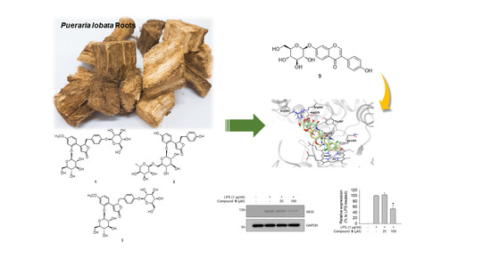
Lobatamunsolides A–C, Norlignans from the Roots of Pueraria lobata and their Nitric Oxide Inhibitory Activities in Macrophages

1
School of Pharmacy, Sungkyunkwan University, Suwon 16419, Korea
2
College of Pharmacy, Chonnam National University, Gwangju 61186, Korea
*
Author to whom correspondence should be addressed.
Biomolecules 2019, 9(12), 755; https://doi.org/10.3390/biom9120755
Submission received: 31 October 2019 / Revised: 15 November 2019 / Accepted: 17 November 2019 / Published: 20 November 2019

Abstract

:
Phytochemical investigation of the methanol (MeOH) extract of Pueraria lobata roots, known as “kudzu”, combined with liquid chromatography/mass spectrometry (LC/MS)-based analysis, resulted in the identification of four norlignans (14), including three new norlignans, lobatamunsolides A–C (13), and five known isoflavonoids (59). The structures of the new compounds were elucidated by a combination of spectroscopic methods, including 1D and 2D nuclear magnetic resonance (NMR) and high resolution (HR)-electrospray ionization mass spectrometry (ESIMS), and their absolute configurations were determined by chemical reaction and quantum chemical electronic circular dichroism (ECD) calculations. The isolated compounds (19) were evaluated for their inhibitory effects on nitric oxide (NO) production in lipopolysaccharide (LPS)-stimulated RAW 264.7 macrophages. Compound 9 displayed the strongest NO inhibitory effect and compound 2 showed a weak effect. The potential mechanism of the effect of compound 9 was investigated by analysis of its molecular docking on the active site of inducible nitric oxide synthase (iNOS), which showed the potential interactions of compound 9 with key amino acid residues and the heme cofactor of iNOS. The mechanism as the inhibition of transcriptional iNOS protein expression was confirmed by western blotting experiments.

Graphical Abstract

1. Introduction

Pueraria lobata (Willd.) Ohwi is a creeping, climbing, and trailing perennial vine belonging to the plant family Leguminosae, and is distributed in Southeast Asia, the Pacific, East Asia (Korea, China, Japan, and Russian far east), and the United States [1,2]. P. lobata, commonly known as “kudzu”, has been used in Korean traditional medicine for treating headache, diarrhea, fever, and cardiovascular disease [3,4]. In particular, the root of P. lobata has been used as a major medicinal and food ingredient in East Asia. Starch from P. lobata root is consumed as a popular drink in Vietnam and is used in many dishes, including kuzumochi and kuzuyu, in Japan, indicating the high nutritional value of the P. lobata root [5]. Previous studies have shown that extracts of P. lobata roots prevent obesity, improve glucose metabolism [6], and reduce oxidative stress [7]. In addition, previous phytochemical studies on the roots of P. lobata have revealed that its major bioactive compounds are isoflavones such as daidzein, daidzin, puerarin, and genistein [8]. These isoflavones exhibit diverse pharmacological effects, such as anti-inflammatory, antimicrobial, antioxidant, and antidipsotropic effects [9,10]. The P. lobata root has recently become commercially available in Western dietary supplements for the treatment of menopausal symptoms, and P. lobata root isoflavones such as puerarin have shown promise as natural alternatives to hormone replacement therapy for postmenopausal symptoms, with reduced risks of cancer [11,12].
As part of ongoing projects to identify structurally and/or biologically novel compounds from natural sources [13,14,15,16,17], we investigated the active compounds from P. lobata root that have both estrogen-like and anti-breast cancer cell proliferative activities. We used the bioactivity-guided isolation approach employed in a recent report that demonstrated that genistein from P. lobata root has estrogen-like effects dependent on estrogen receptor (ER) pathway activation, and anti-proliferative effects mediated by the apoptosis pathway in MCF-7 breast cancer cells [18]. These findings prompted us to investigate other potential bioactive components from P. lobata roots. Chemical analysis of the methanol (MeOH) extract of P. lobata root combined with liquid chromatography/mass spectrometry (LC/MS)-based analysis using an in-house ultraviolet (UV) spectra library led to the isolation and identification of four norlignans (14), including three new norlignans (13) (Figure 1), and five isoflavonoids (59) from the n-butanol-soluble fraction of the extract. The structures of the new norlignans were elucidated by a combination of 1D and 2D nuclear magnetic resonance (NMR) spectroscopic experiments, high resolution (HR)-electrospray ionization mass spectrometry (ESIMS), chemical reactions, and quantum chemical electronic circular dichroism (ECD) calculations. Furthermore, we elucidated the inhibitory effects of compounds 19 on nitric oxide (NO) production in lipopolysaccharide (LPS)-activated RAW 264.7 macrophages.

2. Materials and Methods

2.1. Plant Material

P. lobata roots grown in Geochang, Gyeongnam Province, Korea in 2014 were purchased from Okchundang Co., Ltd. A sample specimen of this material (GK-14-063) was confirmed by one of the authors (K. H. Kim) and stored in the herbarium of the School of Pharmacy, Sungkyunkwan University, Suwon, Korea.

2.2. Extraction and Isolation

The dried P. lobata roots (500 g) were extracted using 80% MeOH (20 h × 3) at room temperature. The extract was finely filtered and evaporated under reduced pressure using a rotary evaporator to obtain the MeOH extract (206.7 g). An in-depth description regarding the isolation of compounds from MeOH extract using the LC/MS guided isolation method can be found in the Supplementary Materials.

2.2.1. Lobatamunsolide A (1)

White, amorphous powder; [ α ] D 25 −38.8 (c 0.08, MeOH); UV (MeOH) λmax (log ε) 195 (4.0), 215 (3.2), 310 (2.7) nm; ECD (MeOH) λmaxε) 228 (−9.76), 250 (+7.01), 282 (−15.76) nm; IR (KBr) νmax 3705, 2966, 2833, 2492, 2704, 1698, 1516, 1024 cm−1; 1H (850 MHz) and 13C (212.5 MHz) NMR data, see Table 1; (+)−ESIMS m/z 659 [M + Na]+; (+)−HRESIMS m/z 637.2143 [M + H]+ (calculated for C30H37O15 637.2132).

2.2.2. Lobatamunsolide B (2)

White, amorphous powder; [ α ] D 25 +23.1 (c 0.08, MeOH); UV (MeOH) λmax (log ε) 195 (4.0), 215 (3.2), 310 (2.7) nm; ECD (MeOH) λmaxε) 230 (−5.19), 254 (+8.34), 284 (−19.77) nm; IR (KBr) νmax 3705, 3330, 2947, 2828, 2513, 2047, 1453, 1029 cm−1; 1H (850 MHz) and 13C (212.5 MHz) NMR data, see Table 1; (+)−ESIMS m/z 629 [M + Na]+; (+)−HRESIMS m/z 607.2018 [M + H]+ (calculated for C29H35O14 607.2027).

2.2.3. Lobatamunsolide C (3)

White, amorphous powder; [ α ] D 25 −50.1 (c 0.09, MeOH); UV (MeOH) λmax (log ε) 200 (3.5), 215 (3.8), 310 (3.7) nm; ECD (MeOH) λmaxε) 228 (+1.55), 247 (−27.72), 284 (+24.02) nm; IR (KBr) νmax 3709, 3320, 2940, 2827, 1750, 1446, 1127, 1024 cm−1; 1H (850 MHz) and 13C (212.5 MHz) NMR data, see Table 1; (+)–ESIMS m/z 659 [M + Na]+; (+)–HRESIMS m/z 637.2126 [M + H]+ (calculated for C30H37O15 637.2132).

2.3. Acid Hydrolysis and Determination of the Absolute Configuration of Sugar Moieties

The absolute configuration of the sugar moieties was determined using an LC/MS–UV-based method, which is described in detail in the Supplementary Materials.

2.4. Computational Analysis

Conformational optimization of 1a/1b was carried out using computational density functional theory (DFT) calculations. The first structural energy minimization of 1a/1b was carried out using Avogadro 1.2.0 with universal force field (UFF) setting. The ground-state geometries of 1a/1b were established by Tmolex 4.3.1 with DFT settings at B3-LYP functional/M3 grid size, geometry optimization options were set at energy 10−6 hartree, gradient norm |dE/dxyz| = 10−3 hartree/bohr, and the basis set was def-SV(P) by default for all atoms. The details of this analysis are described in the Supplementary Materials.

2.5. NO Production Assay

RAW 264.7 cells (6.0 × 10⁴ cells/well) in 96 well plates were incubated overnight and then stimulated with LPS (1 µg/mL) after treatment with the indicated concentrations of compounds. After 100 µL, cultured media were transferred to a new 96 well plate, and 100 µL Griess reagent (a mixture of 1% sulfanilamide in 2.5% phosphoric acid (H3PO4) and 0.1% N-(1-raphthyl) ethylenediamine in distilled water) was added to each well. Sodium nitrite was used to generate a standard curve. Absorbance was measured at 540 nm using a Synergy H1 Microplate Reader.

2.6. Cell Viability Assay

Cell viability was measured using the EZ-Cytox cell viability assay kit (DAEIL lab, Seoul, Korea) according to the manufacturer’s instruction. RAW 264.7 cells (6.0 × 10⁴ cells/well) were seeded into a 96 well plate. After overnight incubation, cells were treated with different concentrations of compounds. After incubation for 24 h, the cells were incubated for 1 h at 37 °C by adding EZ-Cytox diluent. Absorbance of culture medium was measured at 450 nm using a Synergy H1 Microplate Reader (BioTek Instruments, Winooski, VT, USA).

2.7. Preparation of Ligand and Receptor for Docking

All the procedures for docking modeling were carried out using the Tripos Sybyl-X 2.1.1 (Tripos Inc, St Louis, MO, USA) molecular modeling package with a Windows 7 professional K operating system. The structure of compound 9 was sketched and saved in the MOL2 format. Sequentially, Gasteiger–Hückel charges were assigned to all the atoms. To optimize the structure, energy minimization was conducted under a standard tripos force field with convergence to maximum derivatives of 0.001 kcal mol−1−1. As a target receptor for 9, the X-ray structure of murine iNOS in complex with the inhibitor AR-C124355 (PDB id: 3E6O) was downloaded at a resolution of 2.6 Å from Protein Data Bank (https://www.rcsb.org). To prepare the receptor, all the water molecules and duplicated chains were deleted, hydrogen atoms were attached, and Amber 7 FF99 charges were assigned using the biopolymer module in the Sybyl program.

2.8. Molecular Docking Analysis

Flexible docking was performed using a Surflex-Dock embedded in Tripos Sybyl X 2.1.1. The active site was generated as a protomol based on a native ligand (AR-C124355) extracted from the co-crystal structure of iNOS (PDB id: 3E6O). The protomol was built using the hydrogen-containing protein mol2 file. All parameters were applied by default to the docking simulation and 50 conformers were generated for the ligand. The binding affinity (−log Kd) of each conformer was calculated by the Surflex-Dock scoring function, and consensus scores were obtained based on five scores (Total Score, PMF score, ChemScore, G score, D score). The best docking model was picked out considering the calculated binding affinity, consensus score ≥4, and the number of intermolecular interactions.

2.9. Western Blot Analysis

RAW 264.7 cells (2.0 × 10⁶ cells/well) were seeded into 6 well plates and incubated overnight for adhesion. Cells were simultaneously treated with selected concentrations (25 µM and 100 µM) of compounds 2 and 9 and LPS (1 µg/mL) and incubated for 24 h. After washing cells with ice-cold PBS, radioimmunoprecipitation assay (RIPA) buffer was applied to the cells and the supernatants obtained after centrifugation were collected as total protein lysates. The same quantities of proteins were prepared with protein sample loading buffer and subjected to SDS-PAGE. Proteins separated on the gel were transferred onto nitrocellulose (NC) membranes, which were then blocked with 5% non-fat dried milk. The membranes were incubated overnight at 4 °C with iNOS and GAPDH primary antibodies, following which secondary antibodies conjugated with HRP were applied to the membranes. Finally, the membranes were blotted with an enhanced chemiluminescence reagent, and the intensities of bands obtained were quantified using an Amersham Imager 680 (GE Healthcare, Chicago, IL, United States).

2.10. Statistical Analysis

Significance of the data was analyzed by employing Student’s t-test. Results for experimental replicates (n = 9 for NO assay and cell viability; n = 3 for western blotting) were compared with those for the LPS-treated control group. p < 0.05 was considered statistically significant, and groups showing statistically significant differences are indicated with an asterisk (*).

3. Results and Discussion

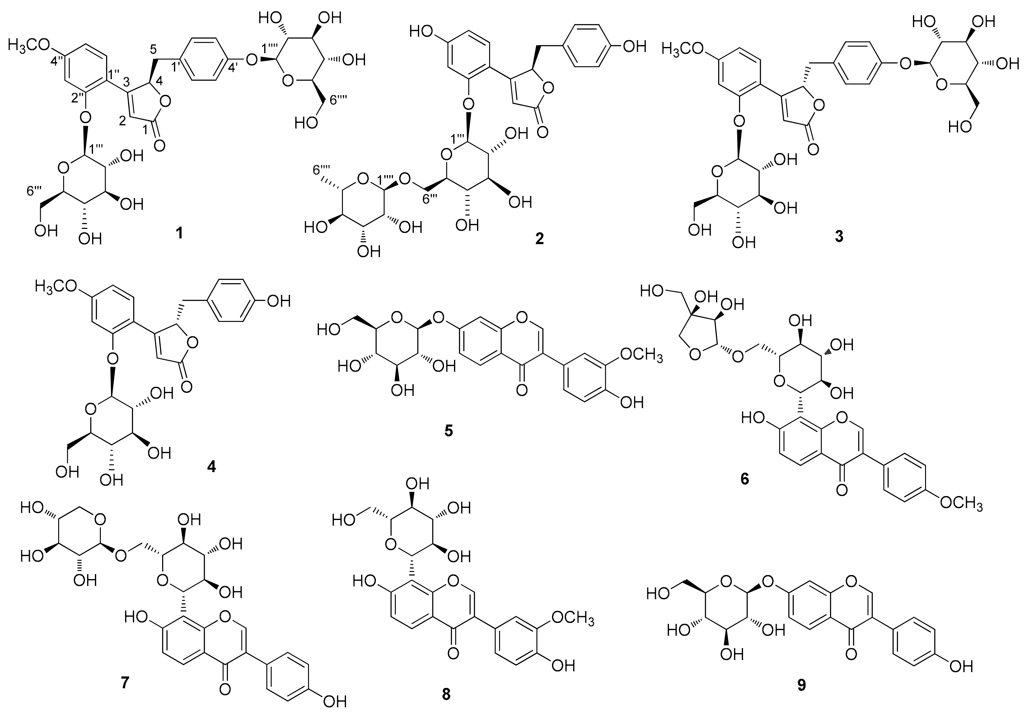
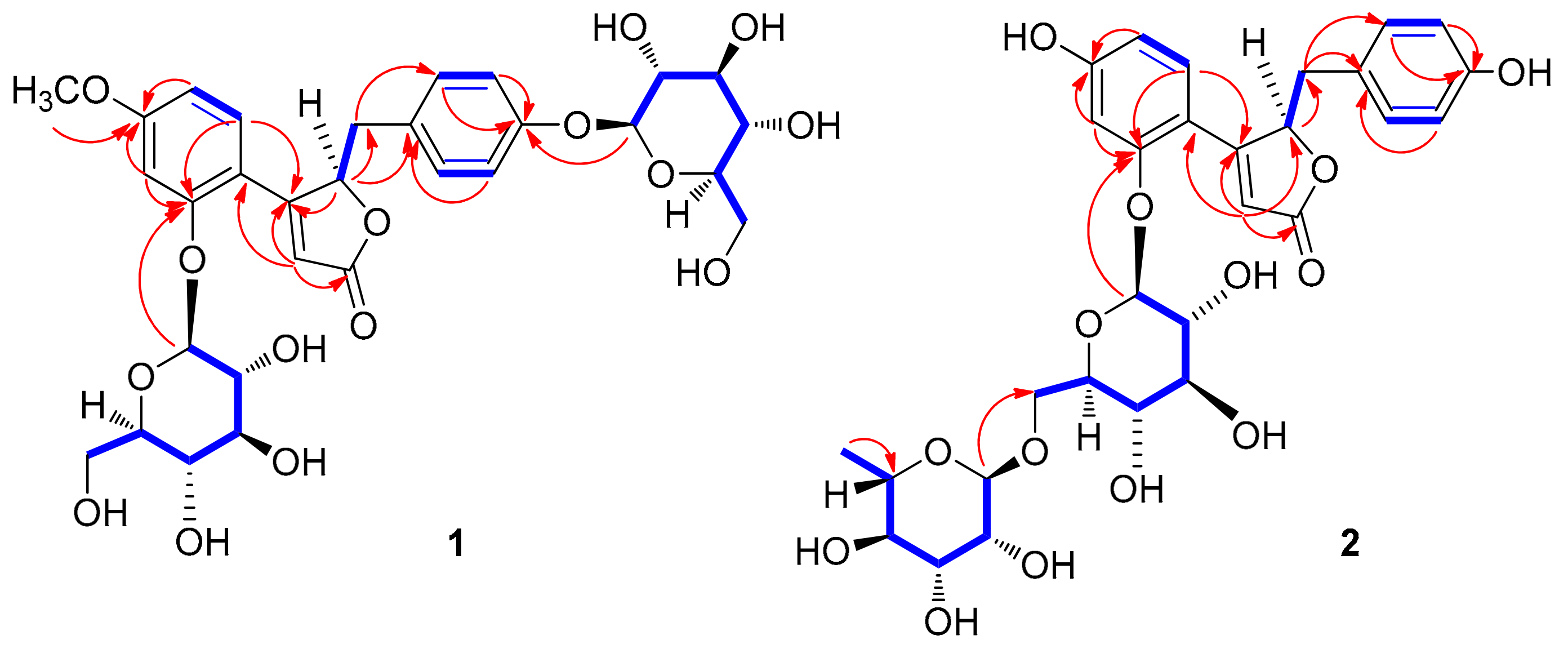
3.1. Isolation of Compounds

LC/MS-guided phytochemical investigation of the MeOH extract using series of column chromatography, such as silica gel, RP-C18 silica, Sephadex LH-20, and HP-20, along with preparative and semi-preparative HPLC, were used to isolate and identify four norlignans (14), including three new norlignans (13), and five isoflavonoids (59) from the n-butanol-soluble fraction of the extract.

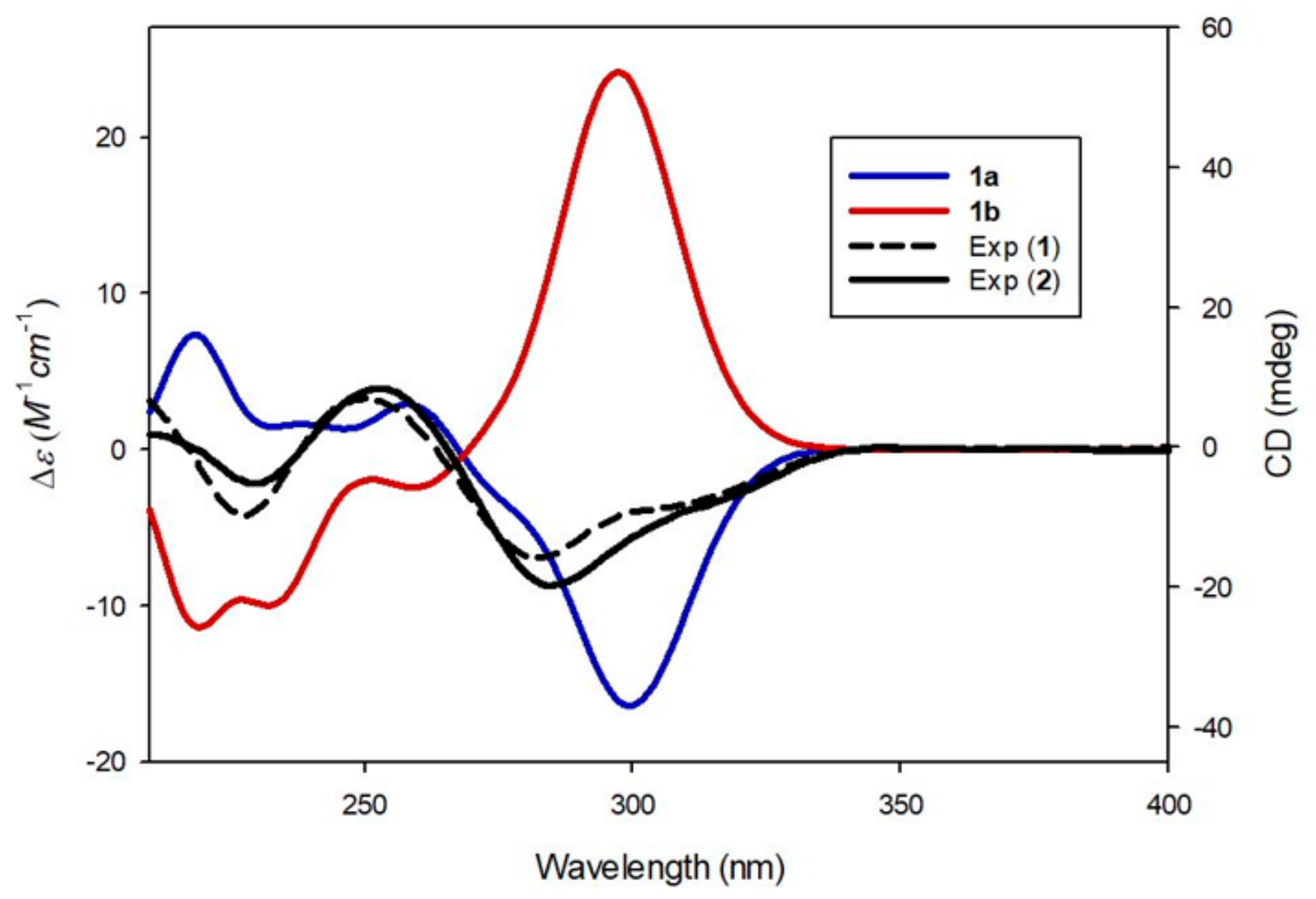
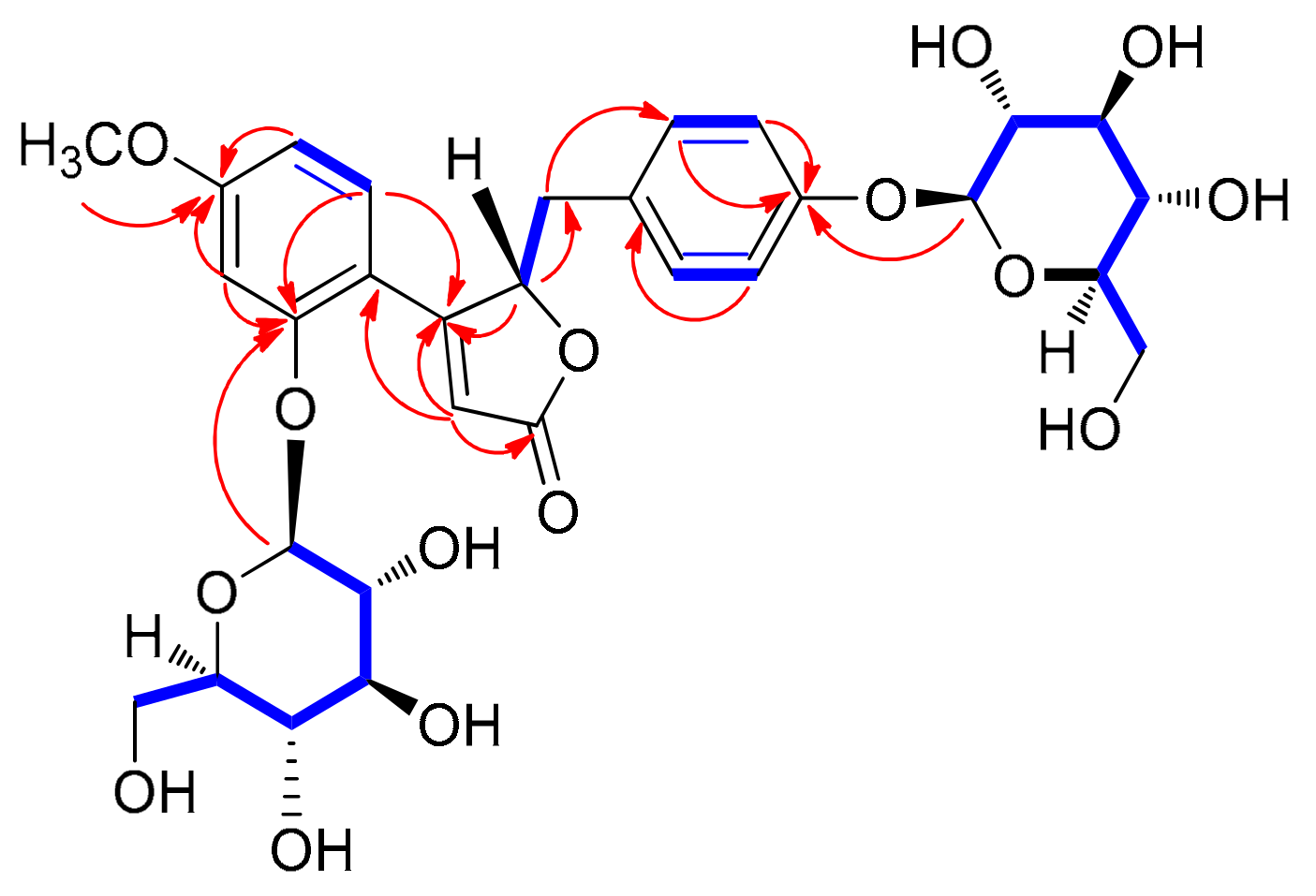
3.2. Structure Elucidation of Compounds
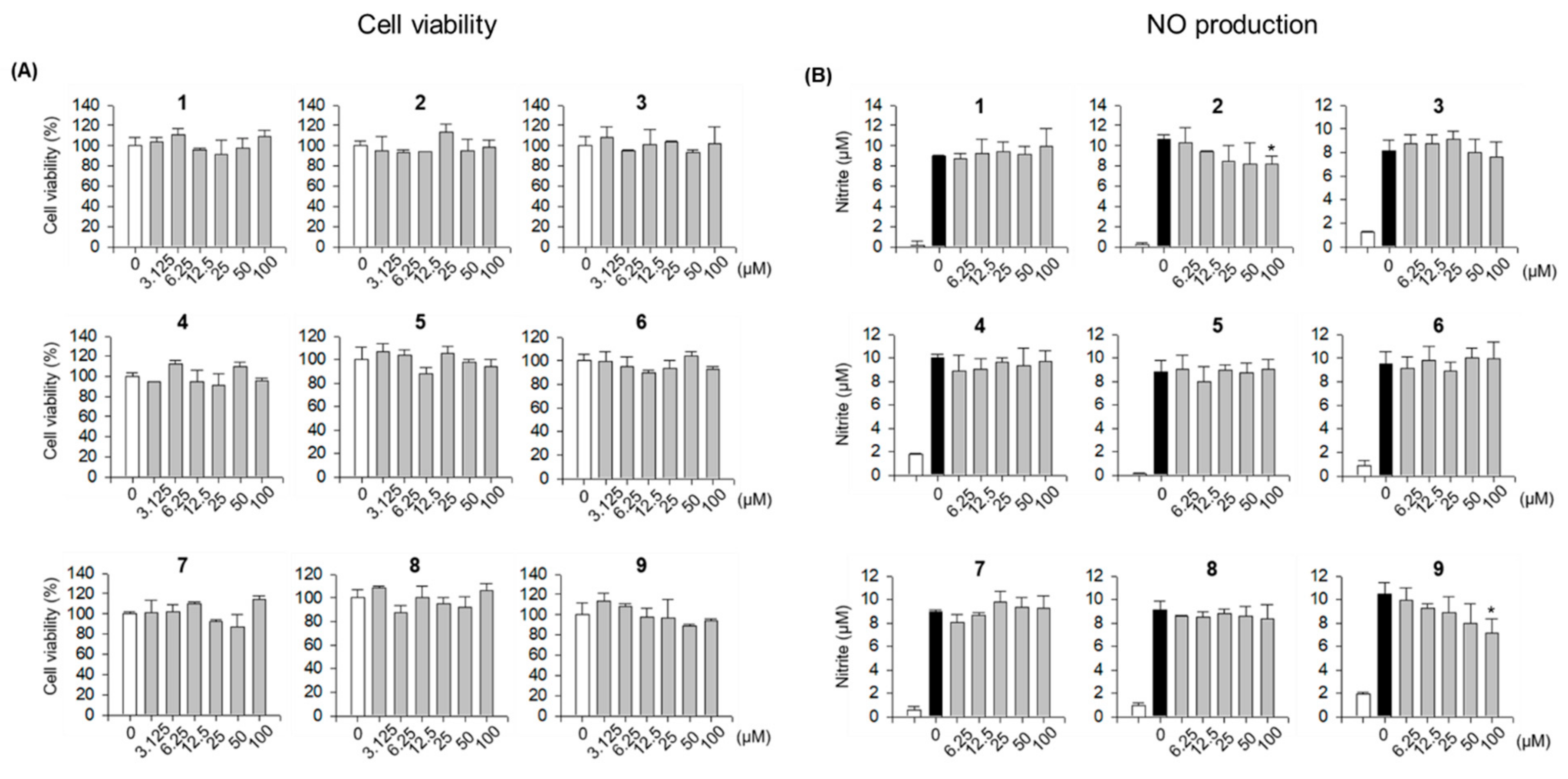
Compound 1 was isolated as a white amorphous powder. Its molecular formula was determined to be C30H36O15 based on the [M + H]+ peak at m/z 637.2143 (calculated for C30H37O15 637.2132) in the positive-ion HR-ESI-MS spectrum. The IR spectrum of 1 showed absorption bands for hydroxy (3356 cm−1) and α,β-unsaturated ketone (1677 cm−1) functional groups. The 1H NMR spectrum of 1 (Table 1) showed signals for a proton set of 1,4-disubstituted benzene at δH 6.89 (2H, d, J = 8.5 Hz) and 6.85 (2H, d, J = 8.5 Hz); another proton set of 1,3,4-trisubstituted benzene at δH 7.34 (1H, d, J = 8.5 Hz), 6.96 (1H, d, J = 2.5 Hz), and 6.73 (1H, dd, J = 8.5, 2.5 Hz); one olefinic proton at δH 6.30 (1H, d, J = 1.0 Hz); one oxygenated proton at δH 6.07 (1H, td, J = 4.5, 1.0 Hz); one methylene proton at δH 3.24 (1H, dd, J = 15.0, 4.5 Hz) and 2.92 (1H, dd, J = 15.0, 4.5 Hz); one methoxy group at δH 3.88 (3H, s); as well as signals attributable to two sugar moieties, including two anomeric protons at δH 5.12 (1H, d, J = 7.5 Hz) and 4.84 (1H, d, J = 7.5 Hz). The 13C NMR data (Table 1), obtained with the aid of heteronuclear single quantum coherence (HSQC) and heteronuclear multiple bond correlation (HMBC) spectra, displayed resonances of 18 carbons for aglycone, including 12 carbons of aromatic rings, α,β-unsaturated ketone (δC 176.4, 166.7, and 115.9), one methylene (δC 38.7), one oxygenated methine (δC 84.6), and one methoxy group (δC 56.0), together with 12 carbons of two sugar groups. Detailed inspection of 1H and 13C NMR data revealed that our NMR data closely coincided with those for kuzubutenolide A [19], except for the signals for an additional sugar moiety and one methoxy group. Both sugar units were determined to be glucopyranose upon comparing our values with those reported previously [19]; moreover, the coupling constant (J = 7.5 Hz) of anomeric proton signals was indicative of the β-form of glucopyranose [20]. The locations of the two β-glucopyranosyl groups were clearly confirmed as C-2″ and C-4′ through HMBC correlations of the two anomeric protons with C-2″ (δC 158.5) and C-4′ (δC 158.0), respectively (Figure 2). The HMBC correlations of the methoxy group (δH 3.88) with C-4′’ (δC 164.8) allowed us to confirm its location as C-4″ (Figure 2). The gross planar structure of 1 was completely elucidated through 2D NMR analysis (1H–1H correlation spectroscopy (COSY), HSQC, and HMBC) (Figure 2). The absolute configuration of sugar moieties was determined using an LC/MS–UV-based method [21,22,23], and acid hydrolysis of 1 was carried out to yield a glucopyranose. The absolute configuration of the sugar moieties was determined as d-glucopyranose by comparing the retention times of the thiocarbamoyl-thiazolidine derivative of their acid hydrolysates with that of the standard d-glucopyranose sample through LC/MS analysis. In order to confirm the absolute configuration, quantum chemical ECD calculations were performed by comparing the experimental ECD spectrum of 1 with the calculated ECD spectra of two possible enantiomers, 1a (4R) and 1b (4S) (Figure 3). The experimental ECD data of 1 matched well with the calculated ECD data for 1a. Accordingly, the chemical structure of 1, including its absolute configuration, was elucidated as shown in Figure 1, and the compound was named lobatamunsolide A.
Compound 2 was isolated as a white amorphous powder with a molecular formula of C29H34O14, as deduced from the HRESIMS data at m/z 607.2018 [M + H]+ (calculated for C29H35O14 607.2027) and NMR data (Table 1). The 1H and 13C NMR data of 2 showed signals quite similar to those of 1, with noticeable differences being the absence of signals for the methoxy group in 1 and the presence of an additional characteristic methyl group at δH 1.18 (3H, d, J = 6.0 Hz)/δC 17.6. The characteristic methyl group was determined to be C-6 of rhamnopyranose, and the two sugar moieties present in 2 were identified as rutinose [6-O-α-rhamnosyl-(1→6)-β-glucopyranoside] upon comparison with previously reported values [24] and analysis of the coupling constants of the anomeric proton signals at δH 5.03 (1H, d, J = 7.5 Hz) and 4.70 (1H, d, J = 1.0 Hz). The complete planar structure of 1, including the position of the rutinosyl group, was determined by 2D NMR analysis (1H–1H COSY, HSQC, and HMBC) (Figure 2). The absolute configuration of 2 was determined via chemical reactions and quantum chemical ECD calculations. Acid hydrolysis of 2 yielded a glucopyranose and a rhamnopyranose, the absolute configurations of which were determined as d-glucopyranose and l-rhamnopyranose by LC/MS analysis of their thiocarbamoyl–thiazolidine derivatives. To establish the absolute configuration of C-4 in 2, computationally calculated ECD data of two enantiomers, 1a (4R) and 1b (4R), were compared with the experimental ECD data of 2 (Figure 3). The ECD data of 2 were well matched to the calculated ECD data of 1a and the experimental ECD data of 1. Collectively, these findings allowed the elucidation of the absolute structure of 2 as shown in Figure 1, and the compound was named lobatamunsolide B.
Compound 3 was isolated as a white amorphous powder, and its molecular formula was determined as C30H36O15 based on HRESIMS data at m/z 637.2126 [M + H]+ (calculated for C30H37O15 637.2132) and NMR data (Table 1). The 1H and 13C NMR spectra of 3 (Table 1) were almost identical to those of 1, with slight differences in the chemical shifts assigned to the butenolide moiety. Their superimposable NMR data and the fact that they have the same molecular formula suggested that compound 3 is a stereoisomer of 1. The complete structure of 3 was further confirmed by analysis of cross-peaks through HMBC and 1H–1H COSY experiments (Figure 4). Acid hydrolysis of 3 yielded a glucopyranose. The absolute configuration of this glucopyranose was established as d-form through the LC/MS analysis of the thiocarbamoyl–thiazolidine derivatives of its acid hydrolysate. The ECD spectrum of 3 displayed opposite Cotton effects (247 (negative), 284 (positive) nm) to those of the ECD spectrum of 1, suggesting that the absolute configuration of C-4 in 3 is opposite to that of 1. Moreover, the experimental ECD data of 3 were in agreement with the calculated ECD data of 1b (4S) rather than those of 1a (4R) (Figure 5). Therefore, the absolute structure of 3 was determined as shown in Figure 1, and the compound was named lobatamunsolide C.
The remaining six isolates from the MeOH extract of P. lobata roots were known compounds, which were identified as (+)-puerarol B-2-O-glucoside (4) [25], 3′-methoxydaidzin (5) [19], formononetin 8-C-apiofuranosyl-(1→6)-glucoside (6) [26], puerarin xyloside (7) [27], 3′-methoxypuerarin (8) [27], and daidzin (9) [27], by comparing their NMR spectroscopic data with those reported earlier and by LC/MS analysis.

3.3. Inhibitory Effects of Compounds 16 on LPS-Induced NO Production in RAW 264.7 Cells

Nitric oxide (NO) is known to have a close association with inflammation and possesses a dual regulatory function in the inflammatory reaction [28,29]. Under normal physiological conditions, NO plays anti-inflammatory roles; however, upon stimulation with LPS, inducible NO synthase (iNOS) is transcriptionally activated and, subsequently, NO is produced by macrophages as a first-line pro-inflammatory mediator. Excessive production of NO may lead to high expression of proinflammatory mediators and result in a variety of inflammatory diseases. Therefore, the discovery of agents that can inhibit excessive NO production might be an effective strategy to treat inflammatory disorders. Considering the goal to obtain NO inhibitors to treat inflammatory disorders and the traditional efficacy of P. lobate for this purpose, the inhibitory effects of compounds 19 isolated from P. lobate roots on NO production in LPS-stimulated RAW 264.7 macrophages were evaluated. Before evaluating the inhibitory activities, the effect of all compounds on cell viability at concentrations ranging from 3.125 to 100 μM was tested using water-soluble tetrazolium, and no cytotoxic effects were observed (Figure 6A). NO assays were then carried out at the range of 6.25 to 100 μM. Compound 9 exhibited the strongest NO inhibitory effect among all the isolated compounds, whereas compound 2 displayed a weak inhibitory effect on NO production in LPS-induced RAW 264.7 cells (Figure 6B).

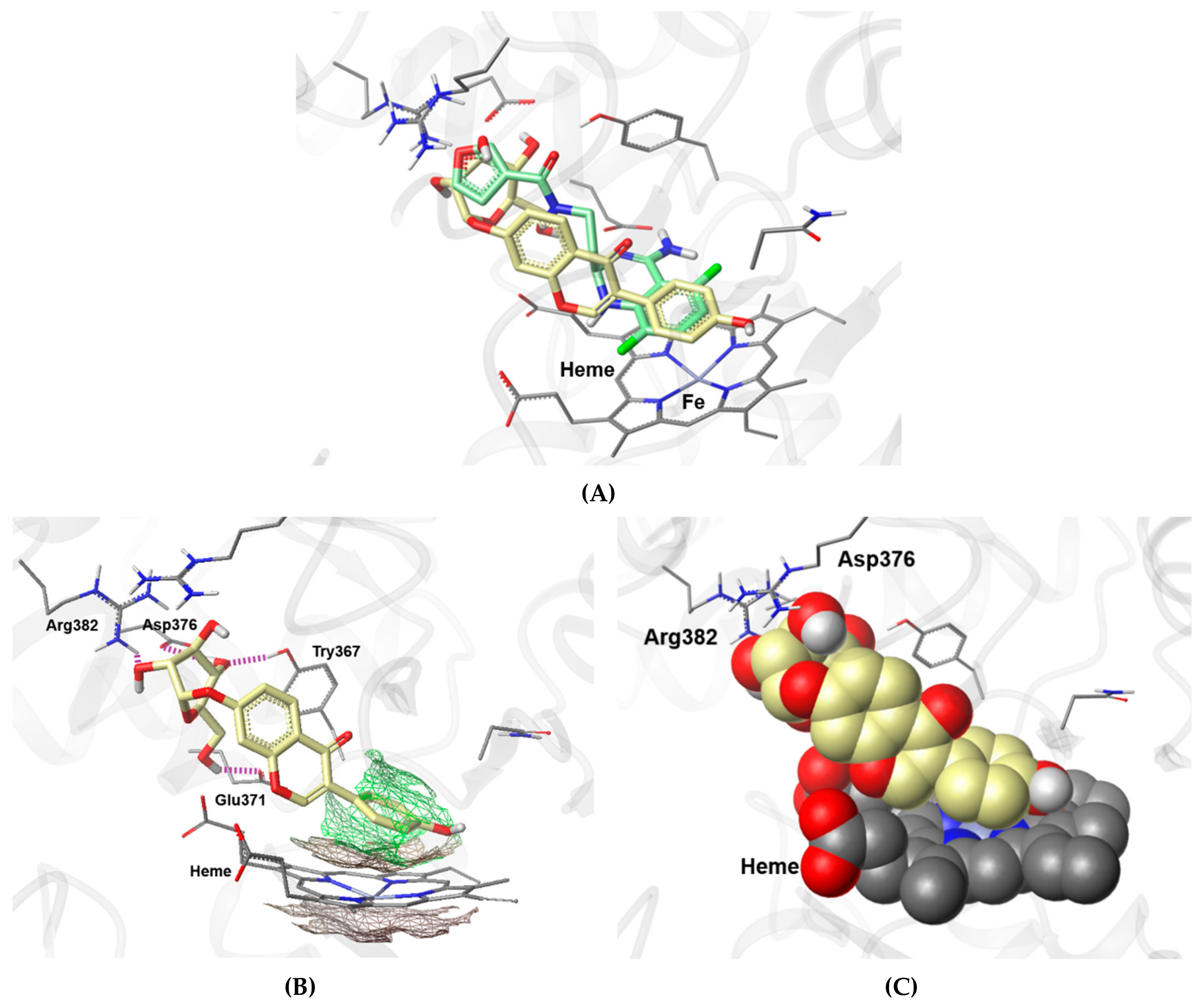
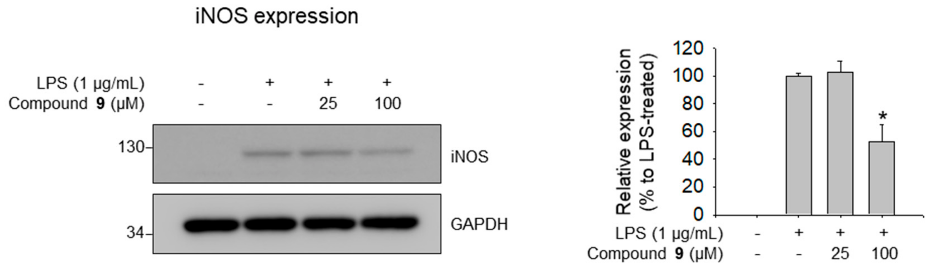
3.4. Molecular Docking Analysis of 9 and Its Inhibitory Effects on iNOS Expression

To elucidate the possible mechanism through which compound 9 inhibits LPS-induced NO production, the mode of binding of compound 9 within the active site of iNOS was investigated. Compound 9 was subjected to molecular docking analysis using the published X-ray structure of iNOS complexed with a difluoroquinazoline inhibitor AR-C124355 (N-[2-(4-Amino-5,8-difluoro-1,2-dihydroquinazolin-2-yl)ethyl]-3-furamide) [30]. The key interactions between AR-C124355 and amino acid residues in the active site of iNOS are as follows: (i) π–π stacking between the diflorophenyl ring and heme cofactor, (ii) anchoring bidentate hydrogen bonds (H-bonds) between the cis-amidine moiety of difluoroquinazoline and the side chain carboxylate group of Glu371, and (iii) π–cation interaction between the furan moiety and guanidium group of Arg260 [30]. As shown in Figure 7A, compound 9 fits snugly into the binding pocket of AR-C124355 via heme stacking and H-bonding to Glu371. Upon looking at the docked pose of 9 in detail (Figure 7B,C), it can be seen that the phenol ring of the daidzein moiety is positioned in parallel with the porphyrin plane of heme to form a π–π interaction, and that the three hydroxyl groups (C3, C5, and C6) of the d-glucopyranosyl moiety form H-bonds with Arg382, Asp376, and Glu371, respectively. The docking model suggests that 9 fits into the common binding pocket for known iNOS inhibitors, forming key interactions with Glu371 and the heme cofactor in the active site of iNOS, thus acting as an iNOS inhibitor.
To confirm whether the protein expression levels of iNOS were decreased by compound 9, western blotting experiments were performed. Corresponding to the results for the effects of compound 9 on NO production and its molecular docking with iNOS, compound 9 showed strong inhibition of the expression of iNOS protein in LPS-stimulated RAW 264.7 macrophages (Figure 8); however, compound 2 did not show such inhibitory effects.

4. Conclusions

Phytochemical investigation of the MeOH extract of P. lobata roots combined with LC/MS-based analysis led to the discovery of four norlignans (14), including three new norlignans, lobatamunsolides A–C (13), and five known isoflavonoids (59). Their structures were established based on NMR spectroscopic data, chemical reactions, and quantum chemical ECD calculations. Compound 9 displayed significant NO inhibitory effect in LPS-stimulated RAW 264.7 macrophages. The potential mechanism of the effect was also investigated using molecular docking and western blot analyses, which showed that the mechanism of 9 was mediated by the inhibition of iNOS protein expression. Overall, the present findings suggest the potential of P. lobata roots for use as a functional food and medicine ingredient, and indicate that compound 9 could be a valuable compound for the development of promising anti-inflammatory agents.

Supplementary Materials

The following are available online at https://www.mdpi.com/2218-273X/9/12/755/s1, HR-ESIMS, and 1D and 2D NMR spectra of compounds 13, general experimental procedures, extraction and isolation, acid hydrolysis and determination of the absolute configuration of sugar moieties, and computational analysis are available free of charge on the Internet.

Author Contributions

Conceptualization, Y.-C.C., H.-J.P., and K.H.K.; formal analysis, M.S.J., J.S.Y., and S.L.; investigation, M.S.J., J.S.Y., J.C.L., and S.L.; data curation, M.S.J., S.L., Y.-C.C., H.-J.P., and K.H.K.; writing—original draft preparation, M.S.J., Y.-C.C., H.-J.P., and K.H.K.; writing—review and editing, M.S.J. and K.H.K.; supervision, Y.-C.C., H.-J.P., and K.H.K.; funding acquisition, K.H.K.

Funding

This work was supported by a National Research Foundation of Korea (NRF) grant funded by the Korea government (MSIT) (2018R1A2B2006879) and (2019R1A5A2027340). This research was supported by the Bio & Medical Technology Development Program of the National Research Foundation of Korea (NRF) funded by the Korean government (MSIT) (NRF-2012M3A9C4048775).

Acknowledgments

We would like to thank Young Hye Kim (KBSI) for the ESIMS analysis.

Conflicts of Interest

The authors declare no conflict of interest.

References

  1. Prasain, J.K.; Jones, K.; Kirk, M.; Wilson, L.; Smith-Johnson, M.; Weaver, C.; Barnes, S. Profiling and quantification of isoflavonoids in kudzu dietary supplements by high-performance liquid chromatography and electrospray ionization tandem mass spectrometry. J. Agric. Food Chem. 2018, 51, 4213–4218. [Google Scholar] [CrossRef] [PubMed]
  2. Patterson, D.T. The history and distribution of five exotic weeds in North Carolina. Castanea 1976, 177–180. [Google Scholar]
  3. Tanaka, T.; Tang, H.; Yu, F.; Michihara, S.; Uzawa, Y.; Zaima, N.; Moriyama, T.; Kawamura, Y. Kudzu (Pueraria lobata) vine ethanol extracts improve ovariectomy-induced bone loss in female mice. J. Agric. Food Chem. 2011, 59, 13230–13237. [Google Scholar] [CrossRef] [PubMed]
  4. Tam, W.Y. The efficacy and tolerability of adjunctive alternative herbal medicine (Salvia miltiorrhiza and Pueraria lobata) on vascular function and structure in coronary patients. J. Altern. Complement. Med. 2009, 15, 415–421. [Google Scholar] [CrossRef] [PubMed]
  5. Zhang, Z.; Lam, T.N.; Zuo, Z. Radix Puerariae: An overview of its chemistry, pharmacology, pharmacokinetics, and clinical use. J. Clin. Pharmacol. 2013, 53, 787–811. [Google Scholar] [CrossRef] [PubMed]
  6. Jung, H.; Kang, A.N.; Kang, S.; Park, Y.K.; Song, M. The root extract of Pueraria lobata and its main compound, puerarin, prevent obesity by increasing the energy metabolism in skeletal muscle. Nutrients 2017, 9, 33. [Google Scholar] [CrossRef] [PubMed]
  7. Bebrevska, L.; Foubert, K.; Hermans, N.; Chatterjee, S.; Van Marck, E.; De Meyer, G.; Vlietinck, A.; Pieters, L.; Apers, S. In vivo antioxidative activity of a quantified Pueraria lobata root extract. J. Ethnopharmacol. 2010, 127, 112–117. [Google Scholar] [CrossRef]
  8. Reppert, A.; Yousef, G.G.; Rogers, R.B.; Lila, M.A. Isolation of radiolabeled isoflavones from kudzu (Pueraria lobata) root cultures. J. Agric. Food Chem. 2008, 56, 7860–7865. [Google Scholar] [CrossRef]
  9. Jin, S.E.; Son, Y.K.; Min, B.S.; Jung, H.A.; Choi, J.S. Anti-inflammatory and antioxidant activities of constituents isolated from Pueraria lobata roots. Arch. Pharm. Res. 2012, 35, 823–837. [Google Scholar] [CrossRef]
  10. Keung, W.M.; Vallee, B.L. Kudzu root: An ancient Chinese source of modern antidipsotropic agents. Phytochemistry 1998, 47, 499–506. [Google Scholar] [CrossRef]
  11. Kayano, S.I.; Matsumura, Y.; Kitagawa, Y.; Kobayashi, M.; Nagayama, A.; Kawabata, N.; Kikuzaki, H.; Kitada, Y. Isoflavone C-glycosides isolated from the root of kudzu (Pueraria lobata) and their estrogenic activities. Food Chem. 2012, 134, 282–287. [Google Scholar] [CrossRef]
  12. Zhou, Y.-X.; Zhang, H.; Peng, C. Puerarin: A review of pharmacological effects. Phytother. Res. 2014, 28, 961–975. [Google Scholar] [CrossRef] [PubMed]
  13. Yu, J.S.; Lee, D.; Lee, S.R.; Lee, J.W.; Choi, C.-I.; Jang, T.S.; Kang, K.S.; Kim, K.H. Chemical characterization of cytotoxic indole acetic acid derivative from Mulberry fruit (Morus alba L.) against human cervical cancer. Bioorg. Chem. 2018, 76, 28–36. [Google Scholar] [CrossRef] [PubMed]
  14. So, H.M.; Eom, H.J.; Lee, D.; Kim, S.; Kang, K.S.; Lee, I.K.; Baek, K.-H.; Park, J.Y.; Kim, K.H. Bioactivity evaluations of betulin identified from the bark of Betula platyphylla var. japonica for cancer therapy. Arch. Pharm. Res. 2018, 41, 815–822. [Google Scholar] [PubMed]
  15. Yu, J.S.; Roh, H.-S.; Baek, K.-H.; Lee, S.; Kim, S.; So, H.M.; Moon, E.; Pang, C.; Jang, T.S.; Kim, K.H. Bioactivity-guided isolation of ginsenosides from Korean Red Ginseng with cytotoxic activity against human lung adenocarcinoma cells. J. Ginseng Res. 2018, 42, 562–570. [Google Scholar] [CrossRef] [PubMed]
  16. Lee, S.R.; Song, J.H.; Song, J.-H.; Ko, H.-J.; Baek, J.Y.; Trinh, T.A.; Beemelmanns, C.; Yamabe, N.; Kim, K.H. Chemical identification of isoflavonoids from a termite-associated Streptomyces sp. RB1 and their neuroprotective effects in murine hippocampal HT22 cell line. Int. J. Mol. Sci. 2018, 19, 2640. [Google Scholar] [CrossRef]
  17. Baek, S.C.; Choi, E.; Eom, H.J.; Jo, M.S.; Kim, S.; So, H.M.; Kim, S.H.; Kang, K.S.; Kim, K.H. LC/MS-based analysis of bioactive compounds from the bark of Betula platyphylla var. japonica and their effects on regulation of adipocyte and osteoblast differentiation. Nat. Prod. Sci. 2018, 24, 235–240. [Google Scholar]
  18. Ahn, S.-Y.; Jo, M.S.; Lee, D.; Baek, S.-E.; Baek, J.; Yu, J.S.; Jo, J.; Yun, H.; Kang, K.S.; Yoo, J.-E.; et al. Dual effects of isoflavonoids from Pueraria lobata roots on estrogenic activity and anti-proliferation of MCF-7 human breast carcinoma cells. Bioorg. Chem. 2019, 83, 135–144. [Google Scholar] [CrossRef]
  19. Hirakura, K.; Morita, M.; Nakajima, K.; Sugama, K.; Takagi, K.; Niitsu, K.; Ikeya, Y.; Maruno, M.; Okada, M. Phenolic glucosides from the root of Pueraria lobata. Phytochemistry 1997, 46, 921–928. [Google Scholar] [CrossRef]
  20. Coxon, B. Two-dimensional J.-resolved proton nuclear magnetic resonance spectrometry of hydroxyl-coupled α- and β-d-glucose. Anal. Chem. 1983, 55, 2361–2366. [Google Scholar] [CrossRef]
  21. Tanaka, T.; Nakashima, T.; Ueda, T.; Tomii, K.; Kouno, I. Facile discrimination of aldose enantiomers by reversed-phase HPLC. Chem. Pharm. Bull. 2007, 55, 899–901. [Google Scholar] [CrossRef] [PubMed]
  22. Muhit, M.A.; Umehara, K.; Mori-Yasumoto, K.; Noguchi, H. Furofuran lignan glucosides with estrogen-inhibitory properties from the Bangladeshi medicinal plant Terminalia citrina. J. Nat. Prod. 2016, 79, 1298–1307. [Google Scholar] [CrossRef] [PubMed]
  23. Kil, Y.S.; Kim, S.M.; Kang, U.; Chung, H.Y.; Seo, E.K. Peroxynitrite-scavenging glycosides from the stem bark of Catalpa ovata. J. Nat. Prod. 2017, 80, 2240–2251. [Google Scholar] [CrossRef] [PubMed]
  24. Aquino, R.; Behar, I.; D’agostino, M.; Simone, F.D.; Schettino, O.; Pizza, C. Phytochemical investigation on Mercurialis annua. Biochem. Syst. Ecol. 1987, 15, 667–669. [Google Scholar] [CrossRef]
  25. Ding, H.Y.; Chen, Y.P.; Chang, W.L.; Lin, H.C. Isoflavonoids and but-2-enolides from the roots of Pueraria lobata. Chin. Pharm. J. 2004, 56, 31–35. [Google Scholar]
  26. Wang, F.R.; Ge, X.Z.; Yang, X.W. Chemical constituents of Tongmai formula. Chin. J. Exp. Tradit. Med. Formulae 2011, 17, 61–69. [Google Scholar]
  27. Kinjo, J.E.; Furusawa, J.I.; Baba, J.; Takeshita, T.; Yamasaki, M.; Nohara, T. Studies on the constituents of Pueraria lobata. III. Isoflavonoids and related compounds in the roots and the voluble stems. Chem. Pharm. Bull. 1987, 35, 4846–4850. [Google Scholar] [CrossRef]
  28. Lee, S.; Lee, D.; Lee, J.C.; Kang, K.S.; Ryoo, R.; Park, H.J.; Kim, K.H. Bioactivity-guided Isolation of Anti-inflammatory Constituents of the Rare Mushroom Calvatia nipponica in LPS-stimulated RAW264.7 Macrophages. Chem. Biodivers. 2018, 15, e1800203. [Google Scholar] [CrossRef]
  29. Lee, S.; Lee, D.; Lee, S.O.; Ryu, J.Y.; Choi, S.Z.; Kang, K.S.; Kim, K.H. Anti-inflammatory activity of the sclerotia of edible fungus, Poria cocos Wolf and their active lanostane triterpenoids. J. Funct. Foods 2017, 32, 27–36. [Google Scholar] [CrossRef]
  30. Garcin, E.D.; Arvai, A.S.; Rosenfeld, R.J.; Kroeger, M.D.; Crane, B.R.; Andersson, G.; Andrews, G.; Hamley, P.J.; Mallinder, P.R.; Nicholls, D.J.; et al. Anchored plasticity opens doors for selective inhibitor design in nitric oxide synthase. Nat. Chem. Biol. 2008, 4, 700–707. [Google Scholar]
Figure 1. Structures of compounds 19 isolated from P. lobata roots.
Biomolecules 09 00755 g001
Figure 2. 1H–1H correlation spectroscopy (COSY) (blue bold lines) and key heteronuclear multiple bond correlation (HMBC) (red arrows) correlations of 1 and 2.
Biomolecules 09 00755 g002
Figure 3. Experimental and calculated electronic circular dichroism (ECD) spectra of compounds 1 and 2.
Biomolecules 09 00755 g003
Figure 4. 1H–1H COSY (blue bold lines) and key HMBC correlations (red arrows) of 3.
Biomolecules 09 00755 g004
Figure 5. Experimental and calculated ECD spectra of compounds 1 and 3.
Biomolecules 09 00755 g005
Figure 6. (A) Cell viabilities of the isolated compounds (19) measured using the EZ-Cytox cell viability assay. (B) Inhibitory effects of compounds 19 on LPS-induced NO production in RAW 264.7 cells. Cell viability data represent relative cell viability compared to that of the untreated group (100%). NO assay data represent NO production levels calculated by applying absorbance values at 540 nm to a nitrite standard curve. p < 0.05 relative to the LPS-treated control group.
Biomolecules 09 00755 g006
Figure 7. A putative binding mode of 9 in the active site of iNOS. (A) Superposition of the docked pose of 9 onto the co-crystalized pose of the inhibitor AR-C124355 in the active site of iNOS oxygenase (PDB id: 3E6O). The colors of the molecules are presented by atom type except for carbon atoms. The carbon atoms are colored green-blue (AR-C124355), yellow (9), and gray (amino acids); nitrogen is blue, oxygen is red, fluorine is green, hydrogen is white, and iron is light blue. (B) A docking pose of 9 fitted in the lipophilic domain. The amino acid residues involved in hydrogen bonds (H-bonds) are labeled and H-bonds are represented by purple dotted lines. The lipophilic potential surface, calculated by MOLCAD™ (a module in Sybyl-X 2.1.1), for heme and phenol moiety of 9 are stacked in parallel. (C) A view rendered in space-filling spheres, revealing a parallel stacking interaction between 9 and the co-factor heme. The atoms of docked 9 and heme are represented in Van der Waals spheres generated by Corey-Pauling-Koltun (CPK) rendering method.
Biomolecules 09 00755 g007
Figure 8. Inhibitory effects of compound 9 on LPS-induced iNOS expression in RAW 264.7 cells. Data represent relative iNOS protein expression levels compared to those in the LPS-treated group (100%). p < 0.05 relative to the LPS-treated control group.
Biomolecules 09 00755 g008
Table 1. 1H and 13C nuclear magnetic resonance (NMR) data of Compounds 13 in CD3OD a.
Position123
δHδC δHδC δHδC
1 176.4s 176.7s 176.0s
26.30 d (1.0)115.9d6.26 s114.9d6.17 s114.4d
3 166.7s 167.3s 168.1s
46.07 td (4.5, 1.0)84.6d6.04 td (5.0, 1.0)84.9d6.16 m85.5d
5a2.92 dd (15.0, 5.0)38.7t2.86 dd (15.0, 5.0)38.9t2.85 dd (15.0. 6.0)38.8t
5b3.24 dd (15.0, 4.5) 3.20 dd (15.0, 4.0) 3.24 dd (15.0, 3.5)
1′ 129.6s 126.7s 130.8s
2′6.85 d (8.5)131.9d6.78 d (8.5)131.9d7.00 d (8.5)131.7d
3′6.89 d (8.5)117.0d6.60 d (8.5)115.5d6.95 d (8.5)117.1d
4′ 158.0s 157.0s 157.9s
5′6.89 d (8.5)117.0d6.60 d (8.5)115.5d6.95 d (8.5)117.1d
6′6.85 d (8.5)131.9d6.78 d (8.5)131.9d7.00 d (8.5)131.7d
1″ 113.9s 113.1s 113.7s
2″ 158.5s 158.5s 158.1s
3″6.96 d (2.5)102.7d6.77 d (2.5)104.0d6.96 d (2.5)102.3d
4″ 164.8s 163.0s 165.0s
5″6.73 dd (8.5, 2.5)109.5d6.60 dd (8.5, 2.5)110.9d6.74 dd (8.5, 2.5)109.9d
6″7.34 d (8.5)131.8d7.26 d (8.5)132.1d7.45 d (8.5)132.2d
1‴5.12 d (7.5)101.6d5.03 d (7.5)101.6d5.07 d (7.5)101.8d
2‴3.48 m74.6d3.49 m74.5d3.48 m74.4d
3‴3.42 m77.9d3.46 m78.3d3.48 m78.0d
4‴3.36 m71.1d3.40 m71.0d3.36 m71.2d
5‴3.52 m78.4d3.59 m76.9d3.55 m78.4d
6‴a3.92 dd (12.0, 2.0)62.4t4.03 dd (11.5, 2.0)67.5t3.95 dd (12.0, 2.0)62.4t
6‴b 3.68 dd (12.0, 6.0) 3.65 dd (11.5, 6.0) 3.71 dd (12.0, 6.0)
1⁗4.84 d (7.5)102.1d4.70 d (1.0)102.1d4.85 d (7.5)102.1d
2⁗3.42 m74.8d3.88 m71.8d3.44 m74.6d
3⁗3.44 m77.8d3.69 dd (9.5, 3.5)72.0d3.48 m78.0d
4⁗3.38 m71.1d3.33 m73.8d3.44 m71.1d
5⁗3.48 m78.5d3.63 dd (9.6, 6.5)69.6d3.39 m78.3d
6⁗a3.86 dd (12.0, 2.0)62.3t1.18 d (6.0)17.6q3.90 dd (12.0, 2.0)62.2t
6⁗b 3.68 dd (12.0, 6.0) 3.69 dd (12.0, 6.0)
4″-OCH33.88 s56.0q 3.88 s55.8q
a Signal multiplicity and coupling constants (Hz) are in given parentheses; the assignments were based on heteronuclear single quantum coherence (HSQC), HMBC, and 1H–1H COSY experiments.

