PFAS Exposure and Metabolic Disorders: Mechanistic Insights into Lipid and Glucose Homeostasis

 , , , and
Abstract
1. Introduction
2. PFAS Exposure Induces Lipid Metabolic Disorders
2.1. Lipid-Related Epidemiological Evidence
2.2. Underlying Mechanism in Lipid Disturbances
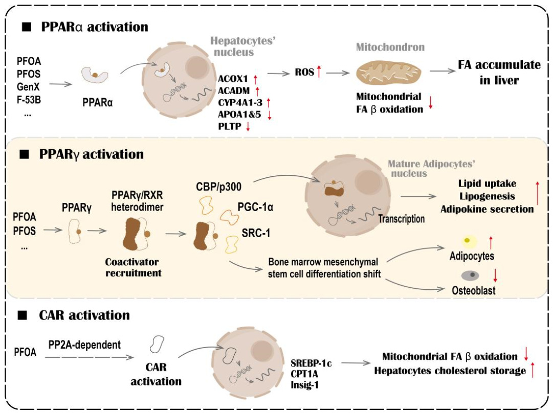
2.2.1. Core Dysregulation via Nuclear Receptor Signaling
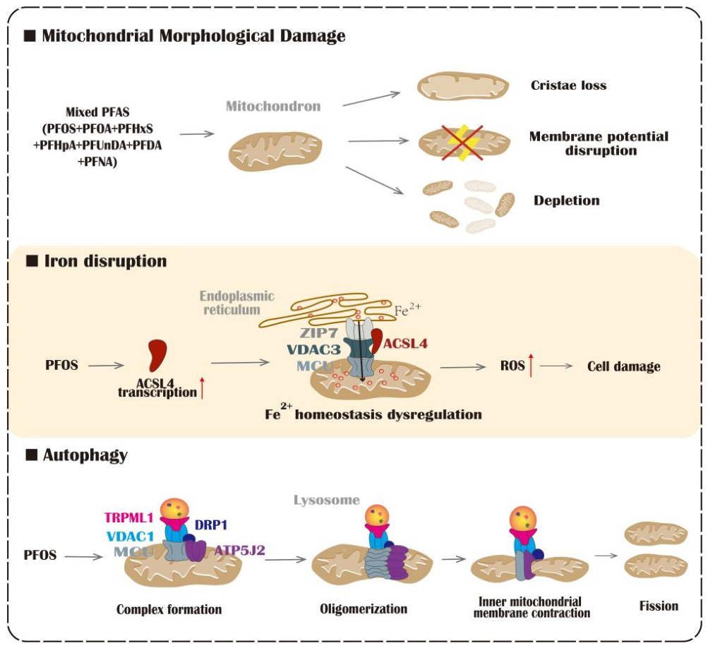
2.2.2. Metabolic Oxidative Stress
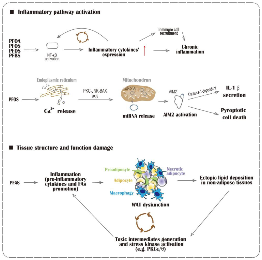
2.2.3. Systemic Inflammation and Intertissue Communication
3. PFAS Exposure Induces Glucose Metabolic Disorders
3.1. Glucose-Related Epidemiological Evidence
3.2. Underlying Mechanism in Glucose Disturbances
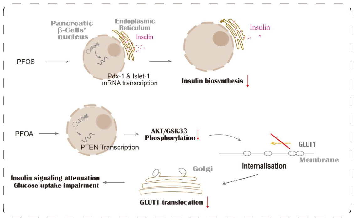
3.2.1. Direct Effect on Pancreatic β-Cell Function and Insulin Signaling
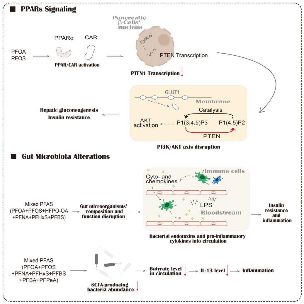
3.2.2. PPARs Signaling Dysregulation
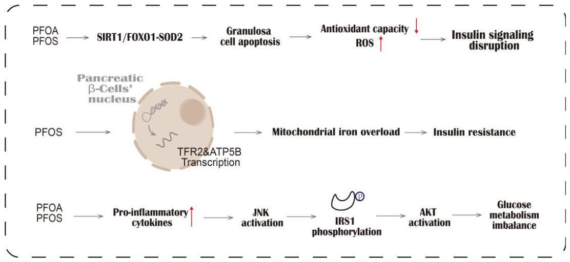
3.2.3. Oxidative Stress and Inflammatory Activation
3.2.4. Gut Microbiota Alterations
4. Discussion
4.1. Integrated Mechanisms Underlying PFAS-Disturbed Glucose and Lipid Metabolic Homeostasis
4.2. Research Limitations and Future Computational and Technological Perspectives
5. Conclusions
Author Contributions
Funding
Institutional Review Board Statement
Informed Consent Statement
Data Availability Statement
Conflicts of Interest
Abbreviations
| 6:2 Cl-PFAES | 6:2 Chlorinated polyfluoroalkyl ether sulfonate |
| ACADM | Acyl-CoA Dehydrogenase Medium Chain |
| ACOX1 | Acyl-CoA Oxidase 1 |
| ACSL4 | Acyl-CoA synthetase long-chain family member 4 |
| Akt | Protein kinase B |
| AMPK | AMP-activated protein kinase |
| APOA | Apolipoprotein A |
| ATP5B | ATP synthase β subunit |
| CAR | The constitutive androstane receptor |
| CPT1A | Carnitine palmitoyltransferase 1A |
| CYP4A | Cytochrome P450 Family 4 Subfamily A |
| DAG | Diacylglycerol |
| F-53B | 6:2 Chlorinated polyfluorinated ether sulfonate |
| FA | Fatty acid |
| FAO | Fatty acid oxidation |
| FFAs | Free fatty acids |
| FPG | Fasting plasma glucose |
| FXR | The farnesoid X receptor |
| GDM | Gestational diabetes mellitus |
| GenX | 2,3,3,3-Tetrafluoro-2-(heptafluoropropoxy)propanoic acid |
| GLP-1 | Glucagon-like peptide-1 |
| GLUT4 | Glucose transporter type 4 |
| GPAT | Glycerol-3-phosphate acyltransferase |
| GPBAR1 | G protein-coupled bile acid receptor 1 |
| GSK3β | Glycogen synthase kinase 3β |
| HDL-C | High-density lipoprotein cholesterol |
| HFPO-TA | Hexafluoropropylene oxide trimer acid |
| HOMA-IR | Homeostatic model assessment of insulin resistance |
| IDF | The International Diabetes Federation |
| IL-10 | Interleukin-10 |
| IL-1β | Interleukin-1β |
| IL-4 | Interleukin-4 |
| IL-6 | Interleukin-6 |
| Insig-1 | Insulin-induced gene 1 |
| InsR | Insulin receptor |
| Islet-1 | Insulin enhancer-binding protein 1 |
| LDL-C | Low-density lipoprotein cholesterol |
| LPS | Lipopolysaccharide |
| LXR | The liver X receptor |
| MCU | Mitochondrial calcium uniporter |
| Me-PFOSA-AcOH | 2-(N-methyl-perfluorooctane sulfonamido)acetic acid |
| mtDNAcn | Mitochondrial DNA copy number |
| NAFLD | Non-alcoholic fatty liver disease |
| NF-κB | Nuclear factor-kappaB |
| NRs | Nuclear receptors |
| OXPHOS | Oxidative phosphorylation |
| Pdx-1 | Pancreatic and duodenal homeobox 1 |
| PFAS | Per- and polyfluoroalkyl substances |
| PFBA | Perfluorobutyric acid |
| PFBS | Perfluorobutanesulfonic acid |
| PFCAs | Perfluoroalkyl carboxylic acids |
| PFDeA | Perfluorodecanoic acid |
| PFHxS | Perfluorohexane sulfonic acid |
| PFNA | Perfluorononanoic acid |
| PFOA | Perfluorooctanoic acid |
| PFOS | Perfluorooctane sulfonic acid |
| PFSAs | Perfluoroalkane sulfonic acids |
| PI3K | Phosphatidylinositol 3-kinase |
| PIP3 | Phosphatidylinositol 3,4,5-trisphosphate |
| PLTP | Phospholipid Transfer Protein |
| PMOH | Perfluoro-2-methoxyhexanoic acid |
| POPs | Persistent organic pollutants |
| PPAR | The peroxisome proliferator-activated receptor |
| PPREs | Peroxisome proliferator response elements |
| PXR | The pregnane X receptor |
| ROS | Reactive oxygen species |
| RXR | Retinoid X receptor |
| SREBP-1c | Sterol regulatory element-binding protein 1c |
| T2DM | Type 2 diabetes mellitus |
| TC | Total cholesterol |
| TFR2 | Transferrin receptor 2 |
| TNF-α | Tumor necrosis factor-α |
| VDAC3 | Voltage-dependent anion channel 3 |
| WAT | White adipose tissue |
| ZIP7 | Zrt-/Irt-like Protein 7 |
References
- Wang, Z.; Buser, A.M.; Cousins, I.T.; Demattio, S.; Drost, W.; Johansson, O.; Ohno, K.; Patlewicz, G.; Richard, A.M.; Walker, G.W.; et al. A New OECD Definition for Per- and Polyfluoroalkyl Substances. Environ. Sci. Technol. 2021, 55, 15575–15578. [Google Scholar] [CrossRef] [Scilit] [PubMed]
- Buck, R.C.; Franklin, J.; Berger, U.; Conder, J.M.; Cousins, I.T.; de Voogt, P.; Jensen, A.A.; Kannan, K.; Mabury, S.A.; van Leeuwen, S.P.J. Perfluoroalkyl and Polyfluoroalkyl Substances in the Environment: Terminology, Classification, and Origins. Integr. Environ. Assess. Manag. 2011, 7, 513–541. [Google Scholar] [CrossRef] [Scilit] [PubMed]
- Gagliano, E.; Sgroi, M.; Falciglia, P.P.; Vagliasindi, F.G.A.; Roccaro, P. Removal of Poly- and Perfluoroalkyl Substances (PFAS) from Water by Adsorption: Role of PFAS Chain Length, Effect of Organic Matter and Challenges in Adsorbent Regeneration. Water Res. 2020, 171, 115381. [Google Scholar] [CrossRef] [Scilit] [PubMed]
- Qin, Y.; Gu, T.; Ling, J.; Luo, J.; Zhao, J.; Hu, B.; Hua, L.; Wan, C.; Jiang, S. PFOS Facilitates Liver Inflammation and Steatosis: An Involvement of NLRP3 Inflammasome-Mediated Hepatocyte Pyroptosis. J. Appl. Toxicol. 2022, 42, 806–817. [Google Scholar] [CrossRef] [Scilit] [PubMed]
- Qiu, L.; Wang, H.; Dong, T.; Huang, J.; Li, T.; Ren, H.; Wang, X.; Qu, J.; Wang, S. Perfluorooctane Sulfonate (PFOS) Disrupts Testosterone Biosynthesis via CREB/CRTC2/StAR Signaling Pathway in Leydig Cells. Toxicology 2021, 449, 152663. [Google Scholar] [CrossRef] [Scilit] [PubMed]
- Gao, X.-X.; Zuo, Q.-L.; Fu, X.-H.; Song, L.-L.; Cen, M.-Q.; Wu, J. Association between Prenatal Exposure to Per- and Polyfluoroalkyl Substances and Neurodevelopment in Children: Evidence Based on Birth Cohort. Environ. Res. 2023, 236, 116812. [Google Scholar] [CrossRef] [Scilit] [PubMed]
- Naumann, A.; Alesio, J.; Poonia, M.; Bothun, G.D. PFAS Fluidize Synthetic and Bacterial Lipid Monolayers Based on Hydrophobicity and Lipid Charge. J. Environ. Chem. Eng. 2022, 10, 107351. [Google Scholar] [CrossRef] [Scilit] [PubMed]
- Li, X.; Hou, M.; Zhang, F.; Ji, Z.; Cai, Y.; Shi, Y. Per- and Polyfluoroalkyl Substances and Female Health Concern: Gender-Based Accumulation Differences, Adverse Outcomes, and Mechanisms. Environ. Sci. Technol. 2025, 59, 1469–1486. [Google Scholar] [CrossRef] [Scilit] [PubMed]
- Evich, M.G.; Davis, M.J.B.; McCord, J.P.; Acrey, B.; Awkerman, J.A.; Knappe, D.R.U.; Lindstrom, A.B.; Speth, T.F.; Tebes-Stevens, C.; Strynar, M.J.; et al. Per- and Polyfluoroalkyl Substances in the Environment. Science 2022, 375, eabg9065. [Google Scholar] [CrossRef] [Scilit] [PubMed]
- Ateia, M.; Maroli, A.; Tharayil, N.; Karanfil, T. The Overlooked Short- and Ultrashort-Chain Poly- and Perfluorinated Substances: A Review. Chemosphere 2019, 220, 866–882. [Google Scholar] [CrossRef] [Scilit] [PubMed]
- Wang, S.; Huang, J.; Yang, Y.; Hui, Y.; Ge, Y.; Larssen, T.; Yu, G.; Deng, S.; Wang, B.; Harman, C. First Report of a Chinese PFOS Alternative Overlooked for 30 Years: Its Toxicity, Persistence, and Presence in the Environment. Environ. Sci. Technol. 2013, 47, 10163–10170. [Google Scholar] [CrossRef] [Scilit] [PubMed]
- Mahoney, H.; Ankley, P.; Roberts, C.; Lamb, A.; Schultz, M.; Zhou, Y.; Giesy, J.P.; Brinkmann, M. Unveiling the Molecular Effects of Replacement and Legacy PFASs: Transcriptomic Analysis of Zebrafish Embryos Reveals Surprising Similarities and Potencies. Environ. Sci. Technol. 2024, 58, 18554–18565. [Google Scholar] [CrossRef] [Scilit] [PubMed]
- Lin, P.-I.D.; Cardenas, A.; Hauser, R.; Gold, D.R.; Kleinman, K.P.; Hivert, M.-F.; Fleisch, A.F.; Calafat, A.M.; Sanchez-Guerra, M.; Osorio-Yáñez, C.; et al. Dietary Characteristics Associated with Plasma Concentrations of Per- and Polyfluoroalkyl Substances among Adults with Pre-Diabetes: Cross-Sectional Results from the Diabetes Prevention Program Trial. Environ. Int. 2020, 137, 105217. [Google Scholar] [CrossRef] [Scilit] [PubMed]
- Geng, D.; Musse, A.A.; Wigh, V.; Carlsson, C.; Engwall, M.; Orešič, M.; Scherbak, N.; Hyötyläinen, T. Effect of Perfluorooctanesulfonic Acid (PFOS) on the Liver Lipid Metabolism of the Developing Chicken Embryo. Ecotoxicol. Environ. Saf. 2019, 170, 691–698. [Google Scholar] [CrossRef] [Scilit] [PubMed]
- Ling, J.; Hua, L.; Qin, Y.; Gu, T.; Jiang, S.; Zhao, J. Perfluorooctane Sulfonate Promotes Hepatic Lipid Accumulation and Steatosis in High-Fat Diet Mice through AMP-Activated Protein Kinase/Acetyl-CoA Carboxylase (AMPK/ACC) Pathway. J. Appl. Toxicol. JAT 2023, 43, 312–322. [Google Scholar] [CrossRef] [Scilit] [PubMed]
- Qu, J.; Han, Y.; Zhao, Z.; Wu, Y.; Lu, Y.; Chen, G.; Jiang, J.; Qiu, L.; Gu, A.; Wang, X. Perfluorooctane Sulfonate Interferes with Non-Genomic Estrogen Receptor Signaling Pathway, Inhibits ERK1/2 Activation and Induces Apoptosis in Mouse Spermatocyte-Derived Cells. Toxicology 2021, 460, 152871. [Google Scholar] [CrossRef] [Scilit] [PubMed]
- Kahn, L.G.; Philippat, C.; Nakayama, S.F.; Slama, R.; Trasande, L. Endocrine-Disrupting Chemicals: Implications for Human Health. Lancet Diabetes Endocrinol. 2020, 8, 703–718. [Google Scholar] [CrossRef] [Scilit] [PubMed]
- Abarca-Gómez, L.; Abdeen, Z.A.; Hamid, Z.A.; Abu-Rmeileh, N.M.; Acosta-Cazares, B.; Acuin, C.; Adams, R.J.; Aekplakorn, W.; Afsana, K.; Aguilar-Salinas, C.A.; et al. Worldwide Trends in Body-Mass Index, Underweight, Overweight, and Obesity from 1975 to 2016: A Pooled Analysis of 2416 Population-Based Measurement Studies in 128·9 Million Children, Adolescents, and Adults. Lancet 2017, 390, 2627–2642. [Google Scholar] [CrossRef] [Scilit] [PubMed]
- Kumanyika, S.; Dietz, W.H. Solving Population-Wide Obesity—Progress and Future Prospects. N. Engl. J. Med. 2020, 383, 2197–2200. [Google Scholar] [CrossRef] [Scilit] [PubMed]
- Roumeliotis, S.; Divani, M.; Stamellou, E.; Liakopoulos, V. Genomics in Diabetic Kidney Disease: A 2024 Update. Curr. Genom. 2024, 25, 153–157. [Google Scholar] [CrossRef] [Scilit] [PubMed]
- Zheng, Y.; Xu, Y.; Ji, L.; San, W.; Shen, D.; Zhou, Q.; Meng, G.; Shi, J.; Chen, Y. Roles of Distinct Nuclear Receptors in Diabetic Cardiomyopathy. Front. Pharmacol. 2024, 15, 1423124. [Google Scholar] [CrossRef] [Scilit] [PubMed]
- Shen, J.; San, W.; Zheng, Y.; Zhang, S.; Cao, D.; Chen, Y.; Meng, G. Different Types of Cell Death in Diabetic Endothelial Dysfunction. Biomed. Pharmacother. 2023, 168, 115802. [Google Scholar] [CrossRef] [Scilit] [PubMed]
- World Obesity Federation. World Obesity Atlas; World Obesity Federation: London, UK, 2024. [Google Scholar]
- Genitsaridi, I.; Salpea, P.; Salim, A.; Sajjadi, S.F.; Tomic, D.; James, S.; Thirunavukkarasu, S.; Issaka, A.; Chen, L.; Basit, A.; et al. 11th Edition of the IDF Diabetes Atlas: Global, Regional, and National Diabetes Prevalence Estimates for 2024 and Projections for 2050. Lancet Diabetes Endocrinol. 2026, 14, 149–156. [Google Scholar] [CrossRef] [Scilit] [PubMed]
- Cioana, M.; Deng, J.; Nadarajah, A.; Hou, M.; Qiu, Y.; Chen, S.S.J.; Rivas, A.; Banfield, L.; Toor, P.P.; Zhou, F.; et al. The Prevalence of Obesity among Children with Type 2 Diabetes: A Systematic Review and Meta-Analysis. JAMA Netw. Open 2022, 5, e2247186. [Google Scholar] [CrossRef] [Scilit] [PubMed]
- Sen, P.; Fan, Y.; Schlezinger, J.J.; Ehrlich, S.D.; Webster, T.F.; Hyötyläinen, T.; Pedersen, O.; Orešič, M. Exposure to Environmental Toxicants Is Associated with Gut Microbiome Dysbiosis, Insulin Resistance and Obesity. Environ. Int. 2024, 186, 108569. [Google Scholar] [CrossRef] [Scilit] [PubMed]
- Le, M.-L.; Xue, J.-Y.; Wu, L.-H.; Zhao, X.-Y.; Ding, J.; Yuan, Y.-Y.; Wan, Y.-Q.; Zhang, D.-L. Hexafluoropropylene Oxide Homologues, the Novel Alternatives to PFOA, Induce Mitochondrial Dysfunction and Cytotoxicity in Leydig Cells through Disrupting SIRT1/PGC-1α Signaling Pathway. Toxicology 2025, 518, 154282. [Google Scholar] [CrossRef] [Scilit] [PubMed]
- Zhang, J.; Sun, P.; Ren, H.; Guo, X.; Ding, E.; Ma, X.; Li, C.; Li, C.; Xu, Y.; Cao, K.; et al. Exposure to Per- and Polyfluoroalkyl Substances and Abnormal Glycometabolism in Healthy Elderly: Multiomics Evidence from the China BAPE Study. Environ. Sci. Technol. 2026, 60, 4171–4183. [Google Scholar] [CrossRef] [Scilit] [PubMed]
- Kingsley, S.L.; Walker, D.I.; Calafat, A.M.; Chen, A.; Papandonatos, G.D.; Xu, Y.; Jones, D.P.; Lanphear, B.P.; Pennell, K.D.; Braun, J.M. Metabolomics of Childhood Exposure to Perfluoroalkyl Substances: A Cross-Sectional Study. Metabolomics Off. J. Metabolomic Soc. 2019, 15, 95. [Google Scholar] [CrossRef] [Scilit] [PubMed]
- Ortega, M.A.; Fraile-Martínez, O.; Naya, I.; García-Honduvilla, N.; Álvarez-Mon, M.; Buján, J.; Asúnsolo, Á.; de la Torre, B. Type 2 Diabetes Mellitus Associated with Obesity (Diabesity). The Central Role of Gut Microbiota and Its Translational Applications. Nutrients 2020, 12, 2749. [Google Scholar] [CrossRef] [Scilit] [PubMed]
- DeFronzo, R.A.; Ferrannini, E. Insulin Resistance. A Multifaceted Syndrome Responsible for NIDDM, Obesity, Hypertension, Dyslipidemia, and Atherosclerotic Cardiovascular Disease. Diabetes Care 1991, 14, 173–194. [Google Scholar] [CrossRef] [Scilit] [PubMed]
- Ho, S.H.; Soh, S.X.H.; Wang, M.X.; Ong, J.; Seah, A.; Wong, Y.; Fang, Z.; Sim, S.; Lim, J.T. Perfluoroalkyl Substances and Lipid Concentrations in the Blood: A Systematic Review of Epidemiological Studies. Sci. Total Environ. 2022, 850, 158036. [Google Scholar] [CrossRef] [Scilit] [PubMed]
- Liu, B.; Zhu, L.; Wang, M.; Sun, Q. Associations between Per- and Polyfluoroalkyl Substances Exposures and Blood Lipid Levels among Adults-a Meta-Analysis. Environ. Health Perspect. 2023, 131, 56001. [Google Scholar] [CrossRef] [Scilit] [PubMed]
- Lin, C.-Y.; Chen, P.-C.; Lin, Y.-C.; Lin, L.-Y. Association among Serum Perfluoroalkyl Chemicals, Glucose Homeostasis, and Metabolic Syndrome in Adolescents and Adults. Diabetes Care 2009, 32, 702–707. [Google Scholar] [CrossRef] [Scilit] [PubMed]
- Wang, Z.; Zhang, B.; Zhang, J.; Xu, S.; Dai, Y.; Ding, J.; Guo, J.; Qi, X.; Chang, X.; Wu, C.; et al. Prenatal Exposure to Per- and Polyfluoroalkyl Substances and Sex-Specific Associations with Offspring Adiposity at 10 Years of Age: Metabolic Perturbation Plays a Role. Environ. Int. 2024, 192, 109037. [Google Scholar] [CrossRef] [Scilit] [PubMed]
- Averina, M.; Brox, J.; Huber, S.; Furberg, A.-S. Exposure to Perfluoroalkyl Substances (PFAS) and Dyslipidemia, Hypertension and Obesity in Adolescents. The Fit Futures Study. Environ. Res. 2021, 195, 110740. [Google Scholar] [CrossRef] [Scilit] [PubMed]
- Sun, S.; Xie, Z.; Song, X.; Wen, S.; Yuan, W.; Miao, M.; Ji, H.; Liang, H. Prenatal Exposure to Per- and Polyfluoroalkyl Substances and Adiposity Measures of Children at 4 and 6 Years: A Prospective Birth Cohort in China. Ecotoxicol. Environ. Saf. 2024, 269, 115751. [Google Scholar] [CrossRef] [Scilit] [PubMed]
- Frigerio, G.; Ferrari, C.M.; Fustinoni, S. Prenatal and Childhood Exposure to Per-/Polyfluoroalkyl Substances (PFASs) and Its Associations with Childhood Overweight and/or Obesity: A Systematic Review with Meta-Analyses. Environ. Health 2023, 22, 56. [Google Scholar] [CrossRef] [Scilit] [PubMed]
- Schillemans, T.; Donat-Vargas, C.; Åkesson, A. Per- and Polyfluoroalkyl Substances and Cardiometabolic Diseases: A Review. Basic Clin. Pharmacol. Toxicol. 2024, 134, 141–152. [Google Scholar] [CrossRef] [Scilit] [PubMed]
- India-Aldana, S.; Yao, M.; Midya, V.; Colicino, E.; Chatzi, L.; Chu, J.; Gennings, C.; Jones, D.P.; Loos, R.J.F.; Setiawan, V.W.; et al. PFAS Exposures and the Human Metabolome: A Systematic Review of Epidemiological Studies. Curr. Pollut. Rep. 2023, 9, 511–568. [Google Scholar] [CrossRef] [Scilit] [PubMed]
- Gaillard, L.; Barouki, R.; Blanc, E.; Coumoul, X.; Andréau, K. Per- and Polyfluoroalkyl Substances as Persistent Pollutants with Metabolic and Endocrine-Disrupting Impacts. Trends Endocrinol. Metab. TEM 2025, 36, 249–261. [Google Scholar] [CrossRef] [Scilit] [PubMed]
- Du, X.; Li, D.-L.; Xu, X.; Wu, Y.; Du, Z.; Liang, G.; Li, Y.-Z.; Zheng, Y.-J.; Qin, Y.; Qian, K.; et al. Effects of Mixed Exposure to PFAS on Adolescent Non-Alcoholic Fatty Liver Disease: Integrating Evidence from Human Cohorts, Toxicogenomics, and Animal Models to Uncover Mechanisms and Potential Target Sites. J. Hazard. Mater. 2025, 485, 136854. [Google Scholar] [CrossRef] [Scilit] [PubMed]
- Wu, Y.; Cheng, Z.; Zhang, W.; Yin, C.; Sun, J.; Hua, H.; Long, X.; Wu, X.; Wang, Y.; Ren, X.; et al. Association between Per- and Poly-Fluoroalkyl Substances and Nonalcoholic Fatty Liver Disease: A Nested Case-Control Study in Northwest China. Environ. Pollut. (Barking Essex 1987) 2024, 350, 123937. [Google Scholar] [CrossRef] [Scilit] [PubMed]
- Volle, D.H. Nuclear Receptors in Physiology and Pathophysiology. Mol. Asp. Med. 2021, 78, 100956. [Google Scholar] [CrossRef] [Scilit] [PubMed]
- Fragki, S.; Dirven, H.; Fletcher, T.; Grasl-Kraupp, B.; Bjerve Gützkow, K.; Hoogenboom, R.; Kersten, S.; Lindeman, B.; Louisse, J.; Peijnenburg, A.; et al. Systemic PFOS and PFOA Exposure and Disturbed Lipid Homeostasis in Humans: What Do We Know and What Not? Crit. Rev. Toxicol. 2021, 51, 141–164. [Google Scholar] [CrossRef] [Scilit] [PubMed]
- Issemann, I.; Green, S. Activation of a Member of the Steroid Hormone Receptor Superfamily by Peroxisome Proliferators. Nature 1990, 347, 645–650. [Google Scholar] [CrossRef] [Scilit] [PubMed]
- Lefebvre, P.; Chinetti, G.; Fruchart, J.-C.; Staels, B. Sorting out the Roles of PPAR Alpha in Energy Metabolism and Vascular Homeostasis. J. Clin. Investig. 2006, 116, 571–580. [Google Scholar] [CrossRef] [Scilit] [PubMed]
- Xia, B.; Li, Q.; Zheng, K.; Wu, J.; Huang, C.; Liu, K.; You, Q.; Yuan, X. Down-Regulation of Hrd1 Protects against Myocardial Ischemia-Reperfusion Injury by Regulating PPARα to Prevent Oxidative Stress, Endoplasmic Reticulum Stress, and Cellular Apoptosis. Eur. J. Pharmacol. 2023, 954, 175864. [Google Scholar] [CrossRef] [Scilit] [PubMed]
- Hashimoto, T.; Cook, W.S.; Qi, C.; Yeldandi, A.V.; Reddy, J.K.; Rao, M.S. Defect in Peroxisome Proliferator-Activated Receptor Alpha-Inducible Fatty Acid Oxidation Determines the Severity of Hepatic Steatosis in Response to Fasting. J. Biol. Chem. 2000, 275, 28918–28928. [Google Scholar] [CrossRef] [Scilit] [PubMed]
- Lecka-Czernik, B.; Moerman, E.J.; Grant, D.F.; Lehmann, J.M.; Manolagas, S.C.; Jilka, R.L. Divergent Effects of Selective Peroxisome Proliferator-Activated Receptor-Gamma 2 Ligands on Adipocyte versus Osteoblast Differentiation. Endocrinology 2002, 143, 2376–2384. [Google Scholar] [CrossRef] [PubMed]
- Kennedy, G.L., Jr.; Butenhoff, J.L.; Olsen, G.W.; O’Connor, J.C.; Seacat, A.M.; Perkins, R.G.; Biegel, L.B.; Murphy, S.R.; Farrar, D.G. The Toxicology of Perfluorooctanoate. Crit. Rev. Toxicol. 2004, 34, 351–384. [Google Scholar] [CrossRef] [Scilit] [PubMed]
- Vanden Heuvel, J.P.; Thompson, J.T.; Frame, S.R.; Gillies, P.J. Differential Activation of Nuclear Receptors by Perfluorinated Fatty Acid Analogs and Natural Fatty Acids: A Comparison of Human, Mouse, and Rat Peroxisome Proliferator-Activated Receptor-Alpha, -Beta, and -Gamma, Liver X Receptor-Beta, and Retinoid X Receptor-Alpha. Toxicol. Sci. Off. J. Soc. Toxicol. 2006, 92, 476–489. [Google Scholar] [CrossRef] [Scilit] [PubMed]
- Lake, B.G. Mechanisms of Hepatocarcinogenicity of Peroxisome-Proliferating Drugs and Chemicals. Annu. Rev. Pharmacol. Toxicol. 1995, 35, 483–507. [Google Scholar] [CrossRef] [Scilit]
- Yang, W.; Ling, X.; He, S.; Cui, H.; Yang, Z.; An, H.; Wang, L.; Zou, P.; Chen, Q.; Liu, J.; et al. PPARα/ACOX1 as a Novel Target for Hepatic Lipid Metabolism Disorders Induced by per- and Polyfluoroalkyl Substances: An Integrated Approach. Environ. Int. 2023, 178, 108138. [Google Scholar] [CrossRef] [Scilit] [PubMed]
- Wan, H.T.; Zhao, Y.G.; Wei, X.; Hui, K.Y.; Giesy, J.P.; Wong, C.K.C. PFOS-Induced Hepatic Steatosis, the Mechanistic Actions on β-Oxidation and Lipid Transport. Biochim. Biophys. Acta Gen. Subj. 2012, 1820, 1092–1101. [Google Scholar] [CrossRef] [Scilit] [PubMed]
- Hari, A.; AbdulHameed, M.D.M.; Balik-Meisner, M.R.; Mav, D.; Phadke, D.P.; Scholl, E.H.; Shah, R.R.; Casey, W.; Auerbach, S.S.; Wallqvist, A.; et al. Exposure to PFAS Chemicals Induces Sex-Dependent Alterations in Key Rate-Limiting Steps of Lipid Metabolism in Liver Steatosis. Front. Toxicol. 2024, 6, 1390196. [Google Scholar] [CrossRef] [Scilit] [PubMed]
- Brahimaj, A.; Ligthart, S.; Ikram, M.A.; Hofman, A.; Franco, O.H.; Sijbrands, E.J.G.; Kavousi, M.; Dehghan, A. Serum Levels of Apolipoproteins and Incident Type 2 Diabetes: A Prospective Cohort Study. Diabetes Care 2017, 40, 346–351. [Google Scholar] [CrossRef] [Scilit] [PubMed]
- Zhao, G.-J.; Yin, K.; Fu, Y.-C.; Tang, C.-K. The Interaction of ApoA-I and ABCA1 Triggers Signal Transduction Pathways to Mediate Efflux of Cellular Lipids. Mol. Med. 2012, 18, 149–158. [Google Scholar] [CrossRef] [Scilit] [PubMed]
- Zheng, X.-Y.; Zhao, S.-P.; Yan, H. The Role of Apolipoprotein A5 in Obesity and the Metabolic Syndrome. Biol. Rev. 2013, 88, 491–498. [Google Scholar] [CrossRef] [Scilit] [PubMed]
- Yang, Y.; Beigneux, A.P.; Song, W.; Nguyen, L.P.; Jung, H.; Tu, Y.; Weston, T.A.; Tran, C.M.; Xie, K.; Yu, R.G.; et al. Hypertriglyceridemia in Apoa5−/− Mice Results from Reduced Amounts of Lipoprotein Lipase in the Capillary Lumen. J. Clin. Investig. 2023, 133, e172600. [Google Scholar] [CrossRef] [Scilit] [PubMed]
- Yazdanyar, A.; Jiang, X.-C. Liver Phospholipid Transfer Protein (PLTP) Expression with a PLTP-Null Background Promotes Very Low-Density Lipoprotein Production in Mice. Hepatology 2012, 56, 576–584. [Google Scholar] [CrossRef] [Scilit] [PubMed]
- Jiang, X.C.; Bruce, C.; Mar, J.; Lin, M.; Ji, Y.; Francone, O.L.; Tall, A.R. Targeted Mutation of Plasma Phospholipid Transfer Protein Gene Markedly Reduces High-Density Lipoprotein Levels. J. Clin. Investig. 1999, 103, 907–914. [Google Scholar] [CrossRef] [Scilit] [PubMed]
- Chan, A.P.; Jarrett, K.E.; Lai, R.W.; Brearley-Sholto, M.C.; Cheng, A.S.; Taveras, M.O.; Iwata, A.M.; Steel, M.E.; Lau, A.; Whang, E.C.; et al. Bile Acids Regulate Lipid Metabolism through Selective Actions on Fatty Acid Absorption. Cell Metab. 2026, 38, 263–280.e10. [Google Scholar] [CrossRef] [Scilit] [PubMed]
- Zhang, H.; Zhou, X.; Sheng, N.; Cui, R.; Cui, Q.; Guo, H.; Guo, Y.; Sun, Y.; Dai, J. Subchronic Hepatotoxicity Effects of 6:2 Chlorinated Polyfluorinated Ether Sulfonate (6:2 Cl-PFESA), a Novel Perfluorooctanesulfonate (PFOS) Alternative, on Adult Male Mice. Environ. Sci. Technol. 2018, 52, 12809–12818. [Google Scholar] [CrossRef] [Scilit] [PubMed]
- Gabal, E.; Azaizeh, M.; Baloni, P. Investigating Lipid and Energy Dyshomeostasis Induced by Per- and Polyfluoroalkyl Substances (PFAS) Congeners in Mouse Model Using Systems Biology Approaches. Metabolites 2025, 15, 499. [Google Scholar] [CrossRef] [Scilit] [PubMed]
- Su, S.; Billy, L.J.; Chang, S.; Gonzalez, F.J.; Patterson, A.D.; Peters, J.M. The Role of Mouse and Human Peroxisome Proliferator-Activated Receptor-α in Modulating the Hepatic Effects of Perfluorooctane Sulfonate in Mice. Toxicology 2022, 465, 153056. [Google Scholar] [CrossRef] [Scilit] [PubMed]
- Rowan-Carroll, A.; Reardon, A.; Leingartner, K.; Gagné, R.; Williams, A.; Meier, M.J.; Kuo, B.; Bourdon-Lacombe, J.; Moffat, I.; Carrier, R.; et al. High-Throughput Transcriptomic Analysis of Human Primary Hepatocyte Spheroids Exposed to per- and Polyfluoroalkyl Substances as a Platform for Relative Potency Characterization. Toxicol. Sci. Off. J. Soc. Toxicol. 2021, 181, 199–214. [Google Scholar] [CrossRef] [Scilit] [PubMed]
- Zhang, W.; Tian, Y.; Chen, B.; Xu, S.; Wu, L. PFOA/PFOS Facilitated Intestinal Fatty Acid Absorption by Activating the PPARα Pathway: Insights from Organoids Model. Environ. Health 2024, 2, 85–94. [Google Scholar] [CrossRef] [Scilit] [PubMed]
- Qin, H.; Niu, Y.; Luan, H.; Li, M.; Zheng, L.; Pan, Y.; Liu, W. Effects of Legacy and Emerging Per- and Polyfluoroalkyl Substances on PPARα/β/γ Regulation and Osteogenic/Adipogenic Differentiation. Environ. Int. 2022, 170, 107584. [Google Scholar] [CrossRef] [Scilit] [PubMed]
- Rosen, E.D.; Sarraf, P.; Troy, A.E.; Bradwin, G.; Moore, K.; Milstone, D.S.; Spiegelman, B.M.; Mortensen, R.M. PPAR Gamma Is Required for the Differentiation of Adipose Tissue in Vivo and in Vitro. Mol. Cell 1999, 4, 611–617. [Google Scholar] [CrossRef] [Scilit] [PubMed]
- Evans, R.M.; Barish, G.D.; Wang, Y.-X. PPARs and the Complex Journey to Obesity. Nat. Med. 2004, 10, 355–361. [Google Scholar] [CrossRef] [Scilit] [PubMed]
- Takacs, M.L.; Abbott, B.D. Activation of Mouse and Human Peroxisome Proliferator-Activated Receptors (Alpha, Beta/Delta, Gamma) by Perfluorooctanoic Acid and Perfluorooctane Sulfonate. Toxicol. Sci. Off. J. Soc. Toxicol. 2007, 95, 108–117. [Google Scholar] [CrossRef] [Scilit] [PubMed]
- Ganesh, H.; Moran, J.; Roy, S.; Mathew, J.; Ackah-Blay, J.; Costello, E.; Shan, P.; Dakshanamurthy, S. Impact of Persistent Endocrine-Disrupting Chemicals on Human Nuclear Receptors: Insights from in Silico and Experimental Characterization. Int. J. Mol. Sci. 2025, 26, 2879. [Google Scholar] [CrossRef] [Scilit] [PubMed]
- Li, C.-H.; Ren, X.-M.; Guo, L.-H. Adipogenic Activity of Oligomeric Hexafluoropropylene Oxide (Perfluorooctanoic Acid Alternative) through Peroxisome Proliferator-Activated Receptor γ Pathway. Environ. Sci. Technol. 2019, 53, 3287–3295. [Google Scholar] [CrossRef] [Scilit] [PubMed]
- Tontonoz, P.; Spiegelman, B.M. Fat and beyond: The Diverse Biology of PPARgamma. Annu. Rev. Biochem. 2008, 77, 289–312. [Google Scholar] [CrossRef] [Scilit] [PubMed]
- Rosen, M.B.; Das, K.P.; Rooney, J.; Abbott, B.; Lau, C.; Corton, J.C. PPARα-Independent Transcriptional Targets of Perfluoroalkyl Acids Revealed by Transcript Profiling. Toxicology 2017, 387, 95–107. [Google Scholar] [CrossRef] [Scilit] [PubMed]
- Liu, S.; Yang, R.; Yin, N.; Wang, Y.-L.; Faiola, F. Environmental and Human Relevant PFOS and PFOA Doses Alter Human Mesenchymal Stem Cell Self-Renewal, Adipogenesis and Osteogenesis. Ecotoxicol. Environ. Saf. 2019, 169, 564–572. [Google Scholar] [CrossRef] [Scilit] [PubMed]
- Qin, W.; Henneberger, L.; Glüge, J.; König, M.; Escher, B.I. Baseline Toxicity Model to Identify the Specific and Nonspecific Effects of Per- and Polyfluoroalkyl Substances in Cell-Based Bioassays. Environ. Sci. Technol. 2024, 58, 5727–5738. [Google Scholar] [CrossRef] [Scilit] [PubMed]
- Yang, H.; Wang, H. Signaling Control of the Constitutive Androstane Receptor (CAR). Protein Cell 2014, 5, 113–123. [Google Scholar] [CrossRef] [Scilit] [PubMed]
- Abe, T.; Takahashi, M.; Kano, M.; Amaike, Y.; Ishii, C.; Maeda, K.; Kudoh, Y.; Morishita, T.; Hosaka, T.; Sasaki, T.; et al. Activation of Nuclear Receptor CAR by an Environmental Pollutant Perfluorooctanoic Acid. Arch. Toxicol. 2017, 91, 2365–2374. [Google Scholar] [CrossRef] [Scilit] [PubMed]
- De Battistis, F.; Djordjevic, A.B.; Saso, L.; Mantovani, A. Constitutive Androstane Receptor, Liver Pathophysiology and Chemical Contaminants: Current Evidence and Perspectives. Front. Endocrinol. 2025, 16, 1472563. [Google Scholar] [CrossRef] [Scilit] [PubMed]
- Dong, B.; Saha, P.K.; Huang, W.; Chen, W.; Abu-Elheiga, L.A.; Wakil, S.J.; Stevens, R.D.; Ilkayeva, O.; Newgard, C.B.; Chan, L.; et al. Activation of Nuclear Receptor CAR Ameliorates Diabetes and Fatty Liver Disease. Proc. Natl. Acad. Sci. USA 2009, 106, 18831–18836. [Google Scholar] [CrossRef] [Scilit] [PubMed]
- Ouyang, S.; Zhuo, S.; Yang, M.; Zhu, T.; Yu, S.; Li, Y.; Ying, H.; Le, Y. Glycerol Kinase Drives Hepatic de Novo Lipogenesis and Triglyceride Synthesis in Nonalcoholic Fatty Liver by Activating SREBP-1c Transcription, Upregulating DGAT1/2 Expression, and Promoting Glycerol Metabolism. Adv. Sci. 2024, 11, e2401311. [Google Scholar] [CrossRef] [Scilit] [PubMed]
- Shizu, R.; Otsuka, Y.; Ishii, C.; Ezaki, K.; Yoshinari, K. PPARα Induces the Expression of CAR That Works as a Negative Regulator of PPARα Functions in Mouse Livers. Int. J. Mol. Sci. 2023, 24, 3953. [Google Scholar] [CrossRef] [Scilit] [PubMed]
- Moreau, A.; Vilarem, M.J.; Maurel, P.; Pascussi, J.M. Xenoreceptors CAR and PXR Activation and Consequences on Lipid Metabolism, Glucose Homeostasis, and Inflammatory Response. Mol. Pharm. 2008, 5, 35–41. [Google Scholar] [CrossRef] [Scilit] [PubMed]
- Zhang, H.; Kong, X.; Wang, W.; Zhou, H.; Qu, H.; Guan, Z.; Wu, H.; Zhai, X.; Jin, B. TRIM25-Mediated INSIG1 Ubiquitination Promotes MASH Progression through Reprogramming Lipid Metabolism. Adv. Sci. 2025, 12, e2414646. [Google Scholar] [CrossRef] [Scilit] [PubMed]
- Attema, B.; Janssen, A.W.F.; Rijkers, D.; van Schothorst, E.M.; Hooiveld, G.J.E.J.; Kersten, S. Exposure to Low-Dose Perfluorooctanoic Acid Promotes Hepatic Steatosis and Disrupts the Hepatic Transcriptome in Mice. Mol. Metab. 2022, 66, 101602. [Google Scholar] [CrossRef] [Scilit] [PubMed]
- Dubois, L.; Oliviero, F.; Mary, L.; Dauwe, Y.; Comera, C.; Gayard, V.; Mselli-Lakhal, L. P25-09 the Role of Constitutive Androstane Receptor (CAR) in PFOA-Induced Hepatic Steatosis. Toxicol. Lett. 2025, 411, S318–S319. [Google Scholar] [CrossRef] [Scilit]
- Kliewer, S.A.; Moore, J.T.; Wade, L.; Staudinger, J.L.; Watson, M.A.; Jones, S.A.; McKee, D.D.; Oliver, B.B.; Willson, T.M.; Zetterström, R.H.; et al. An Orphan Nuclear Receptor Activated by Pregnanes Defines a Novel Steroid Signaling Pathway. Cell 1998, 92, 73–82. [Google Scholar] [CrossRef] [Scilit] [PubMed]
- Xing, Y.; Yan, J.; Niu, Y. PXR: A Center of Transcriptional Regulation in Cancer. Acta Pharm. Sin. B 2020, 10, 197–206. [Google Scholar] [CrossRef] [Scilit] [PubMed]
- Du, X.; Xu, X.; Yu, H.; Du, Z.; Wu, Y.; Qian, K.; Xu, J.; Tao, G.; Zhang, L.; Zheng, W. Thyrotoxic Effects of Mixed Exposure to Perfluorinated Compounds: Integrating Population-Based, Toxicogenomic, Animal, and Cellular Evidence to Elucidate Molecular Mechanisms and Identify Potential Effector Targets. Environ. Sci. Technol. 2024, 58, 18177–18189. [Google Scholar] [CrossRef] [Scilit] [PubMed]
- Yang, W.; Li, Y.; Feng, R.; Liang, P.; Tian, K.; Hu, L.; Wang, K.; Qiu, T.; Zhang, J.; Sun, X.; et al. PFOS Causes Lysosomes-Regulated Mitochondrial Fission through TRPML1-VDAC1 and Oligomerization of MCU/ATP5J2. J. Hazard. Mater. 2025, 489, 137685. [Google Scholar] [CrossRef] [Scilit] [PubMed]
- Eccles-Miller, J.A.; Williams, L.A.; Leonard, E.; Tharayil, N.; Buslee, E.L.; Fiddler, J.; Baldwin, W.S. Perfluorooctane Sulfonic Acid (PFOS) Perturbs Skeletal Muscle Oxidative Phosphorylation by a Different Mechanism than Liver. Chemosphere 2026, 394, 144815. [Google Scholar] [CrossRef] [Scilit] [PubMed]
- Du, X.; Xu, X.; Dong, X.-X.; Liang, X.; Wu, Y.; Du, Z.; Pan, C.-W.; Liang, G.; Li, Y.-Z.; Zheng, Y.-J.; et al. Integration of Animal, Population, and Toxicogenomic Evidence on the Hematotoxic and Immunosuppressive Effects of Environmental Exposure to PFAS Mixtures in Adolescents. Environ. Sci. Technol. 2025, 59, 10841–10853. [Google Scholar] [CrossRef] [Scilit] [PubMed]
- Li, Z.; Qu, X.; Hong, X.; Wang, G.; Song, M.; Choi, G.; Buckley, J.P.; Wang, X. Prenatal Per- and Polyfluoroalkyl Substances (PFAS) Exposures, Newborn Mitochondrial DNA Copy Number, and the Modifying Role of the Maternal Folate Level. Environ. Sci. Technol. 2025, 59, 20216–20228. [Google Scholar] [CrossRef] [Scilit] [PubMed]
- Zhang, B.; Wang, J.; Zhang, Y.; Liu, M.; Zhang, X. Individual and Joint Associations of Exposure to Per- and Polyfluoroalkyl Substances with Children’s Mitochondrial DNA Copy Number, and Modified by Estimated Glomerular Filtration Rate. Environ. Res. 2025, 266, 120598. [Google Scholar] [CrossRef] [Scilit] [PubMed]
- Rickard, B.P.; Tan, X.; Fenton, S.E.; Rizvi, I. Select Per- and Polyfluoroalkyl Substances (PFAS) Induce Resistance to Carboplatin in Ovarian Cancer Cell Lines. Int. J. Mol. Sci. 2022, 23, 5176. [Google Scholar] [CrossRef] [Scilit] [PubMed]
- Zorov, D.B.; Juhaszova, M.; Sollott, S.J. Mitochondrial Reactive Oxygen Species (ROS) and ROS-Induced ROS Release. Physiol. Rev. 2014, 94, 909–950. [Google Scholar] [CrossRef] [Scilit] [PubMed]
- Li, S.; Qin, S.; Zeng, H.; Chou, W.; Oudin, A.; Kanninen, K.M.; Jalava, P.; Dong, G.; Zeng, X. Adverse Outcome Pathway for the Neurotoxicity of Per- and Polyfluoroalkyl Substances: A Systematic Review. Eco-Environ. Health 2024, 3, 476–493. [Google Scholar] [CrossRef] [Scilit] [PubMed]
- Liang, P.; Tian, K.; Yang, W.; Feng, R.; Li, Y.; Hu, L.; Wang, K.; Qiu, T.; Zhang, J.; Sun, X.; et al. ACSL4-Mediated ZIP7-VDAC3 Interaction Regulates Endoplasmic Reticulum-Mitochondria Iron Transfer in Hepatocytes under PFOS Exposure. Sci. Total Environ. 2024, 957, 177679. [Google Scholar] [CrossRef] [Scilit] [PubMed]
- Jia, B.; Li, J.; Song, Y.; Luo, C. ACSL4-Mediated Ferroptosis and Its Potential Role in Central Nervous System Diseases and Injuries. Int. J. Mol. Sci. 2023, 24, 10021. [Google Scholar] [CrossRef] [Scilit] [PubMed]
- Chen, P.-H.; Wu, J.; Xu, Y.; Ding, C.-K.C.; Mestre, A.A.; Lin, C.-C.; Yang, W.-H.; Chi, J.-T. Zinc Transporter ZIP7 Is a Novel Determinant of Ferroptosis. Cell Death Dis. 2021, 12, 198. [Google Scholar] [CrossRef] [Scilit] [PubMed]
- DeHart, D.N.; Fang, D.; Heslop, K.; Li, L.; Lemasters, J.J.; Maldonado, E.N. Opening of Voltage Dependent Anion Channels Promotes Reactive Oxygen Species Generation, Mitochondrial Dysfunction and Cell Death in Cancer Cells. Biochem. Pharmacol. 2018, 148, 155–162. [Google Scholar] [CrossRef] [Scilit] [PubMed]
- Hurley, J.H. The Human Autophagy Core Complexes. Annu. Rev. Biochem. 2026, 95, 507–524. [Google Scholar] [CrossRef] [Scilit] [PubMed]
- Gangwar, R.S.; Bevan, G.H.; Palanivel, R.; Das, L.; Rajagopalan, S. Oxidative Stress Pathways of Air Pollution Mediated Toxicity: Recent Insights. Redox Biol. 2020, 34, 101545. [Google Scholar] [CrossRef] [Scilit] [PubMed]
- Jones, L.E.; Ghassabian, A.; Yeung, E.; Mendola, P.; Kannan, K.; Bell, E.M. Maternal Exposure to Legacy PFAS Compounds PFOA and PFOS Is Associated with Disrupted Cytokine Homeostasis in Neonates: The Upstate KIDS Study (2008–2010). Environ. Int. 2025, 196, 109288. [Google Scholar] [CrossRef] [Scilit] [PubMed]
- Tan, Y.; Taibl, K.R.; Dunlop, A.L.; Barr, D.B.; Panuwet, P.; Yakimavets, V.; Kannan, K.; Corwin, E.J.; Ryan, P.B.; Eatman, J.A.; et al. Association between a Mixture of Per- and Polyfluoroalkyl Substances (PFAS) and Inflammatory Biomarkers in the Atlanta African American Maternal-Child Cohort. Environ. Sci. Technol. 2023, 57, 13419–13428. [Google Scholar] [CrossRef] [Scilit] [PubMed]
- Dragon, J.; Hoaglund, M.; Badireddy, A.R.; Nielsen, G.; Schlezinger, J.; Shukla, A. Perfluoroalkyl Substances (PFAS) Affect Inflammation in Lung Cells and Tissues. Int. J. Mol. Sci. 2023, 24, 8539. [Google Scholar] [CrossRef] [Scilit] [PubMed]
- Zhang, H.; Fang, W.; Wang, D.; Gao, N.; Ding, Y.; Chen, C. The Role of Interleukin Family in Perfluorooctanoic Acid (PFOA)-Induced Immunotoxicity. J. Hazard. Mater. 2014, 280, 552–560. [Google Scholar] [CrossRef] [Scilit] [PubMed]
- Kumari, P.; Russo, A.J.; Shivcharan, S.; Rathinam, V.A. AIM2 in Health and Disease: Inflammasome and Beyond. Immunol. Rev. 2020, 297, 83–95. [Google Scholar] [CrossRef] [Scilit] [PubMed]
- Sharma, B.R.; Karki, R.; Kanneganti, T.-D. Role of AIM2 Inflammasome in Inflammatory Diseases, Cancer and Infection. Eur. J. Immunol. 2019, 49, 1998–2011. [Google Scholar] [CrossRef] [Scilit] [PubMed]
- Zhou, Y.; Tong, Z.; Jiang, S.; Zheng, W.; Zhao, J.; Zhou, X. The Roles of Endoplasmic Reticulum in NLRP3 Inflammasome Activation. Cells 2020, 9, 1219. [Google Scholar] [CrossRef] [Scilit] [PubMed]
- Chen, X.; Guo, X.; Ge, Q.; Zhao, Y.; Mu, H.; Zhang, J. ER Stress Activates the NLRP3 Inflammasome: A Novel Mechanism of Atherosclerosis. Oxidative Med. Cell. Longev. 2019, 2019, 3462530. [Google Scholar] [CrossRef] [Scilit] [PubMed]
- Corsini, E.; Sangiovanni, E.; Avogadro, A.; Galbiati, V.; Viviani, B.; Marinovich, M.; Galli, C.L.; Dell’Agli, M.; Germolec, D.R. In Vitro Characterization of the Immunotoxic Potential of Several Perfluorinated Compounds (PFCs). Toxicol. Appl. Pharmacol. 2012, 258, 248–255. [Google Scholar] [CrossRef] [Scilit] [PubMed]
- Tang, L.; Qiu, W.; Zhang, S.; Wang, J.; Yang, X.; Xu, B.; Magnuson, J.T.; Xu, E.G.; Wu, M.; Zheng, C. 1Poly- and Perfluoroalkyl Substances Induce Immunotoxicity via the TLR Pathway in Zebrafish: Links to Carbon Chain Length. Environ. Sci. Technol. 2023, 57, 6139–6149. [Google Scholar] [CrossRef] [Scilit] [PubMed]
- Li, F.; Chen, L.; Peng, B.-X.; Lei, Y.; Li, M.; Guo, L.-H. Perfluorooctane Sulfonate Promotes the Migration of Colorectal Cancer Cells by Inducing Epithelial-Mesenchymal Transition. J. Environ. Sci. 2024, 145, 1–12. [Google Scholar] [CrossRef] [Scilit] [PubMed]
- Zhou, C.; Chen, W.; Xuan, R.; Kang, X.; Wang, J.; Magnuson, J.T.; Li, J.; Fang, D.; Qiu, W. 2Perfluorodecanesulfonate (PFDS) Induces Innate Immune Toxicity through the NF-kappaB Pathway in Early Life Stage Zebrafish. Environ. Int. 2025, 202, 109688. [Google Scholar] [CrossRef] [Scilit] [PubMed]
- Karin, M.; Ben-Neriah, Y. Phosphorylation Meets Ubiquitination: The Control of NF-[Kappa]B Activity. Annu. Rev. Immunol. 2000, 18, 621–663. [Google Scholar] [CrossRef] [Scilit] [PubMed]
- Arkoudi, K.; Yuan, Y.; Cumine, A.P.; Dyer, C.; Busch-Nentwich, E.; Bravo, I.; Feng, Y.; Knight, R.D. An NF-kB/TNF-Alpha Signalling Feedback Loop Acts to Coordinate Tissue Regeneration and Macrophage Behaviour in Zebrafish. npj Regen. Med. 2025, 10, 27. [Google Scholar] [CrossRef] [Scilit] [PubMed]
- Megha, K.B.; Joseph, X.; Akhil, V.; Mohanan, P.V. Cascade of Immune Mechanism and Consequences of Inflammatory Disorders. Phytomed. Int. J. Phytother. Phytopharm. 2021, 91, 153712. [Google Scholar] [CrossRef] [Scilit] [PubMed]
- Kany, S.; Vollrath, J.T.; Relja, B. Cytokines in Inflammatory Disease. Int. J. Mol. Sci. 2019, 20, 6008. [Google Scholar] [CrossRef] [Scilit] [PubMed]
- Unger, R.H.; Scherer, P.E. Gluttony, Sloth and the Metabolic Syndrome: A Roadmap to Lipotoxicity. Trends Endocrinol. Metab. TEM 2010, 21, 345–352. [Google Scholar] [CrossRef] [Scilit] [PubMed]
- Samuel, V.T.; Liu, Z.-X.; Wang, A.; Beddow, S.A.; Geisler, J.G.; Kahn, M.; Zhang, X.; Monia, B.P.; Bhanot, S.; Shulman, G.I. 22Inhibition of Protein Kinase Cepsilon Prevents Hepatic Insulin Resistance in Nonalcoholic Fatty Liver Disease. J. Clin. Investig. 2007, 117, 739–745. [Google Scholar] [CrossRef] [Scilit] [PubMed]
- Szendroedi, J.; Yoshimura, T.; Phielix, E.; Koliaki, C.; Marcucci, M.; Zhang, D.; Jelenik, T.; Müller, J.; Herder, C.; Nowotny, P.; et al. 44Role of Diacylglycerol Activation of PKCθ in Lipid-Induced Muscle Insulin Resistance in Humans. Proc. Natl. Acad. Sci. USA 2014, 111, 9597–9602. [Google Scholar] [CrossRef] [Scilit] [PubMed]
- Gassaway, B.M.; Petersen, M.C.; Surovtseva, Y.V.; Barber, K.W.; Sheetz, J.B.; Aerni, H.R.; Merkel, J.S.; Samuel, V.T.; Shulman, G.I.; Rinehart, J. 11PKCε Contributes to Lipid-Induced Insulin Resistance through Cross Talk with p70S6K and through Previously Unknown Regulators of Insulin Signaling. Proc. Natl. Acad. Sci. USA 2018, 115, E8996–E9005. [Google Scholar] [CrossRef] [Scilit] [PubMed]
- Brøns, C.; Grunnet, L.G. 33MECHANISMS IN ENDOCRINOLOGY: Skeletal Muscle Lipotoxicity in Insulin Resistance and Type 2 Diabetes: A Causal Mechanism or an Innocent Bystander? Eur. J. Endocrinol. 2017, 176, R67–R78. [Google Scholar] [CrossRef] [Scilit] [PubMed]
- Matilla-Santander, N.; Valvi, D.; Lopez-Espinosa, M.-J.; Manzano-Salgado, C.B.; Ballester, F.; Ibarluzea, J.; Santa-Marina, L.; Schettgen, T.; Guxens, M.; Sunyer, J.; et al. Exposure to Perfluoroalkyl Substances and Metabolic Outcomes in Pregnant Women: Evidence from the Spanish INMA Birth Cohorts. Environ. Health Perspect. 2017, 125, 117004. [Google Scholar] [CrossRef] [Scilit] [PubMed]
- Cardenas, A.; Gold, D.R.; Hauser, R.; Kleinman, K.P.; Hivert, M.-F.; Calafat, A.M.; Ye, X.; Webster, T.F.; Horton, E.S.; Oken, E. Plasma Concentrations of Per- and Polyfluoroalkyl Substances at Baseline and Associations with Glycemic Indicators and Diabetes Incidence among High-Risk Adults in the Diabetes Prevention Program Trial. Environ. Health Perspect. 2017, 125, 107001. [Google Scholar] [CrossRef] [Scilit] [PubMed]
- Lind, L.; Zethelius, B.; Salihovic, S.; van Bavel, B.; Lind, P.M. Circulating Levels of Perfluoroalkyl Substances and Prevalent Diabetes in the Elderly. Diabetologia 2014, 57, 473–479. [Google Scholar] [CrossRef] [Scilit] [PubMed]
- Chen, Z.; Wei, W.; Hu, Y.; Niu, Q.; Yan, Y. Associations between Co-Exposure to per- and Polyfluoroalkyl Substances and Metabolic Diseases: The Mediating Roles of Inflammation and Oxidative Stress. Sci. Total Environ. 2024, 953, 176187. [Google Scholar] [CrossRef] [Scilit] [PubMed]
- Jia, P.; Yu, X.; Jin, Y.; Wang, X.; Yang, A.; Zhang, L.; Jing, X.; Kang, W.; Zhao, G.; Gao, B. Relationship between per-Fluoroalkyl and Polyfluoroalkyl Substance Exposure and Insulin Resistance in Nondiabetic Adults: Evidence from NHANES 2003-2018. Ecotoxicol. Environ. Saf. 2024, 287, 117260. [Google Scholar] [CrossRef] [Scilit] [PubMed]
- Goodrich, J.A.; Alderete, T.L.; Baumert, B.O.; Berhane, K.; Chen, Z.; Gilliland, F.D.; Goran, M.I.; Hu, X.; Jones, D.P.; Margetaki, K.; et al. Exposure to Perfluoroalkyl Substances and Glucose Homeostasis in Youth. Environ. Health Perspect. 2021, 129, 97002. [Google Scholar] [CrossRef] [Scilit] [PubMed]
- Lundin, J.I.; Peters, U.; Hu, Y.; Ammous, F.; Benjamin, E.J.; Bis, J.C.; Brody, J.A.; Cushman, M.; Fuller, H.; Gignoux, C.; et al. Epigenetic Mechanisms Underlying Variation of IL-6, a Well-Established Inflammation Biomarker and Risk Factor for Cardiovascular Disease. Atherosclerosis 2025, 407, 120219. [Google Scholar] [CrossRef] [Scilit] [PubMed]
- Xu, Y.; Zhang, L.; Wang, P.; Zhou, Y.; Li, Q.; Wang, H.; Zhao, Y.; Xie, Y.; Wang, F.; Zhao, Y.; et al. Effects of Per- and Polyfluoroalkyl Substances on Glucose and Lipid Metabolism in Healthy Adults: The Moderating Role of Caffeine Consumption. Ecotoxicol. Environ. Saf. 2025, 304, 119092. [Google Scholar] [CrossRef] [Scilit] [PubMed]
- Schillemans, T.; Shi, L.; Donat-Vargas, C.; Hanhineva, K.; Tornevi, A.; Johansson, I.; Koponen, J.; Kiviranta, H.; Rolandsson, O.; Bergdahl, I.A.; et al. PLasma Metabolites Associated with Exposure to Perfluoroalkyl Substances and Risk of Type 2 Diabetes—A Nested Case-Control Study. Environ. Int. 2021, 146, 106180. [Google Scholar] [CrossRef] [Scilit] [PubMed]
- Donat-Vargas, C.; Bergdahl, I.A.; Tornevi, A.; Wennberg, M.; Sommar, J.; Kiviranta, H.; Koponen, J.; Rolandsson, O.; Åkesson, A. PErfluoroalkyl Substances and Risk of Type II Diabetes: A Prospective Nested Case-Control Study. Environ. Int. 2019, 123, 390–398. [Google Scholar] [CrossRef] [Scilit] [PubMed]
- Alderete, T.L.; Jin, R.; Walker, D.I.; Valvi, D.; Chen, Z.; Jones, D.P.; Peng, C.; Gilliland, F.D.; Berhane, K.; Conti, D.V.; et al. PErfluoroalkyl Substances, Metabolomic Profiling, and Alterations in Glucose Homeostasis among Overweight and Obese Hispanic Children: A Proof-of-Concept Analysis. Environ. Int. 2019, 126, 445–453. [Google Scholar] [CrossRef] [Scilit] [PubMed]
- Tian, Q.; Yang, Y.; An, Q.; Li, Y.; Wang, Q.; Zhang, P.; Zhang, Y.; Zhang, Y.; Mu, L.; Lei, L. Association of Exposure to Multiple Perfluoroalkyl and Polyfluoroalkyl Substances and Glucose Metabolism in National Health and Nutrition Examination Survey 2017–2018. Front. Public Health 2024, 12, 1370971. [Google Scholar] [CrossRef] [Scilit] [PubMed]
- Preston, E.V.; Rifas-Shiman, S.L.; Hivert, M.-F.; Zota, A.R.; Sagiv, S.K.; Calafat, A.M.; Oken, E.; James-Todd, T. Associations of Per- and Polyfluoroalkyl Substances (PFAS) with Glucose Tolerance during Pregnancy in Project Viva. J. Clin. Endocrinol. Metab. 2020, 105, e2864–2876. [Google Scholar] [CrossRef] [Scilit] [PubMed]
- Ren, X.-M.; Wang, J.; Zhao, F.; Zhang, P.; He, H.; Xu, Z.; Huang, B.; Pan, X. Comparative Cytotoxicity and Toxicological Mechanisms of 6:2 Cl-PFAES and PFOS in Pancreatic β Cells: Implications for Glucose Metabolism Disruption. Environ. Sci. Process. Impacts 2025, 27, 1864–1876. [Google Scholar] [CrossRef] [Scilit] [PubMed]
- Mao, D.; Ding, G.; Wang, Z.; Zhao, J.; Li, H.; Lei, X.; Zheng, J.; Zhang, Y.; Shi, R.; Yuan, T.; et al. Associations of legacy perfluoroalkyl and polyfluoroalkyl substances, alternatives, and isomers with gestational diabetes mellitus and glucose homeostasis among women conceiving through assisted reproduction in Shanghai, China. Environ. Sci. Pollut. Res. 2024, 31, 14088–14102. [Google Scholar] [CrossRef] [Scilit] [PubMed]
- Palaniyandi, J.; Bruin, J.E.; Fisher, M.; Borghese, M.M.; Hoyeck, M.P.; Panagiotopoulos, C.; Ashley-Martin, J. Prenatal Concentrations of Perfluoroalkyl Substances and Maternal Beta Cell Function at 7 to 9 Years of Follow-Up. J. Clin. Endocrinol. Metab. 2025, 110, e4221–e4231. [Google Scholar] [CrossRef] [Scilit] [PubMed]
- Al-Abdulla, R.; Ferrero, H.; Boronat-Belda, T.; Soriano, S.; Quesada, I.; Alonso-Magdalena, P. Exploring the Effects of Metabolism-Disrupting Chemicals on Pancreatic Alpha-Cell Viability, Gene Expression and Function: A Screening Testing Approach. Int. J. Mol. Sci. 2023, 24, 1044. [Google Scholar] [CrossRef] [Scilit] [PubMed]
- Vasiljević, J.; Torkko, J.M.; Knoch, K.-P.; Solimena, M. The Making of Insulin in Health and Disease. Diabetologia 2020, 63, 1981–1989. [Google Scholar] [CrossRef] [Scilit] [PubMed]
- Gao, T.; McKenna, B.; Li, C.; Reichert, M.; Nguyen, J.; Singh, T.; Yang, C.; Pannikar, A.; Doliba, N.; Zhang, T.; et al. Pdx1 Maintains β Cell Identity and Function by Repressing an α Cell Program. Cell Metab. 2014, 19, 259–271. [Google Scholar] [CrossRef] [Scilit] [PubMed]
- Ediger, B.N.; Du, A.; Liu, J.; Hunter, C.S.; Walp, E.R.; Schug, J.; Kaestner, K.H.; Stein, R.; Stoffers, D.A.; May, C.L. Islet-1 Is Essential for Pancreatic β-Cell Function. Diabetes 2014, 63, 4206–4217. [Google Scholar] [CrossRef] [Scilit] [PubMed]
- Wan, H.T.; Cheung, L.Y.; Chan, T.F.; Li, M.; Lai, K.P.; Wong, C.K.C. Characterization of PFOS Toxicity on In-Vivo and Ex-Vivo Mouse Pancreatic Islets. Environ. Pollut. 2021, 289, 117857. [Google Scholar] [CrossRef] [Scilit] [PubMed]
- Saltiel, A.; Kahn, C. Insulin Signalling and the Regulation of Glucose and Lipid Metabolism. Nature 2001, 414, 799–806. [Google Scholar] [CrossRef] [Scilit] [PubMed]
- Accili, D.; Deng, Z.; Liu, Q. Insulin Resistance in Type 2 Diabetes Mellitus. Nat. Rev. Endocrinol. 2025, 21, 413–426. [Google Scholar] [CrossRef] [Scilit] [PubMed]
- Saltiel, A.R. Insulin Signaling in Health and Disease. J. Clin. Investig. 2021, 131, 142241. [Google Scholar] [CrossRef] [Scilit] [PubMed]
- Taniguchi, C.M.; Kondo, T.; Sajan, M.; Luo, J.; Bronson, R.; Asano, T.; Farese, R.; Cantley, L.C.; Kahn, C.R. Divergent Regulation of Hepatic Glucose and Lipid Metabolism by Phosphoinositide 3-Kinase via Akt and PKClambda/Zeta. Cell Metab. 2006, 3, 343–353. [Google Scholar] [CrossRef] [Scilit] [PubMed]
- Sopasakis, V.R.; Liu, P.; Suzuki, R.; Kondo, T.; Winnay, J.; Tran, T.T.; Asano, T.; Smyth, G.; Sajan, M.P.; Farese, R.V.; et al. Specific Roles of the P110alpha Isoform of Phosphatidylinsositol 3-Kinase in Hepatic Insulin Signaling and Metabolic Regulation. Cell Metab. 2010, 11, 220–230. [Google Scholar] [CrossRef] [Scilit] [PubMed]
- Braccini, L.; Ciraolo, E.; Campa, C.C.; Perino, A.; Longo, D.L.; Tibolla, G.; Pregnolato, M.; Cao, Y.; Tassone, B.; Damilano, F.; et al. PI3K-C2γ Is a Rab5 Effector Selectively Controlling Endosomal Akt2 Activation Downstream of Insulin Signalling. Nat. Commun. 2015, 6, 7400. [Google Scholar] [CrossRef] [Scilit] [PubMed]
- Liu, T.-Y.; Shi, C.-X.; Gao, R.; Sun, H.-J.; Xiong, X.-Q.; Ding, L.; Chen, Q.; Li, Y.-H.; Wang, J.-J.; Kang, Y.-M.; et al. Irisin Inhibits Hepatic Gluconeogenesis and Increases Glycogen Synthesis via the PI3K/Akt Pathway in Type 2 Diabetic Mice and Hepatocytes. Clin. Sci. 2015, 129, 839–850. [Google Scholar] [CrossRef] [Scilit] [PubMed]
- Beg, M.; Abdullah, N.; Thowfeik, F.S.; Altorki, N.K.; McGraw, T.E. Distinct Akt Phosphorylation States Are Required for Insulin Regulated Glut4 and Glut1-Mediated Glucose Uptake. eLife 2017, 6, e26896. [Google Scholar] [CrossRef] [Scilit] [PubMed]
- De Toni, L.; Di Nisio, A.; Rocca, M.S.; Guidolin, D.; Della Marina, A.; Bertazza, L.; Sut, S.; Purpura, E.; Pannella, M.; Garolla, A.; et al. Exposure to Perfluoro-Octanoic Acid Associated with Upstream Uncoupling of the Insulin Signaling in Human Hepatocyte Cell Line. Front. Endocrinol. 2021, 12, 632927. [Google Scholar] [CrossRef] [Scilit] [PubMed]
- Yan, S.; Zhang, H.; Zheng, F.; Sheng, N.; Guo, X.; Dai, J. Perfluorooctanoic Acid Exposure for 28 Days Affects Glucose Homeostasis and Induces Insulin Hypersensitivity in Mice. Sci. Rep. 2015, 5, 11029. [Google Scholar] [CrossRef] [Scilit] [PubMed]
- Lin, S.-C.; Hardie, D.G. AMPK: Sensing Glucose as Well as Cellular Energy Status. Cell Metab. 2018, 27, 299–313. [Google Scholar] [CrossRef] [Scilit] [PubMed]
- Towler, M.C.; Hardie, D.G. AMP-Activated Protein Kinase in Metabolic Control and Insulin Signaling. Circ. Res. 2007, 100, 328–341. [Google Scholar] [CrossRef] [Scilit] [PubMed]
- Martinez Calejman, C.; Trefely, S.; Entwisle, S.W.; Luciano, A.; Jung, S.M.; Hsiao, W.; Torres, A.; Hung, C.M.; Li, H.; Snyder, N.W.; et al. mTORC2-AKT Signaling to ATP-Citrate Lyase Drives Brown Adipogenesis and de Novo Lipogenesis. Nat. Commun. 2020, 11, 575. [Google Scholar] [CrossRef] [Scilit] [PubMed]
- Mastrofrancesco, A.; Ottaviani, M.; Cardinali, G.; Flori, E.; Briganti, S.; Ludovici, M.; Zouboulis, C.C.; Lora, V.; Camera, E.; Picardo, M. Pharmacological PPARγ Modulation Regulates Sebogenesis and Inflammation in SZ95 Human Sebocytes. Biochem. Pharmacol. 2017, 138, 96–106. [Google Scholar] [CrossRef] [Scilit] [PubMed]
- Evans, N.; Conley, J.M.; Cardon, M.; Hartig, P.; Medlock-Kakaley, E.; Gray, L.E. In vitro activity of a panel of per- and polyfluoroalkyl substances (PFAS), fatty acids, and pharmaceuticals in peroxisome proliferator-activated receptor (PPAR) alpha, PPAR gamma, and estrogen receptor assays. Toxicol. Appl. Pharmacol. 2022, 449, 116136. [Google Scholar] [CrossRef] [Scilit] [PubMed]
- Haddadi, N.; Lin, Y.; Travis, G.; Simpson, A.M.; Nassif, N.T.; McGowan, E.M. PTEN/PTENP1: “Regulating the Regulator of RTK-Dependent PI3K/Akt Signalling”, New Targets for Cancer Therapy. Mol. Cancer 2018, 17, 37. [Google Scholar] [CrossRef] [Scilit] [PubMed]
- Hong, Y.; Li, J.; Qiu, Y.; Wang, Y.; Du, Z.; Liu, Z.; Mi, Y.; Geng, H.; Xin, S. InTegrative in Silico Analysis of per- and Polyfluorinated Alkyl Substances (PFAS)-Associated Molecular Alterations in Human Cancers: A Multi-Cancer Framework for Predicting Toxicogenomic Disruption. Int. J. Surg. 2025, 111, 5122–5136. [Google Scholar] [CrossRef] [Scilit] [PubMed]
- Qiu, T.; Chen, M.; Sun, X.; Cao, J.; Feng, C.; Li, D.; Wu, W.; Jiang, L.; Yao, X. PeRfluorooctane Sulfonate-Induced Insulin Resistance Is Mediated by Protein Kinase B Pathway. Biochem. Biophys. Res. Commun. 2016, 477, 781–785. [Google Scholar] [CrossRef] [Scilit] [PubMed]
- Gao, M.; Shen, H.; Li, Q.; Gu, X.; Jia, T.; Wang, Y. Perfluorooctane sulfonate (PFOS) induces apoptosis and autophagy by inhibition of PI3K/AKT/mTOR pathway in human granulosa cell line KGN. Environ. Pollut. 2024, 344, 123333. [Google Scholar] [CrossRef] [Scilit] [PubMed]
- Behr, A.; Plinsch, C.; Braeuning, A.; Buhrke, T. Activation of Human Nuclear Receptors by Perfluoroalkylated Substances (PFAS). Toxicol. In Vitro 2020, 62, 104700. [Google Scholar] [CrossRef] [Scilit] [PubMed]
- Nishikawa, T.; Araki, E. Impact of Mitochondrial ROS Production in the Pathogenesis of Diabetes Mellitus and Its Complications. Antioxid. Redox Signal. 2007, 9, 343–353. [Google Scholar] [CrossRef] [Scilit] [PubMed]
- Bloch-Damti, A.; Bashan, N. Proposed Mechanisms for the Induction of Insulin Resistance by Oxidative Stress. Antioxid. Redox Signal. 2005, 7, 1553–1567. [Google Scholar] [CrossRef] [Scilit] [PubMed]
- Wang, J.; Wang, J.; Qiu, T.; Wu, J.; Sun, X.; Jiang, L.; Liu, X.; Yang, G.; Cao, J.; Yao, X. Mitochondrial Iron Overload Mediated by Cooperative Transfer of Plasma Membrane ATP5B and TFR2 to Mitochondria Triggers Hepatic Insulin Resistance under PFOS Exposure. Ecotoxicol. Environ. Saf. 2023, 253, 114662. [Google Scholar] [CrossRef] [Scilit] [PubMed]
- Leermakers, P.A.; Remels, A.H.V.; Zonneveld, M.I.; Rouschop, K.M.A.; Schols, A.M.W.J.; Gosker, H.R. Iron Deficiency-Induced Loss of Skeletal Muscle Mitochondrial Proteins and Respiratory Capacity; the Role of Mitophagy and Secretion of Mitochondria-Containing Vesicles. FASEB J. Off. Publ. Fed. Am. Soc. Exp. Biol. 2020, 34, 6703–6717. [Google Scholar] [CrossRef] [Scilit] [PubMed]
- Picca, A.; Mankowski, R.T.; Kamenov, G.; Anton, S.D.; Manini, T.M.; Buford, T.W.; Saini, S.K.; Calvani, R.; Landi, F.; Bernabei, R.; et al. Advanced Age Is Associated with Iron Dyshomeostasis and Mitochondrial DNA Damage in Human Skeletal Muscle. Cells 2019, 8, 1525. [Google Scholar] [CrossRef] [Scilit] [PubMed]
- Wang, L.-Q.; Liu, T.; Yang, S.; Sun, L.; Zhao, Z.-Y.; Li, L.-Y.; She, Y.-C.; Zheng, Y.-Y.; Ye, X.-Y.; Bao, Q.; et al. Perfluoroalkyl Substance Pollutants Activate the Innate Immune System through the AIM2 Inflammasome. Nat. Commun. 2021, 12, 2915. [Google Scholar] [CrossRef] [Scilit] [PubMed]
- Li, H.; Wang, C.; Zhao, J.; Guo, C. JNK Downregulation Improves Olanzapine-Induced Insulin Resistance by Suppressing IRS1(Ser307) Phosphorylation and Reducing Inflammation. Biomed. Pharmacother. 2021, 142, 112071. [Google Scholar] [CrossRef] [Scilit] [PubMed]
- Cai, D.; Yuan, M.; Frantz, D.F.; Melendez, P.A.; Hansen, L.; Lee, J.; Shoelson, S.E. Local and Systemic Insulin Resistance Resulting from Hepatic Activation of IKK-Beta and NF-kappaB. Nat. Med. 2005, 11, 183–190. [Google Scholar] [CrossRef] [Scilit] [PubMed]
- Guo, C.; Zhang, S.; Zhang, H.; Chen, L.; Gao, S.; Xu, M.; Shi, H.; Zhu, J. Per- and Polyfluoroalkyl Substances (PFAS) Disrupt Gut Microbiome Composition and Metabolism in Metabolic Syndrome: Evidence from a Host-Free in Vitro Colonic Model. Environ. Pollut. 2025, 386, 127189. [Google Scholar] [CrossRef] [Scilit] [PubMed]
- Tulkens, J.; Vergauwen, G.; Van Deun, J.; Geeurickx, E.; Dhondt, B.; Lippens, L.; De Scheerder, M.-A.; Miinalainen, I.; Rappu, P.; De Geest, B.G.; et al. Increased Levels of Systemic LPS-Positive Bacterial Extracellular Vesicles in Patients with Intestinal Barrier Dysfunction. Gut 2020, 69, 191–193. [Google Scholar] [CrossRef] [Scilit] [PubMed]
- Zhang, H.; Wen, J.; Liu, X.; Feng, Y.-Q.; Liu, X.; Ning, K. Microbial Bile Acid Modifications: Current Understandings, Key Problems, and Future Perspectives. Sci. China Life Sci. 2025. [Google Scholar] [CrossRef] [Scilit] [PubMed]
- Olsen, I.A.; Eggesbø, M.; Trivedi, U.; Timmermann, A. Per- and Polyfluoroalkyl Substances and the Gut Microbiota in Infants: A Scoping Review. Environ. Res. 2025, 286, 122937. [Google Scholar] [CrossRef] [Scilit] [PubMed]
- Kimura, I.; Ichimura, A.; Ohue-Kitano, R.; Igarashi, M. Free Fatty Acid Receptors in Health and Disease. Physiol. Rev. 2020, 100, 171–210. [Google Scholar] [CrossRef] [Scilit] [PubMed]
- He, J.; Zhang, P.; Shen, L.; Niu, L.; Tan, Y.; Chen, L.; Zhao, Y.; Bai, L.; Hao, X.; Li, X.; et al. Short-Chain Fatty Acids and Their Association with Signalling Pathways in Inflammation, Glucose and Lipid Metabolism. Int. J. Mol. Sci. 2020, 21, 6356. [Google Scholar] [CrossRef] [Scilit] [PubMed]
- Smarkusz-Zarzecka, J.; Ostrowska, L.; Radziszewska, M. The Impact of Environmental Factors on the Secretion of Gastrointestinal Hormones. Nutrients 2025, 17, 2544. [Google Scholar] [CrossRef] [Scilit] [PubMed]
- Haange, S.-B.; Riesbeck, S.; Aldehoff, A.S.; Engelmann, B.; Jensen Pedersen, K.; Castaneda-Monsalve, V.; Rolle-Kampczyk, U.; von Bergen, M.; Jehmlich, N. Chemical Mixture Effects on the Simplified Human Intestinal Microbiota: Assessing Xenobiotics at Environmentally Realistic Concentrations. J. Hazard. Mater. 2024, 474, 134683. [Google Scholar] [CrossRef] [Scilit] [PubMed]
- Rosso, C.; Kazankov, K.; Younes, R.; Esmaili, S.; Marietti, M.; Sacco, M.; Carli, F.; Gaggini, M.; Salomone, F.; Møller, H.J.; et al. Crosstalk between Adipose Tissue Insulin Resistance and Liver Macrophages in Non-Alcoholic Fatty Liver Disease. J. Hepatol. 2019, 71, 1012–1021. [Google Scholar] [CrossRef] [Scilit] [PubMed]
- Ma, X.; Cai, D.; Chen, Q.; Zhu, Z.; Zhang, S.; Wang, Z.; Hu, Z.; Shen, H.; Meng, Z. Hunting Metabolic Biomarkers for Exposure to Per- and Polyfluoroalkyl Substances: A Review. Metabolites 2024, 14, 392. [Google Scholar] [CrossRef] [Scilit] [PubMed]
- Zhang, J.; Hu, L.; Xu, H. Dietary Exposure to Per- and Polyfluoroalkyl Substances: Potential Health Impacts on Human Liver. Sci. Total Environ. 2024, 907, 167945. [Google Scholar] [CrossRef] [Scilit] [PubMed]
- Yang, K.; Song, M. New Insights into the Pathogenesis of Metabolic-Associated Fatty Liver Disease (MAFLD): Gut-Liver-Heart Crosstalk. Nutrients 2023, 15, 3970. [Google Scholar] [CrossRef] [Scilit] [PubMed]
- de Oliveira Dos Santos, A.R.; de Oliveira Zanuso, B.; Miola, V.F.B.; Barbalho, S.M.; Santos Bueno, P.C.; Flato, U.A.P.; Detregiachi, C.R.P.; Buchaim, D.V.; Buchaim, R.L.; Tofano, R.J.; et al. Adipokines, Myokines, and Hepatokines: Crosstalk and Metabolic Repercussions. Int. J. Mol. Sci. 2021, 22, 2639. [Google Scholar] [CrossRef] [Scilit] [PubMed]
- Ehrlich, V.; Bil, W.; Vandebriel, R.; Granum, B.; Luijten, M.; Lindeman, B.; Grandjean, P.; Kaiser, A.-M.; Hauzenberger, I.; Hartmann, C.; et al. Consideration of Pathways for Immunotoxicity of Per- and Polyfluoroalkyl Substances (PFAS). Environ. Health 2023, 22, 19. [Google Scholar] [CrossRef] [Scilit] [PubMed]
- Starnes, H.M.; Rock, K.D.; Jackson, T.W.; Belcher, S.M. A Critical Review and Meta-Analysis of Impacts of per- and Polyfluorinated Substances on the Brain and Behavior. Front. Toxicol. 2022, 4, 881584. [Google Scholar] [CrossRef] [Scilit] [PubMed]
- Zhang, W.; Wang, Z.; Reid, O.; Harris, F.; Man, K.; Wang, M.; Li, S.; Armand, L.C.; Lane, A.; Patel, G.; et al. Per- and Polyfluoroalkyl Substances Induce Cardiotoxicity and Alter Protein Profiles of Extracellular Matrix, Metabolism, and Mitochondrial Function in Human Cardiomyocytes. Chem. Res. Toxicol. 2026, 39, 31–48. [Google Scholar] [CrossRef] [Scilit] [PubMed]
- Cao, Y.; Ng, C. Absorption, Distribution, and Toxicity of per- and Polyfluoroalkyl Substances (PFAS) in the Brain: A Review. Environ. Sci. Process. Impacts 2021, 23, 1623–1640. [Google Scholar] [CrossRef] [Scilit] [PubMed]
- Ko, M.Y.; Min, E.; Hyun, S.-A.; Ka, M. Impact of Per- and Polyfluoroalkyl Substances (PFAS) on Brain Development and Function. Behav. Brain Res. 2026, 514, 116348. [Google Scholar] [CrossRef] [Scilit] [PubMed]
- Hoxie, T.; Zhang, S.; Herkert, N.J.; Bauer, R.A.; Guo, Y.; Bhattacharya, A.; Carignan, C.C.; Hoffman, K.; Higgins, C.P.; Stapleton, H.M. Silicone Wristbands as a Personal Passive Sampler to Evaluate Indoor Exposure to Volatile and Non-Volatile PFASs. Environ. Sci. Technol. 2024, 58, 16316–16326. [Google Scholar] [CrossRef] [Scilit] [PubMed]
- Niu, S.; Zhu, X.; Chen, R.; Winchell, A.; Gao, P.; Barchowsky, A.; Buchanich, J.M.; Ng, C. Personal Wearable Sampler for Per- and Polyfluoroalkyl Substances Exposure Assessment. Environ. Sci. Technol. Lett. 2024, 11, 301–307. [Google Scholar] [CrossRef] [Scilit]
- Huo, T.; Sarkar, D.; Andreescu, S.; Du, H. Advances in Nanosensor Technologies for PFAS Detection: A Review. IEEE Sens. Rev. 2025, 2, 498–510. [Google Scholar] [CrossRef] [Scilit]
- Hari, A.; Balik-Meisner, M.R.; Mav, D.; Phadke, D.P.; Scholl, E.H.; Shah, R.R.; Casey, W.; Auerbach, S.S.; Wallqvist, A.; Pannala, V.R. Genome-Scale Metabolic Modeling Predicts per- and Polyfluoroalkyl Substance-Mediated Early Perturbations in Liver Metabolism. Toxics 2025, 13, 684. [Google Scholar] [CrossRef] [Scilit] [PubMed]
- Li, W.; Bischel, H.N. Bioactivity Classification of 2649 per- and Polyfluoroalkyl Substances (PFASs) via Quantitative Structure-Activity Relationships and Molecular Docking to Health-Relevant Proteins. Environ. Sci. Technol. Lett. 2026, 13, 671–676. [Google Scholar] [CrossRef] [Scilit] [PubMed]
- Kowalska, D.; Sosnowska, A.; Bulawska, N.; Stępnik, M.; Besselink, H.; Behnisch, P.; Puzyn, T. How the Structure of Per- and Polyfluoroalkyl Substances (PFAS) Influences Their Binding Potency to the Peroxisome Proliferator-Activated and Thyroid Hormone Receptors-an in Silico Screening Study. Molecules 2023, 28, 479. [Google Scholar] [CrossRef] [Scilit] [PubMed]




| Compound | Abbreviation | Condensed Formula |
|---|---|---|
| Perfluoroalkyl substances | ||
| Perfluoroalkyl carboxylic acids (PFCAs) | ||
| Perfluorobutanoic acid | PFBA | CF3(CF2)2COOH |
| Perfluorononanoic acid | PFOA | CF3(CF2)6COOH |
| Perfluorodecanoic acid | PFNA | CF3(CF2)7COOH |
| Perfluorooctanoic acid | PFDA | CF3(CF2)8COOH |
| Perfluoroalkane sulfonic acids (PFSAs) | ||
| Perfluorobutane sulfonic acid | PFBS | CF3(CF2)3SO3H |
| Perfluorohexane sulfonic acid | PFHxS | CF3(CF2)5SO3H |
| Perfluorooctane sulfonic acid | PFOS | CF3(CF2)7SO3H |
| Polyfluoroalkyl substances | ||
| Perfluoroalkane sulfonamido derivatives | ||
| 2-(N-methyl-perfluorooctane sulfonamido)acetic acid | MeFOSAA | CF3(CF2)7SO2 |
| -N(CH3)CH2COOH | ||
| Emerging/alternatives PFAS | ||
| Chlorinated polyfluoroalkyl ether sulfonates | ||
| 6:2 Chlorinated polyfluoroalkyl ether sulfonate | 6:2 Cl-PFAES | Cl(CF2)6O(CF2)2SO3H |
| 6:2 Chlorinated polyfluorinated ether sulfonate | F-53B | Cl(CF2)3O(CF2)5SO3H |
| Hexafluoropropylene oxide (HFPO)-derived carboxylic acids | ||
| 2,3,3,3-Tetrafluoro-2-(heptafluoropropoxy)propanoic acid | GenX | CF3CF(CF3)OCF(CF3)COOH |
| Hexafluoropropylene oxide trimer acid | HFPO-TA | CF3CF(CF3)O−CF(CF3)OCF(CF3)COOH |
| Other alternative polyfluoroalkyl substances | ||
| Perfluoro-2-methoxyhexanoic acid | PFMOHA | CF3(CF2)3CF(OCH3)−COOH |
Disclaimer/Publisher’s Note: The statements, opinions and data contained in all publications are solely those of the individual author(s) and contributor(s) and not of MDPI and/or the editor(s). MDPI and/or the editor(s) disclaim responsibility for any injury to people or property resulting from any ideas, methods, instructions or products referred to in the content. |
© 2026 by the authors. Licensee MDPI, Basel, Switzerland. This article is an open access article distributed under the terms and conditions of the Creative Commons Attribution (CC BY) license.
Share and Cite
Chen, X.; Wen, W.; Gu, S.; Guo, F.; Mo, Z.; Chen, Z.; Yan, S.; Wang, X. PFAS Exposure and Metabolic Disorders: Mechanistic Insights into Lipid and Glucose Homeostasis. Biomolecules 2026, 16, 1056. https://doi.org/10.3390/biom16071056
Chen X, Wen W, Gu S, Guo F, Mo Z, Chen Z, Yan S, Wang X. PFAS Exposure and Metabolic Disorders: Mechanistic Insights into Lipid and Glucose Homeostasis. Biomolecules. 2026; 16(7):1056. https://doi.org/10.3390/biom16071056
Chicago/Turabian StyleChen, Xinyi, Weijing Wen, Simeng Gu, Fanjia Guo, Zhe Mo, Zhijian Chen, Sujun Yan, and Xiaofeng Wang. 2026. "PFAS Exposure and Metabolic Disorders: Mechanistic Insights into Lipid and Glucose Homeostasis" Biomolecules 16, no. 7: 1056. https://doi.org/10.3390/biom16071056
APA StyleChen, X., Wen, W., Gu, S., Guo, F., Mo, Z., Chen, Z., Yan, S., & Wang, X. (2026). PFAS Exposure and Metabolic Disorders: Mechanistic Insights into Lipid and Glucose Homeostasis. Biomolecules, 16(7), 1056. https://doi.org/10.3390/biom16071056