AP1 Transcription Factor in the Regulation of the Urokinase Plasminogen Activation System


Abstract
1. Introduction
2. AP1 Transcription Factor and Its Regulation
2.1. AP1 Transcription Factor Structure
2.2. Regulation of AP1 Transcription Factor
3. Urokinase Plasminogen Activator Promoter
3.1. Regulation of the Urokinase Plasminogen Activator Promoter
3.2. AP1 Regulation of the PLAU Promoter
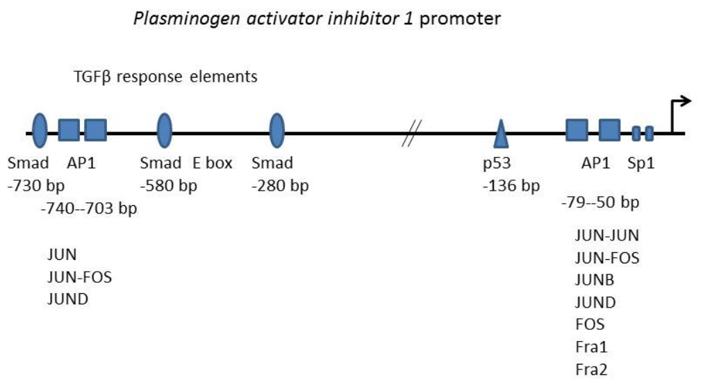
4. Plasminogen Activator Inhibitor 1 Promoter
4.1. Regulation of the Plasminogen Activator Inhibitor 1 Promoter
4.2. AP1 Regulation of the PAI1 Promoter
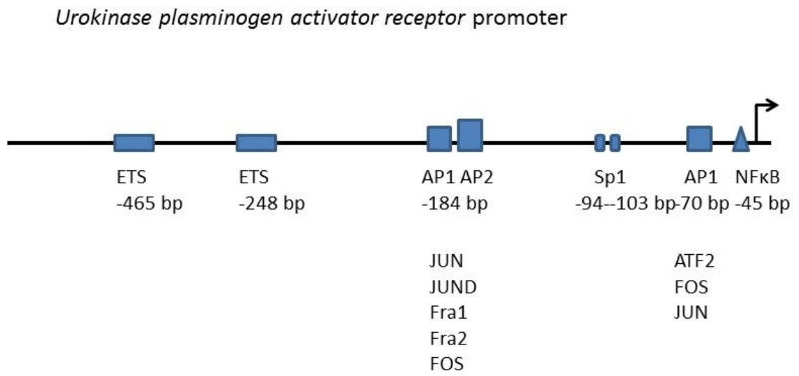
5. uPAR Promoter
5.1. Regulation of the uPAR Promoter
5.2. AP1 Regulation of the uPAR Promoter
6. Discussion
7. Conclusions
Author Contributions
Funding
Institutional Review Board Statement
Informed Consent Statement
Data Availability Statement
Conflicts of Interest
References
- Nagamine, Y.; Medcalf, R.L.; Muñoz-Cánoves, P. Transcriptional and posttranscriptional regulation of the plasminogen activator system. Thromb. Haemost. 2005, 93, 661–675. [Google Scholar] [CrossRef]
- Roselli, H.T.; Su, M.; Washington, K.; Kerins, D.M.; Vaughan, D.E.; Russell, W.E. Liver regeneration is transiently impaired in urokinase-deficient mice. Am. J. Physiol. 1998, 275, G1472–G1479. [Google Scholar] [CrossRef] [PubMed]
- Lin, Z.; Jiang, L.; Yuan, C.; Jensen, J.K.; Zhang, X.; Luo, Z.; Furie, B.C.; Furie, B.; Andreasen, P.A.; Huang, M. Structural basis for recognition of urokinase-type plasminogen activator by plasminogen activator inhibitor-1. J. Biol. Chem. 2011, 286, 7027–7032. [Google Scholar] [CrossRef]
- Zhang, D.; Zhang, J.W.; Xu, H.; Chen, X.; Gao, Y.; Jiang, H.G.; Wang, Y.; Wu, H.; Yang, L.; Wang, W.B.; et al. Therapy-induced senescent tumor cell-derived extracellular vesicles promote colorectal cancer progression through SERPINE1-mediated NF-κB p65 nuclear translocation. Mol. Cancer 2024, 23, 70. [Google Scholar] [CrossRef]
- Nanbu, R.; Menoud, P.A.; Nagamine, Y. Multiple instability-regulating sites in the 3′ untranslated region of the urokinase-type plasminogen activator mRNA. Mol. Cell. Biol. 1994, 14, 4920–4928. [Google Scholar] [CrossRef] [PubMed]
- Montero, L.; Nagamine, Y. Regulation by p38 mitogen-activated protein kinase of adenylate- and uridylate-rich element-mediated urokinase-type plasminogen activator (uPA) messenger RNA stability and uPA-dependent in vitro cell invasion. Cancer Res. 1999, 59, 5286–5293. [Google Scholar]
- Tran, H.; Maurer, F.; Nagamine, Y. Stabilization of urokinase and urokinase receptor mRNAs by HuR is linked to its cytoplasmic accumulation induced by activated mitogen-activated protein kinase-activated protein kinase 2. Mol. Cell. Biol. 2003, 23, 7177–7188. [Google Scholar] [CrossRef]
- Velusamy, T.; Shetty, P.; Bhandary, Y.P.; Liu, M.C.; Shetty, S. Posttranscriptional regulation of urokinase receptor expression by heterogeneous nuclear ribonuclear protein C. Biochemistry 2008, 47, 6508–6517. [Google Scholar] [CrossRef]
- Rømer, J.; Lund, L.R.; Eriksen, J.; Ralfkiaer, E.; Zeheb, R.; Gelehrter, T.D.; Danø, K.; Kristensen, P. Differential expression of urokinase-type plasminogen activator and its type-1 inhibitor during healing of mouse skin wounds. J. Investig. Dermatol. 1991, 97, 803–811. [Google Scholar] [CrossRef] [PubMed]
- Diaz, A.; Martin-Jimenez, C.; Xu, Y.; Merino, P.; Woo, Y.; Torre, E.; Yepes, M. Urokinase-type plasminogen activator-mediated crosstalk between N-cadherin and β-catenin promotes wound healing. J. Cell Sci. 2021, 134, jcs255919. [Google Scholar] [CrossRef] [PubMed]
- Su, J.; Morgani, S.M.; David, C.J.; Wang, Q.; Er, E.E.; Huang, Y.H.; Basnet, H.; Zou, Y.; Shu, W.; Soni, R.K.; et al. TGF-β orchestrates fibrogenic and developmental EMTs via the RAS effector RREB1. Nature 2020, 577, 566–571. [Google Scholar] [CrossRef]
- Bejjani, F.; Evanno, E.; Zibara, K.; Piechaczyk, M.; Jariel-Encontre, I. The AP-1 transcriptional complex: Local switch or remote command? Biochim. Biophys. Acta Rev. Cancer 2019, 1872, 11–23. [Google Scholar] [CrossRef] [PubMed]
- Angel, P.; Karin, M. The role of Jun, Fos and the AP-1 complex in cell-proliferation and transformation. Biochim. Biophys. Acta 1991, 1072, 129–157. [Google Scholar] [CrossRef] [PubMed]
- Piu, F.; Aronheim, A.; Katz, S.; Karin, M. AP-1 repressor protein JDP-2: Inhibition of UV-mediated apoptosis through p53 down-regulation. Mol. Cell. Biol. 2001, 21, 3012–3024. [Google Scholar] [CrossRef]
- Chinenov, Y.; Kerppola, T.K. Close encounters of many kinds: Fos-Jun interactions that mediate transcription regulatory specificity. Oncogene 2001, 20, 2438–2452. [Google Scholar] [CrossRef] [PubMed]
- Eferl, R.; Wagner, E.F. AP-1: A double-edged sword in tumorigenesis. Nat. Rev. Cancer 2003, 3, 859–868. [Google Scholar] [CrossRef]
- Oh, I.Y.; Albea, D.M.; Goodwin, Z.A.; Quiggle, A.M.; Baker, B.P.; Guggisberg, A.M.; Geahlen, J.H.; Kroner, G.M.; de Guzman Strong, C. Regulation of the dynamic chromatin architecture of the epidermal differentiation complex is mediated by a c-Jun/AP-1-modulated enhancer. J. Investig. Dermatol. 2014, 134, 2371–2380. [Google Scholar] [CrossRef]
- Obier, N.; Cauchy, P.; Assi, S.A.; Gilmour, J.; Lie-A-Ling, M.; Lichtinger, M.; Hoogenkamp, M.; Noailles, L.; Cockerill, P.N.; Lacaud, G.; et al. Cooperative binding of AP-1 and TEAD4 modulates the balance between vascular smooth muscle and hemogenic cell fate. Development 2016, 143, 4324–4340. [Google Scholar] [CrossRef]
- Hogan, N.T.; Whalen, M.B.; Stolze, L.K.; Hadeli, N.K.; Lam, M.T.; Springstead, J.R.; Glass, C.K.; Romanoski, C.E. Transcriptional networks specifying homeostatic and inflammatory programs of gene expression in human aortic endothelial cells. eLife 2017, 6, e22536. [Google Scholar] [CrossRef]
- Ghisletti, S.; Barozzi, I.; Mietton, F.; Polletti, S.; De Santa, F.; Venturini, E.; Gregory, L.; Lonie, L.; Chew, A.; Wei, C.L.; et al. Identification and characterization of enhancers controlling the inflammatory gene expression program in macrophages. Immunity 2010, 32, 317–328. [Google Scholar] [CrossRef]
- Velazquez, F.N.; Caputto, B.L.; Boussin, F.D. c-Fos importance for brain development. Aging 2015, 7, 1028–1029. [Google Scholar] [CrossRef] [PubMed]
- Cohen, A.J.; Saiakhova, A.; Corradin, O.; Luppino, J.M.; Lovrenert, K.; Bartels, C.F.; Morrow, J.J.; Mack, S.C.; Dhillon, G.; Beard, L.; et al. Hotspots of aberrant enhancer activity punctuate the colorectal cancer epigenome. Nat. Commun. 2017, 8, 14400. [Google Scholar] [CrossRef]
- Morrow, J.J.; Bayles, I.; Funnell, A.P.W.; Miller, T.E.; Saiakhova, A.; Lizardo, M.M.; Bartels, C.F.; Kapteijn, M.Y.; Hung, S.; Mendoza, A.; et al. Positively selected enhancer elements endow osteosarcoma cells with metastatic competence. Nat. Med. 2018, 24, 176–185. [Google Scholar] [CrossRef]
- Bakiri, L.; Lallemand, D.; Bossy-Wetzel, E.; Yaniv, M. Cell cycle-dependent variations in c-Jun and JunB phosphorylation: A role in the control of cyclin D1 expression. EMBO J. 2000, 19, 2056–2068. [Google Scholar] [CrossRef] [PubMed]
- Schreiber, M.; Kolbus, A.; Piu, F.; Szabowski, A.; Möhle-Steinlein, U.; Tian, J.; Karin, M.; Angel, P.; Wagner, E.F. Control of cell cycle progression by c-Jun is p53 dependent. Genes Dev. 1999, 13, 607–619. [Google Scholar] [CrossRef]
- Owen, T.A.; Cosenza, S.C.; Soprano, D.R.; Soprano, K.J. Time of c-fos and c-myc expression in human diploid fibroblasts stimulated to proliferate after prolonged periods in quiescence. J. Biol. Chem. 1987, 262, 15111–15117. [Google Scholar] [CrossRef]
- Ozanne, B.W.; Spence, H.J.; McGarry, L.C.; Hennigan, R.F. Transcription factors control invasion: AP-1 the first among equals. Oncogene 2007, 26, 1–10. [Google Scholar] [CrossRef] [PubMed]
- Selvaraj, N.; Budka, J.A.; Ferris, M.W.; Plotnik, J.P.; Hollenhorst, P.C. Extracellular signal-regulated kinase signaling regulates the opposing roles of JUN family transcription factors at ETS/AP-1 sites and in cell migration. Mol. Cell. Biol. 2015, 35, 88–100. [Google Scholar] [CrossRef]
- Reichmann, E.; Schwarz, H.; Deiner, E.M.; Leitner, I.; Eilers, M.; Berger, J.; Busslinger, M.; Beug, H. Activation of an inducible c-FosER fusion protein causes loss of epithelial polarity and triggers epithelial-fibroblastoid cell conversion. Cell 1992, 71, 1103–1116. [Google Scholar] [CrossRef]
- Fialka, I.; Schwarz, H.; Reichmann, E.; Oft, M.; Busslinger, M.; Beug, H. The estrogen-dependent c-JunER protein causes a reversible loss of mammary epithelial cell polarity involving a destabilization of adherens junctions. J. Cell Biol. 1996, 132, 1115–1132. [Google Scholar] [CrossRef]
- Wang, W.D.; Gralla, J.D. Differential ability of proximal and remote element pairs to cooperate in activating RNA polymerase II transcription. Mol. Cell. Biol. 1991, 11, 4561–4571. [Google Scholar] [CrossRef]
- Jochum, W.; Passegué, E.; Wagner, E.F. AP-1 in mouse development and tumorigenesis. Oncogene 2001, 20, 2401–2412. [Google Scholar] [CrossRef]
- Kalampounias, G.; Androutsopoulou, T.; Katsoris, P. JUNB and JUND in Urological Cancers: A Literature Review. Curr. Issues Mol. Biol. 2025, 47, 741. [Google Scholar] [CrossRef] [PubMed]
- Marconcini, L.; Marchio, S.; Morbidelli, L.; Cartocci, E.; Albini, A.; Ziche, M.; Bussolino, F.; Oliviero, S. c-fos-induced growth factor/vascular endothelial growth factor D induces angiogenesis In Vivo and In Vitro. Proc. Natl. Acad. Sci. USA 1999, 96, 9671–9676. [Google Scholar] [CrossRef]
- Liu, Z.G.; Jiang, G.; Tang, J.; Wang, H.; Feng, G.; Chen, F.; Tu, Z.; Liu, G.; Zhao, Y.; Peng, M.J.; et al. c-Fos over-expression promotes radioresistance and predicts poor prognosis in malignant glioma. Oncotarget 2016, 7, 65946–65956. [Google Scholar] [CrossRef] [PubMed]
- Phanstiel, D.H.; Van Bortle, K.; Spacek, D.; Hess, G.T.; Shamim, M.S.; Machol, I.; Love, M.I.; Aiden, E.L.; Bassik, M.C.; Snyder, M.P. Static and Dynamic DNA Loops form AP-1-Bound Activation Hubs during Macrophage Development. Mol. Cell 2017, 67, 1037–1048.e6. [Google Scholar] [CrossRef] [PubMed]
- Chronis, C.; Fiziev, P.; Papp, B.; Butz, S.; Bonora, G.; Sabri, S.; Ernst, J.; Plath, K. Cooperative Binding of Transcription Factors Orchestrates Reprogramming. Cell 2017, 168, 442–459.e20. [Google Scholar] [CrossRef]
- Patrick, R.; Naval-Sanchez, M.; Deshpande, N.; Huang, Y.; Zhang, J.; Chen, X.; Yang, Y.; Tiwari, K.; Esmaeili, M.; Tran, M.; et al. The activity of early-life gene regulatory elements is hijacked in aging through pervasive AP-1-linked chromatin opening. Cell Metab. 2024, 36, 1858–1881.e23. [Google Scholar] [CrossRef]
- Hai, T.W.; Liu, F.; Allegretto, E.A.; Karin, M.; Green, M.R. A family of immunologically related transcription factors that includes multiple forms of ATF and AP-1. Genes Dev. 1988, 2, 1216–1226. [Google Scholar] [CrossRef]
- van Dam, H.; Castellazzi, M. Distinct roles of Jun: Fos and Jun: ATF dimers in oncogenesis. Oncogene 2001, 20, 2453–2464. [Google Scholar] [CrossRef]
- Mathas, S.; Hinz, M.; Anagnostopoulos, I.; Krappmann, D.; Lietz, A.; Jundt, F.; Bommert, K.; Mechta-Grigoriou, F.; Stein, H.; Dörken, B.; et al. Aberrantly expressed c-Jun and JunB are a hallmark of Hodgkin lymphoma cells, stimulate proliferation and synergize with NF-kappa B. EMBO J. 2002, 21, 4104–4113. [Google Scholar] [CrossRef] [PubMed]
- Ott, R.G.; Simma, O.; Kollmann, K.; Weisz, E.; Zebedin, E.M.; Schorpp-Kistner, M.; Heller, G.; Zöchbauer, S.; Wagner, E.F.; Freissmuth, M.; et al. JunB is a gatekeeper for B-lymphoid leukemia. Oncogene 2007, 26, 4863–4871. [Google Scholar] [CrossRef] [PubMed]
- Seldeen, K.L.; Deegan, B.J.; Bhat, V.; Mikles, D.C.; McDonald, C.B.; Farooq, A. Energetic coupling along an allosteric communication channel drives the binding of Jun-Fos heterodimeric transcription factor to DNA. FEBS J. 2011, 278, 2090–2104. [Google Scholar] [CrossRef] [PubMed]
- Hong, S.; Wang, D.; Horton, J.R.; Zhang, X.; Speck, S.H.; Blumenthal, R.M.; Cheng, X. Methyl-dependent and spatial-specific DNA recognition by the orthologous transcription factors human AP-1 and Epstein-Barr virus Zta. Nucleic Acids Res. 2017, 45, 2503–2515. [Google Scholar] [CrossRef]
- Arthur-Farraj, P.J.; Morgan, C.C.; Adamowicz, M.; Gomez-Sanchez, J.A.; Fazal, S.V.; Beucher, A.; Razzaghi, B.; Mirsky, R.; Jessen, K.R.; Aitman, T.J. Changes in the Coding and Non-coding Transcriptome and DNA Methylome that Define the Schwann Cell Repair Phenotype after Nerve Injury. Cell Rep. 2017, 20, 2719–2734. [Google Scholar] [CrossRef]
- Yin, Z.; Machius, M.; Nestler, E.J.; Rudenko, G. Activator Protein-1: Redox switch controlling structure and DNA-binding. Nucleic Acids Res. 2017, 45, 11425–11436. [Google Scholar] [CrossRef]
- Iwafuchi-Doi, M.; Zaret, K.S. Cell fate control by pioneer transcription factors. Development 2016, 143, 1833–1837. [Google Scholar] [CrossRef]
- He, X.; Chatterjee, R.; John, S.; Bravo, H.; Sathyanarayana, B.K.; Biddie, S.C.; FitzGerald, P.C.; Stamatoyannopoulos, J.A.; Hager, G.L.; Vinson, C. Contribution of nucleosome binding preferences and co-occurring DNA sequences to transcription factor binding. BMC Genom. 2013, 14, 428. [Google Scholar] [CrossRef]
- Qiao, Y.; Shiue, C.N.; Zhu, J.; Zhuang, T.; Jonsson, P.; Wright, A.P.; Zhao, C.; Dahlman-Wright, K. AP-1-mediated chromatin looping regulates ZEB2 transcription: New insights into TNFα-induced epithelial-mesenchymal transition in triple-negative breast cancer. Oncotarget 2015, 6, 7804–7814. [Google Scholar] [CrossRef]
- Zanconato, F.; Forcato, M.; Battilana, G.; Azzolin, L.; Quaranta, E.; Bodega, B.; Rosato, A.; Bicciato, S.; Cordenonsi, M.; Piccolo, S. Genome-wide association between YAP/TAZ/TEAD and AP-1 at enhancers drives oncogenic growth. Nat. Cell Biol. 2015, 17, 1218–1227. [Google Scholar] [CrossRef]
- Behre, G.; Whitmarsh, A.J.; Coghlan, M.P.; Hoang, T.; Carpenter, C.L.; Zhang, D.E.; Davis, R.J.; Tenen, D.G. c-Jun is a JNK-independent coactivator of the PU.1 transcription factor. J. Biol. Chem. 1999, 274, 4939–4946. [Google Scholar] [CrossRef]
- Wasylyk, B.; Wasylyk, C.; Flores, P.; Begue, A.; Leprince, D.; Stehelin, D. The c-ets proto-oncogenes encode transcription factors that cooperate with c-Fos and c-Jun for transcriptional activation. Nature 1990, 346, 191–193. [Google Scholar] [CrossRef] [PubMed]
- Gum, R.; Lengyel, E.; Juarez, J.; Chen, J.H.; Sato, H.; Seiki, M.; Boyd, D. Stimulation of 92-kDa gelatinase B promoter activity by ras is mitogen-activated protein kinase kinase 1-independent and requires multiple transcription factor binding sites including closely spaced PEA3/ets and AP-1 sequences. J. Biol. Chem. 1996, 271, 10672–10680. [Google Scholar] [CrossRef]
- Gutman, A.; Wasylyk, B. The collagenase gene promoter contains a TPA and oncogene-responsive unit encompassing the PEA3 and AP-1 binding sites. EMBO J. 1990, 9, 2241–2246. [Google Scholar] [CrossRef] [PubMed]
- Nerlov, C.; De Cesare, D.; Pergola, F.; Caracciolo, A.; Blasi, F.; Johnsen, M.; Verde, P. A regulatory element that mediates co-operation between a PEA3-AP-1 element and an AP-1 site is required for phorbol ester induction of urokinase enhancer activity in HepG2 hepatoma cells. EMBO J. 1992, 11, 4573–4582. [Google Scholar] [CrossRef]
- Chang, H.; Liu, Y.; Xue, M.; Liu, H.; Du, S.; Zhang, L.; Wang, P. Synergistic action of master transcription factors controls epithelial-to-mesenchymal transition. Nucleic Acids Res. 2016, 44, 2514–2527. [Google Scholar] [CrossRef]
- Stein, B.; Baldwin, A.S., Jr.; Ballard, D.W.; Greene, W.C.; Angel, P.; Herrlich, P. Cross-coupling of the NF-kappa B p65 and Fos/Jun transcription factors produces potentiated biological function. EMBO J. 1993, 12, 3879–3891. [Google Scholar] [CrossRef]
- Kawana, M.; Lee, M.E.; Quertermous, E.E.; Quertermous, T. Cooperative interaction of GATA-2 and AP1 regulates transcription of the endothelin-1 gene. Mol. Cell. Biol. 1995, 15, 4225–4231. [Google Scholar] [CrossRef]
- Kerppola, T.K.; Luk, D.; Curran, T. Fos is a preferential target of glucocorticoid receptor inhibition of AP-1 activity In Vitro. Mol. Cell. Biol. 1993, 13, 3782–3791. [Google Scholar] [CrossRef] [PubMed]
- Nead, M.A.; Baglia, L.A.; Antinore, M.J.; Ludlow, J.W.; McCance, D.J. Rb binds c-Jun and activates transcription. EMBO J. 1998, 17, 2342–2352. [Google Scholar] [CrossRef]
- Herrlich, P. Cross-talk between glucocorticoid receptor and AP-1. Oncogene 2001, 20, 2465–2475. [Google Scholar] [CrossRef]
- Panne, D.; Maniatis, T.; Harrison, S.C. An atomic model of the interferon-beta enhanceosome. Cell 2007, 129, 1111–1123. [Google Scholar] [CrossRef]
- Murphy, L.O.; Smith, S.; Chen, R.H.; Fingar, D.C.; Blenis, J. Molecular interpretation of ERK signal duration by immediate early gene products. Nat. Cell Biol. 2002, 4, 556–564. [Google Scholar] [CrossRef]
- Lim, W.; Mayer, B.; Pawson, T. Cell Signaling: Principles and Mechanisms; Garland Science: New York, NY, USA; Taylor and Francis Group: Oxfordshire, UK, 2015. [Google Scholar]
- Pérez-Benavente, B.; Farràs, R. Regulation of GSK3β-FBXW7-JUNB axis. Oncotarget 2013, 4, 956–957. [Google Scholar] [CrossRef]
- Lavoie, H.; Gagnon, J.; Therrien, M. ERK signalling: A master regulator of cell behaviour, life and fate. Nat. Rev. Mol. Cell Biol. 2020, 21, 607–632. [Google Scholar] [CrossRef]
- Morrison, D.K. MAP kinase pathways. Cold Spring Harb. Perspect. Biol. 2012, 4, a011254. [Google Scholar] [CrossRef]
- Murakami, M.; Ui, M.; Iba, H. Fra-2-positive autoregulatory loop triggered by mitogen-activated protein kinase (MAPK) and Fra-2 phosphorylation sites by MAPK. Cell Growth Differ. Mol. Biol. J. Am. Assoc. Cancer Res. 1999, 10, 333–342. [Google Scholar]
- Sutherland, C.; Cohen, P. The alpha-isoform of glycogen synthase kinase-3 from rabbit skeletal muscle is inactivated by p70 S6 kinase or MAP kinase-activated protein kinase-1 In Vitro. FEBS Lett. 1994, 338, 37–42. [Google Scholar] [CrossRef]
- Li, T.; Zhang, J.; Zhu, F.; Wen, W.; Zykova, T.; Li, X.; Liu, K.; Peng, C.; Ma, W.; Shi, G.; et al. P21-activated protein kinase (PAK2)-mediated c-Jun phosphorylation at 5 threonine sites promotes cell transformation. Carcinogenesis 2011, 32, 659–666. [Google Scholar] [CrossRef] [PubMed]
- Sevilla, A.; Santos, C.R.; Barcia, R.; Vega, F.M.; Lazo, P.A. c-Jun phosphorylation by the human vaccinia-related kinase 1 (VRK1) and its cooperation with the N-terminal kinase of c-Jun (JNK). Oncogene 2004, 23, 8950–8958. [Google Scholar] [CrossRef] [PubMed]
- Baker, S.J.; Kerppola, T.K.; Luk, D.; Vandenberg, M.T.; Marshak, D.R.; Curran, T.; Abate, C. Jun is phosphorylated by several protein kinases at the same sites that are modified in serum-stimulated fibroblasts. Mol. Cell. Biol. 1992, 12, 4694–4705. [Google Scholar] [CrossRef]
- Boyle, W.J.; Smeal, T.; Defize, L.H.; Angel, P.; Woodgett, J.R.; Karin, M.; Hunter, T. Activation of protein kinase C decreases phosphorylation of c-Jun at sites that negatively regulate its DNA-binding activity. Cell 1991, 64, 573–584. [Google Scholar] [CrossRef]
- Zhu, F.; Choi, B.Y.; Ma, W.Y.; Zhao, Z.; Zhang, Y.; Cho, Y.Y.; Choi, H.S.; Imamoto, A.; Bode, A.M.; Dong, Z. COOH-terminal Src kinase-mediated c-Jun phosphorylation promotes c-Jun degradation and inhibits cell transformation. Cancer Res. 2006, 66, 5729–5736. [Google Scholar] [CrossRef]
- Li, B.; Tournier, C.; Davis, R.J.; Flavell, R.A. Regulation of IL-4 expression by the transcription factor JunB during T helper cell differentiation. EMBO J. 1999, 18, 420–432. [Google Scholar] [CrossRef] [PubMed]
- Wang, H.; Xie, Z.; Scott, R.E. JunD phosphorylation, and expression of AP-1 DNA binding activity modulated by serum growth factors in quiescent murine 3T3T cells. Oncogene 1996, 13, 2639–2647. [Google Scholar] [PubMed]
- Pakneshan, P.; Szyf, M.; Farias-Eisner, R.; Rabbani, S.A. Reversal of the hypomethylation status of urokinase (uPA) promoter blocks breast cancer growth and metastasis. J. Biol. Chem. 2004, 279, 31735–31744. [Google Scholar] [CrossRef]
- Boudreau, N.J.; Varner, J.A. The homeobox transcription factor Hox D3 promotes integrin alpha5beta1 expression and function during angiogenesis. J. Biol. Chem. 2004, 279, 4862–4868. [Google Scholar] [CrossRef] [PubMed]
- D’Orazio, D.; Besser, D.; Marksitzer, R.; Kunz, C.; Hume, D.A.; Kiefer, B.; Nagamine, Y. Cooperation of two PEA3/AP1 sites in uPA gene induction by TPA and FGF-2. Gene 1997, 201, 179–187. [Google Scholar] [CrossRef]
- Verde, P.; Boast, S.; Franzè, A.; Robbiati, F.; Blasi, F. An upstream enhancer and a negative element in the 5’ flanking region of the human urokinase plasminogen activator gene. Nucleic Acids Res. 1998, 16, 10699–10716. [Google Scholar] [CrossRef]
- Benasciutti, E.; Pagès, G.; Kenzior, O.; Folk, W.; Blasi, F.; Crippa, M.P. MAPK and JNK transduction pathways can phosphorylate Sp1 to activate the uPA minimal promoter element and endogenous gene transcription. Blood 2004, 104, 256–262. [Google Scholar] [CrossRef]
- Belaguli, N.S.; Aftab, M.; Rigi, M.; Zhang, M.; Albo, D.; Berger, D.H. GATA6 promotes colon cancer cell invasion by regulating urokinase plasminogen activator gene expression. Neoplasia 2010, 12, 856–865. [Google Scholar] [CrossRef] [PubMed]
- Soubt, M.K.; Marksitzer, R.; Menoud, P.A.; Nagamine, Y. Role of tissue-specific transcription factor LFB3 in a cyclic AMP-responsive enhancer of the urokinase-type plasminogen activator gene in LLC-PK1 cells. Mol. Cell. Biol. 1998, 18, 4698–4706. [Google Scholar] [CrossRef]
- Sánchez-Tilló, E.; de Barrios, O.; Siles, L.; Amendola, P.G.; Darling, D.S.; Cuatrecasas, M.; Castells, A.; Postigo, A. ZEB1 Promotes invasiveness of colorectal carcinoma cells through the opposing regulation of uPA and PAI-1. Clin. Cancer Res. Off. J. Am. Assoc. Cancer Res. 2013, 19, 1071–1082. [Google Scholar] [CrossRef]
- Hiendlmeyer, E.; Regus, S.; Wassermann, S.; Hlubek, F.; Haynl, A.; Dimmler, A.; Koch, C.; Knoll, C.; van Beest, M.; Reuning, U.; et al. Beta-catenin up-regulates the expression of the urokinase plasminogen activator in human colorectal tumors. Cancer Res. 2004, 64, 1209–1214. [Google Scholar] [CrossRef]
- Shimizu, M.; Cohen, B.; Goldvasser, P.; Berman, H.; Virtanen, C.; Reedijk, M. Plasminogen activator uPA is a direct transcriptional target of the JAG1-Notch receptor signaling pathway in breast cancer. Cancer Res. 2011, 71, 277–286. [Google Scholar] [CrossRef]
- Ito, T.; Takenaka, K.; Sakai, H.; Yoshimura, S.; Hayashi, K.; Noda, S.; Sakai, N. Elevation of mRNA levels of tissue-type plasminogen activator and urokinase-type plasminogen activator in hippocampus and cerebral cortex following middle cerebral artery occlusion in rats. Neurol. Res. 2000, 22, 413–419. [Google Scholar] [CrossRef]
- Cannio, R.; Rennie, P.S.; Blasi, F. A cell-type specific and enhancer-dependent silencer in the regulation of the expression of the human urokinase plasminogen activator gene. Nucleic Acids Res. 1991, 19, 2303–2308. [Google Scholar] [CrossRef] [PubMed]
- Kunz, C.; Pebler, S.; Otte, J.; von der Ahe, D. Differential regulation of plasminogen activator and inhibitor gene transcription by the tumor suppressor p53. Nucleic Acids Res. 1995, 23, 3710–3717. [Google Scholar] [CrossRef]
- Lee, J.S.; von der Ahe, D.; Kiefer, B.; Nagamine, Y. Cytoskeletal reorganization and TPA differently modify AP-1 to induce the urokinase-type plasminogen activator gene in LLC-PK1 cells. Nucleic Acids Res. 1993, 21, 3365–3372. [Google Scholar] [CrossRef] [PubMed]
- Lee, J.S.; Favre, B.; Hemmings, B.A.; Kiefer, B.; Nagamine, Y. Okadaic acid-dependent induction of the urokinase-type plasminogen activator gene associated with stabilization and autoregulation of c-Jun. J. Biol. Chem. 1994, 269, 2887–2894. [Google Scholar] [CrossRef]
- Botteri, F.M.; Ballmer-Hofer, K.; Rajput, B.; Nagamine, Y. Disruption of cytoskeletal structures results in the induction of the urokinase-type plasminogen activator gene expression. J. Biol. Chem. 1990, 265, 13327–13334. [Google Scholar] [CrossRef]
- Cuevas, B.D.; Uhlik, M.T.; Garrington, T.P.; Johnson, G.L. MEKK1 regulates the AP-1 dimer repertoire via control of JunB transcription and Fra-2 protein stability. Oncogene 2005, 24, 801–809. [Google Scholar] [CrossRef]
- Besser, D.; Presta, M.; Nagamine, Y. Elucidation of a signaling pathway induced by FGF-2 leading to uPA gene expression in NIH 3T3 fibroblasts. Cell Growth Differ. Mol. Biol. J. Am. Assoc. Cancer Res. 1995, 6, 1009–1017. [Google Scholar]
- Moquet-Torcy, G.; Tolza, C.; Piechaczyk, M.; Jariel-Encontre, I. Transcriptional complexity and roles of Fra-1/AP-1 at the uPA/Plau locus in aggressive breast cancer. Nucleic Acids Res. 2014, 42, 11011–11024. [Google Scholar] [CrossRef]
- Rørth, P.; Nerlov, C.; Blasi, F.; Johnsen, M. Transcription factor PEA3 participates in the induction of urokinase plasminogen activator transcription in murine keratinocytes stimulated with epidermal growth factor or phorbol-ester. Nucleic Acids Res. 1990, 18, 5009–5017. [Google Scholar] [CrossRef]
- Bernhart, E.; Damm, S.; Wintersperger, A.; DeVaney, T.; Zimmer, A.; Raynham, T.; Ireson, C.; Sattler, W. Protein kinase D2 regulates migration and invasion of U87MG glioblastoma cells In Vitro. Exp. Cell Res. 2013, 319, 2037–2048. [Google Scholar] [CrossRef]
- Irigoyen, J.P.; Besser, D.; Nagamine, Y. Cytoskeleton reorganization induces the urokinase-type plasminogen activator gene via the Ras/extracellular signal-regulated kinase (ERK) signaling pathway. J. Biol. Chem. 1997, 272, 1904–1909. [Google Scholar] [CrossRef] [PubMed]
- Irigoyen, J.P.; Nagamine, Y. Cytoskeletal reorganization leads to induction of the urokinase-type plasminogen activator gene by activating FAK and Src and subsequently the Ras/Erk signaling pathway. Biochem. Biophys. Res. Commun. 1999, 262, 666–670. [Google Scholar] [CrossRef]
- Silberman, S.; Janulis, M.; Schultz, R.M. Characterization of downstream Ras signals that induce alternative protease-dependent invasive phenotypes. J. Biol. Chem. 1997, 272, 5927–5935. [Google Scholar] [CrossRef] [PubMed]
- Ried, S.; Jäger, C.; Jeffers, M.; Vande Woude, G.F.; Graeff, H.; Schmitt, M.; Lengyel, E. Activation mechanisms of the urokinase-type plasminogen activator promoter by hepatocyte growth factor/scatter factor. J. Biol. Chem. 1999, 274, 16377–16386. [Google Scholar] [CrossRef] [PubMed]
- Lee, K.H.; Choi, E.Y.; Koh, S.A.; Kim, M.K.; Kim, K.O.; Lee, S.H.; Jang, B.I.; Kim, S.W.; Kim, S.W.; Song, S.K.; et al. Down-regulation of survivin suppresses uro-plasminogen activator through transcription factor JunB. Exp. Mol. Med. 2011, 43, 501–509. [Google Scholar] [CrossRef] [PubMed]
- Fowles, L.F.; Martin, M.L.; Nelsen, L.; Stacey, K.J.; Redd, D.; Clark, Y.M.; Nagamine, Y.; McMahon, M.; Hume, D.A.; Ostrowski, M.C. Persistent activation of mitogen-activated protein kinases p42 and p44 and ets-2 phosphorylation in response to colony-stimulating factor 1/c-fms signaling. Mol. Cell. Biol. 1998, 18, 5148–5156. [Google Scholar] [CrossRef] [PubMed]
- Stacey, K.J.; Fowles, L.F.; Colman, M.S.; Ostrowski, M.C.; Hume, D.A. Regulation of urokinase-type plasminogen activator gene transcription by macrophage colony-stimulating factor. Mol. Cell. Biol. 1995, 15, 3430–3441. [Google Scholar] [CrossRef]
- Cirillo, G.; Casalino, L.; Vallone, D.; Caracciolo, A.; De Cesare, D.; Verde, P. Role of distinct mitogen-activated protein kinase pathways and cooperation between Ets-2, ATF-2, and Jun family members in human urokinase-type plasminogen activator gene induction by interleukin-1 and tetradecanoyl phorbol acetate. Mol. Cell. Biol. 1999, 19, 6240–6252. [Google Scholar] [CrossRef]
- Seddighzadeh, M.; Zhou, J.N.; Kronenwett, U.; Shoshan, M.C.; Auer, G.; Sten-Linder, M.; Wiman, B.; Linder, S. ERK signalling in metastatic human MDA-MB-231 breast carcinoma cells is adapted to obtain high urokinase expression and rapid cell proliferation. Clin. Exp. Metastasis 1999, 17, 649–654. [Google Scholar] [CrossRef]
- Miskin, R.; Ben-Ishai, R. Induction of plasminogen activator by UV light in normal and xeroderma pigmentosum fibroblasts. Proc. Natl. Acad. Sci. USA 1981, 78, 6236–6240. [Google Scholar] [CrossRef]
- Miralles, F.; Parra, M.; Caelles, C.; Nagamine, Y.; Félez, J.; Muñoz-Cánoves, P. UV irradiation induces the murine urokinase-type plasminogen activator gene via the c-Jun N-terminal kinase signaling pathway: Requirement of an AP1 enhancer element. Mol. Cell. Biol. 1998, 18, 4537–4547. [Google Scholar] [CrossRef]
- Madunić, J.; Antica, M.; Cvjetko, P.; Požgaj, L.; Matulić, M. Modulation of urokinase plasminogen activator system by poly(ADP-ribose)polymerase-1 inhibition. Cytotechnology 2016, 68, 783–794. [Google Scholar] [CrossRef]
- Tanos, T.; Marinissen, M.J.; Leskow, F.C.; Hochbaum, D.; Martinetto, H.; Gutkind, J.S.; Coso, O.A. Phosphorylation of c-Fos by members of the p38 MAPK family. Role in the AP-1 response to UV light. J. Biol. Chem. 2005, 280, 18842–18852. [Google Scholar] [CrossRef] [PubMed]
- Niiya, M.; Niiya, K.; Shibakura, M.; Asaumi, N.; Yoshida, C.; Shinagawa, K.; Teshima, T.; Ishimaru, F.; Ikeda, K.; Tanimoto, M. Involvement of ERK1/2 and p38 MAP kinase in doxorubicin-induced uPA expression in human RC-K8 lymphoma and NCI-H69 small cell lung carcinoma cells. Oncology 2004, 67, 310–319. [Google Scholar] [CrossRef]
- Lengyel, E.; Wang, H.; Stepp, E.; Juarez, J.; Wang, Y.; Doe, W.; Pfarr, C.M.; Boyd, D. Requirement of an upstream AP-1 motif for the constitutive and phorbol ester-inducible expression of the urokinase-type plasminogen activator receptor gene. J. Biol. Chem. 1996, 271, 23176–23184. [Google Scholar] [CrossRef]
- Cáceres, M.; Tobar, N.; Guerrero, J.; Smith, P.C.; Martínez, J. c-jun-NH2JNK mediates invasive potential and EGFR activation by regulating the expression of HB-EGF in a urokinase-stimulated pathway. J. Cell. Biochem. 2008, 103, 986–993. [Google Scholar] [CrossRef]
- Chen, H.; Padia, R.; Li, T.; Li, Y.; Li, B.; Jin, L.; Huang, S. Signaling of MK2 sustains robust AP1 activity for triple negative breast cancer tumorigenesis through direct phosphorylation of JAB1. npj Breast Cancer 2021, 7, 91. [Google Scholar] [CrossRef]
- Talotta, F.; Casalino, L.; Verde, P. The nuclear oncoprotein Fra-1: A transcription factor knocking on therapeutic applications’ door. Oncogene 2020, 39, 4491–4506. [Google Scholar] [CrossRef]
- Schorpp-Kistner, M.; Wang, Z.Q.; Angel, P.; Wagner, E.F. JunB is essential for mammalian placentation. EMBO J. 1999, 18, 934–948. [Google Scholar] [CrossRef]
- De Cesare, D.; Palazzolo, M.; Blasi, F. Functional characterization of COM, a DNA region required for cooperation between AP-1 sites in urokinase gene transcription. Oncogene 1996, 13, 2551–2562. [Google Scholar]
- Palazzolo, M.; Berthelsen, J.; De Cesare, D.; Blasi, F. Oct-1 specifically binds the UEF4 site of the human AP1-regulated urokinase enhancer. Eur. J. Biochem. 2000, 267, 5427–5437. [Google Scholar] [CrossRef]
- Dunn, S.E.; Torres, J.V.; Oh, J.S.; Cykert, D.M.; Barrett, J.C. Up-regulation of urokinase-type plasminogen activator by insulin-like growth factor-I depends upon phosphatidylinositol-3 kinase and mitogen-activated protein kinase kinase. Cancer Res. 2001, 61, 1367–1374. [Google Scholar]
- Huang, W.S.; Chin, C.C.; Chen, C.N.; Kuo, Y.H.; Chen, T.C.; Yu, H.R.; Tung, S.Y.; Shen, C.H.; Hsieh, Y.Y.; Guo, S.E.; et al. Stromal cell-derived factor-1/CXC receptor 4 and β1 integrin interaction regulates urokinase-type plasminogen activator expression in human colorectal cancer cells. J. Cell. Physiol. 2012, 227, 1114–1122. [Google Scholar] [CrossRef]
- Grabinski, N.; Bartkowiak, K.; Grupp, K.; Brandt, B.; Pantel, K.; Jücker, M. Distinct functional roles of Akt isoforms for proliferation, survival, migration and EGF-mediated signalling in lung cancer derived disseminated tumor cells. Cell. Signal. 2011, 23, 1952–1960. [Google Scholar] [CrossRef]
- Moreau, M.; Mourah, S.; Dosquet, C. β-Catenin and NF-κB cooperate to regulate the uPA/uPAR system in cancer cells. Int. J. Cancer 2011, 128, 1280–1292. [Google Scholar] [CrossRef]
- Tai, Y.T.; Podar, K.; Mitsiades, N.; Lin, B.; Mitsiades, C.; Gupta, D.; Akiyama, M.; Catley, L.; Hideshima, T.; Munshi, N.C.; et al. CD40 induces human multiple myeloma cell migration via phosphatidylinositol 3-kinase/AKT/NF-kappa B signaling. Blood 2003, 101, 2762–2769. [Google Scholar] [CrossRef]
- Ibañez-Tallon, I.; Caretti, G.; Blasi, F.; Crippa, M.P. In Vivo analysis of the state of the human uPA enhancer following stimulation by TPA. Oncogene 1999, 18, 2836–2845. [Google Scholar] [CrossRef]
- Marksitzer, R.; Stief, A.; Menoud, P.A.; Nagamine, Y. Role of LFB3 in cell-specific cAMP induction of the urokinase-type plasminogen activator gene. J. Biol. Chem. 1995, 270, 21833–21838. [Google Scholar] [CrossRef]
- Mira-Y-Lopez, R.; Jaramillo, S.; Jing, Y. Synergistic transcriptional activation of the mouse urokinase plasminogen activator (uPA) gene and of its enhancer activator protein 1 (AP1) site by cAMP and retinoic acid. Biochem. J. 1998, 331, 909–916. [Google Scholar] [CrossRef]
- Miralles, F.; Ibáñez-Tallon, I.; Parra, M.; Crippa, M.; Blasi, F.; Besser, D.; Nagamine, Y.; Muñoz-Cánoves, P. Transcriptional regulation of the murine urokinase-type plasminogen activator gene in skeletal myoblasts. Thromb. Haemost. 1999, 81, 767–774. [Google Scholar] [CrossRef]
- Santibañez, J.F. JNK mediates TGF-beta1-induced epithelial mesenchymal transdifferentiation of mouse transformed keratinocytes. FEBS Lett. 2006, 580, 5385–5391. [Google Scholar] [CrossRef]
- Tanaka, Y.; Kobayashi, H.; Suzuki, M.; Kanayama, N.; Terao, T. Transforming growth factor-beta1-dependent urokinase up-regulation and promotion of invasion are involved in Src-MAPK-dependent signaling in human ovarian cancer cells. J. Biol. Chem. 2004, 279, 8567–8576. [Google Scholar] [CrossRef]
- Hansen, S.K.; Nerlov, C.; Zabel, U.; Verde, P.; Johnsen, M.; Baeuerle, P.A.; Blasi, F. A novel complex between the p65 subunit of NF-kappa B and c-Rel binds to a DNA element involved in the phorbol ester induction of the human urokinase gene. EMBO J. 1992, 11, 205–213. [Google Scholar] [CrossRef]
- Li, H.; Ye, X.; Mahanivong, C.; Bian, D.; Chun, J.; Huang, S. Signaling mechanisms responsible for lysophosphatidic acid-induced urokinase plasminogen activator expression in ovarian cancer cells. J. Biol. Chem. 2005, 280, 10564–10571. [Google Scholar] [CrossRef]
- Slivova, V.; Zaloga, G.; DeMichele, S.J.; Mukerji, P.; Huang, Y.S.; Siddiqui, R.; Harvey, K.; Valachovicova, T.; Sliva, D. Green tea polyphenols modulate secretion of urokinase plasminogen activator (uPA) and inhibit invasive behavior of breast cancer cells. Nutr. Cancer 2005, 52, 66–73. [Google Scholar] [CrossRef]
- Valachovicova, T.; Slivova, V.; Bergman, H.; Shuherk, J.; Sliva, D. Soy isoflavones suppress invasiveness of breast cancer cells by the inhibition of NF-kappaB/AP-1-dependent and -independent pathways. Int. J. Oncol. 2004, 25, 1389–1395. [Google Scholar]
- Das, R.; Mahabeleshwar, G.H.; Kundu, G.C. Osteopontin induces AP-1-mediated secretion of urokinase-type plasminogen activator through c-Src-dependent epidermal growth factor receptor transactivation in breast cancer cells. J. Biol. Chem. 2004, 279, 11051–11064. [Google Scholar] [CrossRef] [PubMed]
- Dos Santos, S.; Delattre, A.I.; De Longueville, F.; Bult, H.; Raes, M. Gene expression profiling of LPS-stimulated murine macrophages and role of the NF-kappaB and PI3K/mTOR signaling pathways. Ann. N. Y. Acad. Sci. 2007, 1096, 70–77. [Google Scholar] [CrossRef]
- Hapke, S.; Kessler, H.; Arroyo de Prada, N.; Benge, A.; Schmitt, M.; Lengyel, E.; Reuning, U. Integrin alpha(v)beta(3)/vitronectin interaction affects expression of the urokinase system in human ovarian cancer cells. J. Biol. Chem. 2001, 276, 26340–26348. [Google Scholar] [CrossRef] [PubMed]
- Kessler, T.L.; Markus, G. Epidermal growth factor and 12-tetradecanoyl phorbol 13-acetate induction of urokinase in A431 cells. Semin. Thromb. Hemostasis 1991, 17, 217–224. [Google Scholar] [CrossRef]
- Shin, B.A.; Yoo, H.G.; Kim, H.S.; Kim, M.H.; Hwang, Y.S.; Chay, K.O.; Lee, K.Y.; Ahn, B.W.; Jung, Y.D. P38 MAPK pathway is involved in the urokinase plasminogen activator expression in human gastric SNU-638 cells. Oncol. Rep. 2003, 10, 1467–1471. [Google Scholar] [CrossRef] [PubMed]
- Secchia, S.; Beilinson, V.; Chen, X.; Gucwa, M.; Denson, L.A.; Miraldi, E.R.; Weirauch, M.T.; Ikegami, K. Starvation activates ECM-remodeling gene transcription and putative enhancers in fibroblasts despite inducing quiescence. Cell Rep. 2025, 44, 115896. [Google Scholar] [CrossRef]
- Seiffert, D.; Loskutoff, D.J. Type 1 plasminogen activator inhibitor induces multimerization of plasma vitronectin. A suggested mechanism for the generation of the tissue form of vitronectin In Vivo. J. Biol. Chem. 1996, 271, 29644–29651. [Google Scholar] [CrossRef]
- Nagamine, Y. Transcriptional regulation of the plasminogen activator inhibitor type 1—With an emphasis on negative regulation. Thromb. Haemost. 2008, 100, 1007–1013. [Google Scholar] [CrossRef]
- Mansfield, M.W.; Stickland, M.H.; Grant, P.J. Environmental and genetic factors in relation to elevated circulating levels of plasminogen activator inhibitor-1 in Caucasian patients with non-insulin-dependent diabetes mellitus. Thromb. Haemost. 1995, 74, 842–847. [Google Scholar] [CrossRef] [PubMed]
- Dennler, S.; Pendaries, V.; Tacheau, C.; Costas, M.A.; Mauviel, A.; Verrecchia, F. The steroid receptor co-activator-1 (SRC-1) potentiates TGF-beta/Smad signaling: Role of p300/CBP. Oncogene 2005, 24, 1936–1945. [Google Scholar] [CrossRef]
- Massagué, J. How cells read TGF-beta signals. Nat. Rev. Mol. Cell Biol. 2000, 1, 169–178. [Google Scholar] [CrossRef]
- Datta, P.K.; Blake, M.C.; Moses, H.L. Regulation of plasminogen activator inhibitor-1 expression by transforming growth factor-beta -induced physical and functional interactions between smads and Sp1. J. Biol. Chem. 2000, 275, 40014–40019. [Google Scholar] [CrossRef]
- Riccio, A.; Lund, L.R.; Sartorio, R.; Lania, A.; Andreasen, P.A.; Danø, K.; Blasi, F. The regulatory region of the human plasminogen activator inhibitor type-1 (PAI-1) gene. Nucleic Acids Res. 1988, 16, 2805–2824. [Google Scholar] [CrossRef]
- Grinberg, A.V.; Kerppola, T. Both Max and TFE3 cooperate with Smad proteins to bind the plasminogen activator inhibitor-1 promoter, but they have opposite effects on transcriptional activity. J. Biol. Chem. 2003, 278, 11227–11236. [Google Scholar] [CrossRef]
- Rossato, V.V.; Silveira, D.A.; Gupta, S.; Mombach, J.C.M. Towards the contribution of the p38MAPK pathway to the dual role of TGFβ in cancer: A boolean model approach. Comput. Biol. Med. 2019, 104, 235–240. [Google Scholar] [CrossRef]
- Kutz, S.M.; Providence, K.M.; Higgins, P.J. Antisense targeting of c-fos transcripts inhibits serum- and TGF-beta 1-stimulated PAI-1 gene expression and directed motility in renal epithelial cells. Cell Motil. Cytoskelet. 2001, 48, 163–174. [Google Scholar] [CrossRef]
- Samarakoon, R.; Dobberfuhl, A.D.; Cooley, C.; Overstreet, J.M.; Patel, S.; Goldschmeding, R.; Meldrum, K.K.; Higgins, P.J. Induction of renal fibrotic genes by TGF-β1 requires EGFR activation, p53 and reactive oxygen species. Cell. Signal. 2013, 25, 2198–2209. [Google Scholar] [CrossRef] [PubMed]
- Pawlus, M.R.; Wang, L.; Murakami, A.; Dai, G.; Hu, C.J. STAT3 or USF2 contributes to HIF target gene specificity. PLoS ONE 2013, 8, e72358. [Google Scholar] [CrossRef] [PubMed]
- Pawlus, M.R.; Hu, C.J. Enhanceosomes as integrators of hypoxia inducible factor (HIF) and other transcription factors in the hypoxic transcriptional response. Cell. Signal. 2013, 25, 1895–1903. [Google Scholar] [CrossRef] [PubMed]
- Dimova, E.Y.; Kietzmann, T. The MAPK pathway and HIF-1 are involved in the induction of the human PAI-1 gene expression by insulin in the human hepatoma cell line HepG2. Ann. N. Y. Acad. Sci. 2006, 1090, 355–367. [Google Scholar] [CrossRef]
- Kietzmann, T.; Jungermann, K.; Görlach, A. Regulation of the hypoxia-dependent plasminogen activator inhibitor 1 expression by MAP kinases. Thromb. Haemost. 2003, 89, 666–673. [Google Scholar] [CrossRef]
- Fink, T.; Kazlauskas, A.; Poellinger, L.; Ebbesen, P.; Zachar, V. Identification of a tightly regulated hypoxia-response element in the promoter of human plasminogen activator inhibitor-1. Blood 2002, 99, 2077–2083. [Google Scholar] [CrossRef]
- Sato, M.; Tanaka, T.; Maemura, K.; Uchiyama, T.; Sato, H.; Maeno, T.; Suga, T.; Iso, T.; Ohyama, Y.; Arai, M.; et al. The PAI-1 gene as a direct target of endothelial PAS domain protein-1 in adenocarcinoma A549 cells. Am. J. Respir. Cell Mol. Biol. 2004, 31, 209–215. [Google Scholar] [CrossRef] [PubMed]
- Dimova, E.Y.; Jakubowska, M.M.; Kietzmann, T. CREB binding to the hypoxia-inducible factor-1 responsive elements in the plasminogen activator inhibitor-1 promoter mediates the glucagon effect. Thromb. Haemost. 2007, 98, 296–303. [Google Scholar] [CrossRef]
- Samoylenko, A.; Roth, U.; Jungermann, K.; Kietzmann, T. The upstream stimulatory factor-2a inhibits plasminogen activator inhibitor-1 gene expression by binding to a promoter element adjacent to the hypoxia-inducible factor-1 binding site. Blood 2001, 97, 2657–2666. [Google Scholar] [CrossRef]
- Gross, C.; Buchwalter, G.; Dubois-Pot, H.; Cler, E.; Zheng, H.; Wasylyk, B. The ternary complex factor net is downregulated by hypoxia and regulates hypoxia-responsive genes. Mol. Cell. Biol. 2007, 27, 4133–4141. [Google Scholar] [CrossRef]
- Buchwalter, G.; Gross, C.; Wasylyk, B. The ternary complex factor Net regulates cell migration through inhibition of PAI-1 expression. Mol. Cell. Biol. 2005, 25, 10853–10862. [Google Scholar] [CrossRef]
- Alessi, M.C.; Poggi, M.; Juhan-Vague, I. Plasminogen activator inhibitor-1, adipose tissue and insulin resistance. Curr. Opin. Lipidol. 2007, 18, 240–245. [Google Scholar] [CrossRef] [PubMed]
- Koziczak, M.; Müller, H.; Helin, K.; Nagamine, Y. E2F1-mediated transcriptional inhibition of the plasminogen activator inhibitor type 1 gene. Eur. J. Biochem. 2001, 268, 4969–4978. [Google Scholar] [CrossRef]
- Banfi, C.; Eriksson, P.; Giandomenico, G.; Mussoni, L.; Sironi, L.; Hamsten, A.; Tremoli, E. Transcriptional regulation of plasminogen activator inhibitor type 1 gene by insulin: Insights into the signaling pathway. Diabetes 2001, 50, 1522–1530. [Google Scholar] [CrossRef]
- Venugopal, J.; Hanashiro, K.; Nagamine, Y. Regulation of PAI-1 gene expression during adipogenesis. J. Cell. Biochem. 2007, 101, 369–380. [Google Scholar] [CrossRef]
- Özcan, S.; Alessio, N.; Acar, M.B.; Mert, E.; Omerli, F.; Peluso, G.; Galderisi, U. Unbiased analysis of senescence associated secretory phenotype (SASP) to identify common components following different genotoxic stresses. Aging 2016, 8, 1316–1329. [Google Scholar] [CrossRef] [PubMed]
- Kortlever, R.M.; Higgins, P.J.; Bernards, R. Plasminogen activator inhibitor-1 is a critical downstream target of p53 in the induction of replicative senescence. Nat. Cell Biol. 2006, 8, 877–884. [Google Scholar] [CrossRef]
- Samarakoon, R.; Higgins, S.P.; Higgins, C.E.; Higgins, P.J. The TGF-β1/p53/PAI-1 Signaling Axis in Vascular Senescence: Role of Caveolin-1. Biomolecules 2019, 9, 341. [Google Scholar] [CrossRef]
- Parra, M.; Jardí, M.; Koziczak, M.; Nagamine, Y.; Muñoz-Cánoves, P. p53 Phosphorylation at serine 15 is required for transcriptional induction of the plasminogen activator inhibitor-1 (PAI-1) gene by the alkylating agent N-methyl-N’-nitro-N-nitrosoguanidine. J. Biol. Chem. 2001, 276, 36303–36310. [Google Scholar] [CrossRef] [PubMed]
- Omer, A.; Patel, D.; Lian, X.J.; Sadek, J.; Di Marco, S.; Pause, A.; Gorospe, M.; Gallouzi, I.E. Stress granules counteract senescence by sequestration of PAI-1. EMBO Rep. 2018, 19, e44722. [Google Scholar] [CrossRef]
- Smith, L.H.; Coats, S.R.; Qin, H.; Petrie, M.S.; Covington, J.W.; Su, M.; Eren, M.; Vaughan, D.E. Differential and opposing regulation of PAI-1 promoter activity by estrogen receptor alpha and estrogen receptor beta in endothelial cells. Circ. Res. 2004, 95, 269–275. [Google Scholar] [CrossRef] [PubMed]
- Marquard, S.; Thomann, S.; Weiler, S.M.E.; Bissinger, M.; Lutz, T.; Sticht, C.; Tóth, M.; de la Torre, C.; Gretz, N.; Straub, B.K.; et al. Yes-associated protein (YAP) induces a secretome phenotype and transcriptionally regulates plasminogen activator Inhibitor-1 (PAI-1) expression in hepatocarcinogenesis. Cell Commun. Signal. 2020, 18, 166. [Google Scholar] [CrossRef]
- Maemura, K.; de la Monte, S.M.; Chin, M.T.; Layne, M.D.; Hsieh, C.M.; Yet, S.F.; Perrella, M.A.; Lee, M.E. CLIF, a novel cycle-like factor, regulates the circadian oscillation of plasminogen activator inhibitor-1 gene expression. J. Biol. Chem. 2000, 275, 36847–36851. [Google Scholar] [CrossRef]
- Bosma, P.J.; Kooistra, T. Different induction of two plasminogen activator inhibitor 1 mRNA species by phorbol ester in human hepatoma cells. J. Biol. Chem. 1991, 266, 17845–17849. [Google Scholar] [CrossRef]
- Dennler, S.; Itoh, S.; Vivien, D.; ten Dijke, P.; Huet, S.; Gauthier, J.M. Direct binding of Smad3 and Smad4 to critical TGF beta-inducible elements in the promoter of human plasminogen activator inhibitor-type 1 gene. EMBO J. 1998, 17, 3091–3100. [Google Scholar] [CrossRef] [PubMed]
- Arts, J.; Grimbergen, J.; Bosma, P.J.; Rahmsdorf, H.J.; Kooistra, T. Role of c-Jun and proximal phorbol 12-myristate-13-acetate-(PMA)-responsive elements in the regulation of basal and PMA-stimulated plasminogen-activator inhibitor-1 gene expression in HepG2. Eur. J. Biochem. 1996, 241, 393–402. [Google Scholar] [CrossRef] [PubMed]
- Descheemaeker, K.A.; Wyns, S.; Nelles, L.; Auwerx, J.; Ny, T.; Collen, D. Interaction of AP-1-, AP-2-, and Sp1-like proteins with two distinct sites in the upstream regulatory region of the plasminogen activator inhibitor-1 gene mediates the phorbol 12-myristate 13-acetate response. J. Biol. Chem. 1992, 267, 15086–15091. [Google Scholar] [CrossRef] [PubMed]
- Kasza, A.; Kiss, D.L.; Gopalan, S.; Xu, W.; Rydel, R.E.; Koj, A.; Kordula, T. Mechanism of plasminogen activator inhibitor-1 regulation by oncostatin M and interleukin-1 in human astrocytes. J. Neurochem. 2002, 83, 696–703. [Google Scholar] [CrossRef] [PubMed]
- Pontrelli, P.; Ranieri, E.; Ursi, M.; Ghosh-Choudhury, G.; Gesualdo, L.; Paolo Schena, F.; Grandaliano, G. jun-N-terminal kinase regulates thrombin-induced PAI-1 gene expression in proximal tubular epithelial cells. Kidney Int. 2004, 65, 2249–2261. [Google Scholar] [CrossRef]
- Lee, K.S.; Nam, G.S.; Baek, J.; Kim, S.; Nam, K.S. Inhibition of TPA-induced metastatic potential by morin hydrate in MCF-7 human breast cancer cells via the Akt/GSK-3β/c-Fos signaling pathway. Int. J. Oncol. 2020, 56, 630–640. [Google Scholar] [CrossRef]
- Olman, M.A.; Hagood, J.S.; Simmons, W.L.; Fuller, G.M.; Vinson, C.; White, K.E. Fibrin fragment induction of plasminogen activator inhibitor transcription is mediated by activator protein-1 through a highly conserved element. Blood 1999, 94, 2029–2038. [Google Scholar] [CrossRef]
- Yang, C.; Patel, K.; Harding, P.; Sorokin, A.; Glass, W.F., 2nd. Regulation of TGF-beta1/MAPK-mediated PAI-1 gene expression by the actin cytoskeleton in human mesangial cells. Exp. Cell Res. 2007, 313, 1240–1250. [Google Scholar] [CrossRef]
- Samarakoon, R.; Higgins, P.J. Pp60c-src mediates ERK activation/nuclear localization and PAI-1 gene expression in response to cellular deformation. J. Cell. Physiol. 2003, 195, 411–420. [Google Scholar] [CrossRef]
- Al-Nedawi, K.N.; Czyz, M.; Bednarek, R.; Szemraj, J.; Swiatkowska, M.; Cierniewska-Cieslak, A.; Wyczolkowska, J.; Cierniewski, C.S. Thymosin beta 4 induces the synthesis of plasminogen activator inhibitor 1 in cultured endothelial cells and increases its extracellular expression. Blood 2004, 103, 1319–1324. [Google Scholar] [CrossRef]
- Milde-Langosch, K.; Röder, H.; Andritzky, B.; Aslan, B.; Hemminger, G.; Brinkmann, A.; Bamberger, C.M.; Löning, T.; Bamberger, A.M. The role of the AP-1 transcription factors c-Fos, FosB, Fra-1 and Fra-2 in the invasion process of mammary carcinomas. Breast Cancer Res. Treat. 2004, 86, 139–152. [Google Scholar] [CrossRef]
- He, W.; Tan, R.; Dai, C.; Li, Y.; Wang, D.; Hao, S.; Kahn, M.; Liu, Y. Plasminogen activator inhibitor-1 is a transcriptional target of the canonical pathway of Wnt/beta-catenin signaling. J. Biol. Chem. 2010, 285, 24665–24675. [Google Scholar] [CrossRef]
- Keeton, M.R.; Curriden, S.A.; van Zonneveld, A.J.; Loskutoff, D.J. Identification of regulatory sequences in the type 1 plasminogen activator inhibitor gene responsive to transforming growth factor beta. J. Biol. Chem. 1991, 266, 23048–23052. [Google Scholar] [CrossRef]
- Zhang, Y.; Feng, X.H.; Derynck, R. Smad3 and Smad4 cooperate with c-Jun/c-Fos to mediate TGF-beta-induced transcription. Nature 1998, 394, 909–913. [Google Scholar] [CrossRef]
- Yingling, J.M.; Datto, M.B.; Wong, C.; Frederick, J.P.; Liberati, N.T.; Wang, X.F. Tumor suppressor Smad4 is a transforming growth factor beta-inducible DNA binding protein. Mol. Cell. Biol. 1997, 17, 7019–7028. [Google Scholar] [CrossRef]
- Sundqvist, A.; Zieba, A.; Vasilaki, E.; Herrera Hidalgo, C.; Söderberg, O.; Koinuma, D.; Miyazono, K.; Heldin, C.H.; Landegren, U.; Ten Dijke, P.; et al. Specific interactions between Smad proteins and AP-1 components determine TGFβ-induced breast cancer cell invasion. Oncogene 2013, 32, 3606–3615. [Google Scholar] [CrossRef]
- Pakay, J.L.; Diesch, J.; Gilan, O.; Yip, Y.Y.; Sayan, E.; Kolch, W.; Mariadason, J.M.; Hannan, R.D.; Tulchinsky, E.; Dhillon, A.S. A 19S proteasomal subunit cooperates with an ERK MAPK-regulated degron to regulate accumulation of Fra-1 in tumour cells. Oncogene 2012, 31, 1817–1824. [Google Scholar] [CrossRef]
- Frey, R.S.; Mulder, K.M. TGFbeta regulation of mitogen-activated protein kinases in human breast cancer cells. Cancer Lett. 1997, 117, 41–50. [Google Scholar] [CrossRef]
- Guo, B.; Inoki, K.; Isono, M.; Mori, H.; Kanasaki, K.; Sugimoto, T.; Akiba, S.; Sato, T.; Yang, B.; Kikkawa, R.; et al. MAPK/AP-1-dependent regulation of PAI-1 gene expression by TGF-beta in rat mesangial cells. Kidney Int. 2005, 68, 972–984. [Google Scholar] [CrossRef]
- Westerhausen, D.R., Jr.; Hopkins, W.E.; Billadello, J.J. Multiple transforming growth factor-beta-inducible elements regulate expression of the plasminogen activator inhibitor type-1 gene in Hep G2 cells. J. Biol. Chem. 1991, 266, 1092–1100. [Google Scholar] [CrossRef]
- Song, M.Y.; Lee, D.Y.; Yun, S.M.; Kim, E.H. GLUT3 Promotes Epithelial-Mesenchymal Transition via TGF-β/JNK/ATF2 Signaling Pathway in Colorectal Cancer Cells. Biomedicines 2022, 10, 1837. [Google Scholar] [CrossRef]
- Song, X.; Thalacker, F.W.; Nilsen-Hamilton, M. Synergistic and multidimensional regulation of plasminogen activator inhibitor type 1 expression by transforming growth factor type β and epidermal growth factor. J. Biol. Chem. 2012, 287, 12520–12528. [Google Scholar] [CrossRef]
- Strippoli, R.; Benedicto, I.; Perez Lozano, M.L.; Pellinen, T.; Sandoval, P.; Lopez-Cabrera, M.; del Pozo, M.A. Inhibition of transforming growth factor-activated kinase 1 (TAK1) blocks and reverses epithelial to mesenchymal transition of mesothelial cells. PLoS ONE 2012, 7, e31492. [Google Scholar] [CrossRef]
- Boon, R.A.; Fledderus, J.O.; Volger, O.L.; van Wanrooij, E.J.; Pardali, E.; Weesie, F.; Kuiper, J.; Pannekoek, H.; ten Dijke, P.; Horrevoets, A.J. KLF2 suppresses TGF-beta signaling in endothelium through induction of Smad7 and inhibition of AP-1. Arterioscler. Thromb. Vasc. Biol. 2007, 27, 532–539. [Google Scholar] [CrossRef]
- Park, J.S.; Jung, I.A.; Choi, H.S.; Kim, D.H.; Choi, H.I.; Bae, E.H.; Ma, S.K.; Kim, S.W. Anti-fibrotic effect of 6-bromo-indirubin-3’-oxime (6-BIO) via regulation of activator protein-1 (AP-1) and specificity protein-1 (SP-1) transcription factors in kidney cells. Biomed. Pharmacother. 2022, 145, 112402. [Google Scholar] [CrossRef]
- Mukai, Y.; Wang, C.Y.; Rikitake, Y.; Liao, J.K. Phosphatidylinositol 3-kinase/protein kinase Akt negatively regulates plasminogen activator inhibitor type 1 expression in vascular endothelial cells. Am. J. Physiol. Heart Circ. Physiol. 2007, 292, H1937–H1942. [Google Scholar] [CrossRef] [PubMed]
- Koziczak, M.; Krek, W.; Nagamine, Y. Pocket protein-independent repression of urokinase-type plasminogen activator and plasminogen activator inhibitor 1 gene expression by E2F1. Mol. Cell. Biol. 2000, 20, 2014–2022. [Google Scholar] [CrossRef] [PubMed]
- Venugopal, J.; Hanashiro, K.; Yang, Z.Z.; Nagamine, Y. Identification and modulation of a caveolae-dependent signal pathway that regulates plasminogen activator inhibitor-1 in insulin-resistant adipocytes. Proc. Natl. Acad. Sci. USA 2004, 101, 17120–17125. [Google Scholar] [CrossRef] [PubMed]
- Griffiths, M.R.; Black, E.J.; Culbert, A.A.; Dickens, M.; Shaw, P.E.; Gillespie, D.A.; Tavaré, J.M. Insulin-stimulated expression of c-fos, fra1 and c-jun accompanies the activation of the activator protein-1 (AP-1) transcriptional complex. Biochem. J. 1998, 335, 19–26. [Google Scholar] [CrossRef] [PubMed]
- Suzuki, M.; Akimoto, K.; Hattori, Y. Glucose upregulates plasminogen activator inhibitor-1 gene expression in vascular smooth muscle cells. Life Sci. 2002, 72, 59–66. [Google Scholar] [CrossRef] [PubMed]
- Isono, M.; Chen, S.; Hong, S.W.; Iglesias-de la Cruz, M.C.; Ziyadeh, F.N. Smad pathway is activated in the diabetic mouse kidney and Smad3 mediates TGF-beta-induced fibronectin in mesangial cells. Biochem. Biophys. Res. Commun. 2002, 296, 1356–1365. [Google Scholar] [CrossRef]
- Andrew, A.S.; Klei, L.R.; Barchowsky, A. AP-1-dependent induction of plasminogen activator inhibitor-1 by nickel does not require reactive oxygen. Am. J. Physiol. Lung Cell. Mol. Physiol. 2001, 281, L616–L623. [Google Scholar] [CrossRef]
- Hu, Y.B.; Lin, Z.; Feng, D.Y.; Li, X.; Chu, L.; Jiang, H.Y.; Peng, J.W. Silica Induces Plasminogen Activator Inhibitor-1 Expression through a MAPKs/AP-1-Dependent Mechanism in Human Lung Epithelial Cells. Toxicol. Mech. Methods 2008, 18, 561–567. [Google Scholar] [CrossRef] [PubMed]
- Vulin, A.I.; Stanley, F.M. Oxidative stress activates the plasminogen activator inhibitor type 1 (PAI-1) promoter through an AP-1 response element and cooperates with insulin for additive effects on PAI-1 transcription. J. Biol. Chem. 2004, 279, 25172–25178. [Google Scholar] [CrossRef]
- Vayalil, P.K.; Iles, K.E.; Choi, J.; Yi, A.K.; Postlethwait, E.M.; Liu, R.M. Glutathione suppresses TGF-beta-induced PAI-1 expression by inhibiting p38 and JNK MAPK and the binding of AP-1, SP-1, and Smad to the PAI-1 promoter. Am. J. Physiol. Lung Cell. Mol. Physiol. 2007, 293, L1281–L1292. [Google Scholar] [CrossRef]
- Chen, H.C.; Feener, E.P. MEK1, 2 response element mediates angiotensin II-stimulated plasminogen activator inhibitor-1 promoter activation. Blood 2004, 103, 2636–2644. [Google Scholar] [CrossRef]
- Pelayo, B.A.; Fu, Y.M.; Meadows, G.G. Decreased tissue plasminogen activator and increased plasminogen activator inhibitors and increased activator protein-1 and specific promoter 1 are associated with inhibition of invasion in human A375 melanoma deprived of tyrosine and phenylalanine. Int. J. Oncol. 2001, 18, 877–883. [Google Scholar] [CrossRef]
- Geh, E.; Meng, Q.; Mongan, M.; Wang, J.; Takatori, A.; Zheng, Y.; Puga, A.; Lang, R.A.; Xia, Y. Mitogen-activated protein kinase kinase kinase 1 (MAP3K1) integrates developmental signals for eyelid closure. Proc. Natl. Acad. Sci. USA 2011, 108, 17349–17354. [Google Scholar] [CrossRef]
- Ahn, J.D.; Morishita, R.; Kaneda, Y.; Lee, K.U.; Park, J.Y.; Jeon, Y.J.; Song, H.S.; Lee, I.K. Transcription factor decoy for activator protein-1 (AP-1) inhibits high glucose- and angiotensin II-induced type 1 plasminogen activator inhibitor (PAI-1) gene expression in cultured human vascular smooth muscle cells. Diabetologia 2001, 44, 713–720. [Google Scholar] [CrossRef]
- Lee, K.L.; Chen, W.L.; Chen, R.J.; Lai, K.S.; Chung, C.L. Lipoteichoic acid upregulates plasminogen activator inhibitor-1 expression in parapneumonic effusions. Respirology 2018, 23, 89–95. [Google Scholar] [CrossRef]
- Patel, N.; Sundaram, N.; Yang, M.; Madigan, C.; Kalra, V.K.; Malik, P. Placenta growth factor (PlGF), a novel inducer of plasminogen activator inhibitor-1 (PAI-1) in sickle cell disease (SCD). J. Biol. Chem. 2010, 285, 16713–16722. [Google Scholar] [CrossRef]
- Gilder, A.S.; Natali, L.; Van Dyk, D.M.; Zalfa, C.; Banki, M.A.; Pizzo, D.P.; Wang, H.; Klemke, R.L.; Mantuano, E.; Gonias, S.L. The Urokinase Receptor Induces a Mesenchymal Gene Expression Signature in Glioblastoma Cells and Promotes Tumor Cell Survival in Neurospheres. Sci. Rep. 2018, 8, 2982. [Google Scholar] [CrossRef]
- Chauhan, S.; Boyd, D.D. Regulation of u-PAR gene expression by H2A.Z is modulated by the MEK-ERK/AP-1 pathway. Nucleic Acids Res. 2012, 40, 600–613. [Google Scholar] [CrossRef] [PubMed]
- Soravia, E.; Grebe, A.; De Luca, P.; Helin, K.; Suh, T.T.; Degen, J.L.; Blasi, F. A conserved TATA-less proximal promoter drives basal transcription from the urokinase-type plasminogen activator receptor gene. Blood 1995, 86, 624–635. [Google Scholar] [CrossRef] [PubMed]
- Wang, H.; Yang, L.; Jamaluddin, M.S.; Boyd, D.D. The Kruppel-like KLF4 transcription factor, a novel regulator of urokinase receptor expression, drives synthesis of this binding site in colonic crypt luminal surface epithelial cells. J. Biol. Chem. 2004, 279, 22674–22683. [Google Scholar] [CrossRef]
- Leupold, J.H.; Yang, H.S.; Colburn, N.H.; Asangani, I.; Post, S.; Allgayer, H. Tumor suppressor Pdcd4 inhibits invasion/intravasation and regulates urokinase receptor (u-PAR) gene expression via Sp-transcription factors. Oncogene 2007, 26, 4550–4562. [Google Scholar] [CrossRef]
- Leupold, J.H.; Asangani, I.; Maurer, G.D.; Lengyel, E.; Post, S.; Allgayer, H. Src induces urokinase receptor gene expression and invasion/intravasation via activator protein-1/p-c-Jun in colorectal cancer. Mol. Cancer Res. 2007, 5, 485–496. [Google Scholar] [CrossRef]
- Hapke, S.; Gawaz, M.; Dehne, K.; Köhler, J.; Marshall, J.F.; Graeff, H.; Schmitt, M.; Reuning, U.; Lengyel, E. beta(3)A-integrin downregulates the urokinase-type plasminogen activator receptor (u-PAR) through a PEA3/ets transcriptional silencing element in the u-PAR promoter. Mol. Cell. Biol. 2001, 21, 2118–2132. [Google Scholar] [CrossRef] [PubMed]
- Zhang, X.; Ding, Y.; Zhang, C.; Han, W.; Yu, X.; Wang, Y.; Xu, F. Reducing the NFAT-uPAR signal of podocyte injury exerts the effect of Danggui-Shaoyao-San in improving proteinuria in nephrotic syndrome. J. Ethnopharmacol. 2026, 357, 120942. [Google Scholar] [CrossRef]
- Zhang, C.; Chen, S.; Wang, W.; Gao, Y. YY1 regulates the proliferation and invasion of triple-negative breast cancer via activating PLAUR. Funct. Integr. Genom. 2023, 23, 269. [Google Scholar] [CrossRef] [PubMed]
- Allgayer, H.; Wang, H.; Gallick, G.E.; Crabtree, A.; Mazar, A.; Jones, T.; Kraker, A.J.; Boyd, D.D. Transcriptional induction of the urokinase receptor gene by a constitutively active Src. Requirement of an upstream motif (-152/-135) bound with Sp1. J. Biol. Chem. 1999, 274, 18428–18437. [Google Scholar] [CrossRef]
- Shetty, S.; Velusamy, T.; Idell, S.; Shetty, P.; Mazar, A.P.; Bhandary, Y.P.; Shetty, R.S. Regulation of urokinase receptor expression by p53: Novel role in stabilization of uPAR mRNA. Mol. Cell. Biol. 2007, 27, 5607–5618. [Google Scholar] [CrossRef] [PubMed]
- Wang, G.J.; Collinge, M.; Blasi, F.; Pardi, R.; Bender, J.R. Posttranscriptional regulation of urokinase plasminogen activator receptor messenger RNA levels by leukocyte integrin engagement. Proc. Natl. Acad. Sci. USA 1998, 95, 6296–6301. [Google Scholar] [CrossRef]
- Yang, X.; Wang, X.; Sang, L. uPAR Is Regulated via miR-561-3p and Affects the Progression and Aggressiveness of CRC Cells. Ann. Clin. Lab. Sci. 2025, 55, 929–938. [Google Scholar] [PubMed]
- Maity, A.; Solomon, D. Both increased stability and transcription contribute to the induction of the urokinase plasminogen activator receptor (uPAR) message by hypoxia. Exp. Cell Res. 2000, 255, 250–257. [Google Scholar] [CrossRef]
- Wang, H.; Yan, C.; Asangani, I.; Allgayer, H.; Boyd, D.D. Identification of an histone H3 acetylated/K4-methylated-bound intragenic enhancer regulatory for urokinase receptor expression. Oncogene 2007, 26, 2058–2070. [Google Scholar] [CrossRef]
- Gum, R.; Juarez, J.; Allgayer, H.; Mazar, A.; Wang, Y.; Boyd, D. Stimulation of urokinase-type plasminogen activator receptor expression by PMA requires JNK1-dependent and -independent signaling modules. Oncogene 1998, 17, 213–225. [Google Scholar] [CrossRef]
- Dang, J.; Boyd, D.; Wang, H.; Allgayer, H.; Doe, W.F.; Wang, Y. A region between -141 and -61 bp containing a proximal AP-1 is essential for constitutive expression of urokinase-type plasminogen activator receptor. Eur. J. Biochem. 1999, 264, 92–99. [Google Scholar] [CrossRef]
- Okan, E.; Drewett, V.; Shaw, P.E.; Jones, P. The small-GTPase RalA activates transcription of the urokinase plasminogen activator receptor (uPAR) gene via an AP1-dependent mechanism. Oncogene 2001, 20, 1816–1824. [Google Scholar] [CrossRef]
- Su, F.; Kozak, K.R.; Herschman, H.; Reddy, S.T.; Farias-Eisner, R. Characterization of the rat urokinase plasminogen activator receptor promoter in PC12 cells. J. Neurosci. Res. 2007, 85, 1952–1958. [Google Scholar] [CrossRef]
- Yue, J.; Sun, B.; Liu, G.; Mulder, K.M. Requirement of TGF-beta receptor-dependent activation of c-Jun N-terminal kinases (JNKs)/stress-activated protein kinases (Sapks) for TGF-beta up-regulation of the urokinase-type plasminogen activator receptor. J. Cell. Physiol. 2004, 199, 284–292. [Google Scholar] [CrossRef]
- Schewe, D.M.; Biller, T.; Maurer, G.; Asangani, I.A.; Leupold, J.H.; Lengyel, E.R.; Post, S.; Allgayer, H. Combination analysis of activator protein-1 family members, Sp1 and an activator protein-2alpha-related factor binding to different regions of the urokinase receptor gene in resected colorectal cancers. Clin. Cancer Res. Off. J. Am. Assoc. Cancer Res. 2005, 11, 8538–8548. [Google Scholar] [CrossRef]
- Lian, S.; Xia, Y.; Nguyen, T.T.; Ung, T.T.; Yoon, H.J.; Kim, N.H.; Kim, K.K.; Jung, Y.D. Docosahexaenoic Acid Inhibits Tumor Promoter-Induced Urokinase-Type Plasminogen Activator Receptor by Suppressing PKCδ- and MAPKs-Mediated Pathways in ECV304 Human Endothelial Cells. PLoS ONE 2016, 11, e0163395. [Google Scholar] [CrossRef]
- Lian, S.; Xia, Y.; Ung, T.T.; Khoi, P.N.; Yoon, H.J.; Lee, S.G.; Kim, K.K.; Jung, Y.D. Prostaglandin E2 stimulates urokinase-type plasminogen activator receptor via EP2 receptor-dependent signaling pathways in human AGS gastric cancer cells. Mol. Carcinog. 2017, 56, 664–680. [Google Scholar] [CrossRef]
- Park, J.S.; Park, J.H.; Khoi, P.N.; Joo, Y.E.; Jung, Y.D. MSP-induced RON activation upregulates uPAR expression and cell invasiveness via MAPK, AP-1 and NF-κB signals in gastric cancer cells. Carcinogenesis 2011, 32, 175–181. [Google Scholar] [CrossRef] [PubMed]
- Baek, M.K.; Kim, M.H.; Jang, H.J.; Park, J.S.; Chung, I.J.; Shin, B.A.; Ahn, B.W.; Jung, Y.D. EGF stimulates uPAR expression and cell invasiveness through ERK, AP-2011, 1, and NF-kappaB signaling in human gastric carcinoma cells. Oncol. Rep. 2008, 20, 1569–1575. [Google Scholar] [PubMed]
- Mann, B.; Gelos, M.; Siedow, A.; Hanski, M.L.; Gratchev, A.; Ilyas, M.; Bodmer, W.F.; Moyer, M.P.; Riecken, E.O.; Buhr, H.J.; et al. Target genes of beta-catenin-T cell-factor/lymphoid-enhancer-factor signaling in human colorectal carcinomas. Proc. Natl. Acad. Sci. USA 1999, 96, 1603–1608. [Google Scholar] [CrossRef] [PubMed]
- Wang, Q.; Sun, Z.X.; Allgayer, H.; Yang, H.S. Downregulation of E-cadherin is an essential event in activating beta-catenin/Tcf-dependent transcription and expression of its target genes in Pdcd4 knockdown cells. Oncogene 2010, 29, 128–138. [Google Scholar] [CrossRef]
- Jamaluddin, M.S. Aspirin upregulates expression of urokinase type plasminogen activator receptor (uPAR) gene in human colon cancer cells through AP1. Biochem. Biophys. Res. Commun. 2006, 348, 618–627. [Google Scholar] [CrossRef]
- Khoi, P.N.; Xia, Y.; Lian, S.; Kim, H.D.; Kim, D.H.; Joo, Y.E.; Chay, K.O.; Kim, K.K.; Jung, Y.D. Cadmium induces urokinase-type plasminogen activator receptor expression and the cell invasiveness of human gastric cancer cells via the ERK-1/2, NF-κB, and AP-1 signaling pathways. Int. J. Oncol. 2014, 45, 1760–1768. [Google Scholar] [CrossRef]
- Marschall, C.; Lengyel, E.; Nobutoh, T.; Braungart, E.; Douwes, K.; Simon, A.; Magdolen, V.; Reuning, U.; Degitz, K. UVB increases urokinase-type plasminogen activator receptor (uPAR) expression. J. Investig. Dermatol. 1999, 113, 69–76. [Google Scholar] [CrossRef]
- Huang, J.; Gurung, B.; Wan, B.; Matkar, S.; Veniaminova, N.A.; Wan, K.; Merchant, J.L.; Hua, X.; Lei, M. The same pocket in menin binds both MLL and JUND but has opposite effects on transcription. Nature 2012, 482, 542–546. [Google Scholar] [CrossRef]
- Al-Khayyat, W.; Pirkkanen, J.; Dougherty, J.; Laframboise, T.; Dickinson, N.; Khaper, N.; Lees, S.J.; Mendonca, M.S.; Boreham, D.R.; Tai, T.C.; et al. Overexpression of FRA1 (FOSL1) Leads to Global Transcriptional Perturbations, Reduced Cellular Adhesion and Altered Cell Cycle Progression. Cells 2023, 12, 2344. [Google Scholar] [CrossRef]



| Promoter Site | Transcription Factor | Signaling Pathway | Interfering Pathways | Cell Type | References |
|---|---|---|---|---|---|
| AP1/ETS-COM-AP1 (~1900 bp) | JUN-JUN JUN-JUND JUN-FOS Fra1 Fra2 JUNB JUN-ATF2 | basal conditions | mouse fibroblasts aggressive breast cancer | [79,94,95] | |
| AP1/ETS-COM-AP1 (~1900 bp) | JUN JUND ATF2 | basal conditions | CRE element binding SRF | myoblasts | [127] |
| AP1/ETS-COM-AP1 (~1900 bp) | JUN-JUN JUN-JUND ↓ FOS ↑ ATF2 | FGF2 → TPA → PKC → ERK JNK | ETS | mouse transformed fibroblasts keratinocytes | [93,96] |
| AP1/ETS-COM-AP1 (~1900 bp) | AP1 | TPA | COM region ETS glucocorticoid response element | hepatocyte | [55,117] |
| AP1/ETS-COM-AP1 (~1900 bp) | ↑ JUN | FAK-Src-MAPK (→ERK, JNK) (cytoskeletal disruption) | ETS | pig kidney epithelial cells | [90,98] |
| AP1/ETS-COM-AP1 (~1900 bp) | ↑ JUN-FOS ↑ JUN | phosphatase inhibitor Tyr phosphatase inhibitor | mouse keratinocytes pig kidney cells | [91] | |
| AP1/ETS-COM-AP1 (~1900 bp) | JUND | Tyr phosphatase inhibitor (decrease in urokinase expression) | breast cancer cells | [106] | |
| AP1/ETS-COM-AP1 (~1900 bp) | ↑ JUN | UV irradiation JNK | cooperation of two AP1 sites | mouse transformed fibroblasts teratocarcinoma | [108] |
| AP1/ETS-COM-AP1 (~1900 bp) | ↑ JUN, FOS, JUND JUNB | HGF-Met receptor | ETS | canine kidney cells mouse fibroblasts | [94,101,102] |
| AP1/ETS-COM-AP1 (~1900 bp) | ↑ AP1 | CSF1 TPA | ETS | macrophages | [103,104] |
| AP1/ETS-COM-AP1 (~1900 bp) | ↑ ATF2 JUN, JUND | IL1 → JNK TPA → ERK, JNK | ETS | squamous cell carcinoma | [105,137] |
| AP1/ETS-COM-AP1 (~1900 bp) | FOS JUND | ERK | squamous cell carcinoma | [112] | |
| AP1/ETS-COM-AP1 (~1900 bp) | ↑ AP1 | EGFR → JNK | prostatic cell line | [113] | |
| AP1/ETS-COM-AP1 (~1900 bp) | ↑ AP1 AP1/JAB1 | MK2 → p38, ERK | gastric carcinoma breast carcinoma | [114,138] | |
| AP1/ETS-COM-AP1 (~1900 bp) | AP1 | TPA | NF-κB | hepatocytes | [126] |
| AP1 (4.1 kb) | Fra1 JUN JUNB JUND | basal conditions | aggressive breast cancer | [95] | |
| AP1/ETS (~5300 bp) | JUN JUND | basal conditions | ETS | mouse transformed fibroblasts | [79,94] |
| AP1/ETS (~5300 bp) | ↑ FOS | FGF2 → PKC → ERK JNK | cooperation of two AP1 sites, ETS | mouse transformed fibroblasts | [79,94] |
| AP1/ETS | JUND JUN JUNB | oncogenic ETS activation by JUN inhibition by JUND | oncogenic ETS | prostate cell line | [28] |
| AP1/ETS | JUND JUN JUNB JUN > JUND | low ERK | basal conditions, low ERK | prostate cell line | [28] |
| AP1/ETS | JUND JUN JUNB pJUND > JUN | ERK | ERK signaling cooperation with ETS | prostate cell line | [28] |
| AP1/ETS | AP1 | IGF1 → ERK, PI3K | ETS | breast cancer cells | [119] |
| AP1 | AP1 | SDF → CXCR4 → p38PI3K | AP1 + Sp1 signaling | colon cancer cells | [110] |
| AP1/ETS | AP1 | β-catenin-TCF4 | β-catenin ETS | colorectal tumors | [85] |
| AP1 | JUN | TGFβ-JNK | transformed keratinocytes | [128] | |
| proximal and distal AP1 | AP1 | cAMP, retinoic acid | cis elements | mouse mammary carcinoma cells | [126] |
| AP1 | AP1, NFκB | TGFβ → Src-NFκB Src-MAPK | dominant NFκB | ovarian cancer | [131] |
| CRE (−3.4 kb) | CREB | LFB3/HNF1B | crosstalk between cAMP and AP1 signaling | kidney cells | [125] |
| Promoter Site | Transcription Factor | Pathway | Interfering Pathways | Cell Type | References |
|---|---|---|---|---|---|
| AP1 (~−79–50 bp) | FOS JUN | basal condition TPA cytokines | STAT NFκB | astrocytes | [175,176,177] |
| AP1 (~−79–50 bp) | FOS/JUND | fibrin proteolysis | fibroblast | [180] | |
| AP1 (~−79–50 bp) | FOS JUN | thymosin β4 → ERK, JNK | endothelial cells | [183] | |
| AP1 (~−79–50 bp) | FOS JUN | oxydative stress JNK | insulin pathways | pituitary cells | [207] |
| AP1 (~−79–50 bp) | JUN FOS | angiotensin II → MEK1,2 angiotensin II, high glucose—PKC | Sp1 | vascular smooth muscle cells | [209,212] |
| AP1 | JUN FOS | deprivation of aminoacids | cooperation with Sp1 | melanoma cells | [210] |
| AP1 | JUN | JNK | eyelid development | [211] | |
| AP1 | ATF2/JUN | lipoteichoic acid | mesothelial cells | [213] | |
| AP1 | JUN | thrombin-JNK | kidney cells | [178] | |
| AP1 | Fra1, Fra2, FOS | overexpression | breast cancer | [184] | |
| AP1 (~−740–703 bp) | AP1 | TGFβ | Smad alone or in cooperation with AP1 | hepatoma cells | [187] |
| AP1 (~−740–703 bp) | JUN JUN/FOS JUND | placenta growth factor → JNK, HIFα, NADPH oxidase | HRE binding sites | endothelial cells | [214] |
| AP1 | JUN, JUNB, FOS, Fra1 | TGFβ | Smad3, Smad4, Smad2 | breast cancer cells | [189] |
| AP1 | AP1 | TGFβ → ERK | Smad3/4 | mouse fibroblasts | [174] |
| AP1 | ATF2 | TGFβ → JNK | colorectal cancer | [194] | |
| AP1 | AP1 | TGFβ + EGF → p38 | Smad | hepatocarcinoma | [195] |
| AP1 | JUN | TGFβ + IL1β → ERK, Smad3 | Smad | primary mesothelial cells | [196] |
| AP1 | FOS | TGFβ + serum | renal epithelial cells | [149] | |
| AP1 | JUN ↓ | basal and TGFβ induced KLF2 upregulation | Smad2 | endothelial cells | [197] |
| AP1 | JUN, FOS | fibrosis → JNK, ERK, p38, TGFβ | fibroblasts, kidney epithelial cells | [198] | |
| AP1 | AP1 | high glucose, MEK, PKC glucose + TGFβ | vascular smooth muscle cells | [203,204] | |
| AP1 | AP1 | oxidative stress | airway epithelial cells | [205] | |
| AP1 | AP1 ↓ | actin cytoskeleton disruption → ERK, JNK ↓ | mesangial cells | [181] | |
| AP1 | AP1 | cytokine-mediated disruption—Src ERK ↑ | kidney cells | [182] | |
| AP1 | JUN | silica treatment ERK | lung epithelial cells | [206] |
| Promoter Site | Transcription Factor | Pathway | Interfering Pathways | Cell Type | References |
|---|---|---|---|---|---|
| AP1 (−70 bp) | FOS, JUN | basal conditions | Sp1 (Sp3) AP2 | colon cancer | [217,232] |
| AP1 (−184 bp) | JUN, JUND, Fra1, FOS | basal conditions, TPA | NFκB Sp1 (Sp3) AP2 (TPA) | colon cancer breast cancer | [112,178,224,230,236] |
| AP1 | JUN | JNK | Rac1 MEKK1 | ovarian cancer cells | [224,230] |
| AP1 (−70 bp) | ATF2 | RalA, Ras | human embryonic kidney cells colon carcinoma | [224,232] | |
| AP1 (−184 bp) | JUN | RalA → Src, Ras | human embryonic kidney cells colon carcinoma | [224,232] | |
| AP1 (−70 bp) | FOS JUN | NGF | pheochromocytoma cells | [233] | |
| AP1 (−184 bp) | JUN JUND, Fra1 | Src → JNK | colorectal cancer cells | [220] | |
| AP1 (−184 bp) | JUND | TGFβ → MKK4, JNK | intestinal epithelial cells | [234] | |
| AP1 (−184 bp) | AP1 | basal conditions | Sp1 and AP2 sites | colorectal cancers | [235] |
| AP1 | AP1 | prostaglandin E2 → Src-EGFR-JNK, ERK, p38 | NFκB | gastric cells | [236] |
| AP1 | AP1 | Macrophage-stimulating protein | [239] | ||
| AP1 AP1 (−70 bp) | JUN, Fra1 | Wnt → β-catenin | β-catenin β-catenin + NFκB and Sp1 | colon carcinoma cells | [240,241] |
| AP1 (−184 bp) | JUN, Fra1 | acetylsalicylic acid | colon carcinoma cells | [242] | |
| AP1 | AP1 | cadmium, hypoxia → MAPK | gastric cancer cells | [243] | |
| AP1 (−184 bp) | AP1 | UV B | keratinocyte cell line | [244] |
Disclaimer/Publisher’s Note: The statements, opinions and data contained in all publications are solely those of the individual author(s) and contributor(s) and not of MDPI and/or the editor(s). MDPI and/or the editor(s) disclaim responsibility for any injury to people or property resulting from any ideas, methods, instructions or products referred to in the content. |
© 2026 by the authors. Licensee MDPI, Basel, Switzerland. This article is an open access article distributed under the terms and conditions of the Creative Commons Attribution (CC BY) license.
Share and Cite
Korać, P.; Antica, M.; Matulić, M. AP1 Transcription Factor in the Regulation of the Urokinase Plasminogen Activation System. Biomolecules 2026, 16, 778. https://doi.org/10.3390/biom16060778
Korać P, Antica M, Matulić M. AP1 Transcription Factor in the Regulation of the Urokinase Plasminogen Activation System. Biomolecules. 2026; 16(6):778. https://doi.org/10.3390/biom16060778
Chicago/Turabian StyleKorać, Petra, Mariastefania Antica, and Maja Matulić. 2026. "AP1 Transcription Factor in the Regulation of the Urokinase Plasminogen Activation System" Biomolecules 16, no. 6: 778. https://doi.org/10.3390/biom16060778
APA StyleKorać, P., Antica, M., & Matulić, M. (2026). AP1 Transcription Factor in the Regulation of the Urokinase Plasminogen Activation System. Biomolecules, 16(6), 778. https://doi.org/10.3390/biom16060778