Proteoglycan Dynamics and Bone Quality: Molecular Regulation to Age-Related Fragility




Abstract
1. Introduction
2. PG Regulation in Bone
2.1. PG Metabolism
2.1.1. PG Synthesis, Secretion, and Locations
2.1.2. Catabolic Enzymatic and Non-Enzymatic Mechanisms
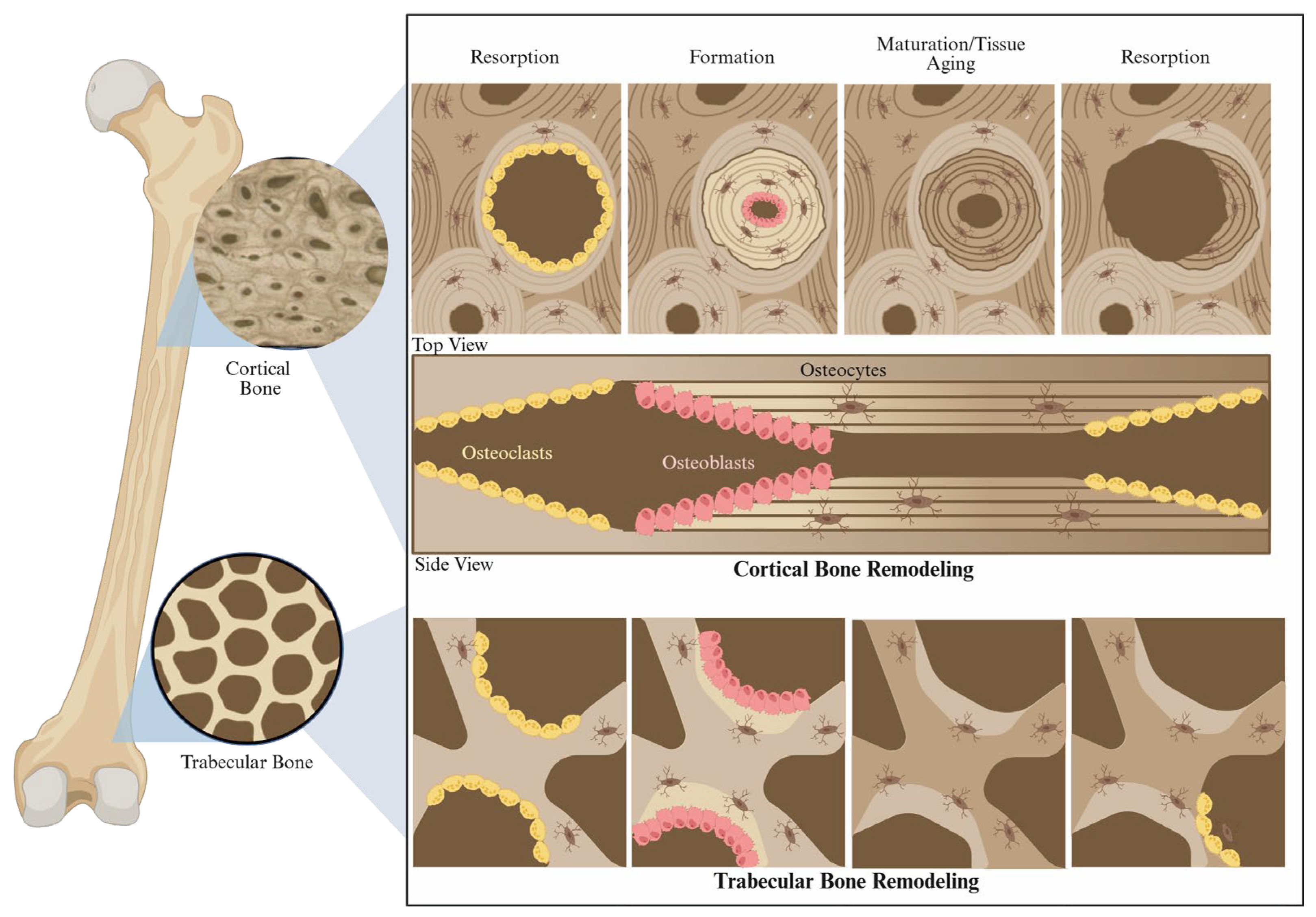
2.2. PG Dynamics in Bone Remodeling
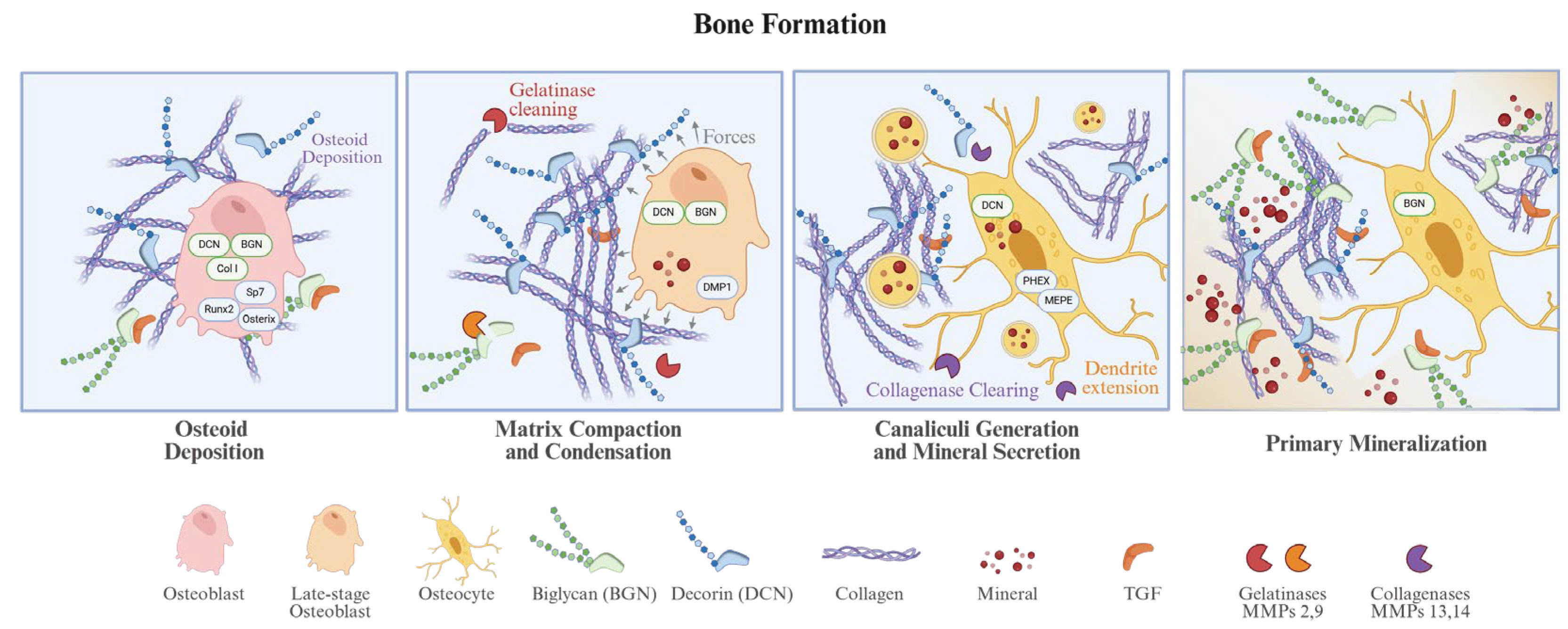
2.2.1. PGs in Bone Formation
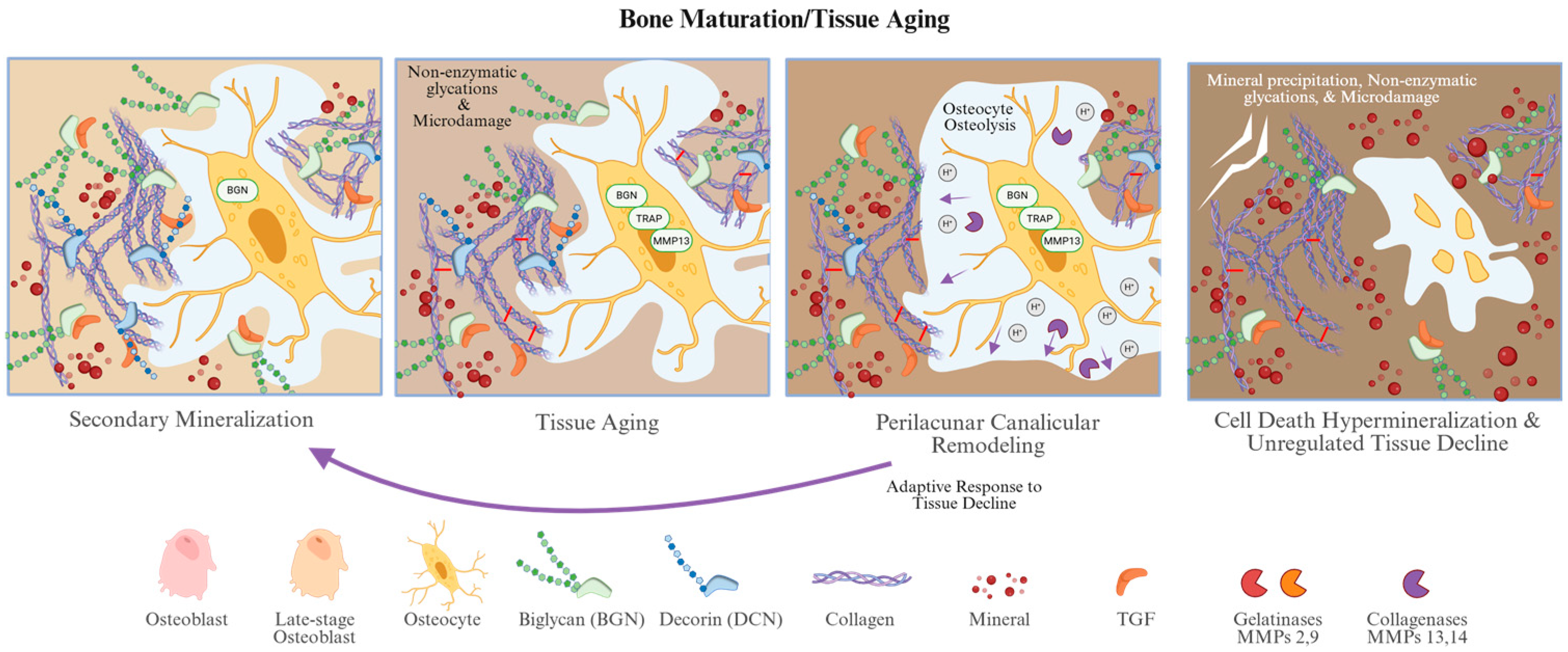
2.2.2. PGs in Tissue Aging and Perilacunar Remodeling
2.2.3. PGs and Osteocytes in Bone Resorption or Unregulated Tissue Decline
3. Age-Related Changes in PGs in Bone
3.1. Bulk Changes in GAGs/PGs in Bone
3.1.1. PG Levels During Skeletal Development and Maturation
3.1.2. Age-Related Changes in GAG/PG Content, Type, and Distribution
3.1.3. Systemic Modulators of Bone Matrix Aging
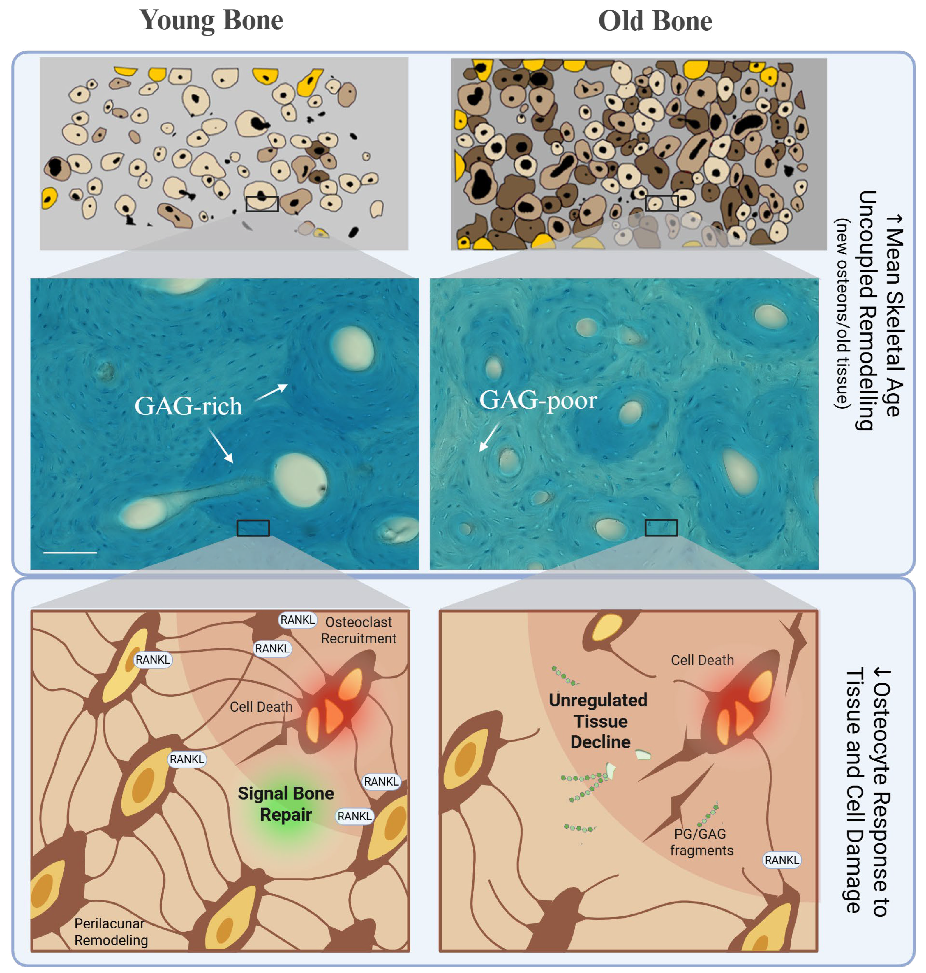
3.2. Age-Related PG Changes in Local Osteocyte Function and Bone Remodeling
Osteocyte PG Dysfunction
3.3. PGs in Mean Skeletal Aging
4. Implications for PG Loss in Bone
4.1. Role of PGs and GAGs in Endochondral Ossification
4.2. Role of PG and GAG Loss in Matrix Quality and Organization
4.3. Role of PG and GAG Loss in Mechanical Behavior
4.4. Functional Role of PGs and GAGs Loss in Cell Signaling
4.5. Functional Role of Catabolic Products of PG and GAG Loss in Matrix Regulation
5. Potential Interventions for Age-Related GAG/PG Loss
5.1. Pharmacological and Biological Replenishment
5.2. Osteoblast and Osteocyte Anabolic Stimulation and Turnover
5.3. Diagnostic Stratification and Inhibiting Catabolic Signaling
6. Conclusions
Author Contributions
Funding
Institutional Review Board Statement
Informed Consent Statement
Data Availability Statement
Acknowledgments
Conflicts of Interest
Abbreviations
| AGEs | Advanced Glycation End-products |
| ADAMTS | A Disintegrin and Metalloproteinase with Thrombospondin Motifs |
| BAP | Bone-specific Alkaline Phosphatase |
| BFR-v | Bone Formation Rate (per bone volume) |
| Bgn/BGN | Biglycan |
| BGP | Bone Gla-Protein (Osteocalcin) |
| BMD | Bone Mineral Density |
| BMSCs | Bone Marrow-derived Mesenchymal Stem Cells |
| BRU/BMU | Bone Remodeling Unit/Bone Multicellular Unit |
| CS | Chondroitin Sulfate |
| DAMPs | Damage-Associated Molecular Patterns |
| Dcn/DCN | Decorin |
| Dmp1 | Dentin Matrix Protein 1 |
| DS | Dermatan Sulfate |
| DXA | Dual-energy X-ray Absorptiometry |
| ECM | Extracellular Matrix |
| EFM | Extrafibrillar Matrix |
| EGFR | Epidermal Growth Factor Receptor |
| ERK | Extracellular Signal-Regulated Kinase |
| FGF | Fibroblast Growth Factor |
| FMOD | Fibromodulin |
| GAGs | Glycosaminoglycans |
| HA | Hyaluronic Acid |
| HS | Heparan Sulfate |
| IGF-IR | Insulin-like Growth Factor I Receptor |
| KS | Keratan Sulfate |
| LCN | Lacunocanalicular Network |
| LRP6 | Low-density Lipoprotein Receptor-related Protein 6 |
| LUM | Lumican |
| MAPK | Mitogen-Activated Protein Kinase |
| MEPE | Matrix Extracellular Phosphoglycoprotein |
| MMP | Matrix Metalloproteinase |
| OB | Osteoblast |
| OC | Osteoclast |
| OCY | Osteocyte |
| OPG | Osteoprotegerin |
| PCM | Pericellular Matrix |
| PGs | Proteoglycans |
| PHEX | Phosphate-regulating Endopeptidase Homolog, X-linked |
| PLR/PLM | Perilacunar Remodeling/Perilacunar Mineralization |
| PTH | Parathyroid Hormone |
| RANKL | Receptor Activator of Nuclear Factor Kappa-B Ligand |
| ROS | Reactive Oxygen Species |
| SLRPs | Small Leucine-Rich Proteoglycans |
| TIMP | Tissue Inhibitor of Metalloproteinases |
| TLR | Toll-Like Receptor |
| TNAP | Tissue Non-specific Alkaline Phosphatase |
References
- Koester, K.J.; Barth, H.D.; Ritchie, R.O. Effect of aging on the transverse toughness of human cortical bone: Evaluation by R-curves. J. Mech. Behav. Biomed. Mater. 2011, 4, 1504–1513. [Google Scholar] [CrossRef] [Scilit] [PubMed]
- Nalla, R.K.; Kruzic, J.J.; Ritchie, R.O. On the origin of the toughness of mineralized tissue: Microcracking or crack bridging? Bone 2004, 34, 790–798. [Google Scholar] [CrossRef] [Scilit]
- Wang, X.D.; Masilamani, N.S.; Mabrey, J.D.; Alder, M.E.; Agrawal, C.M. Changes in the fracture toughness of bone may not be reflected in its mineral density, porosity, and tensile properties. Bone 1998, 23, 67–72. [Google Scholar] [CrossRef] [Scilit]
- McCalden, R.W.; McGeough, J.A.; Court-Brown, C.M. Age-related changes in the compressive strength of cancellous bone. The relative importance of changes in density and trabecular architecture. J. Bone Jt. Surg. Am. 1997, 79, 421–427. [Google Scholar] [CrossRef] [Scilit]
- McCalden, R.W.; McGeough, J.A.; Barker, M.B.; Court-Brown, C.M. Age-related changes in the tensile properties of cortical bone. The relative importance of changes in porosity, mineralization, and microstructure. J. Bone Jt. Surg. Am. 1993, 75, 1193–1205. [Google Scholar] [CrossRef] [Scilit]
- Sözen, T.; Özışık, L.; Başaran, N. An overview and management of osteoporosis. Eur. J. Rheumatol. 2017, 4, 46–56. [Google Scholar] [CrossRef] [Scilit]
- Nevitt, M.C. Type of fall and risk of hip and wrist fractures: The study of osteoporotic fractures. The Study of Osteoporotic Fractures Research Group. J. Am. Geriatr. Soc. 1993, 41, 1226–1234. [Google Scholar] [CrossRef] [Scilit] [PubMed]
- Reinbold, W.D. Bone mineral content in early-postmenopausal and postmenopausal osteoporotic women: Comparison of measurement methods. Radiology 1986, 160, 469–478. [Google Scholar] [CrossRef] [Scilit] [PubMed]
- Alho, A. Bone mineral content and mechanical strength. An ex vivo study on human femora at autopsy. Clin. Orthop. Relat. Res. 1988, 227, 292–297. [Google Scholar] [CrossRef] [Scilit] [PubMed]
- Recker, R.R. Architecture and vertebral fracture. Calcif. Tissue Int. 1993, 53, S139–S142. [Google Scholar] [CrossRef] [Scilit]
- Hui, S.L.; Slemenda, C.W.; Johnston, C.C., Jr. Age and bone mass as predictors of fracture in a prospective study. J. Clin. Investig. 1988, 81, 1804–1809. [Google Scholar] [CrossRef] [Scilit]
- Kanis, J.A.; Johnell, O.; Oden, A.; Johansson, H.; McCloskey, E. FRAX and the assessment of fracture probability in men and women from the UK. Osteoporos. Int. 2008, 19, 385–397. [Google Scholar] [CrossRef] [Scilit]
- García Martín, A.; de la Higuera López-Frías, M.; Cortés Berdonces, M.; Jodar Gimeno, E.; Ávila Rubio, V.; Alhambra, M.R.; Muñoz Torres, M. New technologies in the evaluation of bone fragility and its application in Endocrinology. Endocrinol. Diabetes Nutr. (Engl. Ed.) 2020, 67, 602–610. [Google Scholar] [CrossRef] [Scilit]
- Wang, X.; Hua, R.; Ahsan, A.; Ni, Q.; Huang, Y.; Gu, S.; Jiang, J.X. Age-Related Deterioration of Bone Toughness Is Related to Diminishing Amount of Matrix Glycosaminoglycans (Gags). JBMR Plus 2018, 2, 164–173. [Google Scholar] [CrossRef] [Scilit] [PubMed]
- Hua, R.; Ni, Q.; Eliason, T.D.; Han, Y.; Gu, S.; Nicolella, D.P.; Wang, X.; Jiang, J.X. Biglycan and chondroitin sulfate play pivotal roles in bone toughness via retaining bound water in bone mineral matrix. Matrix Biol. J. Int. Soc. Matrix Biol. 2020, 94, 95–109. [Google Scholar] [CrossRef] [Scilit]
- Iozzo, R.V.; Schaefer, L. Proteoglycan form and function: A comprehensive nomenclature of proteoglycans. Matrix Biol. J. Int. Soc. Matrix Biol. 2015, 42, 11–55. [Google Scholar] [CrossRef] [Scilit] [PubMed]
- Esko, J.D. Chapter 11: Proteoglycans and Glycosaminoglycans. In Essentials of Glycobiology; Varki, A., Cummings, R.D., Esko, J.D., Stanley, P., Hart, G.W., Aebi, M., Mohnen, D., Kinoshita, T., Packer, N.H., Prestegard, J.H., et al., Eds.; Cold Spring Harbor Laboratory Press: Cold Spring Harbor, NY, USA, 1999. [Google Scholar]
- Chen, Y.-H.; Narimatsu, Y.; Clausen, T.M.; Gomes, C.; Karlsson, R.; Steentoft, C.; Spliid, C.B.; Gustavsson, T.; Salanti, A.; Persson, A.; et al. The GAGOme: A cell-based library of displayed glycosaminoglycans. Nat. Methods 2018, 15, 881–888. [Google Scholar] [CrossRef] [Scilit]
- Stanley, P. Golgi glycosylation. Cold Spring Harb. Perspect. Biol. 2011, 3, a005199. [Google Scholar] [CrossRef] [Scilit]
- Jalava, N.A.-O.; Arponen, M.A.-O.; Widjaja, N.A.-O.; Heino, T.A.-O.; Ivaska, K.A.-O. Short- and long-term exposure to high glucose induces unique transcriptional changes in osteoblasts in vitro. Biol. Open 2024, 13, bio060239. [Google Scholar] [CrossRef] [Scilit] [PubMed]
- Huang, Y.F.; Mizumoto, S.; Fujita, M. Novel Insight Into Glycosaminoglycan Biosynthesis Based on Gene Expression Profiles. Front. Cell Dev. Biol. 2021, 9, 709018. [Google Scholar] [CrossRef] [Scilit]
- Lowe, J.B.; Marth, J.D. A Genetic Approach to Mammalian Glycan Function. Annu. Rev. Biochem. 2003, 72, 643–691. [Google Scholar] [CrossRef] [Scilit] [PubMed]
- Sammon, D.; Krueger, A.; Busse-Wicher, M.; Morgan, R.M.; Haslam, S.M.; Schumann, B.; Briggs, D.C.; Hohenester, E. Molecular mechanism of decision-making in glycosaminoglycan biosynthesis. Nat. Commun. 2023, 14, 6425. [Google Scholar] [CrossRef] [Scilit] [PubMed]
- Kusche-Gullberg, M.; Kjellén, L. Sulfotransferases in glycosaminoglycan biosynthesis. Curr. Opin. Struct. Biol. 2003, 13, 605–611. [Google Scholar] [CrossRef] [Scilit] [PubMed]
- Best, S.M.; Duer, M.J.; Reid, D.G.; Wise, E.R.; Zou, S. Towards a model of the mineral-organic interface in bone: NMR of the structure of synthetic glycosaminoglycan- and polyaspartate-calcium phosphate composites. Magn. Reson. Chem. 2008, 46, 323–329. [Google Scholar] [CrossRef] [Scilit]
- Hashimoto, Y.; Lester, G.E.; Caterson, B.; Yamauchi, M. EDTA-insoluble, calcium-binding proteoglycan in bovine bone. Calcif. Tissue Int. 1995, 56, 398–402. [Google Scholar] [CrossRef] [Scilit]
- Scott, I.C.; Imamura, Y.; Pappano, W.N.; Pappano, W.N.; Troedel, J.M.; Recklies, A.D.; Roughley, P.J.; Greenspan, D.S. Bone morphogenetic protein-1 processes probiglycan. J. Biol. Chem. 2000, 275, 30504–30511. [Google Scholar] [CrossRef] [Scilit]
- Huntley, R.; Jensen, E.; Gopalakrishnan, R.; Mansky, K.C. Bone morphogenetic proteins: Their role in regulating osteoclast differentiation. Bone Rep. 2019, 10, 100207. [Google Scholar] [CrossRef] [Scilit]
- von Marschall, Z.; Fisher, L.W. Decorin is processed by three isoforms of bone morphogenetic protein-1 (BMP1). Biochem. Biophys. Res. Commun. 2010, 391, 1374–1378. [Google Scholar] [CrossRef] [Scilit]
- Paiva, K.B.S.; Granjeiro, J.M. Chapter Six-Matrix Metalloproteinases in Bone Resorption, Remodeling, and Repair. In Progress in Molecular Biology and Translational Science; Khalil, R.A., Ed.; Academic Press: New York, NY, USA, 2017; Volume 148, pp. 203–303. [Google Scholar]
- Prideaux, M.; Staines, K.A.; Jones, E.R.; Riley, G.P.; Pitsillides, A.A.; Farquharson, C. MMP and TIMP temporal gene expression during osteocytogenesis. Gene Expr. Patterns 2015, 18, 29–36. [Google Scholar] [CrossRef] [Scilit]
- Aziz, A.H.; Wilmoth, R.L.; Ferguson, V.L.; Bryant, S.J. IDG-SW3 Osteocyte Differentiation and Bone Extracellular Matrix Deposition Are Enhanced in a 3D Matrix Metalloproteinase-Sensitive Hydrogel. ACS Appl. Bio Mater. 2020, 3, 1666–1680. [Google Scholar] [CrossRef] [Scilit]
- Genovese, F.; Barascuk, N.; Larsen, L.; Larsen, M.R.; Nawrocki, A.; Li, Y.; Zheng, Q.; Wang, J.; Veidal, S.S.; Leeming, D.J.; et al. Biglycan fragmentation in pathologies associated with extracellular matrix remodeling by matrix metalloproteinases. Fibrogenesis Tissue Repair. 2013, 6, 9. [Google Scholar] [CrossRef] [Scilit]
- Bhutada, S.; Li, L.; Willard, B.; Muschler, G.; Piuzzi, N.; Apte, S.S. Forward and reverse degradomics defines the proteolytic landscape of human knee osteoarthritic cartilage and the role of the serine protease HtrA1. Osteoarthr. Cartil. 2022, 30, 1091–1102. [Google Scholar] [CrossRef] [Scilit]
- Melching, L.I.; Fisher, W.D.; Lee, E.R.; Mort, J.S.; Roughley, P.J. The cleavage of biglycan by aggrecanases. Osteoarthr. Cartil. 2006, 14, 1147–1154. [Google Scholar] [CrossRef] [Scilit] [PubMed]
- Monfort, J.; Tardif, G.; Reboul, P.; Mineau, F.; Roughley, P.; Pelletier, J.-P.; Martel-Pelletier, J. Degradation of small leucine-rich repeat proteoglycans by matrix metalloprotease-13: Identification of a new biglycan cleavage site. Arthritis Res. Ther. 2006, 8, R26. [Google Scholar] [CrossRef] [Scilit]
- Schurman, C.A.; Kaya, S.; Dole, N.; Luna, N.M.M.; Castillo, N.; Potter, R.; Rose, J.P.; Bons, J.; King, C.D.; Burton, J.B.; et al. Aging impairs the osteocytic regulation of collagen integrity and bone quality. Bone Res. 2024, 12, 13. [Google Scholar] [CrossRef] [Scilit] [PubMed]
- Daquinag, A.C.; Gao, Z.; Fussell, C.; Sun, K.; Kolonin, M.G. Glycosaminoglycan Modification of Decorin Depends on MMP14 Activity and Regulates Collagen Assembly. Cells 2020, 9, 2646. [Google Scholar] [CrossRef] [Scilit]
- Stanton, H.; Melrose, J.; Little, C.B.; Fosang, A.J. Proteoglycan degradation by the ADAMTS family of proteinases. Biochim. Biophys. Acta (BBA)-Mol. Basis Dis. 2011, 1812, 1616–1629. [Google Scholar] [CrossRef] [Scilit] [PubMed]
- Ernst, S.; Langer, R.; Cooney, C.L.; Sasisekharan, R. Enzymatic degradation of glycosaminoglycans. Crit. Rev. Biochem. Mol. Biol. 1995, 30, 387–444. [Google Scholar] [CrossRef] [Scilit] [PubMed]
- Moseley, R.; Waddington, R.J.; Embery, G.; Rees, S.G. The Modification of Alveolar Bone Proteoglycans by Reactive Oxygen Species In Vitro. Connect. Tissue Res. 1998, 37, 13–28. [Google Scholar] [CrossRef] [Scilit] [PubMed]
- Moseley, R.; Waddington, R.; Evans, P.; Halliwell, B.; Embery, G. The chemical modification of glycosaminoglycan structure by oxygen-derived species in vitro. Biochim. Biophys. Acta (BBA)-General. Subj. 1995, 1244, 245–252. [Google Scholar] [CrossRef] [Scilit]
- Fuchs, B.; Schiller, J. Glycosaminoglycan Degradation by Selected Reactive Oxygen Species. Antioxid. Redox Signal. 2014, 21, 1044–1062. [Google Scholar] [CrossRef] [Scilit]
- Imai, K.; Hiramatsu, A.; Fukushima, D.; Pierschbacher, M.D.; Okada, Y. Degradation of decorin by matrix metalloproteinases: Identification of the cleavage sites, kinetic analyses and transforming growth factor-beta1 release. Biochem. J. 1997, 322, 809–814. [Google Scholar] [CrossRef] [Scilit]
- Tocchi, A.; Parks, W.C. Functional interactions between matrix metalloproteinases and glycosaminoglycans. FEBS J. 2013, 280, 2332–2341. [Google Scholar] [CrossRef] [Scilit]
- Kemberi, M.A.-O.; Minns, A.A.-O.; Santamaria, S.A.-O. Soluble Proteoglycans and Proteoglycan Fragments as Biomarkers of Pathological Extracellular Matrix Remodeling. Proteoglycan Res. 2024, 2, e70011. [Google Scholar] [CrossRef] [Scilit]
- Mori, S.; Burr, D.B. Increased intracortical remodeling following fatigue damage. Bone 1993, 14, 103–109. [Google Scholar] [CrossRef] [Scilit]
- Rumpler, M.; Würger, T.; Roschger, P.; Zwettler, E.; Peterlik, H.; Fratzl, P.; Klaushofer, K. Microcracks and Osteoclast Resorption Activity In Vitro. Calcif. Tissue Int. 2012, 90, 230–238. [Google Scholar] [CrossRef] [Scilit]
- Vashishth, D.; Behiri, J.C.; Bonfield, W. Crack growth resistance in cortical bone: Concept of microcrack toughening. J. Biomech. 1997, 30, 763–769. [Google Scholar] [CrossRef] [Scilit] [PubMed]
- O’Brien, F.J.; Taylor, D.; Clive Lee, T.; Clive Lee, T. The effect of bone microstructure on the initiation and growth of microcracks. J. Orthop. Res. 2005, 23, 475–480. [Google Scholar] [CrossRef] [Scilit] [PubMed]
- van Oers, R.F.M.; Ruimerman, R.; Tanck, E.; Hilbers, P.A.J.; Huiskes, R. A unified theory for osteonal and hemi-osteonal remodeling. Bone 2008, 42, 250–259. [Google Scholar] [CrossRef] [Scilit] [PubMed]
- Thelin, M.A.; Bartolini, B.; Axelsson, J.; Gustafsson, R.; Tykesson, E.; Pera, E.; Oldberg, Å.; Maccarana, M.; Malmstrom, A. Biological functions of iduronic acid in chondroitin/dermatan sulfate. FEBS J. 2013, 280, 2431–2446. [Google Scholar] [CrossRef] [Scilit]
- Shiflett, L.A.; Tiede-Lewis, L.M.; Xie, Y.; Lu, Y.; Ray, E.C.; Dallas, S.L. Collagen Dynamics During the Process of Osteocyte Embedding and Mineralization. Front. Cell Dev. Biol. 2019, 7, 178. [Google Scholar] [CrossRef] [Scilit]
- Waddington, R.J.; Roberts, H.C.; Sugars, R.V.; Schönherr, E. Differential roles for small leucine-rich proteoglycans in bone formation. Eur. Cell Mater. 2003, 6, 12–21; discussion 21. [Google Scholar] [CrossRef] [Scilit] [PubMed]
- Hao, J.-X.; Wan, Q.-Q.; Mu, Z.; Gu, J.-T.; Yu, W.-W.; Qin, W.; Li, Y.-T.; Wang, C.-Y.; Ma, Y.-X.; Jiao, K.; et al. A seminal perspective on the role of chondroitin sulfate in biomineralization. Carbohydr. Polym. 2023, 310, 120738. [Google Scholar] [CrossRef] [Scilit]
- Murshed, M.; Harmey, D.; Millán, J.L.; McKee, M.D.; Karsenty, G. Unique coexpression in osteoblasts of broadly expressed genes accounts for the spatial restriction of ECM mineralization to bone. Genes Dev. 2005, 19, 1093–1104. [Google Scholar] [CrossRef] [Scilit]
- Oftadeh, R.; Perez-Viloria, M.; Villa-Camacho, J.C.; Vaziri, A.; Nazarian, A. Biomechanics and mechanobiology of trabecular bone: A review. J. Biomech. Eng. 2015, 137, 0108021–01080215. [Google Scholar] [CrossRef] [Scilit]
- Bergot, C.; Wu, Y.; Jolivet, E.; Zhou, L.Q.; Laredo, J.D.; Bousson, V. The degree and distribution of cortical bone mineralization in the human femoral shaft change with age and sex in a microradiographic study. Bone 2009, 45, 435–442. [Google Scholar] [CrossRef] [Scilit]
- Nyman, J.S.; Makowski, A.J.; Patil, C.A.; Masui, T.P.; O’Quinn, E.C.; Bi, X.; Guelcher, S.A.; Nicollela, D.P.; Mahadevan-Jansen, A. Measuring differences in compositional properties of bone tissue by confocal Raman spectroscopy. Calcif. Tissue Int. 2011, 89, 111–122. [Google Scholar] [CrossRef] [Scilit] [PubMed]
- Taylor, E.A.; Mileti, C.J.; Ganesan, S.; Kim, J.H.; Donnelly, E. Measures of Bone Mineral Carbonate Content and Mineral Maturity/Crystallinity for FT-IR and Raman Spectroscopic Imaging Differentially Relate to Physical–Chemical Properties of Carbonate-Substituted Hydroxyapatite. Calcif. Tissue Int. 2021, 109, 77–91. [Google Scholar] [CrossRef] [Scilit] [PubMed]
- Nyman, J.S.; Roy, A.; Acuna, R.L.; Gayle, H.J.; Reyes, M.J.; Tyler, J.H.; Dean, D.D.; Wang, X. Age-related effect on the concentration of collagen crosslinks in human osteonal and interstitial bone tissue. Bone 2006, 39, 1210–1217. [Google Scholar] [CrossRef] [Scilit]
- Sroga, G.E.; Karim, L.; Colón, W.; Vashishth, D. Biochemical Characterization of Major Bone-Matrix Proteins Using Nanoscale-Size Bone Samples and Proteomics Methodology. Mol. Cell. Proteom. 2011, 10, M110.006718. [Google Scholar] [CrossRef] [Scilit]
- Dong, X.N.; Qin, A.; Xu, J.; Wang, X. In situ accumulation of advanced glycation endproducts (AGEs) in bone matrix and its correlation with osteoclastic bone resorption. Bone 2011, 49, 174–183. [Google Scholar] [CrossRef] [Scilit]
- Ingram, R.T.; Clarke, B.L.; Fisher, L.W.; Fitzpatrick, L.A. Distribution of noncollagenous proteins in the matrix of adult human bone: Evidence of anatomic and functional heterogeneity. J. Bone Miner. Res. 1993, 8, 1019–1029. [Google Scholar] [CrossRef] [Scilit]
- Kerschnitzki, M.; Kollmannsberger, P.; Burghammer, M.; Duda, G.N.; Weinkamer, R.; Wagermaier, W.; Fratzl, P. Architecture of the osteocyte network correlates with bone material quality. J. Bone Miner. Res. 2013, 28, 1837–1845. [Google Scholar] [CrossRef] [Scilit] [PubMed]
- Grzesik, W.J.; Frazier, C.R.; Shapiro, J.R.; Sponseller, P.D.; Robey, P.G.; Fedarko, N.S. Age-related changes in human bone proteoglycan structure. Impact of osteogenesis imperfecta. J. Biol. Chem. 2002, 277, 43638–43647. [Google Scholar] [CrossRef] [Scilit] [PubMed]
- Fedarko, N.S.; Vetter, U.K.; Weinstein, S.; Robey, P.G. Age-related changes in hyaluronan, proteoglycan, collagen, and osteonectin synthesis by human bone cells. J. Cell Physiol. 1992, 151, 215–227. [Google Scholar] [CrossRef] [Scilit] [PubMed]
- Roughley, P.J.; White, R.J.; Magny, M.C.; Liu, J.; Pearce, R.H.; Mort, J.S. Non-proteoglycan forms of biglycan increase with age in human articular cartilage. Biochem. J. 1993, 295, 421–426. [Google Scholar] [CrossRef] [Scilit]
- Paschalis, E.P.; Gamsjaeger, S.; Klaushofer, K.; Shane, E.; Cohen, A.; Stepan, J.; Pavo, I.; Eriksen, E.F.; Taylor, K.A.; Dempster, D.W. Treatment of postmenopausal osteoporosis patients with teriparatide for 24 months reverts forming bone quality indices to premenopausal healthy control values. Bone 2022, 162, 116478. [Google Scholar] [CrossRef] [Scilit] [PubMed]
- Pei, S.; Wang, S.; Martinez, J.R.; Parajuli, A.; Kirn-Safran, C.B.; Farach-Carson, M.C.; Lu, X.L.; Wang, L. Osteocytic Pericellular Matrix (PCM): Accelerated Degradation under In Vivo Loading and Unloading Conditions Using a Novel Imaging Approach. Genes 2022, 13, 72. [Google Scholar] [CrossRef] [Scilit]
- Kaya, S.A.-O.; Schurman, C.A.; Dole, N.A.-O.; Evans, D.S.; Alliston, T.A.-O. Prioritization of Genes Relevant to Bone Fragility Through the Unbiased Integration of Aging Mouse Bone Transcriptomics and Human GWAS Analyses. J. Bone Miner. Res. 2022, 37, 804–817. [Google Scholar] [CrossRef] [Scilit]
- Vahidi, G.; Boone, C.; Hoffman, F.; Heveran, C. Aging decreases osteocyte peri-lacunar-canalicular system turnover in female C57BL/6JN mice. Bone 2024, 186, 117163. [Google Scholar] [CrossRef] [Scilit]
- Hagan, M.L.; Yu, K.; Zhu, J.; Vinson, B.N.; Roberts, R.L.; Montesinos Cartagena, M.; Johnson, M.H.; Wang, L.; Isales, C.M.; Hamrick, M.W.; et al. Decreased pericellular matrix production and selection for enhanced cell membrane repair may impair osteocyte responses to mechanical loading in the aging skeleton. Aging Cell 2020, 19, e13056. [Google Scholar] [CrossRef] [Scilit]
- Chang, B.; Liu, X. Osteon: Structure, Turnover, and Regeneration. Tissue Eng. Part B Rev. 2022, 28, 261–278. [Google Scholar] [CrossRef] [Scilit] [PubMed]
- Johnstone, B.; Markopoulos, M.; Neame, P.; Caterson, B. Identification and characterization of glycanated and non-glycanated forms of biglycan and decorin in the human intervertebral disc. Biochem. J. 1993, 292, 661–666. [Google Scholar] [CrossRef] [Scilit] [PubMed]
- Kram, V.; Shainer, R.; Jani, P.; Meester, J.A.N.; Loeys, B.; Young, M.F. Biglycan in the Skeleton. J. Histochem. Cytochem. 2020, 68, 747–762. [Google Scholar] [CrossRef] [Scilit] [PubMed]
- Miguez, P.A. Evidence of biglycan structure-function in bone homeostasis and aging. Connect. Tissue Res. 2020, 61, 19–33. [Google Scholar] [CrossRef] [Scilit] [PubMed]
- Melrose, J.; Fuller, E.S.; Roughley, P.J.; Smith, M.M.; Kerr, B.; Hughes, C.E.; Caterson, B.; Little, C.B. Fragmentation of decorin, biglycan, lumican and keratocan is elevated in degenerate human meniscus, knee and hip articular cartilages compared with age-matched macroscopically normal and control tissues. Arthritis Res. Ther. 2008, 10, R79. [Google Scholar] [CrossRef] [Scilit]
- Gorni, D.; Finco, A.A.-O. Oxidative stress in elderly population: A prevention screening study. Aging Med. 2020, 3, 205–213. [Google Scholar] [CrossRef] [Scilit]
- Almeida, M.; Han, L.; Martin-Millan, M.; O’Brien, C.A.; Manolagas, S.C. Oxidative Stress Antagonizes Wnt Signaling in Osteoblast Precursors by Diverting β-Catenin from T Cell Factor-to Forkhead Box O-mediated Transcription. J. Biol. Chem. 2007, 282, 27298–27305. [Google Scholar] [CrossRef] [Scilit]
- Liguori, I.; Russo, G.; Curcio, F.; Bulli, G.; Aran, L.; Della-Morte, D.; Gargiulo, G.; Testa, G.; Cacciatore, F.; Bonaduce, D.; et al. Oxidative stress, aging, and diseases. Clin. Interv. Aging 2018, 13, 757–772. [Google Scholar] [CrossRef] [Scilit]
- Stinghen, A.E.; Massy, Z.A.; Vlassara, H.; Striker, G.E.; Boullier, A. Uremic Toxicity of Advanced Glycation End Products in CKD. J. Am. Soc. Nephrol. 2016, 27, 354–370. [Google Scholar] [CrossRef] [Scilit]
- Thomas, M.C.; Tsalamandris, C.; Macisaac, R.; Medley, T.; Kingwell, B.; Cooper, M.E.; Jerums, G.; Alberti, D. Low-molecular-weight AGEs are associated with GFR and anemia in patients with type 2 diabetes. Kidney Int. 2004, 66, 1167–1172. [Google Scholar] [CrossRef] [Scilit]
- Hein, G.; Wiegand, R.; Lehmann, G.; Stein, G.; Franke, S. Advanced glycation end-products pentosidine and Nϵ-carboxymethyllysine are elevated in serum of patients with osteoporosis. Rheumatology 2003, 42, 1242–1246. [Google Scholar] [CrossRef] [Scilit]
- Penc, S.F.; Pomahac, B.; Winkler, T.; Dorschner, R.A.; Eriksson, E.; Herndon, M.; Gallo, R.L. Dermatan Sulfate Released after Injury Is a Potent Promoter of Fibroblast Growth Factor-2 Function. J. Biol. Chem. 1998, 273, 28116–28121. [Google Scholar] [CrossRef] [Scilit]
- Trowbridge, J.M.; Rudisill, J.A.; Ron, D.; Gallo, R.L. Dermatan Sulfate Binds and Potentiates Activity of Keratinocyte Growth Factor (FGF-7). J. Biol. Chem. 2002, 277, 42815–42820. [Google Scholar] [CrossRef] [Scilit] [PubMed]
- Schaefer, L.; Babelova, A.; Kiss, E.; Hausser, H.-J.; Baliova, M.; Krzyzankova, M.; Marsche, G.; Young, M.F.; Mihalik, D.; Götte, M.; et al. The matrix component biglycan is proinflammatory and signals through Toll-like receptors 4 and 2 in macrophages. J. Clin. Investig. 2005, 115, 2223–2233. [Google Scholar] [CrossRef] [Scilit]
- Clark, D.; Nakamura, M.; Miclau, T.; Marcucio, R. Effects of Aging on Fracture Healing. Curr. Osteoporos. Rep. 2017, 15, 601–608. [Google Scholar] [CrossRef] [Scilit] [PubMed]
- Huiskes, R.; Ruimerman, R.; van Lenthe, G.H.; van Lenthe, G.H.; Janssen, J.D. Effects of mechanical forces on maintenance and adaptation of form in trabecular bone. Nature 2000, 405, 704–706. [Google Scholar] [CrossRef] [Scilit]
- Lee, S.W.; Kim, W.Y.; Song, J.H.; Kim, J.H.; Lee, H.A.-O. Factors Affecting Periprosthetic Bone Loss after Hip Arthroplasty. Hip Pelvis 2021, 33, 53–61. [Google Scholar] [CrossRef] [Scilit] [PubMed]
- Vashishth, D.; Verborgt, O.; Divine, G.; Schaffler, M.B.; Fyhrie, D.P. Decline in osteocyte lacunar density in human cortical bone is associated with accumulation of microcracks with age. Bone 2000, 26, 375–380. [Google Scholar] [CrossRef] [Scilit]
- Vashishth, D.; Gibson, G.J.; Fyhrie, D.P. Sexual dimorphism and age dependence of osteocyte lacunar density for human vertebral cancellous bone. Anat. Rec. Part A Discov. Mol. Cell. Evol. Biol. 2005, 282A, 157–162. [Google Scholar] [CrossRef] [Scilit]
- Mullender, M.G.; van der Meer, D.D.; Huiskes, R.; Lips, P. Osteocyte density changes in aging and osteoporosis. Bone 1996, 18, 109–113. [Google Scholar] [CrossRef] [Scilit]
- Gauthier, R.; Follet, H.; Olivier, C.; Mitton, D.; Peyrin, F. 3D analysis of the osteonal and interstitial tissue in human radii cortical bone. Bone 2019, 127, 526–536. [Google Scholar] [CrossRef] [Scilit] [PubMed]
- Sieverts, M.; Yee, C.; Nemani, M.; Parkinson, D.Y.; Alliston, T.; Acevedo, C. Spatial control of perilacunar canalicular remodeling during lactation. Sci. Rep. 2024, 14, 14655. [Google Scholar] [CrossRef] [Scilit]
- Ford, S.; Tiedemann, K.; Shapiro, R.; Komarova, S.V.; Jähn-Rickert, K.; Zimmermann, E.A. Fluorescent mapping of osteocyte-driven bone formation at pre-osteocyte and mature osteocyte lacunae. Acta Biomater. 2025, 207, 444–455. [Google Scholar] [CrossRef] [Scilit] [PubMed]
- Hattner, R.F.; Frost, H.M. Mean skeletal age: Its calculation, and theoretical effects on skeletal tracer physiology and on the physical characteristics of bone. Henry Ford Hosp. Med. Bull. 1963, 11, 201–216. [Google Scholar]
- Wang, X.; Li, X.; Shen, X.; Shen, X.; Agrawal, C.M.; Agrawal, C.M. Age-related changes of noncalcified collagen in human cortical bone. Ann. Biomed. Eng. 2003, 31, 1365–1371. [Google Scholar] [CrossRef] [Scilit]
- Granke, M.; Makowski, A.J.; Uppuganti, S.; Nyman, J.S. Prevalent role of porosity and osteonal area over mineralization heterogeneity in the fracture toughness of human cortical bone. J. Biomech. 2016, 49, 2748–2755. [Google Scholar] [CrossRef] [Scilit] [PubMed]
- Cool, S.M.; Hendrikz, J.K.; Wood, W.B. Microscopic age changes in the human occipital bone. J. Forensic Sci. 1995, 40, 789–796. [Google Scholar] [CrossRef] [Scilit] [PubMed]
- Wu, K.; Schubeck, K.E.; Frost, H.M.; Villanueva, A. Haversian bone formation rates determined by a new method in a mastodon, and in human diabetes mellitus and osteoporosis. Calcif. Tissue Res. 1970, 6, 204–219. [Google Scholar] [CrossRef] [Scilit]
- Goliath, J.R.; Stewart, M.C.; Stout, S.D. Variation in osteon histomorphometrics and their impact on age-at-death estimation in older individuals. Forensic Sci. Int. 2016, 262, 282.e1–282.e6. [Google Scholar] [CrossRef] [Scilit]
- Ten Cate, A.R. Bone. In Ten Cate’s Oral Histology; Nanci, A., Ed.; Mosby Elsevier: St. Louis, MO, USA, 2008; pp. 95–121. [Google Scholar]
- Jee, W. The Skeletal Tissues, Histology: Cell and Tissue Biology; Elsevier Science Ltd.: New York, NY, USA, 1983. [Google Scholar]
- Eastell, R.; Delmas, P.D.; Hodgson, S.F.; Eriksen, E.F.; Mann, K.G.; Riggs, B.L. Bone Formation Rate in Older Normal Women: Concurrent Assessment with Bone Histomorphometry, Calcium Kinetics, and Biochemical Markers. J. Clin. Endocrinol. Metab. 1988, 67, 741–748. [Google Scholar] [CrossRef] [Scilit] [PubMed]
- Fatayerji, D.; Eastell, R. Age-related changes in bone turnover in men. J. Bone Miner. Res. 1999, 14, 1203–1210. [Google Scholar] [CrossRef] [Scilit] [PubMed]
- Jenkins, N.; Black, M.; Paul, E.; Pasco, J.A.; Kotowicz, M.A.; Schneider, H.G. Age-related reference intervals for bone turnover markers from an Australian reference population. Bone 2013, 55, 271–276. [Google Scholar] [CrossRef] [Scilit]
- Piemontese, M.; Almeida, M.; Robling, A.G.; Kim, H.-N.; Xiong, J.; Thostenson, J.D.; Weinstein, R.S.; Manolagas, S.C.; O’Brien, C.A.; Jilka, R.L. Old age causes de novo intracortical bone remodeling and porosity in mice. JCI Insight 2017, 2, e93771. [Google Scholar] [CrossRef] [Scilit] [PubMed]
- Monti, M.; Scazzoso, A.; Calzaferri, G.; Santi, I.; D’Aprile, E.; Cunietti, E. Tartrate-resistant acid phosphatase (TRAP) activity in serum: Potential use in assessing bone resorption in patients with multiple myeloma. Int. J. Biol. Markers 1990, 5, 61–64. [Google Scholar] [CrossRef] [Scilit]
- Jones, K.L.; Schwarze, U.; Adam, M.P.; Byers, P.H.; Mefford, H.C. A homozygous B3GAT3 mutation causes a severe syndrome with multiple fractures, expanding the phenotype of linkeropathy syndromes. Am. J. Med. Genet. A 2015, 167A, 2691–2696. [Google Scholar] [CrossRef] [Scilit]
- Baasanjav, S.; Al-Gazali, L.; Hashiguchi, T.; Mizumoto, S.; Fischer, B.; Horn, D.; Seelow, D.; Ali, B.R.; Aziz, S.A.A.; Langer, R.; et al. Faulty initiation of proteoglycan synthesis causes cardiac and joint defects. Am. J. Hum. Genet. 2011, 89, 15–27. [Google Scholar] [CrossRef] [Scilit]
- Budde, B.S.; Mizumoto, S.; Kogawa, R.; Becker, C.; Altmüller, J.; Thiele, H.; Rüschendorf, F.; Toliat, M.R.; Kaleschke, G.; Hämmerle, J.M.; et al. Skeletal dysplasia in a consanguineous clan from the island of Nias/Indonesia is caused by a novel mutation in B3GAT3. Hum. Genet. 2015, 134, 691–704. [Google Scholar] [CrossRef] [Scilit]
- von Oettingen, J.E.; Tan, W.H.; Dauber, A. Skeletal dysplasia, global developmental delay, and multiple congenital anomalies in a 5-year-old boy-report of the second family with B3GAT3 mutation and expansion of the phenotype. Am. J. Med. Genet. A 2014, 164A, 1580–1586. [Google Scholar] [CrossRef] [Scilit] [PubMed]
- Yauy, K.; Tran Mau-Them, F.; Willems, M.; Coubes, C.; Blanchet, P.; Herlin, C.; Taleb Arrada, I.; Sanchez, E.; Faure, J.M.; Le Gac, M.P.; et al. B3GAT3-related disorder with craniosynostosis and bone fragility due to a unique mutation. Genet. Med. 2018, 20, 269–274. [Google Scholar] [CrossRef] [Scilit]
- Izumikawa, T.; Kanagawa, N.; Watamoto, Y.; Okada, M.; Saeki, M.; Sakano, M.; Sugahara, K.; Sugihara, K.; Asano, M.; Kitagawa, H. Impairment of embryonic cell division and glycosaminoglycan biosynthesis in glucuronyltransferase-I-deficient mice. J. Biol. Chem. 2010, 285, 12190–12196. [Google Scholar] [CrossRef] [Scilit]
- Meester, J.A.N.; Hebert, A.; Bastiaansen, M.; Rabaut, L.; Bastianen, J.; Boeckx, N.; Ashcroft, K.; Atwal, P.S.; Benichou, A.; Billon, C.; et al. Expanding the clinical spectrum of biglycan-related Meester-Loeys syndrome. NPJ Genom. Med. 2024, 9, 22. [Google Scholar] [CrossRef] [Scilit]
- Meester, J.A.; Vandeweyer, G.; Pintelon, I.; Lammens, M.A.-O.; Van Hoorick, L.A.-O.; De Belder, S.A.-O.; Waitzman, K.; Young, L.; Markham, L.A.-O.X.; Vogt, J.; et al. Loss-of-function mutations in the X-linked biglycan gene cause a severe syndromic form of thoracic aortic aneurysms and dissections. Genet. Med. 2017, 19, 386–395. [Google Scholar] [CrossRef] [Scilit]
- Hua, R.; Han, Y.; Ni, Q.; Fajardo, R.J.; Iozzo, R.V.; Ahmed, R.; Nyman, J.A.-O.; Wang, X.; Jiang, J.A.-O. Pivotal roles of biglycan and decorin in regulating bone mass, water retention, and bone toughness. Bone Res. 2025, 13, 2. [Google Scholar] [CrossRef] [Scilit] [PubMed]
- Wuyts, W.; Van Hul, W.; De Boulle, K.; Hendrickx, J.; Bakker, E.; Vanhoenacker, F.; Mollica, F.; Lüdecke, H.J.; Sayli, B.S.; Pazzaglia, U.E.; et al. Mutations in the EXT1 and EXT2 genes in hereditary multiple exostoses. Am. J. Hum. Genet. 1998, 62, 346–354. [Google Scholar] [CrossRef] [Scilit] [PubMed]
- Hilton, M.J.; Gutiérrez, L.; Martinez, D.A.; Wells, D.E. EXT1 regulates chondrocyte proliferation and differentiation during endochondral bone development. Bone 2005, 36, 379–386. [Google Scholar] [CrossRef] [Scilit] [PubMed]
- Cartault, F.; Munier, P.; Jacquemont, M.L.; Vellayoudom, J.; Doray, B.; Payet, C.; Randrianaivo, H.; Laville, J.M.; Munnich, A.; Cormier-Daire, V. Expanding the clinical spectrum of B4GALT7 deficiency: Homozygous p.R270C mutation with founder effect causes Larsen of Reunion Island syndrome. Eur. J. Hum. Genet. 2015, 23, 49–53. [Google Scholar] [CrossRef] [Scilit]
- Huber, C.; Oulès, B.; Bertoli, M.; Chami, M.; Fradin, M.; Alanay, Y.; Al-Gazali, L.I.; Ausems, M.G.E.M.; Bitoun, P.; Cavalcanti, D.P.; et al. Identification of CANT1 Mutations in Desbuquois Dysplasia. Am. J. Hum. Genet. 2009, 85, 706–710. [Google Scholar] [CrossRef] [Scilit]
- Bui, C.; Huber, C.; Tuysuz, B.; Alanay, Y.; Bole-Feysot, C.; Leroy, J.G.; Mortier, G.; Nitschke, P.; Munnich, A.; Cormier-Daire, V. XYLT1 mutations in Desbuquois dysplasia type 2. Am. J. Hum. Genet. 2014, 94, 405–414. [Google Scholar] [CrossRef] [Scilit]
- Minatogawa, M.; Unzaki, A.; Morisaki, H.; Syx, D.; Sonoda, T.; Janecke, A.R.; Slavotinek, A.; Voermans, N.C.; Lacassie, Y.; Mendoza-Londono, R.; et al. Clinical and molecular features of 66 patients with musculocontractural Ehlers−Danlos syndrome caused by pathogenic variants in CHST14 (mcEDS-CHST14). J. Med. Genet. 2022, 59, 865. [Google Scholar] [CrossRef] [Scilit] [PubMed]
- Klüppel, M.; Wight, T.N.; Chan, C.; Hinek, A.; Wrana, J.L. Maintenance of chondroitin sulfation balance by chondroitin-4-sulfotransferase 1 is required for chondrocyte development and growth factor signaling during cartilage morphogenesis. Development 2005, 132, 3989–4003. [Google Scholar] [CrossRef] [Scilit] [PubMed]
- Lautrup, C.K.; Teik, K.W.; Unzaki, A.; Mizumoto, S.; Syx, D.; Sin, H.H.; Nielsen, I.K.; Markholt, S.; Yamada, S.; Malfait, F.; et al. Delineation of musculocontractural Ehlers-Danlos Syndrome caused by dermatan sulfate epimerase deficiency. Mol. Genet. Genom. Med. 2020, 8, e1197. [Google Scholar] [CrossRef] [Scilit]
- Gustafsson, R.; Stachtea, X.; Maccarana, M.; Grottling, E.; Eklund, E.; Malmström, A.; Oldberg, A. Dermatan sulfate epimerase 1 deficient mice as a model for human abdominal wall defects. Birth Defects Res. A Clin. Mol. Teratol. 2014, 100, 712–720. [Google Scholar] [CrossRef] [Scilit] [PubMed]
- Shimbo, M.; Suzuki, R.; Fuseya, S.; Sato, T.; Kiyohara, K.; Hagiwara, K.; Okada, R.; Wakui, H.; Tsunakawa, Y.; Watanabe, H.; et al. Postnatal lethality and chondrodysplasia in mice lacking both chondroitin sulfate N-acetylgalactosaminyltransferase-1 and -2. PLoS ONE 2017, 12, e0190333. [Google Scholar] [CrossRef] [Scilit]
- Ichikawa, S.; Sorenson, A.H.; Austin, A.M.; Mackenzie, D.S.; Fritz, T.A.; Moh, A.; Hui, S.L.; Econs, M.J. Ablation of the Galnt3 gene leads to low-circulating intact fibroblast growth factor 23 (Fgf23) concentrations and hyperphosphatemia despite increased Fgf23 expression. Endocrinology 2009, 150, 2543–2550. [Google Scholar] [CrossRef] [Scilit]
- Ichikawa, S.; Gray, A.K.; Padgett, L.R.; Allen, M.R.; Clinkenbeard, E.L.; Sarpa, N.M.; White, K.E.; Econs, M.J. Genetic rescue of glycosylation-deficient Fgf23 in the Galnt3 knockout mouse. Endocrinology 2014, 155, 3891–3898. [Google Scholar] [CrossRef] [Scilit]
- Izumikawa, T.; Sato, B.; Kitagawa, H. Chondroitin sulfate is indispensable for pluripotency and differentiation of mouse embryonic stem cells. Sci. Rep. 2014, 4, 3701. [Google Scholar] [CrossRef] [Scilit]
- Job, F.; Mizumoto, S.; Smith, L.; Couser, N.; Brazil, A.; Saal, H.; Patterson, M.; Gibson, M.I.; Soden, S.; Miller, N.; et al. Functional validation of novel compound heterozygous variants in B3GAT3 resulting in severe osteopenia and fractures: Expanding the disease phenotype. BMC Med. Genet. 2016, 17, 86. [Google Scholar] [CrossRef] [Scilit]
- Berendsen, A.D.; Pinnow, E.L.; Maeda, A.; Brown, A.C.; McCartney-Francis, N.; Kram, V.; Owens, R.T.; Robey, P.G.; Holmbeck, K.; de Castro, L.F.; et al. Biglycan modulates angiogenesis and bone formation during fracture healing. Matrix Biol. 2014, 35, 223–231. [Google Scholar] [CrossRef] [Scilit]
- Courtney, T.H. Mechanical Behavior of Materials, 2nd ed.; Waveland Press: Long Grove, IL, USA, 2005. [Google Scholar]
- Heath, S.; Han, Y.; Hua, R.; Roy, A.; Jiang, J.; Nyman, J.S.; Wang, X. Assessment of glycosaminoglycan content in bone using Raman spectroscopy. Bone 2023, 171, 116751. [Google Scholar] [CrossRef] [Scilit]
- Kirkham, J.; Brookes, S.; Shore, R.; Bonass, W.; Robinson, C. The effect of glycosylaminoglycans on the mineralization of sheep periodontal ligament in vitro. Connect. Tissue Res. 1995, 33, 23–29. [Google Scholar] [CrossRef] [Scilit] [PubMed]
- Xiao, P.; Roy, A.; Wang, X. In-silico simulation of nanoindentation on bone using a 2D cohesive finite element model. J. Mech. Behav. Biomed. Mater. 2024, 151, 106403. [Google Scholar] [CrossRef] [Scilit]
- Han, Y.; Gomez, J.; Hua, R.; Xiao, P.; Gao, W.; Jiang, J.X.; Wang, X. Removal of glycosaminoglycans affects the in situ mechanical behavior of extrafibrillar matrix in bone. J. Mech. Behav. Biomed. Mater. 2021, 123, 104766. [Google Scholar] [CrossRef] [Scilit] [PubMed]
- Al Makhzoomi, A.K.; Kirk, T.B.; Dye, D.E.; Allison, G.T. Contribution of glycosaminoglycans to the structural and mechanical properties of tendons–A multiscale study. J. Biomech. 2021, 128, 110796. [Google Scholar] [CrossRef] [Scilit]
- Roth, J.A.-O.X.; Hoop, C.A.-O.; Williams, J.K.; Hayes, R.; Baum, J. Probing the effect of glycosaminoglycan depletion on integrin interactions with collagen I fibrils in the native extracellular matrix environment. Protein Sci. 2023, 32, e4508. [Google Scholar] [CrossRef] [Scilit] [PubMed]
- Lujan, T.J.; Underwood, C.J.; Jacobs, N.T.; Weiss, J.A. Contribution of glycosaminoglycans to viscoelastic tensile behavior of human ligament. J. Appl. Physiol. 2009, 106, 423–431. [Google Scholar] [CrossRef] [Scilit]
- Blank, J.A.-O.; Eekhoff, J.D.; Soslowsky, L.J. Glycosaminoglycans influence regional mechanics in young but not old Achilles tendons. J. Physiol. 2025, 603, 7589–7601. [Google Scholar] [CrossRef] [Scilit]
- Li, M.; Yang, S.; Xu, D.A.-O. Heparan Sulfate Regulates the Structure and Function of Osteoprotegerin in Osteoclastogenesis. J. Biol. Chem. 2016, 291, 24160–24171. [Google Scholar] [CrossRef] [Scilit] [PubMed]
- Li, M.; Xu, D. Antiresorptive activity of osteoprotegerin requires an intact heparan sulfate-binding site. Proc. Natl. Acad. Sci. USA 2020, 117, 17187–17194. [Google Scholar] [CrossRef] [Scilit] [PubMed]
- Timmen, M.; Hidding, H.; Götte, M.; Khassawna, T.E.; Kronenberg, D.; Stange, R. The heparan sulfate proteoglycan Syndecan-1 influences local bone cell communication via the RANKL/OPG axis. Sci. Rep. 2020, 10, 20510. [Google Scholar] [CrossRef] [Scilit]
- Irie, A.; Habuchi, H.; Kimata, K.; Sanai, Y. Heparan sulfate is required for bone morphogenetic protein-7 signaling. Biochem. Biophys. Res. Commun. 2003, 308, 858–865. [Google Scholar] [CrossRef] [Scilit] [PubMed]
- Diehl, V.; Huber, L.S.; Trebicka, J.; Wygrecka, M.; Iozzo, R.V.; Schaefer, L. The Role of Decorin and Biglycan Signaling in Tumorigenesis. Front. Oncol. 2021, 11, 801801. [Google Scholar] [CrossRef] [Scilit]
- Giatagana, E.-M.; Berdiaki, A.; Gaardløs, M.; Samsonov, S.A.; Tzanakakis, G.N.; Nikitovic, D. Biglycan Interacts with Type I Insulin-like Receptor (IGF-IR) Signaling Pathway to Regulate Osteosarcoma Cell Growth and Response to Chemotherapy. Cancers 2022, 14, 1196. [Google Scholar] [CrossRef] [Scilit] [PubMed]
- Jung, Y.-K.; Park, H.-R.; Cho, H.-J.; Jang, J.-A.; Lee, E.-J.; Han, M.-S.; Kim, G.-W.; Han, S. Degrading products of chondroitin sulfate can induce hypertrophy-like changes and MMP-13/ADAMTS5 production in chondrocytes. Sci. Rep. 2019, 9, 15846. [Google Scholar] [CrossRef] [Scilit]
- Goodall, K.J.; Poon, I.K.; Phipps, S.; Hulett, M.D. Soluble heparan sulfate fragments generated by heparanase trigger the release of pro-inflammatory cytokines through TLR-4. PLoS ONE 2014, 9, e109596. [Google Scholar] [CrossRef] [Scilit]
- Taylor, K.R.; Gallo, R.L. Glycosaminoglycans and their proteoglycans: Host-associated molecular patterns for initiation and modulation of inflammation. FASEB J. 2006, 20, 9–22. [Google Scholar] [CrossRef] [Scilit]
- Zhu, M.; Wu, S.C.M.; Tam, W.-K.; Wong, C.K.; Liao, P.; Cheah, K.S.; Chan, D.; James, A.W.; Leung, V.Y. Biglycan fragment modulates TGF-β activity in intervertebral disc via an eIF6-coupled intracellular path. Sci. Adv. 2025, 11, eadq8545. [Google Scholar] [CrossRef] [Scilit] [PubMed]
- Adepu, S.; Ekman, S.; Leth, J.; Johansson, U.; Lindahl, A.; Skiöldebrand, E. Biglycan neo-epitope (BGN262), a novel biomarker for screening early changes in equine osteoarthritic subchondral bone. Osteoarthr. Cartil. 2022, 30, 1328–1336. [Google Scholar] [CrossRef] [Scilit]
- Iozzo, R.V.; Moscatello, D.K.; McQuillan, D.J.; Eichstetter, I. Decorin is a biological ligand for the epidermal growth factor receptor. J. Biol. Chem. 1999, 274, 4489–4492. [Google Scholar] [CrossRef] [Scilit] [PubMed]
- Wang, B.; Lai, X.; Price, C.; Thompson, W.R.; Li, W.; Quabili, T.R.; Tseng, W.-J.; Liu, X.S.; Zhang, H.; Pan, J.; et al. Perlecan-containing pericellular matrix regulates solute transport and mechanosensing within the osteocyte lacunar-canalicular system. J. Bone Miner. Res. 2014, 29, 878–891. [Google Scholar] [CrossRef] [Scilit]
- Venkatesan, N.; Barré, L.; Benani, A.; Netter, P.; Magdalou, J.; Fournel-Gigleux, S.; Ouzzine, M. Stimulation of proteoglycan synthesis by glucuronosyltransferase-I gene delivery: A strategy to promote cartilage repair. Proc. Natl. Acad. Sci. USA 2004, 101, 18087–18092. [Google Scholar] [CrossRef] [Scilit]
- Silagi, E.S.; Shapiro, I.M.; Risbud, M.V. Glycosaminoglycan synthesis in the nucleus pulposus: Dysregulation and the pathogenesis of disc degeneration. Matrix Biol. J. Int. Soc. Matrix Biol. 2018, 71–72, 368–379. [Google Scholar] [CrossRef] [Scilit]
- Rehn, A.P.; Cerny, R.; Sugars, R.V.; Kaukua, N.; Wendel, M. Osteoadherin is upregulated by mature osteoblasts and enhances their in vitro differentiation and mineralization. Calcif. Tissue Int. 2008, 82, 454–464. [Google Scholar] [CrossRef] [Scilit] [PubMed]
- Bellido, T.; Saini, V.; Pajevic, P.D. Effects of PTH on osteocyte function. Bone 2013, 54, 250–257. [Google Scholar] [CrossRef] [Scilit]
- Wein, M.N. Parathyroid Hormone Signaling in Osteocytes. JBMR Plus 2018, 2, 22–30. [Google Scholar] [CrossRef] [Scilit]
- Liu, X.S.; Wang, L.; de Bakker, C.M.J.; Lai, X. Mechanical Regulation of the Maternal Skeleton during Reproduction and Lactation. Curr. Osteoporos. Rep. 2019, 17, 375–386. [Google Scholar] [CrossRef] [Scilit] [PubMed]
- Manhard, M.K.; Horch, R.A.; Harkins, K.D.; Gochberg, D.F.; Nyman, J.S.; Does, M.D. Validation of quantitative bound- and pore-water imaging in cortical bone. Magn. Reson. Med. 2014, 71, 2166–2171. [Google Scholar] [CrossRef] [Scilit] [PubMed]
- Bae, W.C.; Chen, P.C.; Chung, C.B.; Masuda, K.; D’Lima, D.; Du, J. Quantitative ultrashort echo time (UTE) MRI of human cortical bone: Correlation with porosity and biomechanical properties. J. Bone Miner. Res. 2012, 27, 848–857. [Google Scholar] [CrossRef] [Scilit] [PubMed]




| Category | Specific Genes/Enzymes | Major Function | Ref |
|---|---|---|---|
| Proteoglycan Core Proteins | BGN, DCN, ASPN, FMOD, LUM | Major bone matrix PG core proteins (e.g., Bgn, Dcn, asporin, FMOD, lumican (LUM) | [16] |
| Synthesis of GAG Backbone (linkage region and polymerization) | XYLT1, XYLT2, B4GALT7, FAM20B, B3GALT6, B3GAT3, PXYLP1 *, EXT1/2, EXTL3, CSGALNACT1/2, CHSY1/3, CHPF, CHPF2 * | Initiates GAG attachment to Serine residues on the core protein and supports chain elongation | [21,22,23] |
| CS/DS Post-Translational Modifications | CHST11, CHST12 *, CHST13 *, CHST14, UST, CHST3, CHST15, DSE, DSEL | Sulfates CS chains and supports epimerization to dermatan sulfate (DS) | [21,22,23,24] |
| HS Post-Translational Modifications | NDST1-4, GLCE, HS2ST1, HS6ST1-3, HS3ST1-6, SULF1, SULF2 | Modifies HS (e.g., on syndecans and perlecan | [21,22,23,24] |
| Regulators & Metabolic Support | PAPSS1, PAPSS2, CANT1 *, IMPAD1, GOLPH3, SPPL3 * | Provides sulfate donor supply and general support for GAG | [21] |
| Transporters | SLC35B2, SLC35B3, SLC35B4, SLC35A2 | Transports nucleotide-sugar and sulfate-related precursors required for PG/GAG biosynthesis | [21] |
| Class | Factor | Specific PG/Core Target | OB | OCY | OC | Cleavage | Location | Ref |
|---|---|---|---|---|---|---|---|---|
| Enzymatic | BMP-1 | Dcn, Bgn | +++ | ++ | ++ | Converts pro-forms into mature Bgn and Dcn via N-terminal cleavage | Extracellular matrix | [27,28,29] |
| MMP-2 | Bgn | + | +++ | +++ | Multiple cleavage sites reported | Extracellular matrix | [20,30,31,32] | |
| MMP-3 | Bgn, Dcn | - | + | ++ | Activates other MMPs; cleaves near N and C-termini | Extracellular matrix | [33,34,35,36] | |
| MMP-7 | Dcn | ++ | - | ++ | Multiple cleavage sites reported | Extracellular matrix | [20,30,31,32] | |
| MMP-9 | Bgn | ++ | + | +++ | Multiple cleavage sites reported | Extracellular matrix | [20,30,31,32] | |
| MMP-12 | Bgn, Dcn | +++ | + | ++ | Multiple cleavage sites reported | Extracellular matrix | [20,30,31,32] | |
| MMP-13 | Bgn, Dcn | +++ | ++ | + | Several cleavage sites reported | Extracellular matrix | [20,32,37] | |
| MMP-14 | Bgn, Dcn | ++ | +++ | + | Membrane-tethered; activates other MMPs and cleaves SLRPs (including N-terminal regions) | Cell Membrane | [37,38] | |
| ADAMTS-4/5 | Bgn, Dcn | + | +++ | + | Cleaves within the core region resulting in a “half-moon” structure of SLRPs | Extracellular matrix | [39] | |
| Heparinase | HS | + | ++ | ++ | Depolymerizes HS | Extracellular matrix | [40] | |
| Chondroitinase | CS | - | - | - | Depolymerizes CS | Bacterial systems (experimental) | [40] | |
| Hyaluronidase | HA | + | ++ | +++ | Depolymerizes HA | Lysosome/ extracellular | [40] | |
| Non-Enzymatic | Reactive Oxygen Species (ROS) | Nonspecific | + | ++ | ++ | Oxidative fragmentation of GAG chains (often preferential for less-sulfated regions), and core protein damage | Extracellular matrix | [41,42,43] |
| Mechanical Damage | Nonspecific | + | +++ | - | Mechanical loading-associated fragmentation of GAG chains and proteolysis with damage accumulation | Extracellular matrix |
| Species | Study Focus/Condition | Age Range/Group | Observed Change/Phenotype | Ref |
|---|---|---|---|---|
| Human | Skeletal Development | Juvenile vs. Skeletal Maturity (~30 years) | Total GAG amount reduced by ~50% by skeletal maturity. CS PGs and Bgn are the major proportion in early development. | [66] |
| Human | Juvenile Synthesis | <15 years | Juvenile osteoblasts produce PGs with longer GAG chains and synthesis rates 3–4x higher than donors > 30 years. | [67] |
| Human | Bone Sulfation | Juvenile vs. Adult | Young matrix possesses a high ratio of Chondroitin-6-Sulfate (C6S) relative to Chondroitin-4-Sulfate (C4S). | [54] |
| Human | Iduronic Substitution | Fetal to 60 years | 1.5-fold linear increase in iduronic acid substitution in CS chains with age (transition to DS-like). | [66] |
| Human | Bulk Cortical Bone | Young vs. Mid-Aged vs. Elderly | GAG content decreases by up to 17% between young (avg. 24 years) and elderly (avg. 73 years); associated with loss of bound water and toughness. | [14] |
| Human | Tissue Aging | Adults | Decline in Osteopontin, Osteocalcin, and Decorin (Dcn) in older interstitial tissue vs. younger osteons. | [64] |
| Human | Articular Cartilage | Elderly | Elevated fragmentation of Dcn (14–38 kDa) and Bgn (16–45 kDa) compared to normal controls. | [68] |
| Human | Intervertebral Discs | Healthy vs. Degenerate | Healthy discs have higher Bgn fragments; degenerate discs have full-length Bgn that upregulates FGF-17. | [68] |
| Human | Menopause (Iliac Crest) | Pre- vs. Post-menopausal | Post-menopausal women (avg. 70 years) have significantly lower GAG levels than pre-menopausal (avg. 40 years). | [69] |
| Human | Bone Marrow (Lumican) | Elderly (Hip Fracture) | Lumican 16.9% lower in fracture patients; low LUM correlates with decreased bone mass. | [37] |
| Mouse | Pericellular Matrix (PCM) | 15-week vs. 65-week | Older bone shows more homogenous PG distribution in the osteocyte PCM. | [70] |
| Mouse | Osteocyte Gene Expression | 2-month vs. 1, 2, and 2.5-year | Downregulation of PG core proteins (Bgn, Dcn, Lum). Upregulation of Mmp-2, -8, -14 at 2 years. | [71] |
| Mouse | Lacunar–Canalicular Turnover | 5-month vs. 22-month | Reduced fraction of osteocytes undergoing PLM and fewer MMP-14-positive cells. | [72] |
| Mouse | Osteocyte Mechanosensing | 3-month vs. 24-month | Primary osteocytes show reduced PCM formation and reduced mechanosensitivity. | [73] |
| Bone Remodeling Properties | Change with Age | Ref | |
|---|---|---|---|
| Bone Formation Rate | Bone Formation (% new tissue volume/year) | 70% ↑ serum BAP 50% ↑ serum BGP 100% ↑ BFR-v | [105] |
| Bone formation markers (osteocalcin & propeptide type 1 procollagen) | ↓ | [106,107] | |
| Osteoid Volume | ↑ | [105] | |
| Mineral Apposition Rate | ↑ | [105,108] | |
| Osteoblast Number | No change in number between 7 and 21 month old mice | [108] | |
| Bone Resorption Rate | Bone resorption remnants (C- and N-Terminal Telopeptides of Type I Collagen, Pyridinolines) | ↓ after 30 years | [107] |
| Osteoclast Activity (secreted Tartrate-resistant acid phosphatase) | Highest before 30 years | [109] | |
| Osteoclast Number | ↑numberbetween 7 and 21 month old mice | [108] | |
| Gene | Species | Genotype/Defect | Skeletal Phenotype | Ref |
|---|---|---|---|---|
| B3GAT3 | Human | Homozygous mutation | Linkeropathy. Multiple fractures, severe osteopenia, short stature, and radio-ulnar synostosis. | [110] |
| Human | Faulty initiation of PG synthesis | Joint dislocations, short stature, and cardiac defects. | [111] | |
| Human | Novel mutation (Nias/Indonesia) | Skeletal dysplasia in a consanguineous clan. | [112] | |
| Human | Second family report | Skeletal dysplasia, global developmental delay, and multiple congenital anomalies in a 5-year-old. | [113] | |
| Human | Unique mutation | Craniosynostosis and bone fragility. | [114] | |
| Mouse | B3GAT3 knockout | Embryonic mortality; impairment of embryonic cell division | [115] | |
| BGN | Human | Loss-of-function (X-linked) | Meester-Loeys syndrome; mild skeletal dysplasia, pectus deformities, and flat vertebral bodies. | [116,117] |
| Mouse | Bgn knockout | Significant long bone shortening, skeletal dysplasia, and loss of regulated collagen-GAG spacing | [15] | |
| DCN | Mouse | Dcn knockout | Minimal effect on bone structure but impaired local and bulk mechanical properties | [118] |
| BGN/DCN | Mouse | Bgn/Dcn double knockout | Severely disorganized matrix and irregular collagen fibril diameters; extreme matrix fragility | [118] |
| EXT | Human | EXT1/2 deficiency | Hereditary Multiple Exostoses; multiple bony outgrowths (osteochondromas), short stature, and irregular limb length. | [119] |
| Mouse | Ext1 knockout | Increased chondrocyte proliferation and significant delay in skeletal maturation/differentiation | [120] | |
| B4GALT7 | Human | B4GALT7 deficiency | Larsen-like Syndrome; Short long bones, joint dislocations or laxity, and scoliosis. | [121] |
| CANT1 | Human | Homozygous or compound heterozygous mutations | Desbuquois Dysplasia Type 1. Lethal dwarfism, “Swedish key” or “monkey wrench” proximal femur. | [122] |
| XYLT1 | Human | Homozygous loss-of-function mutations | Desbuquois Dysplasia Type 2. Severe short stature, joint laxity, and advanced carpal ossification without characteristic hand anomalies. | [123] |
| CHST | Human | CHST14 deficiency | mcEDS-CHST14. Progressive foot/ankle deformities, recurrent joint dislocations (80% by age 10), and kyphoscoliosis. | [124] |
| Mouse | CHST11 knockout | Impaired mineralization and skeletal development | [125] | |
| DSE | Human | DSE deficiency | mcEDS-DSE. Similar to mcEDS-CHST14 but often with a lower symptom burden; includes progressive clubfoot and spinal deformities. | [126] |
| Mouse | DSE knockout | Altered collagen fibril diameter and irregular shapes | [127] | |
| CSGALNACT | Mouse | CSGalNACT knockout | Postnatal lethality, impaired endochondral ossification, malocclusion, and skin hyperextension | [128] |
| GALNT3 | Mouse | Galnt3 knockout | Hyperphosphatemia, tumor calcinosis, and hyperostosis; elevated bone volume (more prominent in males) | [129,130] |
Disclaimer/Publisher’s Note: The statements, opinions and data contained in all publications are solely those of the individual author(s) and contributor(s) and not of MDPI and/or the editor(s). MDPI and/or the editor(s) disclaim responsibility for any injury to people or property resulting from any ideas, methods, instructions or products referred to in the content. |
© 2026 by the authors. Licensee MDPI, Basel, Switzerland. This article is an open access article distributed under the terms and conditions of the Creative Commons Attribution (CC BY) license.
Share and Cite
Heath, S.; Hua, R.; Wang, X.; Jiang, J. Proteoglycan Dynamics and Bone Quality: Molecular Regulation to Age-Related Fragility. Biomolecules 2026, 16, 572. https://doi.org/10.3390/biom16040572
Heath S, Hua R, Wang X, Jiang J. Proteoglycan Dynamics and Bone Quality: Molecular Regulation to Age-Related Fragility. Biomolecules. 2026; 16(4):572. https://doi.org/10.3390/biom16040572
Chicago/Turabian StyleHeath, Savannah, Rui Hua, Xiaodu Wang, and Jean Jiang. 2026. "Proteoglycan Dynamics and Bone Quality: Molecular Regulation to Age-Related Fragility" Biomolecules 16, no. 4: 572. https://doi.org/10.3390/biom16040572
APA StyleHeath, S., Hua, R., Wang, X., & Jiang, J. (2026). Proteoglycan Dynamics and Bone Quality: Molecular Regulation to Age-Related Fragility. Biomolecules, 16(4), 572. https://doi.org/10.3390/biom16040572