Review

From Native Glycosaminoglycans to Mimetics: Design, Mechanisms, and Biomedical Applications

Institute of Biophysics, Center of Integrative Physiology and Molecular Medicine (CIPMM), Saarland University, 66421 Homburg, Germany
*
Author to whom correspondence should be addressed.
Biomolecules 2025, 15(11), 1518; https://doi.org/10.3390/biom15111518 (registering DOI)
Submission received: 19 September 2025 / Revised: 23 October 2025 / Accepted: 24 October 2025 / Published: 27 October 2025
(This article belongs to the Special Issue Advances in Glycosaminoglycans (GAGs) and Mimetics)

Abstract

Glycosaminoglycans (GAGs) are essential regulators of numerous biological processes through their interactions with growth factors, chemokines, cytokines, and enzymes. Their structural diversity and heterogeneity, however, limit reproducibility and translational use, as native GAGs are typically obtained from animal-derived sources with notable batch-to-batch variability. To overcome these challenges, a wide range of GAG mimetics has been developed with the aim of replicating or modulating the biological functions of native GAGs while offering improved structural definition, accessibility, and therapeutic potential. Polysaccharide-based GAG mimetics, including derivatives of heparan sulfate, hyaluronan, dextran, and other natural glycans, represent one major strategy, whereas non-saccharide-based mimetics provide alternative scaffolds with enhanced stability and selectivity. Both approaches have yielded compounds that serve as valuable tools for dissecting GAG/protein interactions and as candidates for therapeutic development. Biomedical applications of GAG mimetics span diverse areas such as cancer, cardiovascular and inflammatory diseases, bone and cartilage regeneration, wound healing, and infectious diseases. This mini-review summarizes key developments in the design and synthesis of GAG mimetics, highlights their potential biomedical applications, and discusses current challenges and future perspectives in advancing them toward clinical translation.

Graphical Abstract

1. Introduction

This review provides an overview of the current state of glycosaminoglycan (GAG) mimetics, highlighting their structural design, mechanisms of action, and biomedical applications. Native GAGs are linear polysaccharides with essential regulatory functions in processes such as cell proliferation, angiogenesis, inflammation, and tissue repair, mediated through interactions with a broad range of proteins, including growth factors, chemokines, and enzymes [1,2,3,4]. Recent advances in glycan microarray technologies have enabled systematic mapping of GAG/protein interactions, thereby expanding our understanding of sulfation-specific binding motifs [5]. However, their inherent heterogeneity, dependence on animal-derived sources, and limited availability pose significant challenges for reproducibility and large-scale biomedical use. To address these limitations, chemically synthesized, chemo-enzymatically produced, and bioengineered GAG mimetics have emerged as promising alternatives. These compounds aim to replicate or modulate the biological functions of native GAGs while offering improved structural definition, accessibility, and therapeutic potential. Importantly, GAG mimetics are increasingly investigated not only as research tools for dissecting GAG/protein interactions but also as candidates for clinical translation in diverse fields ranging from regenerative medicine to cancer therapy [6]. This review first outlines polysaccharide-based mimetics, followed by non-saccharide approaches, and concludes with a discussion of their biomedical applications, clinical translation, and safety considerations.

2. Learning from Nature: Synthesis and Interaction of Native GAGs

GAGs are a family of linear polysaccharides composed of repeating disaccharide units, typically of an amino sugar (glucosamine or galactosamine) and a uronic acid (glucuronic (GlcA) or iduronic acid (IdoA)) or galactose in the case of keratan sulfate (KS); the major mammalian types include heparan sulfate (HS), heparin, chondroitin sulfate (CS), dermatan sulfate (DS), KS, and the non-sulfated hyaluronan (HA) (Figure 1). They are found ubiquitously on cell surfaces (as components of proteoglycans), in the extracellular matrix (ECM), and in various bodily fluids, where their distribution is highly tissue- and cell-type specific. The biosynthesis of sulfated GAGs (except for HA) takes place in the Golgi apparatus via sequential action of glycosyltransferases, epimerases, and sulfotransferases, starting from core protein linkage and elongation of disaccharide repeats, followed by modifications such as acetylation, sulfation (at multiple positions), and epimerization (e.g., conversion of GlcA to IdoA in HS/DS) [7,8]. These multiple enzymatic steps, which are under genetic, developmental, and environmental regulation, cause each GAG chain to differ in length, sulfation pattern, acetylation state, and epimerization. This intrinsic “microheterogeneity” (and sometimes macro variation between tissues or species) is a major reason for the challenges in consistency, purification, and functional characterization of native GAGs [9]. At the molecular level, GAG interactions with proteins are dominated by ionic interactions between negatively charged sulfate/carboxylate groups on the GAGs and clusters of positively charged amino acid residues (lysine, arginine) on proteins, hydrogen bonding, and van der Waals contacts. Furthermore, the three-dimensional conformation of the GAG (including flexibility/rigidity, orientation of sulfates, conformation of uronic acid residues) also contributes importantly to affinity and specificity [10]. These interactions enable GAGs to modulate a wide array of biological processes: growth factor activity, cell adhesion, receptor binding, morphogen gradients, ECM assembly, and even protection of ligands from proteolytic degradation [11].

3. Rationale for GAG Mimetics and Synthetic and Bioengineering Strategies

The extensive structural diversity of native GAGs as well as their limited availability due to the requirement of extraction from different animal-derived tissues are drawbacks limiting their broad use in biomedical applications. The variability regarding their chain length and especially their degree of modification due to acetylation, sulfation, and epimerization reactions give rise to a broad heterogeneity of native GAGs. As a result, notable batch-to-batch variations need to be considered while working with native GAGs [12]. To overcome these limitations, several GAG mimetics have been developed to analyze their interaction for HS-binding proteins, to modify the binding and release profiles of surfaces and biomaterials, and as therapeutics to interfere with disorders accompanied by altered HS/protein interactions. In this review, we define a GAG mimetic as a non-native, chemically defined molecule or engineered polymer (saccharide or non-saccharide) that reproduces or modulates mammalian GAG/protein interactions. This category includes (i) chemically modified natural GAG backbones, such as sulfated hyaluronan (sHA), which does not occur naturally but imitates the functions of sulfated GAGs; (ii) fully synthetic oligosaccharides, exemplified by fondaparinux, which reproduces the antithrombin-binding pentasaccharide of heparin; and (iii) non-saccharide scaffolds, such as sulfated cyclitols or flavonoids, designed to mimic sulfate spacing and charge density of sulfated GAGs. By contrast, native mammalian GAGs (e.g., HS, CS, DS, KS, HA) and bioengineered polysaccharides identical to them (e.g., recombinant CS-A or CS-C produced in E. coli) are not considered mimetics here but rather native equivalents. Natural GAG analogs from non-mammalian sources (e.g., fucosylated CS from marine organisms) are discussed when used experimentally as surrogates for mammalian GAGs.
Different strategies, such as the chemical and chemo-enzymatic synthesis [13,14] and the bioengineered production [15] of GAG mimetics, are currently explored. The use of GAG mimetics with defined sulfation motifs should improve the understanding and dissecting of structural binding motifs required for the pleiotropic functions of native GAGs. Novel synthetic strategies now also explore conjugation approaches, such as cholesterol-modified GAG mimetics, to improve oral bioavailability and delivery [16]. Ideally, GAG mimetics as druggable compounds should be (i) homogeneous with (ii) enhanced selectivity for HS interaction partners compared to native HS, (iii) easy to synthetize, (iv) cost-effective in production, and (v) available in large quantities, with (vi) no or only few off-target and side effects. Overall, GAG mimetics can be grouped into polysaccharide-based derivatives and non-saccharide-based compounds.

3.1. HS Mimetics Based on Modified Polysaccharides

Different polysaccharides have been explored for chemical modification to derive HS mimetics. The oligosaccharide mixture phosphomannopentaose sulfate (PI-88) is the sulfated reaction product of yeast-derived phosphomannan containing 60% tetra- and 30% pentasaccharides [17]. Besides its function as a heparinase inhibitor and its anticoagulative potential, it has been reported to bind HS-binding proteins such as vascular endothelial growth factor-A (VEGF-A), and fibroblast growth factor-1 (FGF-1) and -2 (FGF-2), thereby competing with native HS for these bioactive mediator proteins [17,18]. Further PI-88-related HS mimetics have been developed with improved pharmacokinetic properties and decreased anticoagulative activity [19,20]. A rationally designed octasaccharide synthesized by Kuhnast et al. was shown to inhibit the binding of vascular VEGF-A, FGF-2, platelet derived growth factor-β (PDGF-β), and stromal cell-derived factor-1α (SDF-1α) to heparin, with IC50 values in the low nM range [21].
Moreover, different bacteria-derived polysaccharides have been used as starting materials for the synthesis of GAG mimetics. For example, Matou et al. over-sulfated an exopolysaccharide secreted by Alteromonas infernus. Compared to the non-sulfated polysaccharide, this GAG mimetic stimulated the proliferation, migration, and tube formation of human endothelial cells in vitro, suggesting an interplay with angiogenic factors such as FGF-2 and VEGF-A [22]. A biotechnological–chemical process developed by the Schiraldi group gave rise to over-sulfated CS-like polysaccharides [23]. Thereby, a chondroitin-like polysaccharide was extracted after synthesis by E. coli O5:K4:H4 and sulfated using the SO3-pyridine complex [24]. ELISA studies showed a higher VEGF-A-binding capacity for surfaces with immobilized over-sulfated CS derivatives compared to native GAGs [25].
Beyond chemo-enzymatic routes, recent synthetic biology advances achieved the complete in vivo biosynthesis of sulfated chondroitin in E. coli. This was achieved by co-expressing the chondroitin backbone pathway together with the PAPS sulfate donor system and functional sulfotransferases, yielding defined CS motifs directly in cells [26]. We therefore distinguish bioengineered natural CS (same as mammalian CS) from chemically modified CS-like polymers (e.g., over-sulfated derivatives), which we classify as GAG mimetics in this review [27]. Pioneering CS glycomimetics from the de Paz/Nieto group introduced aromatic substituents and multivalent presentations to enhance affinity for CS-binding proteins such as midkine/pleiotrophin, establishing design principles later adopted broadly in GAG mimetic libraries [28,29].
Sheng et al. described the synthesis of HS glycomimetics based on a core disaccharide precursor, which is later used to generate differently sulfated HS mimetics. In particular, a trisulfated glycopolymer strongly competed with heparin for binding to CC-chemokine ligand 5 (CCL5), a proinflammatory chemokine [30].
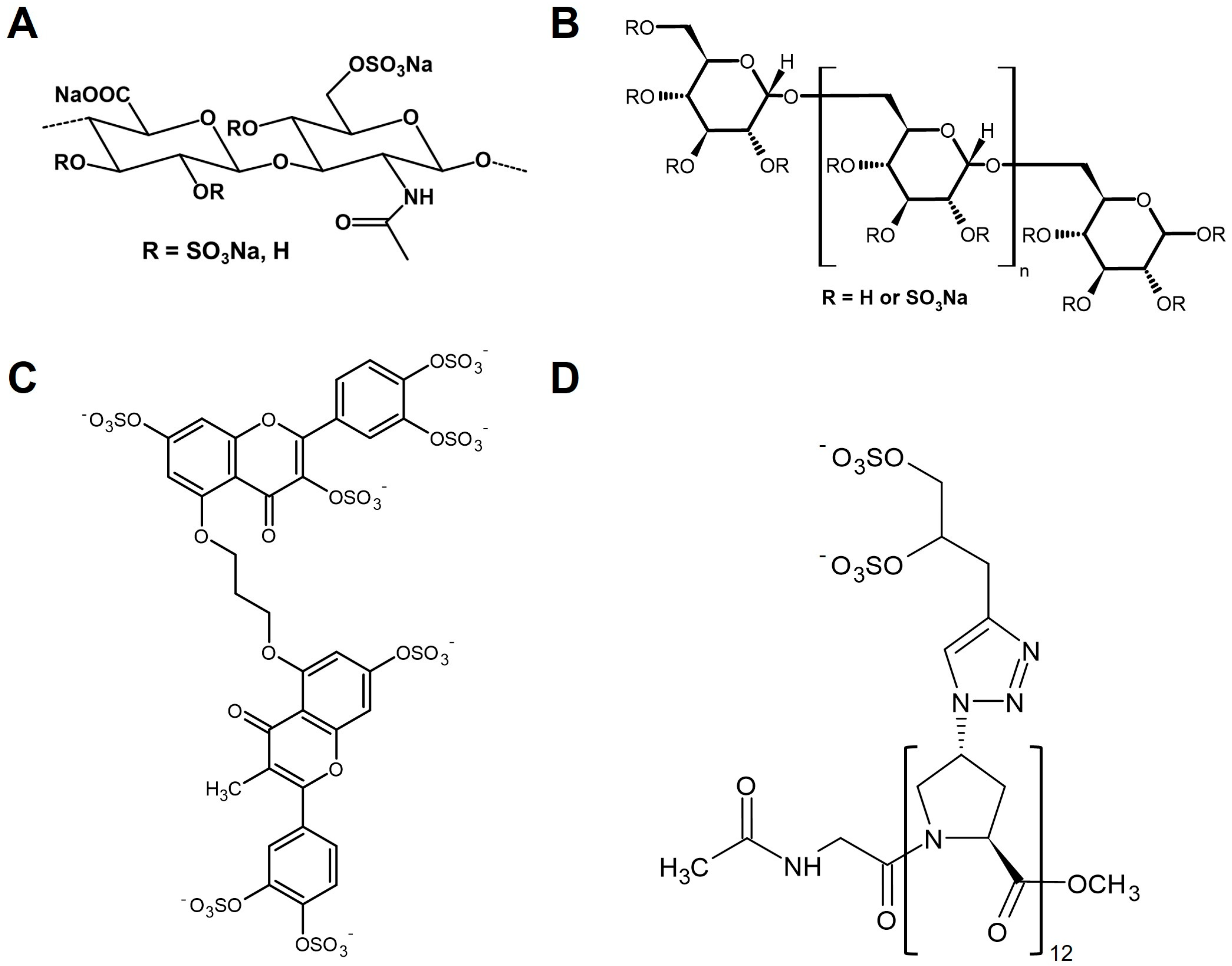
A further widely explored group of GAG mimetics is based on HA, which can be produced on a large scale by Streptococcus zooepidemicus. Several reviews summarize the various strategies for HA modification and functionalization [31,32]. It is of note that especially highly sulfated hyaluronan (sHA, Figure 2A) derivatives showed even higher binding levels for various biologically active proteins, such as growth factors like transforming growth factor-β1 (TGF-β1) [33], bone morphogenic protein-2 (BMP-2) [34] and -4 (BMP-4) [35], VEGF-A [36], heparin-binding epidermal growth factor-like growth factor (HB-EGF) [37], inhibitors of matrix degradation and angiogenesis-like tissue inhibitor of metalloproteinases-3 (TIMP-3) [38], and modulators of receptor activator of NF-κB ligand (RANKL)/RANK and Wnt signaling such as osteoprotegerin [39] and sclerostin [40], in surface plasmon resonance (SPR) studies compared to native GAGs such as CS and heparin or carboxymethylated HA derivatives. Furthermore, sHA was shown to inhibit the activity of mouse and human heparinase to enzymatically degrade fluorescein-labeled HS [41]. Modeling studies pointed out an electrostatic interaction between an sHA tetrasaccharide and the heparin-binding domain-1 of heparinase. Computational and structural studies of the HS mimetic pixatimod (PG545) further illustrate how defined sulfation motifs can modulate viral spike protein interactions, underscoring the translational potential of engineered GAG mimetics [42].
Dextran, a branched polysaccharide, is frequently chemically functionalized with sulfate, carboxylate, or acetate groups to generate GAG mimetics, which are commonly employed to investigate interactions with heparin-binding proteins such as stromal cell-derived factor-1α (SDF-1α) [43]. While carboxymethyl–dextran sulfate GAG mimetics had no influence on the expression of HS biosynthetic enzymes, they were shown to reduce the mRNA and protein level of heparinase-1 in Huh7 human hepatoma cells. However, there were no detectable differences in the heparinase activity in the cell lysates after treatment with these dextran derivatives using a heparin/FGF-2 detection system. In addition, GAG mimetics like dextran sulfate-500 (DS-500, Figure 2B), suramin, and pentosan polysulfate are used to evaluate the role of sulfated GAGs as cofactors of prion protein accumulation, highlighting the role of sulfate residues in altering protein interactions and functions [44]. GAG mimetics from the RGTA® family act as ReGeneraTing Agents and are often dextran-based. Dextran sulfate has also been increasingly explored as a nanocarrier for bioactive molecules, broadening its potential beyond anticoagulation to applications in drug delivery and tissue engineering [45]. Moreover, clinical observations with RGTA® formulations demonstrate resistance against enzymatic degradation and promising effects in wound healing [46].
In addition, other natural glycans, such as alginate, fucans, xylan, chitosan, and levan, have been chemically modified to obtain GAG mimetics [47,48,49,50,51]. Erginer et al. used sulfated derivatives of levan, a natural polymer composed of β(2-6)-linked fructose, as heparin mimetic glycan. They showed concentration-dependent anticoagulative activity via thrombin inhibition, suggesting a potential for cardiac tissue engineering applications [52]. 6-O-sulfated chitosan was crosslinked to fabricate wound dressings, which were able to scavenge interleukin-6 (IL-6) and TGF-β1 and promote vascularization and re-epithelialization of full-thickness wounds in vivo [53].
Beyond chemically modified GAG mimetics, GAG-functionalized nanomaterials represent an emerging class of bioactive scaffolds. For instance, chitosan- and HA-modified nanocomposites have demonstrated both antimicrobial properties and enhanced wound closure in murine models [54]. Other studies showed that heparin mimetic sulfated alginate carriers can sequester and sustain the release of heparin-binding growth factors (e.g., VEGF, FGF-2, CTGF/IGF-I), while dextran sulfate–based nanoformulations display intrinsic antibacterial activity [51,55,56,57]. These examples underscore the translational potential of combining native GAG structures with nanotechnology to address challenges in infection control and tissue repair.

3.2. Non-Saccharide-Based HS Mimetics

A detailed overview of different sulfated non-saccharide-based GAG mimetics was given by Afosah and Al-Horani [58]. In particular, exploiting the GAG/protein interface by drug discovery programs is a promising option for developing novel drug candidates modulating GAG-dependent protein functions. Synthetic HS mimetics composed of sulfated cyclitol subunits coupled by different linkers have been developed by Freeman et al. as highly stable compounds [59]. SPR interaction studies showed the inhibitory potential of these HS mimetics to interfere with the binding of the growth factors FGF-1, FGF-2, and VEGF-A and the chemokine IL-8 to heparin surfaces. Furthermore, those sulfated cyclitols, which resembled pentasaccharides, were as potent as PI-88 for inhibiting the activity of the HS-degrading enzyme heparinase, while all sulfated HS mimetics blocked the activity of cathepsin G. Current efforts in small-molecule discovery have yielded novel heparinase inhibitors with improved potency and selectivity, reinforcing the promise of non-saccharide scaffolds [60]. Structure–activity studies further confirm that such mimetics can mimic critical sulfate spacing requirements while offering higher stability than native saccharides [16].
Raman et al. reported the development of polysulfonated small-molecule GAG mimetics as angiogenesis inhibitors competing with cell-surface HS for the binding of pro-angiogenic proteins [61]. Here, the authors found that the inhibitory activity of these GAG mimetics towards vascular tube formation required two sulfate residues at a distance of 5–10 Å and did not correlate with the number of sulfate groups. A screening of 53 compounds as potential sulfated non-saccharide GAG mimetics for the selective targeting of colon cancer stem cells identified compound G2.2 (Figure 2C), a dimeric sulfated flavonoid, as the lead drug for inhibiting the spheroid formation of cancer cells, with IC50 values in the µM range [62]. Mechanistical studies suggested that G2.2 induced cancer cell apoptosis and reduced their ability for self-renewal. These data were confirmed by in vivo experiments that demonstrated that the inhibitory effect of G2.2 on colon cancer stem cells is mediated through p38 MAP kinase activation [63].
A further strategy for the synthesis of GAG mimetics is based on polyproline residues via amide coupling conjugated sulfate residues. Polyproline-based GAG mimetics were thus obtained that model the length and periodicity of GAG disaccharide units without showing any factor IIa activity [64]. The lead molecule {Z}12 (Figure 2D) showed comparable activity to heparin in reducing hematogenous metastasis in mice, which was suggested to be primarily associated with the inhibition of P-selectin.
In addition, surfen (bis-2-methyl-4-amino-quinolyl-6-carbamide), a synthetic bis-quinoline derivative, acts as a small-molecule antagonist of HS/protein interactions and has been widely employed as a research tool to probe HS-dependent signaling pathways [65].

4. Selected Biomedical Applications of GAG Mimetics

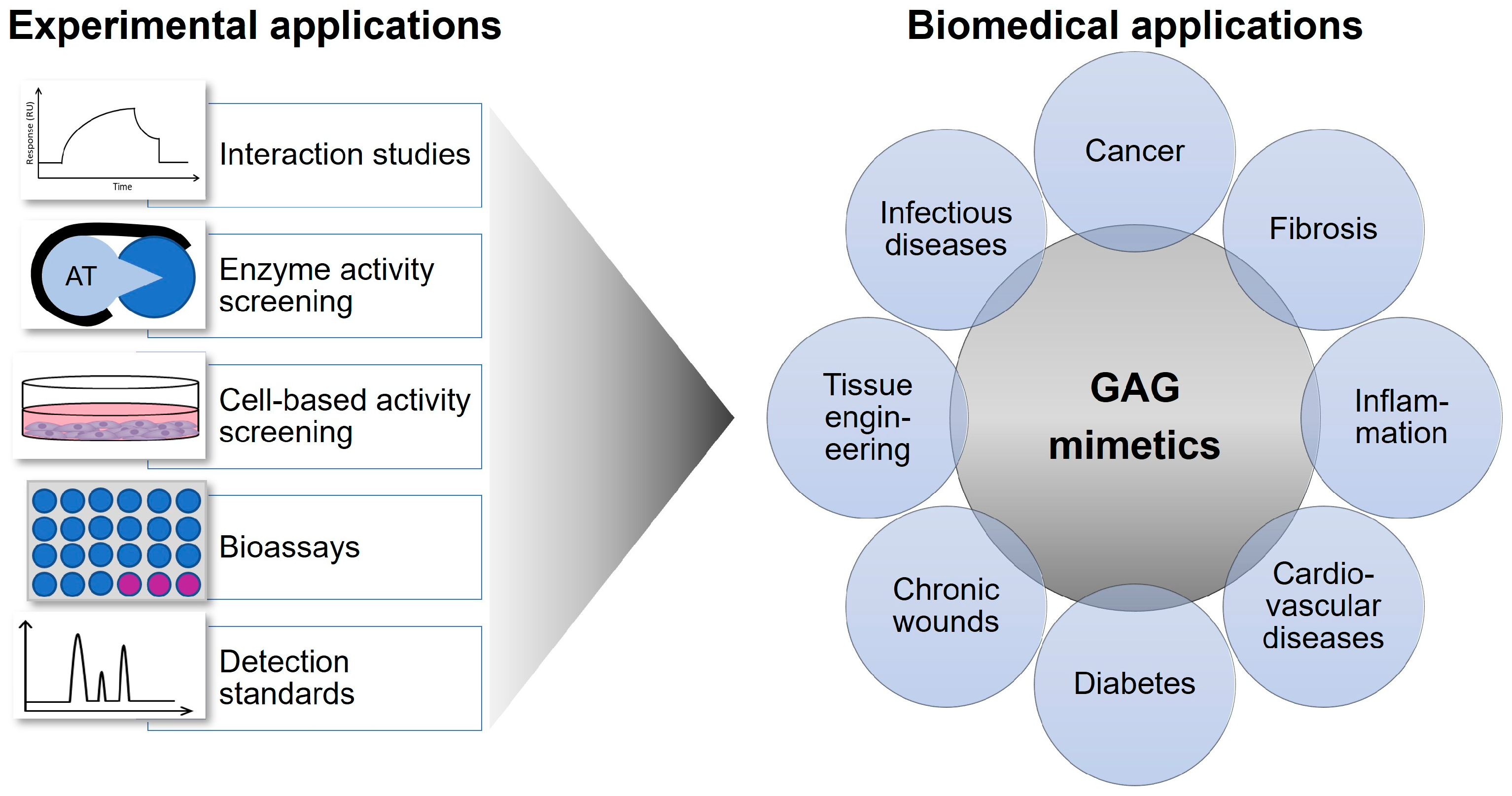
Due to the broad functions of HS chains in orchestrating key biological processes via their interaction with mediator proteins, various HS mimetics have been explored for biomedical applications [66] (Figure 3). For example, recent work confirmed the antiviral activity of pixatimod (PG545) through direct interference with the SARS-CoV-2 spike protein, highlighting the broader utility of GAG mimetics in infectious diseases [42]. Furthermore, updated reviews on heparinase inhibitors provide compelling evidence for their role in reducing cancer progression and metastasis [52]. A summary of studies focusing on the application of heparin derivatives and HS mimetics in cancer therapy is given in Refs. [67,68], while the effects of sHA derivatives in the field of bone and skin regeneration are reviewed in Refs. [69,70,71,72]. Table 1 highlights some currently studied potential biomedical applications of polysaccharide-based GAG mimetics, and Table 2 summarizes the biomedical uses of selected non-saccharide-based GAG mimetics.

5. Glycomimetics in Late-Stage Development and Clinical Translation

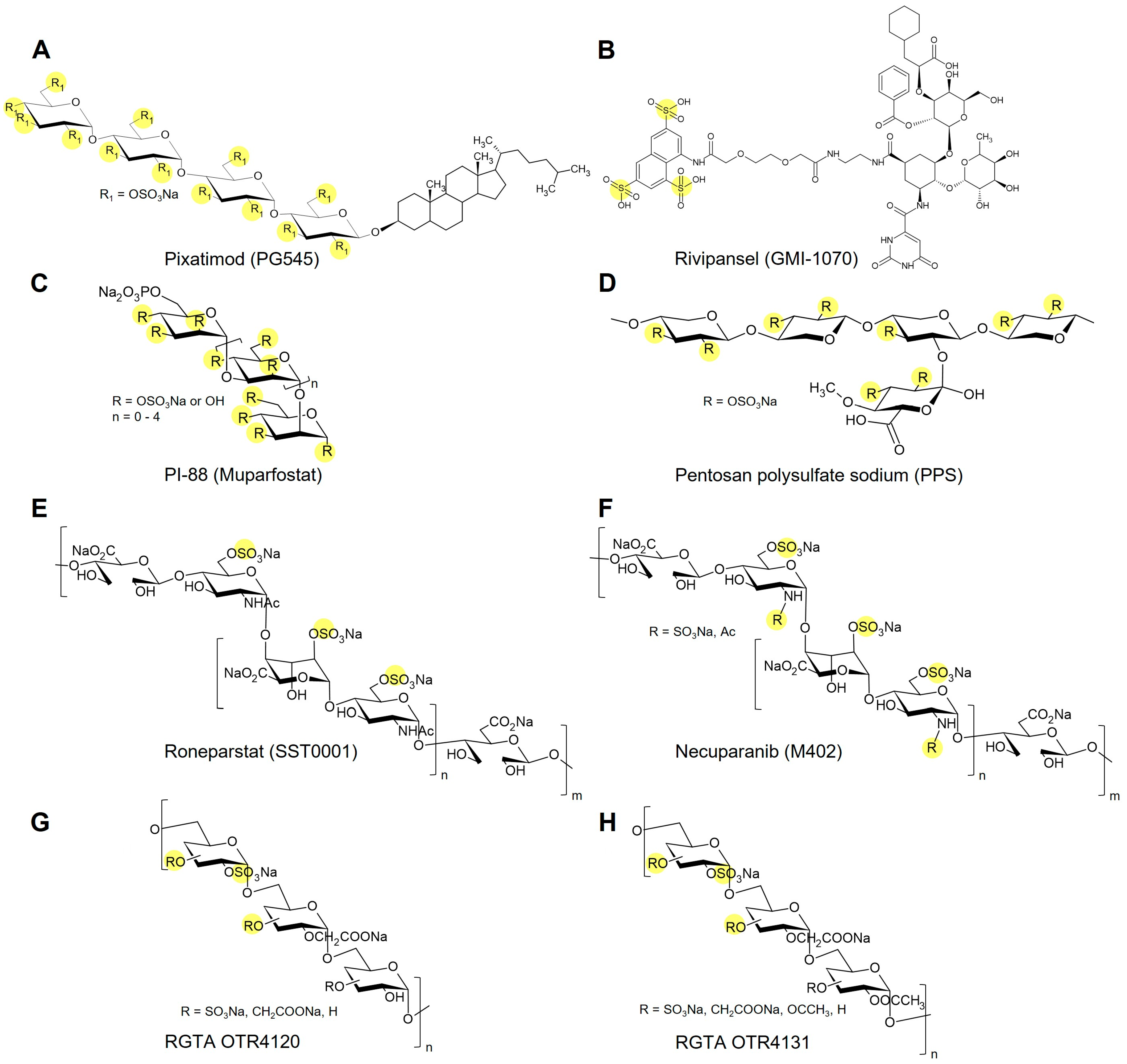
Several glycomimetics have advanced beyond preclinical evaluation and entered late-stage development or clinical testing (Figure 4). The synthetic pentasaccharide fondaparinux (Arixtra) exemplifies a clinically approved GAG mimetic that reproduces the heparin antithrombin-binding sequence with single-entity chemical definition and predictable pharmacology [95,96]. PI-88 (muparfostat), a sulfated oligosaccharide mixture from yeast phosphomannan, reached Phase III trials in hepatocellular carcinoma as a heparanase inhibitor, though further development was halted due to limited efficacy [18,20,97]. A next-generation derivative, pixatimod (PG545), combines a sulfated saccharide backbone with a lipophilic moiety, improving stability and pharmacokinetics. It has completed Phase I/II oncology trials and showed antiviral activity against SARS-CoV-2 in preclinical models [76,79,98,99,100]. In regenerative medicine, RGTA formulations (e.g., OTR4120, OTR4131), based on dextran sulfate, have shown promising results in early clinical studies on chronic wounds and tissue repair [46,101]. Among non-saccharide mimetics, rivipansel (GMI-1070), an E-selectin antagonist, advanced to Phase III trials for vaso-occlusive crises in sickle cell disease. Although the primary endpoint was not achieved, clinical data confirmed safety and biological activity [91]. The approved pentosan polysulfate sodium (PPS) is a heparin-like polysulfate that is routinely used to treat interstitial cystitis/bladder pain syndrome, where it acts as a replacement for the damaged urothelial GAG layer, as supported by clinical trials [102,103]. Roneparstat (SST0001), a non-anticoagulant, glycol-split heparin, showed heparanase inhibition and clinical activity in a Phase I multiple-myeloma study [104]. Necuparanib (M402), a heparan sulfate mimetic, completed Phase I and Phase II trials in pancreatic cancer. Although the development program was later discontinued, these studies yielded valuable data on pharmacokinetics, dosing, and biomarker correlations [105]. Sevuparin, a low-anticoagulant heparinoid with anti-adhesive activity, advanced to clinical testing in sickle cell vaso-occlusive crises and as adjunct therapy in malaria [106,107]. These agents illustrate both feasibility and remaining challenges for translating structurally defined GAG mimetics to the clinic.

6. Safety and Immunogenicity Considerations

The translational success of GAG mimetics critically depends on their safety and immunological profile. The heparin contamination crisis of 2007/2008, which was traced to over-sulfated CS, underscored the risk of adverse immune reactions when structural heterogeneity or impurities are present [108,109]. Fully synthetic, single-entity agents (e.g., fondaparinux) demonstrate that chemical definition and homogeneity can reduce unpredictability relative to complex mixtures [95,96]. Defined mimetics, however, can display favorable or even beneficial immunomodulatory properties. For example, sulfated hyaluronan (sHA) has been shown to reduce pro-inflammatory macrophage functions, attenuate NF-κB signaling, and promote tissue repair in diabetic wound models, suggesting that it can mitigate rather than exacerbate inflammatory responses [110,111,112]. Similarly, pixatimod (PG545) not only functions as a heparinase inhibitor but also exerts immune-regulatory effects, including suppression of effector T-cell activity and promotion of regulatory T-cells [76,113]. Clinical experience with rivipansel (GMI-1070) further indicates good tolerability across multiple trials, with no major immune-related safety signals despite lack of efficacy in Phase III. In regenerative applications, RGTAs like OTR4120 have been reported to reduce inflammation and fibrosis while accelerating wound closure [46,114,115]. Together, these examples illustrate that the immunological impact of GAG mimetics varies with their chemical design. While polydisperse or contaminated preparations carry risk, structurally defined mimetics may offer anti-inflammatory or immune-modulatory benefits in addition to their primary biological activity.

7. Conclusions

Recent technological advances for the complex analysis of glycans foster the continuous expansion of our knowledge about GAGs. In particular, computational simulation studies targeting the GAG/protein interface combined with glycan array technologies will help to rationally engineer novel classes of GAG mimetics that can function, for example, as artificial decoy receptors to control dysregulated cell signaling or prevent cell attachment of pathogens, which would advance, e.g., antiviral drug developments. The integration of these in silico strategies with scalable synthesis approaches should ultimately pave the way for clinically viable, structurally defined mimetics. The goal is still to increase the specificity and to allow for a large-scale production of defined GAG mimetics in order to promote their broad biomedical application.

Author Contributions

Conceptualization, S.R.; resources, S.R.; writing—original draft preparation, F.J. and S.R.; writing—review and editing, F.J. and S.R.; visualization, S.R.; supervision, S.R.; project administration, S.R.; funding acquisition, S.R. All authors have read and agreed to the published version of the manuscript.

Funding

This research was funded by Dr. Rolf M. Schwiete Stiftung, grant number 2024-021.

Institutional Review Board Statement

Not applicable.

Informed Consent Statement

Not applicable.

Data Availability Statement

No new data are included.

Conflicts of Interest

The authors declare no conflicts of interest. The funders had no role in the design of the study; in the collection, analyses, or interpretation of data; in the writing of the manuscript; or in the decision to publish the results.

References

  1. Köwitsch, A.; Zhou, G.; Groth, T. Medical Application of Glycosaminoglycans: A Review. J. Tissue Eng. Regen. Med. 2017, 12, e23–e41. [Google Scholar] [CrossRef]
  2. Xu, D.; Esko, J.D. Demystifying Heparan Sulfate-Protein Interactions. Annu. Rev. Biochem. 2014, 83, 129–157. [Google Scholar] [CrossRef]
  3. Esko, J.D.; Selleck, S.B. Order Out of Chaos: Assembly of Ligand Binding Sites in Heparan Sulfate. Annu. Rev. Biochem. 2002, 71, 435–471. [Google Scholar] [CrossRef]
  4. Pessentheiner, A.R.; Ducasa, G.M.; Gordts, P.L.S.M. Proteoglycans in Obesity-Associated Metabolic Dysfunction and Meta-Inflammation. Front. Immunol. 2020, 11, 769. [Google Scholar] [CrossRef] [PubMed]
  5. Chittum, J.E.; Thompson, A.; Desai, U.R. Glycosaminoglycan Microarrays for Studying Glycosaminoglycan-Protein Systems. Carbohydr. Polym. 2024, 335, 122106. [Google Scholar] [CrossRef]
  6. Portillo, G.; Li, D.R.; Goyal, Y.; Rowan, N.; Al-Horani, B.G.; Anbalagan, R.A.; Heparin, M.; Gatica Portillo, D.R.; Li, Y.; Goyal, N.; et al. Heparin, Heparin-like Molecules, and Heparin Mimetics in Breast Cancer: A Concise Review. Biomolecules 2025, 15, 1034. [Google Scholar] [CrossRef] [PubMed]
  7. Shi, D.; Sheng, A.; Chi, L. Glycosaminoglycan-Protein Interactions and Their Roles in Human Disease. Front. Mol. Biosci. 2021, 8, 639666. [Google Scholar] [CrossRef] [PubMed]
  8. Vallet, S.D.; Clerc, O.; Ricard-Blum, S. Glycosaminoglycan-Protein Interactions: The First Draft of the Glycosaminoglycan Interactome. J. Histochem. Cytochem. 2021, 69, 93–104. [Google Scholar] [CrossRef]
  9. Le Pennec, J.; Picart, C.; Vivès, R.R.; Migliorini, E. Sweet but Challenging: Tackling the Complexity of GAGs with Engineered Tailor-Made Biomaterials. Adv. Mater. 2024, 36, 2312154. [Google Scholar] [CrossRef]
  10. Zhang, L. Glycosaminoglycan (GAG) Biosynthesis and GAG-Binding Proteins. Prog. Mol. Biol. Transl. Sci. 2010, 93, 1–17. [Google Scholar] [CrossRef]
  11. Sarrazin, S.; Lamanna, W.C.; Esko, J.D. Heparan Sulfate Proteoglycans. Cold Spring Harb. Perspect. Biol. 2011, 3, a004952. [Google Scholar] [CrossRef] [PubMed]
  12. Shojania, A.M.; Tetreault, J.; Turnbull, G. The Variations between Heparin Sensitivity of Different Lots of Activated Partial Thromboplastin Time Reagent Produced by the Same Manufacturer. Am. J. Clin. Pathol. 1988, 89, 19–23. [Google Scholar] [CrossRef]
  13. Mende, M.; Bednarek, C.; Wawryszyn, M.; Sauter, P.; Biskup, M.B.; Schepers, U.; Bräse, S. Chemical Synthesis of Glycosaminoglycans. Chem. Rev. 2016, 116, 8193–8255. [Google Scholar] [CrossRef]
  14. Deangelis, P.L.; Liu, J.; Linhardt, R.J. Chemoenzymatic Synthesis of Glycosaminoglycans: Re-Creating, Re-Modeling and Re-Designing Nature’s Longest or Most Complex Carbohydrate Chains. Glycobiology 2013, 23, 764–777. [Google Scholar] [CrossRef]
  15. DeAngelis, P.L. Glycosaminoglycan Polysaccharide Biosynthesis and Production: Today and Tomorrow. Appl. Microbiol. Biotechnol. 2012, 94, 295–305. [Google Scholar] [CrossRef] [PubMed]
  16. Morla, S.; Ravikumar, O.; O’Hara, C.; Boothello, R.; Vera, A.; Abdelfadiel, E.I.; Fayyad, R.; Afosah, D.K.; Sharon, C.; Fernandez, L.; et al. Designing Synthetic, Sulfated Glycosaminoglycan Mimetics That Are Orally Bioavailable and Exhibiting In Vivo Anticancer Activity. J. Med. Chem. 2023, 66, 1321–1338. [Google Scholar] [CrossRef]
  17. Khachigian, L.M.; Parish, C.R. Phosphomannopentaose Sulfate (PI-88): Heparan Sulfate Mimetic with Clinical Potential in Multiple Vascular Pathologies. Cardiovasc. Drug Rev. 2004, 22, 1–6. [Google Scholar] [CrossRef] [PubMed]
  18. Francis, D.J.; Parish, C.R.; McGarry, M.; Santiago, F.S.; Lowe, H.C.; Brown, K.J.; Bingley, J.A.; Hayward, I.P.; Cowden, W.B.; Campbell, J.H.; et al. Blockade of Vascular Smooth Muscle Cell Proliferation and Intimal Thickening after Balloon Injury by the Sulfated Oligosaccharide PI-88: Phosphomannopentaose Sulfate Directly Binds FGF-2, Blocks Cellular Signaling, and Inhibits Proliferation. Circ. Res. 2003, 92, 8. [Google Scholar] [CrossRef] [PubMed]
  19. Morla, S. Glycosaminoglycans and Glycosaminoglycan Mimetics in Cancer and Inflammation. Int. J. Mol. Sci. 2019, 20, 1963. [Google Scholar] [CrossRef]
  20. Chhabra, M.; Ferro, V. PI-88 and Related Heparan Sulfate Mimetics. Adv. Exp. Med. Biol. 2020, 1221, 473–491. [Google Scholar] [CrossRef]
  21. Kuhnast, B.; el Hadri, A.; Boisgard, R.; Hinnen, F.; Richard, S.; Caravano, A.; Nancy-Portebois, V.; Petitou, M.; Tavitian, B.; Dollé, F. Synthesis, Radiolabeling with Fluorine-18 and Preliminary in Vivo Evaluation of a Heparan Sulphate Mimetic as Potent Angiogenesis and Heparanase Inhibitor for Cancer Applications. Org. Biomol. Chem. 2016, 14, 1915–1920. [Google Scholar] [CrossRef] [PubMed]
  22. Matou, S.; Colliec-Jouault, S.; Galy-Fauroux, I.; Ratiskol, J.; Sinquin, C.; Guezennec, J.; Fischer, A.M.; Helley, D. Effect of an Oversulfated Exopolysaccharide on Angiogenesis Induced by Fibroblast Growth Factor-2 or Vascular Endothelial Growth Factor in Vitro. Biochem. Pharmacol. 2005, 69, 751–759. [Google Scholar] [CrossRef]
  23. Schiraldi, C.; Cimini, D.; de Rosa, M. Production of Chondroitin Sulfate and Chondroitin. Appl. Microbiol. Biotechnol. 2010, 87, 1209–1220. [Google Scholar] [CrossRef]
  24. Bedini, E.; Decastro, C.; Derosa, M.; Dinola, A.; Iadonisi, A.; Restaino, O.F.; Schiraldi, C.; Parrilli, M. A Microbiological–Chemical Strategy to Produce Chondroitin Sulfate A,C. Angew. Chem. Int. Ed. 2011, 50, 6160–6163. [Google Scholar] [CrossRef] [PubMed]
  25. Corsuto, L.; Rother, S.; Koehler, L.; Bedini, E.; Moeller, S.; Schnabelrauch, M.; Hintze, V.; Schiraldi, C.; Scharnweber, D. Sulfation Degree Not Origin of Chondroitin Sulfate Derivatives Modulates Keratinocyte Response. Carbohydr. Polym. 2018, 191, 53–64. [Google Scholar] [CrossRef]
  26. Badri, A.; Williams, A.; Awofiranye, A.; Datta, P.; Xia, K.; He, W.; Fraser, K.; Dordick, J.S.; Linhardt, R.J.; Koffas, M.A.G. Complete Biosynthesis of a Sulfated Chondroitin in Escherichia Coli. Nat. Commun. 2021, 12, 1389. [Google Scholar] [CrossRef]
  27. Couto, M.R.; Rodrigues, J.L.; Rodrigues, L.R. Heterologous Production of Chondroitin. Biotechnol. Rep. 2022, 33, e00710. [Google Scholar] [CrossRef]
  28. Domínguez-Rodríguez, P.; Reina, J.J.; Gil-Caballero, S.; Nieto, P.M.; de Paz, J.L.; Rojo, J. Glycodendrimers as Chondroitin Sulfate Mimetics: Synthesis and Binding to Growth Factor Midkine. Chemistry 2017, 23, 11338–11345. [Google Scholar] [CrossRef]
  29. García-Jiménez, M.J.; Gil-Caballero, S.; Maza, S.; Corzana, F.; Juárez-Vicente, F.; Miles, J.R.; Sakamoto, K.; Kadomatsu, K.; García-Domínguez, M.; de Paz, J.L.; et al. Midkine Interaction with Chondroitin Sulfate Model Synthetic Tetrasaccharides and Their Mimetics: The Role of Aromatic Interactions. Chem.—A Eur. J. 2021, 27, 12395–12409. [Google Scholar] [CrossRef] [PubMed]
  30. Sheng, G.J.; Oh, Y.I.; Chang, S.K.; Hsieh-Wilson, L.C. Tunable Heparan Sulfate Mimetics for Modulating Chemokine Activity. J. Am. Chem. Soc. 2013, 135, 10898–10901. [Google Scholar] [CrossRef]
  31. Hintze, V.; Schnabelrauch, M.; Rother, S. Chemical Modification of Hyaluronan and Their Biomedical Applications. Front. Chem. 2022, 10, 830671. [Google Scholar] [CrossRef]
  32. Burdick, J.A.; Prestwich, G.D. Hyaluronic Acid Hydrogels for Biomedical Applications. Adv. Mater. 2011, 23, H41–H56. [Google Scholar] [CrossRef] [PubMed]
  33. Hintze, V.; Miron, A.; Moeller, S.; Schnabelrauch, M.; Wiesmann, H.-P.; Worch, H.; Scharnweber, D. Sulfated Hyaluronan and Chondroitin Sulfate Derivatives Interact Differently with Human Transforming Growth Factor-Β1 (TGF-Β1). Acta Biomater. 2012, 8, 2144–2152. [Google Scholar] [CrossRef] [PubMed]
  34. Hintze, V.; Samsonov, S.A.; Anselmi, M.; Moeller, S.; Becher, J.; Schnabelrauch, M.; Scharnweber, D.; Pisabarro, M.T. Sulfated Glycosaminoglycans Exploit the Conformational Plasticity of Bone Morphogenetic Protein 2 (BMP-2) and Alter the Interaction Pro Fi Le with Its Receptor. Biomacromolecules 2014, 15, 3083–3092. [Google Scholar] [CrossRef] [PubMed]
  35. Hintze, V.; Moeller, S.; Schnabelrauch, M.; Bierbaum, S.; Viola, M.; Worch, H.; Scharnweber, D. Modifications of Hyaluronan Influence the Interaction with Human Bone Morphogenetic Protein-4 (HBMP-4). Biomacromolecules 2009, 10, 3290–3297. [Google Scholar] [CrossRef]
  36. Koehler, L.; Ruiz-Gómez, G.; Balamurugan, K.; Rother, S.; Freyse, J.; Möller, S.; Schnabelrauch, M.; Köhling, S.; Djordjevic, S.; Scharnweber, D.; et al. Dual Action of Sulfated Hyaluronan on Angiogenic Processes in Relation to Vascular Endothelial Growth Factor-A. Sci. Rep. 2019, 9, 18143. [Google Scholar] [CrossRef]
  37. Thönes, S.; Rother, S.; Wippold, T.; Blaszkiewicz, J.; Balamurugan, K.; Moeller, S.; Ruiz-gómez, G.; Schnabelrauch, M.; Scharnweber, D.; Saalbach, A.; et al. Hyaluronan/Collagen Hydrogels Containing Sulfated Hyaluronan Improve Wound Healing by Sustained Release of Heparin-Binding EGF-like Growth Factor. Acta Biomater. 2019, 86, 135–147. [Google Scholar] [CrossRef]
  38. Rother, S.; Samsonov, S.A.; Hofmann, T.; Blaszkiewicz, J.; Köhling, S.; Moeller, S.; Schnabelrauch, M.; Rademann, J.; Kalkhof, S.; von Bergen, M.; et al. Structural and Functional Insights into the Interaction of Sulfated Glycosaminoglycans with Tissue Inhibitor of Metalloproteinase-3—A Possible Regulatory Role on Extracellular Matrix Homeostasis. Acta Biomater. 2016, 45, 143–154. [Google Scholar] [CrossRef]
  39. Salbach-Hirsch, J.; Kraemer, J.; Rauner, M.; Samsonov, S.A.; Pisabarro, M.T.; Moeller, S.; Schnabelrauch, M.; Scharnweber, D.; Hofbauer, L.C.; Hintze, V. The Promotion of Osteoclastogenesis by Sulfated Hyaluronan through Interference with Osteoprotegerin and Receptor Activator of NF-ΚB Ligand/Osteoprotegerin Complex Formation. Biomaterials 2013, 34, 7653–7661. [Google Scholar] [CrossRef]
  40. Salbach-Hirsch, J.; Samsonov, S.A.; Hintze, V.; Hofbauer, C.; Picke, A.; Rauner, M.; Gehrcke, J.-P.; Moeller, S.; Schnabelrauch, M.; Scharnweber, D.; et al. Structural and Functional Insights into Sclerostin-Glycosaminoglycan Interactions in Bone. Biomaterials 2015, 67, 335–345. [Google Scholar] [CrossRef]
  41. Shi, J.; Kanoya, R.; Tani, Y.; Ishikawa, S.; Maeda, R.; Suzuki, S.; Kawanami, F.; Miyagawa, N.; Takahashi, K.; Oku, T.; et al. Sulfated Hyaluronan Binds to Heparanase and Blocks Its Enzymatic and Cellular Actions in Carcinoma Cells. Int. J. Mol. Sci. 2022, 23, 5055. [Google Scholar] [CrossRef]
  42. Guimond, S.E.; Mycroft-West, C.J.; Gandhi, N.S.; Tree, J.A.; Le, T.T.; Spalluto, C.M.; Humbert, M.v.; Buttigieg, K.R.; Coombes, N.; Elmore, M.J.; et al. Synthetic Heparan Sulfate Mimetic Pixatimod (PG545) Potently Inhibits SARS-CoV-2 by Disrupting the Spike–ACE2 Interaction. ACS Cent. Sci. 2022, 8, 527–545. [Google Scholar] [CrossRef]
  43. Friand, V.; Haddad, O.; Papy-Garcia, D.; Hlawaty, H.; Vassy, R.; Hamma-Kourbali, Y.; Perret, G.Y.; Courty, J.; Baleux, F.; Oudar, O.; et al. Glycosaminoglycan Mimetics Inhibit SDF-1/CXCL12-Mediated Migration and Invasion of Human Hepatoma Cells. Glycobiology 2009, 19, 1511–1524. [Google Scholar] [CrossRef] [PubMed]
  44. Wolf, H.; Graßmann, A.; Bester, R.; Hossinger, A.; Möhl, C.; Paulsen, L.; Groschup, M.H.; Schätzl, H.; Vorberg, I. Modulation of Glycosaminoglycans Affects PrPSc Metabolism but Does Not Block PrPSc Uptake. J. Virol. 2015, 89, 9853–9864. [Google Scholar] [CrossRef]
  45. Ramasundaram, S.; Saravanakumar, G.; Sobha, S.; Oh, T.H. Dextran Sulfate Nanocarriers: Design, Strategies and Biomedical Applications. Int. J. Mol. Sci. 2022, 24, 355. [Google Scholar] [CrossRef] [PubMed]
  46. Barritault, D.; Gilbert-Sirieix, M.; Rice, K.L.; Siñeriz, F.; Papy-Garcia, D.; Baudouin, C.; Desgranges, P.; Zakine, G.; Saffar, J.L.; van Neck, J. RGTA® or ReGeneraTing Agents Mimic Heparan Sulfate in Regenerative Medicine: From Concept to Curing Patients. Glycoconj. J. 2017, 34, 325–338. [Google Scholar] [CrossRef] [PubMed]
  47. Arlov, Ø.; Aachmann, F.L.; Sundan, A.; Espevik, T.; Skjåk-Bræk, G. Heparin-like Properties of Sulfated Alginates with Defined Sequences and Sulfation Degrees. Biomacromolecules 2014, 15, 2744–2750. [Google Scholar] [CrossRef]
  48. Arlov, Ø.; Aachmann, F.L.; Feyzi, E.; Sundan, A.; Skjåk-Bræk, G. The Impact of Chain Length and Flexibility in the Interaction between Sulfated Alginates and HGF and FGF-2. Biomacromolecules 2015, 16, 3417–3424. [Google Scholar] [CrossRef]
  49. Stone, A.L.; Melton, D.J.; Lewis, M.S. Structure-Function Relations of Heparin-Mimetic Sulfated Xylan Oligosaccharides: Inhibition of Human Immunodeficiency Virus-1 Infectivity in Vitro. Glycoconj. J. 1998, 15, 697–712. [Google Scholar] [CrossRef]
  50. Zayed, A.; Avila-Peltroche, J.; El-Aasr, M.; Ulber, R. Sulfated Galactofucans: An Outstanding Class of Fucoidans with Promising Bioactivities. Mar. Drugs 2022, 20, 412. [Google Scholar] [CrossRef]
  51. Maatouk, B.; Jaffa, M.A.; Karam, M.; Fahs, D.; Nour-Eldine, W.; Hasan, A.; Jaffa, A.A.; Mhanna, R. Sulfated Alginate/Polycaprolactone Double-Emulsion Nanoparticles for Enhanced Delivery of Heparin-Binding Growth Factors in Wound Healing Applications. Colloids Surf. B Biointerfaces 2021, 208, 112105. [Google Scholar] [CrossRef]
  52. Erginer, M.; Akcay, A.; Coskunkan, B.; Morova, T.; Rende, D.; Bucak, S.; Baysal, N.; Ozisik, R.; Eroglu, M.S.; Agirbasli, M.; et al. Sulfated Levan from Halomonas Smyrnensis as a Bioactive, Heparin-Mimetic Glycan for Cardiac Tissue Engineering Applications. Carbohydr. Polym. 2016, 149, 289–296. [Google Scholar] [CrossRef]
  53. Sahraneshin-Samani, F.; Kazemi-Ashtiani, M.; Karimi, H.; Shiravandi, A.; Baharvand, H.; Daemi, H. Regioselective Sulfated Chitosan Produces a Biocompatible and Antibacterial Wound Dressing with Low Inflammatory Response. Biomater. Adv. 2022, 139, 213020. [Google Scholar] [CrossRef]
  54. Gan, H.-Q.; Gui, W.-Z.; Hu, X.-L.; Zheng, Y.-J.; He, X.-P. Trinary Dressing Material Formed between Polysaccharides and Gold Nanoparticles for Synergistic Wound Disinfection and Repair. ACS Appl. Bio Mater. 2025, 8, 9072–9080. [Google Scholar] [CrossRef] [PubMed]
  55. Freeman, I.; Kedem, A.; Cohen, S. The Effect of Sulfation of Alginate Hydrogels on the Specific Binding and Controlled Release of Heparin-Binding Proteins. Biomaterials 2008, 29, 3260–3268. [Google Scholar] [CrossRef]
  56. Gionet-Gonzales, M.; Casella, A.; Diloretto, D.; Ginnell, C.; Griffin, K.H.; Bigot, A.; Leach, J.K. Sulfated Alginate Hydrogels Prolong the Therapeutic Potential of MSC Spheroids by Sequestering the Secretome. Adv. Heal. Mater. 2021, 10, e2101048. [Google Scholar] [CrossRef]
  57. Madkhali, O.A.; Sivagurunathan Moni, S.; Sultan, M.H.; Bukhary, H.A.; Ghazwani, M.; Alhakamy, N.A.; Meraya, A.M.; Alshahrani, S.; Alqahtani, S.S.; Bakkari, M.A.; et al. Formulation and Evaluation of Injectable Dextran Sulfate Sodium Nanoparticles as a Potent Antibacterial Agent. Sci. Rep. 2021, 11, 9914. [Google Scholar] [CrossRef] [PubMed]
  58. Afosah, D.K.; Al-Horani, R.A. Sulfated Non-Saccharide Glycosaminoglycan Mimetics as Novel Drug Discovery Platform for Various Pathologies. Curr. Med. Chem. 2020, 27, 3412. [Google Scholar] [CrossRef]
  59. Freeman, C.; Liu, L.; Banwell, M.G.; Brown, K.J.; Bezos, A.; Ferro, V.; Parish, C.R. Use of Sulfated Linked Cyclitols as Heparan Sulfate Mimetics to Probe the Heparin/Heparan Sulfate Binding Specificity of Proteins. J. Biol. Chem. 2005, 280, 8842–8849. [Google Scholar] [CrossRef] [PubMed]
  60. Zhang, Y.; Cui, L. Discovery and Development of Small-Molecule Heparanase Inhibitors. Bioorg. Med. Chem. 2023, 90, 117335. [Google Scholar] [CrossRef]
  61. Raman, K.; Karuturi, R.; Swarup, V.P.; Desai, U.R.; Kuberan, B. Discovery of Novel Sulfonated Small Molecules That Inhibit Vascular Tube Formation. Bioorg. Med. Chem. Lett. 2012, 22, 4467–4470. [Google Scholar] [CrossRef] [PubMed]
  62. Patel, N.J.; Karuturi, R.; Al-Horani, R.A.; Baranwal, S.; Patel, J.; Desai, U.R.; Patel, B.B. Synthetic, Non-Saccharide, Glycosaminoglycan Mimetics Selectively Target Colon Cancer Stem Cells. ACS Chem. Biol. 2014, 9, 1826–1833. [Google Scholar] [CrossRef]
  63. Boothello, R.S.; Patel, N.J.; Sharon, C.; Abdelfadiel, E.I.; Morla, S.; Brophy, D.F.; Robert Lippman, H.; Desai, U.R.; Patel, B.B. A Unique Nonsaccharide Mimetic of Heparin Hexasaccharide Inhibits Colon Cancer Stem Cells via P38 MAP Kinase Activation. Mol. Cancer Ther. 2019, 18, 51–61. [Google Scholar] [CrossRef]
  64. Lim, T.C.; Cai, S.; Huber, R.G.; Bond, P.J.; Siew Chia, P.X.; Khou, S.L.; Gao, S.; Lee, S.S.; Lee, S.G. Facile Saccharide-Free Mimetics That Recapitulate Key Features of Glycosaminoglycan Sulfation Patterns. Chem. Sci. 2018, 9, 7940–7947. [Google Scholar] [CrossRef]
  65. Schuksz, M.; Fuster, M.M.; Brown, J.R.; Crawford, B.E.; Ditto, D.P.; Lawrence, R.; Glass, C.A.; Wang, L.; Tor, Y.; Esko, J.D. Surfen, a Small Molecule Antagonist of Heparan Sulfate. Proc. Natl. Acad. Sci. USA 2008, 105, 13075–13080. [Google Scholar] [CrossRef]
  66. Mohamed, S.; Coombe, D.R. Heparin Mimetics: Their Therapeutic Potential. Pharmaceuticals 2017, 10, 78. [Google Scholar] [CrossRef] [PubMed]
  67. Lanzi, C.; Cassinelli, G. Heparan Sulfate Mimetics in Cancer Therapy: The Challenge to Define Structural Determinants and the Relevance of Targets for Optimal Activity. Molecules 2018, 23, 2915. [Google Scholar] [CrossRef] [PubMed]
  68. Ma, S.N.; Mao, Z.X.; Wu, Y.; Liang, M.X.; Wang, D.D.; Chen, X.; Chang, P.A.; Zhang, W.; Tang, J.H. The Anti-Cancer Properties of Heparin and Its Derivatives: A Review and Prospect. Cell Adhes. Migr. 2020, 14, 118–128. [Google Scholar] [CrossRef]
  69. Salbach, J.; Rachner, T.D.; Rauner, M.; Hempel, U.; Anderegg, U.; Franz, S.; Simon, J.C.; Hofbauer, L.C. Regenerative Potential of Glycosaminoglycans for Skin and Bone. J. Mol. Med. 2012, 90, 625–635. [Google Scholar] [CrossRef]
  70. Salbach-Hirsch, J.; Rauner, M.; Hofbauer, C.; Hofbauer, L.C. New Insights into the Role of Glycosaminoglycans in the Endosteal Bone Microenvironment. Biol. Chem. 2021, 402, 1415–1425. [Google Scholar] [CrossRef]
  71. Anderegg, U.; Halfter, N.; Schnabelrauch, M.; Hintze, V. Collagen/Glycosaminoglycan-Based Matrices for Controlling Skin Cell Responses. Biol. Chem. 2021, 402, 1325–1335. [Google Scholar] [CrossRef]
  72. Scharnweber, D.; Hübner, L.; Rother, S.; Hempel, U.; Anderegg, U.; Samsonov, S.A.; Pisabarro, M.T.; Hofbauer, L.; Schnabelrauch, M.; Franz, S.; et al. Glycosaminoglycan Derivatives: Promising Candidates for the Design of Functional Biomaterials. J. Mater. Sci. Mater. Med. 2015, 26, 232. [Google Scholar] [CrossRef]
  73. van der Smissen, A.; Samsonov, S.; Hintze, V.; Scharnweber, D.; Moeller, S.; Schnabelrauch, M.; Pisabarro, M.T.; Anderegg, U. Artificial Extracellular Matrix Composed of Collagen I and Highly Sulfated Hyaluronan Interferes with TGFβ1 Signaling and Prevents TGFβ1-Induced Myofibroblast Differentiation. Acta Biomater. 2013, 9, 7775–7786. [Google Scholar] [CrossRef] [PubMed]
  74. Syanda, A.M.; Kringstad, V.I.; Blackford, S.J.I.; Kjesbu, J.S.; Ng, S.S.; Ma, L.; Xiao, F.; Coron, A.E.; Rokstad, A.M.A.; Modi, S.; et al. Sulfated Alginate Reduces Pericapsular Fibrotic Overgrowth on Encapsulated CGMP-Compliant HPSC-Hepatocytes in Mice. Front. Bioeng. Biotechnol. 2022, 9, 816542. [Google Scholar] [CrossRef]
  75. Nonaka, M.; Bao, X.; Matsumura, F.; Götze, S.; Kandasamy, J.; Kononov, A.; Broide, D.H.; Nakayama, J.; Seeberger, P.H.; Fukuda, M. Synthetic Di-Sulfated Iduronic Acid Attenuates Asthmatic Response by Blocking T-Cell Recruitment to Inflammatory Sites. Proc. Natl. Acad. Sci. USA 2014, 111, 8173–8178. [Google Scholar] [CrossRef]
  76. Koliesnik, I.O.; Kuipers, H.F.; Medina, C.O.; Zihsler, S.; Liu, D.; Van Belleghem, J.D.; Bollyky, P.L. The Heparan Sulfate Mimetic PG545 Modulates T Cell Responses and Prevents Delayed-Type Hypersensitivity. Front. Immunol. 2020, 11, 132. [Google Scholar] [CrossRef]
  77. Sutton, A.; Friand, V.; Papy-Garcia, D.; Dagouassat, M.; Martin, L.; Vassy, R.; Haddad, O.; Sainte-Catherine, O.; Kraemer, M.; Saffar, L.; et al. Glycosaminoglycans and Their Synthetic Mimetics Inhibit RANTES-Induced Migration and Invasion of Human Hepatoma Cells. Mol. Cancer Ther. 2007, 6, 2948–2958. [Google Scholar] [CrossRef]
  78. al Matari, N.; Deeb, G.; Mshiek, H.; Sinjab, A.; Kadara, H.; Abou-Kheir, W.; Mhanna, R. Anti-Tumor Effects of Biomimetic Sulfated Glycosaminoglycans on Lung Adenocarcinoma Cells in 2D and 3D In Vitro Models. Molecules 2020, 25, 2595. [Google Scholar] [CrossRef]
  79. Ferro, V.; Liu, L.; Johnstone, K.D.; Wimmer, N.; Karoli, T.; Handley, P.; Rowley, J.; Dredge, K.; Li, C.P.; Hammond, E.; et al. Discovery of PG545: A Highly Potent and Simultaneous Inhibitor of Angiogenesis, Tumor Growth, and Metastasis. J. Med. Chem. 2012, 55, 3804–3813. [Google Scholar] [CrossRef] [PubMed]
  80. Lemech, C.; Dredge, K.; Bampton, D.; Hammond, E.; Clouston, A.; Waterhouse, N.J.; Stanley, A.C.; Leveque-El Mouttie, L.; Chojnowski, G.M.; Haydon, A.; et al. Phase Ib Open-Label, Multicenter Study of Pixatimod, an Activator of TLR9, in Combination with Nivolumab in Subjects with Microsatellite-Stable Metastatic Colorectal Cancer, Metastatic Pancreatic Ductal Adenocarcinoma and Other Solid Tumors. J. Immunother. Cancer 2023, 11, e006136. [Google Scholar] [CrossRef] [PubMed]
  81. Lu, G.; Rausell-Palamos, F.; Zhang, J.; Zheng, Z.; Zhang, T.; Valle, S.; Rosselot, C.; Berrouet, C.; Conde, P.; Spindler, M.P.; et al. Dextran Sulfate Protects Pancreatic β-Cells, Reduces Autoimmunity, and Ameliorates Type 1 Diabetes. Diabetes 2020, 69, 1692–1707. [Google Scholar] [CrossRef]
  82. Chevalier, F.; Lavergne, M.; Negroni, E.; Ferratge, S.; Carpentier, G.; Gilbert-Sirieix, M.; Siñeriz, F.; Uzan, G.; Albanese, P. Glycosaminoglycan Mimetic Improves Enrichment and Cell Functions of Human Endothelial Progenitor Cell Colonies. Stem Cell Res. 2014, 12, 703–715. [Google Scholar] [CrossRef]
  83. Picke, A.K.; Salbach-Hirsch, J.; Hintze, V.; Rother, S.; Rauner, M.; Kascholke, C.; Möller, S.; Bernhardt, R.; Rammelt, S.; Pisabarro, M.T.; et al. Sulfated Hyaluronan Improves Bone Regeneration of Diabetic Rats by Binding Sclerostin and Enhancing Osteoblast Function. Biomaterials 2016, 96, 11–23. [Google Scholar] [CrossRef]
  84. Frescaline, G.; Bouderlique, T.; Huynh, M.B.; Papy-Garcia, D.; Courty, J.; Albanese, P. Glycosaminoglycans Mimetics Potentiate the Clonogenicity, Proliferation, Migration and Differentiation Properties of Rat Mesenchymal Stem Cells. Stem Cell Res. 2012, 8, 180–192. [Google Scholar] [CrossRef]
  85. Rother, S.; Samsonov, S.A.; Hempel, U.; Vogel, S.; Moeller, S.; Blaszkiewicz, J.; Köhling, S.; Schnabelrauch, M.; Rademann, J.; Pisabarro, M.T.; et al. Sulfated Hyaluronan Alters the Interaction Profile of TIMP-3 with the Endocytic Receptor LRP-1 Clusters II and IV and Increases the Extracellular TIMP-3 Level of Human Bone Marrow Stromal Cells. Biomacromolecules 2016, 17, 3252–3261. [Google Scholar] [CrossRef]
  86. Groah, S.L.; Libin, A.; Spungen, M.; Nguyen, K.L.; Woods, E.; Nabili, M.; Ramella-Roman, J.; Barritault, D. Regenerating Matrix-Based Therapy for Chronic Wound Healing: A Prospective within-Subject Pilot Study. Int. Wound J. 2011, 8, 85–95. [Google Scholar] [CrossRef] [PubMed]
  87. Huang, G.P.; Molina, A.; Tran, N.; Collins, G.; Arinzeh, T.L. Investigating Cellulose Derived Glycosaminoglycan Mimetic Scaffolds for Cartilage Tissue Engineering Applications. J. Tissue Eng. Regen. Med. 2018, 12, e592–e603. [Google Scholar] [CrossRef] [PubMed]
  88. Menezes, R.; Hashemi, S.; Vincent, R.; Collins, G.; Meyer, J.; Foston, M.; Arinzeh, T.L. Investigation of Glycosaminoglycan Mimetic Scaffolds for Neurite Growth. Acta Biomater. 2019, 90, 169–178. [Google Scholar] [CrossRef]
  89. Chang, J.; Patton, J.T.; Sarkar, A.; Ernst, B.; Magnani, J.L.; Frenette, P.S. GMI-1070, a Novel Pan-Selectin Antagonist, Reverses Acute Vascular Occlusions in Sickle Cell Mice. Blood 2010, 116, 1779–1786. [Google Scholar] [CrossRef]
  90. Telen, M.J.; Wun, T.; McCavit, T.L.; de Castro, L.M.; Krishnamurti, L.; Lanzkron, S.; Hsu, L.L.; Smith, W.R.; Rhee, S.; Magnani, J.L.; et al. Randomized Phase 2 Study of GMI-1070 in SCD: Reduction in Time to Resolution of Vaso-Occlusive Events and Decreased Opioid Use. Blood 2015, 125, 2656–2664. [Google Scholar] [CrossRef]
  91. Dampier, C.D.; Telen, M.J.; Wun, T.; Brown, R.C.; Desai, P.; El Rassi, F.; Fuh, B.; Kanter, J.; Pastore, Y.; Rothman, J.; et al. A Randomized Clinical Trial of the Efficacy and Safety of Rivipansel for Sickle Cell Vaso-Occlusive Crisis. Blood 2023, 141, 168–179. [Google Scholar] [CrossRef]
  92. de Boer, C.; Armstrong, Z.; Lit, V.A.J.; Barash, U.; Ruijgrok, G.; Boyango, I.; Weitzenberg, M.M.; Schroder, S.P.; Sarris, A.J.C.; Meeuwenoord, N.J.; et al. Mechanism-Based Heparanase Inhibitors Reduce Cancer Metastasis in Vivo. Proc. Natl. Acad. Sci. USA 2022, 119, e2203167119. [Google Scholar] [CrossRef]
  93. Lee, E.C.; Nguyen, C.T.H.; Strounina, E.; Davis-Poynter, N.; Ross, B.P. Structure-Activity Relationships of GAG Mimetic-Functionalized Mesoporous Silica Nanoparticles and Evaluation of Acyclovir-Loaded Antiviral Nanoparticles with Dual Mechanisms of Action. ACS Omega 2018, 3, 1689–1699. [Google Scholar] [CrossRef]
  94. Tansik, G.; Kilic, E.; Beter, M.; Demiralp, B.; Kiziltas Sendur, G.; Can, N.; Ozkan, H.; Ergul, E.; Guler, M.O.; Tekinay, A.B. A Glycosaminoglycan Mimetic Peptide Nanofiber Gel as an Osteoinductive Scaffold. Biomater. Sci. 2016, 4, 1328–1339. [Google Scholar] [CrossRef]
  95. Zhang, L.; Liu, Y.; Xu, Z.; Hao, T.; Wang, P.G.; Zhao, W.; Li, T. Design and Synthesis of Neutralizable Fondaparinux. JACS Au 2022, 2, 2791–2799. [Google Scholar] [CrossRef]
  96. Samama, M.M.; Gerotziafas, G.T. Evaluation of the Pharmacological Properties and Clinical Results of the Synthetic Pentasaccharide (Fondaparinux). Thromb. Res. 2003, 109, 1–11. [Google Scholar] [CrossRef]
  97. Ferro, V.; Dredge, K.; Liu, L.; Hammond, E.; Bytheway, I.; Li, C.; Johnstone, K.; Karoli, T.; Davis, K.; Copeman, E.; et al. PI-88 and Novel Heparan Sulfate Mimetics Inhibit Angiogenesis. Semin. Thromb. Hemost. 2007, 33, 557–562. [Google Scholar] [CrossRef]
  98. Brennan, T.V.; Lin, L.; Brandstadter, J.D.; Rendell, V.R.; Dredge, K.; Huang, X.; Yang, Y. Heparan Sulfate Mimetic PG545-Mediated Antilymphoma Effects Require TLR9-Dependent NK Cell Activation. J. Clin. Investig. 2016, 126, 207–219. [Google Scholar] [CrossRef]
  99. Bendersky, V.; Yang, Y.; Brennan, T.V. Immunomodulatory Activities of the Heparan Sulfate Mimetic PG545. Adv. Exp. Med. Biol. 2020, 1221, 461–470. [Google Scholar] [CrossRef]
  100. Weissmann, M.; Bhattacharya, U.; Feld, S.; Hammond, E.; Ilan, N.; Vlodavsky, I. The Heparanase Inhibitor PG545 Is a Potent Anti-Lymphoma Drug: Mode of Action. Matrix Biol. 2019, 77, 58–72. [Google Scholar] [CrossRef]
  101. Xeroudaki, M.; Peebo, B.; Germundsson, J.; Fagerholm, P.; Lagali, N. RGTA in Corneal Wound Healing after Transepithelial Laser Ablation in a Rabbit Model: A Randomized, Blinded, Placebo-Controlled Study. Acta Ophthalmol. 2016, 94, 685–691. [Google Scholar] [CrossRef]
  102. Nickel, J.C.; Herschorn, S.; Whitmore, K.E.; Forrest, J.B.; Hu, P.; Friedman, A.J.; Baseman, A.S. Pentosan Polysulfate Sodium for Treatment of Interstitial Cystitis/Bladder Pain Syndrome: Insights from a Randomized, Double-Blind, Placebo Controlled Study. J. Urol. 2015, 193, 857–862. [Google Scholar] [CrossRef]
  103. van Ophoven, A.; Vonde, K.; Koch, W.; Auerbach, G.; Maag, K.P. Efficacy of Pentosan Polysulfate for the Treatment of Interstitial Cystitis/Bladder Pain Syndrome: Results of a Systematic Review of Randomized Controlled Trials. Curr. Med. Res. Opin. 2019, 35, 1495–1503. [Google Scholar] [CrossRef]
  104. Galli, M.; Chatterjee, M.; Grasso, M.; Specchia, G.; Magen, H.; Einsele, H.; Celeghini, I.; Barbieri, P.; Paoletti, D.; Pace, S.; et al. Phase I Study of the Heparanase Inhibitor Roneparstat: An Innovative Approach for Ultiple Myeloma Therapy. Haematologica 2018, 103, e469–e472. [Google Scholar] [CrossRef]
  105. O’Reilly, E.M.; Roach, J.; Miller, P.; Yu, K.H.; Tjan, C.; Rosano, M.; Krause, S.; Avery, W.; Wolf, J.; Flaherty, K.; et al. Safety, Pharmacokinetics, Pharmacodynamics, and Antitumor Activity of Necuparanib Combined with Nab-Paclitaxel and Gemcitabine in Patients with Metastatic Pancreatic Cancer: Phase I Results. Oncologist 2017, 22, 1429. [Google Scholar] [CrossRef]
  106. Biemond, B.J.; Tombak, A.; Kilinc, Y.; Al-Khabori, M.; Abboud, M.; Nafea, M.; Inati, A.; Wali, Y.; Kristensen, J.; Kowalski, J.; et al. Sevuparin for the Treatment of Acute Pain Crisis in Patients with Sickle Cell Disease: A Multicentre, Randomised, Double-Blind, Placebo-Controlled, Phase 2 Trial. Lancet Haematol. 2021, 8, e334–e343. [Google Scholar] [CrossRef]
  107. Maitland, K.; Hamaluba, M.; Obonyo, N.; Oguda, E.; Mogoka, C.; Williams, T.N.; Chaponda, M.; Miti, S.; Kamavu, L.K.; Jonathan Gwasupika, J.; et al. SEVUparin as a Potential Adjunctive Treatment in Children with Severe Malaria: A Phase I Trial Safety and Dose Finding Trial (SEVUSMAART). Wellcome Open Res. 2024, 8, 484. [Google Scholar] [CrossRef]
  108. Kishimoto, T.K.; Viswanathan, K.; Ganguly, T.; Elankumaran, S.; Smith, S.; Pelzer, K.; Lansing, J.C.; Sriranganathan, N.; Zhao, G.; Galcheva-Gargova, Z.; et al. Contaminated Heparin Associated with Adverse Clinical Events and Activation of the Contact System. New Engl. J. Med. 2008, 358, 2457–2467. [Google Scholar] [CrossRef]
  109. Blossom, D.B.; Kallen, A.J.; Patel, P.R.; Elward, A.; Robinson, L.; Gao, G.; Langer, R.; Perkins, K.M.; Jaeger, J.L.; Kurkjian, K.M.; et al. Outbreak of Adverse Reactions Associated with Contaminated Heparin. New Engl. J. Med. 2008, 359, 2674–2684. [Google Scholar] [CrossRef]
  110. Hauck, S.; Zager, P.; Halfter, N.; Wandel, E.; Torregrossa, M.; Kakpenova, A.; Rother, S.; Ordieres, M.; Räthel, S.; Berg, A.; et al. Collagen/Hyaluronan Based Hydrogels Releasing Sulfated Hyaluronan Improve Dermal Wound Healing in Diabetic Mice via Reducing Inflammatory Macrophage Activity. Bioact. Mater. 2021, 6, 4342–4359. [Google Scholar] [CrossRef]
  111. Franz, S.; Allenstein, F.; Kajahn, J.; Forstreuter, I.; Hintze, V.; Möller, S.; Simon, J.C. Artificial Extracellular Matrices Composed of Collagen i and High-Sulfated Hyaluronan Promote Phenotypic and Functional Modulation of Human pro-Inflammatory M1 Macrophages. Acta Biomater. 2013, 9, 5621–5629. [Google Scholar] [CrossRef] [PubMed]
  112. Jouy, F.; Lohmann, N.; Wandel, E.; Ruiz-Gómez, G.; Pisabarro, M.T.; Beck-Sickinger, A.G.; Schnabelrauch, M.; Möller, S.; Simon, J.C.; Kalkhof, S.; et al. Sulfated Hyaluronan Attenuates Inflammatory Signaling Pathways in Macrophages Involving Induction of Antioxidants. Proteomics 2017, 17, 1700082. [Google Scholar] [CrossRef] [PubMed]
  113. Hammond, E.; Haynes, N.M.; Cullinane, C.; Brennan, T.V.; Bampton, D.; Handley, P.; Karoli, T.; Lanksheer, F.; Lin, L.; Yang, Y.; et al. Immunomodulatory Activities of Pixatimod: Emerging Nonclinical and Clinical Data, and Its Potential Utility in Combination with PD-1 Inhibitors. J. Immunother. Cancer 2018, 6, 54. [Google Scholar] [CrossRef]
  114. Garcia-Filipe, S.; Barbier-Chassefiere, V.; Alexakis, C.; Huet, E.; Ledoux, D.; Kerros, M.E.; Petit, E.; Barritault, D.; Caruelle, J.P.; Kern, P. RGTA OTR4120, a Heparan Sulfate Mimetic, Is a Possible Long-Term Active Agent to Heal Burned Skin. J. Biomed. Mater. Res. A 2007, 80, 75–84. [Google Scholar] [CrossRef]
  115. Zakine, G.; Barbier, V.; Garcia-Filipe, S.; Luboinski, J.; Papy-Garcia, D.; Chachques, J.C.; Carpentier, A.; Barritault, D. Matrix Therapy with RGTA OTR4120 Improves Healing Time and Quality in Hairless Rats with Deep Second-Degree Burns. Plast. Reconstr. Surg. 2011, 127, 541–550. [Google Scholar] [CrossRef] [PubMed]
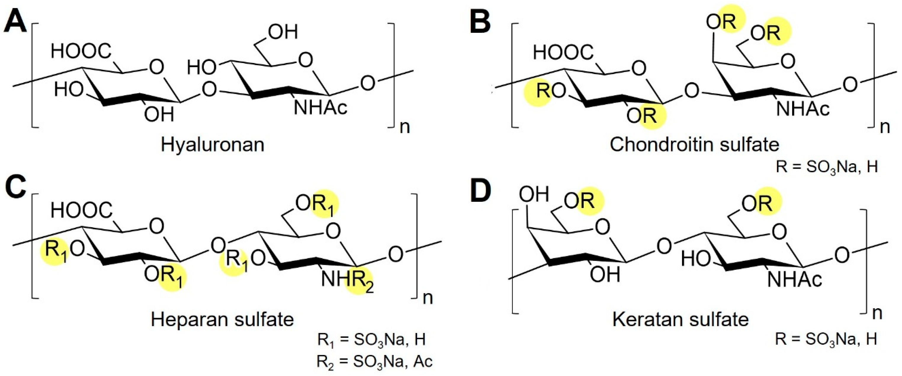
Figure 1. Representative disaccharide structures of major GAGs. (A) HA consists of β-D-glucuronic acid (GlcA) and β-D-N-acetylglucosamine (GlcNAc) linked by alternating β(1→3) and β(1→4) bonds. (B) CS contains β-D-glucuronic acid (GlcA) and β-D-N-acetylgalactosamine (GalNAc) units joined by β(1→3)/β(1→4) linkages, variably sulfated at C4 and/or C6 of GalNAc. (C) HS consists of alternating α-L-iduronic or β-D-glucuronic acid (IdoA/GlcA) and α-D-glucosamine (GlcN) residues connected through α(1→4)/β(1→4) linkages and extensively N- and O-sulfated. (D) Keratan sulfate is composed of β-D-galactose (Gal) and β-D-N-acetylglucosamine (GlcNAc) linked by β(1→3)/β(1→4) bonds and O-sulfated mainly at C6. Sulfated or potentially sulfated groups are highlighted in yellow. Structural differences in monosaccharide composition and sulfation pattern define the diversity of GAG function.
Biomolecules 15 01518 g001
Figure 2. Structures of selected GAG mimetics. (A) Sulfated hyaluronan (sHA), a chemically modified hyaluronan; (B) DS-500, a highly sulfated dextran; (C) G2.2, a synthetic small-molecule mimetic; and (D) {Z}12, a glycomimetic with defined sulfation patterns.
Biomolecules 15 01518 g002
Figure 3. Potential experimental and biomedical applications of GAG mimetics. The schematic highlights both the methodological approaches and biomedical fields in which GAG mimetics are applied. On the left, typical experimental assays are shown: interaction studies (e.g., binding kinetics), enzyme activity screening (e.g., antithrombin/heparinase assays), cell-based activity screening (cell culture models), bioassays (high-throughput activity profiling), and detection standards (analytical characterization). On the right, biomedical application areas are summarized: GAG mimetics are explored, for example, in cancer therapy, fibrosis, inflammation, cardiovascular diseases, diabetes, chronic wound healing, tissue engineering, and infectious diseases.
Biomolecules 15 01518 g003
Figure 4. Selected GAG mimetics in late-stage development and clinical translation. Sulfated or potentially sulfated regions are highlighted in yellow. (A) Pixatimod (PG545) is a sulfated tetrasaccharide conjugated to a cholestanyl aglycone that combines HS-like interactions with improved lipophilicity; evaluated in oncology and antiviral studies. (B) Rivipansel (GMI-1070) is a multivalent glycomimetic bearing carboxylate and sulfate groups that inhibit selectin-mediated adhesion; developed for vaso-occlusive crises in sickle cell disease. (C) PI-88 (Muparfostat) is a phosphosulfated oligosaccharide heparanase inhibitor derived from yeast mannan; evaluated in hepatocellular carcinoma treatment. (D) Pentosan polysulfate sodium (PPS) is a semisynthetic, highly sulfated heparin analog with anticoagulant and anti-inflammatory activity; approved for interstitial cystitis/bladder pain syndrome. (E) Roneparstat (SST0001) is a glycol-split heparin derivative that inhibits heparinase; investigated in clinical trials for the treatment of multiple myeloma. (F) Necuparanib (M402) is a HS mimetic that modulates tumor-associated signaling; evaluated in clinical studies for pancreatic cancer therapy. The ReGeneraTing Agents OTR4120 (G) and OTR4131 (H) are engineered polysaccharides with sulfate and carboxylate substitutions mimicking HS binding to growth factors and ECM components; studied in regenerative medicine and tissue repair.
Biomolecules 15 01518 g004
Table 1. Studies investigating biomedical applications of saccharide-based GAG mimetics.
Target ProteinGAG
Mimetic
Cells/ModelOutcome/LimitationRef.
Fibrosis
TGF-β1sHA
  • Human dermal fibroblasts
  • Collagen/sHA-based surface coatings
  • Reduced myofibroblast differentiation
  • sHA blocked TGF-β1 mediated α-smooth muscle actin, collagen, and fibronectin expression (in vitro)
[73]
-Sulfated
alginate
  • Sulfated alginate microbeads
  • Human-induced pluripotent stem cell-derived hepatocytes
  • C57BL/6 J mice
  • Reduced pericapsular fibrotic outgrowth on microbeads with sulfated alginate compared to non-sulfated microbeads (ex vivo)
[74]
Inflammation
CCL20, L-selectin2,4-O-di-
sulfated
iduronic
acid
(Di-S-IdoA)
  • Glycan micro array
  • Ovalbumin-induced asthma model in wild-type mice
  • Di-S-IdoA binds CCL20, inhibits the HS/CCL20 interaction, blocks binding of L-selectin to F2 endothelial cells
  • Reduced total number of leukocytes and decreased airway inflammation in ovalbumin-challenged wild-type mice (in vivo)
[75]
ERK1/2 signalingPG545
  • Mouse primary T cells
  • Model of Th1/Th17-dependent inflammation:
  • Methylated bovine serum albumin-induced delayed-type hypersensitivity (DTH) mice
  • Enhanced induction of anti-inflammatory regulator T-cells
  • Inhibition of ERK1/2 signaling
  • Blocks Th17 polarization in vitro and in vivo
[76]
Cancer
CCL5Carboxylated
dextran
sulfates
OTR4120,
OTR4131
  • Human hepatoma cells Huh7
  • Direct binding of GAG mimetics to CCL5
  • Inhibited CCL5-induced cell migration and invasion (in vitro)
[77]
Growth factors (not specified)Sulfated
alginate
(degree of
substitution
22.0, 2.7)
  • Human lung adenocarcinoma cells (H1792)
  • Mouse lung adenocarcinoma cells (MDA-F471)
  • Decreased migration of lung adenocarcinoma cells in 2D-scratch assay
  • Inhibited sphere formation
  • Decreased expression of CCL20
  • Suggested scavenging of growth factors required for proliferation (not specified, analyzed) (in vitro)
[78]
FGF-1, FGF-2, VEGF-A, heparinase, TLR9PG545
  • Human umbilical vein endothelial cells (HUVECs)
  • Rat aortic rings in Matrigel
  • Patients with metastatic colorectal/pancreatic cancer, solid tumors
  • SPR binding affinities for FGF-1, FGF-2, VEGF-A in the nM-range
  • Ki for heparinase: 6.1 ± 2.5 nM
  • Inhibition of HUVEC tube formation
  • Reduced microvessel outgrowths from aortic ring explants
  • Combination with pixatimod showed signs of clinical benefit for patients with colorectal cancer
  • In vitro, ex vivo, phase 1b clinical trial
[79,80]
Diabetes
Proinflammatory cytokines (IL-1β, TNF-α, IFN-γ)Low-
molecular-
weight
dextran
sulfate
  • β-cells from dispersed human and mouse islets
  • Prediabetic female NOD mice
  • Reduced proinflammatory cytokine-induced signaling and β-cell death
  • Increased heparan sulfate proteoglycan staining in islets
  • Daily treatment i.p. reduced insulitis and β-cell death in NOD mice
  • No long-term prevention of diabetes in NOD mice after four weeks of treatment (in vitro, in vivo)
[81]
Cardiovascular diseases
FGF-2, VEGF-ADextran
sulfate
OTR4131
  • Human endothelial progenitor cells from umbilical cord blood
  • Female NOD/SCID mice
  • Enhanced colony formation efficiency, adhesion, and migration of endothelial progenitor cells
  • Enhanced mitogenic effects of FGF-2 and VEGF-A under low serum conditions
  • No effects on tube formation on Matrigel (in vitro and vivo)
[82]
Infectious diseases
SARS-CoV-2PG545
  • SARS-CoV-2 spike protein receptor-binding domain (S1 RBD)
  • Clinical isolates of SARS-CoV-2
  • Monkey Vero E6 cells
  • Human bronchial epithelial cells A549
  • K18-hACE2 mouse model
  • Direct binding of PG545 to S1 RBD
  • Blocked binding of SARS-CoV-2 S1 RBD to A549 cells and binding to ACE2 protein
  • Reduced infection of A549 and Vero E6 cells with SARS-CoV-2 isolates (EC50 0.9–13.2 µg/mL)
  • Inhibited infection with SARS-CoV-2 in K18-hACE2 transgenic mice (in vitro, in vivo)
[42]
Bone regeneration
SclerostinHigh-
sulfated
HA (sHA3)
  • Non-diabetic + diabetic ZDF rats
  • 3 mm femoral defect
  • Lactide-based scaffolds coated with collagen/sHA3 or collagen/HA
  • Improved bone defect regeneration for scaffolds with collagen/sHA3 and collagen/HA in diabetic rats
  • No improvement for non-diabetic animals
  • Increased sclerostin binding to collagen/sHA3 coatings in vivo for diabetic rats
[83]
FGF-2Dextran
sulfate
OTR4131,
OTR4120
  • Rat mesenchymal stem cells (rMSCs)
  • Enhanced rMSC migration in Boyden chamber model
  • OTR4120 but not OTR4131 increased expression of osteogenic marker genes compared to control cells in basal medium (in vitro)
[84]
Chronic wounds
TIMP-3sHA3,
over-
sulfated
CS
  • SPR, enzyme kinetics, hydrogen/deuterium exchange mass spectrometry
  • Human bone marrow stromal cells
  • Higher binding capacity of sHA3 and over-sulfated CS for TIMP-3 compared to heparin
  • No interference of GAG-binding to TIMP-3 with MMP-1/-2 inhibition by TIMP-3
  • Reduced binding of TIMP-3/GAG complexes to endocytic receptor LRP-1
  • Accumulation of TIMP-3 in pericellular space of human bone marrow stromal cells after sHA treatment (in vitro)
[38,85]
GAG-binding growth factors (not specified)Cacipliq20
(RGTA)
  • 16 African American patients
  • 18 analyzed wounds due to diabetes, pressure, vascular or burn wounds
  • 22% full wound closure after four weeks of treatment
  • Reduced wound sizes by 15–18%
  • Decreased wound-related pain (60–70%)
  • Clinical trial (prospective pilot study, each patient served as own control)
[86]
Cartilage tissue engineering
TGF-β3, lysozymeFully
sulfated
sodium
cellulose
sulfate
  • Human mesenchymal stem cells (hMSCs)
  • Higher binding of TGF-β3 to gelatin scaffolds containing cellulose sulfate compared gelatin control scaffold
  • Increased gene expression of collagen type II and staining of sulfated GAGs after hMSC pellet culture with 0.01% soluble cellulose sulfate
  • Higher concentrations (>0.1%) of cellulose sulfate in gelatin gels decreased collagen type 2 production and reduced expression of chondrogenic marker genes (in vitro)
[87]
Neural tissue engineering
Nerve growth factor (NGF)Fully and
partially
sulfated
cellulose
sulfate
  • Dorsal root ganglion (DRG) neurons
  • Higher binding of NGF to gelatin-based scaffolds containing cellulose sulfate compared to those with CS
  • Enhanced neurite extension (in vitro)
[88]
Table 2. Studies investigating biomedical applications of non-saccharide-based GAG mimetics.
Target ProteinGAG MimeticCells/ModelOutcome/LimitationRef.
Inflammation
E-selectinGMI-1070
(rivipansel)
  • Male sickle cell mice
  • SCD patients with vaso-occlusive crises
  • IC50 for E-selectin binding in ELISA: 4.3 µM
  • Sustained blood flow rates and improved survival of SCD mice after surgical trauma
  • Phase II study: compound safe in acute vaso-occlusion, reduced time to resolution of vaso-occlusive crises, significantly decreased opioid use
  • Phase III clinical study: primary end point of readiness for discharge was not achieved; however, E-selectin was reduced 61% in the intervention group with rivipansel compared to baseline
[89,90,91]
Cancer
HeparinaseCyclo-
phellitol-
derived
heparinase
inhibitors
  • Cancer cell lines (U87 cells, B16 melanoma cells, 4T1 breast cancer cells)
  • Murine metastasis models (in vivo)
  • Reduced heparinase activity
  • Decreased cancer cell aggressiveness (proliferation/invasion) in vitro
  • Significantly reduced metastasis in mice
  • ECM-heparan sulfate preserved; less release of mitogens (in vitro, in vivo)
[92]
Infectious diseases
Herpes simplex virus (HSV)Benzene
sulfonate-
functionalized
mesoporous
silica
nanoparticles
  • HSV-1 and HSV-2
  • Benzene sulfonate-functionalized mesoporous silica nanoparticles loaded with acyclovir decreased infection in plaque reduction assay
  • Benzene sulfonate functionalization reduced acyclovir drug release (in vitro)
[93]
Bone regeneration
GAG-binding growth factors (especially BMP-2)Peptide
amphiphile
molecule
(Lauryl-
VVAGEGD
(Kp-
sulfo-
benzoate)S)
-Am)
  • Rat mesenchymal stem cells (rMSCs)
  • Rabbit tibial bone defect model
  • Increased expression of osteogenic marker genes and alkaline phosphatase activity after rMSC culture on GAG mimetic peptide nanofibers
  • Improved formation of cortical bone compared to treatment with physiological saline
  • In vitro, in vivo, no standard of care included in bone defect model, no binding data for BMP-2 shown
[94]
Disclaimer/Publisher’s Note: The statements, opinions and data contained in all publications are solely those of the individual author(s) and contributor(s) and not of MDPI and/or the editor(s). MDPI and/or the editor(s) disclaim responsibility for any injury to people or property resulting from any ideas, methods, instructions or products referred to in the content.