Article

The Role of BRCT Domain from LmjPES in Leishmania major Pathogenesis

by
Esther Larrea
1,
José Peña-Guerrero
2,
Celia Fernández-Rubio
2,
Aroia Burguete-Mikeo
2,
Elizabeth Guruceaga
3 and
Paul Nguewa
2,*
1
Unit of Translational Medicine, IdiSNA (Navarra Institute for Health Research), University of Navarra, 31009 Pamplona, Navarra, Spain
2
Department of Microbiology and Parasitology, IdiSNA (Navarra Institute for Health Research), University of Navarra, 31009 Pamplona, Navarra, Spain
3
Bioinformatics Platform, Center for Applied Medical Research, IdiSNA (Navarra Institute for Health Research), University of Navarra, 31009 Pamplona, Navarra, Spain
*
Author to whom correspondence should be addressed.
Biomolecules 2025, 15(8), 1191; https://doi.org/10.3390/biom15081191
Submission received: 10 July 2025 / Revised: 14 August 2025 / Accepted: 15 August 2025 / Published: 19 August 2025
(This article belongs to the Special Issue Cellular and Molecular Basis of Parasite Infection)

Abstract

Leishmaniasis is caused by protozoan parasites from the genus Leishmania and remains one of the major threats to global health, impacting millions of people worldwide as well as animals including dogs. Several treatments have been used for managing leishmaniasis; nevertheless, drug resistance has emerged as an important obstacle to disease control. Therefore, there is an urgent need to discover new therapeutic targets. The aim of this work was to study the role played by the breast cancer associated 1 C-terminal (BRCT) domain from LmjPES protein (Pescadillo ribosomal biogenesis factor) in Leishmania major‘s pathogenesis through the construction of novel genomic tools. For this purpose, Leishmania integrative plasmids that were able to express the BRCT domain from LmjPES and a hypothetical defective LmjPES lacking this BRCT domain were constructed. It was observed that the overexpression of the aforementioned BRCT domain in L. major dysregulated the mRNA expression of 152 genes (95 up-regulated and 57 down-regulated) in respect to control parasites. Furthermore, clustering studies of these altered genes revealed an enrichment in genes related to metabolic processes, transporter activity, response to stimuli, and protein folding, which are categories described to be associated with the metacyclogenesis process and parasite survival. Interestingly, these genes reached normal levels of expression in parasites transfected with a defective LmjPES (a mutated gene lacking the coding sequence of the BRCT domain). In addition, it was found that the footpad of mice inoculated with LmjPES BRCT-overexpressing parasites had significantly greater inflammation compared to the size of the footpad of animals infected with the control parasites. Based on all these results, it was suggested that the BRCT domain from LmjPES might play a role in L. major‘s infection process and pathogenesis.

1. Introduction

Protozoan parasites belonging to the genus Leishmania have been described as the causative agents of leishmaniasis [1]. At least 20 different Leishmania spp. are responsible for a variety of clinical manifestations ranging from self-healing skin ulcers (CL) to life-threatening visceral diseases (VL). Leishmaniasis is endemic in around 100 countries, with one billion people at risk. The World Health Organization (WHO) estimated 50,000–90,000 new cases of VL (but only 25–45% of these are registered) per year and 600,000–1 million new cases of CL annually (with around 200,000 reported to the WHO). These data represent cases of human disease. Currently, leishmaniasis is increasingly recognized as a One Health issue. Animals are also infected including dogs, the most important reservoirs of Leishmania infantum in Europe [2].
After the bite of infected female phlebotomine sand flies, neutrophils and monocytes migrate to the site of infection. Neutrophils further entrap the parasites [3,4]. Interestingly, during the early stages of the infection, no remarkable changes take place in the epidermis, although it is known that the local microenvironment plays a crucial role in further immune response development [5]. Afterwards, Leishmania infection progresses to a silent stage in a few weeks to months, and parasites proliferate without causing any apparent pathology. The silent phase ends with extensive inflammation, with the infiltration of eosinophils, macrophages, and neutrophils, and lesion formation at the inoculation site [6]. Interestingly, the highest burden of parasites is observed just prior to lesion development, suggesting that the effects of the immune response for parasite clearance are the main factors leading to ulcerations and tissue damage [6,7,8].
The life cycle of Leishmania parasites consists of two stages with different morphologies. The parasite has a non-motile intracellular amastigote form in the initial stage, which can be detected in mammalian hosts’ phagocytes and circulatory systems. In the second stage, it has a promastigote extracellular elongated and flagellated form, which can be seen in the gut of infected female sand flies [9]. The conversion from non-infective procyclic to infective metacyclic promastigotes is related to biochemical and molecular changes that are not currently well known [10].
The complexity of the Leishmania life cycle is partly responsible for the lack of an effective vaccine available against this disease. The control of the parasite relies on treatments that are unfortunately associated with toxicity, difficult to administer, have high costs, or demonstrate drug resistance [11,12]. Therefore, there is a need to investigate new therapeutic targets. One of the most important advancements in the field of leishmanicidal drug development took place decades ago, with the sequencing of the complete genome of L. major [13], which enabled researchers to search for potential drug targets.
Recently, the homologue of the human oncogene PES1 (pescadillo ribosomal biogenesis factor 1) in L. major (LmjPES) was identified. This was the first study to suggest that this gene is involved in leishmaniasis pathology [14]. Interestingly, the gene expression levels in L. major parasites dramatically increased in the metacyclic stage (more virulent, infectious, and disease-inducing forms of Leishmania) compared to the procyclic stage (with forms exhibiting a low virulence). Therefore, LmjPES-overexpressing parasites showed higher in vitro infections as well as increased and faster footpad inflammation in BALB/c mice compared to non-overexpressing parasites. Furthermore, in vivo infection with LmjPES-overexpressing parasites correlated with a significantly greater lesion size, supporting the role of LmjPES in Leishmania virulence [14]. In mammals, PES1 is known to participate in biological processes such as ribosomal biogenesis [15] and cell growth [16]. Additionally, PES1 is implicated in pathological processes like tumor development [17]. This protein encloses a breast cancer associated 1 (BRCA1) C-terminal (BRCT) domain that has been reported in proteins related to cell cycle regulation and DNA repair [18,19]. It has been described that the accuracy of this domain plays an important role in cell homeostasis and regulation [20,21]. Several investigations have shown that the BRCT domain from PES protein displays important roles in biological cellular processes [15,22]. In Leishmania, a BRCT domain in LmjPES protein (LmjPES BRCT) was identified [23]. It was observed that the expression of the BRCT domain from LmjPES in mammal cells induced both higher survival and replication rates [24]. Moreover, mammal cells expressing this LmjPES BRCT domain were more resistant to genotoxic drugs [24]. In this work, the goal was to further analyze the role of the LmjPES BRCT domain in Leishmania major‘s biology and pathogenesis.

2. Materials and Methods

2.1. Plasmid Construction

For this study, two expression vectors were performed as follows. One contained the LmjPES BRCT domain and the other one contained LmjPES ∆BRCT sequence.
The BRCT domain sequence from LmjPES gene [23,24] was inserted in the pLEXSY-Hyg expression vector (Jena Bioscience, Jena, Germany) using the In-fusion cloning kit (Clontech, Montain View, CA, USA). After extracting L. major‘s total genomic DNA following a stablished protocol [25], LmjPES BRCT sequence was amplified through PCR using primers designed with the In-Fusion cloning software version PR133833 (Takara, Shiga, Japan). The sense primer (BRCTF1, Table 1) included the sequence from the start of this domain, a start codon (ATG), a kozak sequence (ACACC), and the sequence of the end of Nco I (New England Biolabs, Ipswich, MA, USA) digested pLEXSY-Hyg vector. The antisense primer (BRCTF2, Table 1) included the sequence from the end of this BRCT domain and the sequence of the end of Kpn I (New England Biolabs) digested pLEXSY-Hyg vector. PCR product size was checked by gel electrophoresis and ligated into the previous Nco I and Kpn I digested pLEXSY-Hyg vector, obtaining the pLEXSY-LmjPES BRCT plasmid.
LmjPES ∆BRCT sequence was achieved with two sets of PCR reactions using the previously described and constructed pLEXSY-Hyg-lmjPES vector as template [13]. PES∆BRCTF1 and PES∆BRCTR2 primers (Table 1) were designed to cover the beginning of LmjPES coding sequence to the first codon of BRCT domain, whereas PES∆BRCTF3 and PES∆BRCTR4 primers (Table 1) covered the end of BRCT domain to the end of LmjPES coding sequence. Since PES∆BRCTF3 oligo was designed to be complementary with the end of PES∆BRCTR2 oligo, the reaction products were joined by a third PCR reaction, using PES∆BRCTF1 and PES∆BRCTR4 primers. This final PCR product was cut (Nco I and Kpn I) and then ligated into the previous Nco I and Kpn I digested pLEXSY-Hyg vector using T4 DNA ligase (Invitrogen, Vilnius, Lithuania), obtaining the pLEXSY-LmjPESBRCT plasmid.
The presence of the inserts in the vectors was checked by PCR, and the sequence was verified by DNA sequencing carried out at CIMA LAB Diagnostic (Pamplona, Spain).

2.2. Cell Culture Conditions

L. major (Lv39c5) promastigotes were grown with agitation at 26 °C in supplemented M199 medium. Prior to in vivo infectivity assays, promastigotes were maintained under agitation in Schneider’s medium (Gibco Laboratories, Grand Islands, NE, USA) supplemented with 10% (vol/vol) heat-inactivated fetal bovine serum (FBS; Gibco Laboratories, Grand Islands, NE, USA), 50 U/mL penicillin, and 50 µg/mL streptomycin (Sigma-Aldrich, St. Louis, MO, USA). In addition, the following cell lines were grown as mentioned below: L. majorLmjPES BRCT#54, L. majorLmjPES BRCT#55, L. majorLmjPES BRCT#56, L. majorLmjPES ∆BRCT#1, L. majorLmjPES ∆BRCT#2, L. majorLmjPES ∆BRCT#3, L. major–MC#4, L. major–MC#5, and L. major–MC#6. Then, metacyclic L. major promastigotes isolated by the peanut agglutinin method [26] were collected and used for in vivo experiments.

2.3. Generation of the Transgenic L. major Cell Lines

A total of 108 log-phase L. major parasites were transfected by electroporation with the Swa I (New England Biolabs, Ipswich, MA, USA) digested pLEXSY-LmjPES BRCT, pLEXSY-LmjPESBRCT, or pLEXSY-Hyg plasmids [27]. Transfected colonies were isolated from M199 plates supplemented with Agar Noble (Sigma-Aldrich, St. Louis, MO, USA) and 100 µg/mL of hygromicin B Gold (InvivoGen Europe, Toulouse, France), and later, they grew in complete Schneider’s medium plus hygromicin B Gold. We obtained several overexpressing cell lines. We selected and used the following three cell lines: L. majorLmjPES BRCT#54, L. majorLmjPES BRCT#55, and L. majorLmjPES BRCT#56 (parasites transfected with Swa I digested pLEXSY-LmjPES BRCT plasmid); three additional cell lines: L. majorLmjPES ∆BRCT#1, L. majorLmjPES ∆BRCT#2, and L. majorLmjPES ∆BRCT#3 (parasites transfected with Swa I digested pLEXSY-LmjPES ∆BRCT plasmid); and three control cell lines: L. major–MC#4, L. major–MC#5, and L. major–MC#6 (parasites transfected with Swa I digested pLEXSY-Hyg plasmid).

2.4. RNA-Sequencing Analysis

Total RNA from parasites was extracted using the automated Maxwell system (Promega, Madrid, Spain). Then, RNA quality control was carried out to verify the required standards for RNA quantity and integrity. The samples were shipped on dry ice to Macrogen Company (Seoul, Republic of Korea), where they were sequenced. RNA-seq was performed using Illumina platform following a configuration of paired-end reads of 100 base pairs. RNA sequencing data analysis was performed using the following workflow: (1) the quality of the samples was verified using FastQC software version 0.11.8 (https://www.bioinformatics.babraham.ac.uk/projects/fastqc/) (accessed on 15 November 2022) and the trimming of the reads was performed using trimmomatic [28]; (2) alignment against the Leishmania major reference genome (ASM272v2.45) was performed using STAR [29]; (3) gene expression quantification using read counts of exonic gene regions was carried out with featureCounts [30]; (4) the gene annotation reference was Ensembl Bacteria v55 [31]; and (5) differential expression statistical analysis was performed using R/Bioconductor [32]. Data are publicly available in GEO database with the accession number GSE234048.
First, gene expression data were normalized with edgeR [33] and voom [34]. After quality assessment and outlier detection using R/Bioconductor [32], a filtering process was performed. Genes with read counts lower than 4 in more than 50% of the samples for all the studied conditions were considered as not expressed in the experiment under study. LIMMA (Linear Models for Microarray Data) [34] was used to identify genes with significant differential expressions between experimental conditions. Genes were selected as differentially expressed using a B cut off of B > 0. Further functional and clustering analyses were performed and graphical representations were created using R/Bioconductor [32], clusterProfiler [35], and functional annotation databases [12,36].

2.5. Quantitative Real Time-PCR (qPCR)

Total RNA was extracted from a culture of 10 × 106 parasites in exponential growth phase using TRIZOL reagent (Sigma Aldrich, St. Louis, MO, USA) and subsequently quantified in a NanoDrop spectrophotometer (Thermo Scientific, Waltham, MA USA). Then, one µg of RNA was treated with Ambion DNA-free Kit (Invitrogen, Waltham, MA, USA) and retrotranscribed with SUPERscript II Reverse Transcriptase (Invitrogen, Waltham, MA, USA) at 42 °C for one hour. The obtained cDNA was used for the qPCR assays using a 7500 real-time PCR system (Applied Biosystems, Foster City, CA, USA), 96-well plates (Applied Biosystems, Foster City, CA, USA), and SYBR Green PCR master mix (Applied Biosystems, Foster City, CA, USA). The sequence of primers used in this study is shown in Table 2, and all qPCR tests were performed at 60 °C (primer annealing temperature). To monitor the specificity, the final PCR products were analyzed by melting curves and electrophoresis gel. The PCR efficiency was in the range of 95–105% for all genes analyzed. The amount of each transcript was expressed relative to the reference gene GAPDH as 2∆Cq, where ∆Cq represents the difference in quantification cycle between the control GAPDH and target genes. Then, those data were normalized to the control mRNA expression values.

2.6. In Vivo Infection and Evaluation

Female BALB/c mice were purchased from Harlan Interfauna Ibérica S.A. (Barcelona, Spain). All the procedures involving animals were approved by the Animal Care Ethics Commission of the University of Navarra (approval number: 086-20; 30 March 2021).
Eighteen BALB/c mice were divided into three groups (6 per group). Depending on the Leishmania cell lines used for the challenge, the groups of animals were named as follows: L. majorLmjPES BRCT (mice infected with L. majorLmjPES BRCT cell lines), L. major–MC (animals infected with L. major–MC control cell lines), or sterile PBS (uninfected mice). The subcutaneous inoculation was performed in the right hind footpad using an insulin syringe. The infection process was realized three times in successive weeks, using 500 metacyclic promastigotes (PNA[-]) dissolved in PBS [37]. The uninfected animals were injected with equal sterile PBS solutions. Footpad swelling was measured weekly with a digital caliper, and the “net swelling” was determined as the difference between the infected (right) and non-infected (left) footpad of each mouse. All animals were euthanized five weeks after the last inoculation.

2.7. Hematoxylin and Eosin Staining

Hematoxylin and eosin analyses were performed as previously described [37]. Briefly, footpads were formalin-fixed, decalcified in Osteosoft solution (Merck Millipore, Burlington, MA, USA; 1017281000) for 72 h, paraffin-embedded, and cut into 3 µm thick sections. Later, the sections were stained with hematoxylin and eosin.

2.8. Statistical Analysis

Statistical studies were performed using the GraphPad Prism software (version 5.0). Normality was assessed with Shapiro–Wilk W test. A two-sided unpaired Student’s t-test or Mann–Whitney U-test was used according to sample distribution. RNA-seq statistical tests were performed using R/Bioconductor as described above. Data are presented as means ± SD. p-values < 0.05 were considered statistically significant. The statistical significance was also indicated by * for p < 0.05; ** for p < 0.01; and “ns” for non-significant differences.

3. Results

3.1. Generation of L. major Parasites Exhibiting a Constitutive Overexpression of BRCT Domain from LmjPES (LmjPES BRCT) or an Expression of LmjPES Lacking BRCT (LmjPES ΔBRCT)

In order to understand the implications of the BRCT domain from LmjPES on L. major‘s biology, we decided to overexpress this domain (LmjPES BRCT). In addition, by using an integrative expression vector, the expression levels of a mutated gene (lacking the coding sequence of the BRCT domain) were also assessed. The corresponding constructed plasmids are represented in Figure 1A,B. After L. major‘s transfection with the SwaI-linearized DNA expression cassette containing the hygromycin resistance sequence and either the LmjPES BRCT or LmjPES ∆BRCT inserts, or without an insert, several cell lines were obtained. In this work, we present the results corresponding to those cell lines with the highest mRNA expression levels of the BRCT domain. As shown in Figure 1C, fold changes of 119.09 ± 9.06 (mean ± SD) for the LmjPES BRCT-overexpressing cell lines (L. majorLmjPES BRCT#54, #55, and #56 harboring pLEXSY-LmjPES BRCT) and 1.73 ± 0.52 for the LmjPES ∆BRCT-expressing cell lines (L. majorLmjPES ∆BRCT #1, #2, and #3 containing pLEXSY-LmjPES ∆BRCT) with respect to the control cell lines (L. major–MC#4, #5, and #6) were registered.

3.2. BRCT Domain from LmjPES May Regulate Genes Involved in Metabolic Pathways, Transporter Activity, and Protein Folding

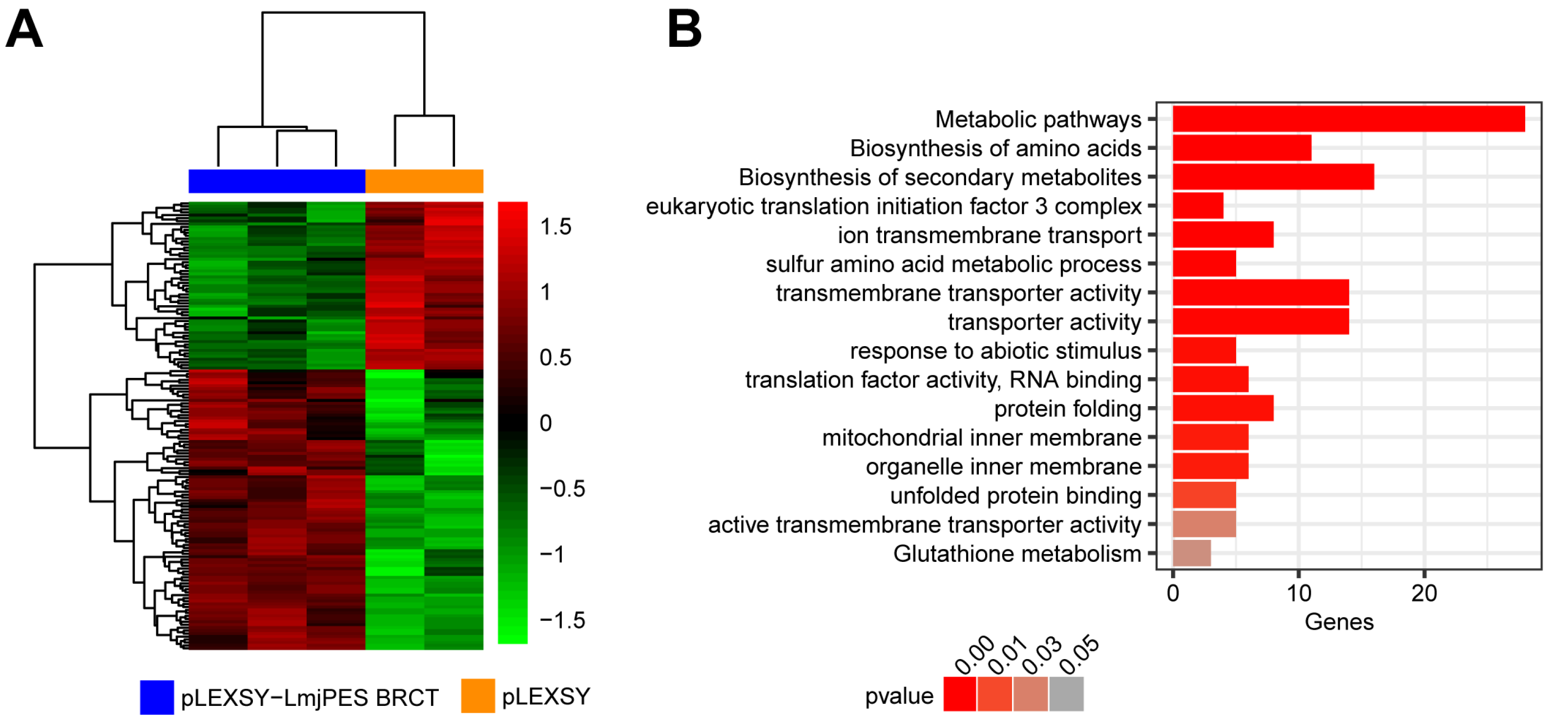
In a previous work, we described that the expression of the BRCT domain from LmjPES (the LmjPES BRCT domain) in HEK293T and NIH/3T3 cell lines altered the expression of proliferation-, survival- and chemoresistance-related genes in those mammalian cells [24]. The current study aimed to further characterize the biological implications of the LmjPES BRCT domain in the parasite. Therefore, high-throughput RNA sequencing of L. major overexpressing the LmjPES BRCT domain (L. majorLmjPES BRCT#54, #55, and #56 cell lines) versus the control parasites (L. major–MC#4 and #5 control cell lines) was performed. The data analysis, based on a B statistic cut-off >0, revealed 152 altered (95 up-regulated and 57 down-regulated) gene mRNA levels in LmjPES BRCT-overexpressing parasites with respect to the control parasites (Figure 2A and Supplementary Table S1). In addition, functional and clustering analyses of these dysregulated genes were performed using the Gene Ontology (GO) and KEEG (Kyoto Encyclopedia of Genes and Genomes) databases. We mainly found an enrichment in categories related to metabolic processes, transporter activity, response to stimuli, and protein folding (Figure 2B). The aforementioned categories had been described to be involved in the metacyclogenesis process [38] and parasite survival [39].
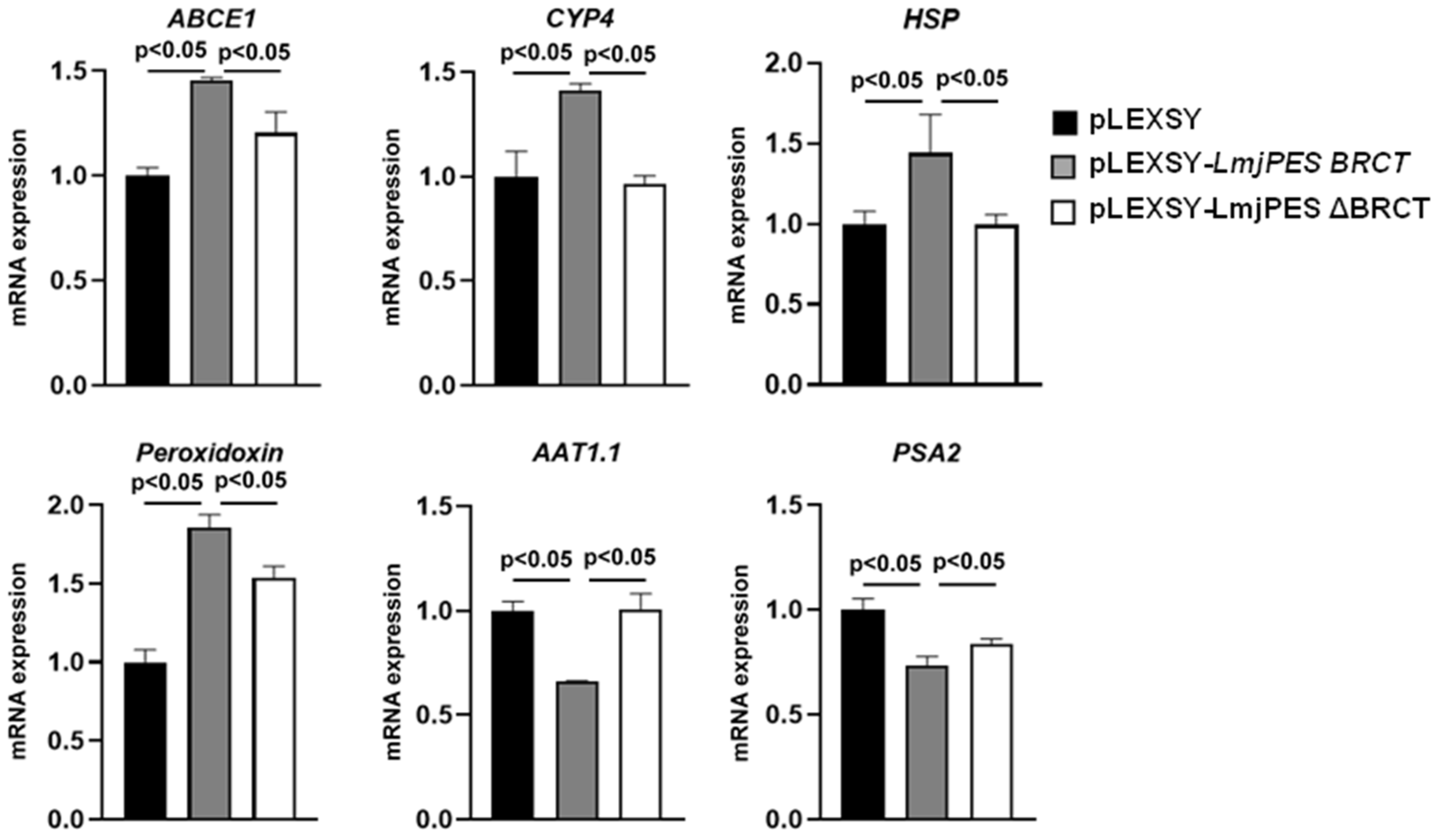
Afterwards, six genes belonging to the significantly enriched categories from Figure 2B (four up-regulated and two down-regulated in LmjPES BRCT-overexpressing parasites) were selected to further analyze the role played by this BRCT domain in parasite gene expression. Moreover, the mRNA levels of these genes were studied by real time-PCR in parasites with an overexpressed BRCT domain from LmjPES and in parasites expressing a mutated LmjPES lacking the coding sequence of BRCT (LmjPES ΔBRCT), compared to parasites transfected with the DNA expression cassette containing only the hygromycin resistance sequence. As observed in the RNAseq, the four up-regulated genes (LMJF_21_0710: ABCE1, LMJF_33_1630: CYP4, LMJF_33_2390: HSP, and LMJF_23_0040: peroxidoxin) and the two down-regulated genes (LMJF_31_0320: AAT1.1 and LMJF_12_0940: PSA2) were validated as expected (Figure 3). In addition, ABCE1, CYP4, HSP, and peroxidoxin mRNA expression levels decreased significantly in the parasites expressing a mutated LmjPES lacking BRCT, in comparison to LmjPES BRCT-overexpressing parasites (Figure 3). Similarly, the mRNA levels of AAT1.1 and PSA2 significantly increased in parasites expressing LmjPES ∆BRCT with respect to LmjPES BRCT-overexpressing parasites (Figure 3). Altogether, these data reinforced the role of the BRCT domain from LmjPES in the biology of the parasites.

3.3. BRCT Domain from LmjPES May Be Involved in L. major Pathogenesis

To better understand the implication of the BRCT domain from LmjPES on leishmaniasis outcomes, we subcutaneously inoculated mice with PBS or parasites harboring a DNA expression cassette containing the hygromycin resistance sequence and the LmjPES BRCT insert (or without such an insert for the control) by serial inoculations as described in Section 2. Materials and Methods. Animals were monitored weekly, and footpad swellings were measured with a digital caliper.
The first reports of swelling were detected three weeks after the last inoculation. The inflammation generated after the infection by the L. major–LmjPES BRCT cell line (exhibiting an overexpression of BRCT from LmjPES) was significantly greater than the inflammation induced by L. major—MC cells (control cell lines; parasites transfected with pLEXSY without the insert) after weeks 4 and 5. The weekly monitorization of in vivo infections is presented in Figure 4A,B.
In agreement with these data, hematoxylin–eosin staining also showed a higher infiltrate area in the footpad inoculated with LmjPES BRCT-overexpressing parasites compared to the footpad infected with control parasites (LmMC, L. major transfected with pLEXSY without the insert) (Figure 4C).

4. Discussion

The aim of this paper was to study the potential role of the BRCT domain from LmjPES in Leishmania‘s biology. Integrative expression vectors were then designed and constructed to overcome two limitations of episomal constructs. Firstly, transgenic cells may lose the plasmid once there is no antibiotic pressure, for example, during a medium- or long-term murine infection [40]. Secondly, the expression of the transgene can be highly heterogenous within the population of engineered parasites, due to copy number variations [40]. Therefore, the transfection of DNA cassettes into the genome parasite has been proposed as an alternative to overcome these limitations [41].
After obtaining Leishmania constitutively overexpressing the BRCT domain from LmjPES (L. majorLmjPES BRCT#54, #55, and #56 cell lines), RNAseq was performed to assess if this domain can alter the L. major gene expression levels. We found 152 genes differentially expressed. According to the GO and KEGG enrichment results, genes involved in metabolic processes were mostly affected, suggesting the role of the BRCT domain from LmjPES in those pathways. It has been described that metacyclogenesis mechanisms are associated with metabolic pathways to maintain and regulate changes in cellular morphology [38]. In addition, energy metabolism is required for the movement and infectivity of metacyclic promastigotes [38]. Through our results, we observed that peroxidoxin was one of the deregulated genes included in the metabolic pathway category. Peroxidoxin contributes in cell resistance to free radicals and has been described as a virulence factor [42,43]. Therefore, the up-regulation of this gene in our study may support the participation of the BRCT domain from LmjPES in the virulence of the parasite. On the other hand, RNAseq data showed a deregulation of genes clustered in protein folding, such as HSP and CYP4. Unfolded proteins can be generated under cellular stress conditions [44,45]. The increased levels of HSP and CYP4 found in the overexpressed LmjPES BRCT domain in Leishmania suggested that this domain might be involved in replication processes, elevated levels of reactive oxygen species (ROS), and cell stress-related situations.
In addition, the expression levels of genes encoding some transporters such as ABCE1 and AAT1.1 were altered. These proteins have key physiological functions and play essential roles in response to drugs [46,47,48], indicating a putative effect of the BRCT domain from LmjPES on Leishmania viability. Another category affected was the response to stimuli, including the PSA2 gene, a Leishmania antigen, which can induce Th1-mediated protection against murine leishmaniasis [49]. The down-regulation of PSA2 might lead to disease development.
Interestingly, the depletion of the BRCT domain from LmjPES (L. majorLmjPES ∆BRCT #1, #2, and #3 cell lines) recovered the expression of these genes to normal levels, corroborating the role of this domain in the pathophysiology of leishmaniasis. After observing the RNAseq results, we were prompted to evaluate the implications of the LmjPES BRCT domain in L. major in vivo infectivity and virulence. The inflammation caused by L. majorLmjPES BRCT-overexpressing cell lines became significantly larger than those of the control by weeks 4 and 5. The correlation between the in vivo lesion size and genetic overexpression have been extensively reported in L. major for genes implicated in both disease progression [50] and a reduction in virulence [37,51,52]. Interestingly, footpad swelling is thought to be linked with both local inflammation and parasite replication [37]. Furthermore, in mammalian PES, it has been described that point mutations within the BRCT domain of such a protein had a negative impact on rRNA processing and protein stability and prevented mammalian PES1 from being incorporated into the PeBoW complex [22]. Moreover, it is known that the addition of an extra PES allele lacking the whole BRCT domain can induce negative effects on cell fitness [15]. Thus, this underlines the importance of this domain for the adequate function of protein.

5. Conclusions

Taken together, and considering the previous data showing the role of LmjPES in L. major in vitro infectivity [14], our results highly support the link between the LmjPES BRCT domain and Leishmania infectivity and pathogenesis. This work highlighted the LmjPES BRCT domain as a virulence factor, playing a key role during the L. major infection process and pathogenesis, while significant inflammation in mice infected with parasites overexpressing this domain was observed. Finally, further experiments need to be conducted to analyze this domain in depth as a potentially relevant therapeutic target.

Supplementary Materials

The following supporting information can be downloaded at https://www.mdpi.com/article/10.3390/biom15081191/s1: Table S1: Differential gene expression analysis.

Author Contributions

Conceptualization, E.L. and P.N.; methodology, E.L., J.P.-G., C.F.-R., A.B.-M., E.G. and P.N.; software, E.L., J.P.-G., E.G. and P.N.; validation, E.L.; formal analysis, E.L., J.P.-G., C.F.-R., A.B.-M., E.G. and P.N.; investigation, E.L., J.P.-G., C.F.-R., A.B.-M., E.G. and P.N.; resources, P.N.; data curation, E.L., J.P.-G., C.F.-R., A.B.-M., E.G. and P.N.; writing—original draft preparation, E.L. and P.N.; writing—review and editing, E.L. and P.N.; visualization, E.L., E.G. and P.N.; supervision, P.N.; project administration, P.N.; funding acquisition, P.N. All authors have read and agreed to the published version of the manuscript.

Funding

The authors gratefully acknowledge the financial support provided by “Ministerio de Ciencia e Innovación” from Spain (MICIU/AEI/10.13039/501100011033 (grant number PID2023-147765OB-C21, -C22)) and Fundación Roviralta (Chair “Maria Francisca de Roviralta of Molecular Parasitology, Leishmaniasis and One Health”).

Institutional Review Board Statement

The animal study protocol was approved by the Animal Care Ethics Commission of the University of Navarra (approval number: 086-20; 30 March 2021).

Informed Consent Statement

Not applicable.

Data Availability Statement

RNA sequencing data are publicly available in the GEO database with the accession number GSE234048.

Acknowledgments

We would like to thank COST (European Cooperation in Science and Technology) actions CA21105 and CA21111.

Conflicts of Interest

The authors declare no conflicts of interest.

Abbreviations

The following abbreviations were used in this manuscript:
BRCTBreast cancer associated 1 C-terminal domain,
CLCutaneous Leishmaniasis
PES1Pescadillo ribosomal biogenesis factor 1
VLVisceral Leishmaniasis
WHOWorld Health Organization

References

  1. Leishmaniasis. Available online: https://www.who.int/news-room/fact-sheets/detail/leishmaniasis (accessed on 24 February 2025).
  2. Burguete-Mikeo, A.; Fernández-Rubio, C.; Peña-Guerrero, J.; El-Dirany, R.; Gainza, L.; Carasa Buj, B.; Nguewa, P.A. Characterization of Leishmania parasites isolated from naturally infected mammals. Animals 2023, 13, 2153. [Google Scholar] [CrossRef]
  3. Serafim, T.D.; Coutinho-Abreu, I.V.; Dey, R.; Kissinger, R.; Valenzuela, J.G.; Oliveira, F.; Kamhawi, S. Leishmaniasis: The act of transmission. Trends Parasitol. 2021, 76, 976–987. [Google Scholar] [CrossRef] [PubMed]
  4. Ranatunga, M.; Deacon, A.; Harbige, L.S.; Dyer, P.; Boateng, J.; Getti, G.T.M. Ex vivo analysis of the association of GFP-expressing L. aethiopica and L. mexicana with human peripheral blood-derived (PBD) leukocytes over 24 hours. Microorganisms 2024, 12, 1909. [Google Scholar] [CrossRef] [PubMed]
  5. dos Santos Meira, C.; Gedamu, L. Protective or detrimental? understanding the role of host immunity in leishmaniasis. Microorganisms 2019, 7, 695. [Google Scholar] [CrossRef]
  6. Belkaid, Y.; Mendez, S.; Lira, R.; Kadambi, N.; Milon, G.; Sacks, D. A natural model of Leishmania major infection reveals a prolonged “Silent” phase of parasite amplification in the skin before the onset of lesion formation and immunity. J. Immunol. 2000, 165, 969–977. [Google Scholar] [CrossRef]
  7. Kumar, R.; Bumb, R.A.; Salotra, P. Correlation of parasitic load with interleukin-4 response in patients with cutaneous leishmaniasis due to Leishmania tropica. FEMS Immunol. Med. Microbiol. 2009, 57, 239–246. [Google Scholar] [CrossRef]
  8. Hanau, S.; Maritati, M.; Contini, C.; Trentini, A.; Manfrinato, M.C.; Almugadam, S.H. Commentary on the issue of Leishmania infection: Focus on some pathogenetic, clinical, and epidemiological aspects. Vet. Sci. 2025, 12, 536. [Google Scholar] [CrossRef]
  9. Kumari, D.; Singh, K. Exploring the paradox of defense between host and Leishmania parasite. Int. Immunopharmacol. 2022, 102, 108400. [Google Scholar] [CrossRef]
  10. Karamysheva, Z.N.; Gutierrez Guarnizo, S.A.; Karamyshev, A.L. Regulation of translation in the protozoan parasite Leishmania. Int. J. Mol. Sci. 2020, 21, 2981. [Google Scholar] [CrossRef]
  11. Domagalska, M.A.; Barrett, M.P.; Dujardin, J.C. Drug resistance in Leishmania: Does it really matter? Trends Parasitol. 2023, 39, 251–259. [Google Scholar] [CrossRef]
  12. Cosma, C.; Maia, C.; Khan, N.; Infantino, M.; Del Riccio, M. Leishmaniasis in humans and animals: A one health approach for surveillance, prevention and control in a changing world. Trop. Med. Infect. Dis. 2024, 9, 258. [Google Scholar] [CrossRef]
  13. Ivens, A.C.; Peacock, C.S.; Worthey, E.A.; Murphy, L.; Aggarwal, G.; Berriman, M.; Sisk, E.; Rajandream, M.A.; Adlem, E.; Aert, R.; et al. The genome of the kinetoplastid parasite, Leishmania major. Science 2005, 309, 436–442. [Google Scholar] [CrossRef] [PubMed]
  14. Algarabel, M.; Fernández-rubio, C.; Musilova, K.; Peña-guerrero, J.; Vacas, A.; Larrea, E.; Nguewa, P.A. In Leishmania major, the homolog of the oncogene Pes1 may play a critical role in parasite infectivity. Int. J. Mol. Sci. 2021, 22, 12592. [Google Scholar] [CrossRef]
  15. Yuan, S.; Xu, N.; Yang, J.; Yuan, B. Emerging role of PES1 in disease: A promising therapeutic target? Gene 2025, 932, 48896. [Google Scholar] [CrossRef] [PubMed]
  16. Fan, P.; Wang, B.; Meng, Z.; Zhao, J.; Jin, X. PES1 Is Transcriptionally regulated by BRD4 and promotes cell proliferation and glycolysis in hepatocellular carcinoma. Int. J. Biochem. Cell Biol. 2018, 104, 1–8. [Google Scholar] [CrossRef]
  17. Li, Y.Z.; Zhang, C.; Pei, J.P.; Zhang, W.C.; Zhang, C.D.; Dai, D.Q. The functional role of Pescadillo ribosomal biogenesis factor 1 in cancer. J. Cancer 2022, 13, 268–277. [Google Scholar] [CrossRef]
  18. Kinoshita, Y.; Jarell, A.D.; Flaman, J.M.; Foltz, G.; Schuster, J.; Sopher, B.L.; Irvin, D.K.; Kanning, K.; Kornblum, H.I.; Nelson, P.S.; et al. Pescadillo, a novel cell cycle regulatory protein abnormally expressed in malignant cells. J. Biol. Chem. 2001, 276, 6656–6665. [Google Scholar] [CrossRef]
  19. Dai, L.; Dai, Y.; Han, J.; Huang, Y.; Wang, L.; Huang, J.; Zhou, Z. Structural insight into BRCA1-BARD1 complex recruitment to damaged chromatin. Mol. Cell 2021, 81, 2765–2777. [Google Scholar] [CrossRef]
  20. Glover, J.N.M. Insights into the molecular basis of human hereditary breast cancer from studies of the BRCA1 BRCT domain. Fam. Cancer 2006, 5, 89–93. [Google Scholar] [CrossRef]
  21. Masi, A.; Antoccia, A. NBS1 heterozygosity and cancer Risk. Curr. Genom. 2008, 9, 275–281. [Google Scholar] [CrossRef]
  22. Hölzel, M.; Grimm, T.; Rohrmoser, M.; Malamoussi, A.; Harasim, T.; Gruber-Eber, A.; Kremmer, E.; Eick, D. The BRCT domain of mammalian Pes1 is crucial for nucleolar localization and rRNA processing. Nucleic Acids Res. 2007, 35, 789–800. [Google Scholar] [CrossRef]
  23. Peña-Guerrero, J.; Fernández-Rubio, C.; Burguete-Mikeo, A.; El-Dirany, R.; García-Sosa, A.T.; Nguewa, P. Discovery and validation of Lmj_04_BRCT domain, a novel therapeutic target: Identification of candidate drugs for leishmaniasis. Int. J. Mol. Sci. 2021, 22, 10493. [Google Scholar] [CrossRef]
  24. Larrea, E.; Fernández-Rubio, C.; Peña-Guerrero, J.; Guruceaga, E.; Nguewa, P.A. The BRCT domain from the homologue of the oncogene PES1 in Leishmania major (LmjPES) promotes malignancy and drug resistance in mammalian cells. Int. J. Mol. Sci. 2022, 23, 13203. [Google Scholar] [CrossRef]
  25. Medina-Acosta, E.; Cross, G.A.M. Rapid Isolation of DNA from Trypanosomatid protozoa using a simple “mini-Prep” procedure. Mol. Biochem. Parasitol. 1993, 59, 327–329. [Google Scholar] [CrossRef] [PubMed]
  26. Sacks, D.L.; Hieny, S.; Sher, A. Identification of cell surface carbohydrate and antigenic changes between noninfective and infective developmental stages of Leishmania major promastigotes. J. Immunol. 1985, 135, 564–569. [Google Scholar] [CrossRef] [PubMed]
  27. Robinson, K.A.; Beverley, S.M. Improvements in transfection efficiency and tests of RNA interference (RNAi) approaches in the protozoan parasite Leishmania. Mol. Biochem. Parasitol. 2003, 128, 217–228. [Google Scholar] [CrossRef] [PubMed]
  28. Bolger, A.M.; Lohse, M.; Usadel, B. Trimmomatic: A flexible trimmer for Illumina sequence data. Bioinformatics 2014, 30, 2114–2120. [Google Scholar] [CrossRef]
  29. Dobin, A.; Davis, C.A.; Schlesinger, F.; Drenkow, J.; Zaleski, C.; Jha, S.; Batut, P.; Chaisson, M.; Gingeras, T.R. STAR: Ultrafast universal RNA-Seq aligner. Bioinformatics 2013, 29, 15–21. [Google Scholar] [CrossRef]
  30. Liao, Y.; Smyth, G.K.; Shi, W. FeatureCounts: An efficient general purpose program for assigning sequence reads to genomic features. Bioinformatics 2014, 30, 923–930. [Google Scholar] [CrossRef]
  31. Yates, A.D.; Allen, J.; Amode, R.M.; Azov, A.G.; Barba, M.; Becerra, A.; Bhai, J.; Campbell, L.I.; Carbajo Martinez, M.; Chakiachvili, M.; et al. Ensembl Genomes 2022: An expanding genome resource for non-vertebrates. Nucleic Acids Res. 2022, 50, D996–D1003. [Google Scholar] [CrossRef]
  32. Heinemann, J. Cluster analysis of untargeted metabolomic experiments. Methods Mol. Biol. 2019, 1859, 275–285. [Google Scholar] [CrossRef]
  33. Robinson, M.D.; McCarthy, D.J.; Smyth, G.K. EdgeR: A bioconductor package for differential expression analysis of digital gene expression data. Bioinformatics 2010, 26, 139–140. [Google Scholar] [CrossRef] [PubMed]
  34. Ritchie, M.E.; Phipson, B.; Wu, D.; Hu, Y.; Law, C.W.; Shi, W.; Smyth, G.K. Limma powers differential expression analyses for RNA-sequencing and microarray studies. Nucleic Acids Res. 2015, 43, e47. [Google Scholar] [CrossRef] [PubMed]
  35. Yu, G.; Wang, L.-G.; Han, Y.; He, Q.-Y. ClusterProfiler: An R package for comparing biological themes among gene clusters. OMICS 2012, 16, 284–287. [Google Scholar] [CrossRef] [PubMed]
  36. Kanehisa, M.; Furumichi, M.; Sato, Y.; Kawashima, M.; Ishiguro-Watanabe, M. KEGG for taxonomy-based analysis of pathways and genomes. Nucleic Acids Res. 2022, 51, D587–D592. [Google Scholar] [CrossRef]
  37. Vacas, A.; Fernández-Rubio, C.; Larrea, E.; Peña-Guerrero, J.; Nguewa, P.A. LmjF.22.0810 from Leishmania major modulates the Th2-type immune response and is involved in leishmaniasis outcome. Biomedicines 2020, 8, 452. [Google Scholar] [CrossRef]
  38. Amiri-Dashatan, N.; Rezaei-Tavirani, M.; Zali, H.; Koushki, M.; Ahmadi, N. Quantitative proteomic analysis reveals differentially expressed proteins in Leishmania major metacyclogenesis. Microb. Pathog. 2020, 149, 104557. [Google Scholar] [CrossRef]
  39. Pramanik, P.K.; Alam, M.N.; Roy Chowdhury, D.; Chakraborti, T. Drug resistance in protozoan parasites: An incessant wrestle for survival. J. Glob. Antimicrob. Resist. 2019, 18, 1–11. [Google Scholar] [CrossRef]
  40. Bolhassani, A.; Taheri, T.; Taslimi, Y.; Zamanilui, S.; Zahedifard, F.; Seyed, N.; Torkashvand, F.; Vaziri, B.; Rafati, S. Fluorescent Leishmania species: Development of stable GFP expression and its application for in vitro and in vivo studies. Exp. Parasitol. 2011, 127, 637–645. [Google Scholar] [CrossRef]
  41. Mißlitz, A.; Mottram, J.C.; Overath, P.; Aebischer, T. Targeted integration into a rRNA locus results in uniform and high level expression of transgenes in Leishmania amastigotes. Mol. Biochem. Parasitol. 2000, 107, 251–261. [Google Scholar] [CrossRef]
  42. Gadelha, F.R.; Gonçalves, C.C.; Mattos, E.C.; Alves, M.J.M.; Piñeyro, M.D.; Robello, C.; Peloso, E.F. Release of the cytosolic tryparedoxin peroxidase into the incubation medium and a different profile of cytosolic and mitochondrial peroxiredoxin expression in H2O2-treated Trypanosoma cruzi tissue culture-derived trypomastigotes. Exp. Parasitol. 2013, 133, 287–293. [Google Scholar] [CrossRef]
  43. Da Fonseca Pires, S.; Fialho, L.C.; Silva, S.O.; Melo, M.N.; De Souza, C.C.; Tafuri, W.L.; Bruna Romero, O.; De Andrade, H.M. Identification of virulence factors in Leishmania infantum strains by a proteomic approach. J. Proteome Res. 2014, 13, 1860–1872. [Google Scholar] [CrossRef] [PubMed]
  44. Alcolea, P.J.; Alonso, A.; García-Tabares, F.; Larraga, J.; Martins, L.T.C.; Loayza, F.J.; Ruiz-García, S.; Larraga, V. An insight into differential protein abundance throughout Leishmania donovani promastigote growth and differentiation. Int. Microbiol. 2023, 26, 25–42. [Google Scholar] [CrossRef] [PubMed]
  45. Clos, J.; Grünebast, J.; Holm, M. Promastigote-to-amastigote conversion in Leishmania spp.-A molecular view. Pathogens 2022, 11, 1052. [Google Scholar] [CrossRef] [PubMed]
  46. Boozhmehrani, M.J.; Eslami, G.; Khamesipour, A.; Jafari, A.A.; Vakili, M.; Hosseini, S.S.; Askari, V. The role of ATP-binding cassette transporter genes expression in treatment failure cutaneous leishmaniasis. AMB Express 2022, 12, 78. [Google Scholar] [CrossRef]
  47. Roy, G.; Bhattacharya, A.; Leprohon, P.; Ouellette, M. Decreased glutamate transport in acivicin resistant Leishmania tarentolae. PLoS Negl. Trop. Dis. 2021, 15, e0010046. [Google Scholar] [CrossRef]
  48. Leprohon, P.; Légaré, D.; Girard, I.; Papadopoulou, B.; Ouellette, M. Modulation of Leishmania ABC protein gene expression through life stages and among drug-resistant parasites. Eukaryot. Cell 2006, 5, 1713–1725. [Google Scholar] [CrossRef]
  49. Kemp, M.; Handman, E.; Kemp, K.; Ismail, A.; Mustafa, M.D.; Kordofani, A.Y.; Bendtzen, K.; Kharazmi, A.; Theander, T.G. The Leishmania promastigote surface antigen-2 (PSA-2) is specifically recognised by Th1 cells in humans with naturally acquired immunity to L. major. FEMS Immunol. Med. Microbiol. 1998, 20, 209–218. [Google Scholar] [CrossRef]
  50. Reiling, L.; Chrobak, M.; Schmetz, C.; Clos, J. Overexpression of a single Leishmania major gene enhances parasite infectivity in vivo and in vitro. Mol. Microbiol. 2010, 76, 1175–1190. [Google Scholar] [CrossRef]
  51. Smirlis, D.; Bisti, S.N.; Xingi, E.; Konidou, G.; Thiakaki, M.; Soteriadou, K.P. Leishmania histone H1 overexpression delays parasite cell-cycle progression, parasite differentiation and reduces Leishmania infectivity in vivo. Mol. Microbiol. 2006, 60, 1457–1473. [Google Scholar] [CrossRef]
  52. Mckean, P.G.; Denny, P.W.; Knuepfer, E.; Keen, J.K.; Smith, D.F. Phenotypic changes associated with deletion and overexpression of a stage-regulated gene family in Leishmania. Cell. Microbiol. 2001, 3, 511–523. [Google Scholar] [CrossRef]
Figure 1. Constructed plasmids and BRCT mRNA expression levels. (A) pLEXSY-LmjPES BRCT construction. (B) pLEXSY-LmjPES ∆BRCT. 5′ ssu and 3′ ssu: Plasmid regions for genomic integration in the parasite chromosome. Utr1, 2, and 3: Untranslated regions for expression enhancement. Hyg: Hygromycin resistance marker gene for selection in Leishmania. Ori: Origin of replication. AmpR: Ampicillin resistance marker gene for selection in bacteria. Yellow: AmpR promoter, The dashed line indicates the number of base pairs mentioned within the plasmid sequence. (C) mRNA expression levels of LmjPES BRCT domain in several cell lines; L. major transfected with DNA expression cassette containing the hygromycin resistance sequence and either LmjPES BRCT insert (L. majorLmjPES BRCT#54, #55, and #56 cell lines) or LmjPES ∆BRCT insert (L. majorLmjPES ∆BRCT #1, #2, and #3 cell lines) or without insert (pLEXSY) (L. major–MC#4, #5, and #6 control cell lines). Data are represented as means of triplicates of each cell line (±SD). p-values < 0.05 were considered statistically significant.
Biomolecules 15 01191 g001
Figure 2. RNA sequencing analysis of LmjPES BRCT-overexpressing L. major. (A) Heatmap of 152 dysregulated genes in LmjPES BRCT-overexpressing (pLEXSY-LmjPES BRCT) L. major (L. majorLmjPES BRCT#54, #55, and #56 cell lines) with respect to control (pLEXSY) parasites (L. major–MC#4 and #5 control cell lines). (B) Bar plot of GO and KEGG categories enriched by altered genes observed in the RNAseq analysis.
Biomolecules 15 01191 g002
Figure 3. Gene expression quantification. mRNA levels of ABCE1, CYP4, HSP, peroxidoxin, AAT1.1, and PSA2 genes in control (pLEXSY) parasites (L. major–MC#4, #5, and #6 control cell lines) and in L. major parasites overexpressing BRCT domain from LmjPES (L. majorLmjPES BRCT#54, #55, and #56 cell lines) or expressing a defective LmjPES lacking BRCT (LmjPES ∆BRCT) (L. majorLmjPES ∆BRCT #1, #2, and #3 cell lines). Data are represented as means of triplicates of each cell line (±SD). p-values < 0.05 were considered statistically significant.
Biomolecules 15 01191 g003
Figure 4. Net footpad swelling and hematoxylin–eosin staining of BALB/c mice infected with LmjPES BRCT-overexpressing parasites or control parasites. (A) The net footpad swelling corresponded to the difference between the infected (right) and non-infected (left) footpad, measured weekly until 35 days after the last parasite inoculation. Data are represented as means (±SD) of different animals in each group. ** p < 0.01 with respect to L. major—control cell line (LmMC, transfected with pLEXSY without insert). * p < 0.05 with respect to uninfected animals (inoculated with PBS). (B) Representative pictures of mouse footpad inoculated (right) and not inoculated (left) with L. major—LmjPES BRCT (overexpressing LmjPES BRCT), L. major—control (LmMC, transfected with pLEXSY without insert), and uninfected animals (inoculated with PBS). (C) Hematoxylin–eosin staining in footpad sections from mice infected with L. major overexpressing LmjPES BRCT or L. major control (LmMC) or non-infected mice inoculated with PBS.
Biomolecules 15 01191 g004
Table 1. Oligos used for plasmids’ construction.
Oligo NameOligo Sequence (5′-3′)
BRCTF1ACCAGATCTGCCATGGACACCATGCGCGGGCTAACCTTCTTCATATCG
BRCTR2TGGTGATGGTGGTGGGTACCGCGGTAGCCCGTCACCGG
PES∆BRCTF1CCATGGGAAATGGTCCATAAGAAGCAGGCA
PES∆BRCTR2GCGGAACAGCTCGCGCA
PES∆BRCTF3TGCGCGAGCTGTTCCGCAACGCGCGGCTGGTG
PES∆BRCTR4GGTACCCTGCACCCACTTGGGCAGTTT
In bold: BRCTF1, sequence from the start of BRCT domain; BRCTR2, sequence from the end of BRCT domain; PES∆BRCTF1, the beginning of LmjPES coding sequence; PES∆BRCTR2, sequence until the first codon of BRCT domain; PES∆BRCTF3, the end sequence of BRCT domain; PES∆BRCTR4, the end of LmjPES coding sequence. Underlined: Endonuclease restriction site. Italics: Start codon plus Kozak sequence.
Table 2. Primers used for quantitative real time-PCR.
Target GeneSense Primer (5′-3′)Antisense Primer (5′-3′)Amplicon Length (pb)
AAT1.1GCAGGTGATTATGCCGTATGGCACAAAGGAGTAAATCGCC170
HSPCCTTTAAAGTGACGGAGTGCTCGACAGTGTTTACCTTGCC235
ABCE1TTCGTATCATCAACCTCCCCCCCAGGCTCATTCATGTATC209
CYP4TTCACTGAAAGTGTCCCTCCTTGAAGAGCTCCATCTCGAC162
PSA2GCACTCGATGACATCTTTGGTTAAGAGAGACGGAAGCCAG254
PeroxidoxinACATGAACGACTACAAGGGCGATTCTTCGATCAGCACACC279
GAPDHCATCAAGTGCGTGAAGGCGCCGTCGGCGAGTACTCGTGCTG216
LmjPES BRCTTCTTCATATCGCGTGAGGTGCATGCTTTTTCATCCCTGGC147
Disclaimer/Publisher’s Note: The statements, opinions and data contained in all publications are solely those of the individual author(s) and contributor(s) and not of MDPI and/or the editor(s). MDPI and/or the editor(s) disclaim responsibility for any injury to people or property resulting from any ideas, methods, instructions or products referred to in the content.