Article

Chemical Composition, In Vitro Antioxidant Activities, and Inhibitory Effects of the Acetylcholinesterase of Liparis nervosa (Thunb.) Lindl. Essential Oil

1
SDU-ANU Joint Science College, Shandong University, Weihai 264209, China
2
Marine College, Shandong University, Weihai 264209, China
*
Author to whom correspondence should be addressed.
Biomolecules 2023, 13(7), 1089; https://doi.org/10.3390/biom13071089
Submission received: 8 June 2023 / Revised: 23 June 2023 / Accepted: 26 June 2023 / Published: 7 July 2023
(This article belongs to the Section Natural and Bio-inspired Molecules)

Abstract

:
The present study aimed to investigate the essential oil composition of Liparis nervosa (Thunb.) Lindl., grown in China, and to determine its antioxidant and inhibitory effects on acetylcholinesterase. The essential oil was obtained by hydrodistillation, and the chemical compounds were analyzed by GC-MS and GC-FID. We used 2,2′-azino-bis(3-ethylbenzothiazoline-6-sulfonic acid) (ABTS), 2,2-diphenyl-1-picrylhydrazyl (DPPH), and ferric reducing assay power (FRAP) to evaluate the antioxidant activity. The anti-acetylcholinesterase activity of the essential oil was also examined. Sixty-seven compounds were identified, representing 98.50 % of the total essential oil, which was shown to be rich in methyl (9E,11E)-octadeca-9,11-dienoate (31.69%), n-hexadecanoic acid (15.08%), isopropyl palmitate (12.44%), propyl tetradecanoate (7.20%), tetradecanoic acid (4.01%), 17-octadecynoic acid (3.71%), and pentacosane (2.24%). Its antioxidant ability was analyzed via ABTS (IC50 = 721.95 ± 9.93 μg/mL), DPPH scavenging capacity (IC50 > 10,000 μg/mL), and the FRAP method (Trolox equivalent antioxidant concentration 39.64 ± 3.38 μM/g). Acetylcholinesterase inhibition effects were evaluated and had an IC50 value of 51.96 ± 14.26 μg/mL. The results show that this essential oil has interesting biological potential, encouraging further investigations, especially regarding the mechanisms of action of its antioxidant and anti-acetylcholinesterase activity. This is the first time that the chemical composition, antioxidant activity, and acetylcholinesterase inhibition effects of essential oil from L. nervosa have been studied.

1. Introduction

The genus Liparis of Orchidaceae is extensive. Liparis plants, of which there are approximately 320 species around the world, are generally perennial herbs, with most of these species scattering from the tropics and subtropics to temperate and alpine regions [1]. Liparis nervosa was selected since it is a widely cultivated commercial medicinal plant. In China, since ancient times, it has been commonly used for medicinal purposes, such as clearing heat fire, cooling the blood, removing toxicity, curing circulatory collapse, and relieving swelling and convulsion [2]. Most of the diseases mentioned above are not caused by a single mechanism but are instead the result of several biological processes. Therefore, traditional herbal medicine is gaining popularity as an approach to treatment.
There is a wealth of scientific evidence supporting the therapeutic effects of plant-derived molecules, and the exploration of natural product repositories has led to the development of some major conventional medicines, such as the anticancer drug Taxol [3]. Essential oils (EOs) are mixtures of volatile organic compounds containing monoterpenes, sesquiterpenes, alcohols, aldehydes, ketones, acids, phenols, ethers, and esters [4]. In addition, previous research has revealed that aromatherapy with various essential oils can alleviate mental health problems, such as sleep disturbances and anxiety, by affecting the inner nervous and circulatory systems [5]. However, there may be some volatile chemical emissions from essential oils, such as acetaldehyde, limonene, and methanol, which could have adverse health effects, such as allergies. Therefore, these oils should be utilized carefully in therapy and other applications [6,7]. Due to their chemical complexity and diversity, EOs have experienced renewed interest in many fields of study; they have interesting physicochemical characteristics and high added value with respect to the environment [8].
Alzheimer’s disease (AD) is a progressive, degenerative, neurological disorder resulting in impaired memory and behavior. Through inflammation, immune response, and mitochondrial damage, imbalances in the in vivo generation of reactive oxygen species (ROS), reactive nitrogen species (RNS), and antioxidative elimination can lead to oxidative stress, which may promote the accumulation of protein-fragment beta-amyloid (Aβ) plaques in the synaptic space. This accumulation of insoluble proteins is recognized as an indicator of neurotoxicity, resulting in neuronal death and, consequently, Alzheimer’s disease [9,10]. Moreover, previous reviews have shown improved cognitive behavior in animals suffering from AD and treated with antioxidants such as curcumin [11]. The plant-derived free-radical scavenger, ferulic acid, has also been demonstrated as a potential therapeutic agent for AD [12]. In addition, considering that acetylcholine is rapidly hydrolyzed and inactivated by cholinesterase, its inhibition couples with the elevation of the neurotransmitter acetylcholine in the synaptic cleft. Hence, cholinesterase is considered a potential therapeutic target with the interference of inhibitory molecules. Accordingly, antioxidant agents and cholinesterase inhibitors are crucial for AD therapeutics [13,14]. Previous research on essential oils’ biological activities also cast new light on their potential therapeutic prospects [15].
Although various studies focus on chemical-compound isolation—including lectin, nervonic acid derivatives, phenylpropanoids, pyrrolizidine alkaloids derivatives, and pyrrolizidine alkaloids, which have anti-fungal, anti-tumor, antioxidant, and α-glucosidase inhibitory properties [16,17,18,19,20], however the chemical composition, antioxidant activities, and acetylcholinesterase inhibitory effects of the EOs of this species remain unexplored. Thus, the present study was carried out to assess the phytochemical composition and biological activities of EO from L. nervosa.

2. Materials and Methods

2.1. Plant Material

Dry leaves and stems of L. nervosa were collected from Haikou Town, Chengjiang City, Kunming, Yunnan Province, China (24.52 N, 102.98 E) in November 2021. This plant was authenticated by Prof. Hong Zhao, Marine College, Shandong University. A voucher specimen was deposited at Marine College with the following registration number: EO2138. The plant material was placed in the shade and maintained under refrigeration (4 °C) until EO extraction.

2.2. EO Isolation

The dried plant (750 g) of L. nervosa was smashed into small pieces and subjected to hydrodistillation with ultrapure water (3.5 L) in a Clevenger-type apparatus for approximately 4 h until translucent distillate was obtained. After flushing the condenser with ether, when no droplet of residue was attached to the arm of the Clevenger-type apparatus, the essential oil was collected and then dried over anhydrous sodium sulfate and Termovap Sample Concentrator. The obtained EO joined the former into glass flasks and was stored at a low temperature (−4 °C) for further analysis.

2.3. GC-MS and GC-FID Analysis

GC-MS analysis was carried out using an Agilent 7890–5975C gas chromatograph–mass spectrometer equipped with a fused silica capillary column type HP-5MS (30 m × 0.25 mm with film thickness, 0.25 microns, Agilent Technologies, Santa Clara, CA, USA). The interface temperature was 280 °C, and the injector temperature was 260 °C. The oven temperature was initially 50 °C. This was maintained for 4 min, programmed from 50 °C to 280 °C at a rate of 6 °C/min, and held steady for 3 min. Helium was used as the carrier gas at a 1.1 mL/min velocity. The mass spectrometer conditions were as follows: electron impact (EI) mode (electron energy = 70 eV), a scan range of 25–500 amu, a scan rate of 4.0 scans/s, and a quadrupole temperature of 150 °C. A 1% w/v sample solution in n-hexane was prepared, and 0.3 μL was injected using splitless mode [21,22]. GC-FID analysis was performed using an Agilent 7890 gas chromatograph with a type HP-5 fused silica capillary column (30 m × 0.25 mm with film thickness of 0.25 microns, Agilent Technologies, USA). The injector temperature was 260 °C, and the detector temperature was 305 °C. The oven temperature was initially 50 °C, maintained for 4 min, and then programmed from 50 °C to 280 °C at a rate of 6 °C/min and held steady for 3 min. Helium was used as the carrier gas at a 1.1 mL/min velocity. Identifying those compounds in EO is primarily based on comparing mass spectrometric data and Kovat retention indices relating to retention time with commercial libraries (NIST 20 and Adams) [23]. Specifically, the Kovat retention indices were calculated using a series of n-alkanes (C8–C30) with linear interpolation.

2.4. Antioxidant Activities Determination

2.4.1. DPPH Method

The DPPH (2,2-diphenyl-1-picrylhydrazyl) ethanolic solution was prepared at a concentration of 0.17 mM. During DPPH free-radical scavenging activity evaluation, 96-well microplates were prepared. Following the addition of 200 µL of the stock DPPH solution, 50 µL of each dilution of the oil in ethanol (50, 25, 10, 5, 2.5, 1, and 0.5 mg mL−1) was added. For comparison, the positive solution was prepared with BHT (butylated hydroxytoluene) or Trolox but without the essential oil. The control was prepared with 200 µL of the stock DPPH solution and 50 µL of ethanol, and the sample blank was prepared with 50 µL of essential oil solution and 200µL of ethanol. After 30 min, readings were taken using a microplate reader (Epoch, Biotech company, Minneapolis, MN, USA) at a wavelength of 517 nm [24]. The DPPH free-radical scavenging capacity was calculated using the following equation:
R S A % = 1 A S a m p l e A S a m p l e   B l a n k A C o n t r o l × 100 %
where RSA% assesses the “radical scavenging activity” of the DPPH radical, ASample corresponds to the absorbance of the solution in the microplate with the sample at different concentrations, and ASample Blank is the absorbance of ethanol without DPPH. AControl is the DPPH solution without the EO sample. After measuring the RSA% at each concentration, IC50 can be calculated.

2.4.2. ABTS Method

The antioxidant of L. nervosa EO against an ABTS (2,2′-Azinobis-(3-ethylbenzothiazolin-6-sulfonic acid) diammonium salt) radical was investigated using the following method: the ABTS radical cation reagent was prepared by mixing 7 mM ABTS solution with 2.6 mM potassium persulfate, which was subsequently kept in the dark at room temperature for at least 12 h [25]. The ethanolic EO solution was prepared according to the following gradients: 25, 10, 5, 2.5, 1, and 0.5 mg mL−1. The absorbance of the solution was determined as 734 nm seven minutes into the reaction using a microplate reader (Epoch, Biotech company, USA). Ethanol was used as a blank and gradient-diluted solvent. BHT and Trolox served as positive controls. The scavenging capacity was calculated using the equation below:
I n h i b i t i o n % = A 0 A A 0 × 100 %
where A0 is the absorbance of 200 μL of diluted ABTS+ solution mixed with 50 μL of ethanol at 734 nm, while A is the absorbance of 200 μL diluted ABTS+ solution mixed with 50 μL of the sample solution at 734 nm. IC50 was then calculated [26].

2.4.3. Ferric-Reducing Antioxidant Power (FRAP) Method

FRAP was assayed according to a previous procedure with some modifications [27]. The stock solutions included (1) pH 3.6 acetate buffer solution, (2) 10 mmol/L TPTZ (2,4,6-tripyridyl-s-triazine) solution, and (3) 20 mmol L−1 Fe3+ solution. The stock solutions were mixed at the proportion of 10: 1: 1 and were diluted 50 times with ethanol. Next, 50 μL of different dilutions of EOs (4000, 2000, 1000, 500, 250, and 100 μg mL−1) and 0.25 mg/mL Trolox solution (2, 5, 10, 15, and 20 μL) were mixed with 200 μL FRAP working reagent in a 96-well microplate. The blank solution was similarly prepared by replacing the EOs with ethanol. All tests were performed in triplicate and the results were averaged. After 30 min of reaction time, the absorbance of the resulting solution was measured at 593 nm using a microplate reader (Epoch, Biotech company, USA) [28].

2.5. Anti-Acetylcholinesterase Activity Test

The acetylcholinesterase inhibitory effect was measured using the previously described method with minor modifications [29]. Then, 140 μL of 0.1 mM, pH 8.0 phosphate-buffered saline (PBS), 20 μL of sample, and 20 μL of acetylcholinesterase solution containing 0.28 U/mL were mixed in a microplate and left to incubate at 4 °C for 20 min. Subsequently, 10 μL of 15 mM AChI (acetylthiocholine iodide) and 10 μL of 2 mM DTNB (5,5′-Dithiobis-(2-nitrobenzoic acid)) were added and then incubated for 20 min. The absorbance at 412 nm was read. A blank reaction was carried out using a PBS solution instead of the EO sample. Next, a complete inhibition reaction was performed by substituting the EO with huperzine A (100 μg/mL), and the sample blank reaction substituted AchE solution with PBS. The acetylcholinesterase inhibition rate is calculated using the following formula:
Inhibition% = [(BC) − (SSB)]/(BC)
where B is the absorbance of the blank reaction, C is the absorbance of the complete inhibition reaction, S is the EO-containing reaction’s absorbance, and SB is the absorbance of the sample blank reaction. The tests were carried out in triplicate, and we calculated the IC50 value then. Huperzine-A was used in the control as a positive reference.

3. Results and Discussion

3.1. EO Yield and Component Analysis

The essential oils of the plant were extracted using a Clevenger-type apparatus. A total of 0.3 mL essential oil was obtained from 750 g L. nervosa biomass via hydrodistillation (0.04 v/w). In a previous study of Rutaceae plant species, Aegle mamelons, the essential oil yield was 0.53%, which is much higher than L. nervosa [30]. However, the EO yield is largely determined by the phylogenetic position of the plant. As for Orchidaceae plants, research on four orchid species showed evaluated yields of 0.03%, 0.02%, 0.52%, and 0.10%, which are akin to the results of the current study [31]. Moreover, a study of Cymbidium sinense found a yield of 0.09% [32]. The yields of other Orchidaceae plants, specifically Orchids sphegodes and Orchids purpurea, were 0.05% and 0.02%, respectively [33]. Collectively, the relatively low EO yield of L. nervosa is reasonable according to previous studies of the same plant family. The essential oils of L. nervosa are yellow, hydrophobic, and have a unique scent. The total ion chromatogram of L. nervosa essential oil is shown in Figure 1.
The identities, relative percentages, retention indices (RI), CAS ID, and identification method of each component are presented in Table 1. Sixty-seven compounds representing 98.50% of the EO were identified from GC-MS and GC-FID analysis. Gas chromatographic analysis of the essential oil revealed methyl (9E,11E)-octadeca-9,11-dienoate (31.69%), n-hexadecanoic acid (15.08%), isopropyl palmitate (12.44%), propyl tetradecanoate (7.20%), tetradecanoic acid (4.01%), 17-octadecynoic acid (3.71%), and pentacosane (2.24%) to be the major constituents.
It is noteworthy that the EO composition is characterized by a primary amount of fatty acids and fatty acid esters (such as methyl (9E,11E)-octadeca-9,11-dienoate, n-hexadecanoic acid, and isopropyl palmitate). The abundance of those compounds indicated that the oil was derived from fatty acid and fatty acid ester chemotypes, which are thought to possess many biological activities, including larvicidal, ovicidal, and repellency activities, as well as a remarkable inhibitory effect in AChE, offering a research direction for further biological activity exploration [34,35]. However, the record of biological activities of (9E,11E)-octadeca-9,11-dienoate (the most abundant component in L. nervosa essential oil) is still lacking in the literature. The next most abundant compound was n-hexadecanoic acid, one of the most common fatty acids occurring in natural fats and oils and an active ingredient in the product ‘pneumotox’. This compound showed cytotoxicity to human leukemic cells but no cytotoxicity to normal human dermal fibroblast (HDF) cells [36]. Moreover, palmitic acid previously exhibited AChE inhibition and larval toxicity [37,38]. The results of other studies indicate that the third major component, isopropyl palmitate, is a model chemical penetration enhancer in drug release systems [39]. Taken together, these results indicate that essential oils from L. nervosa could be considered to possess a wide spectrum of biological activities. However, chemical components of plants’ EO are the result of the combined effects of many factors, including genetics, climate, edaphic, topography, elevation, and crosstalk, as well as the interactions among these factors [40,41].

3.2. Antioxidant Activities Evaluation

Different assays were introduced to measure the antioxidant capacity of our biological samples. According to a previous document clustering study, 2,2-diphenyl-1-picrylhydrazyl (DPPH)-based antioxidant capacity assays, together with ABTS-based and ferric-reducing antioxidant power (FRAP) are the most popular methods. All three are routinely practiced in research laboratories throughout the world. [42] This study evaluates the in vitro antioxidant activities of the EO from L. nervosa using DPPH, ABTS, and FRAP assays. The antioxidant values of the three methods are displayed in Table 2.
Recently, the spectrophotometric assays in the present study have been adopted to measure antioxidant capacity. The three assays in this study employ the same principle: a redox-active compound or synthetic-colored radical is generated, and the radical scavenging ability or redox-active compound reducing the activity of an EO is monitored using a spectrophotometer, with an appropriate standard applied to quantify antioxidant capacity [43].
The DPPH discoloration test is widely employed to evaluate natural products’ free-radical scavenging and antioxidant activities. The action between different free-radical scavengers with DPPH may be different. In plant essential oils, chemical compounds containing conjugated double bonds play a major role in scavenging free radicals [44]. Terpenoids can rapidly terminate DPPH free-radical chain reactions since they contain conjugated double bonds with strong chain-breaking antioxidant activity. However, the proportion of these compounds in L. nervosa essential oil is relatively small, so the result showed a relatively low DPPH scavenging activity with 44.2 ± 2.9% scavenging ability at 10 mg/mL concentration. Its potency seems weaker than those obtained from other species of essential oil [45,46,47].
It was also observed that the EO samples exhibit relatively high ABTS values (IC50 721.95 ± 9.93 μg/mL). L. nervosa essential oil increased in a sigmoidal dose-dependent manner over the concentration range tested in the ABTS assay, as shown in Figure 2. The present results of the ABTS test were better than those obtained from the DPPH test, in accordance with previous research based on the analysis of a large number of food samples. This demonstrates that the ABTS assay can more accurately estimate antioxidant capacity compared to the DPPH assay [43].
An FRAP method using reductants via a redox-linked colorimetric method that employs an easily reduced oxidant in stoichiometric excess could be a simple way of assessing the total antioxidant ability [48]. This assay is simple and quick to perform, and the reaction has a linear relationship with the molar concentration of the present antioxidants [48]. The reducing capacity is related to the degree of hydroxylation and the degree of conjugation of the bonds present in the phenolic compounds found in EOs [49]. The essential oil demonstrated moderate ferric-ion-reducing activity (Trolox equivalent antioxidant concentration).
Generally, the antioxidant activities of essential oils are highly dependent on their chemical composition and content. On the other hand, essential oils are complex mixtures and consist of many compounds; therefore, this complexity generally makes it difficult to characterize the activity pattern. Therefore, Prior et al. suggest using multiple assays to evaluate the overall antioxidant capacity and create an “antioxidant profile” that covers reactivity towards aqueous and lipid/organic radicals through various mechanisms. Using different assays with different mechanisms is recommended for complex samples to fully understand antioxidant action [50].

3.3. Acetylcholinesterase Inhibitory Effects

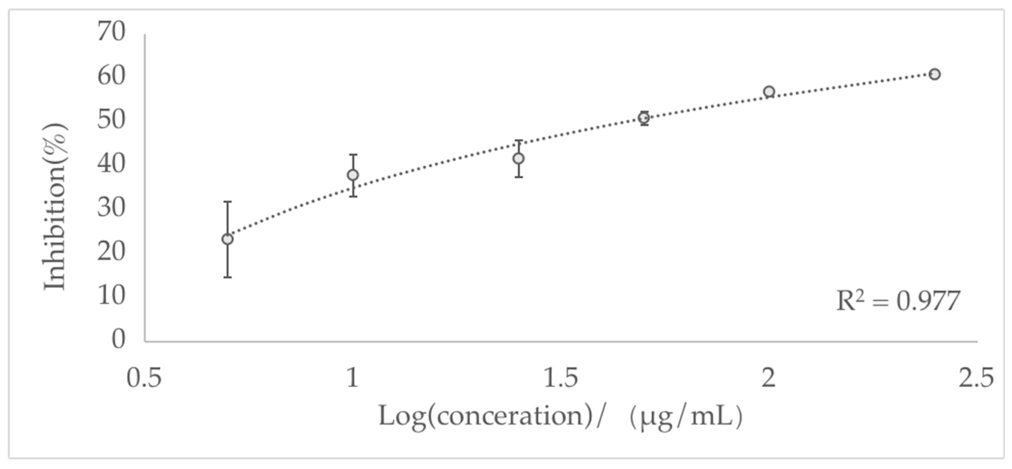
AChE (EC 3.1.1.7) consists of a complex protein of the α/β hydrolase fold type, generally with an ellipsoid shape containing a gorge about 20 Å deep [51]. As shown in Figure 3, the essential oil exhibited highly potent inhibition of the AChE enzyme. The resulting IC50 value was 51.96 ± 14.26 μg/mL, demonstrating effective inhibitory activity against AChE, which is significantly higher than that of essential oils distilled from other species (IC50 = 220, 175, 570, and 152 μg/mL, four Ocimum species) and the reversible inhibitor, physostigmine (IC50 270 μg/mL) [52]. A previous report found that the EO in the leaves and roots of Cymbopogon schoenanthus had an IC50 value of 260 μg/mL, which is also weaker than inhibitory L. nervosa EO [53]. However, a study on EO in Nepeta menthoides showed desirable anti-acetylcholinesterase activity (IC50 of 64.87 μg/mL), similar to the data obtained in this study [54].
Due to the desired enzyme inhibitory effect of L. nervosa essential oil, it was necessary to determine the active component responsible for the anti-acetylcholinesterase activity. The active site of this enzyme is composed of peripheral tryptophan and phenylalanine, tyrosine residues located at the entrance of the gorge. Enzyme amino acid residues may interact with compounds containing multiple aromatic rings due to the affinity of those molecules to residues localized at the entrance of the active site [55].
However, further molecular docking is necessary to reveal how compounds bind to key amino acids in the catalytic domain of AChE.

4. Conclusions

In conclusion, the present study found that the essential oil obtained from L. nervosa has significant anti-acetylcholinesterase activity and possesses moderate antioxidant activity, as evaluated using ABTS and FRAP assays. The results show that this essential oil could be considered a natural source for isolating active constituents for food preservatives and therapeutic applications of AD. However, these results cannot be used to determine which single component is responsible for most biological activity. Therefore, more research on structural chemistry and computational chemistry, as well as further in vivo investigations, is required.

Author Contributions

Conceptualization, J.Z., Z.X. and X.L.; methodology, J.Z., Z.X., P.G. and X.L.; software, J.Z., Z.X. and X.L.; validation, J.Z., Z.X. and X.L.; formal analysis, J.Z., Z.X. and X.L.; investigation, J.Z., Z.X. and X.L.; resources, X.L.; data curation, X.L. and Z.X.; writing—original draft preparation, Z.X.; writing—review and editing, J.Z. and X.L.; visualization, J.Z.; supervision, X.L.; project administration, X.L.; funding acquisition, X.L. All authors have read and agreed to the published version of the manuscript.

Funding

This research received no external funding.

Institutional Review Board Statement

Not applicable.

Informed Consent Statement

Not applicable.

Data Availability Statement

The data presented in this study are available from the corresponding author upon request.

Conflicts of Interest

The authors declare no conflict of interest.

References

  1. Li, L.; Chung, S.W.; Li, B.; Zeng, S.J.; Yan, H.F.; Li, S.J. New insight into the molecular phylogeny of the genus Liparis s.l. (Orchidaceae: Malaxideae) with a new generic segregate: Blepharoglossum. Plant Syst. Evol. 2020, 306, 1–10. [Google Scholar] [CrossRef]
  2. Liang, W.; Guo, X.; Nagle, D.G.; Zhang, W.D.; Tian, X.H. Genus Liparis: A review of its traditional uses in China, phytochemistry and pharmacology. J. Ethnopharmacol. 2019, 234, 154–171. [Google Scholar] [CrossRef] [PubMed]
  3. Thomford, N.E.; Senthebane, D.A.; Rowe, A.; Munro, D.; Seele, P.; Maroyi, A.; Dzobo, K. Natural Products for Drug Discovery in the 21st Century: Innovations for Novel Drug Discovery. Int. J. Mol. Sci. 2018, 19, 1578. [Google Scholar] [CrossRef] [PubMed] [Green Version]
  4. Bakkali, F.; Averbeck, S.; Averbeck, D.; Idaomar, M. Biological effects of essential oils—A review. Food Chem. Toxicol. 2008, 46, 446–475. [Google Scholar] [CrossRef]
  5. Zhong, Y.; Zheng, Q.; Hu, P.Y.; Huang, X.Y.; Yang, M.; Ren, G.L.; Du, Q.; Luo, J.; Zhang, K.N. Sedative and hypnotic effects of compound Anshen essential oil inhalation for insomnia. BMC Complement. Altern. Med. 2019, 19, 306. [Google Scholar] [CrossRef] [PubMed]
  6. Sarkic, A.; Stappen, I. Essential Oils and Their Single Compounds in Cosmetics—A Critical Review. Cosmetics 2018, 5, 11. [Google Scholar] [CrossRef] [Green Version]
  7. Nematollahi, N.; Weinberg, J.L.; Flattery, J.; Goodman, N.; Kolev, S.D.; Steinemann, A. Volatile chemical emissions from essential oils with therapeutic claims. Air Qual. Atmos. Health 2021, 14, 365–369. [Google Scholar] [CrossRef]
  8. El Asbahani, A.; Miladi, K.; Badri, W.; Sala, M.; Addi, E.H.A.; Casabianca, H.; El Mousadik, A.; Hartmann, D.; Jilale, A.; Renaud, F.N.R.; et al. Essential oils: From extraction to encapsulation. Int. J. Pharm. 2015, 483, 220–243. [Google Scholar] [CrossRef]
  9. Greeough, M.A.; Camakaris, J.; Bush, A.I. Metal dyshomeostasis and oxidative stress in Alzheimer’s disease. Neurochem. Int. 2012, 62, 540–555. [Google Scholar] [CrossRef]
  10. Dubey, S.; Singh, E. Antioxidants: An approach for restricting oxidative stress induced neurodegeneration in Alzheimer’s disease. Inflammopharmacology 2023, 31, 717–730. [Google Scholar] [CrossRef]
  11. Reddy, P.H.; Tripathi, R.; Troung, Q.; Tirumala, K.; Reddy, T.P.; Anekonda, V.; Shirendeb, U.P.; Calkins, M.J.; Reddy, A.P.; Mao, P.; et al. Abnormal mitochondrial dynamics and synaptic degeneration as early events in Alzheimer’s disease: Implications to mitochondria-targeted antioxidant therapeutics. Biochim. Biophys. Acta 2012, 1822, 639–649. [Google Scholar] [CrossRef] [PubMed] [Green Version]
  12. Sgarbossa, A.; Giacomazza, D.; Di Carlo, M. Ferulic acid: A hope for Alzheimer’s disease therapy from plants. Nutrients 2015, 7, 5764–5782. [Google Scholar] [CrossRef] [PubMed] [Green Version]
  13. Sharma, K. Cholinesterase inhibitors as Alzheimer’s therapeutics. Mol. Med. Rep. 2019, 20, 1479–1487. [Google Scholar] [CrossRef] [PubMed] [Green Version]
  14. Uriarte-Pueyo, I.; Calvo, M. Flavonoids as acetylcholinesterase inhibitors. Curr. Med. Chem. 2011, 18, 5289–5302. [Google Scholar] [CrossRef] [PubMed]
  15. Benny, A.; Thomas, J. Essential Oils as Treatment Strategy for Alzheimer’s Disease: Current and Future Perspectives. Planta Med. 2019, 85, 239–248. [Google Scholar] [PubMed] [Green Version]
  16. Jiang, N.; Wang, Y.; Zhou, J.; Zheng, R.; Yuan, X.; Wu, M.; Bao, J.; Wu, C. A novel mannose-binding lectin from Liparis nervosa with anti-fungal and anti-tumor activities. Acta Biochim. Biophys. Sin. 2020, 52, 1081–1092. [Google Scholar] [CrossRef]
  17. Huang, S.; Pan, M.F.; Zhou, X.L.; Zhou, Z.L.; Wang, C.J.; Shan, L.H.; Weng, J. Five new nervogenic acid derivatives from Liparis nervosa. Chin. Chem. Lett. 2013, 24, 734–736. [Google Scholar] [CrossRef]
  18. Liu, L.; Zou, M.; Yin, Q.; Zhang, Z.; Zhang, X. Phenylpropanoids from Liparis nervosa and their in vitro antioxidant andα-glucosidase inhibitory activities. Med. Chem. Res. 2021, 30, 1005–1010. [Google Scholar] [CrossRef]
  19. Huang, S.; Zhong, D.; Shan, L.; Zheng, Y.; Zhang, Z.; Bu, Y.; Ma, H.; Zhou, X. Three new pyrrolizidine alkaloids derivatives from Liparis nervosa. Chin. Chem. Lett. 2016, 27, 757–760. [Google Scholar] [CrossRef]
  20. Chen, L.; Li, J.; Huang, S.; Zhou, X.L. Two new pairs of epimeric pyrrolizidine alkaloids from Liparis nervosa. Chem. Nat. Compd. 2019, 55, 305–308. [Google Scholar] [CrossRef]
  21. Sarhadi, E.; Ebrahimi, S.N.; Hadjiakhoondi, A.; Manayi, A. Chemical Composition and Antioxidant Activity of Root Essential Oil of Different Salvia leriifolia Populations. J. Essent. Oil-Bear. Plants 2021, 24, 209–217. [Google Scholar] [CrossRef]
  22. Xu, Z.; Gao, P.; Liu, D.; Song, W.; Zhu, L.; Liu, X. Chemical Composition and In Vitro Antioxidant Activity of Sida rhombifolia L. Volatile Organic Compounds. Molecules 2022, 27, 7067. [Google Scholar] [CrossRef] [PubMed]
  23. Ghorbel, A.; Fakhfakh, J.; Brieudes, V.; Halabalaki, M.; Fontanay, S.; Duval, R.E.; Mliki, K.; Sayadi, S.; Allouche, N. Chemical composition, antibacterial activity using micro-broth dilution method and antioxidant activity of essential oil and water extract from aerial part of Tunisian Thymus algeriensis Boiss & Reut. J. Essent. Oil-Bear. Plants 2022, 24, 1349–1364. [Google Scholar]
  24. Andrade, M.A.; Das Graças Cardoso, M.; De Andrade, J.; Silva, L.F.; Teixeira, M.L.; Valério Resende, J.M.; Da Silva Figueiredo, A.C.; Barroso, J.G. Chemical Composition and Antioxidant Activity of Essential Oils from Cinnamodendron dinisii Schwacke and Siparuna guianensis Aublet. Antioxidants 2013, 2, 384–397. [Google Scholar] [CrossRef] [PubMed] [Green Version]
  25. Sripahco, T.; Khruengsai, S.; Charoensup, R.; Tovaranonte, J.; Pripdeevech, P. Chemical composition, antioxidant, and antimicrobial activity of Elsholtzia beddomei C. B. Clarke ex Hook. f. essential oil. Sci. Rep. 2022, 12, 2225. [Google Scholar] [CrossRef]
  26. Xu, Z.; Gao, P.; Ren, X.; Liu, X. Thermal Treatment (Hydrodistillation) on The Biomass of Ficus hispida L. f.: Volatile Organic Compounds Yield, Phytochemical Composition, and Antioxidant Activity Evaluation. Energies 2022, 15, 8092. [Google Scholar] [CrossRef]
  27. Szafranska, K.; Szewczyk, R.; Janas, K.M. Involvement of melatonin applied to Vigna radiata L. seeds in plant response to chilling stress. Cent. Eur. J. Biol. 2014, 9, 1117–1126. [Google Scholar] [CrossRef] [Green Version]
  28. Tongnuanchan, P.; Benjakul, S.; Prodpran, T. Properties and antioxidant activity of fish skin gelatin film incorporated with citrus essential oils. Food Chem. 2012, 134, 1571–1579. [Google Scholar] [CrossRef]
  29. Ingkaninan, K.; Temkitthawon, P.; Chuenchom, K.; Yuyaem, T.; Thongnoi, W. Screening for acetylcholinesterase inhibitory activity in plants used in Thai traditional rejuvenating and neurotonic remedies. J. Ethnopharmacol. 2003, 89, 261–264. [Google Scholar] [CrossRef]
  30. Aodah, A.H.; Balaha, M.F.; Jawaid, T.; Khan, M.M.; Ansari, M.J.; Alam, A. Aegle marvels (L.) Correa Leaf Essential Oil and Its Phytoconstituents as an Anticancer and Anti-Streptococcus mutans Agent. Antibiotics 2023, 12, 835. [Google Scholar] [CrossRef]
  31. Robustelli della Cuna, F.S.; Calevo, J.; Bari, E.; Giovannini, A.; Boselli, C.; Tava, A. Characterization and Antioxidant Activity of Essential Oil of Four Sympatric Orchid Species. Molecules 2019, 24, 3878. [Google Scholar] [CrossRef] [PubMed] [Green Version]
  32. Li, J.; Zhu, G.F.; Wang, Z.H. Chemical Var-iation in Essential Oil of Cymbidium sinense Flowers from Six Cultivars. J. Essent. Oil Bear. Plants 2017, 20, 385–394. [Google Scholar] [CrossRef]
  33. Robustelli della Cuna, F.S.; Cortis, P.; Esposito, F.; De Agostini, A.; Sottani, C.; Sanna, C. Chemical Composition of Essential Oil from Four Sympatric Orchids in NW-Italy. Plants 2022, 11, 826. [Google Scholar] [CrossRef] [PubMed]
  34. Hadjiakhoondi, A.; Vatandoost, H.; Khanavi, M.; Sadeghipour-Roodsari, H.R.; Vosoughi, M.; Motahareh Kazemi, M.; Abaib, M.R. Fatty acid composition and toxicity of Melia azedarach L. fruits against malaria vector Anopheles stephensi. Iran. J. Pharmceutical Sci. 2006, 9, 97–102. [Google Scholar]
  35. Khaldi, R.; Rehimi, N.; Kharoubi, R.; Soltani, N. Phytochemical composition of almond oil from Melia azedarach L. and its larvicidal, ovicidal, repellent and enzyme activities in Culex pipiens L. Trop. Biomed. 2022, 39, 531–538. [Google Scholar]
  36. Harada, H.; Yamashita, U.; Kurihara, H.; Fukushi, E.; Kawabata, J.; Kamei, Y. Antitumor activity of palmitic acid found as a selective cytotoxic substance in a marine red alga. Anticancer. Res. 2002, 22, 2587–2590. [Google Scholar]
  37. Perumalsamy, H.; Jang, M.J.; Kim, J.-R.; Kadarkarai, M.; Ahn, Y.-J. Larvicidal activity and possible mode of action of four flavonoids and two fatty acids identified in Millettia pinnata seed toward three mosquito species. Parasites Vectors 2015, 8, 237. [Google Scholar] [CrossRef] [Green Version]
  38. Muema, J.M.; Bargul, J.L.; Mutunga, J.M.; Obonyo, M.A.; Asudi, G.O.; Njeru, S.N. Neurotoxic Zanthoxylum chalybeum root constituents invoke mosquito larval growth retardation through ecdysteroidogenic CYP450s transcriptional perturbations. Pestic. Biochem. Physiol. 2021, 178, 104912. [Google Scholar] [CrossRef]
  39. Ruan, J.H.; Wan, X.C.; Quan, P.; Liu, C.; Fang, L. Investigation of Effect of Isopropyl Palmitate on Drug Release from Transdermal Patch and Molecular Dynamics Study. AAPS PharmSciTech 2019, 20, 174. [Google Scholar] [CrossRef]
  40. Pirbalouti, A.G.; Hashemi, M.; Ghahfarokhi, F.T. Essential Oil and Chemical Compositions of Wild and Cultivated Thymus Daenensis Celak and Thymus vulgaris L. Ind. Crops Prod. 2013, 48, 43–48. [Google Scholar] [CrossRef]
  41. Périno-Issartier, S.; Ginies, C.; Cravotto, G.; Chemat, F. A comparison of essential oils obtained from lavandin via different extraction processes: Ultrasound, microwave, turbohydrodistillation, steam and hydrodistillation. J. Chromatogr. A 2013, 1305, 41–47. [Google Scholar] [CrossRef] [PubMed]
  42. Ilyasov, I.R.; Beloborodov, V.L.; Selivanova, I.A.; Terekhov, R.P. ABTS/PP Decolorization Assay of Antioxidant Capacity Reaction Pathways. Int. J. Mol. Sci. 2020, 21, 1131. [Google Scholar] [CrossRef] [PubMed] [Green Version]
  43. Floegel, A.; Kim, D.-O.; Chung, S.-J.; Koo, S.I.; Chun, O.K. Comparison of ABTS/DPPH assays to measure antioxidant capacity in popular antioxidant-rich US foods. J. Food Comp. Anal. 2011, 24, 1043–1048. [Google Scholar] [CrossRef]
  44. Wojtunik, K.A.; Ciesla, L.M.; Waksmundzka-Hajnos, M. Model studies on the antioxidant activity of common terpenoid constituents of essential oils by means of the 2, 2-diphenyl-1-picrylhydrazyl method. J. Agric. Food Chem. 2014, 62, 9088–9094. [Google Scholar] [CrossRef] [PubMed]
  45. Jianu, C.; Rusu, L.-C.; Muntean, I.; Cocan, I.; Lukinich-Gruia, A.T.; Goleț, I.; Horhat, D.; Mioc, M.; Mioc, A.; Șoica, C.; et al. In Vitro and In Silico Evaluation of the Antimicrobial and Antioxidant Potential of Thymus pulegioides Essential Oil. Antioxidants 2022, 11, 2472. [Google Scholar] [CrossRef] [PubMed]
  46. dos Santos, L.G.A.; dos Reis, R.B.; Souza, A.S.D.; Canuto, K.M.; Castro, K.N.D.; Pereira, A.M.L.; Diniz, F.M. Essential oil composition, antioxidant and antibacterial activity against Vibrio parahaemolyticus from five Lamiaceae species. J. Essent. Oil Res. 2022, 34, 313–321. [Google Scholar] [CrossRef]
  47. Rashid, S.; Ahmad, M.; Amin, W.; Ahmad, B. Chemical composition, antimicrobial, cytotoxic and antioxidant activities of the essential oil of Artemisia indica Willd. Food Chem. 2013, 138, 693–700. [Google Scholar] [CrossRef]
  48. Benzie, I.F.F.; Strain, J.J. The Ferric Reducing Ability of Plasma (FRAP) as a Measure of “Antioxidant Power”: The FRAP Assay. Anal. Biochem. 1996, 239, 70–76. [Google Scholar] [CrossRef] [Green Version]
  49. Diniz do Nascimento, L.; Moraes, A.A.B.d.; Costa, K.S.d.; Pereira Galúcio, J.M.; Taube, P.S.; Costa, C.M.L.; Neves Cruz, J.; de Aguiar Andrade, E.H.; Faria, L.J.G.d. Bioactive Natural Compounds and Antioxidant Activity of Essential Oils from Spice Plants: New Findings and Potential Applications. Biomolecules 2020, 10, 988. [Google Scholar] [CrossRef]
  50. Apak, R.; Özyürek, M.; Güçlü, K.; Çapanoğlu, E. Antioxidant Activity/Capacity Measurement. 1. Classification, Physicochemical Principles, Mechanisms, and Electron Transfer (ET)-Based Assays. J. Agric. Food Chem. 2016, 64, 997–1027. [Google Scholar] [CrossRef] [Green Version]
  51. Houghton, P.J.; Ren, Y.; Howes, M.-J. Acetylcholinesterase inhibitors from plants and fungi. Nat. Prod. Rep. 2006, 23, 181–199. [Google Scholar] [CrossRef] [PubMed]
  52. Farag, M.A.; Ezzat, S.M.; Salama, M.M.; Tadros, M.G.; Serya, R.A. Anti-acetylcholinesterase activity of essential oils and their major constituents from four Ocimum species. Z. Für Nat. C 2016, 71, 393–402. [Google Scholar] [CrossRef] [PubMed]
  53. Khadri, A.; Serralheiro, M.; Nogueira, J.; Neffati, M.; Smiti, S.; Araújo, M. Antioxidant and antiacetylcholinesterase activities of essential oils from Cymbopogon schoenanthus L. Spreng. Determination of chemical composition by GC–mass spectrometry and 13C NMR. Food Chem. 2008, 109, 630–637. [Google Scholar] [CrossRef]
  54. Kahkeshani, N.; Razzaghirad, Y.; Ostad, S.N.; Hadjiakhoondi, A.; Shams Ardekani, M.R.; Hajimehdipoor, H.; Attar, H.; Samadi, M.; Jovel, E.; Khanavi, M. Cytotoxic, acetylcholinesterase inhibitor and antioxidant activity of Nepeta menthoides Boiss & Buhse essential oil. J. Essent. Oil-Bear. Plants 2014, 17, 544–552. [Google Scholar]
  55. Falé, P.L.; Borges, C.; Madeira, P.J.A.; Ascensão, L.; Araújo, M.E.M.; Florêncio, M.H.; Serralheiro, M.L.M. Rosmarinic acid, scutellarein 4′-methyl ether 7-O-glucuronide and (16S)-coleon E are the main compounds responsible for the antiacetylcholinesterase and antioxidant activity in herbal tea of Plectranthus barbatus (“falso boldo”). Food Chem. 2009, 114, 798–805. [Google Scholar] [CrossRef]
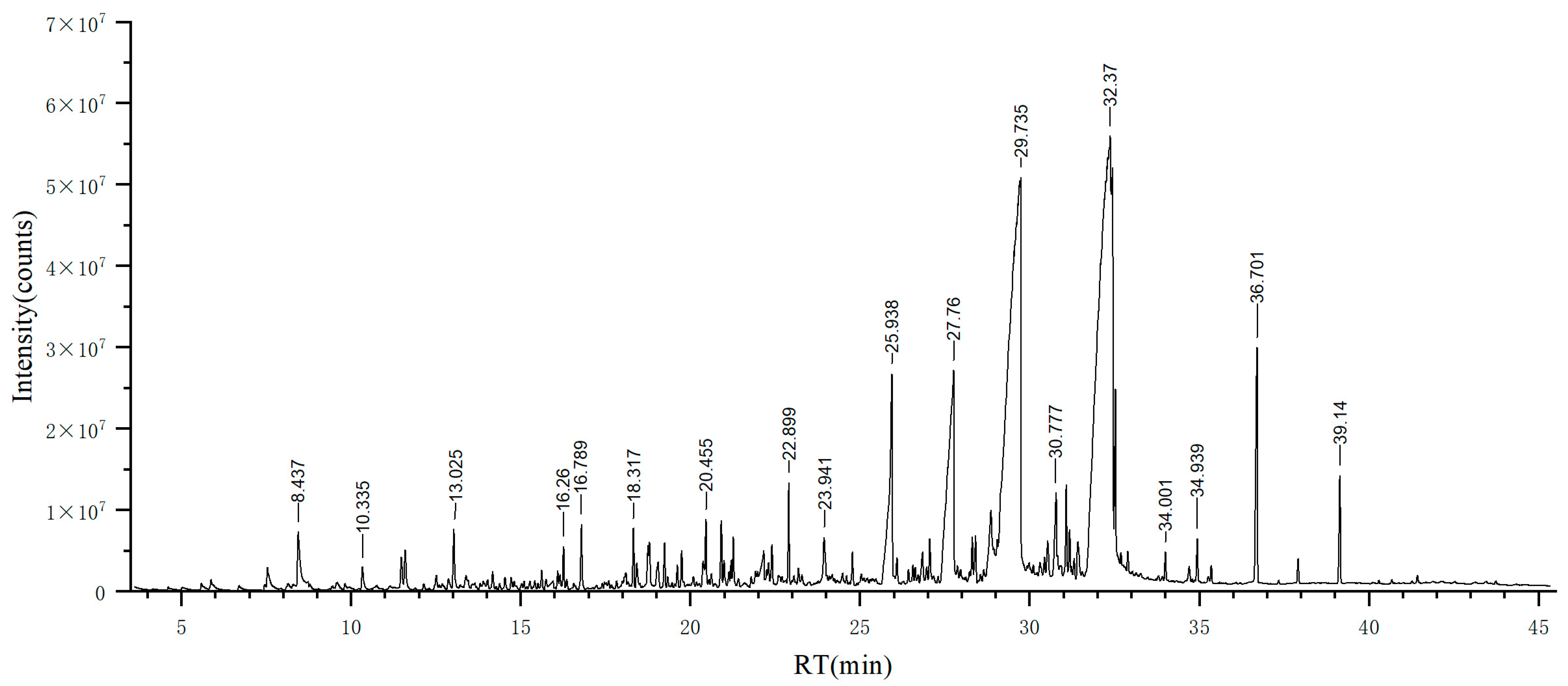
Figure 1. Total ion chromatogram of EO from L. nervosa derived from GC–MS.
Biomolecules 13 01089 g001
Figure 2. Concentration-dependent ABTS scavenging activity of L. nervosa essential oil.
Biomolecules 13 01089 g002
Figure 3. The concentration-dependent anti-acetylcholinesterase activity of L. nervosa essential oil.
Biomolecules 13 01089 g003
Table 1. Chemical composition of EOs distilled from L. nervosa.
No.Retention Time, tR (min)CompoundRI aRI bArea (%)Identification MethodCAS ID
15.867Heptanal9079010.22%RRI, MS111-71-7
27.531Benzaldehyde9689620.43%RRI, MS100-52-7
38.4372-Pentylfuran9959931.08%RRI, MS3777-69-3
49.5832-Octyn-1-ol1038-0.14%MS20739-58-6
510.335(E)-2-Octenal106510600.25%RRI, MS2548-87-0
611.481Linalool110310990.29%RRI, MS78-70-6
711.59Nonanal110811040.39%RRI, MS124-19-6
812.5073-Nonen-2-one112711420.17%RRI, MS14309-57-0
912.872Cucumber aldehyde115911550.16%RRI, MS557-48-2
1013.025(E)-2-Nonenal116511620.56%RRI, MS18829-56-6
1113.394-Ethylbenzaldehyde117811800.18%RRI, MS4748-78-1
1214.171Decanal120912060.17%RRI, MS112-31-2
1315.949Nonanoic acid128312730.17%RRI, MS112-05-0
1416.091Anethole129012860.14%RRI, MS104-46-1
1516.262-Undecanone129612940.31%RRI, MS112-12-9
1616.789(E,E)-2,4-Decadienal132013170.48%RRI, MS25152-84-5
1718.093n-Decanoic acid137913730.19%RRI, MS334-48-5
1818.317β-Damascenone138813860.39%RRI, MS23726-93-4
1918.419(+)-Sativen139313960.23%RRI, MS3650-28-0
2018.748β-Longipinene140814030.33%RRI, MS41432-70-6
2118.791Longifolene141114050.34%RRI, MS475-20-7
2219.048Dihydrodehydro-β-
ionone
142414240.32%RRI, MS20483-36-7
2319.239α-Ionone143314260.31%RRI, MS127-41-3
2419.615Acenaphthylene145114540.15%RRI, MS208-96-8
2519.74Dihydropseudoionone145714560.25%RRI, MS689-67-8
2620.373Curcumene148714830.27%RRI, MS644-30-4
2720.455β-Ionone149114910.45%RRI, MS14901-07-6
2820.908Tridecanal151315120.47%RRI, MS10486-19-8
2920.990Dibenzofuran151815140.15%RRI, MS132-64-9
3021.197δ-Cadinene152815240.13%RRI, MS483-76-1
3122.163Dodecanoic acid157715680.55%RRI, MS143-07-7
3222.304Fluorene158415830.15%RRI, MS86-73-7
3322.403(Z)-α-Bisabolene epoxide158915860.30%RRI, MS111536-37-9
3422.899Tetradecanal161516130.66%RRI, MS124-25-4
3523.183Oxacyclotetradeca-4,11-diyne163016390.14%RRI, MS6568-32-7
3623.941Tridecanoic acid167116660.63%RRI, MS638-53-9
3724.776Pentadecanal171717150.23%RRI, MS2765-11-9
3825.938Tetradecanoic acid178217684.01%RRI, MS544-63-8
3926.085Phenanthrene179017750.22%RRI, MS85-01-8
4026.56Hexadecanal181818170.13%RRI, MS629-80-1
4126.843Isopropyl myristate183518270.32%MS117-27-0
4227.051Hexahydrofarnesyl acetone184718440.35%RRI, MS502-69-2
4327.76Propyl tetradecanoate188918967.20%RRI, MS14303-70-9
4428.311Farnesyl acetone192319190.33%RRI, MS1117-52-8
4528.404Methyl palmitate192919260.34%RRI, MS112-39-0
4628.85611-Hexadecenoic acid195719531.66%RRI, MS2416-20-8
4729.042Dodecenyl succinic anhydride196919680.36%RRI, MS19780-11-1
4829.538n-Hexadecanoic acid-197015.08%MS57-10-3
4929.735Isopropyl palmitate2012202312.44%RRI, MS142-91-6
5030.531Fluoranthene206320540.37%RRI, MS206-44-0
5130.777Heptadecanoic acid207920710.87%RRI, MS506-12-7
5231.077Methyl linoleate209920920.67%RRI, MS112-63-0
5331.175Methyl linolenate210520980.33%RRI, MS301-00-8
5431.427Phytol211221140.39%RRI, MS150-86-7
5532.370methyl (9E,11E)-octadeca-9,11-dienoate2186218731.69%RRI, MS13038-47-6
5632.44117-Octadecynoic acid219021993.71%RRI, MS34450-18-5
5732.5282-Methyl-Z,Z-3,13-
octadecadienol
2196-1.62%MS519002-96-1
5832.686Isopropyl linolenate220722000.20%RRI, MS83918-59-6
5932.75210-trans,12-cis-Linoleic acid221322220.17%RRI, MS2420-56-6
6032.8932,4,5,7-Tetramethylphenanthrene2223-0.21%MS7396-38-5
6134.001Tricosane230023000.18%RRI, MS638-67-5
6234.699Diroleuton235223460.19%RRI, MS1783-84-2
6334.939Octadecanamide237023740.34%RRI, MS124-26-5
6435.348Tetracosane239924000.13%RRI, MS646-31-1
6536.701Pentacosane250325002.24%RRI, MS629-99-2
6637.907Hexacosane259926000.18%RRI, MS630-01-3
6739.140Heptacosane269927000.81%RRI, MS593-49-7
Concentration calculated from the total ion chromatogram. RI a: Calculated retention index. RI b: Retention index obtained from the mass spectral database. RRI: Relative retention indices calculated against n-alkanes. The identification method is based on the relative retention indices (RRI) of authentic compounds on the HP-5MS column. MS was identified based on computer matching of the mass spectra with the NIST/EPA/NIH 2020 Mass Spectral Database, Essential Oils GC/MS Library (Version 4, Robert Adams), and comparison with the literature data.
Table 2. Antioxidant activities of L. nervosa essential oil expressed as IC50 values for DPPH, ABTS, and the antioxidant ability of FRAP assays.
Tested SamplesDPPH 50% Effective
Concentration (μg/mL)
ABTS 50% Effective
Concentration (μg/mL)
FRAP Antioxidant
Capacity (μM/g)
L. nervosa EOs>10,000748.339.64 ± 3.38
BHT37.02 ± 2.2114.69 ± 1.32-
Trolox18.23 ± 1.129.47 ± 1.21-
Disclaimer/Publisher’s Note: The statements, opinions and data contained in all publications are solely those of the individual author(s) and contributor(s) and not of MDPI and/or the editor(s). MDPI and/or the editor(s) disclaim responsibility for any injury to people or property resulting from any ideas, methods, instructions or products referred to in the content.