Article

Altered TRPM7-Dependent Calcium Influx in Natural Killer Cells of Myalgic Encephalomyelitis/Chronic Fatigue Syndrome Patients

by
Stanley Du Preez
1,2,3,4,*,
Natalie Eaton-Fitch
1,2,
Peter K. Smith
4,5 and
Sonya Marshall-Gradisnik
1,2
1
National Centre for Neuroimmunology and Emerging Diseases, Menzies Health Institute, Griffith University, Gold Coast 4215, Australia
2
Consortium Health International for Myalgic Encephalomyelitis, Menzies Health Institute Queensland, Griffith University, Gold Coast 4215, Australia
3
School of Pharmacy and Medical Sciences, Griffith University, Gold Coast 4215, Australia
4
School of Medicine and Dentistry, Griffith University, Gold Coast 4215, Australia
5
Queensland Allergy Services, Gold Coast 4215, Australia
*
Author to whom correspondence should be addressed.
Biomolecules 2023, 13(7), 1039; https://doi.org/10.3390/biom13071039
Submission received: 11 April 2023 / Revised: 19 June 2023 / Accepted: 21 June 2023 / Published: 26 June 2023

Abstract

:
Myalgic encephalomyelitis/chronic fatigue syndrome (ME/CFS) is a disabling multisystemic condition. The pathomechanism of ME/CFS remains unestablished; however, impaired natural killer (NK) cell cytotoxicity is a consistent feature of this condition. Calcium (Ca2+) is crucial for NK cell effector functions. Growing research recognises Ca2+ signalling dysregulation in ME/CFS patients and implicates transient receptor potential ion channel dysfunction. TRPM7 (melastatin) was recently considered in the pathoaetiology of ME/CFS as it participates in several Ca2+-dependent processes that are central to NK cell cytotoxicity which may be compromised in ME/CFS. TRPM7-dependent Ca2+ influx was assessed in NK cells isolated from n = 9 ME/CFS patients and n = 9 age- and sex-matched healthy controls (HCs) using live cell fluorescent imaging techniques. Slope (p < 0.05) was significantly reduced in ME/CFS patients compared with HCs following TRPM7 activation. Half-time of maximal response (p < 0.05) and amplitude (p < 0.001) were significantly reduced in the HCs compared with the ME/CFS patients following TRPM7 desensitisation. Findings from this investigation suggest that TRPM7-dependent Ca2+ influx is reduced with agonism and increased with antagonism in ME/CFS patients relative to the age- and sex-matched HCs. The outcomes reported here potentially reflect TRPM3 dysfunction identified in this condition suggesting that ME/CFS is a TRP ion channelopathy.

1. Introduction

Myalgic encephalomyelitis/chronic fatigue syndrome (ME/CFS) is a debilitating multisystemic condition that demonstrates post-exertional neuroimmune exhaustion as its cardinal feature [1,2]. Symptoms reflecting immune, autonomic, cardiovascular, gastrointestinal, endocrine, metabolic, and cognitive dysfunction also appear in ME/CFS [3,4,5]. The clinical profiles and the illness severity vary, with approximately 25–29% of patients being severely affected [6]. However, no standardised diagnostic test or accepted pharmacotherapeutic for ME/CFS exists as the pathomechanism of this condition remains undefined [7]. Patient diagnosis therefore currently relies on various clinical case definitions, including the International Case Criteria (ICC, 2011) [1] and the Canadian Consensus Criteria (CCC, 2003) [8]. Research has identified natural killer (NK) cells as a reliable cellular model to investigate ME/CFS as NK cell dysfunction is a consistent feature of this condition [9].
NK cells are innate lymphoid cells that contribute to immunosurveillance and the elimination of target cells that are infected with intracellular pathogens or displaying malignant changes [10,11]. Recognition of target cells and subsequent elimination is determined by an overall activating signal communicated downstream of immunoreceptor tyrosine-based activation motif phosphorylation [12]. Once a target cell is identified, an immune synapse forms with the target. Cytoskeletal rearrangements within the NK cell direct the polarisation of cytotoxic granules toward the immune synapse [13]. Perforin and granzyme are released from secretory lysosomes and subsequently catalyse the death of the target cell [14]. The precise regulation of intracellular calcium (Ca2+) is essential to enable these events [15].
Ca2+ acts as a messenger molecule in the signalling cascades of numerous biological processes and is required for all cell types to function [16,17]. Transcription, proliferation, differentiation, metabolism, apoptosis, and effector functions demand Ca2+ at specific concentrations to occur correctly [18]. Hence, several mechanisms ensure that intracellular Ca2+ concentrations and homeostasis are well controlled. Previous investigations have identified Ca2+ dysregulation and impaired cytotoxic activity in the NK cells of ME/CFS patients [19,20,21,22,23,24,25]. Research has indicated that the dysfunction of members belonging to the transient receptor potential (TRP) superfamily of Ca2+-permeable ion channels may be responsible for these observations.
TRPs are polymodal sensors that detect environmental stimuli and mediate adaptive cellular responses through the influx of cations [26]. All tissues express TRPs, with each cell type bearing a unique signature according to its physiological role and environmental conditions [27]. Six subfamilies are described in mammals, with TRP melastatin (TRPM) members garnering particular interest in ME/CFS research. Using the NK cells of ME/CFS patients, numerous single nucleotide polymorphisms were identified across TRP ion channels including sub-members for the canonical (C), melastatin, and vanilloid channel families [28].
Subsequent flow cytometric, patch-clamp electrophysiology, and microscopy investigations revealed a loss of TRPM3 ion channel function, reduced surface expression, and impaired Ca2+ mobilisation in the NK cells of ME/CFS patients compared with healthy controls (HCs) [19,20,21,23,24,25]. Interestingly, the expression levels of a related protein, TRPM2, were elevated on the NK cell membranes of ME/CFS patients according to flow cytometric analyses [22]. Therefore, TRPM3 dysfunction and associated Ca2+ dysregulation may manifest as changes to other TRPM channels in the form of compensatory mechanisms.
The Ca2+- and magnesium (Mg2+)-permeable channel-kinase, TRPM7, was examined in a previous study to extend this hypothesis. Pharmacological inhibition of TRPM7 resulted in significantly higher co-localisation with cortical actin than pharmacological channel activation in ME/CFS patients [29]. This was not observed for HCs. Additionally, the co-localisation of TRPM7 with phosphatidylcholine-4,5-bisphosphate (PIP2) following antagonism treatment appeared to be greater in healthy participants than in those with ME/CFS [29]. These results suggest that TRPM7 may be altered in ME/CFS and thus implicate TRPM7 in the NK cell dysfunction and pathomechanism of this condition. However, it remains unclear from this initial investigation whether TRPM7 itself is functionally impaired, or if the results reflect known TRPM3 dysfunction and Ca2+ dysregulation in the NK cells of ME/CFS patients.
The present study was designed to evaluate TRPM7-dependent Ca2+ influx and elucidate whether the function of the ion channel component of this protein is altered in ME/CFS compared with HCs. This is the first investigation to examine the speed and maximum response of Ca2+ influx mediated via TRPM7 in the NK cells of ME/CFS patients and HCs. Fluorescence live-cell imaging techniques were used to measure these parameters. Outcomes generated from this study will inform future directions to characterise TRPM7 ion channel function and kinase activity. Furthermore, this study contributes to understanding the pathomechanism of ME/CFS and the identification of potential therapeutic targets to assist people affected by this condition.

2. Materials and Methods

2.1. Study Participants

ME/CFS patients and HCs were recruited through the National Centre for Neuroimmunology and Emerging Diseases (NCNED) patient database for this investigation. A comprehensive online questionnaire was administered to screen and define ME/CFS patients according to the ICC [1] and CCC [8] case definitions. ME/CFS patients that fulfilled the ICC or CCC case definitions and reported receiving a diagnosis of ME/CFS by a qualified physician were deemed eligible for this project. Nine ME/CFS patients from south-east Queensland and northern New South Wales were included in this study. Each ME/CFS patient was age- and sex-matched with a HC. The HC cohort was defined as non-fatigued individuals without any diagnosed illnesses. All participants were between 18 and 65 years of age, had a body mass index (BMI) between 18.5 and 30 kg/m2, and were non-smokers.
Exclusionary criteria were applied to all potential participants and included chronic illness (including autoimmune, cardiac, endocrine, metabolic, or primary psychiatric disorders), malignancy, alcohol misuse, pregnancy, breastfeeding, or administering pharmacological agents that are known to directly or indirectly affect TRPM7 channel-kinase activity, Ca2+ signalling, or immune cell activity. All participants provided written informed consent to partake in the present study. Ethics approval was obtained from the Gold Coast Human Research Ethics Committee (HREC/2019/QGC/56469) and the Griffith University Human Research Ethics Committee (GU/2019/1005).
Eligible participants donated 84 mL of whole venous blood, which were collected in ethylenediaminetetraacetic acid (EDTA) tubes between 7:00 a.m. and 11:00 a.m. in the south-east Queensland and Northern New South Wales regions of Australia. Each sample was de-identified by an independent researcher using a unique alpha-numeric code. A full blood count (FBC) was performed within 4 h of collection for each participant as a routine measure for exclusionary haematological abnormalities, including features of anaemia or white cell derangements.
All participants completed an online questionnaire that collected sociodemographic data, medical history, medications, and symptom history of the ME/CFS patients. ME/CFS symptom survey responses were grouped into 10 symptom categories including: (i) cognitive difficulties (slowed thought, impaired concentration, and memory consolidation issues); (ii) pain (headaches, muscle pain, and multi-joint pain); (iii) sleep disturbances (reversed sleep cycle, disturbed sleep cycle, and unrefreshing sleep); (iv) neurosensory disturbances (sensitivity to touch, vibration, taste, odour, sound; and poor coordination or balance); (v) immune disturbances (flu-like symptoms, sore throat, and tender lymph nodes); (vi) gastrointestinal disturbances (abdominal pain, nausea, and bloating); (vii) cardiovascular disturbances (orthostatic intolerances, light-headedness, and heart palpitations); (viii) respiratory disturbances (difficulty breathing, and air hunger); (ix) thermostatic instability (abnormal sweating episodes, hot flushes, and cold extremities); and (x) urinary disturbances (changed urination frequency and painful bladder). In addition, a 36-item short-form health survey (SF-36) and the World Health Organization (WHO) Disability Assessment Schedule (DAS) were used to determine the level of disability and quality of life (QoL).

2.2. Peripheral Blood Mononuclear Cell and Natural Killer Cell Isolation

Eighty mL of whole blood was used for peripheral blood mononuclear cells (PBMC) isolation via density gradient centrifugation using Ficoll (GE Healthcare, Uppsala, Sweden) as previously described [28]. PBMCs were stained with trypan blue (Invitrogen, Carlsbad, CA, USA) to determine cell count and cell viability. PBMCs were adjusted to a final concentration of 5 × 10⁷ cells/mL for NK cell isolation.
NK cells were isolated via immunomagnetic selection using the EasySep Negative Human NK cell Enrichment Kit (Stem Cell Technologies, Vancouver, BC, Canada). A fraction of the NK cell isolate was used to assess purity using flow cytometry. The NK cell population was defined by CD3CD56+ surface expression. NK cells (1 × 10⁵) were labelled with CD3 PE-Cy7 (5 µL/test) and CD56 APC (20 µL/test) monoclonal antibodies (Becton Dickinson [BD] Biosciences, San Jose, CA, USA) for 20 min at room temperature (RT). Cells were then acquired at 10,000 events using the Accuri C6 flow cytometer (BD Biosciences, San Diego, CA, USA). The average NK cell purity for ME/CFS patients was 93.4 ± 0.7 and for HCs was 94.3 ± 1.0 in this study.

2.3. Calcium Imaging

All suspensions were prepared in, and washed with 1.8 mM Ca2+ buffer, unless otherwise stated. The Ca2+ buffer was prepared in milliQ water and contained NaCl 140 mM, KCl 5.4 mM, CaCl2 1.8 mM, MgCl2 1.0 mM, and HEPES 10 mM (Thermofisher, Waltham, MA, USA). The pH was adjusted to 7.40 ± 0.05 using NaOH and the osmolality was adjusted to 300 ± 10 mOsm/L using D-glucose (Thermofisher, Waltham, MA, USA).
NK cells were adhered to 24-well µ-plates (Ibidi, Lochhamer Schlag, Germany) using Corning ® Cell-Tak TM Cell and Tissue Adhesive (BD Biosciences, San Jose, CA, USA) as previously described [24]. Next, NK cells were incubated using 1 µM Fluo-8-acetoxymethyl ester (AM) (Abcam, Cambridge, UK) and 0.02% Pluronic F127 (Thermofisher, Waltham, MA, USA) for 30 min at 37 °C. Cells were then washed and incubated for a further 20 min at RT to allow the de-esterification of Fuo-8-AM.
Fluo-8-loaded cells were imaged using the Nikon A1R microscope (40× objective) and fluorescence emissions were recorded using the iXon Life 888 Electron Multiplying CCD camera. The effects of naltriben (NTB) (Sigma-Aldrich, St. Louis, MO, USA), a TRPM7 ion channel agonist, and NS8593 (Sigma-Aldrich, St. Louis, MO, USA), TRPM7 channel-kinase antagonist, on TRPM7-dependent Ca2+ influx were examined using two separate protocols. The use of NTB and NS8593 is supported by previous research [30,31,32,33,34,35,36,37,38]. NTB modulates TRPM7 channel gating and promotes TRPM7-mediated Ca2+ entry with limited off-target effects [32]. Recently, the binding site of NTB was identified at the inter-subunit interface of TRPM7 tetramers formed by the fourth melastatin homology region of the TRPM7 protein [39]. The same investigation identified the binding site of NS8593 involving the N-terminal portion of S3, C-terminal portion of S4, S4-S5 linker, and the TRPM helix, which locks TRPM7 in its closed conformation [39]. Appropriate concentrations of NTB and NS8593 for this investigation were determined using dose response analysis (Supplementary Figures S1 and S2). All drugs were prepared as suspensions and administered via continuous perfusion methods. Acquisition was performed from the centre of individual wells adjacent to the perfusing line with the aspirating line positioned opposite. Recordings were obtained after flow-induced Ca2+ influx had stabilised. Firstly, baseline Ca2+ was obtained for 2 min before cells were stimulated with 40 µM NTB for 3 min, followed by 1 µM ionomycin for 4 min. In a separate well, fluorescence was recorded for 3 min in the presence of 6 µM NS8593 to desensitise TRPM7, followed by 6 µM NS8593 + 40 µM NTB for 3 min, then 40 µM NTB alone for 3 min, and finally 1 µM ionomycin for 4 min.
Fluorescence recordings were evaluated using NIS Research Elements (Nikon, NIS-Elements V5.2, Tokyo, Japan). Three measures of Ca2+ influx were determined. Amplitude represents the difference in the minimum and the maximum values of TRPM7-dependent Ca2+ influx corresponding to NTB stimulation. From this curve, the half-time of maximum response (T1/2) was also determined. The magnitude of TRPM7-dependent Ca2+ influx was calculated from the initial slope of the curve using OriginLabs. The initial slope is defined in this investigation as the first positive deflection within the first 15 s of the response curve that correlates with the application of NTB or ionomycin and reflects the rate of entry for Ca2+ influx [40]. All measurements were normalised against the ionomycin response curves to ascertain the proportion of the baseline, NTB, and NS8593 responses for individual cells.

2.4. Statistical Analysis

Statistical analysis was conducted using GraphPad Prism V8 (Graphpad Software Inc., Version 8, La Jolla, CA, USA) and OriginLabs (OriginLab Corporation, Northampton, MA, USA). Significance was set at p < 0.05. Normality was assessed using the Shapiro–Wilk test. Definitive outliers were identified using the robust regression and outlier removal method available in GraphPad Prism V8 and were defined as data points that were not represented within the distribution with a false discovery rate set at 0.1%. Cell recordings that returned an outlier value for a particular parameter had all its corresponding values excluded for slope, T1/2, and amplitude to minimise variability. Statistical differences were tested using the Mann–Whitney U non-parametric T test. All data are presented as the mean ± standard deviation (SD) unless otherwise stated.

3. Results

3.1. Participant Characteristics

A total of nine ME/CFS patients and nine age- and sex-matched HCs participated in this study between December 2021 and July 2022. All ME/CFS patients satisfied the CCC case definition and had no other conditions that would explain the symptoms. The participants’ demographic data are presented in Table 1. The average age for the ME/CFS cohort was 40.3 ± 4.5 years and it was 39.8 ± 4.0 years for the HC cohort. All participants were female. The average BMI of the ME/CFS cohort was 23.7 ± 1.1 kg/m2 and 22.0 ± 0.7 kg/m2 for the HC cohort.
QoL was assessed for all participants using the SF-36 and WHODAS inventories and presented in Table 1. SF-36 scores approaching 100 reflect good QoL, whereas scores towards 0 indicate poor QoL for the domain measured. Conversely, WHODAS scores that are closer to 0 suggest no limitations in functioning and values approaching 100 reflect severe functional impairments or disability for the corresponding domain. Mean WHODAS scores collected for the present investigation globally reflect greater impairments in the ME/CFS cohort compared with HCs and were statistically significant in all domains. Similarly, SF-36 inventory comparisons between groups demonstrate significantly reduced scores in the ME/CFS cohort compared with the HCs, excluding the wellbeing measure. The FBC parameters of both groups were within normal range and not significantly different.

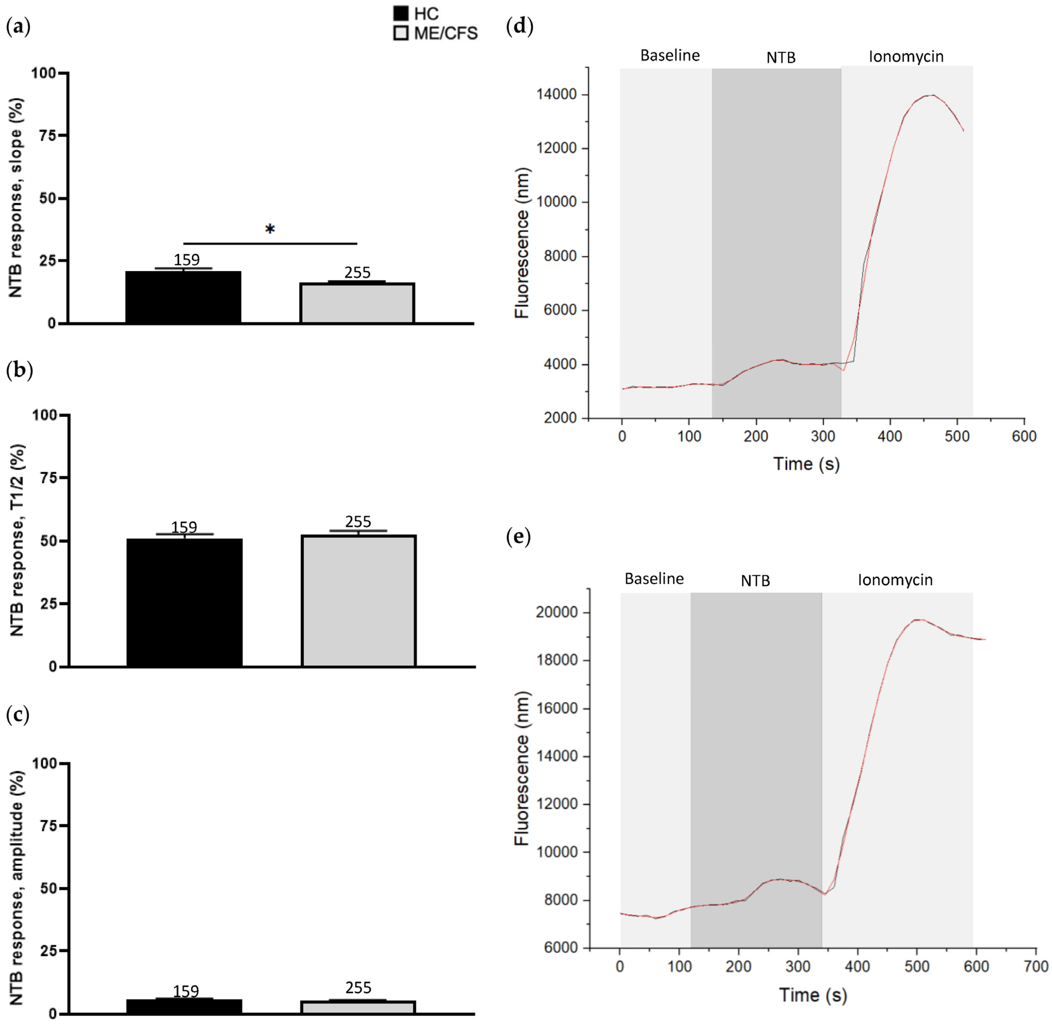
3.2. Effect of Naltriben Treatment on Calcium Influx

NTB (40 µM) was used to stimulate TRPM7-mediated Ca2+ influx of isolated human NK cells (Figure 1). Amplitude (HC 6.0 ± 0.3, ME/CFS 5.4 ± 0.2) and T1/2 (HC 51.1 ± 1.7, ME/CFS 52.7 ± 1.4) were non-significant between the two groups. Slope recordings were lower in ME/CFS (16.2 ± 0.7) patients compared with the HCs (20.9 ± 1.2) and this difference was statistically significant (p < 0.05) (Figure 1).

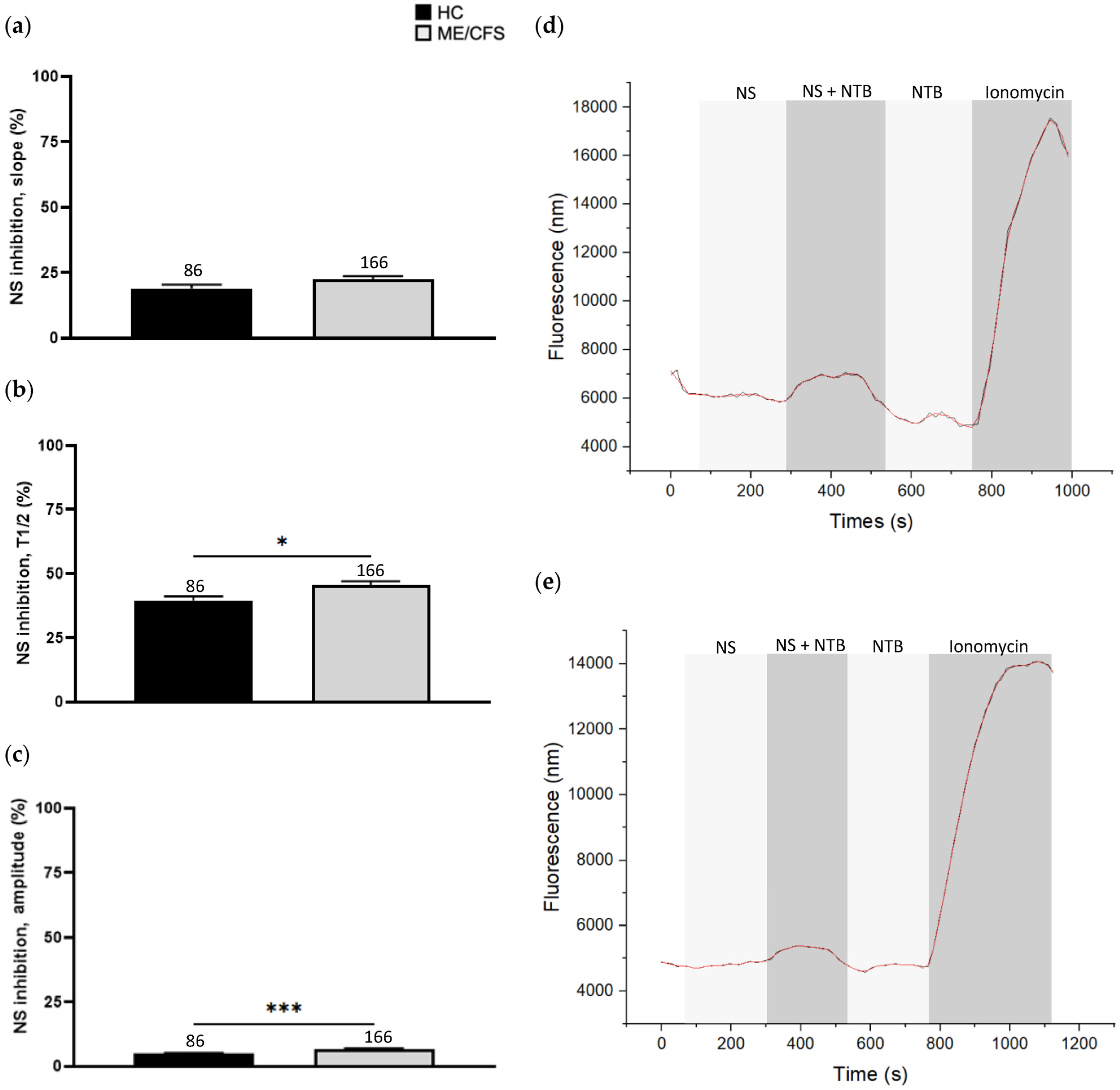
3.3. Effect of NS8593 Treatment on Calcium Influx

Inhibition of TRPM7 using NS8593 (6 µM) did not produce a significant difference in the slope of response between the ME/CFS (22.48 ± 1.22) and HC (19.05 ± 1.36) cohorts. Amplitude (4.97 ± 0.27, ME/CFS 6.68 ± 0.30) (p < 0.001) and T1/2 (HC 39.72 ± 1.61, ME/CFS 45.70 ± 1.52) (p < 0.05), however, were significantly different between the two groups and the HC cohort demonstrated a greater reduction in response relative to the recordings obtained from the ME/CFS patients (Figure 2).

4. Discussion

This pilot investigation is the first report on TRPM7-mediated Ca2+ influx in the NK cells of ME/CFS patients compared with HCs using a live cell Ca2+ imaging technique. Novel findings generated from this study include the demonstration of significantly reduced slope values in ME/CFS patients associated with the TRPM7 chemical agonist naltriben. Significantly impaired desensitisation of the channel-kinase from the T1/2 and amplitude values were also observed in the ME/CFS cohort.
The present investigation identified a significant reduction in slope values in the ME/CFS cohort following NTB stimulation. This parameter likely correlates with initial ion channel opening and stability [41], suggesting that the conformational change which opens and stabilises the TRPM7 ion channel pore may be delayed or impaired in ME/CFS. A previous investigation suggests that TRPC5 ion channel instability may impair Ca2+ influx and thus prevent the activation of Ca2+-dependent processes in neurons [42]. Sustained transmembrane Ca2+ influx is required to promote the translocation of secretory lysosomes, mitochondria, and the microtubule organising centre towards the immune synapse for NK cell degranulation to occur. Hence, TRPM7 instability may similarly constrain the sustained entry of Ca2+ through the ion channel domain, potentially impeding downstream events that are required for NK cell cytotoxic effector functions in ME/CFS patients. T1/2 and amplitude, however, were comparable between the two groups, indicating that peak Ca2+ influx mediated through TRPM7 and the time to achieve this is equivalent in ME/CFS patients and HCs.
By contrast, NS8593 treatment in the ME/CFS cohort indicated significantly reduced desensitisation of the TRPM7 channel-kinase according to the T1/2 and amplitude parameters relative to the HCs. Pooled slope values were statistically equivalent between the ME/CFS and HC cohorts. These findings indicate that the TRPM7 of ME/CFS patients is structurally or functionally resistant to desensitisation and may reflect a compensatory response to dysregulated cellular signalling or metabolic processes in ME/CFS.
As the TRPM7 stimulation and desensitisation protocols were structured differently, direct statistical comparison between the effects of NTB and NS8593 for each group could not be performed for this investigation. However, it is postulated that the findings of significantly greater slope values and significantly reduced T1/2 and amplitude values in the HC cohort compared with the ME/CFS patients collectively reflect underlying differences in the structure or function of TRPM7 in ME/CFS. Notably, pathophysiological changes to the microdomains where NTB or NS8593 act on TRPM7 may cause a reduced binding affinity for these drugs, or conformational instability of the TRPM7 ion channel may limit the duration of action of NTB and NS8593 in the NK cells of ME/CFS patients compared to HCs. Possible cellular and biochemical mechanisms that engage TRPM7 and possibly contribute to these observations in ME/CFS patients may also include impaired store-operated Ca2+ entry (SOCE), dysfunctional ATP production or utilisation in phosphorylation reactions, and abnormal interactions with PIP2 [29,43].
The present investigation employed the immunofluorescent live imaging technique to determine the changes in cytosol Ca2+ and there are certain limitations to it. Though it is important to recognise the limitations of this approach, appropriate pharmacological agents were used to address the concern of changes in Ca2+ independent of the TRPM7 ion channel activity. Methodological steps to mitigate this limitation include the use of the specific pharmacological agents NTB and NS8593 to activate and inhibit TRPM7-mediated Ca2+ influx, respectively. Furthermore, the EC50 for NTB of 40 µM and IC50 for NS8593 of 6 µM were determined using the protocol performed in the present study. This was to ensure that appropriate concentrations of each drug were used to limit potential off-target affects that may influence the results of the current investigation. Patch-clamp electrophysiology and ratiometric Ca2+ imaging is suggested as an additional complementary technique to validate our present findings and further characterise the potential changes in TRPM7-dependent Ca2+ influx in ME/CFS patients relative to HCs, using NTB and NS8593.
Results generated from this investigation support the findings of a previous study that reported significantly increased co-localisation of TRPM7 with cortical actin following desensitisation of the channel-kinase in ME/CFS patients compared with HCs [29]. Treatment of NK cells with NS8593 resulted in this difference, suggesting dysregulation involving both the TRPM7 ion channel and kinase components in ME/CFS rather than the ion channel moiety in isolation as the TRPM7 kinase domain regulates TRPM7 ion channel opening, actin dynamics, and translocation of the TRPM7 protein [43]. Alternatively, the observed differences in co-localisation may also be explained by structural or functional alterations to the microdomains that contribute to TRPM7 ion channel gating in ME/CFS patients where NTB and NS8593 are unable to modulate TRPM7 channel gating to the same degree compared with HCs.
Furthermore, desensitisation of TRPM7 with NS8593 significantly increased the co-localisation of this protein with PIP2 in both the ME/CFS and HC cohorts. Importantly, PIP2 is an essential cofactor for TRPM7 function [44] and the observations collected from this initial study were suggestive of a compensatory pathway to enhance TRPM7 activity in NK cells derived from ME/CFS patients and HCs. It was unclear whether this observation was due to a functional variation in the TRPM7 protein, or was a product of dysregulation of the Ca2+-permeable ion channel, TRPM3, observed in ME/CFS [29]. Accumulating evidence has identified TRPM3 dysregulation as a potential pathomechanism or a notable pathological feature of ME/CFS. Therefore, the findings of the present study of altered Ca2+ influx mediated by the TRPM7 channel-kinase may be additional to the TRPM3 anomalies observed in this condition.
Previous studies have identified significant genetic, phenotypic, and functional dysregulation of TRPM3 in the NK cells of ME/CFS patients relative to matched HCs [19,20,21,23,24,25,28,45]. Findings include significantly reduced surface expression and impaired Ca2+ influx assessed via patch-clamp electrophysiology, and, more recently, live cell imaging techniques. Both TRPM3 and TRPM7 have considerable structural and functional homology including the regulation of Ca2+- and kinase-signalling cascades [41,46].
Altered membrane Ca2+ influx can significantly impair cellular function and physiological processes [16,17,18,30]. Ca2+ is an essential cofactor for several NK cell activities including cytotoxicity [15,30,47,48,49,50]. Ligation of activating receptors at the NK cell surface promotes Ca2+ influx and Ca2+ store release to prolong the signalling cascade [51,52,53,54,55,56]. Moreover, the activation of various kinase cascade mediators including Ras, P38, phosphatidylinositol 4,5-bisphosphate 3-kinase, and mitogen-activated protein kinases is facilitated by Ca2+ [57,58]. Successful transduction of this cascade and associated pathways is required for NK cell cytotoxicity and cytokine production [59,60,61,62]. ME/CFS patients have previously demonstrated marked reductions in the phosphorylation of kinase signalling mediators in activated NK cells [63], indicating that changes in intracellular Ca2+ concentrations significantly alter the signalling capacity of these mediators in ME/CFS. Notably, PIP2 and TRPM7 are crucial molecules required for phosphorylation reactions [43].
TRPM7 participates in several additional cellular processes including the regulation of SOCE, Mg2+ homeostasis, and cellular energy homeostasis [43,64]. In non-excitable cells, including NK cells, SOCE is a major mechanism supplying Ca2+ for signalling cascades [65]. It is currently unclear whether SOCE is also compromised in ME/CFS patients. Further investigations to characterise SOCE and correlations with TRP channel dysfunction in the NK cells of ME/CFS patients is indicated as the SOCE signalling network may also be involved in the pathomechanism of ME/CFS. Assessment of the TRPM7 kinase would support these future investigations as the kinase domain is the principal moiety of TRPM7 that regulates SOCE. Quantification of TRPM7 surface expression is of interest to determine whether there is a relative abundance of TRPM7 to compensate for impaired TRPM7 ion channel stability, or secondary to the Ca2+ signalling dysregulation present in the NK cells of ME/CFS patients compared to HCs as reported for TRPM2 [22]. Conversely, a relative deficit of TRPM7 may exist due to compromised protein trafficking mechanisms and dysregulated Ca2+ signalling in the NK cells of ME/CFS patients compared with HCs as reports of impaired TRPM3 [20]. Furthermore, electrophysiological investigations of TRPM7 in the NK cells of ME/CFS patients would expand on the findings of the present study suggesting impaired TRPM7 channel-kinase function alongside TRPM3 dysfunction as an ion channelopathy in ME/CFS.
Clinical evidence further supports TRPM7 as a potential therapeutic target to improve health outcomes for ME/CFS patients. Aripiprazole is a promising therapeutic agent for the treatment of ME/CFS that is suggested to reduce microglial activation and associated neuroinflammation via TRPM7 [66]. A retrospective study of 101 ME/CFS patients observed a that 74% of the participants experienced a significant improvement in ME/CFS symptoms [67]. Validation of these findings with biological assays and clinical trials may corroborate the results of the present investigation proposing TRPM7 as an additional molecular mechanism and therapeutic target with TRPM3 in ME/CFS.

5. Conclusions

ME/CFS is a complex and disabling neuroimmune disorder with an emerging pathomechanism hypothesis implicating TRP ion channels. Several studies have demonstrated TRPM3 dysfunction in the NK cells of ME/CFS patients, which may alter the activity of other molecules, including TRPM7. The present study identified that Ca2+ influx mediated by TRPM7 is altered in ME/CFS patients relative to HCs. Further research to define the role of the TRPM7 channel-kinase in ME/CFS is therefore required to clarify the pathomechanism of this condition for diagnostic and therapeutic purposes.

Supplementary Materials

The following supporting information can be downloaded at: https://www.mdpi.com/article/10.3390/biom13071039/s1, Figure S1: Naltriben dose responses and EC50 determination; Figure S2: NS8593 dose responses and IC50 determination.

Author Contributions

Conceptualization S.M.-G.; methodology, S.D.P., N.E.-F. and S.M.-G.; formal analysis, S.D.P.; investigation S.D.P. and N.E.-F.; writing—original draft preparation, S.D.P.; writing—review and editing, N.E.-F., P.K.S. and S.M.-G.; visualization, S.D.P.; supervision, N.E.-F., P.K.S. and S.M.-G.; project administration, S.M.-G.; funding acquisition, S.M.-G. All authors have read and agreed to the published version of the manuscript.

Funding

This research was funded by the Stafford Fox Medical Research Foundation (grant number 489798), National Health and Medical Research Council (grant number 1199502), Ian and Talei Stewart, McCusker Charitable Foundation (grant number 49979), Buxton Foundation (grant number 4676), Henty Community (grant number 4879), Henty Lions Club (grant number 4880), Mason Foundation (grant number 47107), Mr Douglas Stutt, Blake Beckett Trust Foundation (grant number 4579), Alison Hunter Memorial Foundation (grant number 4570) and the Change for ME Charity (grant number 4575).

Institutional Review Board Statement

The study was conducted according to the guidelines of the Declaration of Helsinki and approved by the Gold Coast Human Research Ethics Committee (HREC/2019/QGC/56469) and the Griffith University Human Research Ethics Committee (GU/2019/1005).

Informed Consent Statement

Informed consent was obtained from all subjects involved in the study.

Data Availability Statement

The data presented in this study are available on request from the corresponding author. The data are not publicly available due to privacy restrictions.

Acknowledgments

The authors would like to acknowledge those who volunteered their time and blood sample for this project. We acknowledge Leonardo Lucantoni (Technical Partners Health) for providing microscopy and live cell imaging support. Our acknowledgements also extend to Helene Cabanas, Donald Staines, and Katsuhiko Muraki for offering their insights on TRP ion channels and Ca2+ signalling.

Conflicts of Interest

The authors declare that this research was conducted in the absence of any commercial of financial relationships that could be construed as potential conflict of interest.

References

  1. Carruthers, B.M.; van de Sande, M.I.; De Meirleir, K.L.; Klimas, N.G.; Broderick, G.; Mitchell, T.; Staines, D.; Powles, A.P.; Speight, N.; Vallings, R.; et al. Myalgic encephalomyelitis: International Consensus Criteria. J. Intern. Med. 2011, 270, 327–338. [Google Scholar] [CrossRef] [Green Version]
  2. Clayton, E.W. Beyond Myalgic Encephalomyelitis/Chronic Fatigue Syndrome: An IOM Report on Redefining an Illness. JAMA 2015, 313, 1101–1102. [Google Scholar] [CrossRef]
  3. Abdulla, J.; Torpy, B.D. Chronic Fatigue Syndrome. In Endotext; De Groot, L.J., Chrousos, G., Dungan, K., Feingold, K.R., Grossman, A., Hershman, J.M., Eds.; MDText.com, Inc.: South Dartmouth, MA, USA, 2000. [Google Scholar]
  4. Bozzini, S.; Albergati, A.; Capelli, E.; Lorusso, L.; Gazzaruso, C.; Pelissero, G.; Falcone, C. Cardiovascular characteristics of chronic fatigue syndrome. Biomed. Rep. 2018, 8, 26–30. [Google Scholar] [CrossRef] [Green Version]
  5. Monro, J.A.; Puri, B.K. A Molecular Neurobiological Approach to Understanding the Aetiology of Chronic Fatigue Syndrome (Myalgic Encephalomyelitis or Systemic Exertion Intolerance Disease) with Treatment Implications. Mol. Neurobiol. 2018, 55, 7377–7388. [Google Scholar] [CrossRef] [PubMed] [Green Version]
  6. Lim, E.-J.; Ahn, Y.-C.; Jang, E.-S.; Lee, S.-W.; Lee, S.-H.; Son, C.-G. Systematic review and meta-analysis of the prevalence of chronic fatigue syndrome/myalgic encephalomyelitis (CFS/ME). J. Transl. Med. 2020, 18, 100. [Google Scholar] [CrossRef]
  7. Chew-Graham, C.; Dowrick, C.; Wearden, A.; Richardson, V.; Peters, S. Making the diagnosis of Chronic Fatigue Syndrome/Myalgic Encephalitis in primary care: A qualitative study. BMC Fam. Pract. 2010, 11, 16. [Google Scholar] [CrossRef] [PubMed] [Green Version]
  8. Carruthers, B.M.; Jain, A.K.; De Meirleir, K.L.; Peterson, D.L.; Klimas, N.G.; Lerner, A.M.; Bested, A.C.; Flor-Henry, P.; Joshi, P.; Powles, A.P.; et al. Myalgic Encephalomyelitis/Chronic Fatigue Syndrome. J. Chronic Fatigue Syndr. 2003, 11, 7–115. [Google Scholar] [CrossRef]
  9. Eaton-Fitch, N.; du Preez, S.; Cabanas, H.; Staines, D.; Marshall-Gradisnik, S. A systematic review of natural killer cells profile and cytotoxic function in myalgic encephalomyelitis/chronic fatigue syndrome. Syst. Rev. 2019, 8, 279. [Google Scholar] [CrossRef] [PubMed] [Green Version]
  10. Vivier, E.; Nunès, J.A.; Vély, F. Natural Killer Cell Signaling Pathways. Science 2004, 306, 1517–1519. [Google Scholar] [CrossRef]
  11. Smyth, M.J.; Cretney, E.; Kelly, J.M.; Westwood, J.A.; Street, S.E.; Yagita, H.; Takeda, K.; Van Dommelen, S.L.; Degli-Esposti, M.A.; Hayakawa, Y. Activation of NK cell cytotoxicity. Mol. Immunol. 2005, 42, 501–510. [Google Scholar] [CrossRef]
  12. Sim, G.C.; Radvanyi, L.G. The IL-2 cytokine family in cancer immunotherapy. Cytokine Growth Factor Rev. 2014, 25, 377–390. [Google Scholar] [CrossRef] [PubMed]
  13. Griffiths, G.M.; Tsun, A.; Stinchcombe, J.C. The immunological synapse: A focal point for endocytosis and exocytosis. J. Cell Biol. 2010, 189, 399–406. [Google Scholar] [CrossRef] [Green Version]
  14. Krzewski, K.; Coligan, J.E. Human NK cell lytic granules and regulation of their exocytosis. Front. Immunol. 2012, 3, 335. [Google Scholar] [CrossRef] [PubMed] [Green Version]
  15. Hartzell, C.A.; Jankowska, K.I.; Burkhardt, J.K.; Lewis, R.S. Calcium influx through CRAC channels controls actin organization and dynamics at the immune synapse. eLife 2016, 5, e14850. [Google Scholar] [CrossRef] [PubMed]
  16. Peacock, M. Calcium Metabolism in Health and Disease. Clin. J. Am. Soc. Nephrol. 2010, 5, S23–S30. [Google Scholar] [CrossRef] [Green Version]
  17. Piste, P.; Sayaji, D.; Avinash, M. Calcium and its Role in Human Body. Int. J. Res. Pharm. Biomed. Sci. 2012, 4, 2229–3701. [Google Scholar]
  18. Clapham, D.E. Calcium Signaling. Cell 2007, 131, 1047–1058. [Google Scholar] [CrossRef] [Green Version]
  19. Nguyen, T.; Staines, D.; Nilius, B.; Smith, P.; Marshall-Gradisnik, S. Novel identification and characterisation of Transient receptor potential melastatin 3 ion channels on Natural Killer cells and B lymphocytes: Effects on cell signalling in Chronic fatigue syndrome/Myalgic encephalomyelitis patients. Biol. Res. 2016, 49, 27. [Google Scholar] [CrossRef] [Green Version]
  20. Nguyen, T.; Johnston, S.; Clarke, L.; Smith, P.; Staines, D.; Marshall-Gradisnik, S. Impaired calcium mobilization in natural killer cells from chronic fatigue syndrome/myalgic encephalomyelitis patients is associated with transient receptor potential melastatin 3 ion channels. Clin. Exp. Immunol. 2017, 187, 284–293. [Google Scholar] [CrossRef] [Green Version]
  21. Cabanas, H.; Muraki, K.; Eaton, N.; Balinas, C.; Staines, D.; Marshall-Gradisnik, S. Loss of Transient Receptor Potential Melastatin 3 ion channel function in natural killer cells from Chronic Fatigue Syndrome/Myalgic Encephalomyelitis patients. Mol. Med. 2018, 24, 44. [Google Scholar] [CrossRef] [Green Version]
  22. Balinas, C.; Cabanas, H.; Staines, D.; Marshall-Gradisnik, S. Transient receptor potential melastatin 2 channels are overexpressed in myalgic encephalomyelitis/chronic fatigue syndrome patients. J. Transl. Med. 2019, 17, 401. [Google Scholar] [CrossRef]
  23. Cabanas, H.; Muraki, K.; Balinas, C.; Eaton-Fitch, N.; Staines, D.; Marshall-Gradisnik, S. Validation of impaired Transient Receptor Potential Melastatin 3 ion channel activity in natural killer cells from Chronic Fatigue Syndrome/Myalgic Encephalomyelitis patients. Mol. Med. 2019, 25, 14. [Google Scholar] [CrossRef] [PubMed] [Green Version]
  24. Cabanas, H.; Muraki, K.; Staines, D.; Marshall-Gradisnik, S. Naltrexone Restores Impaired Transient Receptor Potential Melastatin 3 Ion Channel Function in Natural Killer Cells From Myalgic Encephalomyelitis/Chronic Fatigue Syndrome Patients. Front. Immunol. 2019, 10, 2545. [Google Scholar] [CrossRef]
  25. Eaton-Fitch, N.; Cabanas, H.; du Preez, S.; Staines, D.; Marshall-Gradisnik, S. The effect of IL-2 stimulation and treatment of TRPM3 on channel co-localisation with PIP2 and NK cell function in myalgic encephalomyelitis/chronic fatigue syndrome patients. J. Transl. Med. 2021, 19, 306. [Google Scholar] [CrossRef]
  26. Nilius, B.; Owsianik, G. The transient receptor potential family of ion channels. Genome Biol. 2011, 12, 218. [Google Scholar] [CrossRef] [Green Version]
  27. Gees, M.; Owsianik, G.; Nilius, B.; Voets, T. TRP Channels. Compr. Physiol. Am. Cancer Soc. 2012, 2, 563–608. [Google Scholar] [CrossRef]
  28. Marshall-Gradisnik, S.M.; Smith, P.; Brenu, E.W.; Nilius, B.; Ramos, S.B.; Staines, D.R. Examination of Single Nucleotide Polymorphisms (SNPs) in Transient Receptor Potential (TRP) Ion Channels in Chronic Fatigue Syndrome Patients. Immunol. Immunogenet. Insights 2015, 7, III-S25147. [Google Scholar] [CrossRef] [Green Version]
  29. Du Preez, S.; Eaton-Fitch, N.; Cabanas, H.; Staines, D.; Marshall-Gradisnik, S. Characterization of IL-2 Stimulation and TRPM7 Pharmacomodulation in NK Cell Cytotoxicity and Channel Co-Localization with PIP2 in Myalgic Encephalomyelitis/Chronic Fatigue Syndrome Patients. Int. J. Environ. Res. Public. Health 2021, 18, 11879. [Google Scholar] [CrossRef] [PubMed]
  30. Faouzi, M.; Kilch, T.; Horgen, F.D.; Fleig, A.; Penner, R. The TRPM7 channel kinase regulates store-operated calcium entry. J. Physiol. 2017, 595, 3165–3180. [Google Scholar] [CrossRef] [Green Version]
  31. Chubanov, V.; Schäfer, S.; Ferioli, S.; Gudermann, T. Natural and Synthetic Modulators of the TRPM7 Channel. Cells 2014, 3, 1089–1101. [Google Scholar] [CrossRef] [Green Version]
  32. Hofmann, T.; Schäfer, S.; Linseisen, M.; Sytik, L.; Gudermann, T.; Chubanov, V. Activation of TRPM7 channels by small molecules under physiological conditions. Pflug. Arch. Eur. J. Physiol. 2014, 466, 2177–2189. [Google Scholar] [CrossRef]
  33. Won, J.; Vang, H.; Kim, J.H.; Lee, P.R.; Kang, Y.; Oh, S.B. TRPM7 Mediates Mechanosensitivity in Adult Rat Odontoblasts. J. Dent. Res. 2018, 97, 1039–1046. [Google Scholar] [CrossRef]
  34. Carvacho, I.; Ardestani, G.; Lee, H.C.; Mcgarvey, K.; Fissore, R.A.; Lykke-Hartmann, K. TRPM7-like channels are functionally expressed in oocytes and modulate post-fertilization embryo development in mouse. Sci. Rep. Nat. Publ. Group. Lond. 2016, 6, 34236. [Google Scholar] [CrossRef] [PubMed] [Green Version]
  35. Tashiro, M.; Inoue, H.; Konishi, M. Effects of an Activator of TRPM7, Naltriben, on Magnesium Influx of Rat Ventricular Myocytes. Biophys. J. 2017, 112, 409a. [Google Scholar] [CrossRef]
  36. Schilling, T.; Miralles, F.; Eder, C. TRPM7 regulates proliferation and polarisation of macrophages. J. Cell Sci. 2014, 127, 4561–4566. [Google Scholar] [CrossRef] [PubMed] [Green Version]
  37. Schäfer, S.; Ferioli, S.; Hofmann, T.; Zierler, S.; Gudermann, T.; Chubanov, V. Mibefradil represents a new class of benzimidazole TRPM7 channel agonists. Pflüg Arch. Eur. J. Physiol. 2016, 468, 623–634. [Google Scholar] [CrossRef]
  38. Zierler, S.; Sumoza-Toledo, A.; Suzuki, S.; Dúill, F.Ó.; Ryazanova, L.V.; Penner, R.; Ryazanov, A.G.; Fleig, A. TRPM7 kinase activity regulates murine mast cell degranulation: TRPM7 kinase in mast cell degranulation. J. Physiol. 2016, 594, 2957–2970. [Google Scholar] [CrossRef] [PubMed] [Green Version]
  39. Nadezhdin, K.D.; Correia, L.; Narangoda, C.; Patel, D.S.; Neuberger, A.; Gudermann, T.; Kurnikova, M.G.; Chubanov, V.; Sobolevsky, A.I. Structural mechanisms of TRPM7 activation and inhibition. Nat. Commun. 2023, 14, 2639. [Google Scholar] [CrossRef] [PubMed]
  40. Cabanas, H.; Harnois, T.; Magaud, C.; Cousin, L.; Constantin, B.; Bourmeyster, N.; Déliot, N. Deregulation of calcium homeostasis in Bcr-Abl-dependent chronic myeloid leukemia. Oncotarget 2018, 9, 26309. [Google Scholar] [CrossRef] [Green Version]
  41. Qin, F. Hill Coefficients of a Polymodal Monod-Wyman-Changeux Model for Ion Channel Gating. Biophys. J. 2010, 99, L29–L31. [Google Scholar] [CrossRef] [Green Version]
  42. Hong, C.; Choi, S.H.; Kwak, M.; Jeong, B.; Ko, J.; Park, H.J.; Choi, S.; Jun, J.Y.; So, I. TRPC5 channel instability induced by depalmitoylation protects striatal neurons against oxidative stress in Huntington’s disease. Biochim. Biophys. Acta (BBA) Mol. Cell. Res. 2020, 1867, 118620. [Google Scholar] [CrossRef]
  43. Du Preez, S.; Cabanas, H.; Staines, D.; Marshall-Gradisnik, S. Potential Implications of Mammalian Transient Receptor Potential Melastatin 7 in the Pathophysiology of Myalgic Encephalomyelitis/Chronic Fatigue Syndrome: A Review. Int. J. Environ. Res. Public Health 2021, 18, 10708. [Google Scholar] [CrossRef] [PubMed]
  44. Schmitz, C.; Perraud, A.L.; Johnson, C.O.; Inabe, K.; Smith, M.K.; Penner, R.; Kurosaki, T.; Fleig, A. Regulation of Vertebrate Cellular Mg2+ Homeostasis by TRPM7. Cell 2003, 114, 191–200. [Google Scholar] [CrossRef] [PubMed] [Green Version]
  45. Eaton-Fitch, N.; Du Preez, S.; Cabanas, H.; Muraki, K.; Staines, D.; Marshall-Gradisnik, S. Impaired TRPM3-dependent calcium influx and restoration using Naltrexone in natural killer cells of myalgic encephalomyelitis/chronic fatigue syndrome patients. J. Transl. Med. 2022, 20, 94. [Google Scholar] [CrossRef] [PubMed]
  46. Thiel, G.; Rubil, S.; Lesch, A.; Guethlein, L.A.; Rössler, O.G. Transient receptor potential TRPM3 channels: Pharmacology, signaling, and biological functions. Pharmacol. Res. 2017, 124, 92–99. [Google Scholar] [CrossRef]
  47. Nicotera, P.; Orrenius, S. The role of calcium in apoptosis. Cell Calcium 1998, 23, 173–180. [Google Scholar] [CrossRef]
  48. Jahn, R.; Scheller, R.H. SNAREs—Engines for membrane fusion. Nat. Rev. Mol. Cell Biol. 2006, 7, 631–643. [Google Scholar] [CrossRef]
  49. Pinton, P.; Giorgi, C.; Siviero, R.; Zecchini, E.; Rizzuto, R. Calcium and apoptosis: ER-mitochondria Ca2+ transfer in the control of apoptosis. Oncogene 2008, 27, 6407–6418. [Google Scholar] [CrossRef] [Green Version]
  50. Law, R.H.; Lukoyanova, N.; Voskoboinik, I.; Caradoc-Davies, T.T.; Baran, K.; Dunstone, M.A.; D’Angelo, M.E.; Orlova, E.V.; Coulibaly, F.; Verschoor, S.; et al. The structural basis for membrane binding and pore formation by lymphocyte perforin. Nature 2010, 468, 447–451. [Google Scholar] [CrossRef]
  51. Broad, L.M.; Braun, F.-J.; Lievremont, J.-P.; Bird, G.S.J.; Kurosaki, T.; Putney, J.W. Role of the Phospholipase C-Inositol 1,4,5-Trisphosphate Pathway in Calcium Release-activated Calcium Current and Capacitative Calcium Entry. J. Biol. Chem. 2001, 276, 15945–15952. [Google Scholar] [CrossRef] [Green Version]
  52. Robert, V.; Triffaux, E.; Savignac, M.; Pelletier, L. Calcium signalling in T-lymphocytes. Biochimie 2011, 93, 2087–2094. [Google Scholar] [CrossRef]
  53. Verkhratsky, A.; Parpura, V. Calcium signalling and calcium channels: Evolution and general principles. Eur. J. Pharmacol. 2014, 739, 1–3. [Google Scholar] [CrossRef] [Green Version]
  54. Hogan, P.G.; Rao, A. Store-operated calcium entry: Mechanisms and modulation. Biochem. Biophys. Res. Commun. 2015, 460, 40–49. [Google Scholar] [CrossRef] [Green Version]
  55. Trebak, M.; Kinet, J.-P. Calcium signalling in T cells. Nat. Rev. Immunol. 2019, 19, 154–169. [Google Scholar] [CrossRef]
  56. Kaschek, L.; Zöphel, S.; Knörck, A.; Hoth, M. A calcium optimum for cytotoxic T lymphocyte and natural killer cell cytotoxicity. Semin. Cell Dev. Biol. 2021, 596, 2681–2698. [Google Scholar] [CrossRef]
  57. Lanier, L.L. Up on the tightrope: Natural killer cell activation and inhibition. Nat. Immunol. 2008, 9, 495–502. [Google Scholar] [CrossRef] [PubMed]
  58. Chuderland, D.; Seger, R. Calcium regulates ERK signaling by modulating its protein-protein interactions. Commun. Integr. Biol. 2008, 1, 4–5. [Google Scholar] [CrossRef] [Green Version]
  59. Chini, C.C.S.; Leibson, P.J. Signal Transduction During Natural Killer Cell Activation. Curr. Protoc. Immunol. 2000, 35, 11.9B.1–11.9B.13. [Google Scholar] [CrossRef]
  60. Watzl, C.; Long, E.O. Signal Transduction During Activation and Inhibition of Natural Killer Cells. Curr. Protoc. Immunol. 2010, 90, 11–19. [Google Scholar] [CrossRef] [Green Version]
  61. Vivier, E.; Raulet, D.H.; Moretta, A.; Caligiuri, M.A.; Zitvogel, L.; Lanier, L.L.; Yokoyama, W.M.; Ugolini, S. Innate or Adaptive Immunity? The Example of Natural Killer Cells. Science 2011, 331, 44–49. [Google Scholar] [CrossRef] [PubMed] [Green Version]
  62. Dong, Z.; Davidson, D.; Pérez-Quintero, L.A.; Kurosaki, T.; Swat, W.; Veillette, A. The Adaptor SAP Controls NK Cell Activation by Regulating the Enzymes Vav-1 and SHIP-1 and by Enhancing Conjugates with Target Cells. Immunity 2012, 36, 974–985. [Google Scholar] [CrossRef] [PubMed] [Green Version]
  63. Huth, T.K.; Staines, D.; Marshall-Gradisnik, S. ERK1/2, MEK1/2 and p38 downstream signalling molecules impaired in CD56dimCD16+ and CD56brightCD16dim/− natural killer cells in Chronic Fatigue Syndrome/Myalgic Encephalomyelitis patients. J. Transl. Med. 2016, 14, 97. [Google Scholar] [CrossRef] [PubMed] [Green Version]
  64. Zou, Z.-G.; Rios, F.J.; Montezano, A.C.; Touyz, R.M. TRPM7, Magnesium, and Signaling. Int. J. Mol. Sci. 2019, 20, 1877. [Google Scholar] [CrossRef] [Green Version]
  65. Staines, D.; Preez, S.D.; Cabanas, H.; Balinas, C.; Eaton, N.; Passmore, R.; Maksoud, R.; Redmayne, J.; Marshall-Gradisnik, S. Transient Receptor Potential Ion Channels in the Etiology and Pathomechanism of Chronic Fatigue Syndrome/Myalgic Encephalomyelitis. Int. J. Clin. Med. 2018, 9, 445. [Google Scholar] [CrossRef] [Green Version]
  66. Sato-Kasai, M.; Kato, T.A.; Ohgidani, M.; Mizoguchi, Y.; Sagata, N.; Inamine, S.; Horikawa, H.; Hayakawa, K.; Shimokawa, N.; Kyuragi, S.; et al. Aripiprazole inhibits polyI:C-induced microglial activation possibly via TRPM7. Schizophr. Res. 2016, 178, 35–43. [Google Scholar] [CrossRef]
  67. Crosby, L.D.; Kalanidhi, S.; Bonilla, A.; Subramanian, A.; Ballon, J.S.; Bonilla, H. Off label use of Aripiprazole shows promise as a treatment for Myalgic Encephalomyelitis/Chronic Fatigue Syndrome (ME/CFS): A retrospective study of 101 patients treated with a low dose of Aripiprazole. J. Transl. Med. 2021, 19, 50. [Google Scholar] [CrossRef]
Figure 1. Ca2+ influx parameters from the stimulation of NK cells using 40 µM NTB. (a) Comparison of pooled slope values. (b) Comparison of pooled T1/2 values. (c) Comparison of pooled amplitude values. (d) Example Ca2+ influx recording with 40 µM NTB for TRPM7 activation and 1 µM ionomycin for non-specific Ca2+ influx in a HC. (e) Example Ca2+ influx recording with 40 µM NTB for TRPM7 activation and 1 µM ionomycin for non-specific Ca2+ influx in a ME/CFS patient. Data were collected from n = 9 HCs matched to n = 9 ME/CFS patients. The Ca2+ influx parameters were calculated from the smoothed (red) graph, which was generated by smoothing the raw (black) recording in OriginLabs displayed in (d,e). Values obtained from NTB stimulation for each cell were normalised to the ionomycin response curve within the same recording. The total number of cells analysed following the exclusion of definitive outliers (number of recordings excluded: HC = 14, ME/CFS = 25) are presented above the corresponding bar graph. All data are presented as mean ± SD with significance as determined using independent Mann–Whitney U tests. * p < 0.05. Abbreviations: calcium (Ca2+), healthy control (HC), myalgic encephalomyelitis/chronic fatigue syndrome (ME/CFS), natural killer (NK), naltriben (NTB), half-time of maximum response (T1/2).
Biomolecules 13 01039 g001
Figure 2. Ca2+ influx parameters from the inhibition and desensitisation of NK cells using 6 µM NS8593. (a) Comparison of pooled slope values. (b) Comparison of pooled T1/2 values. (c) Comparison of pooled amplitude values. (d) Example Ca2+ influx recording with 6 µM NS8593 for TRPM7 activation, 6 µM NS8593 + 40 µM NTB for TRPM7 desensitisation, followed by 40 µM NTB for TRPM7 activation and 1 µM ionomycin for non-specific Ca2+ influx in a HC. (e) Example Ca2+ influx recording with 6 µM NS8593 for TRPM7 activation, 6 µM NS8593 + 40 µM NTB for TRPM7 desensitisation, followed by 40 µM NTB for TRPM7 activation and 1 µM ionomycin for non-specific Ca2+ influx in a ME/CFS patient. Data were collected from n = 9 HCs matched to n = 9 ME/CFS patients. The Ca2+ influx parameters were calculated from the smoothed (red) graph, which was generated by smoothing the raw (black) recording in OriginLabs displayed in (d,e). Values obtained from NS8593 inhibition for each cell were normalised to the ionomycin response curve within the same recording. The total number of cells analysed following the exclusion of definitive outliers (number of recordings excluded: HC = 6, ME/CFS = 7) are presented above the corresponding bar graph. All data are presented as mean ± SD with significance as determined using independent Mann–Whitney U tests. * p < 0.05, *** p < 0.001. Abbreviations: calcium (Ca2+), healthy control (HC), myalgic encephalomyelitis/chronic fatigue syndrome (ME/CFS), natural killer (NK), NS8593 (NS), naltriben (NTB), half-time of maximum response (T1/2).
Biomolecules 13 01039 g002
Table 1. Participants’ demographics, quality of life, disability scores, and full blood count results. Significance denoted by * (p < 0.05), ** (p < 0.01) and *** (p < 0.001).
CategoryItemHCME/CFSp-Value
General demographicsAge (years)39.8 ± 4.040.3 ± 4.50.7263
Gender
Male (%, n)0, 00, 0
Female (%, n)100, 9100, 9
BMI (kg/m2)22.0 ± 0.723.7 ± 1.10.3789
WHODASUnderstanding and communication8.3 ± 2.559.7 ± 8.4***
Mobility2.8 ± 2.861.1 ± 7.3***
Self-care0.0 ± 0.036.1 ± 9.3**
Interpersonal relationships8.3 ± 3.647.2 ± 13.0*
Life activities7.6 ± 3.773.6 ± 7.0***
Participation in work/school4.9 ± 2.573.4 ± 10.3***
Participation in society2.1 ± 0.764.2 ± 8.4***
Illness demographic
(SF-36)
Pain (%)90.5 ± 3.850 ± 9.5**
Physical functioning (%)98.9 ± 1.127.8 ± 7.7***
Role physical (%)98.6 ± 1.415.3 ± 5.2***
General health (%)72.9 ± 4.531.9 ± 5.2*
Social functioning (%)94.4 ± 3.022.2 ± 8.0***
Role emotional (%)92.6 ± 3.256.5 ± 12.3*
Wellbeing (%)71.1 ± 3.257.2 ± 7.90.1868
Vitality (%)66.7 ± 3.96.9 ± 3.2***
Full blood countWhite blood cells5.9 ± 0.46.4 ± 0.50.5353
Lymphocytes1.68 ± 0.121.92 ± 0.150.2005
Neutrophils3.59 ± 0.243.84 ± 0.410.5353
Monocytes0.46 ± 0.030.46 ± 0.030.8259
Eosinophils0.19 ± 0.040.14 ± 0.020.5687
Basophils0.03 ± 0.010.04 ± 0.010.5093
Platelets266 ± 13250 ± 130.5961
Red blood cells4.34 ± 0.094.44 ± 0.100.7263
Haematocrit0.39 ± 0.010.40 ± 0.010.4009
Haemoglobin131 ± 3135 ± 20.7949
Disclaimer/Publisher’s Note: The statements, opinions and data contained in all publications are solely those of the individual author(s) and contributor(s) and not of MDPI and/or the editor(s). MDPI and/or the editor(s) disclaim responsibility for any injury to people or property resulting from any ideas, methods, instructions or products referred to in the content.

