Article

Production of a Human Histamine Receptor for NMR Spectroscopy in Aqueous Solutions

Department of Chemistry, University of Florida, Gainesville, FL 32611, USA
*
Author to whom correspondence should be addressed.
Biomolecules 2021, 11(5), 632; https://doi.org/10.3390/biom11050632
Submission received: 15 March 2021 / Revised: 18 April 2021 / Accepted: 21 April 2021 / Published: 24 April 2021
(This article belongs to the Special Issue Advances in Membrane Proteins 2021)

Abstract

:
G protein-coupled receptors (GPCRs) bind a broad array of extracellular molecules and transmit intracellular signals that initiate physiological responses. The signal transduction functions of GPCRs are inherently related to their structural plasticity, which can be experimentally observed by spectroscopic techniques. Nuclear magnetic resonance (NMR) spectroscopy in particular is an especially advantageous method to study the dynamic behavior of GPCRs. The success of NMR studies critically relies on the production of functional GPCRs containing stable-isotope labeled probes, which remains a challenging endeavor for most human GPCRs. We report a protocol for the production of the human histamine H1 receptor (H1R) in the methylotrophic yeast Pichia pastoris for NMR experiments. Systematic evaluation of multiple expression parameters resulted in a ten-fold increase in the yield of expressed H1R over initial efforts in defined media. The expressed receptor could be purified to homogeneity and was found to respond to the addition of known H1R ligands. Two-dimensional transverse relaxation-optimized spectroscopy (TROSY) NMR spectra of stable-isotope labeled H1R show well-dispersed and resolved signals consistent with a properly folded protein, and 19F-NMR data register a response of the protein to differences in efficacies of bound ligands.

1. Introduction

G protein-coupled receptors (GPCRs) initiate physiological processes by recognizing and binding a wide range of extracellular stimuli, including hormones, neurotransmitters and numerous other small molecules. Over one third of FDA-approved drugs target human GPCRs [1], and GPCRs remain top targets for drug development in both academic and industrial labs [1]. Histamine receptors are class A GPCRs and one of the earliest GPCR targets of rational drug development to treat illness. In humans, four histamine receptor subtypes regulate vital physiological processes including gastric acid secretion, smooth muscle relaxation, the release of neurotransmitters and neutrophil distribution [2]. The human H1 receptor (H1R) subtype is expressed in several different tissue types including in the brain, vascular smooth muscle, airway, liver and in lymphocytes [3]. Activation of H1R elicits physiological processes that include modulation of circadian rhythms [4], responses to allergens, and proinflammatory cytokine production [5]. H1R stimulation has also been implicated in inflammation-associated tumorigenesis [6]. H1R activation by endogenous histamine is blocked by widely recognized drugs known as antihistamines to prevent or reduce allergic responses. The strong interest in identifying novel H1R drugs has led to the development of over 40 H1R antagonists approved for use as therapeutics [7,8]
An H1R complex with the antagonist doxepin, a first-generation antihistamine drug, was among the earliest reported crystal structures of human GPCRs [9]. Molecular docking of antihistamines to the H1R structure provided a structural basis for the improved specificity of second-generation antihistamines, which formed interactions with amino acids unique to the H1R binding pocket that were not present in other GPCRs [9]. Recently a cryo-EM structure of H1R in complex with the Gq signaling protein was solved [10]. However, mechanisms of H1R activation by ligands and the formation of signaling complexes are not yet fully understood.
Nuclear magnetic resonance (NMR) spectroscopic studies of human GPCRs complement crystallographic and cryo-EM structures by providing key insights into the dynamic processes of complex formation and signal transduction [7]. A critical requirement for NMR studies of GPCRs is the production of functional, stable-isotope labeled receptors in milligram to multi-milligram quantities. The methylotrophic yeast Pichia pastoris offers several advantages to overcome impediments of GPCR production for NMR studies. P. pastoris can be cultured in defined media, allowing the incorporation of stable isotopes in the receptor by adding 13C, 15N and 2H stable-isotope reagents to the cell culture medium. Protein overexpression in P. pastoris is tightly regulated by the methanol-inducible AOX1 promoter, and P. pastoris can be grown to high cell densities that enable large-scale protein production. While several dozen GPCRs have been reported to be produced in P. pastoris [11], including adrenergic receptors [12], muscarinic receptors [13], dopamine receptors [14] and opioid receptors [15], a much smaller number of GPCRs have been successfully produced in P. pastoris for NMR experiments. The most studied example is the human A2A adenosine receptor [16,17,18,19,20,21,22,23]. More recently, NMR data have been reported for the cannabinoid receptor type 1 [24] and orexin receptor 2 [24] proteins expressed in P. pastoris.
We demonstrate the production of stable-isotope labeled human H1R in P. pastoris for NMR studies. Optimization of expression conditions resulted in an order-of-magnitude increase in expressed protein yielding 2 to 2.5 mg of purified protein per liter of cell culture. Using analytical size exclusion chromatography and fluorescence thermal shift experiments, we demonstrate that the purified receptor is monodispersed in aqueous solutions containing mixed micelles and interacts with known H1R ligands. NMR spectra of aqueous solutions containing H1R in complex with an antagonist are well dispersed, indicating a properly folded protein. Additional 19F NMR data of H1R complexes with small molecule ligands register a response to the differences in the efficacies of bound drugs, illustrating the potential for future characterization of H1R complexes by NMR.

2. Materials and Methods

2.1. Materials and Reagents

Common reagents were purchased from either MilliporeSigma (Burlington, MA, USA) or Fisher Scientific (Waltham, MA, USA). Stable-isotope labeled reagents, including (15NH4)2SO4 and D2O, were obtained from Cambridge Isotope Laboratories (Andover, MA, USA). n-dodecyl-b-D-maltoside (DDM) and lauryl maltose neopentyl glycol (LMNG) were purchased from Anatrace (Maumee, OH, USA), and cholesterol hemisuccinate (CHS) was purchased from MilliporeSigma (Burlington, MA, USA). The fluorescent dye 7-diethylamino-3-(4-maleimidophenyl)-4-methylcoumarin (CPM) was purchased from MilliporeSigma (Burlington, MA, USA). Electrocompetent cells of the Pichia pastoris Bg12 strain were purchased from Biogrammatics (Carlsbad, CA, USA). The PME1 restriction enzyme was purchased from New England Biolabs (Ipswitch, MA, USA). The gene encoding human H1R (20–229, 399–487) containing an N-terminal FLAG tag and 10 × C-terminal His tag was cloned into a pPIC9k at the BamHI and NotI restriction sites and was purchased from Genscript (Piscataway, NJ, USA). The 100 mL glass douncer and piston ‘A’ used to resuspend membranes were manufactured by Kimble Glass (Millville, NJ, USA) and purchased from MilliporeSigma (Burlington, MA, USA). Protease inhibitor cocktail was prepared with AEBSF (500 µM final concentration), E-64 (1 µM final concentration), leupeptin (1 µM final concentration), and aprotinin (150 nM final concentration), all purchased from GoldBio (St. Loius, MO, USA).

2.2. Transformation and Colony Screening

The H1R plasmid was linearized with PME1 and transformed into electrocompetent Bg12 Pichia pastoris cells. Immediately after electroporation, 1 mL of cold 1M sorbitol was added and the cells were incubated for 1 h at 30 °C and 250 rpm. Following incubation, an aliquot of the culture was plated on YPD agar plates containing 2% dextrose (w/v), 1.7% (w/v) yeast nitrogen base without amino acids, 0.5% (w/v) ammonium sulfate and 0.02% (w/v) biotin and incubated for 48 h at 30 °C.
Fifteen colonies were selected to screen for protein expression. An expression test was carried out by inoculating each colony in 4 mL BMGY media for 3 days and then transferring the cells to fresh BMMY media containing 0.5% methanol (w/v) with additional aliquots of methanol added every 12 h for 48 h total expression time. Individual transformants were then tested for protein expression using a Western blot protocol as described previously [22]. Glycerol stocks of transformants that showed higher levels of expression were prepared and stored at −80 °C for subsequent experiments.

2.3. Optimization of H1R Expression Conditions

To increase expression yields, induction conditions were varied and tested for relative amounts of expressed H1R by a Western blot assay. Small-scale cultures were grown in 4 mL of buffered minimal glycerol yeast (BMGY) media in culture tubes at 30 °C. After 48 h, the cultures were centrifuged and resuspended in 4 mL of buffered minimal methanol yeast (BMMY) media. Induction temperature, duration of protein induction, addition of a stabilizing ligand, and addition of DMSO prior to inducing expression were varied and Western blot analysis was done to determine highest yielding induction conditions. The most intense band was the expression conditions used for all future expression of H1R.

2.4. H1R Production and Purification for NMR Experiments

Several 15 mL culture tubes containing 4 mL buffered minimal glycerol media (BMGY) were inoculated with a glycerol stock of a selected transformant that was determined to have higher levels of protein expression and incubated at 30 °C and 250 rpm until reaching an optical density at 600 nm of 7 to 10. Then, 250 mL baffled flasks containing 50 mL BMGY were inoculated with the 4 mL cultures and grown at 30 °C for 2 days to an optical density of 15–20. Subsequently, 2.8 L baffled flasks containing 500 mL of BMGY were inoculated with the 50 mL cell cultures and grown at 30 °C for 3 days until reaching an optical density of 15–20. The cultures were then centrifuged at 3000× g for 15 min and resuspended in buffered minimal methanol media (BMMY) without methanol and returned to the incubator shaker. The temperature was reduced to 28 °C and after 4 h, doxepin was added to each culture to a final concentration of 20 µM. One hour later, methanol was added to the cultures to a final concentration of 0.5% w/v, and approximately every 12 h after additional aliquots of methanol were added until reaching a total induction time of 36 h. Twelve hours after the final methanol addition, the cells were harvested by centrifuging at 3000× g for 15 min and stored at −80 °C until future use. Uniformly 15N-labeled H1R was expressed by adding 15N ammonium sulfate to the BMGY and BMMY media.
Cell pellets containing H1R were resuspended in lysis buffer (50 mM sodium phosphate pH 7.0, 100 mM NaCl, 5% glycerol (w/v), and in-house-made protease inhibitor cocktail solution) and lysed with a cell disruptor (Pressure Biosciences) operating at 40,000 psi. Membranes were separated and collected by ultracentrifugation at 44,000 rpm (200,000× g), frozen in liquid nitrogen and stored at −80 °C for future use.
Membrane pellets were resuspended using a glass douncer in buffer (10 mM HEPES pH 7.0, 10 mM KCl, 20 mM MgCl2, 1 M NaCl). One hour prior to solubilizing, 20 µM doxepin, in-house-prepared protease inhibitor cocktail solution, and 2 mg/mL iodoacetamide were added to the resuspended membranes. The resuspended membranes were mixed with a buffer containing 0.5% (w/v) 2,2-didecylpropane-1,3-bis-β-D-maltopyranoside (LMNG), 0.025% cholesteryl hemisuccinate (CHS), 50 mM HEPES pH 7.0, and 500 mM NaCl for 6 h at 4 °C. The resulting aqueous solution was separated from insolubilized material using ultracentrifugation at 44,000 rpm (200,000× g) for 30 min. The supernatant was collected and incubated overnight at 4 °C with Co2+-charged affinity resin (Talon, Takara Bio USA, Mountain View, CA, USA) and 30 mM imidazole.
The resin was then washed with 20 column volumes of buffer (25 mM HEPES pH 7.0, 500 mM NaCl, 10 mM MgCl2, 0.1% LMNG, 0.005% CHS, 8 mM ATP, 30 mM imidazole). Resin was then collected and washed two consecutive times with a second buffer (25 mM HEPES pH 7.0, 250 mM NaCl, 0.05% LMNG, 0.0025% CHS, 5% glycerol, 30 mM imidazole and 25 µM doxepin). The protein was then eluted with a third buffer (25 mM HEPES pH 7.0, 250 mM NaCl, 0.05% LMNG, 0.0025% CHS, 5% glycerol, 300 mM imidazole and 25 µM doxepin). A PD-10 desalting column (Cytiva, Marlborough, MA) was used to exchange the sample into NMR buffer (20 mM HEPES pH 7.0, 75 mM NaCl, 0.05% LMNG, 0.025% CHS, and 25 µM doxepin). Samples were concentrated to 280 μL in a Vivaspin-6 concentrator with a 30 kDa MWCO (Sartorius, Goettingen, Germany), 20 μL D2O was added and the sample was transferred to 5 mm Shigemi NMR tube.
19F-labeled H1R was prepared using the in-membrane chemical modification protocol [25]. Subsequent purification steps followed the above described protocol used for producing u-15N H1R.

2.5. Fluorescence Thermal Shift Assays

Fluorescence thermal shift assays were carried out as described previously [26]. H1R was extracted from P. pastoris membranes and purified in aqueous buffer containing LMNG/CHS mixed micelles without addition of any ligands. For each thermal shift assay sample, 5 µg of H1R was added to the buffer (50mM HEPES pH 7.0, 150mM NaCl, 0.05% DDM/CHS). The fluorescent CPM dye was added to each sample at a final concentration of 10 μM. Ligands were dissolved in a minimum amount of DMSO and added to each sample, which were then incubated in the dark on ice for 30 min and then transferred to quartz cuvettes. Experiments were carried out with a Cary Eclipse spectrofluorometer using quartz cuvettes (Starna Cells, Inc., Atascadero, CA, USA) over a linear temperature range from 20 °C to 90 °C heated at a constant rate of 2 °C/min. The excitation wavelength was 387 nm, and the emission wavelength was 463 nm.
Thermal shift data were analyzed using the program Origin (OriginLab Corporation, Northampton, MA, USA). Raw data were fitted to a Boltzmann sigmoidal curve to determine the melting temperatures of each H1R sample.

2.6. NMR Spectroscopy

Two-dimensional [1H,15N] correlation spectra were recorded with a solution containing ~300 µM u-15N human H1R in 20 mM HEPES pH 7.0, 75 mM NaCl, 0.05% LMNG, 0.0025% CHS and 25µM doxepin. Two-dimensional [15N,1H]-transverse relaxation-optimized spectroscopy (TROSY) [27] correlation spectra were measured at 800 MHz 1H nutation frequency on a Bruker Avance III spectrometer running Topspin 3.6.2 and equipped with a 5-mm TXI cryoprobe. Experiments were measured at 42 °C. The sample temperature was calibrated using a standard sample (4% methanol in methanol-d4). The TROSY spectrum was recorded with acquisition periods of 80 ms in 1H and 16 ms in 15N, with a 1 s recycle delay for a total experimental time of about 22 h. NMR data were processed and analyzed in Topspin 3.5pl2 (Bruker Biospin, Billerica, MA, USA). Prior to Fourier transformation, the data matrices were zero filled to 1024 (t1) × 4096 (t2) complex points and multiplied by a Gaussian window function in the acquisition dimension and a 75°-shifted sine bell window function in the indirect dimension.
19F-NMR data were recorded with a solution containing 200 µM H1R in 20 mM HEPES pH 7.0, 75 mM NaCl, 0.05% LMNG, 0.0025% CHS and either 25 µM doxepin or 50 µM histamine. 19F-NMR experiments were measured at 600 MHz 1H nutation frequency on a Bruker Avance III HD spectrometer running Topspin 3.6.2 and equipped with a Bruker 5-mm BBFO probe. Experiments were measured at 7 °C, and the temperature was calibrated using a standard sample. 19F data were acquired with an acquisition period of 360 ms, 16k scans, and 0.3 s recycle delay for a total experimental time of about 3 h per experiment. NMR data were processed and analyzed in Topspin 3.5pl2 (Bruker Biospin, Billerica, MA, USA). Prior to Fourier transformation, the data were zero filled to 64k points and multiplied by an exponential window function with 30 Hz line broadening. NMR signals were referenced to an internal standard of trifluoro acetic acid (TFA), which has a chemical shift of −76.55 ppm relative to trichloro-fluoro-methane.

3. Results

3.1. Design of an H1R Expression Vector for NMR Studies

Construction of a human H1R expression vector for NMR studies was adapted from the design of the vector used to determine the crystal structure of the H1R complex with doxepin [9,28]. This H1R vector includes several modifications from the native receptor sequence: truncation of the first 19 amino acids, including two glycosylation sites, and replacement of the third intracellular loop (ICL3) with T4-lysozyme (T4L) to facilitate crystallization. This H1R vector expressed in Pichia pastoris was demonstrated to bind antagonists and agonists, including the endogenous ligand histamine, with affinities that were highly similar to native H1R expressed in insect cells and mammalian cells [9,28]. To preserve the ligand binding function, we retained all H1R endogenous amino acids present in the vector used for crystallization. For NMR studies, we removed the non-endogenous T4L, as the presence of T4L in ICL3 has been demonstrated to alter that native receptor function-related dynamics [29]. In our new vector, residues numbered 2235.71 to 229 and residues numbered 399 to 4046.24, comprising the intracellular ends of helices V and VI, respectively, were reinserted into the vector (superscripts indicate amino acid positions using the Ballosteros–Weinstein nomenclature [30]). The resulting amino acid sequence is shown in Figure 1.

3.2. Optimization of H1R Expression and Purification Conditions

The H1R gene was placed into the open reading frame of a pPIC9K vector and transformed into electrocompetent P. pastoris Bg12 cells. Transformants containing a higher copy number of the H1R vector were screened in 4 mL cultures, and transformants showing higher expression of H1R relative to other transformants in a Western blot assay were selected for initial expression efforts. Expression of one of these transformants in standard buffered minimal glycerol (BMGY) and buffered minimal methanol (BMMY) media resulted in a yield of less than 0.25 mg of purified protein pure liter cell culture, as estimated from a Bradford assay.
To increase the yield of expressed H1R, multiple expression parameters were explored in 4 mL cell cultures, including induction temperature, duration of protein induction, addition of H1R ligands to the cell culture, and addition of DMSO to the cell culture. Three different temperatures of induction in BMMY media were explored: 22, 28, and 30 °C. Methanol feeding was varied from 24 up to 48 h. The addition of 1% (w/v) and 2% (w/v) of DMSO was also studied. The relative amounts of expressed proteins were analyzed via Western blot (Figure 2 and Table 1).
Most of the tested parameters resulted in no to modest increases in the amount of expressed H1R. The addition of ligand showed the most pronounced effects. The presence of either 20 or 100 µM doxepin in the cell culture resulted in an approximate 10-fold increase in the yield of expressed H1R (Figure 2 and Table 1). The addition of 20 µM doxepin to BMMY media was thus implemented for all subsequent H1R production efforts. Production of H1R with the optimized expression conditions resulted in a yield of approximately 2 to 2.5 mg purified H1R per liter cell culture.
H1R was isolated from P. pastoris membranes following a protocol adapted from the production and purification of human A2AAR from P. pastoris [20,22]. Purification of H1R in aqueous solutions containing doxepin and mixed micelles of either n-dodecyl-b-D-maltoside (DDM) and cholesterol hemisuccinate (CHS) or lauryl maltose neopentyl glycol (LMNG) and CHS resulted in a homogenous preparation. Analytical size exclusion chromatography of the purified H1R product showed a monodispersed peak with an estimated molecular weight above 50 kDa and below 150 kDa (Figure 3), which was similar to the observed elution times of samples of A2AAR prepared in LMNG/CHS mixed micelles.

3.3. H1R Response to Ligand Binding

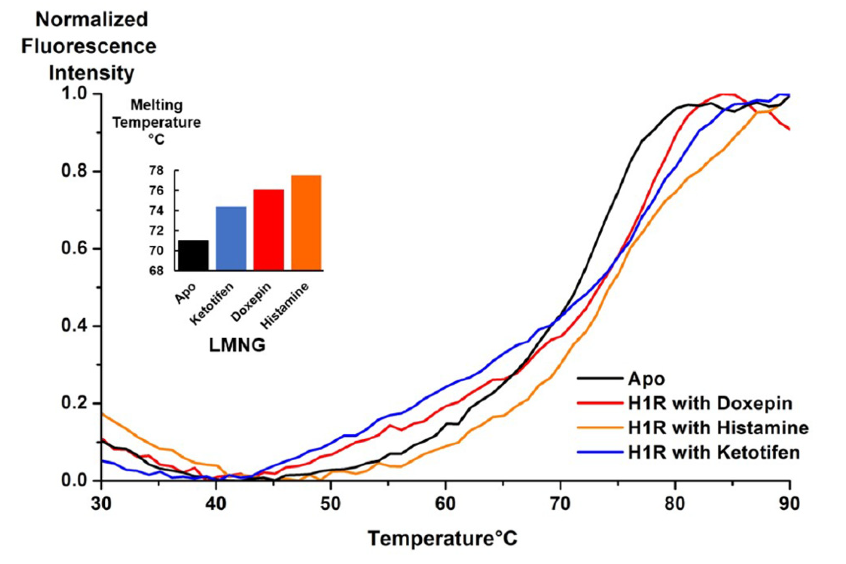
To characterize the ability of purified H1R to bind ligands, we employed a fluorescence thermal shift assay using the thiol-specific fluorophore N-[4-(7-diethylamino-4-methyl-3-coumarinyl)phenyl]maleimide (CPM). This fluorophore interacts with cysteines present in the protein hydrophobic core as they become exposed to aqueous solvent due to thermal unfolding of the protein and has been used to characterize GPCR-ligand interactions [26]. To record the fluorescence thermal shift experiments, H1R was purified in an aqueous solution containing LMNG/CHS and without any ligand added. Several ligands were then individually mixed with aliquots of purified H1R and incubated for half an hour on ice prior to recording the temperature ramp experiments. To determine the melting temperature (TM) of each sample, the fluorescence data were fitted to a Boltzmann sigmoidal curve and the Tm was determined at the inflection point of the curve. All samples with ligands added showed moderate (3–4 °C) to more significant (8 °C) increases in the calculated TM values over the sample with no ligand added (Figure 4), indicating that the purified protein recognized and could bind ligands. The TM for H1R with no ligand added was determined to be 71.0 °C. The H1R sample with histamine added showed the highest determined Tm at 77.2 °C, and the curve showed a clear sigmoidal shape, consistent with cooperative unfolding.

3.4. Spectroscopic Characterization of Solutions Containing Purified H1R by 2-Dimensional TROSY NMR

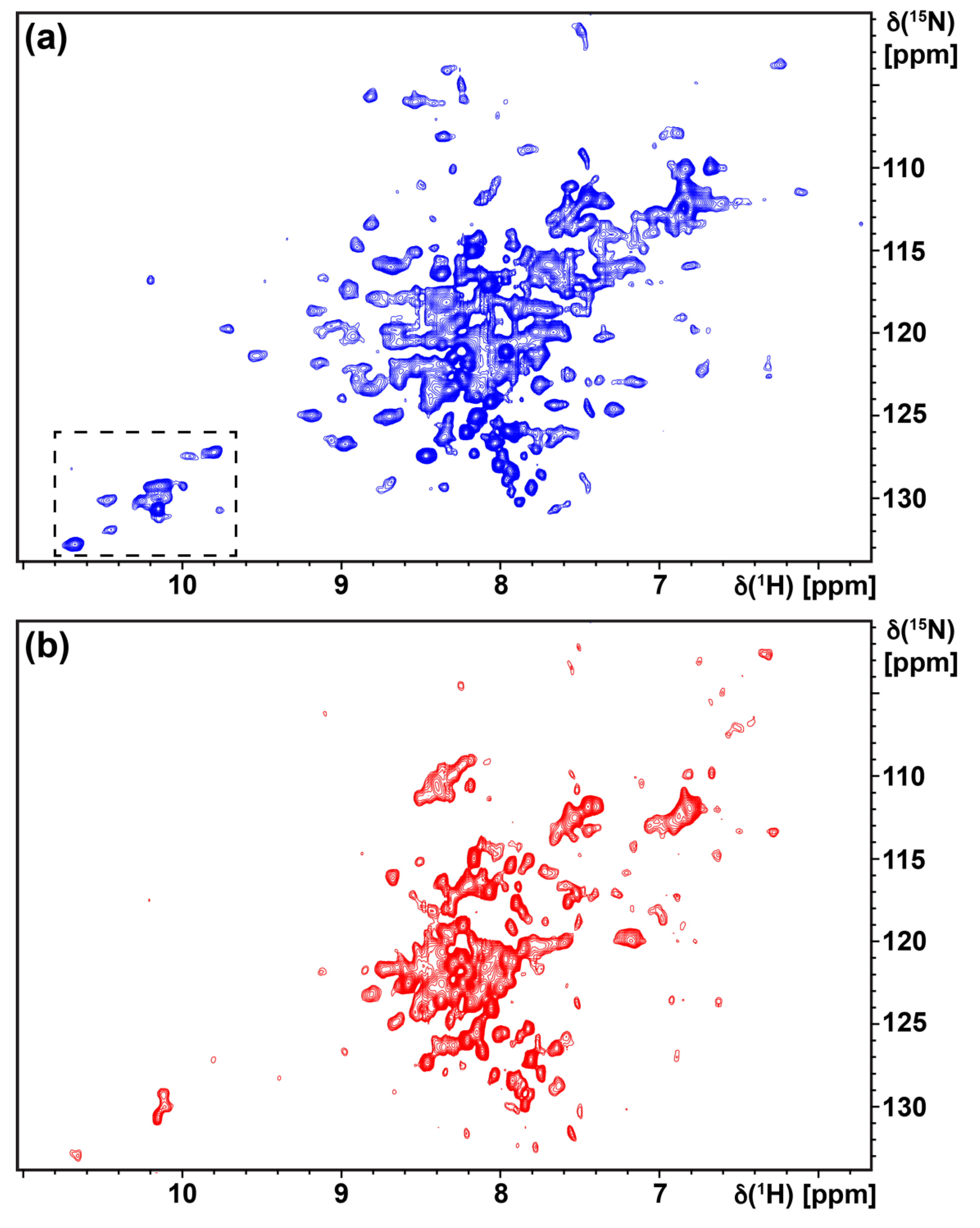
Production of H1R for NMR studies followed the optimized expression and purification conditions described above. To assess the foldedness of purified H1R in solutions containing LMNG/CHS mixed micelles, we recorded [15N,1H]-TROSY two-dimensional spectra of u-15N H1R in complex with the ligands doxepin and histamine (Figure 5a,b). A TROSY spectrum of u-15N H1R in complex with doxepin recorded at 42 °C shows approximately 100 to 130 signals that are well dispersed from 6.5 to 10.5 ppm along the 1H chemical shift axis. About 20 to 25 signals of greater intensity are narrowly dispersed between 7.5 and 8.5 ppm along the 1H axis, which likely arise from more flexible loop regions of the protein. The approximately 25–30 well-resolved signals observed above 8.5 ppm are likely from amide groups in regular secondary structure and indicate that the protein is folded in conditions used for our NMR studies. A TROSY spectrum of u-15N H1R in complex with the agonist histamine shows fewer resolved signals than the complex with doxepin, reminiscent of earlier comparisons of TROSY spectra for antagonist and agonist complexes of human A2AAR [22]; however, a number signals are observed above 8.5 ppm and below 7.5 ppm, consistent with the thermal shift assays that indicate that the complex with histamine is folded.
To provide further interpretation of the NMR data, we compared the TROSY spectrum with the crystal structure of the H1R complex with doxepin and more closely examined signals from the tryptophan indole 15N–1H region (Figure 5 and Figure 6a). There are four resolved signals of greater intensity in the tryptophan indole 15N–1H region of the TROSY spectrum and approximately five to six additional signals that are less intense but resolved. The crystal structure of the H1R complex with doxepin shows 9 tryptophans located in the transmembrane helices and extracellular loops (Figure 6b). Thus the number of observed signals in the Trp indole 15N–1H region of the TROSY spectrum is similar to the number of expected signals from all tryptophans. Comparing the spectra of the agonist and antagonist complexes shows some initial differences, for example in the region of the NMR spectra where glycine and threonine signals are typically observed (Figure 7). These initial spectra are highly encouraging considering that the protein is not deuterated, and thus we anticipate additional improvement in resolution upon incorporation of deuterium in the protein.

3.5. Response to Changes in Efficacy of Bound Drugs Monitored by 19F-NMR Spectroscopy

We assessed the potential for labeling with 19F-trifluoroethanethiol (TET) and whether we could detect a response to changes in the efficacy of bound ligands by preparing samples of H1R complexes with the antagonist doxepin and the endogenous agonist histamine. Based on analysis of the crystal structure of the H1R complex with doxepin, three endogenous cysteines are predicted to be more solvent accessible and thus available for 19F labeling via chemical modification with trifluoroethanethiol (TET). These cysteines are C2215.69, located at the intracellular end of helix V, cysteine 4416.61, located at the extracellular end of helix VI, and C4717.56, located at the intracellular end of helix VII (Figure 8a). While the TROSY experiments were recorded at 42 °C, the 19F NMR data were measured at 7 °C so that we could assess the data in the larger context of many previously reported 19F NMR GPCR studies [21,25,29,32,33]. Both 19F spectra of the complexes with doxepin and histamine show complicated line shapes containing more than three individual components (Figure 8b), suggesting the presence of multiple, simultaneously observed conformers for one or more of the labeled cysteines. Comparison of the 19F data between the complex with doxepin and the complex with histamine shows that individual components appear to have similar chemical shifts, for example the components near 9.0 and 9.8 ppm, with changes observed in the relative intensities of the individual components. This suggests that changes in relative populations of different conformers are potentially observed for complexes with different efficacies of bound ligands and that further 19F-NMR experiments with H1R variants containing single labeled cysteines may be used to characterize these different populations.

4. Discussion

4.1. Production of Stable-Isotope Labeled Human GPCRs in Pichia Pastoris

Production of human GPCRs for structure determination by crystallography or cryo-EM has been achieved mostly using insect cell expression [34], with some full-length class B and class F receptors expressed in mammalian cells. Though P. pastoris has been used to express a much smaller fraction of GPCRs for structure determination so far, the benefits of working with P. pastoris have motivated several studies of the potential to express functional GPCRs in this system. Surveys of the expression of about 30 different human GPCRs demonstrated that a sizeable fraction of functional receptors could be expressed at milligram to multi-milligram scales [13,35]. Systematic evaluation of expression conditions also demonstrated that yields of more poorly expressed receptors could be significantly improved with optimized protocols [11].
P. pastoris offers multiple advantages for the expression of human membrane proteins for NMR studies, which include the ability to produce functional proteins with high levels of deuterium incorporation [36]. A growing number of membrane proteins have been expressed in P. pastoris for NMR experiments in both aqueous solutions and solid state samples, including human aquaporin 1 [37], human aquaporin 2 [38], and fungal rhodopsin from Leptosphaeria maculans [39]. Initial NMR data with the human GPCRs CB1 and OX2 also appear to be promising [24]. The NMR data presented here demonstrate that functional human H1R can be produced at sufficient quantities for NMR studies and establish an important step in the application of NMR to study the energy landscape of additional human GPCRs.

4.2. Tryptophans as NMR Probes of GPCR Structure-Function Relationships

Both endogenous and extrinsically introduced tryptohans are sensitive reporters of function-related changes in the GPCR structure, particularly when nearby other aromatic amino acids that create significant ring current shifts [40]. Further, tryptophan indole 15N–1H signals are typically well-separated from the majority of amide signals in [15N,1H] heteronuclear two-dimensional experiments, which facilitates their assignment by amino acid replacement. We explored the potential use of tryptophans in future NMR studies of H1R structure–function relationships by calculating the ring current shifts for H1R tryptophan indole 15N–1H signals from the crystal structure of the H1R complex with doxepin using the program Molmol [41]. The calculated ring current shifts show a relatively narrow range spanning approximately 0.3 ppm, with the notable exception of Trp 4286.48 (Table 2; superscripted text denotes the Ballosteros–Weinstein nomenclature used to report GPCR amino acid positions relative to a consensus sequence [30]). The calculated ring current shift for Trp 4286.48 shows a pronounced shift of −0.56 and −0.78 ppm for the Hε1 and Nε1 chemical shifts, respectively. Comparably large ring current shifts were also observed for the corresponding Trp2466.48 indole 15N–1H lines in TROSY NMR spectra of human A2AAR [19,22]. The single largest contribution to these ring current shifts was the nearby Phe2426.44 of the highly conserved P-I-F activation motif [19,22], and the chemical shifts of Trp2466.48 were found to be highly sensitive to the efficacy of bound drugs. Thus, we anticipate that Trp 4286.48 in H1R will be a sensitive probe of structure–function relationships.

5. Conclusions

We present a protocol for the expression and purification of the human H1 histamine receptor in Pichia pastoris, which can be applied to economically produce stable-isotope labeled GPCR samples for NMR experiments. Evaluation of protein production parameters revealed that addition of ligand during protein expression resulted in a ten-fold increase in the yield of expressed receptor. Purification of expressed H1R in buffer containing either DDM/CHS mixed micelles or LMNG/CHS mixed micelles yielded a monodispersed and homogeneous preparation. Purified H1R responded to the addition of known ligands, as monitored by fluorescence thermal shift assays, with the addition of the ligand histamine resulting in the largest increase in the protein melting temperature. Two-dimensional [15N,1H]-TROSY NMR spectra of u-15N H1R in buffer containing LMNG/CHS mixed micelles show well-dispersed signals consistent with a folded protein and comparison of TROSY spectra of H1R complexes with an antagonist and an agonist show changes in the distribution of signals. Evaluation of the tryptophan indole 15N–1H region of the TROSY correlation spectrum of the antagonist complex shows the presence of a similar number of signals as expected from the crystal structure of H1R. 19F-NMR spectra of H1R show differences related to changes in the efficacy of bound drugs. Expression of stable-isotope labeled human receptors is a bottleneck in the application of NMR spectroscopy to map the energy landscapes for most human GPCRs. While P. pastoris is a promising host for GPCR expression, only a few receptors have been expressed successfully in P. pastoris for NMR studies. This work demonstrates an additional example of a human GPCR that can be prepared from P. pastoris for NMR experiments.

Author Contributions

Conceptualization, E.M. and M.T.E.; methodology, E.M., A.P.R. and M.T.E.; formal analysis, E.M. and M.T.E.; investigation, E.M. and A.P.R.; writing—original draft preparation, E.M. and A.P.R.; writing—review and editing, M.T.E.; supervision, M.T.E.; project administration, M.T.E.; funding acquisition, M.T.E. All authors have read and agreed to the published version of the manuscript.

Funding

This research was funded by the National Institutes of Health, grant number 1R35GM138291. M.T.E. also acknowledges support from the Oak Ridge Affiliated Universities Ralph E. Powe Junior Faculty Award. NMR data were recorded in the McKnight Brain Institute at the National High Magnetic Field Laboratory’s AMRIS Facility, which is supported by National Science Foundation Cooperative Agreement No. DMR-1644779 and the State of Florida.

Conflicts of Interest

The authors declare no conflict of interest.

References

  1. Hauser, A.S.; Attwood, M.M.; Rask-Andersen, M.; Schiöth, H.B.; Gloriam, D.E. Trends in GPCR drug discovery: New agents, targets and indications. Nat. Rev. Drug Discov. 2017, 16, 829–842. [Google Scholar] [CrossRef] [PubMed]
  2. Hill, S.J.; Ganellin, C.R.; Timmerman, H.; Schwartz, J.C.; Shankley, N.P.; Young, J.M.; Schunack, W.; Levi, R.; Haas, H.L. International Union of Pharmacology. XIII. Classification of histamine receptors. Pharmacol. Rev. 1997, 49, 253–278. [Google Scholar]
  3. Hill, S.J. Distribution, properties, and functional characteristics of three classes of histamine receptor. Pharmacol. Rev. 1990, 42, 45–83. [Google Scholar]
  4. Barbier, A.J.; Bradbury, M.J. Histaminergic control of sleep-wake cycles: Recent therapeutic advances for sleep and wake disorders. CNS Neurol. Disord. Drug Targets 2007, 6, 31–43. [Google Scholar] [CrossRef]
  5. O’Mahony, L.; Akdis, M.; Akdis, C.A. Regulation of the immune response and inflammation by histamine and histamine receptors. J. Allergy Clin. Immunol. 2011, 128, 1153–1162. [Google Scholar] [CrossRef]
  6. Shi, Z.; Fultz, R.S.; Engevik, M.A.; Gao, C.; Hall, A.; Major, A.; Mori-Akiyama, Y.; Versalovic, J. Distinct roles of histamine H1- and H2-receptor signaling pathways in inflammation-associated colonic tumorigenesis. Am. J. Physiol. Gastrointest Liver Physiol. 2019, 316, G205–G216. [Google Scholar] [CrossRef]
  7. Shimada, I.; Ueda, T.; Kofuku, Y.; Eddy, M.T.; Wüthrich, K. GPCR drug discovery: Integrating solution NMR data with crystal and cryo-EM structures. Nat. Rev. Drug Discov. 2019, 18, 59–82. [Google Scholar] [CrossRef]
  8. Klabunde, T.; Hessler, G. Drug design strategies for targeting G-protein-coupled receptors. Chembiochem 2002, 3, 928–944. [Google Scholar] [CrossRef]
  9. Shimamura, T.; Shiroishi, M.; Weyand, S.; Tsujimoto, H.; Winter, G.; Katritch, V.; Abagyan, R.; Cherezov, V.; Liu, W.; Han, G.W.; et al. Structure of the human histamine H1 receptor complex with doxepin. Nature 2011, 475, 65–70. [Google Scholar] [CrossRef] [Green Version]
  10. Xia, R.; Wang, N.; Xu, Z.; Lu, Y.; Song, J.; Zhang, A.; Guo, C.; He, Y. Cryo-EM structure of the human histamine H 1 receptor/G q complex. Nat. Commun. 2021, 12, 1–9. [Google Scholar] [CrossRef]
  11. André, N.; Cherouati, N.; Prual, C.; Steffan, T.; Zeder-Lutz, G.; Magnin, T.; Pattus, F.; Michel, H.; Wagner, R.; Reinhart, C. Enhancing functional production of G protein-coupled receptors in Pichia pastoris to levels required for structural studies via a single expression screen. Protein Sci. 2006, 15, 1115–1126. [Google Scholar] [CrossRef] [PubMed] [Green Version]
  12. Noguchi, S.; Satow, Y. Purification of human beta2-adrenergic receptor expressed in methylotrophic yeast Pichia pastoris. J. Biochem. 2006, 140, 799–804. [Google Scholar] [CrossRef] [PubMed]
  13. Asada, H.; Uemura, T.; Yurugi-Kobayashi, T.; Shiroishi, M.; Shimamura, T.; Tsujimoto, H.; Ito, K.; Sugawara, T.; Nakane, T.; Nomura, N.; et al. Evaluation of the Pichia pastoris expression system for the production of GPCRs for structural analysis. Microb. Cell Fact. 2011, 10, 24. [Google Scholar] [CrossRef] [Green Version]
  14. de Jong, L.A.; Grünewald, S.; Franke, J.P.; Uges, D.R.; Bischoff, R. Purification and characterization of the recombinant human dopamine D2S receptor from Pichia pastoris. Protein Expr. Purif. 2004, 33, 176–184. [Google Scholar] [CrossRef] [PubMed]
  15. Sarramegna, V.; Demange, P.; Milon, A.; Talmont, F. Optimizing functional versus total expression of the human mu-opioid receptor in Pichia pastoris. Protein Expr. Purif. 2002, 24, 212–220. [Google Scholar] [CrossRef]
  16. Ye, L.; Van Eps, N.; Zimmer, M.; Ernst, O.P.; Prosser, R.S. Activation of the A2A adenosine G-protein-coupled receptor by conformational selection. Nature 2016, 533, 265–268. [Google Scholar] [CrossRef]
  17. Clark, L.D.; Dikiy, I.; Chapman, K.; Rödström, K.E.; Aramini, J.; LeVine, M.V.; Khelashvili, G.; Rasmussen, S.G.; Gardner, K.H.; Rosenbaum, D.M. Ligand modulation of sidechain dynamics in a wild-type human GPCR. Elife 2017, 6. [Google Scholar] [CrossRef] [Green Version]
  18. Huang, S.K.; Pandey, A.; Tran, D.P.; Villanueva, N.L.; Kitao, A.; Sunahara, R.K.; Sljoka, A.; Prosser, R.S. Delineating the conformational landscape of the adenosine A2A receptor during G protein coupling. Cell 2021, 184, 1884–1894. [Google Scholar] [CrossRef]
  19. Eddy, M.T.; Martin, B.T.; Wüthrich, K. A2A Adenosine Receptor Partial Agonism Related to Structural Rearrangements in an Activation Microswitch. Structure 2021, 29, 170–176. [Google Scholar] [CrossRef]
  20. Eddy, M.T.; Gao, Z.-G.; Mannes, P.; Patel, N.; Jacobson, K.A.; Katritch, V.; Stevens, R.C.; Wüthrich, K. Extrinsic Tryptophans as NMR Probes of Allosteric Coupling in Membrane Proteins: Application to the A2A Adenosine Receptor. J. Am. Chem. Soc. 2018, 140, 8228–8235. [Google Scholar] [CrossRef]
  21. Sušac, L.; Eddy, M.T.; Didenko, T.; Stevens, R.C.; Wüthrich, K. A2A adenosine receptor functional states characterized by 19F-NMR. Proc. Nat. Acad. Sci. USA 2018, 115, 12733–12738. [Google Scholar] [CrossRef] [Green Version]
  22. Eddy, M.T.; Lee, M.-Y.; Gao, Z.-G.; White, K.L.; Didenko, T.; Horst, R.; Audet, M.; Stanczak, P.; McClary, K.M.; Han, G.W.; et al. Allosteric Coupling of Drug Binding and Intracellular Signaling in the A2A Adenosine Receptor. Cell 2017, 1–26. [Google Scholar] [CrossRef] [PubMed] [Green Version]
  23. Ye, L.; Neale, C.; Sljoka, A.; Lyda, B.; Pichugin, D.; Tsuchimura, N.; Larda, S.T.; Pomès, R.; García, A.E.; Ernst, O.P.; et al. Mechanistic insights into allosteric regulation of the A2A adenosine G protein-coupled receptor by physiological cations. Nat. Commun. 2018, 9, 1372. [Google Scholar] [CrossRef] [PubMed]
  24. Clark, L.; Dikiy, I.; Rosenbaum, D.M.; Gardner, K.H. On the use of Pichia pastoris for isotopic labeling of human GPCRs for NMR studies. J. Biomol. NMR 2018, 71, 203–211. [Google Scholar] [CrossRef] [PubMed]
  25. Sušac, L.; O’Connor, C.; Stevens, R.C.; Wüthrich, K. In-Membrane Chemical Modification (IMCM) for Site-Specific Chromophore Labeling of GPCRs. Angew. Chem. 2015, 127, 15461–15464. [Google Scholar] [CrossRef]
  26. Alexandrov, A.I.; Mileni, M.; Chien, E.Y.T.; Hanson, M.A.; Stevens, R.C. Microscale Fluorescent Thermal Stability Assay for Membrane Proteins. Structure 2008, 16, 351–359. [Google Scholar] [CrossRef] [Green Version]
  27. Pervushin, K.; Riek, R.; Wider, G.; Wüthrich, K. Attenuated T2 relaxation by mutual cancellation of dipole-dipole coupling and chemical shift anisotropy indicates an avenue to NMR structures of very large biological macromolecules in solution. Proc. Nat. Acad. Sci. USA 1997, 94, 12366–12371. [Google Scholar] [CrossRef] [Green Version]
  28. Shiroishi, M.; Kobayashi, T.; Ogasawara, S.; Tsujimoto, H.; Ikeda-Suno, C.; Iwata, S.; Shimamura, T. Production of the stable human histamine H1 receptor in Pichia pastoris for structural determination. Methods 2011, 55, 281–286. [Google Scholar] [CrossRef] [PubMed]
  29. Eddy, M.T.; Didenko, T.; Stevens, R.C.; Wüthrich, K. β2-Adrenergic Receptor Conformational Response to Fusion Protein in the Third Intracellular Loop. Structure 2016, 24, 2190–2197. [Google Scholar] [CrossRef] [Green Version]
  30. Ballesteros, J.A.; Weinstein, H. Integrated methods for the construction of three-dimensional models and computational probing of structure-function relations in G protein-coupled receptors. Methods Neurosci. 1995, 25, 366–428. [Google Scholar]
  31. Munk, C.; Isberg, V.; Mordalski, S.; Harpsøe, K.; Rataj, K.; Hauser, A.; Kolb, P.; Bojarski, A.; Vriend, G.; Gloriam, D. GPCRdb: The G protein-coupled receptor database–an introduction. Br. J. Pharmacol. 2016. [Google Scholar] [CrossRef] [PubMed]
  32. Horst, R.; Liu, J.J.; Stevens, R.C.; Wüthrich, K. β₂-adrenergic receptor activation by agonists studied with ¹⁹F NMR spectroscopy. Angew. Chemie 2013, 52, 10762–10765. [Google Scholar] [CrossRef] [Green Version]
  33. Liu, J.J.; Horst, R.; Katritch, V.; Stevens, R.C.; Wüthrich, K. Biased signaling pathways in β2-adrenergic receptor characterized by 19F-NMR. Science 2012, 335, 1106–1110. [Google Scholar] [CrossRef] [Green Version]
  34. Lv, X.; Liu, J.; Shi, Q.; Tan, Q.; Wu, D.; Skinner, J.J.; Walker, A.L.; Zhao, L.; Gu, X.; Chen, N.; et al. In vitro expression and analysis of the 826 human G protein-coupled receptors. Protein Cell 2016, 7, 325–337. [Google Scholar] [CrossRef] [Green Version]
  35. Yurugi-Kobayashi, T.; Asada, H.; Shiroishi, M.; Shimamura, T.; Funamoto, S.; Katsuta, N.; Ito, K.; Sugawara, T.; Tokuda, N.; Tsujimoto, H.; et al. Comparison of functional non-glycosylated GPCRs expression in Pichia pastoris. Biochem. Biophys. Res. Commun. 2009, 380, 271–276. [Google Scholar] [CrossRef] [PubMed]
  36. Morgan, W.D.; Kragt, A.; Feeney, J. Expression of deuterium-isotope-labelled protein in the yeast pichia pastoris for NMR studies. J. Biomol. NMR 2000, 17, 337–347. [Google Scholar] [CrossRef]
  37. Emami, S.; Fan, Y.; Munro, R.; Ladizhansky, V.; Brown, L.S. Yeast-expressed human membrane protein aquaporin-1 yields excellent resolution of solid-state MAS NMR spectra. J. Biomol. NMR 2013, 55, 147–155. [Google Scholar] [CrossRef]
  38. Munro, R.; de Vlugt, J.; Ladizhansky, V.; Brown, L.S. Improved Protocol for the Production of the Low-Expression Eukaryotic Membrane Protein Human Aquaporin 2 in Pichia pastoris for Solid-State NMR. Biomolecules 2020, 10, 434. [Google Scholar] [CrossRef] [Green Version]
  39. Fan, Y.; Shi, L.; Ladizhansky, V.; Brown, L.S. Uniform isotope labeling of a eukaryotic seven-transmembrane helical protein in yeast enables high-resolution solid-state NMR studies in the lipid environment. J. Biomol. NMR 2011, 49, 151–161. [Google Scholar] [CrossRef]
  40. Liu, D.; Wüthrich, K. Ring current shifts in 19F-NMR of membrane proteins. J. Biomol. NMR 2016, 65, 1–5. [Google Scholar] [CrossRef]
  41. Koradi, R.; Billeter, M.; Wüthrich, K. MOLMOL: A program for display and analysis of macromolecular structures. J. Mol. Graph. 1996, 14, 51–55. [Google Scholar] [CrossRef]
Figure 1. Design of a human H1R amino acid sequence for NMR studies. (a) Snake plot of the H1R receptor employed in the current study, as generated from the GPCRdb [31]. The red dashed line represents residues from the third intracellular loop that have been truncated from the native sequence. The “tag” labels at the N-terminus and C-terminus represent a FLAG tag and polyhistidine tag, respectively; (b) amino acid sequence of the human H1R employed in this study.
Biomolecules 11 00632 g001
Figure 2. Optimization of H1R protein production from a Western blot analysis of protein expression conditions. Each lane represents a distinct set of experimental conditions used to express H1R in 4 mL cell culture (see Table 1). The intensity of a protein band appearing just above the 30 kDa molecular weight marker was used to identify optimal expression conditions. Cell culture conditions tested in lanes 6 and 7 were found to be optimal, and conditions in lane 6 were used for subsequent large-scale protein production.
Biomolecules 11 00632 g002
Figure 3. A representative analytical size exclusion chromatogram of purified H1R in solution containing 20 µM doxepin and LMNG/CHS mixed micelles is shown in black. Superimposed is a chromatogram of a solution containing molecular weight standards, shown in red. A mostly monodispersed population of purified receptor is observed for purified H1R, eluted at 16.3 min between the protein standards with molecular weights of 44 and 158 kDa.
Biomolecules 11 00632 g003
Figure 4. Fluorescence thermal shift assay curves are plotted for purified H1R in complex with several ligands and for a sample with no ligand added (apo). The experimental data were fitted to a Boltzmann sigmoidal function to calculate the unfolding temperatures (TM) of each sample, shown in the inset. Each experimental curve was normalized relative to the maximum observed fluorescence intensity.
Biomolecules 11 00632 g004
Figure 5. Two-dimensional [15N,1H]-TROSY correlation spectra are shown of [u-15N] H1R in complex with (a) the antagonist doxepin and (b) the agonist histamine. The dashed box in panel a represents the tryptophan indole region, which is shown on an expanded scale in Figure 6. Both spectra were recorded with ~300 µM H1R at a 1H NMR frequency of 800 MHz and experimental temperature of 42 °C for a total acquisition time of ~22 h per experiment.
Biomolecules 11 00632 g005
Figure 6. (a) An expanded view is shown of the tryptophan indole 15N–1H region of the TROSY correlation spectrum, corresponding to the dashed box in Figure 5a. (b) The crystal structure of the H1R complex with doxepin is shown in ribbon representation (PDB ID 3RZE [9]), and the 9 tryptophans in H1R are shown in red in stick representation. The blue dashed line represents missing electron density in the crystal structure for amino acids 168 to 174, and the black dotted line represents the location of T4L in the crystal structure. The bound doxepin ligand is shown in brown in stick representation.
Biomolecules 11 00632 g006
Figure 7. A superposition is presented of the TROSY spectra of u-15N H1R in complex with doxepin (blue) and histamine (red), showing an expanded view where signals from glycine and threonine 1H–15N amide signals are typically observed.
Biomolecules 11 00632 g007
Figure 8. (a) The crystal structure of the H1R complex with doxepin is shown in ribbon representation (PDB ID 3RZE [9]). The 3 cysteines available for chemical modification are shown as orange spheres and labeled. (b) Superposition of 19F-NMR spectra of human H1R in complex with the antagonist doxepin (blue) and agonist histamine (red) recorded at 7 °C.
Biomolecules 11 00632 g008
Table 1. Parameters used for testing H1R expression conditions corresponding to the Western blot analysis in Figure 2.
Lane.Induction
Temperature
Methanol Induction TimeConcentration of
Doxepin Added
DMSO%
(w/v)
128 °C36 h00
222 °C36 h00
330 °C36 h00
428 °C24 h00
528 °C48 h00
628 °C36 h20 µM0
728 °C36 h100 µM0
828 °C36 h01%
928 °C36 h02%
Table 2. Calculated tryptophan indole 15N and 1H ring current shifts using the structure of H1R in complex with doxepin (PDB ID 3RZE). Calculated ring current shifts for the conserved “toggle switch” tryptophan, Trp6.48, are highlighted in the grey row.
Trp PositionB-W Notation 1ΔδRC [ppm] Hε1ΔδRC [ppm] Nε1
93ECL10.030.02
1033.28−0.15−0.12
1524.500.090.11
1584.56−0.17−0.11
165ECL20.050.05
1895.370.010.03
2085.560.120.15
4286.48−0.57−0.78
4557.400.110.10
1 Ballosteros–Weinstein nomenclature.
Publisher’s Note: MDPI stays neutral with regard to jurisdictional claims in published maps and institutional affiliations.