Article

Widely Targeted Metabolomic Analysis of Metabolic Differences Among Various Organs of Clematis huchouensis from the Huzhou Production Region


1
Huzhou Vocational & Technical College, Huzhou 313000, China
2
Zhejiang Key Laboratory of Forest Genetics and Breeding, College of Forestry and Biotechnology, Zhejiang A&F University, 666 Wusu Street, Lin’an, Hangzhou 311300, China
*
Author to whom correspondence should be addressed.
Metabolites 2026, 16(8), 561; https://doi.org/10.3390/metabo16080561
Submission received: 26 June 2026 / Revised: 24 July 2026 / Accepted: 4 August 2026 / Published: 9 August 2026
(This article belongs to the Section Plant Metabolism)

Abstract

Background/Objectives: Clematis huchouensis Tamura, a genuine medicinal herb endemic to Huzhou, Zhejiang Province. However, its secondary metabolic profile and organ-specific distribution of bioactive constituents remain largely uncharacterized. This study aims to systematically characterize the metabolic profile of its roots, stems, and leaves, and to elucidate organ-specific accumulation patterns of pharmacologically relevant constituents, thereby providing a scientific basis for resource evaluation and quality control of this regional germplasm. Methods: A widely targeted metabolomics approach was employed to profile metabolites in the roots, stems, and leaves of C. huchouensis. Comprehensive annotation and relative quantification were performed using ultra-performance liquid chromatography–tandem mass spectrometry (UPLC-MS/MS) combined with database matching. Cluster analysis and pathway enrichment were conducted to compare metabolic profiles across organs. Results: A total of 1561 metabolites were identified, exhibiting distinct organ-specific accumulation patterns. Flavonoids, alkaloids, and most phenolic acids were predominantly enriched in the aerial parts, whereas the roots accumulated high levels of glutathione and its related peptides, reflecting their significant antioxidant activity. Amino acids displayed complementary tissue-specific distribution, with peptides enriched in leaves and sulfur-containing amino acids such as L-methionine in stems. Organ-specific metabolic differences were significantly associated with pathways including flavonoid biosynthesis and linoleic acid metabolism. Notably, numerous pharmacologically active compounds, such as hispidulin, diosmetin, trigonelline, colchicoside, and oleanolic acid-3-O-xylosyl(1→3)glucuronide—exhibited marked tissue-selective accumulation. Conclusions: This first metabolomic study of C. huchouensis reveals organ-specific accumulation of bioactive compounds, providing a metabolic foundation for its quality control and rational utilization.

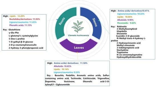
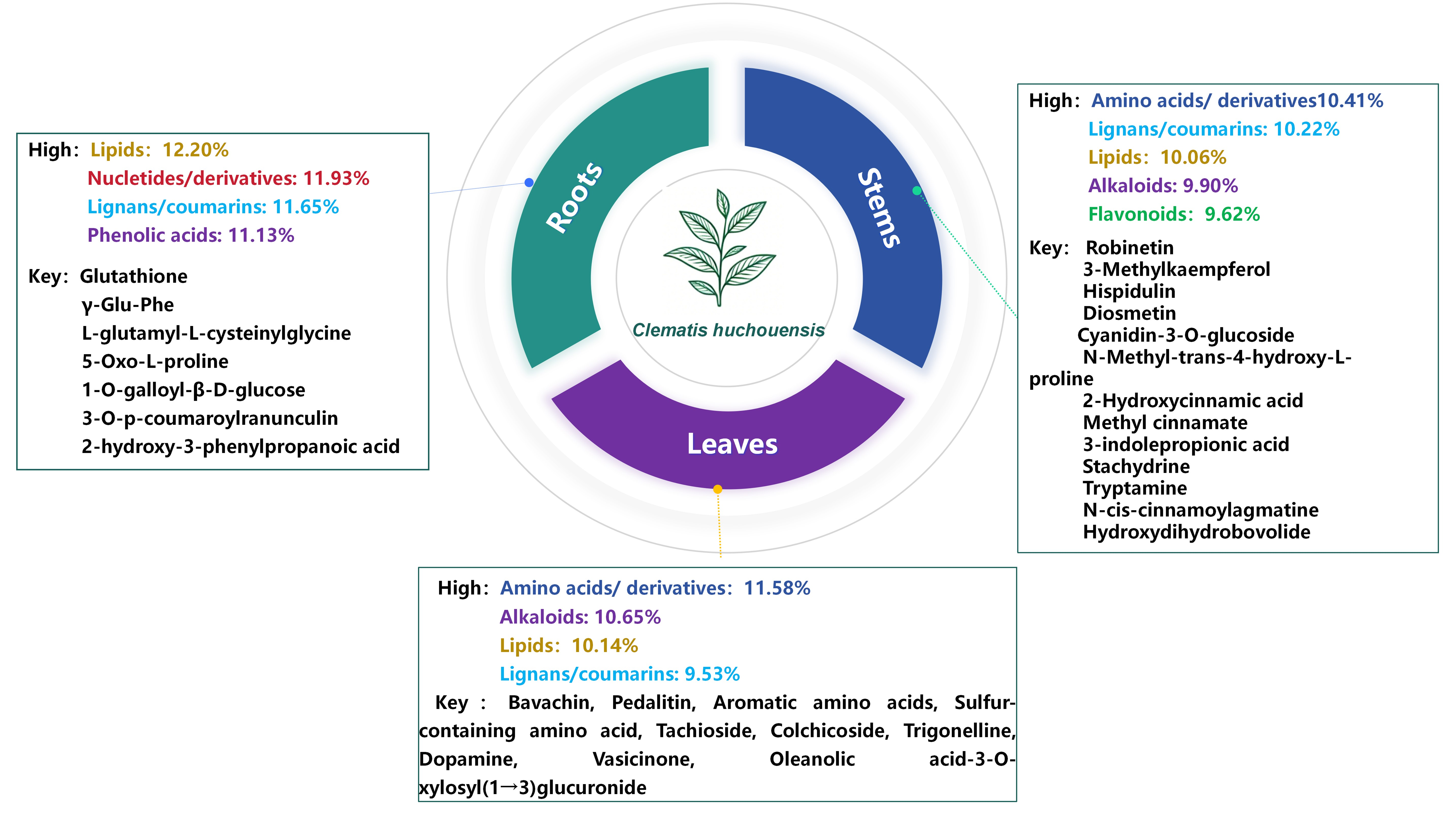
Graphical Abstract

1. Introduction

Clematis belongs to the Ranunculaceae family and is a perennial liana genus with more than 350 species [1,2]. The genus Clematis is primarily distributed in temperate and subtropical regions, with particularly abundant germplasm resources in East Asia [1]. China represents a major distribution center for the genus Clematis, with over 150 species having been documented within its territory [3,4]. Many species within the genus Clematis are valued as ornamental plants in horticulture due to their large and showy flowers. Notable examples include C. vitalba, C. lanuginosa, and C. hancockiana [5,6]. Furthermore, as an important group of medicinal resources, plants of the genus Clematis have been studied and utilized worldwide due to their diverse pharmacological activities [7]. A prominent example is the traditional Chinese medicine “Wei Ling Xian,” prepared from the dried roots and rhizomes of C. chinensis Osbeck, C. hexapetala Pall., or C. manshurica Rupr. This herbal remedy has been traditionally employed as an analgesic, diuretic, and antitumor agent, and is particularly noted for its efficacy in treating rheumatoid arthritis and various inflammatory diseases [6]. With the in-depth advancement of phytochemical and pharmacological research in recent years, researchers have gradually clarified a variety of bioactive constituents contained in Clematis species and revealed their corresponding action mechanisms. For instance, the roots of C. chinensis are rich in saponins and pectin-like polysaccharides. Mechanistic studies have demonstrated that its saponins alleviate osteoarthritis by inhibiting mitochondrial damage and caspase-3 activation [8,9], and the pectin-like polysaccharides alleviate rheumatoid arthritis by suppressing the expression of pro-inflammatory cytokines and matrix metalloproteinase genes [10]. Similarly, clematichinenoside, a compound isolated from the roots of C. hexapetala, exhibits potent immunomodulatory activity. This effect is mediated by the downregulation of TNF-α levels, highlighting its therapeutic potential in chronic inflammation [11]. Further studies have revealed anti-neuroinflammatory properties associated with phenolic compounds and lignans derived from the rhizomes of C. manshurica [12,13]. Triterpenoid saponins from the roots of C. uncinata have demonstrated significant antitumor activity [14], while a novel cyclopeptide alkaloid and coumarins isolated from the roots of C. florida possess anti-inflammatory properties [15]. Beyond applications for internal disorders, the leaves of C. flammula have been traditionally used for the topical treatment of superficial skin burns [16].
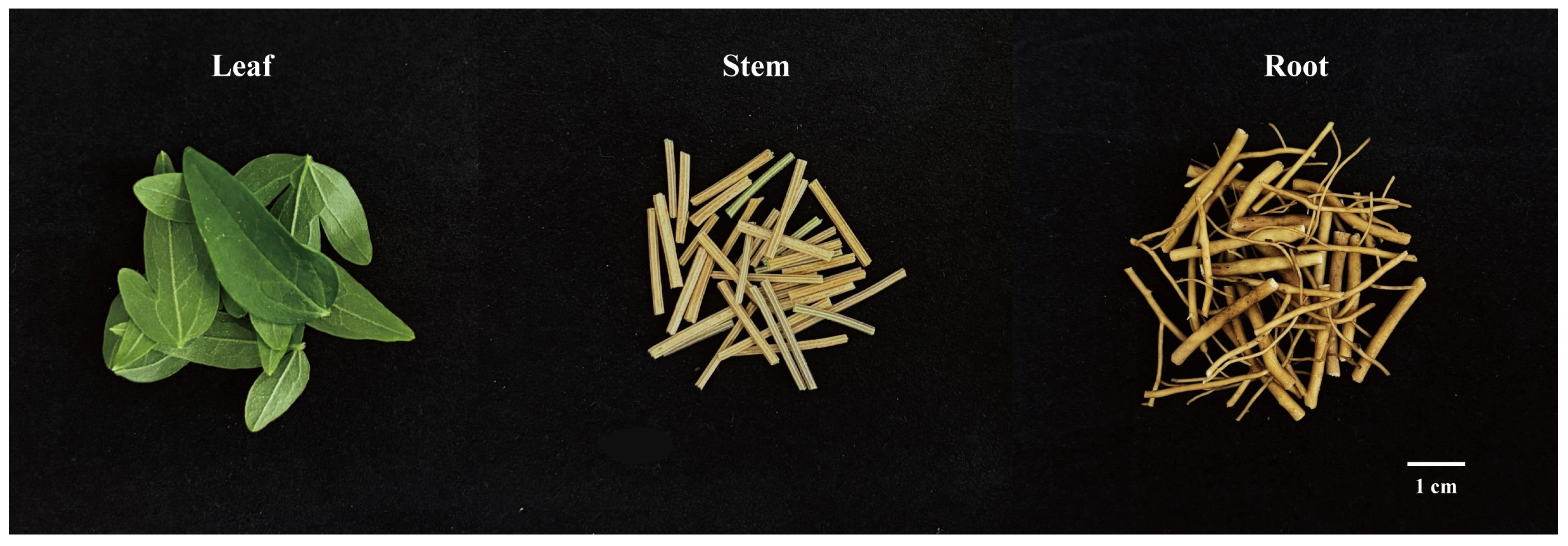
C. huchouensis, also known as “Riverside Wei Ling Xian”, is a genuine medicinal herb native to Huzhou, Zhejiang Province, and its whole plant is used for medicinal purposes (Figure 1). C. huchouensis-based traditional Chinese medicine preparations are widely used clinically for treating rheumatoid arthritis [2]. Moreover, in traditional Chinese medicine practice, its combination with other herbs yields multiple bioactivities, including anti-tumor, anti-inflammatory, and anti-allergic properties in traditional Chinese medicine practice. In traditional folk practice, the herb is commonly pounded and applied topically or boiled into a decoction for oral intake to alleviate pain and address deep-seated abscesses as well as brain tumor-associated manifestations. Meanwhile, it has also found clinical application in the management of toothache, gout, hepatic carcinoma, and lymphadenitis [1,3]. Studies have shown that the polysaccharide extracted from C. huchouensis (CP), as an acidic proteoglycan, exhibits significant scavenging activity against superoxide anions and hydroxyl radicals in vitro, along with strong reducing power, suggesting its promising potential for antioxidant applications.
As a regionally distinctive germplasm, C. huchouensis holds a unique position in local ecosystems and medicinal applications. However, its secondary metabolic characteristics, material basis, and patterns of organ-specific accumulation remain poorly understood. Here, we performed the first systematic analysis using widely targeted metabolomics technology to investigate the metabolite composition and differences across various organs of C. huchouensis, thereby elucidating its metabolomic profile. This discovery fills a gap in metabolomic research on this species, enriches the database for the genus Clematis, and provides a novel metabolic-level basis for studies on phylogenetic evolution, species identification, and resource evaluation within the genus.

2. Materials and Methods

2.1. Plant Material

The C. huchouensis specimen was collected in June 2025 along a riverside in Huzhou City, Zhejiang Province, China. The C. huchouensis growth conditions are consistent with the external environment at the place of origin. The C. huchouensis specimen was collected in June 2025 from Longxi River (Longxi Port, West Tiaoxi) in Huzhou City, Zhejiang Province, China. The geographic coordinates of the sampling location are 120.0252° E, 30.8855° N, and the habitat is riparian wetland along the riverbank. The growth conditions of C. huchouensis match the ambient environment of its native habitat. Three C. huchouensis plants with good growth conditions were randomly selected to obtain three organs: roots, stems, and leaves. They were immediately frozen in liquid nitrogen and placed overnight at −80 °C before being transferred to a pre-cooled freeze dryer (FD-250201GX, FTFDS, Shanghai, China) for freeze-drying.

2.2. Sample Extraction

After freeze-drying, the sample was ground at 50 Hz for 40 s using a grinder (MM 400, Retsch GmbH, Haan, Germany). A 50 mg aliquot of the powder was mixed with 1.2 mL of methanol/water (4:1, v/v) containing an internal standard. The mixture was vortexed and shaken for 30 s, homogenized at 35 Hz for 5 min, and then sonicated in an ice-water bath for 4 min. This homogenization-sonication cycle was repeated three times. The mixture was subsequently kept at 4 °C for 1 h. After centrifugation at 12,000 r/min for 5 min, the supernatant was filtered through a 0.22 μm membrane and transferred into an autosampler vial. Quality control (QC) samples were prepared by pooling equal volumes of each sample for subsequent analysis.

2.3. Ultra-Performance Liquid Chromatography Conditions

The sample extracts were analyzed using a UPLC-ESI-MS/MS system, comprising an UPLC system (ExionLCTM AD, SCIEX, Singapore) and a triple quadrupole-linear ion trap (QTRAP 6500, SCIEX, Singapore) mass spectrometer (both from Sciex). Chromatographic separation was performed on an Agilent SB-C18 column (1.8 µm, 2.1 mm × 100 mm, Agilent Technologies, Santa Clara, CA, USA) at 40 °C, with a flow rate of 0.35 mL/min and an injection volume of 2 µL. The mobile phase consisted of 0.1% formic acid in water (A) and 0.1% formic acid in acetonitrile (B). Separation was achieved using a gradient program: initial conditions of 95% A and 5% B were maintained, followed by a linear gradient to 5% A and 95% B over 9 min, held for 1 min, then returned to 95% A and 5% B within 1.1 min and equilibrated for 2.9 min. The column effluent was introduced into the ESI-QTRAP-MS for detection.

2.4. ESI-Q TRAP-MS/MS

The operating parameters for the ESI source were: temperature, 500 °C; ion spray voltage, ±5500 V (positive)/−4500 V (negative); GS1, GS2, and curtain gas pressures at 50, 60, and 25 psi, respectively; and CAD gas set to high. All QQQ scans were acquired using MRM mode, with the collision gas (nitrogen) pressure set to medium. The declustering potential and collision energy for each individual MRM transition were determined through separate optimization. For each acquisition period, a specific set of MRM transitions was monitored, based on the elution profiles of the target metabolites within that interval.

2.5. Metabolite Identification and Quantification

Metabolites were identified by matching secondary mass spectrometry data against an in-house metabolite database. Prior to analysis, the data were preprocessed to remove isotopic signals, redundant signals caused by K+, Na+, and NH4+ adducts, as well as fragment ions derived from higher molecular weight metabolites. For quantification on a triple quadrupole mass spectrometer, multiple reaction monitoring mode was employed. This mode operates by selectively filtering the precursor ion of interest with the first quadrupole, fragmenting it in the collision cell, and detecting a specific product ion with the third quadrupole. After obtaining mass spectrometry data from all samples, chromatographic peaks of each metabolite were integrated. Peak alignment and integration correction were applied to the same metabolite across different samples based on retention time to ensure data consistency.

2.6. Data Analysis

Unsupervised principal component analysis (PCA) was conducted using the prcomp function in R (www.r-project.org), with unit variance scaling applied to the data prior to analysis. Pearson correlation coefficients (PCC) between samples were calculated by the cor function in R and presented as only heatmaps. The identification of differential metabolites was based on different thresholds depending on the group design. For two-group analyses, metabolites with VIP > 1 and |Log2FC| ≥ 1.0 were considered differential. For multi-group analyses, the criteria were VIP > 1 and ANOVA p-value < 0.05. The OPLS-DA model, from which VIP values and the score/permutation plots were generated, was constructed using the MetaboAnalystR package in R. The input data were log2-transformed and mean-centered before modeling. A permutation test (n = 200) was additionally performed to assess model validity and prevent overfitting. Identified metabolites were annotated using the KEGG Compound database (http://www.kegg.jp/kegg/compound/ accessed on 27 August 2025), and then mapped to the KEGG Pathway database (http://www.kegg.jp/kegg/pathway.html accessed on 10 September 2025).

3. Results

3.1. Total Ion Current Diagram Analysis

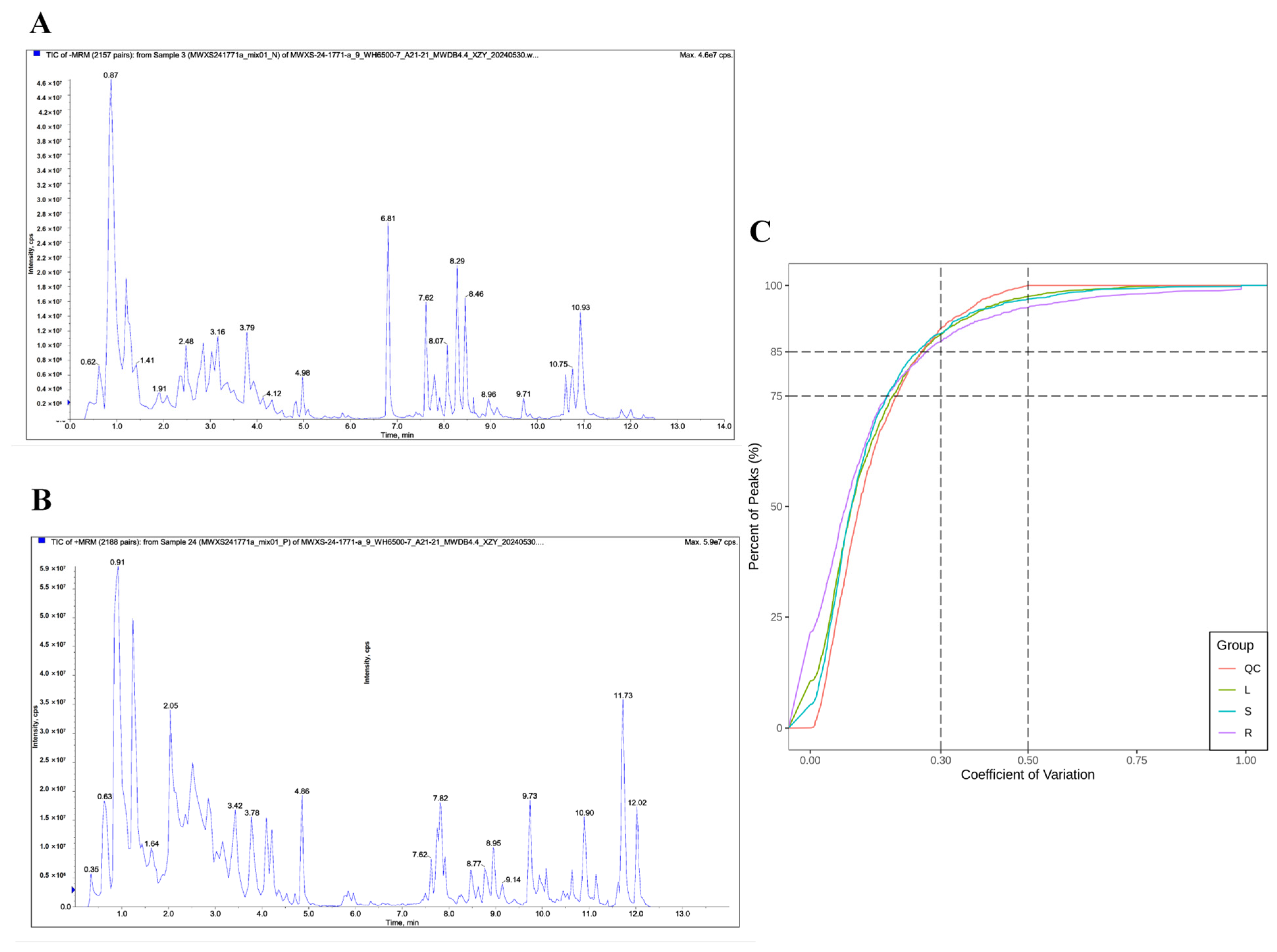
Nearly perfect overlap was observed in the total ion chromatograms (TIC) of QC samples under both positive and negative ion modes, with highly consistent retention times and peak intensities (Figure 2A,B). This indicated that the detection signal was stable, the data were reliable, and the reproducibility was high throughout the analytical run. In the QC samples, more than 85% of the metabolites had CV values below 0.5, and more than 75% had CV values below 0.3, indicating good reproducibility of the experimental data (Figure 2C).

3.2. Overall Characteristics of Metabolites from Different Parts of C. huchouensis

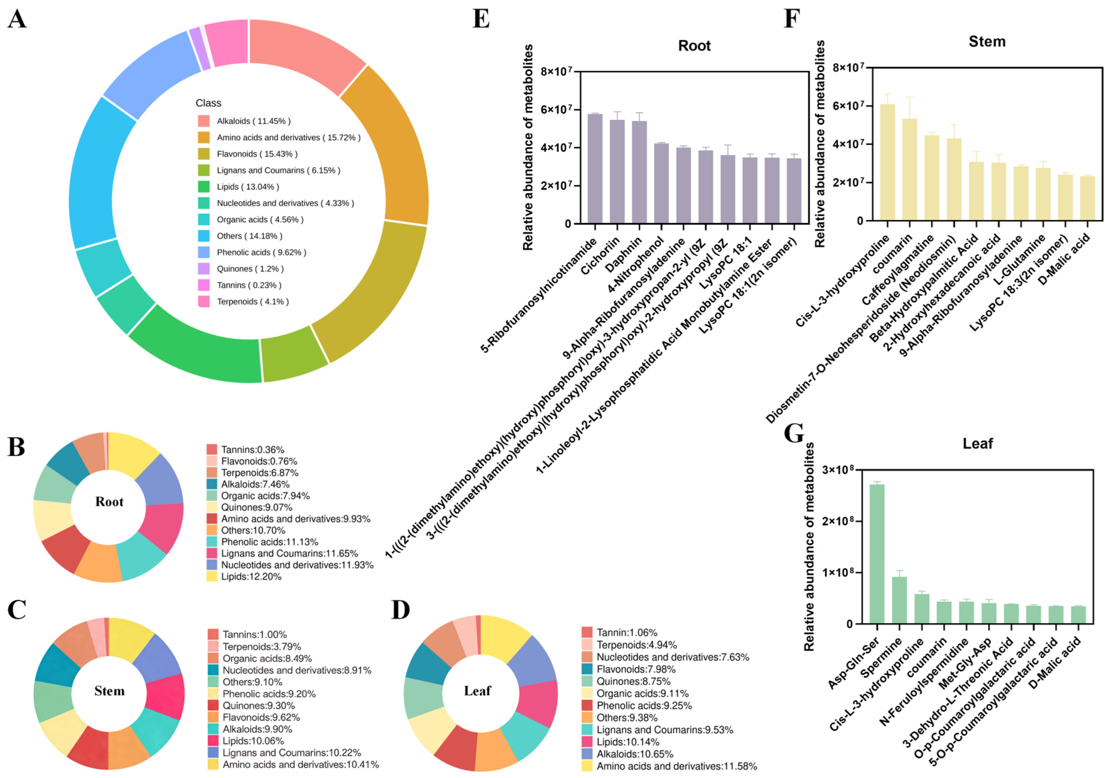
A total of 1561 metabolites were detected in the root, stem, and leaf samples of C. huchouensis. Among them, amino acids and their derivatives (15.72%), flavonoids (15.43%), lipids (13.04%), and alkaloids (11.45%) were relatively abundant, collectively accounting for 55.64% of the total metabolites (Figure 3A). C. huchouensis had high levels of lipids (12.20%), nucleotides and derivatives (11.93%), lignans and coumarins (11.65%), and phenolic metabolites (11.13%) in its roots, while the levels of flavonoids (0.76%) and tannins (0.36%) were low (Figure 3B). The content of amino acids and derivatives (10.41%), lignans and coumarins (10.22%), and lipids (10.06%) in the stem is relatively high, while the content of tannins (1.01%) is the lowest (Figure 3C). The content of amino acids and derivatives (11.58%), alkaloids (10.65%), and lipids (10.14%) in leaves is the highest, while the content of tannins (1.06%) is the lowest (Figure 3D). In the roots of C. huchouensis, the five most abundant metabolites were 5-ribofuranosylnicotinamide, cichoriin, daphnin, 4-nitrophenol, and 9-alpha-ribofuranosyladenine (Figure 3E). In the stems, the top five metabolites were cis-L-3-hydroxyproline, coumarin, caffeoylagmatine, neodiosmin, and β-hydroxypalmitic acid (Figure 3F). In the leaves, the five most abundant metabolites were Asp-Gln-Ser, spermine, cis-L-3-hydroxyproline, coumarin, and N-feruloylspermidine (Figure 3G).

3.3. Multivariate Statistical Analysis

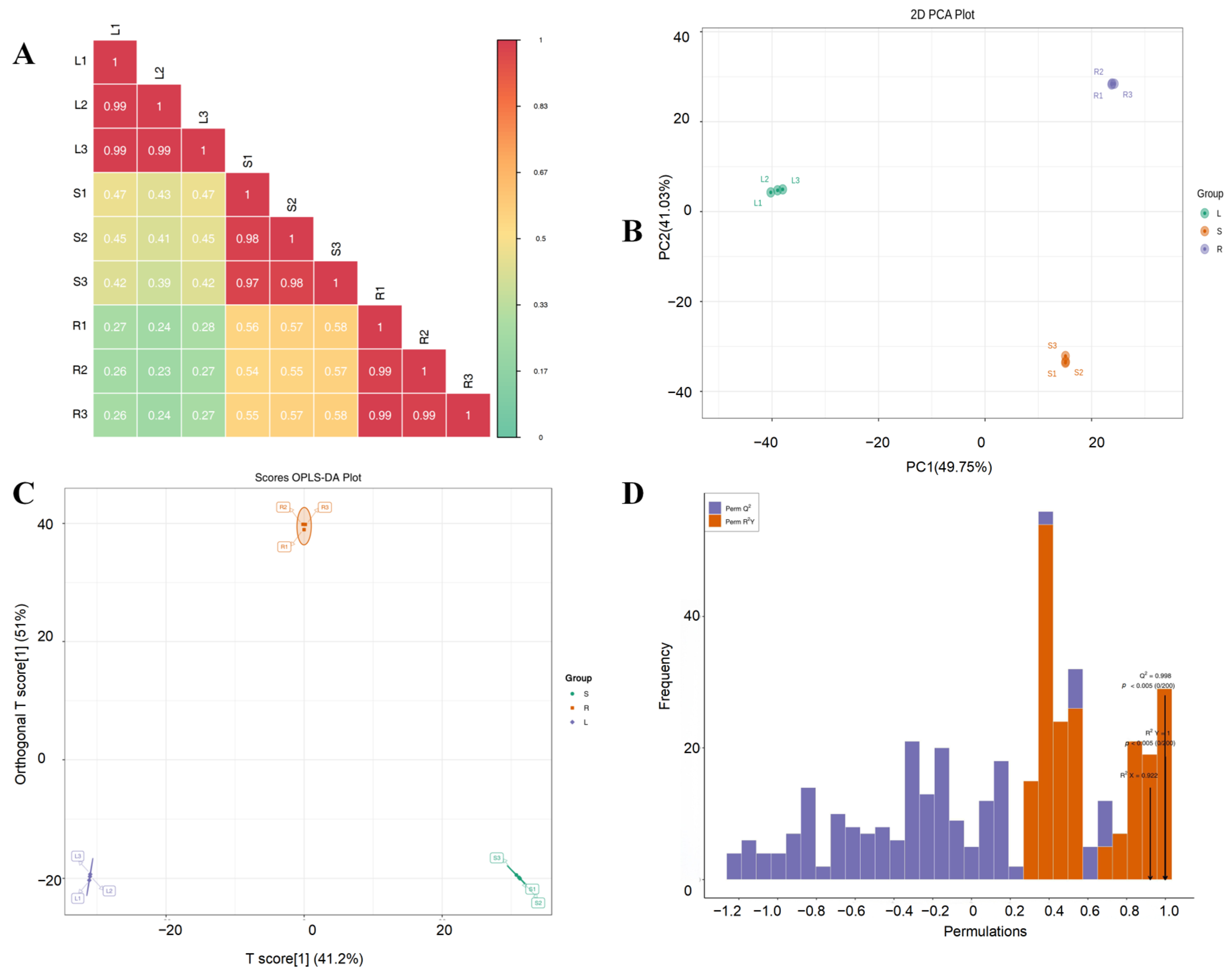
Metabolite differences among different organs of C. huchouensis were analyzed using ultra-high performance liquid chromatography–mass spectrometry (UPLC-MS). Correlation analysis indicated good biological reproducibility and consistency among samples within each group (Figure 4A). The principal component analysis (PCA) score plot revealed that the first principal component (PC1) and the second principal component (PC2) explained 49.75% and 41.02% of the total variance, respectively, cumulatively accounting for 90.77%. This high cumulative variance indicates substantial metabolite differences among the three groups (Figure 4B). The OPLS-DA also demonstrated clear separation of the three metabolite groups (Figure 4C). Furthermore, a high Q2 value (0.998) indicated that the model was well fitted and had strong predictive ability (Figure 4D).

3.4. Screening and Identification of Metabolites from Different Organs of C. huchouensis

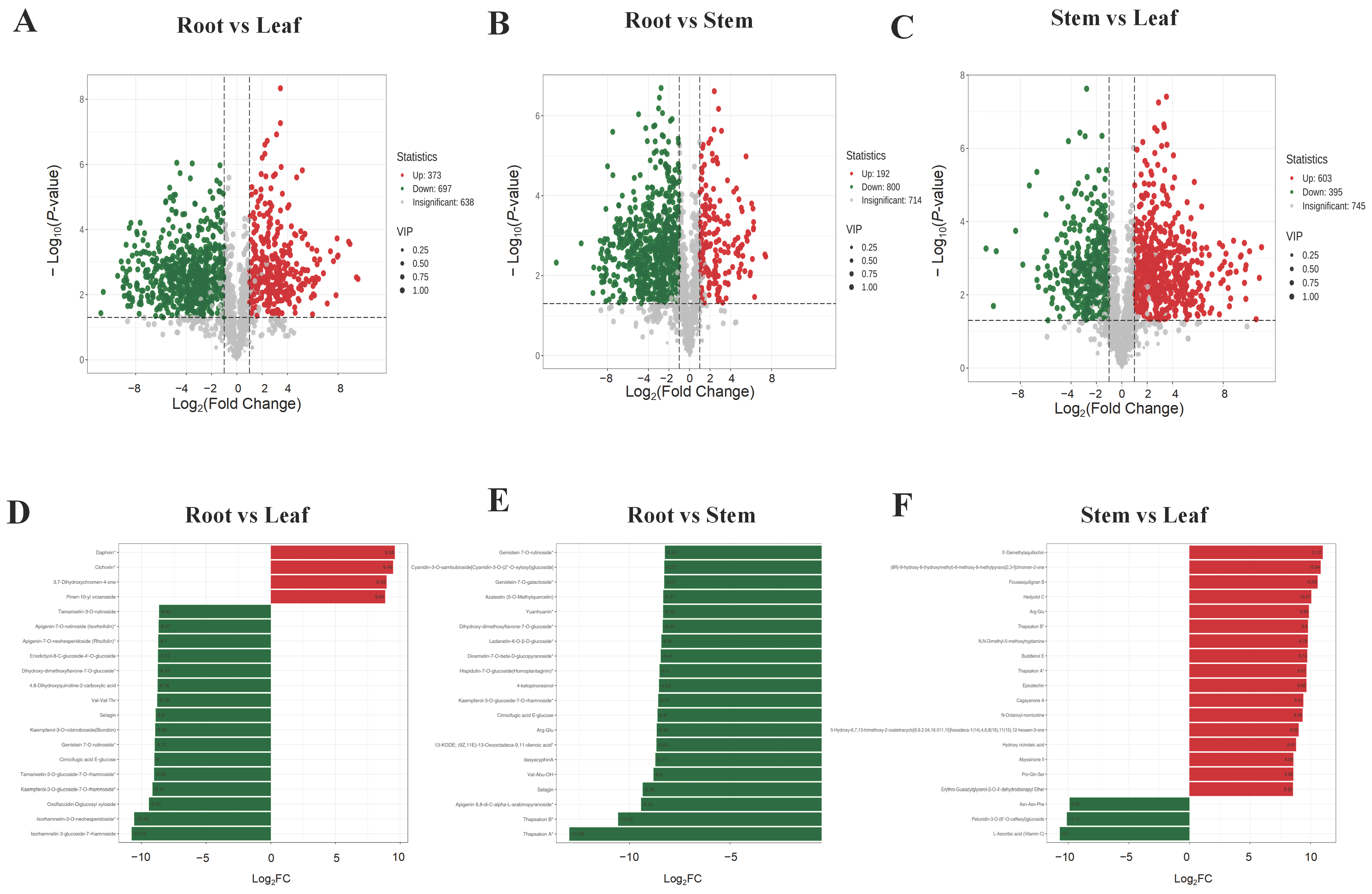
The criteria for screening differential metabolites (DAMs) using the OPLS-DA model were set as VIP ≥ 1 and FC ≥ 2 or ≤0.5. Compared with the root group, 1070 DAMs (373 up-regulated and 696 down-regulated) and 992 DAMs (192 up-regulated and 800 down-regulated) were identified in the leaf group and the stem group, respectively (Figure 5A,B). In the root vs. leaf comparison, among the top 20 metabolites with the greatest fold changes, two coumarins (daphnin and cichoriin), one chromone (3,7-dihydroxychromen-4-one), and one terpenoid (pinen-10-yl vicianoside) showed significantly higher accumulation in the roots, whereas 12 flavonoids, one quinone, one phenolic acid, one alkaloid, and one amino acid metabolite were significantly down-regulated (Figure 5C). In the root vs. stem comparison, among the top 20 DAMs, 12 flavonoids, two amino acids and their derivatives, two alkaloids, one phenolic acid, one lignan, one lipid, and one other metabolite were significantly down-regulated in the roots (Figure 5D). A total of 998 DAMs were detected in the stem vs. leaf comparison (603 up-regulated and 395 down-regulated) (Figure 5E). Compared with the leaf group, among the top 20 metabolites ranked by fold change, six lignans and coumarins, four alkaloids, two flavonoids, two terpenoids, two amino acids and their derivatives, and one lipid metabolite were significantly up-regulated in the stems, whereas one amino acid (Asn-Asn-Phe), one flavonoid (petunidin-3-O-(6″-O-caffeoyl)glucoside), and one other metabolite (L-ascorbic acid) were significantly down-regulated (Figure 5F).

3.5. KEGG Enrichment Analysis of DAMs

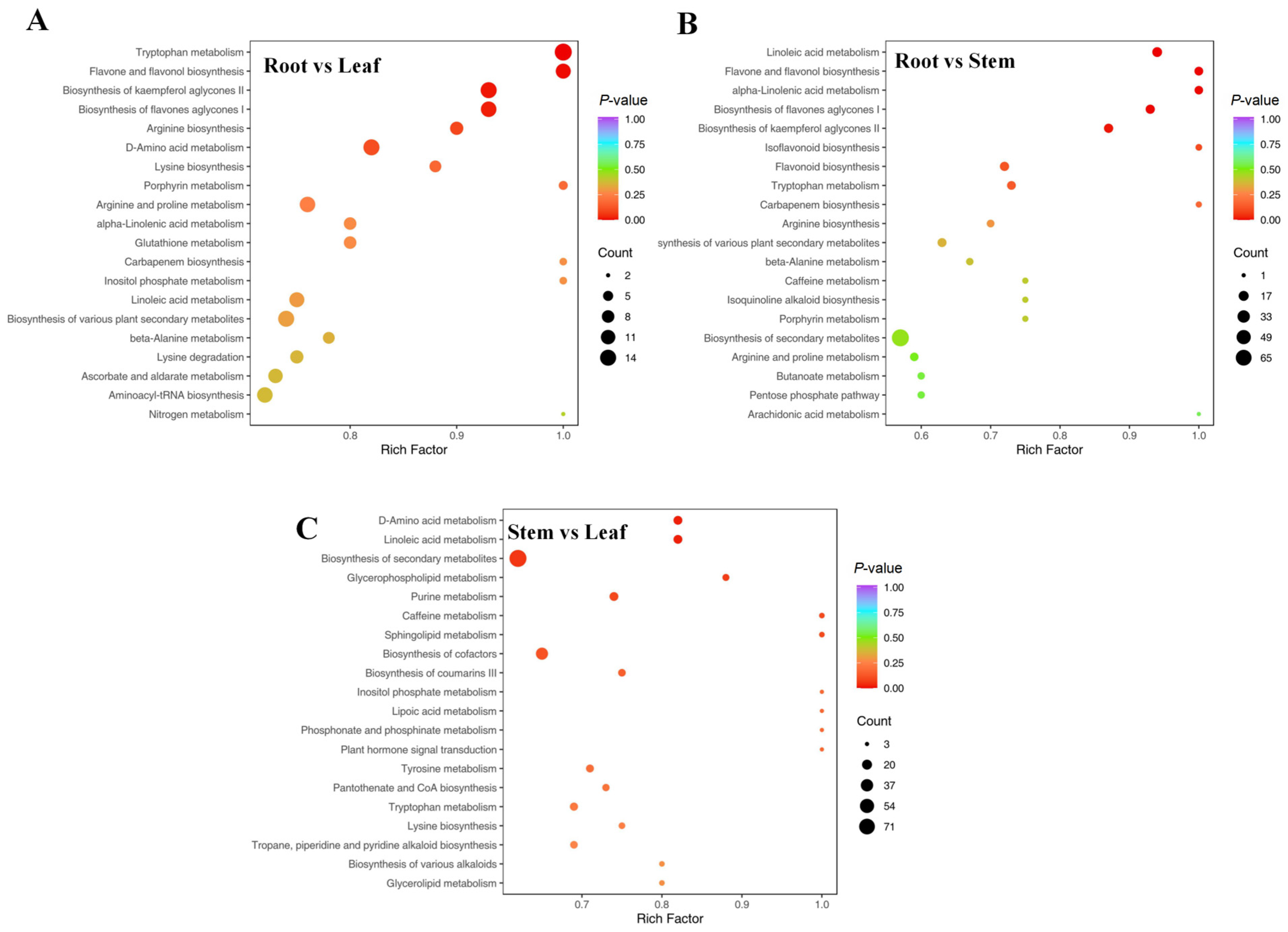
We performed pathway enrichment analysis on the significant DAMs in each comparison to identify the main pathways involved. In the root vs. leaf comparison, the significantly enriched pathways mainly included tryptophan metabolism (16 metabolites), flavone and flavonol biosynthesis (12 metabolites), biosynthesis of kaempferol aglycones II (14 metabolites), and biosynthesis of flavone aglycones I (13 metabolites) (Figure 6A). In the root vs. stem comparison, the significantly enriched pathways mainly comprised linoleic acid metabolism (16 metabolites), biosynthesis of flavone aglycones I (13 metabolites), biosynthesis of kaempferol aglycones II (13 metabolites), flavone and flavonol biosynthesis (11 metabolites), and α-linolenic acid metabolism (10 metabolites) (Figure 6B). In the stem vs. leaf comparison, the significantly enriched pathways included biosynthesis of secondary metabolites (87 metabolites), biosynthesis of cofactors (35 metabolites), D-amino acid metabolism (14 metabolites), and linoleic acid metabolism (14 metabolites) (Figure 6C).

3.6. Comparative Analysis of Relative Contents of Flavonoid Metabolites

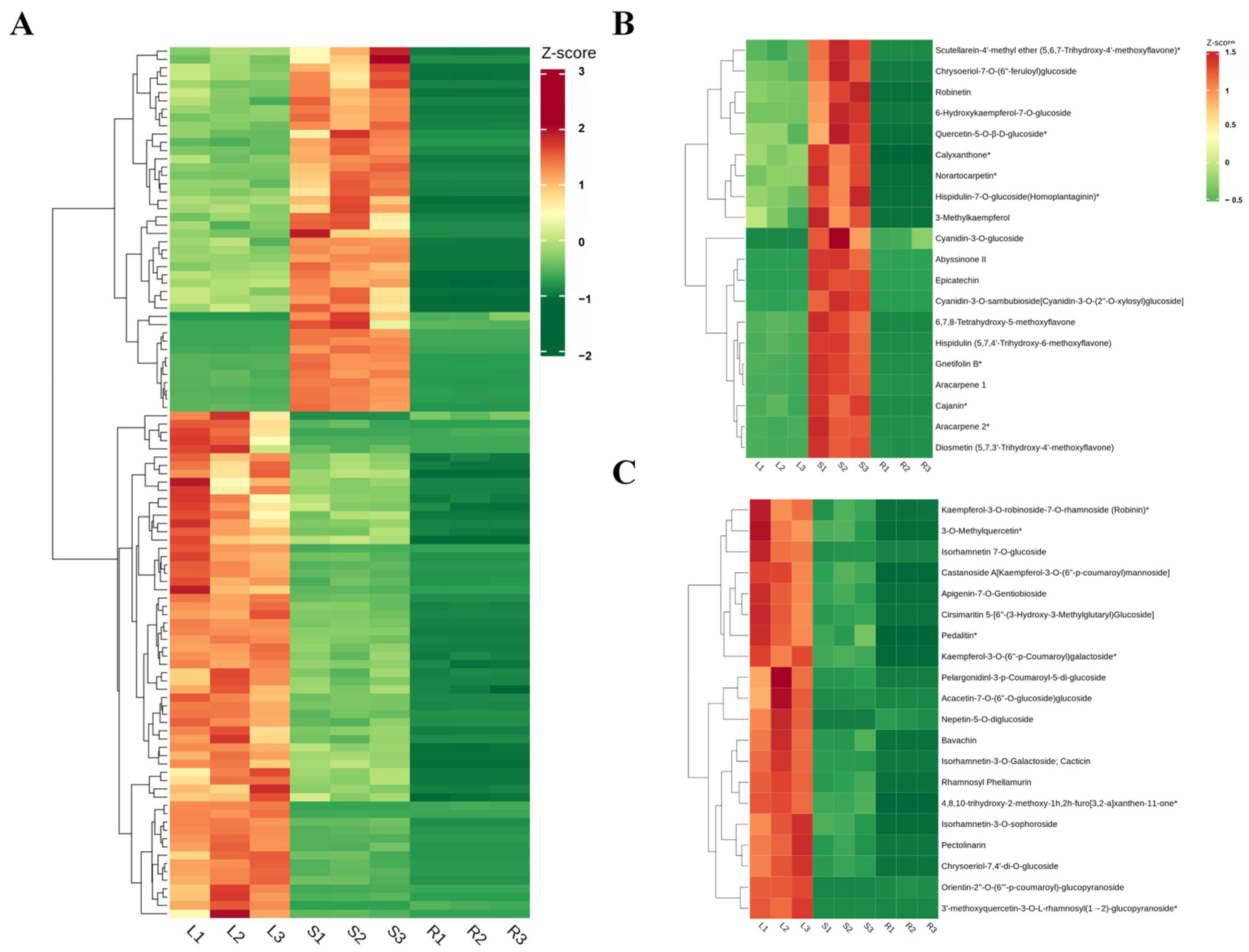
In this study, cluster heatmap analysis of the shared differential flavonoid metabolites among the roots, stems, and leaves revealed a significant tissue-specific accumulation pattern. A total of 105 significantly differentially accumulated flavonoid metabolites were identified across the three organs of C. huchouensis. The majority of flavonoids accumulated at significantly higher levels in the stems (44 flavonoids) and leaves (61 flavonoids), whereas their levels in the roots were generally low (Figure 7A). In the stems, the top 20 significantly up-regulated flavonoid metabolites included 7 flavones (diosmetin (5,7,3′-trihydroxy-4′-methoxyflavone), hispidulin (5,7,4′-trihydroxy-6-methoxyflavone), 6,7,8-tetrahydroxy-5-methoxyflavone, scutellarein-4′-methyl ether (5,6,7-trihydroxy-4′-methoxyflavone), chrysoeriol-7-O-(6″-feruloyl)glucoside, hispidulin-7-O-glucoside (homoplantaginin), and norartocarpetin); 5 flavonols (gnetifolin B, 6-hydroxykaempferol-7-O-glucoside, robinetin, 3-methylkaempferol, and quercetin-5-O-β-D-glucoside); 3 isoflavones (aracarpene 1, aracarpene 2, and cajanin); 2 anthocyanins (cyanidin-3-O-sambubioside [cyanidin-3-O-(2″-O-xylosyl)glucoside] and cyanidin-3-O-glucoside); 1 dihydroflavone (abyssinone II); 1 flavanol (epicatechin); and 1 other flavonoid (calyxanthone) (Figure 7B). In the leaves, the top 20 significantly up-regulated flavonoid metabolites included 8 flavones (orientin-2″-O-(6‴-p-coumaroyl)glucopyranoside, nepetin-5-O-diglucoside, pectolinarin, chrysoeriol-7,4′-di-O-glucoside, cirsimaritin 5-[6″-(3-hydroxy-3-methylglutaryl)glucoside], apigenin-7-O-gentiobioside, acacetin-7-O-(6″-O-glucoside)glucoside, and pedalitin); 8 flavonols (3′-methoxyquercetin-3-O-L-rhamnosyl(1→2)glucopyranoside, isorhamnetin-3-O-galactoside (cacticin), isorhamnetin 7-O-glucoside, kaempferol-3-O-(6″-p-coumaroyl)galactoside, isorhamnetin-3-O-sophoroside, castanoside A [kaempferol-3-O-(6″-p-coumaroyl)mannoside], kaempferol-3-O-robinoside-7-O-rhamnoside (robinin), and 3-O-methylquercetin); 1 dihydroflavonol (rhamnosyl phellamurin); 1 anthocyanin (pelargonidin-3-p-coumaroyl-5-di-glucoside); 1 dihydroflavone (bavachin); and 1 other flavonoid (4,8,10-trihydroxy-2-methoxy-1H,2H-furo[3,2-a]xanthen-11-one) (Figure 7C).

3.7. Comparative Analysis of Relative Contents of Amino Acids

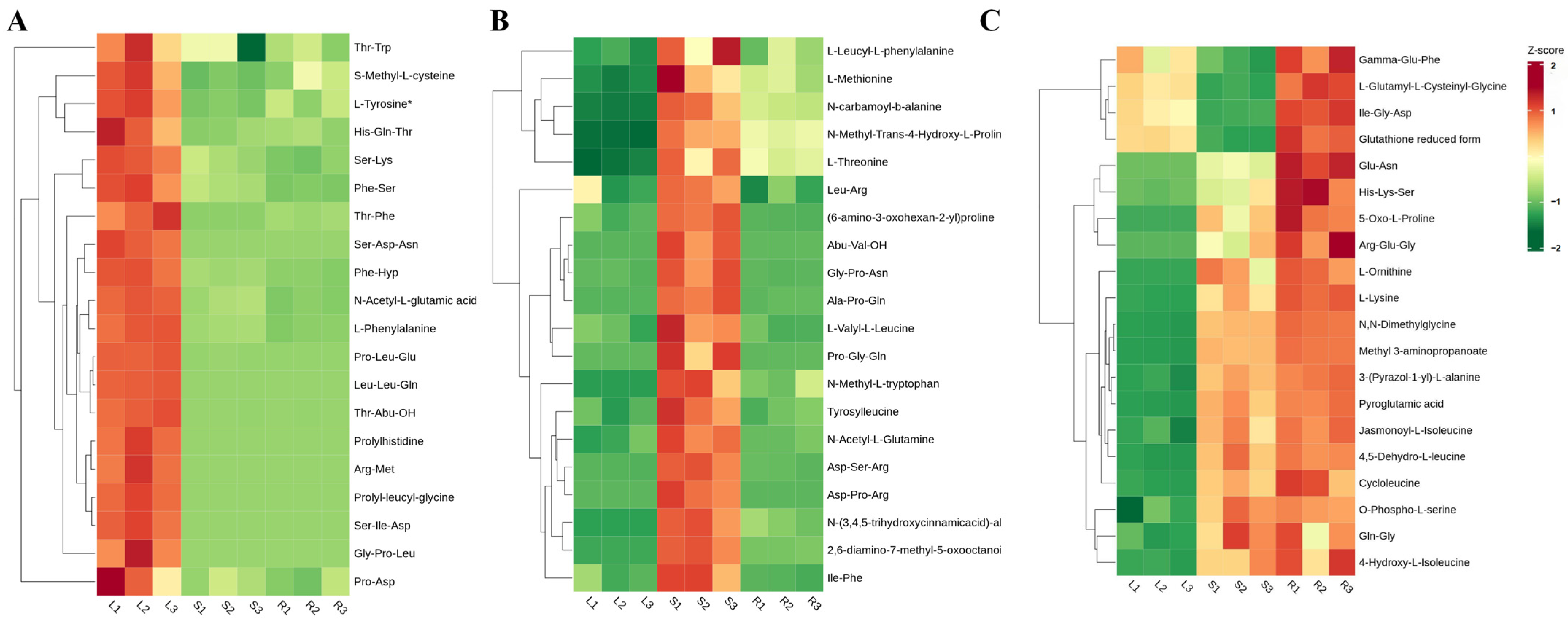
To explore the accumulation characteristics of amino acid metabolites in different organs of C. huchouensis, the differential amino acids in leaves, stems, and roots were compared. A total of 127 significantly differentially expressed amino acid-related metabolites were identified across three organs of C. huchouensis. Significantly higher accumulation of the majority of metabolites occurred in leaves (46 amino acids) and stems (68 amino acids), in contrast to their generally low levels in roots (Figure S1). The results revealed 20 amino acids and their derivatives with significantly high abundance across the three organs. Peptides and modified amino acids, including Thr-Trp, S-methyl-L-cysteine, L-tyrosine, His-Gln-Thr, Ser-Lys, Phe-Ser, Thr-Phe, Ser-Asp-Asn, Phe-Hyp, and N-acetyl-L-glutamic acid, accumulated at markedly higher levels in the leaves (Figure 8A). These metabolites were predominantly dipeptides, tripeptides, and acetylated or methylated derivatives, suggesting active protein turnover and secondary metabolic processes in the leaves. Amino acids such as L-leucyl-L-phenylalanine, L-methionine, N-carbamoyl-β-alanine, N-methyl-trans-4-hydroxy-L-proline, L-threonine, and Leu-Arg were significantly enriched in the stems (Figure 8B). In the roots, amino acids such as γ-Glu-Phe, L-glutamyl-L-cysteinylglycine, Ile-Gly-Asp, reduced glutathione (GSH), Glu-Asn, His-Lys-Ser, 5-oxo-L-proline, and Arg-Glu-Gly were significantly enriched (Figure 8C).

3.8. Comparative Analysis of Relative Contents of Phenolic Acids

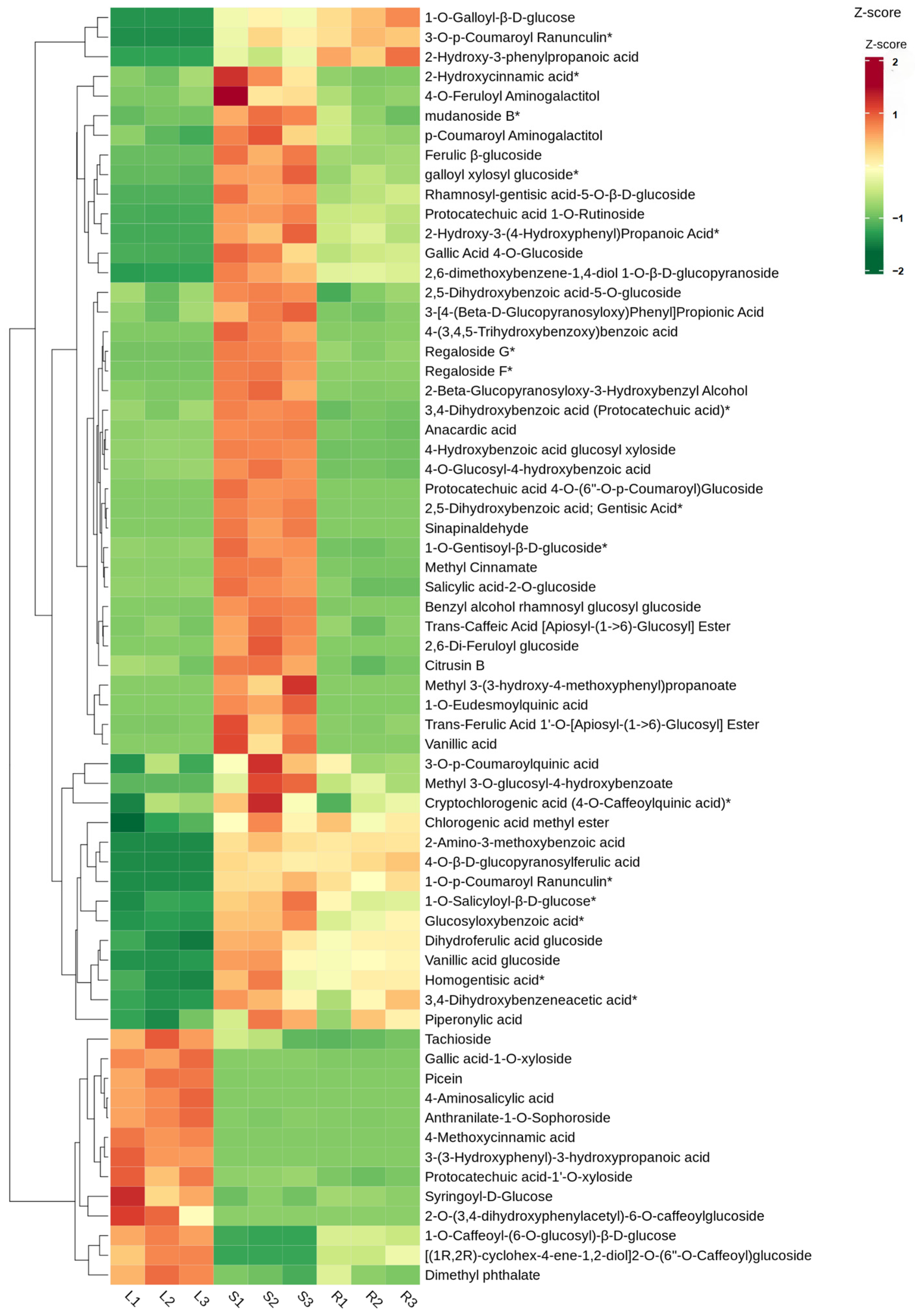
A total of 65 significantly differentially expressed phenolic acid metabolites were detected in the three organs of C. huchouensis (Figure 9). Thirteen phenolic acid metabolites accumulated at significantly higher levels in the leaves, whereas their content remained relatively low in the stems and roots. These leaf-enriched metabolites included tachioside, gallic acid-1-O-xyloside, picein, 4-aminosalicylic acid, anthranilate-1-O-sophoroside, 4-methoxycinnamic acid, 3-(3-hydroxyphenyl)-3-hydroxypropanoic acid, protocatechuic acid-1′-O-xyloside, syringoyl-D-glucose, and 2-O-(3,4-dihydroxyphenylacetyl)-6-O-caffeoylglucoside, among others.
Forty-nine phenolic acid metabolites accumulated predominantly in the stems of C. huchouensis, mainly including 2-hydroxycinnamic acid, 4-O-feruloyl aminogalactitol, mudanoside B, feruloyl β-glucoside, galloyl xylosyl glucoside, gallic acid 4-O-glucoside, regaloside G, regaloside F, sinapinaldehyde, and methyl cinnamate.
In the roots of C. huchouensis, the significantly enriched phenolic acid metabolites were 1-O-galloyl-β-D-glucose, 3-O-p-coumaroylranunculin, and 2-hydroxy-3-phenylpropanoic acid.

3.9. Comparative Analysis of Relative Contents of Other Secondary Metabolites

The cluster heatmap clearly revealed the differential accumulation patterns of 56 common alkaloid metabolites shared among the roots, stems, and leaves across different organs. Overall, the accumulation trends in leaves and stems were highly similar, whereas a clear separation was observed between roots and aboveground parts. This organ-specific distribution pattern reflects the spatial allocation and functional differentiation of alkaloids within the plant (Figure S2). A large number of alkaloids were significantly up-regulated in the leaves (32 alkaloids), followed by the stems (21 alkaloids), indicating that the aboveground organs are the primary sites for alkaloid biosynthesis and accumulation. In contrast, roots showed a distinctly opposing trend to leaves and stems, with the majority of alkaloids displaying significantly reduced abundance in roots. Only three alkaloids (ailanindole, pyrrolidine-2-carboxamide, and caffeine) showed specifically high expression in roots (Figure S2).
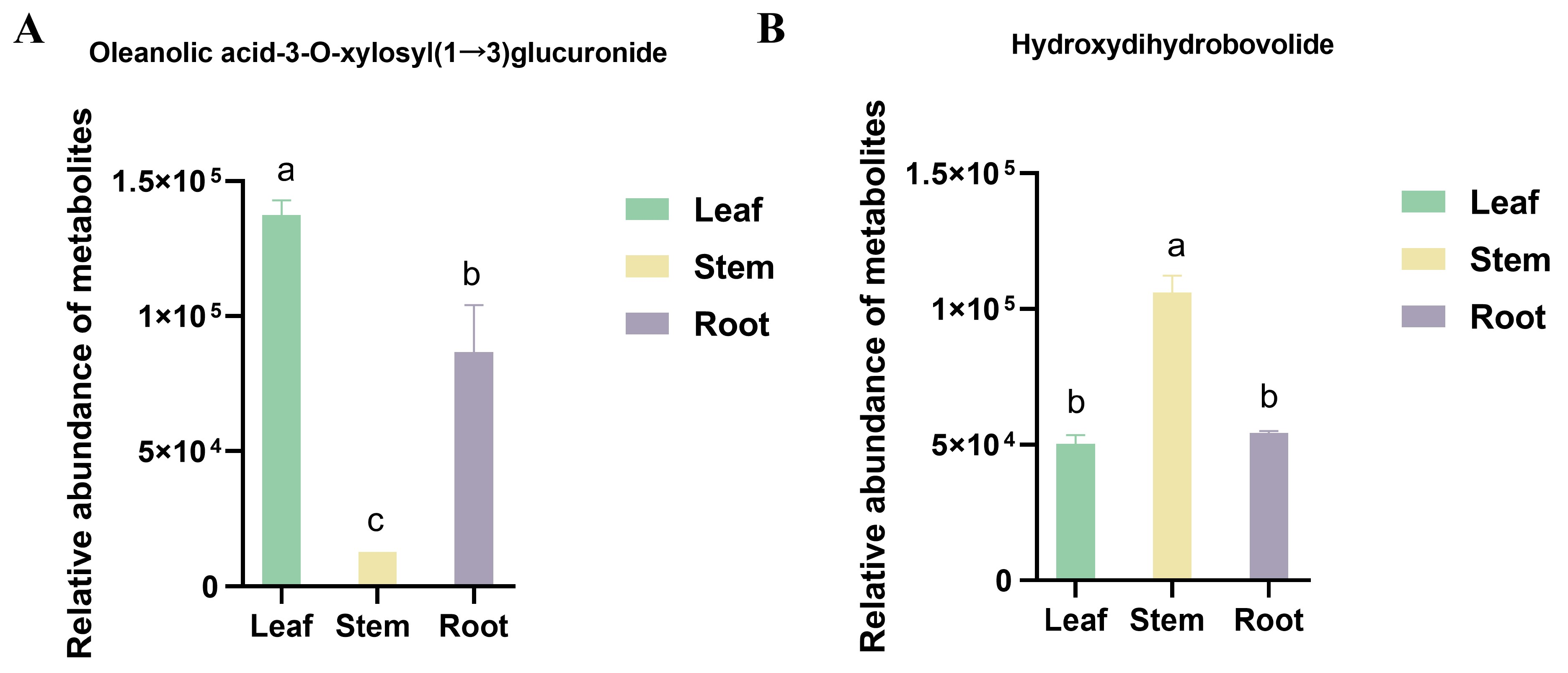
One differential triterpenoid saponin, oleanolic acid-3-O-xylosyl(1→3)glucuronide, and one lactone metabolite, hydroxydihydrobovolide, were detected in the roots, stems, and leaves of C. huchouensis (Figure 10). The relative content of oleanolic acid-3-O-xylosyl(1→3)glucuronide was highest in the leaves, being 10.86-fold higher than in the stems and 1.58-fold higher than in the roots (Figure 10A). The relative content of hydroxydihydrobovolide was highest in the stems, being 2.12-fold higher than in the leaves and 1.95-fold higher than in the roots; it was significantly higher in the stems than in both leaves and roots, whereas no significant difference was observed between leaves and roots (Figure 10B).

4. Discussion

A large number of significantly differentially expressed flavonoid compounds were detected in the stems, and many of them have been confirmed to possess remarkable pharmacological activities. Robinetin not only inhibits HIV-1 integrase and acetylcholinesterase, demonstrating potent antiviral activity, but also exhibits moderate antiproliferative activity in cancer cell lines [17]. 3-Methylkaempferol, isolated from the medicinal plant Psiadia dentata, has been confirmed as an effective inhibitor of poliovirus type 2 replication [18]. Hispidulin triggers reactive oxygen species (ROS)-mediated apoptosis in human non-small cell lung cancer cells via the endoplasmic reticulum stress pathway [19]. Diosmetin alleviates hyperuricemic nephropathy by activating the BNIP3/Nrf2/GPX4 pathway to inhibit ferroptosis of renal tubular epithelial cells induced by monosodium urate crystals [20]. Cyanidin-3-O-glucoside exhibits anti-PRRSV activity [21], while cajanin, a natural small-molecule compound, suppresses osteoclastogenesis and bone resorption both in vitro and in animal models, thereby alleviating pathological bone loss [22]. In addition, bavachin, which was specifically enriched in the leaves, can alleviate rheumatoid arthritis by suppressing the NF-κB signaling pathway and angiogenesis [23], while pedalitin, in combination with amphotericin B, exhibits potent synergistic anticryptococcal activity against Cryptococcus neoformans [24].
Among the amino acid pool identified in the stems, L-tyrosine and L-phenylalanine function as precursors for phenylpropanoid metabolism and flavonoid biosynthesis [25]. The sulfur-containing amino acid S-methyl-L-cysteine has been shown to possess significant pharmacological activities, including antioxidant, anti-inflammatory, and insulin-resistance-improving effects [26]. N-carbamoyl-L-glutamic acid has been approved by the U.S. Food and Drug Administration (FDA) for the treatment of fatal hyperammonemia [27].
Among the amino acids specifically enriched in the leaves, L-methionine, a sulfur-containing amino acid, serves as a core component of the plant heavy metal detoxification network and also as the precursor of S-adenosyl-L-methionine, the principal methyl donor in living organisms, thus contributing to the prevention of heavy metal-induced hepatotoxicity [28]. N-Methyl-trans-4-hydroxy-L-proline also accumulated at high levels in the stems. Studies have demonstrated that it can significantly reduce TNF-α levels and inhibit the activation of nuclear factor-κB (NF-κB), exhibiting potent anti-inflammatory effects [29].
Of particular note is the extremely high accumulation of glutathione and its precursors, γ-Glu-Phe and L-glutamyl-L-cysteinylglycine, in the roots, a phenomenon also observed in rice and alfalfa [30,31]. The remarkably high glutathione levels in roots reflect their role as the primary stress-responsive organ, participating in responses to heavy metal and oxidative stress [30]. 5-Oxo-L-proline also exhibits significant antioxidant activity [32]. The accumulation of these amino acids reflects a robust antioxidant system in the roots of C. huchouensis, suggesting enhanced antioxidant capacity and detoxification potential in this organ.
The phenolic acid components enriched in the leaves also exhibit pharmacological values that should not be overlooked. Among them, tachioside has been attributed with both anti-inflammatory and potent antioxidant activities [33], whereas antioxidant and neuroprotective potential are associated with picein [34]. As a classic anti-tuberculosis drug, 4-aminosalicylic acid exerts anti-mycobacterial effects against Mycobacterium tuberculosis [35]. Turning to the stems, their phenolic acid composition exhibits a distinct functional bias. 2-Hydroxycinnamic acid displays significant antibacterial activity against Staphylococcus aureus [36] and also exhibits distinct neuroprotective properties relevant to Alzheimer’s disease [37]. Methyl cinnamate can activate the CAMKK2-AMPK signaling pathway, thereby inhibiting the differentiation of 3T3-L1 adipocytes and lipid accumulation in HepG2 cells, ultimately reducing triglyceride levels [38].
Although the phenolic acids in the roots are relatively limited in variety, the pharmacological targeting of their individual constituents is equally important. Among these, 1-O-galloyl-β-D-glucose functions as an aldose reductase inhibitor and is considered a promising candidate for the treatment of diabetes [39].
Notably, colchicoside, trigonelline, dopamine, and vasicinone, which accumulated at high levels in the leaves, possess well-defined pharmacological activities. Colchicoside is an anti-gout agent that inhibits microtubule polymerization. This action subsequently blocks neutrophil chemotaxis and the release of inflammatory factors [40]. Trigonelline exhibits hypoglycemic, lipid-lowering, neuroprotective, and antidepressant effects [41,42,43]. Dopamine is a powerful vasoactive medication indicated for hypotension induced by cardiogenic shock or heart failure [44,45]. Vasicinone alleviates asthma and bronchitis by relaxing the airways [46,47]. Similarly, 3-indolepropionic acid, stachydrine, tryptamine, and N-cis-cinnamoylagmatine, which accumulated at high levels in the stems, each possess important functional activities. By scavenging hydroxyl radicals, 3-indolepropionic acid produces neuroprotective effects and has demonstrated efficacy in models of Alzheimer’s and Parkinson’s diseases [48]. Stachydrine, the main active component of Leonurus japonicus, exhibits diuretic, anti-fibrotic, and renal protective effects [49]. Tryptamine is involved in neuromodulation, antioxidation, and immunomodulation [50,51]. N-cis-Cinnamoylagmatine lowers postprandial blood glucose through α-glucosidase inhibition, the same target of the hypoglycemic agent acarbose [52]. Only three alkaloids exhibited specifically high expression in the roots. Among them, caffeine exerts its effects by inhibiting phosphodiesterase 4 to prevent the degradation of cyclic adenosine monophosphate, thereby serving as an adjunct therapy for asthma and chronic obstructive pulmonary disease, while also exhibiting multiple activities including diuretic, antitumor, antioxidant, and skin-protective effects [53].
Oleanolic acid-3-O-xylosyl(1→3)glucuronide is a glycosylated derivative of oleanolic acid and belongs to the oleanane-type triterpenoid saponins. Previous studies have shown that oleanolic acid and its glycoside derivatives exhibit broad-spectrum antitumor effects as well as immunomodulatory activities [54,55]. Studies have identified hydroxydihydrobovolide as a key lactone in Portulaca oleracea that displays cytotoxicity and markedly suppresses the proliferation of the human neuroblastoma SH-SY5Y cell line [56]. Furthermore, hydroxydihydrobovolide isolated from Litsea verticillata has been shown to inhibit HIV, demonstrating significant antiviral potential [56].
C. huchouensis and C. hexapetala share similar chemical profiles, as both are rich in flavonoids and phenolic compounds [57]. Research indicates that these active components exert a marked hypouricemic effect in mouse models of hyperuricemia, while also mitigating renal histopathological injury [57]. The roots of C. chinensis are rich in triterpenoid saponins with novel structural features, which have been demonstrated to exert protective effects on articular cartilage and to effectively suppress local inflammatory responses [9,10]. It is noteworthy that in the mainstream medicinal species of Clematis, such as C. chinensis and Clematis terniflora, the pharmacologically active constituents are predominantly concentrated in the triterpenoid saponins of the roots [9,58]. In contrast, the signature saponin of C. huchouensis, namely oleanolic acid-3-O-xylosyl(1→3)glucuronide, exhibits the highest accumulation in the leaves, followed by the roots. A large number of alkaloids have been isolated from the rhizomes of C. chinensis [59]. However, a substantial number of highly expressed alkaloids, such as colchicine, trigonelline, and dopamine, were also detected in the stems and leaves of C. huchouensis. In marked contrast, the roots exhibited considerably lower alkaloid levels, where only three specifically highly expressed alkaloids (ailanindole, pyrrolidine-2-carboxamide, and caffeine) were detectable. This indicates that the alkaloid resources of this species are predominantly distributed in the above-ground parts. Taken together, these differential distribution patterns suggest that whereas the traditional medicinal use of Clematis is limited to the roots, the stems and leaves of C. huchouensis harbor a diverse array of characteristic bioactive constituents, which provides a chemical rationale for the folk practice of utilizing the whole plant for medicinal purposes.
The results of the present study reveal significant metabolite variations among different organs of C. huchouensis. Nevertheless, such variations are not governed solely by genetic factors. Other factors, including environmental conditions, cultivation practices and developmental stages, may also exert substantial influences on the accumulation of secondary metabolites.
Numerous studies have investigated the effects of environmental factors on the secondary metabolic processes of Clematis species. Ashrafzadeh found that the contents of total phenolics, total flavonoids, and total alkaloids in C. isfahanica under semi-arid climatic conditions were significantly higher than those in plants from arid regions, and that soil pH, electrical conductivity, and total nitrogen content were all positively correlated with metabolite accumulation [60]. Zhang further confirmed that the suitable distribution areas of C. tangutica on the Qinghai–Tibet Plateau are primarily determined by seasonal UV-B radiation and altitude, with flavonoids and phenolic acids reaching peak accumulation at elevations of 2500–3500 m, revealing a coupling mechanism between environmental stress and secondary metabolic pathways [61]. Collectively, these lines of evidence suggest that the distinctive metabolic profiles of C. huchouensis, a geo-authentic herb from Huzhou, may be closely associated with the site-specific hydrological, edaphic, and light conditions of its producing area.
Developmental stage is also a key factor affecting secondary metabolite accumulation. In C. isfahanica, total phenolics, flavonoids, and alkaloids were significantly higher at flowering than during vegetative and mature stages [60]. To minimize developmental variation, all samples in this study were collected during vigorous vegetative growth in June. However, future time-series sampling is still needed to fully assess the dynamic influence of phenological phases on metabolic profiles.
Cultural practices may likewise affect the accumulation of secondary metabolites. Moderate pruning (50%) combined with flower removal is an effective cultivation regulation strategy that can directionally promote the accumulation of bioactive isoflavonoids without compromising root biomass or astragaloside IV content, thereby optimizing the medicinal quality of Astragalus membranaceus var. Mongholicus [62]. The content of taiwanianone A in the tubers of Cynanchum taiwanianum increases with rising potassium fertilizer application, but is reduced by excessive nitrogen fertilization [63].
The wild samples used in this study represent the metabolic traits of the Huzhou Authentic Production Area Specific Habitat. Future work should combine multi-region comparisons, time-series sampling, and controlled cultivation to better understand how genotype–environment interactions shape the metabolic profiles and medicinal quality of C. huchouensis.

5. Conclusions

This study firstly performed widely targeted metabolomic analysis on C. huchouensis from the Huzhou authentic producing area. A total of 1561 metabolites were identified in roots, stems and leaves, showing obvious organ-specific accumulation characteristics. Flavonoids, alkaloids and most phenolic acids were mainly enriched in leaves and stems, while roots accumulated large amounts of glutathione and its precursors, presenting a strong antioxidant and stress defense system. Metabolic differences among organs were mainly involved in flavonoid biosynthesis, linoleic acid metabolism and other key pathways. A variety of metabolites with anti-inflammatory, antitumor, antiviral and other pharmacological activities showed significant tissue-specific distribution. These results revealed the metabolic basis of its medicinal value and provided scientific support for the resource evaluation, quality control and rational utilization of C. huchouensis.

Supplementary Materials

The following supporting information can be downloaded at: https://www.mdpi.com/article/10.3390/metabo16080561/s1, Figure S1. Differential accumulation of amino acids in leaves, stems, and roots of C. huchouensis. The Z-score color scale indicates relative metabolite abundance, with green representing lower abundance and red representing higher abundance. L1, L2, L3: leaves; S1, S2, S3: stems; R1, R2, R3: roots. Figure S2. Differential accumulation of alkaloid in leaves, stems, and roots of C. huchouensis. The Z-score color scale indicates relative metabolite abundance, with green representing lower abundance and red representing higher abundance. L1, L2, L3: leaves; S1, S2, S3: stems; R1, R2, R3: roots.

Author Contributions

M.S. and J.Z.: conceptualization, funding acquisition and writing. G.N. and Y.Z. (Yan Zhang): funding acquisition, supervision and resources. Y.Y. and M.S.: investigation, formal analysis, visualization and writing—original draft. L.Z. and Y.Z. (Yiming Zhang): writing—review and editing. All authors have read and agreed to the published version of the manuscript.

Funding

This study was supported by the High-Level Talent Special Project of Huzhou Vocational & Technical College (Grant No. 2024TS08).

Institutional Review Board Statement

Not applicable.

Informed Consent Statement

Informed consent was obtained from all subjects involved in the study.

Data Availability Statement

The original contributions presented in this study are included in the article. Further inquiries can be directed to the corresponding author.

Conflicts of Interest

The authors declare no conflicts of interest.

References

  1. He, J.; Lin, L.; Chen, Y.; Zhang, X.; Hu, Y.; Feng, L.; Ma, X.; Lin, J.; Hu, Q.; Zheng, J. Research Progress on Molecular Breeding and Application of Clematis Plants. Plants 2025, 14, 3575. [Google Scholar] [CrossRef]
  2. Zhang, Z.; Wang, X.; Zhao, M.; Qian, K. Optimization of polysaccharides extraction from Clematis huchouensis Tamura and its antioxidant activity. Carbohydr. Polym. 2014, 111, 762–767. [Google Scholar] [CrossRef] [PubMed]
  3. Zhao, M.; Da-Wa, Z.-M.; Guo, D.-L.; Fang, D.-M.; Chen, X.-Z.; Xu, H.-X.; Gu, Y.-C.; Xia, B.; Chen, L.; Ding, L.-S.; et al. Cytotoxic triterpenoid saponins from Clematis tangutica. Phytochemistry 2016, 130, 228–237. [Google Scholar] [CrossRef] [PubMed]
  4. Guo, S.; Yang, Y.; Bian, S.; Gao, L.; Zhang, Z.; Zhu, L. A Review of Studies on Clematis Plants in China. J. Agric. 2025, 15, 57–67. [Google Scholar]
  5. Redmond, C.M.; Stout, J.C. Breeding system and pollination ecology of a potentially invasive alien Clematis vitalba L. in Ireland. J. Plant Ecol. 2018, 11, 56–63. [Google Scholar] [CrossRef]
  6. Chawla, R.; Kumar, S.; Sharma, A. The genus Clematis (Ranunculaceae): Chemical and pharmacological perspectives. J. Ethnopharmacol. 2012, 143, 116–150. [Google Scholar] [CrossRef] [PubMed]
  7. Hao, D.; Gu, X.; Xiao, P.; Peng, Y. Chemical and biological research of Clematis medicinal resources. Chin. Sci. Bull. 2013, 58, 1120–1129. [Google Scholar]
  8. Wu, W.; Xu, X.; Dai, Y.; Xia, L. Therapeutic effect of the saponin fraction from Clematis chinensis Osbeck roots on osteoarthritis induced by monosodium iodoacetate through protecting articular cartilage. Phytother. Res. 2010, 24, 538–546. [Google Scholar] [PubMed]
  9. Wu, W.; Gao, X.; Xu, X.; Luo, Y.; Liu, M.; Xia, Y.; Dai, Y. Saponin-rich fraction from Clematis chinensis Osbeck roots protects rabbit chondrocytes against nitric oxide-induced apoptosis via preventing mitochondria impairment and caspase-3 activation. Cytotechnology 2013, 65, 287–295. [Google Scholar] [PubMed][Green Version]
  10. Wu, Z.; Huang, B.; Zhang, D.; Yu, Q.; Yan, C. Structural characterization of a pectin-like polysaccharide from Clematis chinensis Osbeck and its anti-rheumatoid arthritis activity. Int. J. Biol. Macromol. 2025, 307, 141917. [Google Scholar] [CrossRef] [PubMed]
  11. Han, W.; Xiong, Y.; Li, Y.; Fang, W.; Ma, Y.; Liu, L.; Li, F.; Zhu, X. Anti-arthritic effects of clematichinenoside (AR-6) on PI3K/Akt signaling pathway and TNF-α associated with collagen-induced arthritis. Pharm. Biol. 2013, 51, 13–22. [Google Scholar] [PubMed]
  12. Dong, S.; Zhang, J.Y.; Zhao, J.L.; Li, G.Q.; Yan, B.X.; Lv, C.N.; Yuan, J.Z.; Lu, J.C. Three new phenols and one new lignan from Clematis terniflora var. manshurica (Rupr.) Ohwi with their anti-inflammatory activity. Fitoterapia 2024, 177, 106043. [Google Scholar] [CrossRef] [PubMed]
  13. Xiong, J.; Bui, V.B.; Liu, X.H.; Hong, Z.L.; Yang, G.X.; Hu, J.F. Lignans from the stems of Clematis armandii (“Chuan-Mu-Tong”) and their anti-neuroinflammatory activities. J. Ethnopharmacol. 2014, 153, 737–743. [Google Scholar] [CrossRef] [PubMed]
  14. Li, S.-G.; Huang, X.-J.; Li, M.-M.; Wang, M.; Feng, R.-B.; Zhang, W.; Li, Y.-L.; Wang, Y.; Ye, W.-C. Triterpenoid Saponins from the Roots of Clematis uncinata. Chem. Pharm. Bull. 2014, 62, 35–44. [Google Scholar] [CrossRef] [PubMed][Green Version]
  15. Wu, Y.; Zhang, Y.F.; Zhang, H.T.; Ma, X.H.; Yang, N.N.; Feng, S.B.; Zhong, T.H.; Zhang, Y.H. A new cyclopeptide alkaloid from Clematis florida. Nat. Prod. Res. 2022, 36, 1693–1699. [Google Scholar] [PubMed]
  16. Atmani, D.; Chaher, N.; Berboucha, M.; Ayouni, K.; Lounis, H.; Boudaoud, H.; Debbache, N.; Atmani, D. Antioxidant capacity and phenol content of selected Algerian medicinal plants. Food Chem. 2009, 112, 303–309. [Google Scholar] [CrossRef]
  17. Jakimiuk, K. A Comprehensive Review of Robinetin: Distribution, Biological Activity and Pharmacokinetic Parameters. Int. J. Mol. Sci. 2025, 26, 9546. [Google Scholar] [CrossRef] [PubMed]
  18. Robin, V.; Irurzun, A.; Amoros, M.; Boustie, J.; Carrasco, L. Antipoliovirus flavonoids from Psiadia dentata. Antivir. Chem. Chemother. 2001, 12, 283–291. [Google Scholar] [CrossRef] [PubMed]
  19. Lv, L.; Zhang, W.; Li, T.; Jiang, L.; Lu, X.; Lin, J. Hispidulin exhibits potent anticancer activity in vitro and in vivo through activating ER stress in non-small-cell lung cancer cells. Oncol. Rep. 2020, 43, 1995–2003. [Google Scholar] [PubMed]
  20. Hu, C.; Dong, C.; Yang, H.; Yu, Z.; Li, Y.; Qiu, Y.; Mei, Z.; Xu, L. Diosmetin alleviates hyperuricemic nephropathy by activating the BNIP3/Nrf2/GPX4 pathway to inhibit ferroptosis of renal tubular epithelial cells induced by monosodium urate crystals. J. Nutr. Biochem. 2026, 150, 110241. [Google Scholar] [CrossRef] [PubMed]
  21. Chen, X.; Pang, Y.; Amona, F.M.; Liu, Z.; Wang, F.; Liang, Y.; Yang, J.; Zhang, W.; Fang, X.; Chen, X. Self-assembled cyanidin-3-O-glucoside nanoparticles alleviate inflammation and ferroptosis induced by PRRSV infection. J. Virol. 2025, 99, e0095425. [Google Scholar] [CrossRef] [PubMed]
  22. Ma, C.; Mo, L.; Wang, Z.; He, W.; Niu, W.; Goodman, S.B.; Goodnough, L.H.; Liu, Y. Cajanin transcriptionally disrupts the Siglec15/NFATc1 signaling cascade to attenuate osteoclast fusion and bone resorption. Int. Immunopharmacol. 2026, 168, 115726. [Google Scholar] [CrossRef] [PubMed]
  23. Chakraborty, D.; Malik, S.; Sarkar, A.; Mann, S.; Agnihotri, P.; Saquib, M.; Joshi, L.; Kumar, V.; Biswas, S. Bavachin suppresses NF-κB signaling and angiogenic pathways to alleviate rheumatoid arthritis. Arch. Pharmacal Res. 2026, 49, 339–356. [Google Scholar] [CrossRef] [PubMed]
  24. Sangalli-Leite, F.; Scorzoni, L.; Alves de Paula, E.S.A.C.; da Silva, J.F.; de Oliveira, H.C.; de Lacorte Singulani, J.; Gullo, F.P.; Moraes da Silva, R.; Regasini, L.O.; Siqueira da Silva, D.H.; et al. Synergistic effect of pedalitin and amphotericin B against Cryptococcus neoformans by in vitro and in vivo evaluation. Int. J. Antimicrob. Agents 2016, 48, 504–511. [Google Scholar] [CrossRef] [PubMed]
  25. Lee, H.; Lee, S.B.; Park, S.; Song, J.; Kim, B.-G. Biochemical evaluation of molecular parts for flavonoid production using plant synthetic biology. Front. Plant Sci. 2025, 16, 1528122. [Google Scholar] [CrossRef] [PubMed]
  26. Thomas, S.; Senthilkumar, G.P.; Sivaraman, K.; Bobby, Z.; Paneerselvam, S.; Harichandrakumar, K.T. Effect of s-methyl-L-cysteine on oxidative stress, inflammation and insulin resistance in male wistar rats fed with high fructose diet. Iran. J. Med. Sci. 2015, 40, 45–50. [Google Scholar] [PubMed]
  27. Chapel-Crespo, C.C.; Diaz, G.A.; Oishi, K. Efficacy of N-carbamoyl-L-glutamic acid for the treatment of inherited metabolic disorders. Expert. Rev. Endocrinol. Metab. 2016, 11, 467–473. [Google Scholar] [CrossRef] [PubMed]
  28. Friedel, H.A.; Goa, K.L.; Benfield, P. S-Adenosyl-L-Methionine. Drugs 1989, 38, 389–416. [Google Scholar] [CrossRef] [PubMed]
  29. de Aquino, P.E.A.; Magalhães, T.R.; Nicolau, L.A.D.; Leal, L.K.A.M.; de Aquino, N.C.; dos Santos, S.M.; Neves, K.R.T.; Silveira, E.R.; Viana, G.S.d.B. The anti-inflammatory effects of N-methyl-(2S,4R)-trans-4-hydroxy-l-proline from Syderoxylon obtusifolium are related to its inhibition of TNF-alpha and inflammatory enzymes. Phytomedicine 2017, 24, 14–23. [Google Scholar] [CrossRef] [PubMed]
  30. Swapan Kumar, R.; Mollah, N.; Faizur, R. Glutathione: A key frontier of heavy-metal detoxification and tolerance in plants. Plant Trends 2023, 1, 33–42. [Google Scholar] [CrossRef]
  31. Hasanuzzaman, M.; Nahar, K.; Anee, T.I.; Fujita, M. Glutathione in plants: Biosynthesis and physiological role in environmental stress tolerance. Physiol. Mol. Biol. Plants 2017, 23, 249–268. [Google Scholar] [CrossRef] [PubMed]
  32. Lei, S.; Rossi, S.; Yang, Z.; Yu, J.; Huang, B. Metabolic regulation of 5-oxoproline for enhanced heat tolerance in perennial ryegrass. Stress Biol. 2024, 4, 46. [Google Scholar] [CrossRef] [PubMed]
  33. Wang, J.; Liu, R.; Zhang, J.; Su, H.; Yang, Q.; Wulu, J.; Li, J.; Zhang, Z.; Lv, Z. Comparative analysis of phytochemical profile and antioxidant and anti-inflammatory activity of four Gentiana species from the Qinghai-Tibet Plateau. J. Ethnopharmacol. 2024, 326, 117926. [Google Scholar] [CrossRef] [PubMed]
  34. Kesari, K.K.; Dhasmana, A.; Shandilya, S.; Prabhakar, N.; Shaukat, A.; Dou, J.; Rosenholm, J.M.; Vuorinen, T.; Ruokolainen, J. Plant-Derived Natural Biomolecule Picein Attenuates Menadione Induced Oxidative Stress on Neuroblastoma Cell Mitochondria. Antioxidants 2020, 9, 552. [Google Scholar] [CrossRef] [PubMed]
  35. Zheng, J.; Rubin, E.J.; Bifani, P.; Mathys, V.; Lim, V.; Au, M.; Jang, J.; Nam, J.; Dick, T.; Walker, J.R.; et al. para-Aminosalicylic Acid Is a Prodrug Targeting Dihydrofolate Reductase in Mycobacterium tuberculosis. J. Biol. Chem. 2013, 288, 23447–23456. [Google Scholar] [CrossRef] [PubMed]
  36. Yingyongnarongkul, B.-e.; Apiratikul, N.; Aroonrerk, N.; Suksamrarn, A. Solid-phase synthesis and antibacterial activity of hydroxycinnamic acid amides and analogues against methicillin-resistant Staphylococcus aureus and vancomycin-resistant S. aureus. Bioorganic Med. Chem. Lett. 2006, 16, 5870–5873. [Google Scholar] [CrossRef] [PubMed]
  37. Hao, S.; Li, X.; Han, A.; Yang, Y.; Luo, X.; Fang, G.; Wang, H.; Liu, J.; Wang, S. Hydroxycinnamic Acid from Corncob and Its Structural Analogues Inhibit Aβ40 Fibrillation and Attenuate Aβ40-Induced Cytotoxicity. J. Agric. Food Chem. 2020, 68, 8788–8796. [Google Scholar] [CrossRef] [PubMed]
  38. Yingda, F.; Guangbing, L.; Zichen, F.; Jun, L.; Xiaoyu, W.; Tao, W.; Jun, L. Methyl Cinnamate (MC) Alleviates Free Fatty Acids (FFAs) Induced Lipid Accumulation Through the AMPK Pathway in HepG2 Cells. Diabetes Metab. Syndr. Obes. Targets Ther. 2024, 17, 1183–1197. [Google Scholar] [CrossRef] [PubMed]
  39. Puppala, M.; Ponder, J.; Suryanarayana, P.; Reddy, G.B.; Petrash, J.M.; LaBarbera, D.V. The isolation and characterization of β-glucogallin as a novel aldose reductase inhibitor from Emblica officinalis. PLoS ONE 2017, 7, e31399. [Google Scholar]
  40. Leung, Y.Y.; Yao Hui, L.L.; Kraus, V.B. Colchicine—Update on mechanisms of action and therapeutic uses. Semin. Arthritis Rheum. 2015, 45, 341–350. [Google Scholar] [CrossRef] [PubMed]
  41. Sarker, D.K.; Ray, P.; Dutta, A.K.; Rouf, R.; Uddin, S.J. Antidiabetic potential of fenugreek (Trigonella foenum-graecum): A magic herb for diabetes mellitus. Food Sci. Nutr. 2024, 12, 7108–7136. [Google Scholar] [CrossRef] [PubMed]
  42. Majumdar, J.; Chatterjee, A.; Chakraborty, S.B.; Chakraborty, P.; Sarkar, S. Comparative efficacy of four potent bioactive molecules of fenugreek seed in holistic management of type 1 diabetes. Adv. Tradit. Med. 2024, 25, 411–424. [Google Scholar] [CrossRef]
  43. Farid, M.M.; Yang, X.; Kuboyama, T.; Tohda, C. Trigonelline recovers memory function in Alzheimer’s disease model mice: Evidence of brain penetration and target molecule. Sci. Rep. 2020, 10, 16424. [Google Scholar] [CrossRef] [PubMed]
  44. Théroux, P.; Mizgala, H.F.; Bourassa, M.G. Hemodynamic and therapeutic effects of intravenous dopamine. Can. Med. Assoc. J. 1977, 116, 645–648. [Google Scholar] [PubMed]
  45. Stemple, D.R.; Kleiman, J.H.; Harrison, D.C. Combined nitroprusside-dopamine therapy in severe chronic congestive heart failure. Dose-related hemodynamic advantages over single drug infusions. Am. J. Cardiol. 1978, 42, 267–275. [Google Scholar] [PubMed]
  46. Cambridge, G.W.; Jansen, A.B.A.; Jarman, D.A. Bronchodilating Action of Vasicinone and Related Compounds. Nature 1962, 196, 1217. [Google Scholar] [CrossRef] [PubMed]
  47. Qu, L.; Du, W.; Ren, Z.; Chen, M.; Wu, X.; Cao, X.; Rao, G.; Tong, X.; Huang, F.; Sun, Y. Vasicine Attenuates Allergic Asthma by Suppressing Mast Cell Degranulation and Th2 Inflammation via Modulation of the FcεRI/Lyn + Syk/MAPK Pathway. Pharmaceuticals 2026, 19, 190. [Google Scholar] [CrossRef] [PubMed]
  48. Chyan, Y.J.; Poeggeler, B.; Omar, R.A.; Chain, D.G.; Frangione, B.; Ghiso, J.; Pappolla, M.A. Potent neuroprotective properties against the Alzheimer beta-amyloid by an endogenous melatonin-related indole structure, indole-3-propionic acid. J. Biol. Chem. 1999, 274, 21937–21942. [Google Scholar] [CrossRef] [PubMed]
  49. Cheng, F.; Zhou, Y.; Wang, M.; Guo, C.; Cao, Z.; Zhang, R.; Peng, C. A review of pharmacological and pharmacokinetic properties of stachydrine. Pharmacol. Res. 2020, 155, 104755. [Google Scholar] [CrossRef] [PubMed]
  50. Sabnis, R.W.; Sabnis, A.R. Novel Tryptamine Compounds as 5-HT2A Agonists for Treating Mood Disorders such as Depressive Disorders and Bipolar Disorders. ACS Med. Chem. Lett. 2025, 16, 373–374. [Google Scholar] [CrossRef] [PubMed]
  51. Xie, Y.; Liang, S.; Zhang, Y.; Wu, T.; Shen, Y.; Yao, S.; Li, J. Discovery of indole analogues from Periplaneta americana extract and their activities on cell proliferation and recovery of ulcerative colitis in mice. Front. Pharmacol. 2023, 14, 1282545. [Google Scholar] [CrossRef] [PubMed]
  52. Wang, X.; Liu, H.; Liu, Y.; Zhang, D.; Zhu, L.; Wang, G.; Chen, H.; Chai, Y. Natural guanidine alkaloids in tea (Camellia sinensis): Identification, quantification and α-glucosidase inhibitory activity of cis- and trans-cinnamoylagmatines. Food Chem. 2025, 492, 145311. [Google Scholar] [CrossRef] [PubMed]
  53. Sharma, V.; Sharma, A.; Verma, K.; Gaur, P.; Kaushik, R.; Abdali, B. A comprehensive review on pharmacological potentials of caffeine. J. Appl. Pharm. Sci. Res. 2023, 6, 16–26. [Google Scholar] [CrossRef]
  54. Ma, L.; Mahemuti, D.; Lan, Y.; Xu, J.; Li, W.; Su, Z.; Li, J.; Abuduwaili, A.; Maimaitijiang, A. Terpenoids: Emerging Natural Modulators for Reversing ABC Transporter-Mediated Multidrug Resistance in Cancer Chemotherapy. Pharmaceuticals 2026, 19, 146. [Google Scholar] [CrossRef] [PubMed]
  55. Srivastava, R.; Jaiswal, N.; Kharkwal, H.; Dubey, N.K.; Srivastava, R. Phytomedical Properties of Carica papaya for Boosting Human Immunity Against Viral Infections. Viruses 2025, 17, 271. [Google Scholar] [CrossRef] [PubMed]
  56. Zhang, H.J.; Nguyen, V.H.; Nguyen, M.C.; Soejarto, D.D.; Pezzuto, J.M.; Fong, H.H.; Tan, G.T. Sesquiterpenes and butenolides, natural anti-HIV constituents from Litsea verticillata. Planta Medica 2005, 71, 452–457. [Google Scholar] [CrossRef] [PubMed]
  57. Wang, W.; Min, Z.; Luo, C.; Yang, M.; Chen, L. Extract of Clematis hexapetala regulates uric acid metabolism, alleviates renal inflammation and fibrosis via the PI3K/AKT/NF-κB signaling pathway, and improves hyperuricemia in mice. J. Ethnopharmacol. 2026, 367, 121698. [Google Scholar] [CrossRef] [PubMed]
  58. Dong, S.; Lu, A.; Li, K.; Zhao, S.; Zhao, J.; Yuan, J.; Lv, C.; Lu, J. Triterpenoid saponins from the roots and rhizomes of Clematis terniflora var. manshurica (Rupr.) Ohwi and their anti-inflammatory activities. Phytochemistry 2026, 246, 114786. [Google Scholar] [CrossRef] [PubMed]
  59. Jiang, H.; Shao, S.; Wei, Y.; Lin, B.; Li, S. Phenolic glycosides and indole alkaloids isolated from the roots and rhizomes of Clematis chinensis and their anti-inflammatory activity. Phytochemistry 2023, 215, 113832. [Google Scholar] [CrossRef] [PubMed]
  60. Ashrafzadeh, M.; Niknahad Gharmakher, H.; Saharkhiz, M.J.; Ghorbani Nohooji, M. Effects of Phenological Stages and Ecological Factors on Secondary Metabolites of Clematis ispahanica Boiss. Ecopersia 2023, 11, 93–104. [Google Scholar]
  61. Zhang, H.; Guo, X.; Wei, X.; Wang, L.; Zhong, Q.; Sun, X. Potential Distribution Prediction and Metabolite Analysis of Clematis tangutica(Maxim.) Korsh. On the Qinghai Plateau. Ecol. Evol. 2025, 15, e72110. [Google Scholar] [CrossRef] [PubMed]
  62. Law, H.M.; Eyles, A.; Close, D.C. Effects of pruning with flower removal on the growth, starch reserves and secondary metabolites of a medicinal herb, Astragalus mongholicus. S. Afr. J. Bot. 2026, 189, 120–130. [Google Scholar] [CrossRef]
  63. Tseng, H.M.; Lu, T.M.; Ng, L.T. Responses of Cynanchum taiwanianum and its bioactive compound biosynthesis to levels of nitrogen and potassium fertilization. Agronomy 2022, 12, 180. [Google Scholar] [CrossRef]
Figure 1. Morphological illustration of the root, stem, and leaf of C. huchouensis.
Metabolites 16 00561 g001
Figure 2. Overlay of total ion chromatograms from QC samples. (A): Negative ion mode; (B): Positive ion mode; (C): Distribution of CV values in QC samples.
Metabolites 16 00561 g002
Figure 3. Metabolite distribution characteristics. (A) Identify the types and proportions of metabolites; (B) Proportion of metabolite content in roots of C. huchouensis; (C) Proportion of metabolite content in stems of C. huchouensis; (D) Proportion of metabolite content in leaves of C. huchouensis. (E) The top ten metabolites in terms of content in the roots of C. huchouensis; (F) The top ten metabolites in terms of content in the stems of C. huchouensis; (G) The top ten metabolites in terms of content in the leaves of C. huchouensis.
Metabolites 16 00561 g003
Figure 4. Multivariate statistical analysis. (A) Correlation diagram between samples; (B) PCA score plot; (C) OPLS-DA plot; (D) OPLS-DA validation diagram.
Metabolites 16 00561 g004
Figure 5. Metabolite analysis. (A) Volcano plot of DAMs in the root vs. leaf comparison. (B) Volcano plot of DAMs in the root vs. stem comparison. (C) Volcano plot of DAMs in the stem vs. leaf comparison. (D) Top 20 metabolites ranked by log2FC in the root vs. leaf comparison. (E) Top 20 metabolites ranked by log2FC in the root vs. stem comparison. (F) Top 20 metabolites ranked by log2FC in the stem vs. leaf comparison. Log2FC represents the log2-transformed fold change. Red indicates up-regulated metabolites, and green indicates down-regulated metabolites. The marked compounds (*) are isomers that cannot be distinguished by mass spectrometry. Different quantifier ions were selected for certain isomers during library construction, resulting in separate cumulative quantifications.
Metabolites 16 00561 g005
Figure 6. Metabolite analysis. KEGG enrichment plot of DAMs in different organs of C. huchouensis. (A) KEGG enrichment plot of DAMs in the root vs. leaf comparison. (B) KEGG enrichment plot of DAMs in the root vs. stem comparison. (C) KEGG enrichment plot of DAMs in the stem vs. leaf comparison. The color of the dots represents the p-value from the hypergeometric test; the color gradient ranges from purple to red, with more intense red indicating smaller p-values and thus higher statistical significance. The size of the dots denotes the number of differential metabolites in the corresponding pathway, with larger dots indicating a greater number of differential metabolites.
Metabolites 16 00561 g006
Figure 7. Differential flavonoid accumulation in leaves, stems, and roots of C. huchouensis. (A) Heatmap of all flavonoids showing significant differences among the three organs (leaves, stems, and roots). (B) Heatmap of the top 20 significantly up-regulated flavonoids in stems compared to leaves and roots. (C) Heatmap of the top 20 significantly up-regulated flavonoids in leaves compared to stems and roots. The Z-score color scale indicates relative metabolite abundance, with green representing lower abundance and red representing higher abundance. L1, L2, L3: leaves; S1, S2, S3: stems; R1, R2, R3: roots. The marked compounds (*) are isomers that cannot be distinguished by mass spectrometry. Different quantifier ions were selected for certain isomers during library construction, resulting in separate cumulative quantifications.
Metabolites 16 00561 g007
Figure 8. Differential amino acid accumulation in leaves, stems, and roots of C. huchouensis. (A) Heatmap of the top 20 significantly up-regulated amino acids in leaves compared to stems and roots. (B) Heatmap of the top 20 significantly up-regulated amino acids in stems compared to leaves and roots. (C) Heatmap of the top 20 significantly up-regulated amino acids in roots compared to leaves and stems. The Z-score color scale indicates relative metabolite abundance, with green representing lower abundance and red representing higher abundance. L1, L2, L3: leaves; S1, S2, S3: stems; R1, R2, R3: roots. The marked compounds (*) are isomers that cannot be distinguished by mass spectrometry. Different quantifier ions were selected for certain isomers during library construction, resulting in separate cumulative quantifications.
Metabolites 16 00561 g008
Figure 9. Differential accumulation of phenolic acids in leaves, stems, and roots of C. huchouensis. The Z-score color scale indicates relative metabolite abundance, with green representing lower abundance and red representing higher abundance. L1, L2, L3: leaves; S1, S2, S3: stems; R1, R2, R3: roots. The marked compounds (*) are isomers that cannot be distinguished by mass spectrometry. Different quantifier ions were selected for certain isomers during library construction, resulting in separate cumulative quantifications.
Metabolites 16 00561 g009
Figure 10. Relative contents of oleanolic acid-3-O-xylosyl(1→3)glucuronide and hydroxydihydrobovolide in the leaves, stems, and roots of C. huchouensis. (A) Oleanolic acid-3-O-xylosyl(1→3)glucuronide. (B) Hydroxydihydrobovolide. Lowercase letters denote significant differences (p < 0.05) based on one-way ANOVA with Duncan’s multiple comparison test.
Metabolites 16 00561 g010
Disclaimer/Publisher’s Note: The statements, opinions and data contained in all publications are solely those of the individual author(s) and contributor(s) and not of MDPI and/or the editor(s). MDPI and/or the editor(s) disclaim responsibility for any injury to people or property resulting from any ideas, methods, instructions or products referred to in the content.