2. Materials and Methods
2.1. Chemicals and Reagents
Optima-grade methanol was purchased from Thermo Fisher Scientific (Waltham, MA, USA), and distilled water was obtained from Watson’s (Hong Kong, China). Reference standards, including quercetin, euscaphic acid, protocatechuic aldehyde, citric acid, quinic acid, hyperoside, astragalin, kaempferol, kaempferol-3-O-rutinoside, oleanolic acid, asiatic acid, ursolic acid, corilagin, and epicatechin, were purchased from Chengdu Must Bio-Technology Co., Ltd. (Chengdu, China). Malic acid was obtained from Shanghai Standard Technology Service Co., Ltd. (Shanghai, China). Pomolic acid and tartaric acid were purchased from Baoji Herbest Bio-Tech Co., Ltd. (Baoji, China). Raffinose, arginine, and sucrose were sourced from Shanghai Chunyou Bio-Technology Co., Ltd. (Shanghai, China). Rutin, tiliroside, tormentic acid, and isoquercitrin were purchased from Chengdu Herbpurify Co., Ltd. (Chengdu, China). Ellagic acid and gallic acid were obtained from Sichuan Vicky Biotechnology Co., Ltd. (Chengdu, China), while luteoloside was provided by the National Institutes for Food and Drug Control (Beijing, China). The purity of all reference standards was verified to be >98%. Absolute ethanol was purchased from Tianjin Xinbote Chemical Co., Ltd. (Tianjin, China). Sodium formate was obtained from Merck (Darmstadt, Hesse, Germany), and leucine-enkephalin was purchased from Waters Corp. (Milford, MA, USA).
Regarding biological reagents, the 40% kcal high-fat diet (HFD) was purchased from Beijing Boaigang Biological technology (Beijing, China). Commercial kits for total cholesterol (TC), triglycerides (TG), high-density lipoprotein cholesterol (HDL-C), and low-density lipoprotein cholesterol (LDL-C) were obtained from Nanjing Jiancheng Bioengineering Institute (Nanjing, China). Rosuvastatin calcium tablets were purchased from Simcere Pharmaceutical Co., Ltd. (Nanjing, China), and fenofibrate capsules were from Abbott Laboratories (Shanghai, China). Primary antibodies against NFE2L2 (Cat No. 80593-1-RR), NOS3 (Cat No. 27120-1-AP), CYP1A1 (Cat No. 13241-1-AP), PPARG (Cat No. 16643-1-AP), and MAPK1 (Cat No. 51068-1-AP) were purchased from Proteintech Group (Wuhan, China). The GAPDH (Cat No. GB11002-100) antibody was obtained from Servicebio Technology Co., Ltd. (Wuhan, China). Additional supplies, including the one-step gel casting kit, were purchased from Shanghai Yaemei Biotechnology Co., Ltd. (Shanghai, China); the tri-color pre-stained protein marker was from Life-ilab Co., Ltd. (Shanghai, China); the ECL chemiluminescence kit was from Beijing Lanjieco Technology Co., Ltd. (Beijing, China); and PVDF membranes were purchased from Roche (Basel, Switzerland).
2.3. UPLC-QTOF-MS Analysis of Chemical Constituents in FRLE from Different Origins
The FRLE samples were separated using an ultra-performance liquid chromatography (UPLC) system (I-Class, Waters, Milford, MA, USA) coupled with a quadrupole time-of-flight mass spectrometer (QTOF-MS; Xevo G2-XS, Waters, Milford, MA, USA) equipped with an electrospray ionization (ESI) source.
Chromatographic conditions: Samples were injected into an ACQUITY UPLC HSS T3 column (2.1 mm × 100 mm, 1.8 µm; Waters) for separation. The column temperature was maintained at 40 °C, and the injection volume was 1 µL. The flow rate was set at 0.2 mL/min. The mobile phase consisted of methanol (A) and 0.1% formic acid in water (v/v, B). The gradient elution program was optimized as follows: 0–3 min, 0% A; 3–20 min, 0–45% A; 20–23 min, 45% A; 23–28 min, 45–100% A; 28–31 min, 100% A; 31–36 min, 100–0% A; and 36–39 min, 0% A.
Mass spectrometry was performed in MSE mode. For ESI (+), the parameters were set as follows: source temperature, 120 °C; capillary voltage, 3.5 kV; sampling cone voltage, 40 V; source offset voltage, 80 V; cone gas flow, 50 L/h desolvation temperature, 450 °C; and desolvation gas flow, 900 L/h. The collision energy was set at 6 eV for the low-energy function and ramped from 20 to 60 eV for the high-energy function. For real-time mass calibration, leucine-enkephalin was utilized as the lock-spray (ESI+: m/z 556.2771; ESI-: m/z 554.2615), and 5 mM sodium formate was used for multi-point external calibration. The ESI (−) parameters were identical to those of ESI (+), except that the capillary voltage was adjusted to 2.5 kV.
Data acquisition and processing, including the analysis of constituent differences across various origins, were conducted using Progenesis QI software (Ver. 3.0, Waters, Milford, MA, USA).
2.4. Chemical Constituent Analysis of FRLE
The FRLE samples exhibiting the highest number of chemical constituents among the five different origins was selected for further investigation. The corresponding MSE raw data were imported into the UNIFI informatics platform (Ver. 1.9.4, Waters, Milford, MA, USA). Chemical constituents within FRLE were identified by matching against an in-house chemical information database.
The in-house database was established as follows: comprehensive literature reviews were conducted to collect mass spectrometry data (including precursor and fragment ion information) of other medicinal plants within the same genus (
Rosa). The structural information of these compounds in .mol format was retrieved from the ChemSpider database (
https://www.chemspider.com/, accessed on 31 May 2026). These structural files were subsequently imported into the UNIFI compound library, where the collected precursor and fragment ion data were appended as supplementary mass spectrometry parameters.
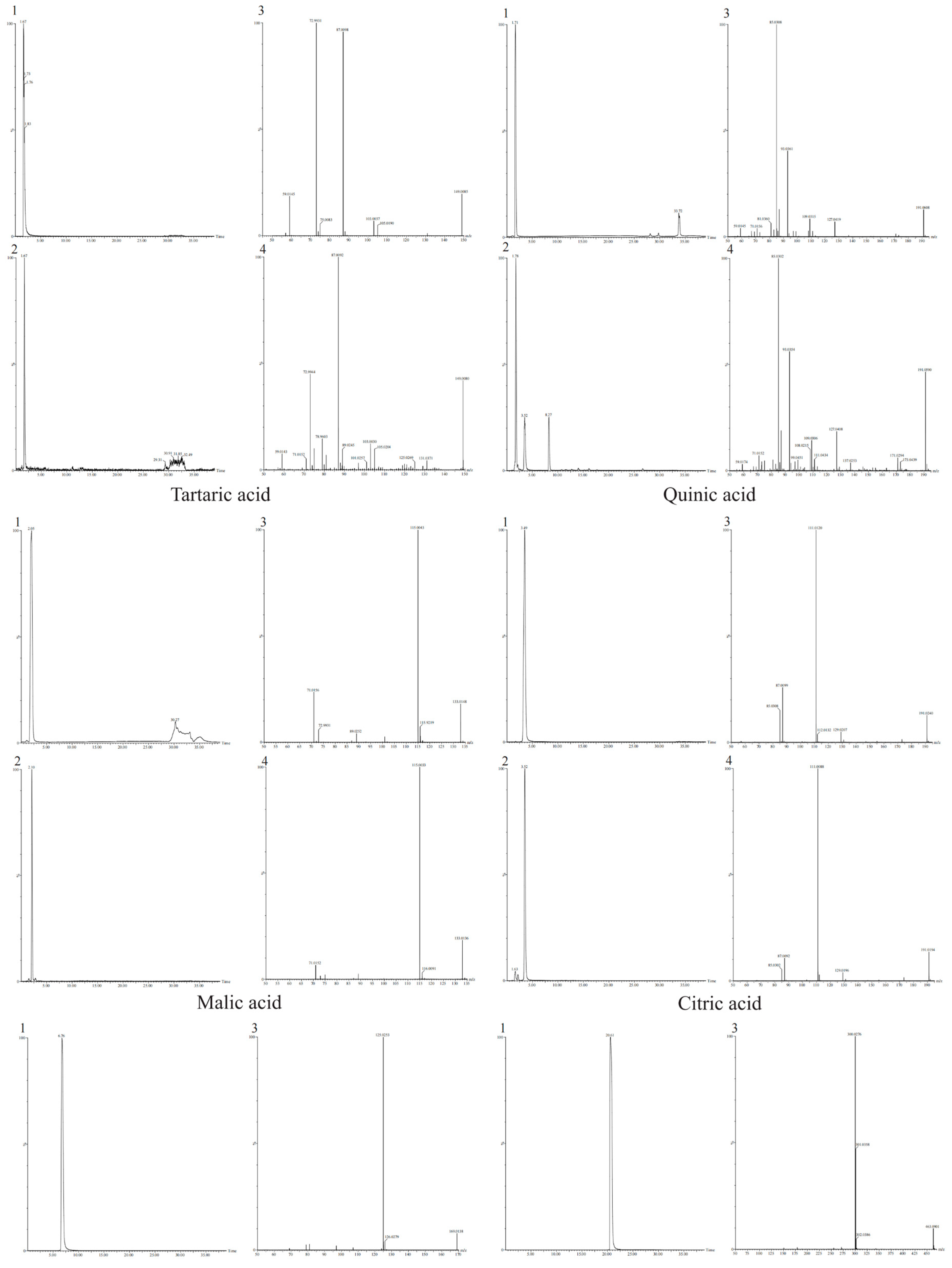
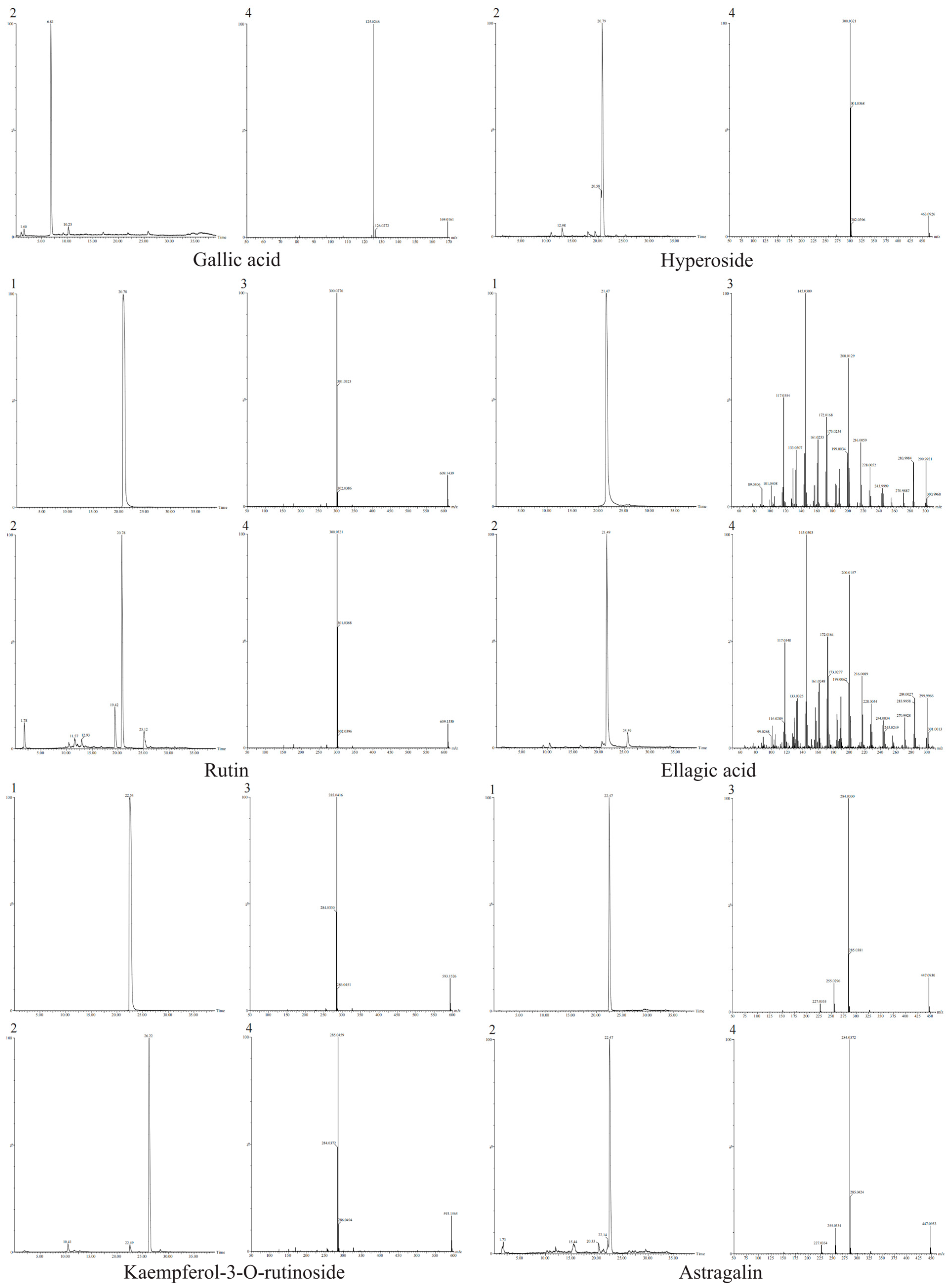
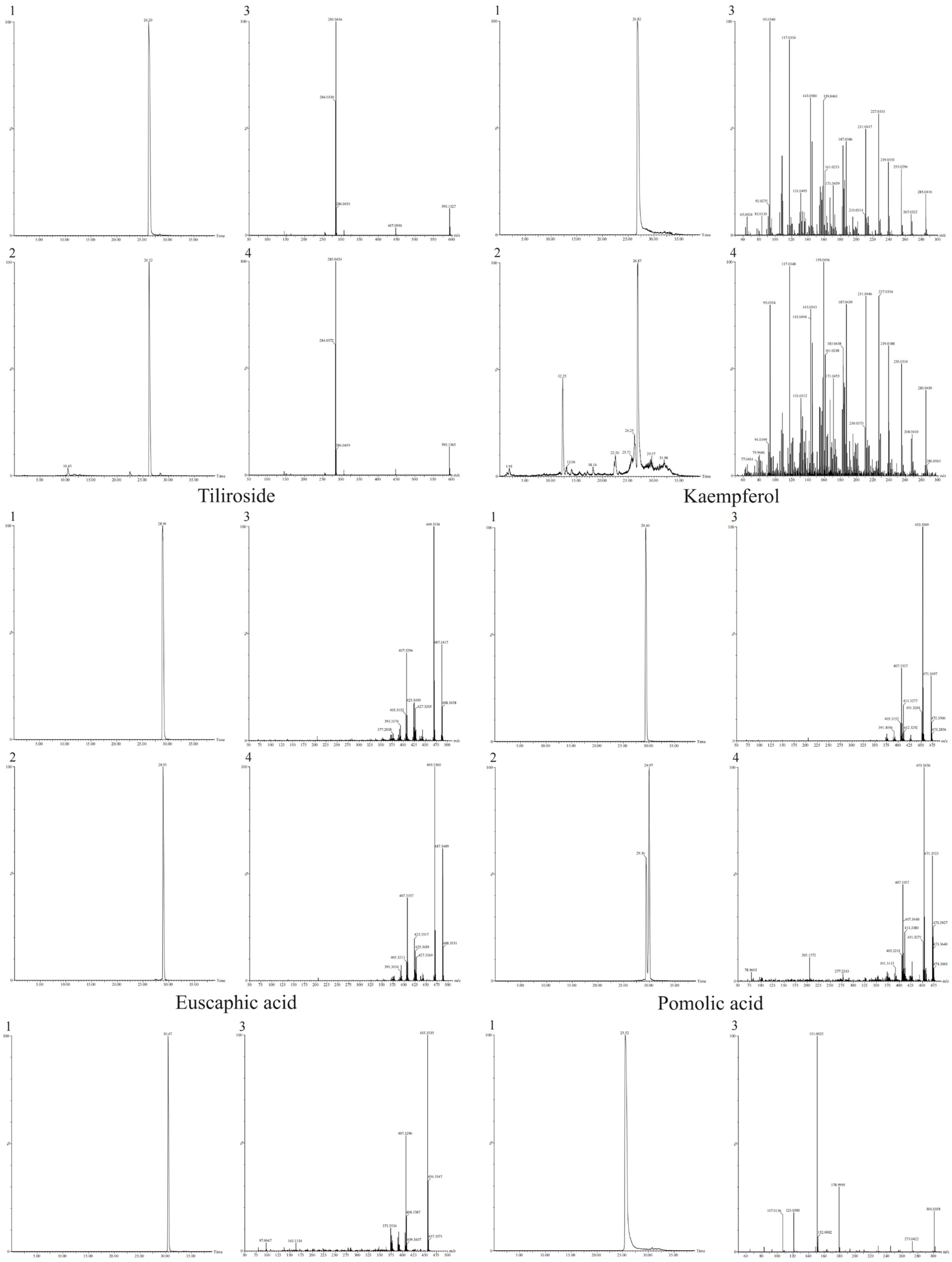
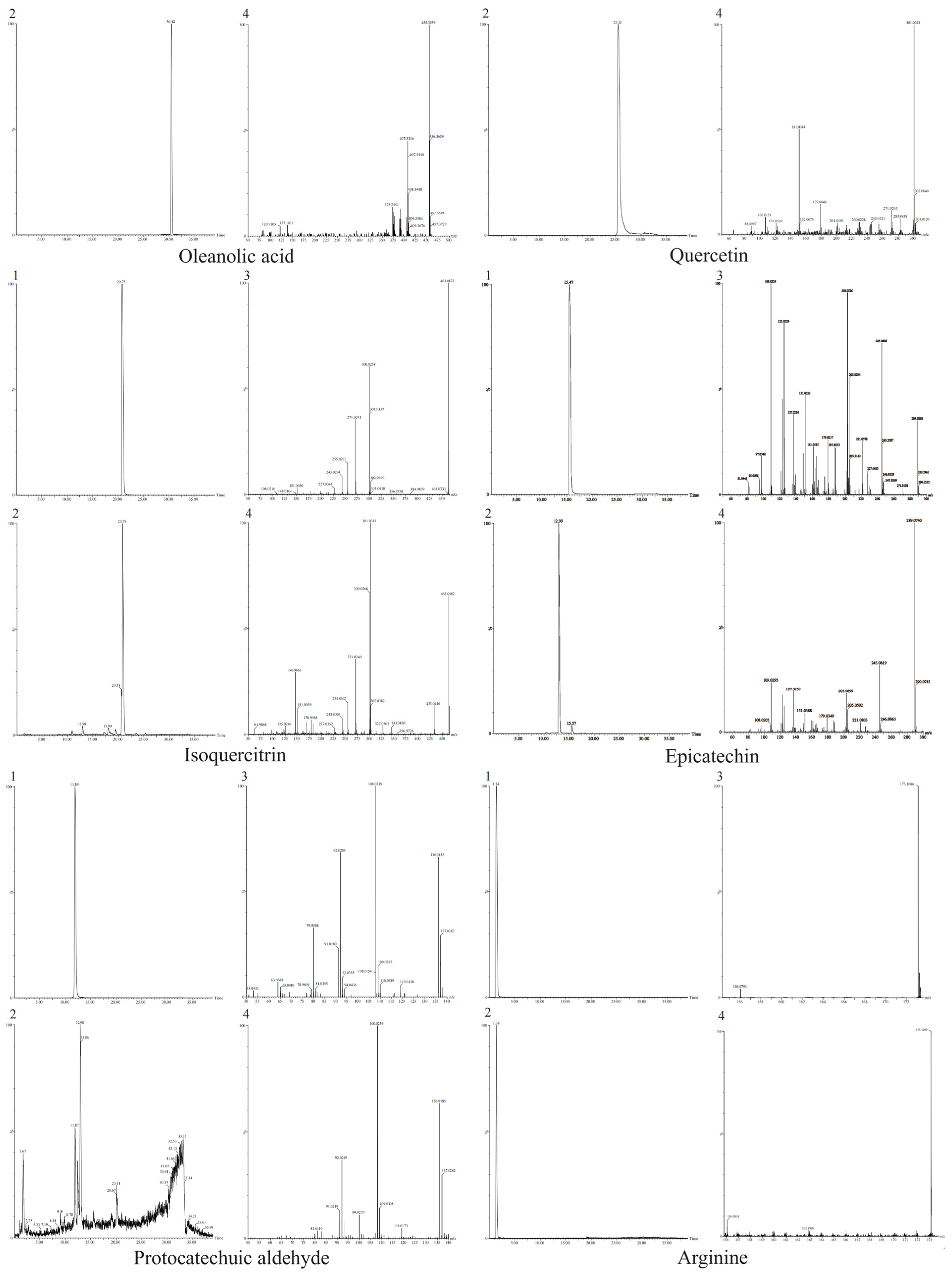
Compound identifications tentative by UNIFI were further validated by comparing their retention times and MS/MS fragmentation patterns with reference standards using the UPLC-QTOF-MS/MS method. Finally, the confirmed positive constituents were quantitatively analyzed using the TOF-MRM (Multiple Reaction Monitoring) mode.
To establish the calibration curves, reference standards were precisely weighed and dissolved in methanol. Stock solutions of astragalin and oleanolic acid were prepared at 0.5 mg/mL, while ellagic acid, tartaric acid, citric acid, gallic acid, pomolic acid, quercetin, kaempferol, kaempferol-3-O-rutinoside, arginine, rutin, hyperoside, euscaphic acid, malic acid, isoquercitrin, quinic acid, epicatechin, tiliroside, and protocatechuic aldehyde were prepared at 0.1 mg/mL. These stock solutions were serially diluted to generate a range of working concentrations: astragalin and oleanolic acid (500, 250, 125, 62.5, and 30.125 μg/mL); citric acid and pomolic acid (100, 50, 25, 12.5, and 6.25 μg/mL); ellagic acid and arginine (50, 25, 12.5, 6.25, and 3.125 μg/mL); tartaric acid, gallic acid, quercetin, epicatechin, euscaphic acid, isoquercitrin, quinic acid, and malic acid (10, 2.5, 0.625, 0.3125, and 0.15625 μg/mL); kaempferol, kaempferol-3-O-rutinoside, hyperoside, and tiliroside (1000, 500, 250, 125, and 62.5 ng/mL); and protocatechuic aldehyde and rutin (100, 50, 25, 12.5, and 6.25 ng/mL).
2.7. Study on Rat Serum Constituents and Their Metabolites
Twelve Sprague-Dawley rats (6 weeks old, 350 ± 10 g) were housed in a standard environment with ad libitum access to food and water. The rats were randomly divided into two groups (n = 6 per group): the blank group and the administration group. FRLE was dissolved in distilled water to prepare a 0.83 g/mL suspension. After 12 h of fasting (with free access to water), rats in the Administration group received a single dose of 1 mL/100 body weight via oral gavage. Blood samples (≈ 0.3 mL) were collected from the retro-orbital venous plexus at 0 (pre-dose), 0.083, 0.25, 0.5, 0.75, 1, 1.5, 2, 3, 4, 6, 8, 12, 24, 36, and 48 h post-administration into heparinized tubes. Water was restricted for 2 h, and food was withheld for 4 h post-dosing. Serum was separated by centrifugation at 3000 rpm for 15 min and stored for analysis.
For protein precipitation, 0.1 mL of serum was mixed with 0.3 mL of acetonitrile containing 0.1% formic acid (v/v) and vortexed for 5 min. The mixture was centrifuged at 13,780 g for 10 min at 4 °C. The supernatant was transferred and evaporated to dryness under nitrogen at 37 °C. The residue was reconstituted in 0.1 mL of methanol, vortexed, and centrifuged again at 13,780 g for 10 min at 4 °C to obtain the final plasma samples.
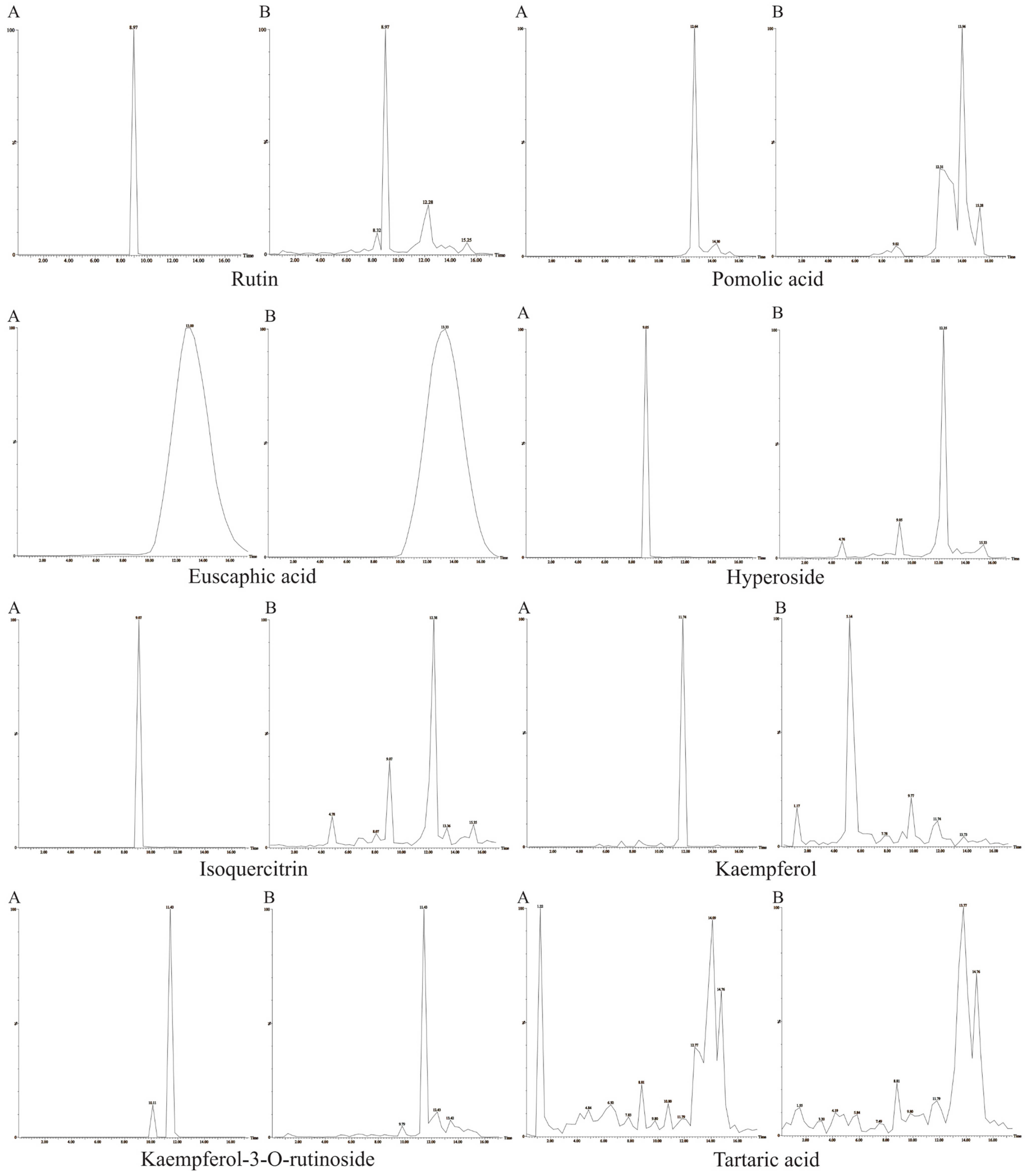
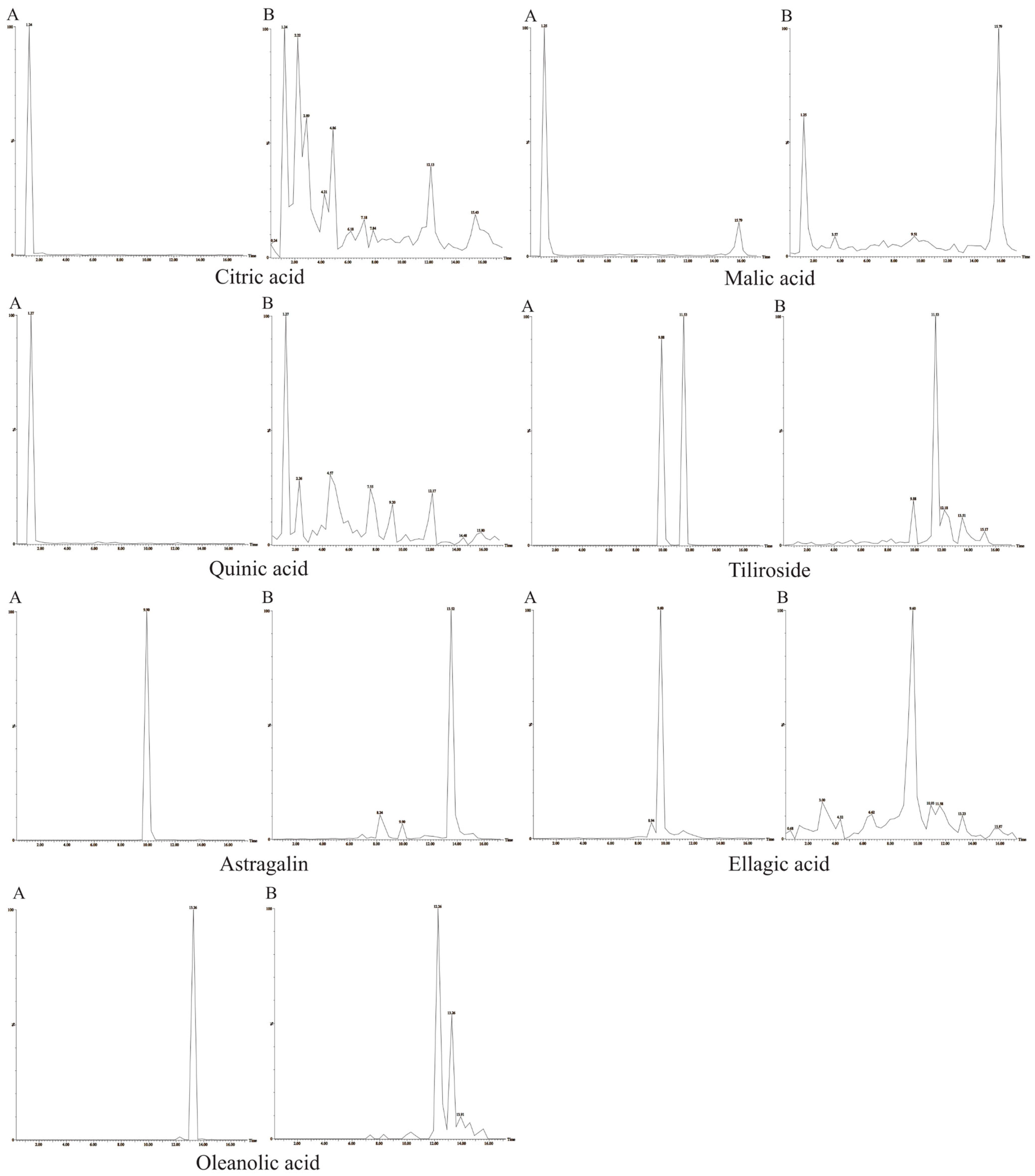
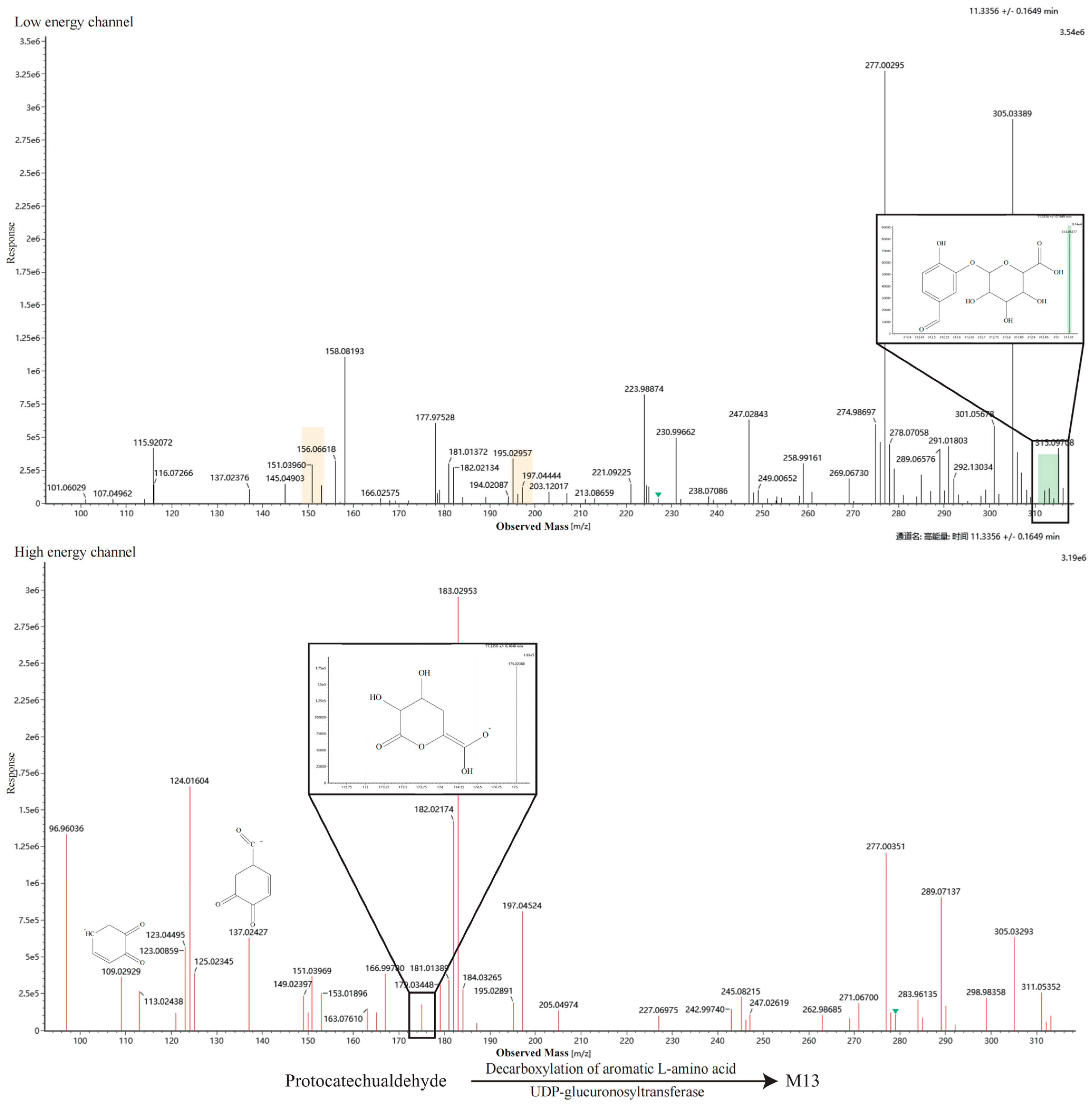
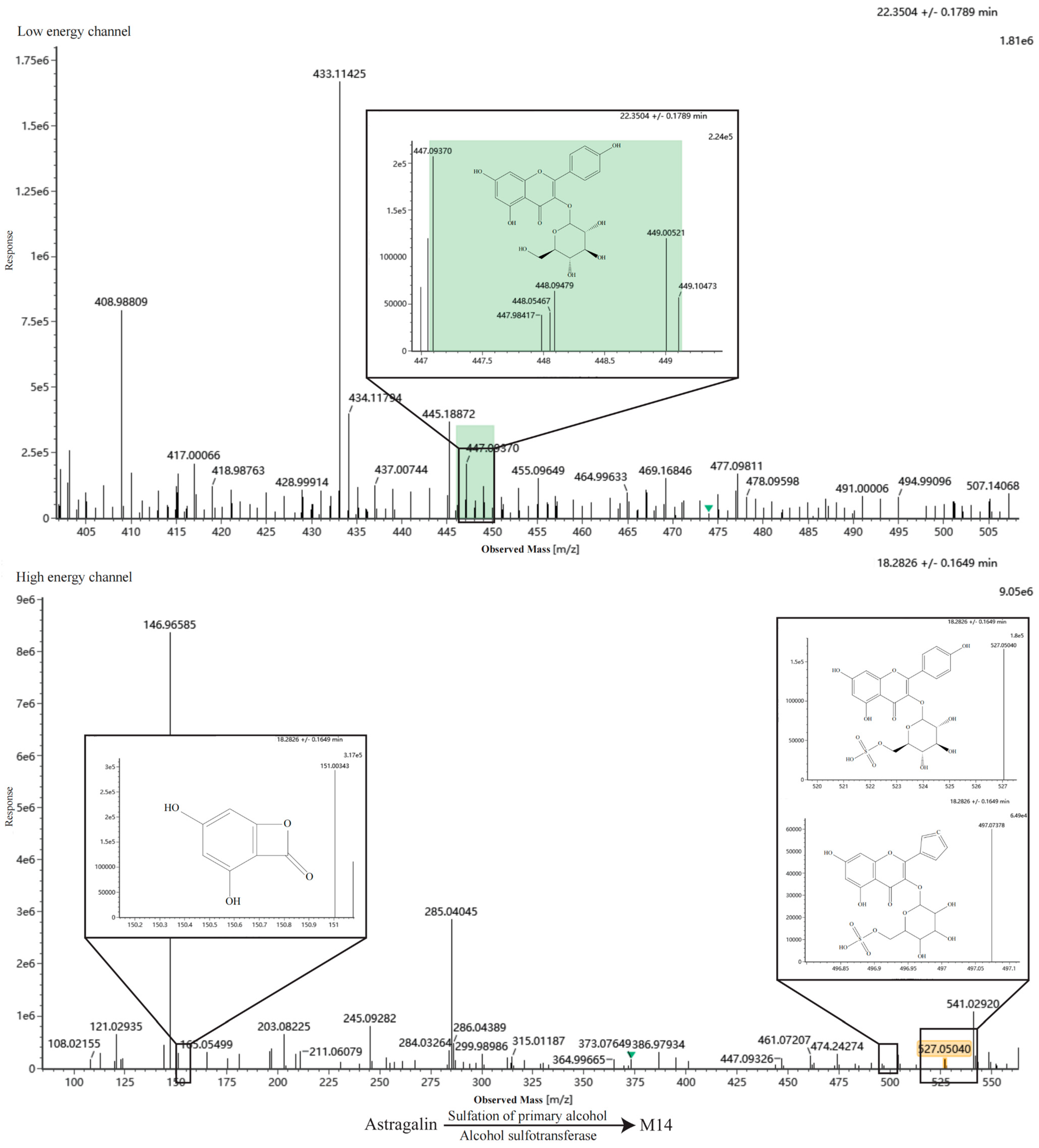
Reference standard stock solutions (1 mg/mL) were prepared in methanol. A mixed standard solution (50 μg/mL) was created by diluting the stocks, and 10 μL of this mixture was spiked into 1 mL of blank serum to prepare the quality control (QC) plasma samples. Samples were analyzed using TOF-MRM mode, and absorbed constituents were identified based on retention time (RT) and characteristic fragment ions.
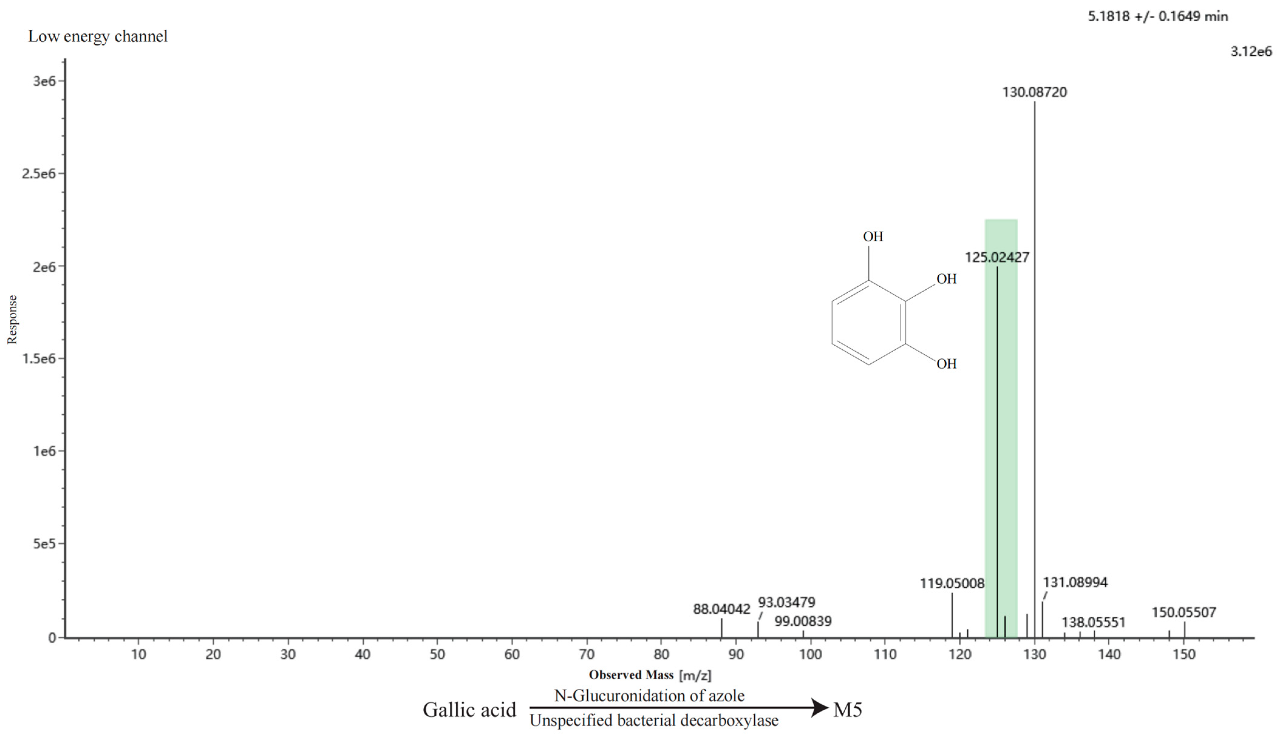
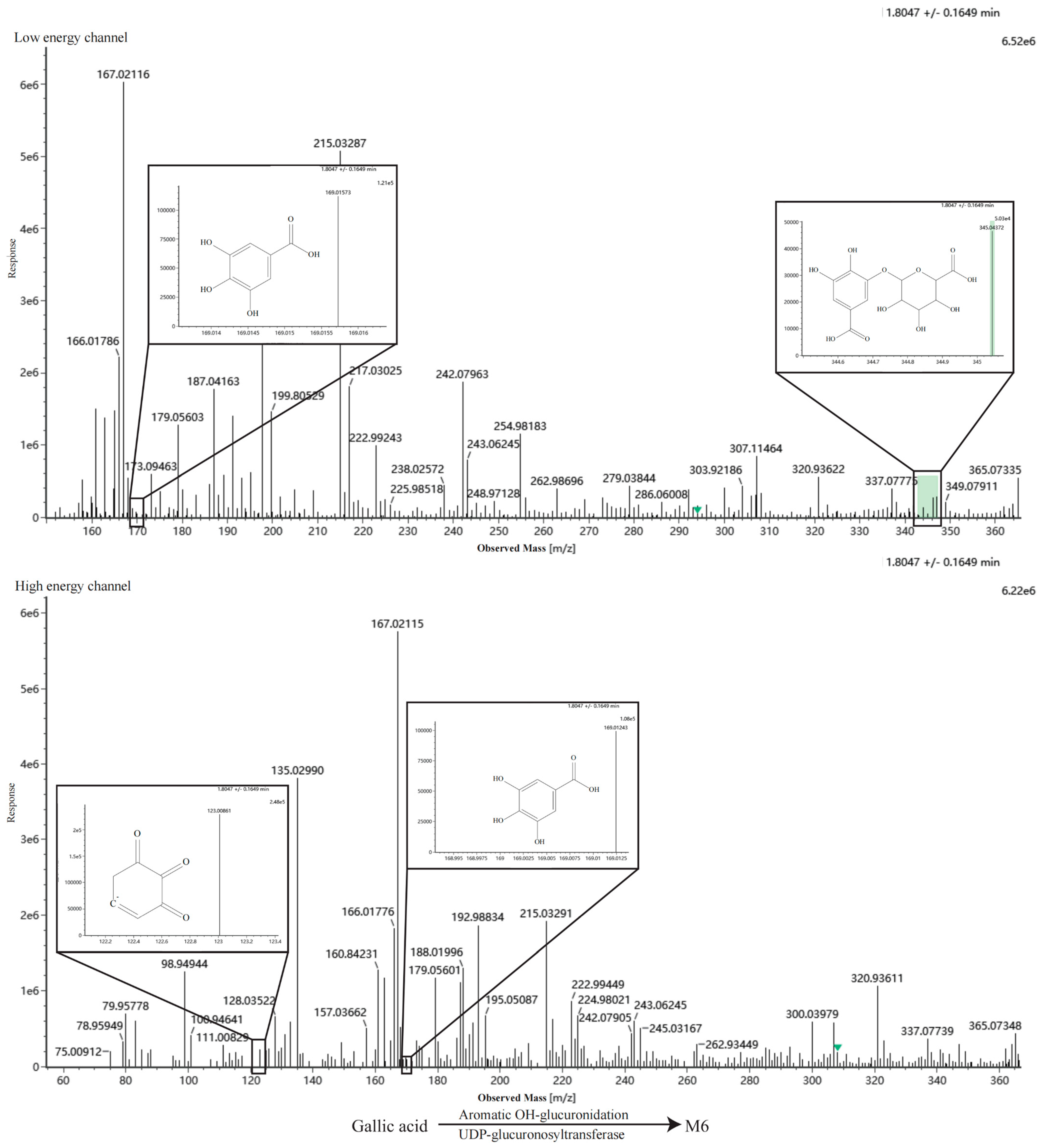
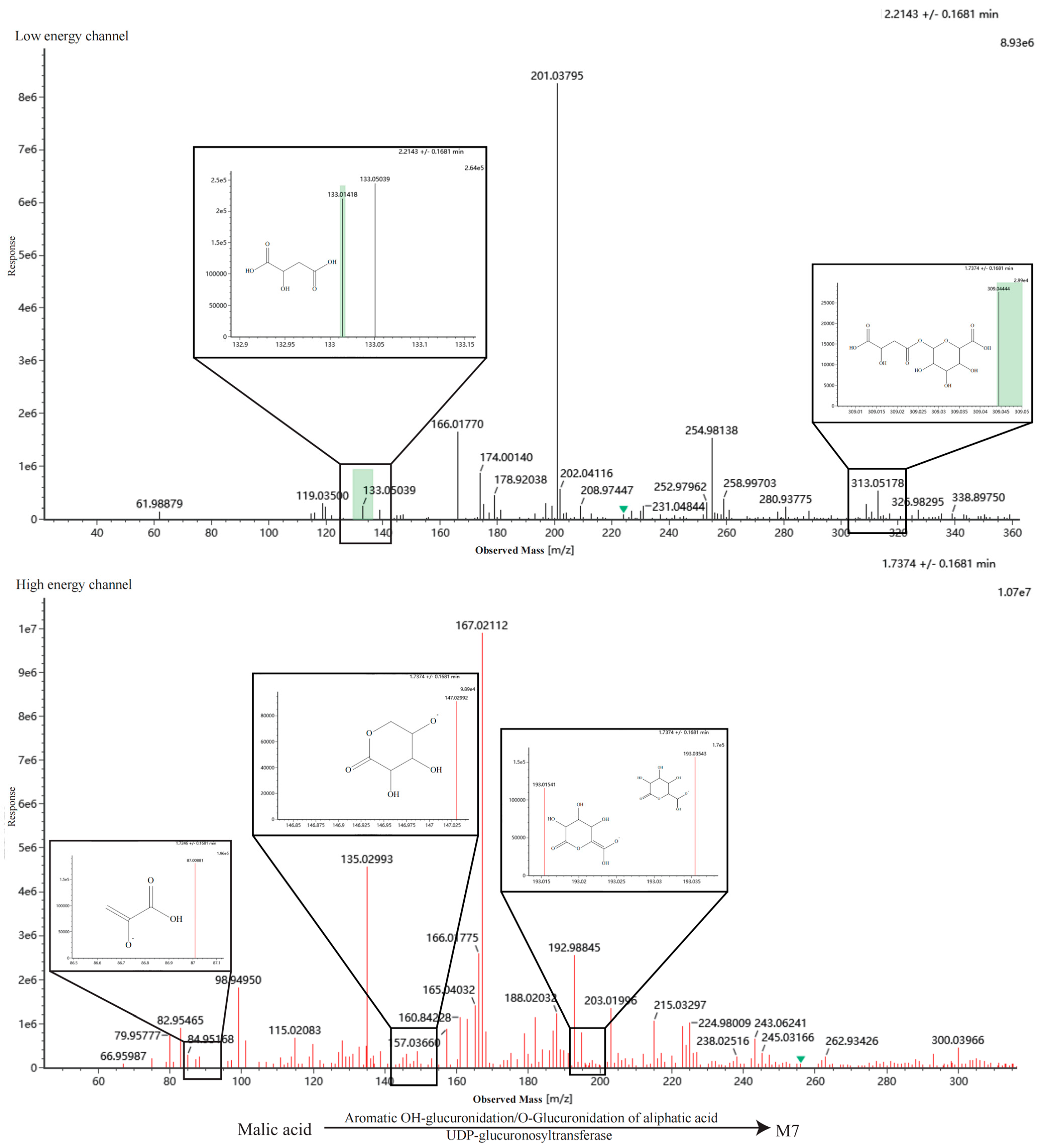
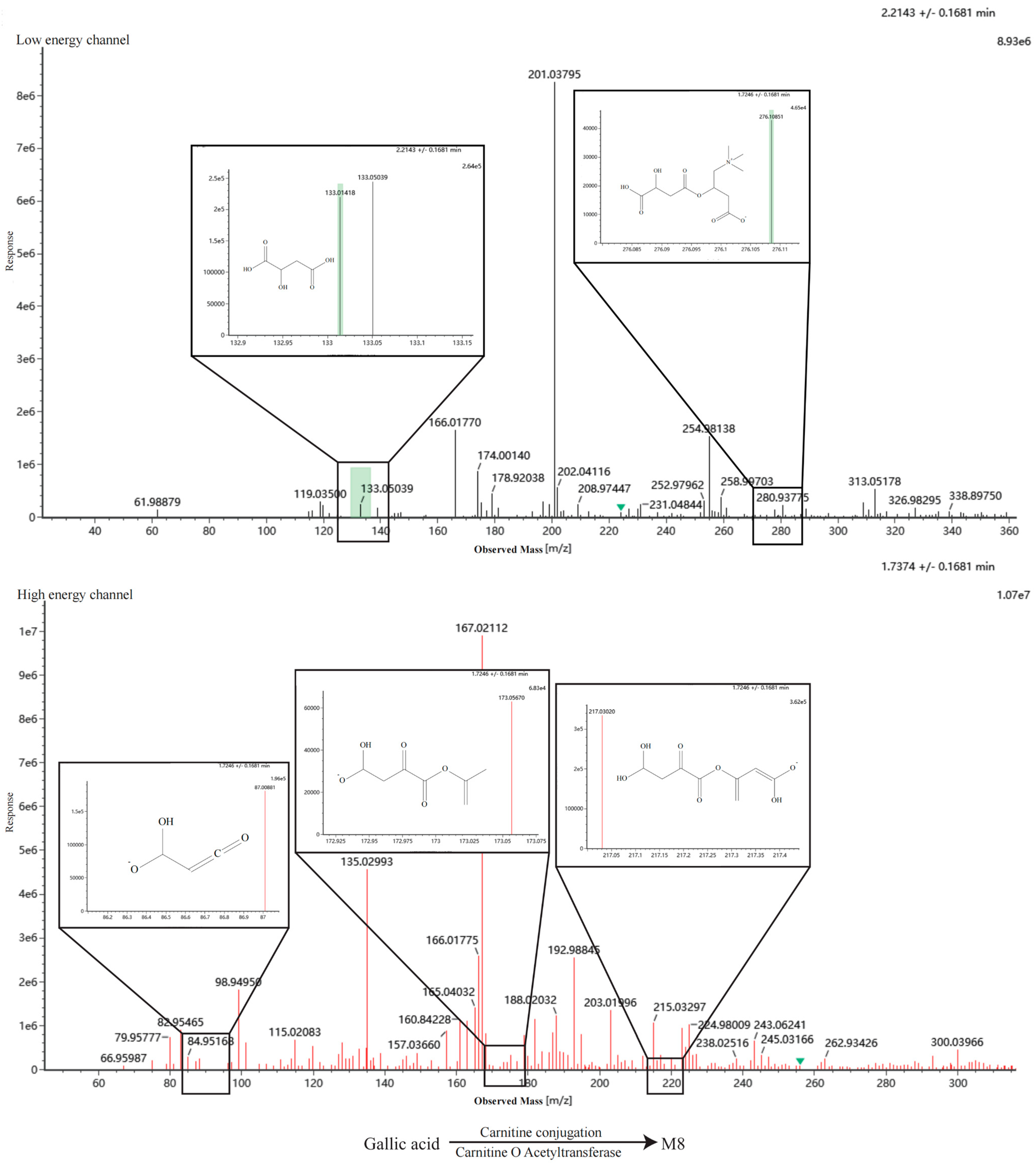
Metabolic pathways were predicted using BioTransformer 3.0 (
https://biotransformer.ca/, accessed on 31 May 2026), a machine learning-based tool [
17,
18]. The chemical structures of 20 identified constituents (including malic acid, quinic acid, and tiliroside, etc.) were converted into SMILES strings via ACD/ChemSketch and processed to predict Phase I and Phase II metabolites, specifically targeting cytochrome P450 enzymatic transformations. To facilitate identification, CFM-ID 4.0 (
https://cfmid.wishartlab.com/, accessed on 31 May 2026) was employed to simulate the fragmentation behavior and generate theoretical MS/MS spectra for the predicted metabolites in ESI- mode [
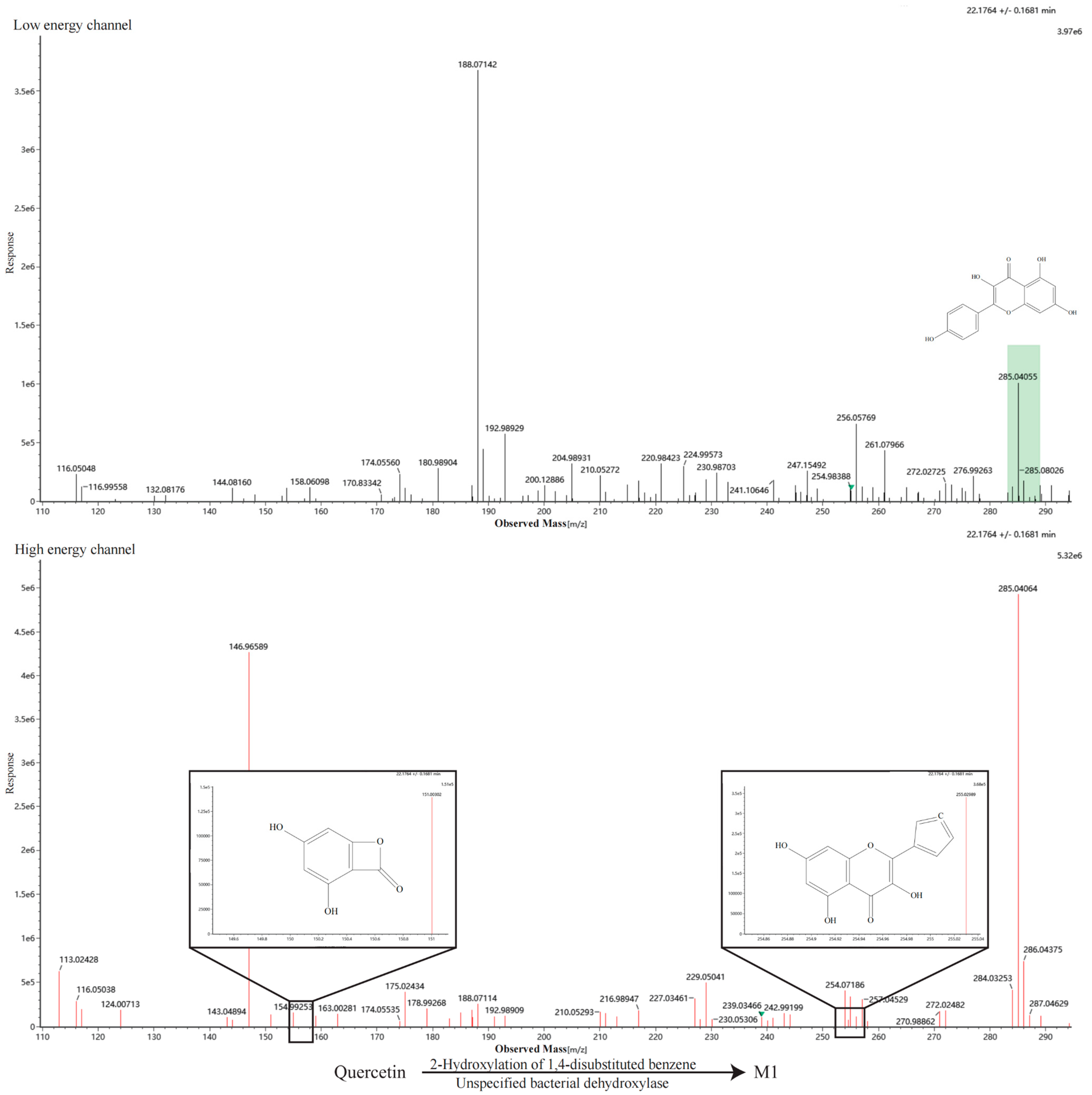
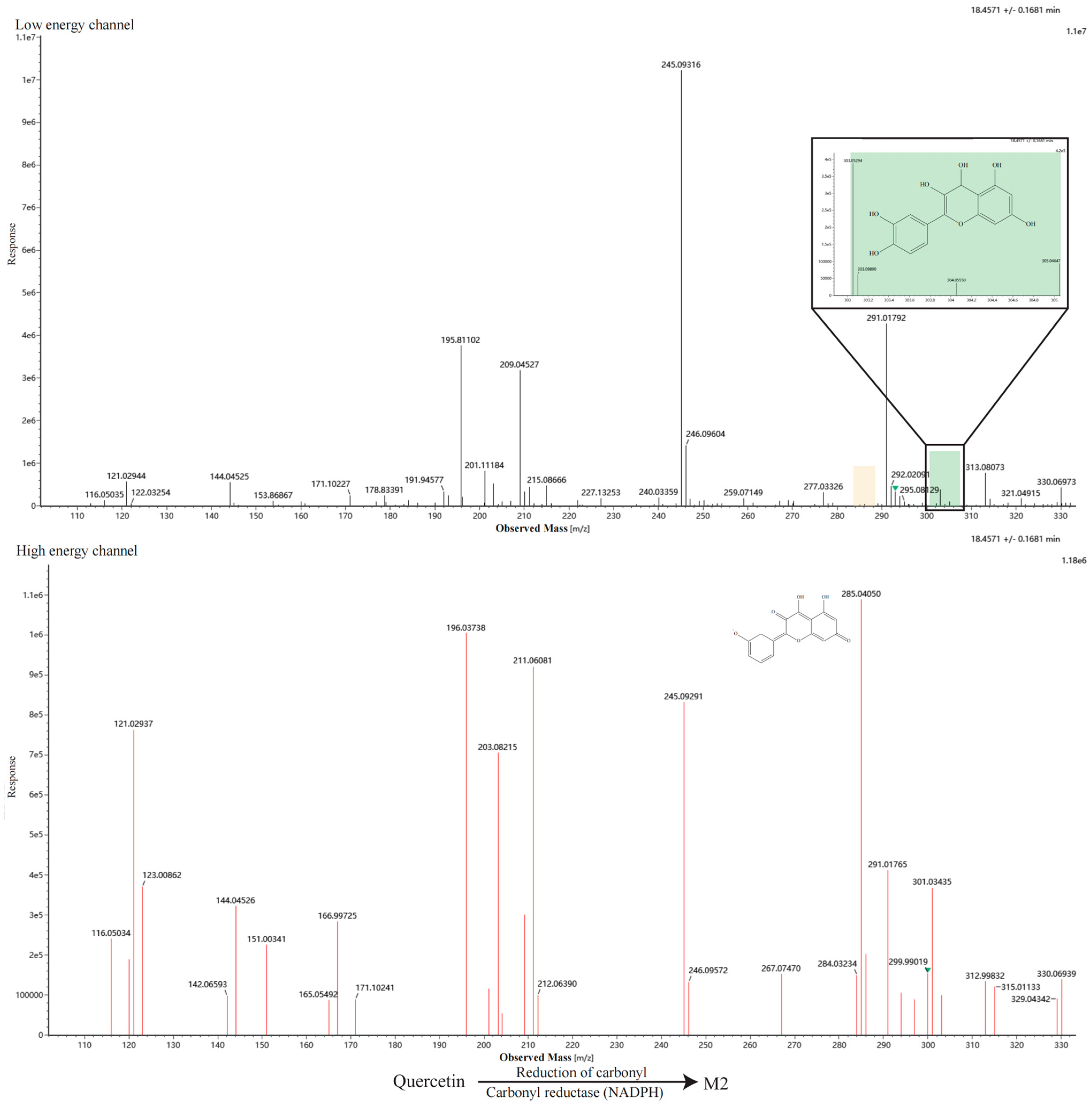
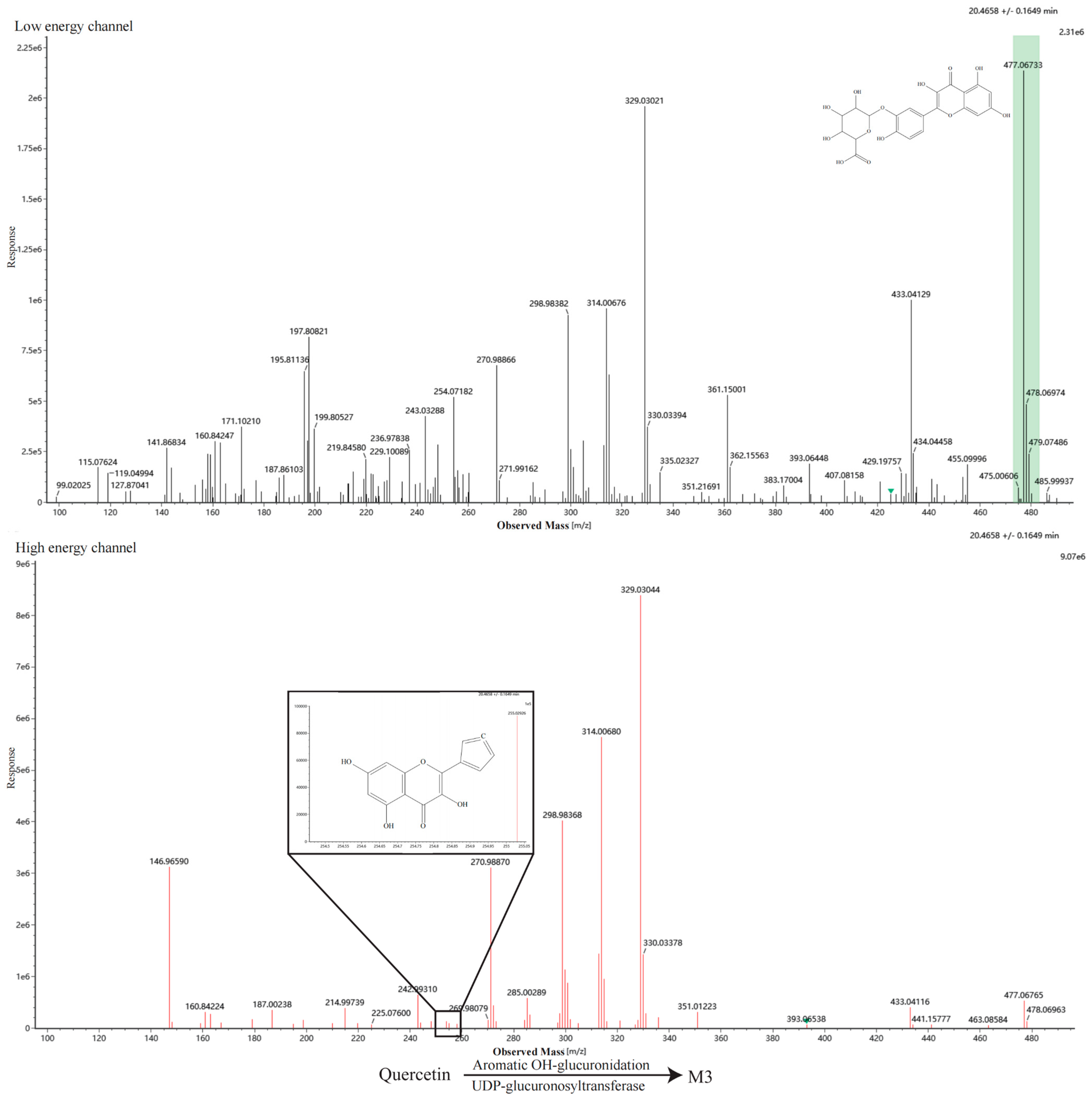
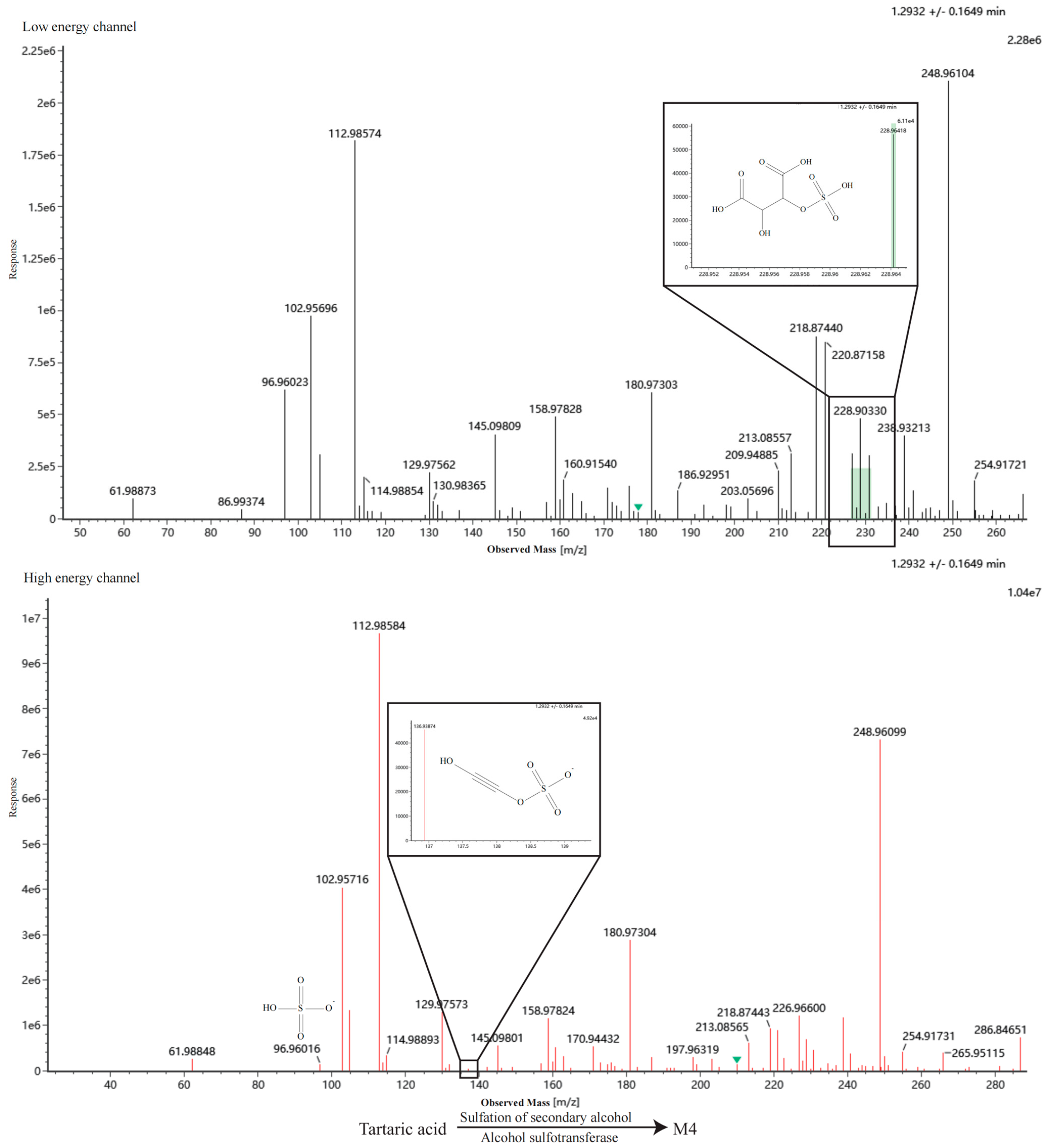
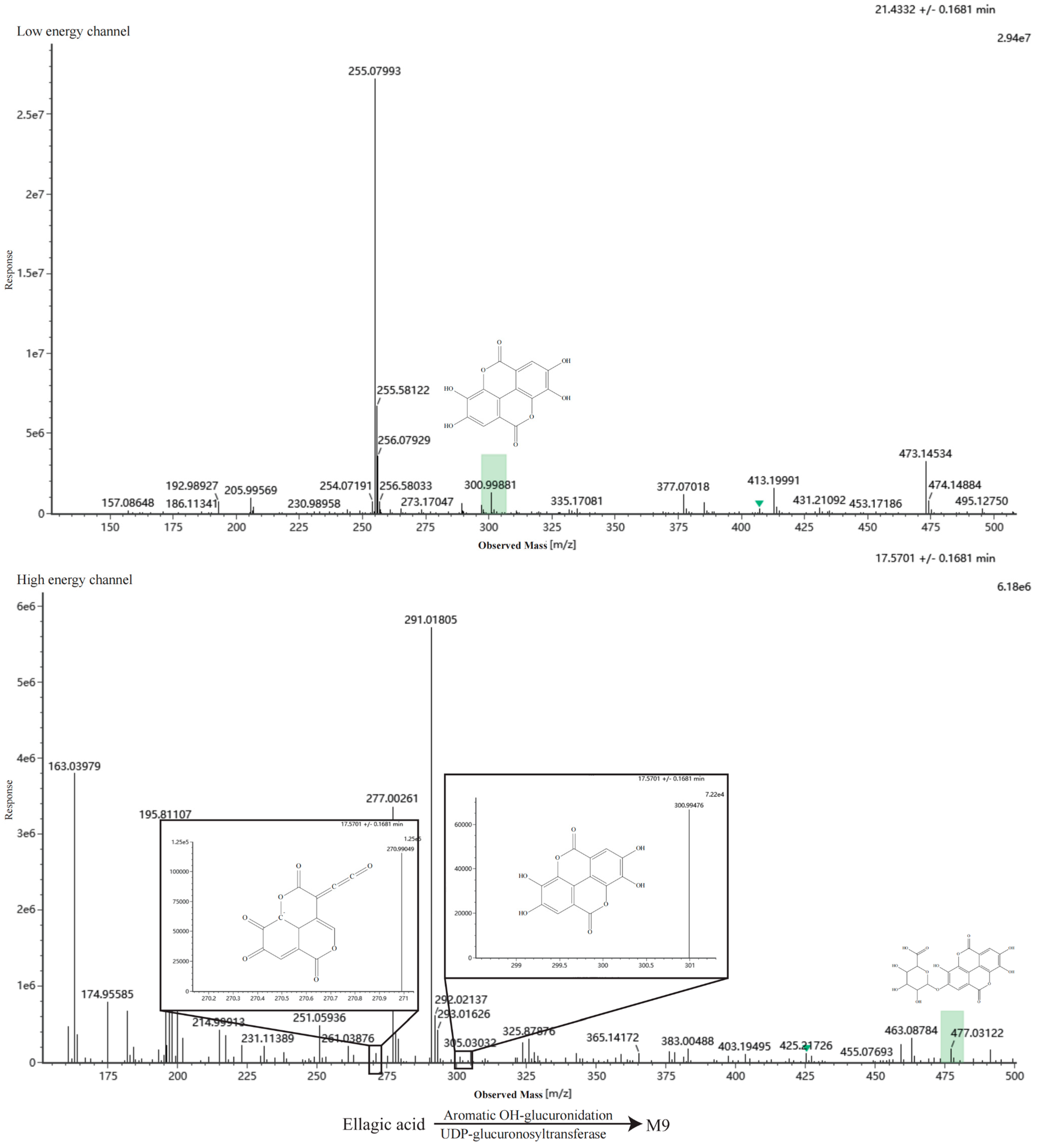
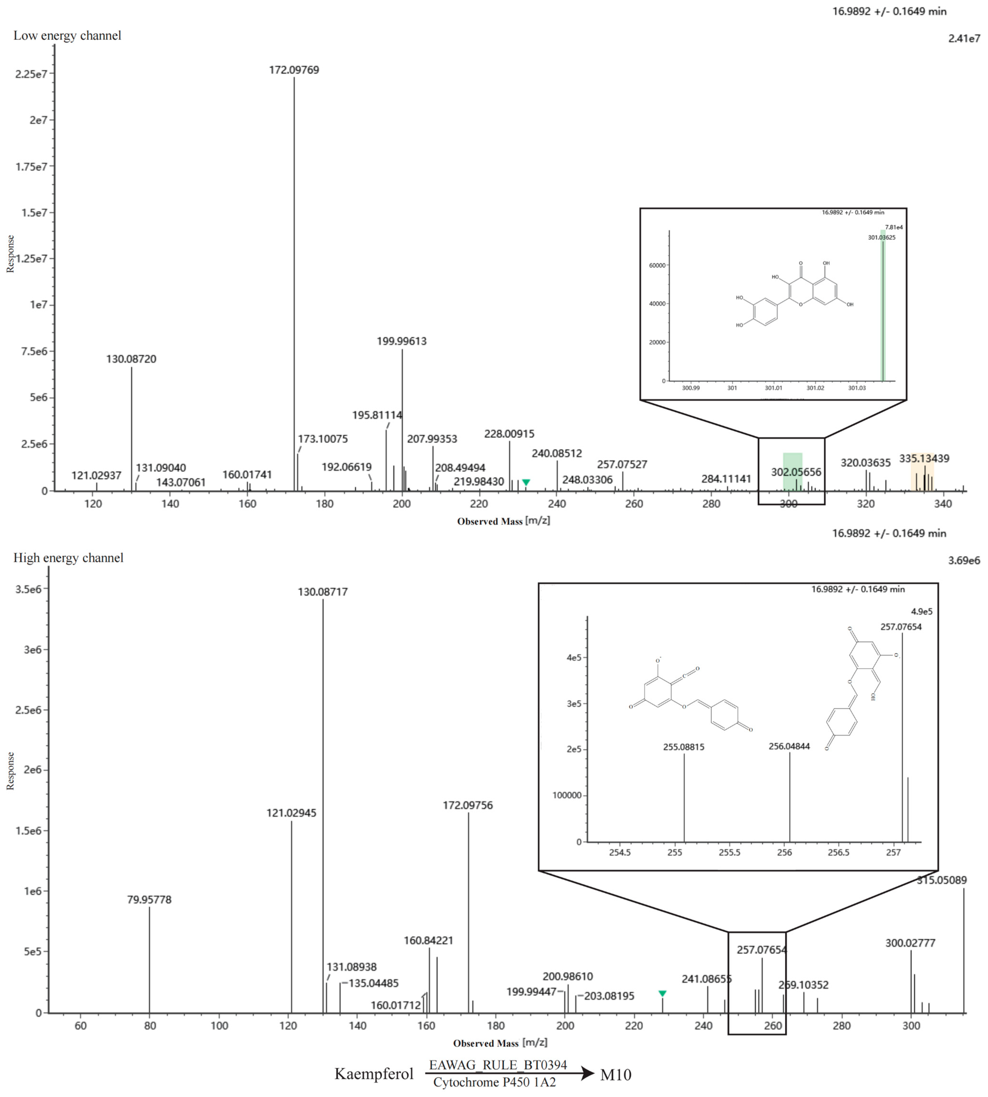
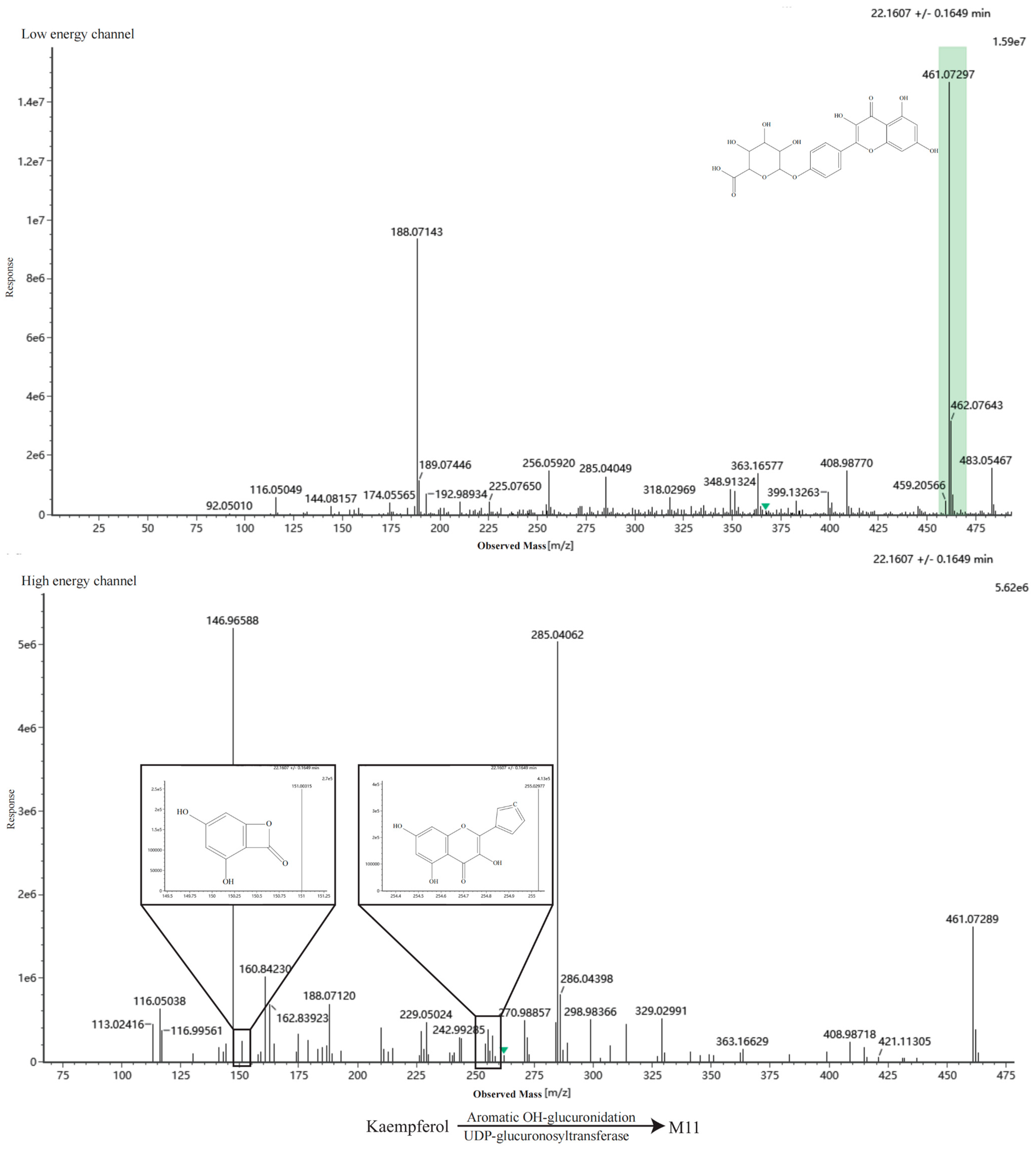
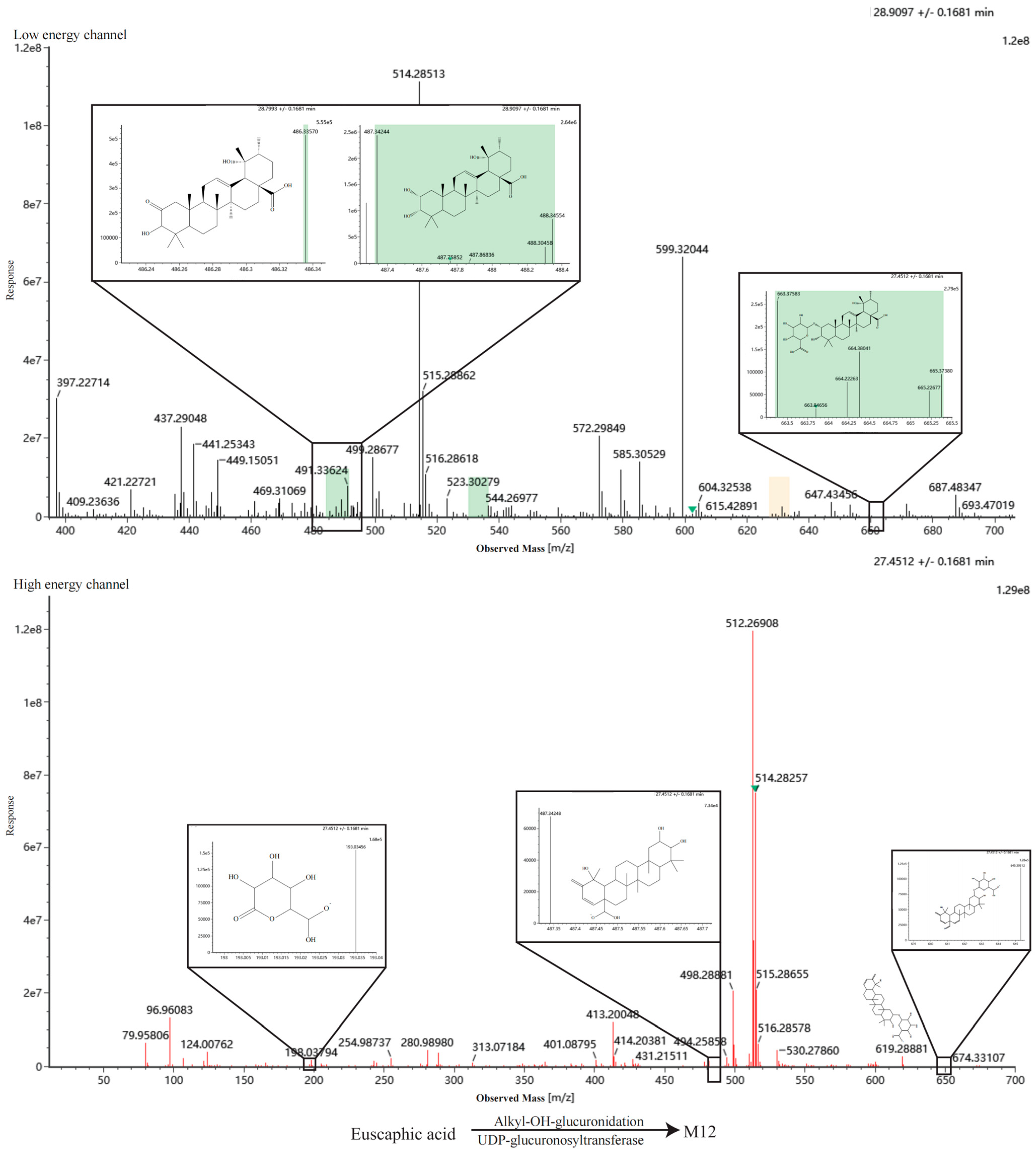
19]. Finally, the absorbed metabolites were identified by integrating Waters UNIFI 1.9.4 with the results from BioTransformer and CFM-ID. A positive identification required at least one characteristic fragment ion to match between the experimental and theoretical MS/MS spectra.
4. Discussion
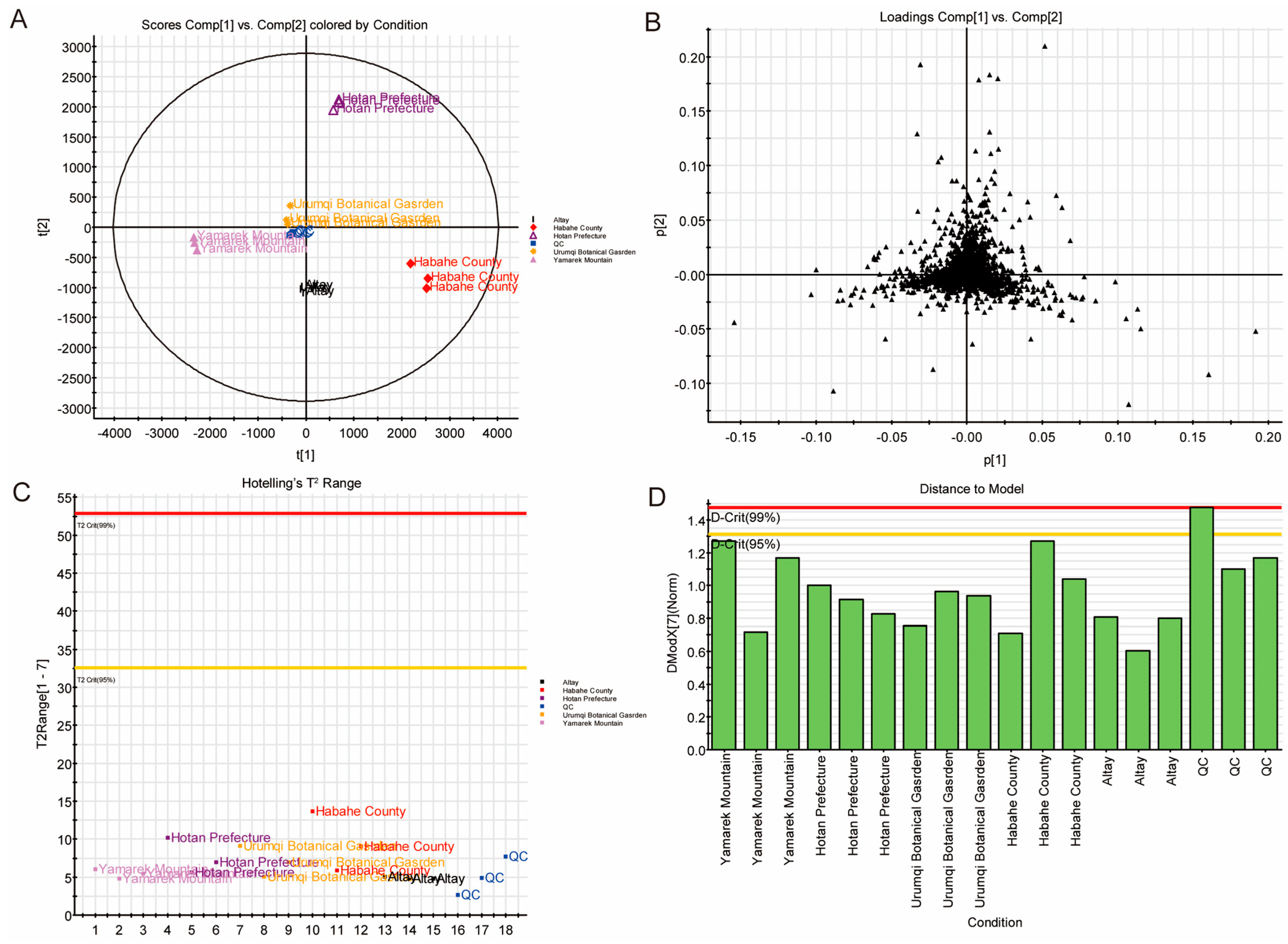
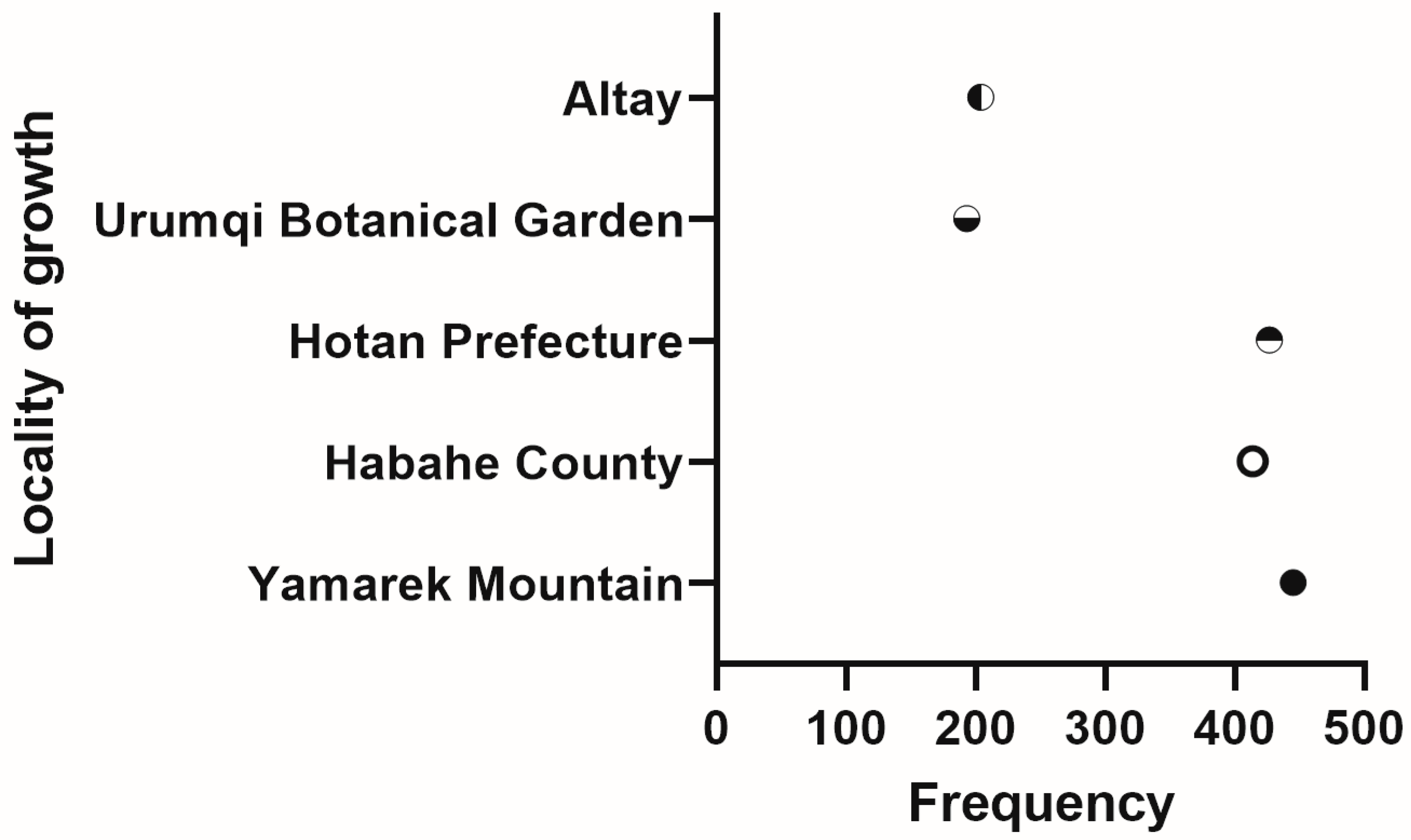
This study confirms that the types and levels of secondary metabolites in FRL are closely related to their growth environments. UPLC-QTOF-MS analysis demonstrated that FRL from Yamalike Mountain possesses the greatest chemical diversity, significantly surpassing samples from the Botanical Garden. This variation likely stems from the semi-alpine habitat of Yamalike Mountain, where the substantial diurnal temperature range promotes the accumulation of secondary metabolites [
32,
33,
34,
35]. In contrast, excessive anthropogenic interventions in the Botanical Garden, such as pruning and intensive irrigation, may lead to the dilution or loss of specific constituents [
36,
37,
38]. It is worth noting that the selection of Yamalike Mountain samples as the primary material for downstream pharmacological experiments was an analytical choice driven by chemical diversity and marker abundance, rather than a pre-demonstrated superiority in biological efficacy. Because the Yamalike Mountain samples possessed the most diverse array and the highest abundance of high-response differential constituents, utilizing this origin maximized our capacity to qualitatively and quantitatively characterize the comprehensive chemical profile of FRLE via UPLC-QTOF-MS. Characterizing such a highly enriched chemical repertoire is a crucial prerequisite for network pharmacology, ensuring that a wider spectrum of potential bioactive components can be captured and evaluated during subsequent target prediction and mechanism elucidation.
The complexity of TCM, characterized by numerous isomers, poses a risk of false positives when relying solely on MS spectral matching. To address this, we established an in-house library integrating RT of reference standards and employed a dual-verification strategy using MSE and DDA modes. This approach overcomes the limitations of RT variability and significantly enhances the accuracy of isomer identification, providing a high-fidelity chemical foundation for studying the pharmacodynamic basis of FRLE.
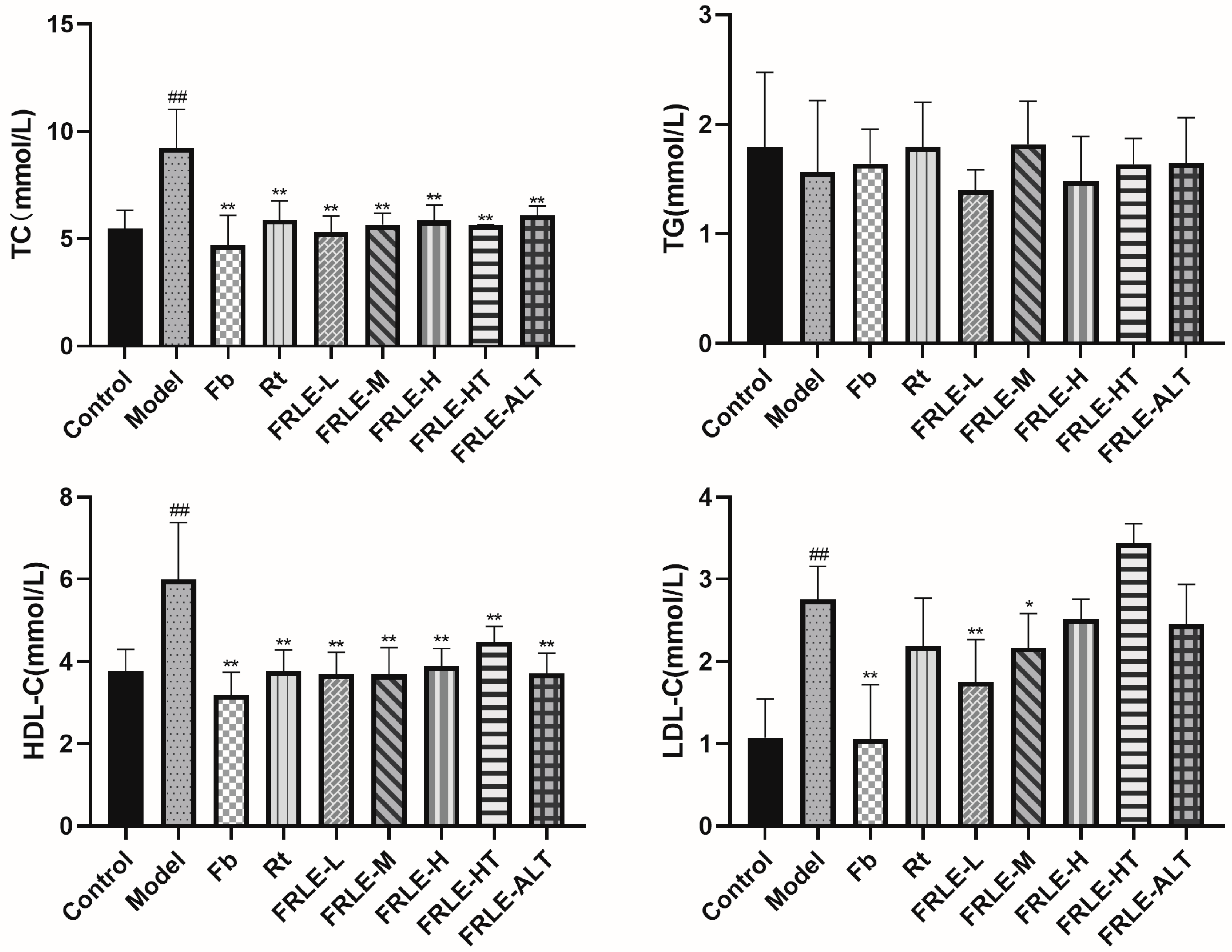
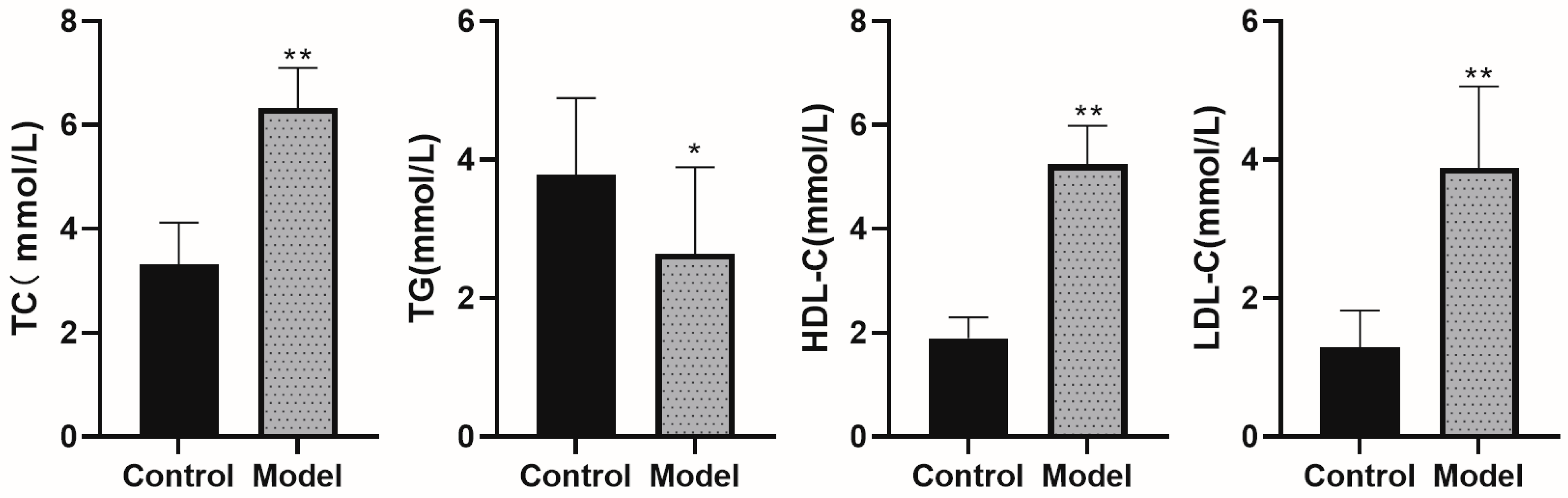
In conventional HLP models, TG levels typically increase while HDL-C levels decrease. However, our model group exhibited unexpected trends. The observed decrease in TG may be attributed to an increase in intrahepatic TG synthesis, which consequently lowers peripheral blood TG levels [
39]. Furthermore, while HDL-C is traditionally viewed as “good cholesterol”, recent studies suggest that excessively high levels do not necessarily confer cardiovascular protection [
40,
41,
42]. In this study, FRLE intervention maintained HDL-C at levels consistent with the control group. This indicates that FRLE exerts a cardiovascular protective effect by normalizing or correcting abnormally elevated HDL-C levels. Ultimately, the absence of significant differences in TG and HDL-C between the treatment groups and the control group suggests that FRLE effectively maintains these parameters within a normal physiological range.
PCA revealed significant chemical variations among FRL from Yamalike Mountain, Hotan, and Altay; however, all three samples significantly reduced TC levels in HLP mice, suggesting that common constituents drive the cholesterol-lowering effect. The pharmacodynamic distinction lies in the lack of LDL-C-lowering efficacy in the Hotan sample, whereas Altay and Yamalike Mountain samples showed similar performance in reducing LDL-C and maintaining HDL-C. Therefore, FRL samples with similar primary chemical profiles can be considered viable candidates for anti-HLP drug development.
Using the UPLC-QTOF-MRM method, 20 constituents were quantified in FRLE, and 15 bioactive components were confirmed in rat plasma. These included flavonoid glycosides (rutin, tiliroside, astragalin, hyperoside, isoquercitrin, kaempferol, and kaempferol-3-O-rutinoside), triterpenoid acids (pomolic acid, euscaphic acid, and oleanolic acid), and organic acids (tartaric, citric, malic, quinic, and ellagic acid). Flavonoids are well-documented for their anti-HLP activities, while triterpenoid acids exert anti-HLP effects by inhibiting bile acid transporter [
43,
44,
45,
46]. These components constitute the material basis for the therapeutic efficacy of FRLE. Arginine was undetected in plasma, likely because as an essential amino acid, it was rapidly utilized in physiological processes post-absorption. Other components, such as gallic acid, quercetin, and protocatechuic aldehyde, were undetected due to their low initial content and subsequent dilution or rapid metabolism in vivo.
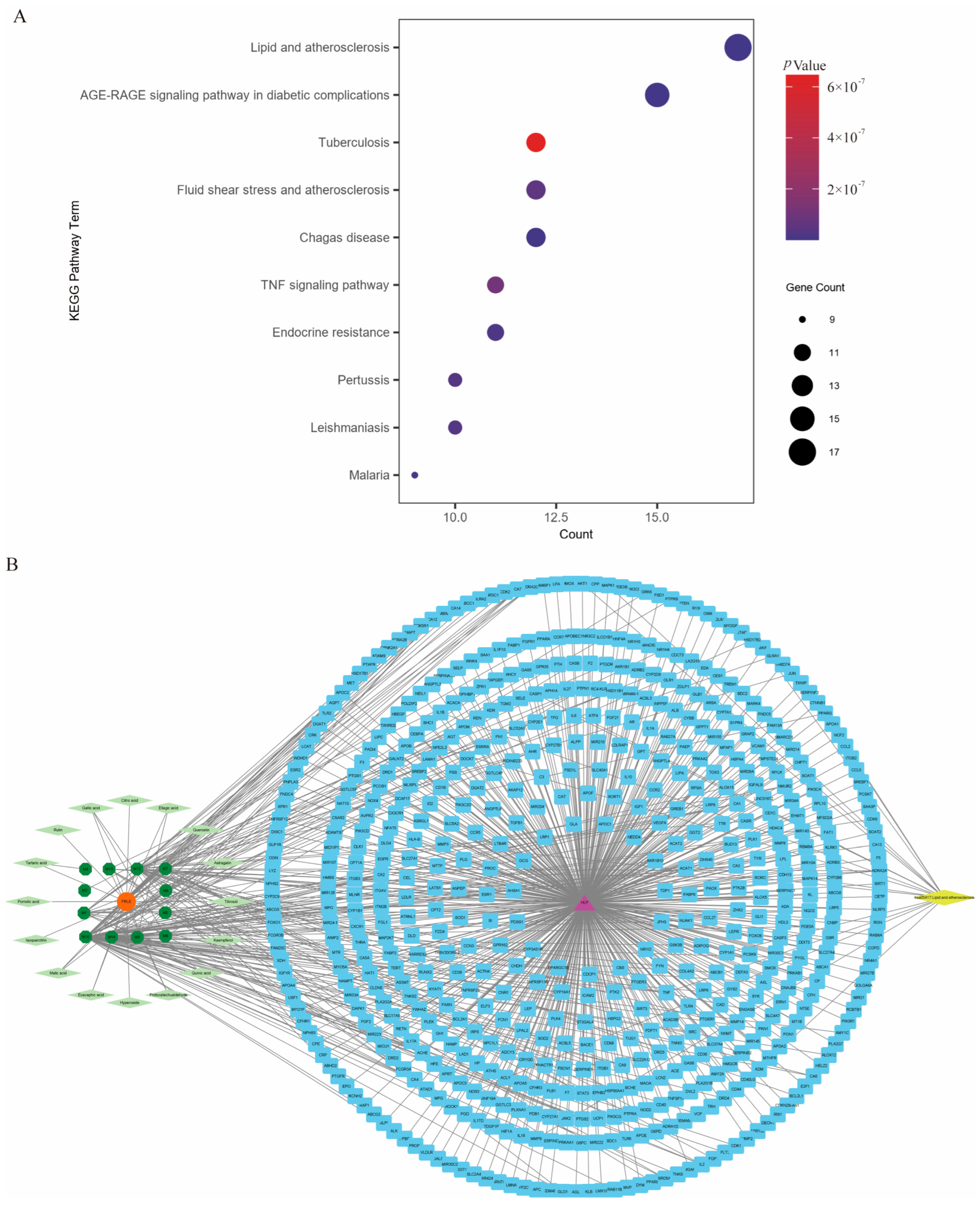
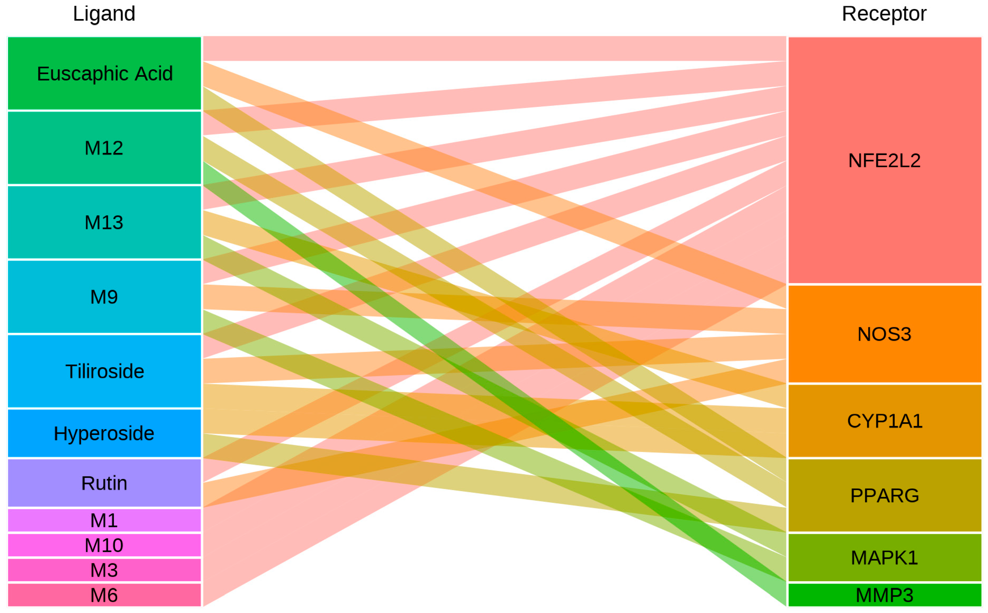
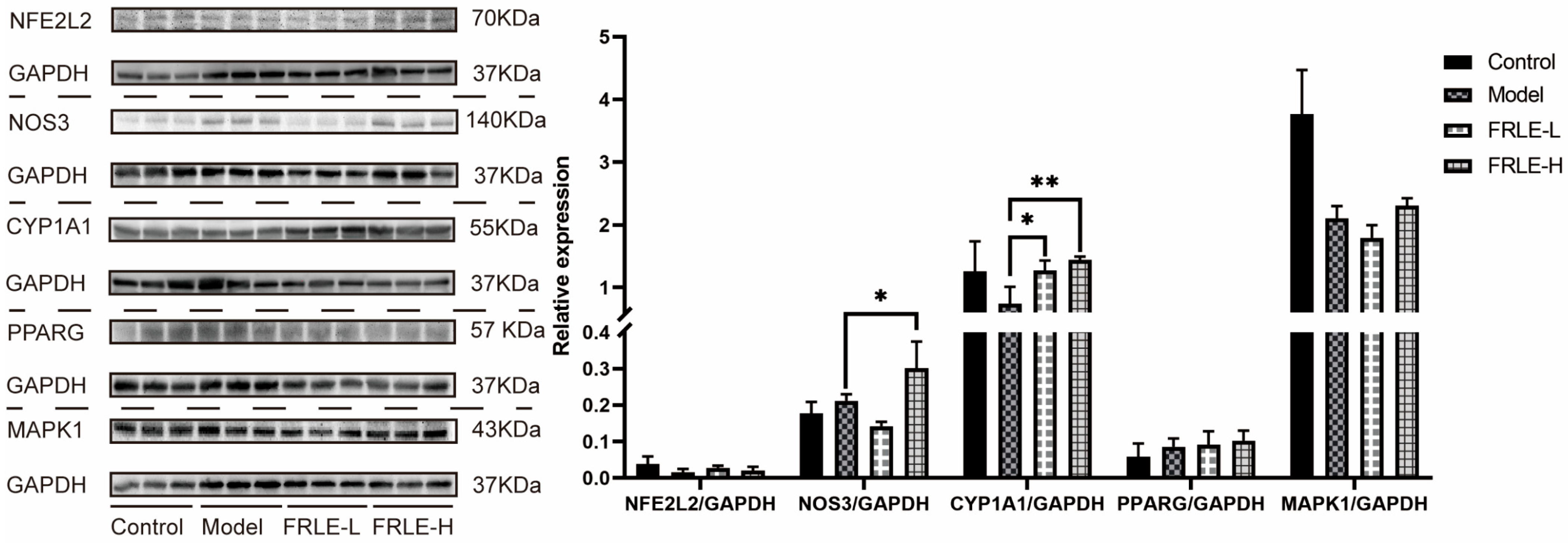
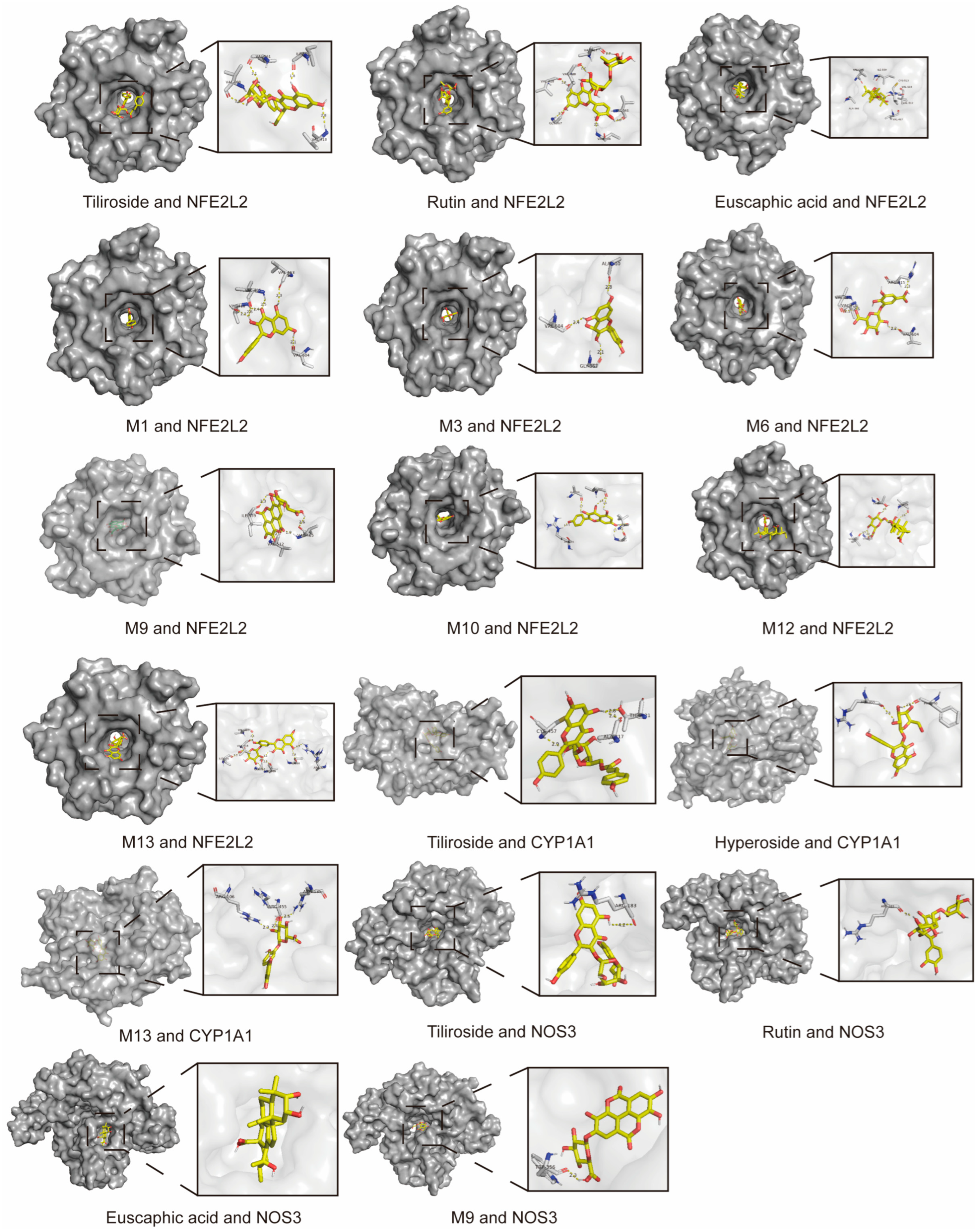
Network pharmacology and molecular docking predicted that the anti-HLP mechanism of FRLE involves 17 key targets (e.g., VCAM1, NOS3, CYP1A1, PPARG, and NFE2L2) within the Lipid and Atherosclerosis pathway (hsa05417). These targets are intricately linked to lipid imbalance, inflammation, oxidative stress, and endothelial dysfunction. Western blot validation further confirmed that the absorbed constituents and their metabolites exert anti-HLP effects by modulating NFE2L2, CYP1A1, NOS3, and MAPK1. Further research is warranted to elucidate the deeper molecular mechanisms involved [
47,
48,
49,
50,
51].
Finally, a limitation of this study is the relatively small sample size (n = 3) utilized for the Western blotting verification. While n = 3 is a standard convention for preliminary screening in pharmacodynamic studies, it inherently possesses low statistical power. This limitation may account for the observed but statistically non-significant (p > 0.05) regulatory trends in certain protein targets, such as NFE2L2, PPARG, and MAPK1. Future studies with a larger cohort are warranted to comprehensively validate these subtle molecular alterations.