Review

Berberine as a Bioactive Alkaloid: Multi-Omics Perspectives on Its Role in Obesity Management

by
Bartłomiej Zieniuk
1 and
Magdalena Pawełkowicz
2,*
1
Department of Chemistry, Institute of Food Sciences, Warsaw University of Life Sciences-SGGW, 159C Nowoursynowska Str., 02-776 Warsaw, Poland
2
Department of Plant Genetics, Breeding and Biotechnology, Institute of Biology, Warsaw University of Life Sciences-SGGW, 159 Nowoursynowska Str., 02-776 Warsaw, Poland
*
Author to whom correspondence should be addressed.
Metabolites 2025, 15(7), 467; https://doi.org/10.3390/metabo15070467
Submission received: 16 June 2025 / Revised: 6 July 2025 / Accepted: 8 July 2025 / Published: 9 July 2025

Abstract

Berberine, a bioactive isoquinoline alkaloid derived from medicinal plants such as Berberis and Coptis species, shows significant promise for managing obesity and associated metabolic disorders. This review synthesizes evidence on its modulation of AMP-activated protein kinase (AMPK) signaling, gut microbiota composition, lipid metabolism, and adipokine networks, elucidating how these actions converge to suppress adipogenesis and improve insulin sensitivity. Metabolomic profiling reveals critical shifts in bile acid metabolism, short-chain fatty acid production, and mitochondrial function. Recent studies also highlight berberine’s anti-inflammatory effects and regulatory influence on glucose homeostasis. Despite its promise, challenges in oral bioavailability and drug interactions necessitate the development of advanced delivery strategies. We further discuss nanoformulations and multi-omics approaches, which integrate data from genomics, transcriptomics, proteomics, and metabolomics, provide new insights into berberine’s mechanisms, and may guide personalized therapeutic applications. While promising, further studies are needed to validate these findings in humans and translate them into effective clinical strategies.

1. Introduction

Obesity is a complex, ongoing, and rapidly growing global health crisis of unprecedented scale. Marked by excessive fat buildup that poses a major health risk, obesity has evolved from a problem in wealthy nations to a widespread issue affecting countries across all income levels. Its roots run deep into the intricate relationships between biology, behavior, environment, and societal structures, making it one of the most significant public health challenges of the 21st century [1,2]. The World Health Organization reports that since 1990, the worldwide rate of obesity has more than doubled, while obesity among adolescents has increased fourfold [3]. Currently, over 1 billion people worldwide (about one in eight) are struggling with obesity, including around 650 million adults, 340 million teens, and 39 million kids [3]. If current trends hold, nearly one in five adults globally (18% of women and 21% of men) will be living with obesity by 2025 [4]. According to the National Health and Nutrition Examination Survey (NHANES), obesity rates among US adults rose to 41.9% in March 2020. Severe obesity affected 9.2% of adults during this time [5]. In Europe, the WHO’s 2022 regional report found that 59% of adults and nearly one-third of children were overweight or obese. The prevalence of adult obesity varies greatly, with rates above 30% in countries such as Türkiye, Malta, and Israel, as reported by the WHO Regional Office for Europe [6].
Obesity is a leading risk factor for a wide range of non-communicable diseases (NCDs), which greatly increases morbidity and mortality, lowers quality of life, and acts as a critical gateway to metabolic dysfunction. It is a primary driver of cardiovascular disease (CVD), including hypertension, dyslipidemia, coronary heart disease, heart failure, and stroke, as excess adipose tissue, particularly visceral fat, promotes inflammation, endothelial dysfunction, and insulin resistance [7]. The link between obesity and type 2 diabetes (T2DM) is exceptionally strong, since adipose tissue dysfunction impairs insulin signaling, leading to insulin resistance, with the majority of T2DM cases attributable to overweight and obesity [8,9]. Furthermore, obesity is associated with an increased risk of at least 13 different types of cancer, such as colon, rectum, gastric cardia, liver, gallbladder, pancreas, kidney, and esophageal adenocarcinoma, where mechanisms involve chronic inflammation, altered hormone metabolism, and elevated insulin levels [10]. Liver disease, specifically metabolic dysfunction-associated steatotic liver disease (MASLD) and its inflammatory form (MASH), is intimately linked to obesity and insulin resistance, potentially progressing to cirrhosis and liver cancer [11]. Finally, obesity is strongly and bidirectionally associated with mental health issues such as depression, anxiety, low self-esteem, and social stigmatization [12,13].
The complexity of obesity has spurred diverse intervention approaches. Modern anti-obesity medications (AOMs) target specific biological pathways implicated in appetite regulation and energy balance, with various mechanisms and indications. Glucagon-like peptide-1 (GLP-1) receptor agonists (e.g., liraglutide, semaglutide) mimic gut hormones to treat patients with obesity or who are overweight. Furthermore, glucose-dependent insulinotropic polypeptide (GIP)/glucagon-like peptide-1 (GLP-1) agonists (e.g., tirzepatide) offer a potent therapeutic option for adults meeting similar criteria. Other options include phentermine and topiramate, naltrexone and bupropion, oral hydrogel (which expands in the stomach and creates a feeling of fullness), and orlistat, which inhibits fat absorption in the intestine [14]. Despite their efficacy, challenges such as long-term safety, cost, access, and the universal need for sustained treatment in conjunction with diet and exercise persist. Concurrently, there is enduring interest in folk medicine and natural products for weight management.
However, berberine’s clinical use is limited by poor solubility and low oral bioavailability. To address these challenges, various carrier agents have been explored to enhance its absorption and therapeutic efficacy. Natural carrier systems such as liposomes, chitosan nanoparticles, and phytosomes have shown promise in improving berberine’s stability and bioavailability due to their biocompatibility and safety profiles [15].
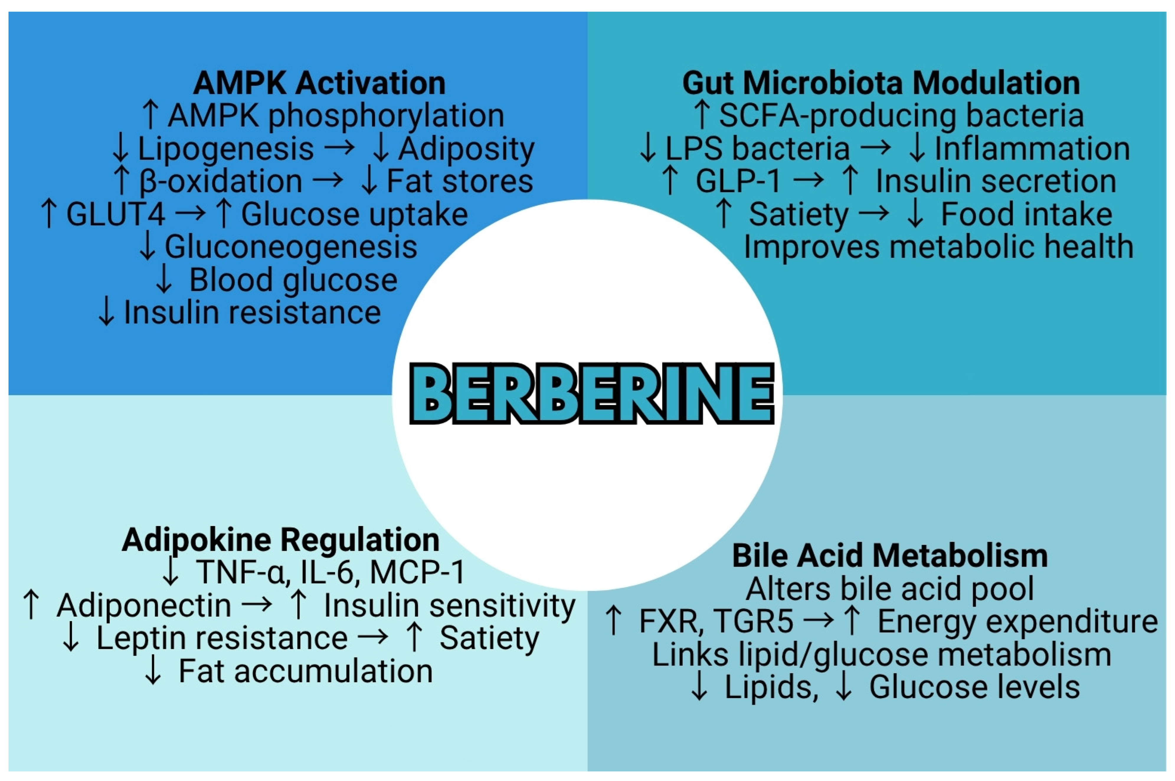
Berberine, an isoquinoline alkaloid found in plants such as Berberis vulgaris L. (barberry) and Coptis chinensis Franch. (goldthread), is a notable example that has been traditionally used in Ayurvedic and Chinese medicine [16,17]. Both preclinical and clinical studies suggest that berberine may modestly reduce body weight and improve metabolic parameters. This is achieved through the activation of AMP-activated protein kinase (AMPK), the modulation of the gut microbiome, and the regulation of lipid metabolism [18] (Figure 1).
This review paper aims to critically evaluate and synthesize the current scientific evidence supporting the anti-obesity potential of berberine, with a specific emphasis on understanding its distinct chemical structure, diverse natural origins, and the multifaceted molecular mechanisms underlying its metabolic effects.

2. Chemical Structure and Physicochemical Properties

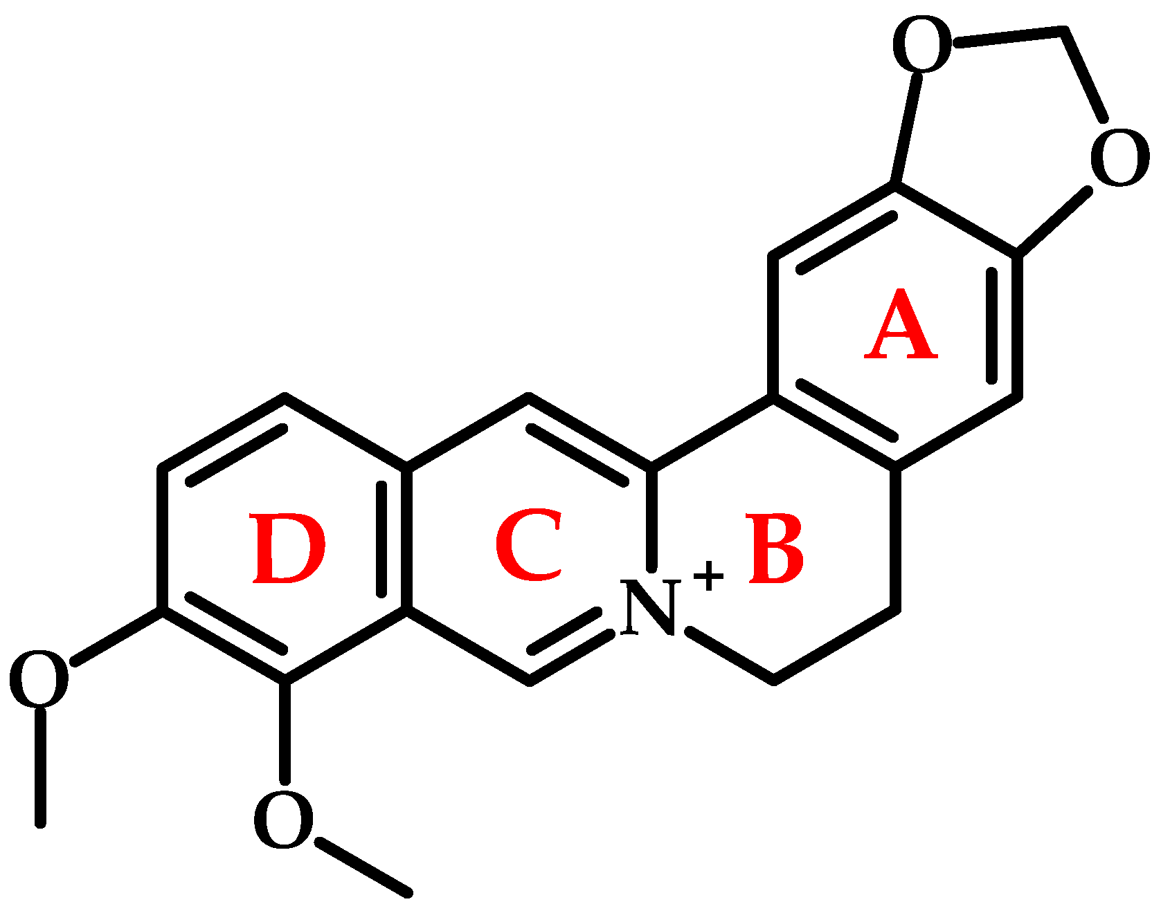
Berberine (Figure 2) is a naturally occurring, yellow-colored, cationic quaternary ammonium alkaloid. Its yellow color has been used in dyeing various materials, and berberine has a Color Index (CI) of 75,160. Chemically, it is defined as 5,6-dihydro-9,10-dimethoxybenzo[g]-1,3-benzodioxolo [5,6-a]quinolizinium, with the formula C20H18NO4+ and a molecular weight of 336.36 g/mol, although its chloride salt form (C20H17NO4HCl·2H2O, MW: 407.646 g/mol) is more commonly utilized.
Its distinct biological activity and fluorescence stem from a unique, planar fused tetracyclic ring system (A–D-rings; Figure 2) forming a benzodioxoloquinolizine core. Key structural features include a methylenedioxy group on the A-ring (C2-C3), a quaternary ammonium moiety on the C-ring, and methoxy groups at C9 and C10 on the D-ring. Interestingly, berberine exhibits fluorescence, with a maximum absorption wavelength of 350 nm and an emission wavelength of 530 nm in sodium dodecyl sulfate solution [19]. Also known as Natural Yellow 18, it is used to stain heparin in mast cells due to its fluorescent properties [20].
Berberine chloride demonstrates excellent solution stability, showing less than 5% degradation over six months across a wide pH range (1.2 to 9.0) at temperatures up to 40 °C [21], which is critical for oral formulation shelf-life and gastrointestinal transit integrity. Its crystalline state standard molar enthalpy of formation is −1335.2 kJ·mol−1, and its standard molar energy of combustion is −9536.2 kJ·mol−1 [19]. The melting point ranges from 145.1 to 146.7 °C.
Berberine chloride displays temperature-dependent solubility. It is readily soluble in hot water but sparingly soluble in cold water or ethanol, and essentially insoluble in nonpolar solvents (e.g., benzene, ether, chloroform) [22]. Solubility is also highly pH-dependent, and this pH-dependence directly impacts bioavailability, peaking at approximately 9.69 mM in phosphate buffer (pH 7.0, 37 °C) and decreasing significantly in acidic buffers. Solubility can be enhanced up to 4.5-fold via complexation with 2-hydroxypropyl-β-cyclodextrin (HPβCD), forming a 1:1 inclusion complex. Such solubility enhancement strategies are crucial for overcoming poor dissolution-limited absorption, a key barrier to therapeutic efficacy. While nonionic surfactants have negligible effects, ionic surfactants like sodium lauryl sulfate can reduce solubility, influencing excipient selection in formulations [21]. Table 1 summarizes critical physicochemical properties of berberine chloride.

3. Natural Occurrence and Sources

Berberine is a naturally occurring secondary metabolite found in many plant species from various plant families. As a bright yellow alkaloid, it is one of the most widely spread protoberberine compounds in the plant kingdom, with records of its occurrence on multiple continents and in different climates [23,24,25,26,27,28,29,30,31,32,33,34,35,36,37,38,39,40,41,42,43,44,45]. The detailed Table 2 below summarizes research findings from 2024 to 2025 on the distribution of berberine in various plant taxa.
This isoquinoline alkaloid shows a remarkable range of taxonomic diversity, occurring in at least six major plant families. The main sources include members of the Berberidaceae, Ranunculaceae, Menispermaceae, Malvaceae, Rutaceae, Annonaceae, and Vitaceae families. This broad phylogenetic distribution suggests that berberine biosynthesis evolved multiple times independently, with recent molecular studies confirming that distantly related lineages followed similar evolutionary paths.
The genus Berberis (Berberidaceae) exhibits substantial interspecific and intraspecific variation in berberine concentration, heavily dependent on the specific plant organ analyzed. Berberis darwinii Hook. roots yielded the highest recorded levels within this genus, reaching up to 26,482.20 µg/g dry weight, significantly surpassing concentrations found in stems (up to 6639.58 µg/g), seeds (up to 1181.75 µg/g), and leaves (up to 511.02 µg/g) [26]. Similarly, B. thunbergii DC. twigs contained notably higher berberine (0.364–0.676%) compared to B. koreana Palib. (0.042–0.067%) and B. × ottawensis “Superba” (0.103%) when extracted under identical ultrasonic conditions with 70% ethanol [38]. B. vulgaris root extracts prepared through hydro-ethanolic reflux yielded 111.06 mg/g using HPTLC quantification [35], whereas another paper showed a lower concentration of 78.95 µg/g dry extract [41]. Roots and stems of B. vulgaris extracted by Accelerated Solvent Extraction (ASE) with methanol yielded detectable berberine quantified by HPLC-ESI-Q-TOF-MS/MS, although specific concentrations were not reported [42].
Within the Ranunculaceae family, Coptis species, particularly their rhizomes and roots, emerge as vibrant sources. C. chinensis rhizomes exhibited berberine levels ranging from 15.2 to 46.2 mg/g dry weight using HPLC after ultrasound-assisted extraction (UAE) with various organic acids [27], up to 77.12 mg/g dry weight using a complex UHPLC-PDA method following DES-UA-MSPD extraction with betaine–acrylic acid [40]. Roots of the same species were reported to contain up to 10% berberine using DES-based coupled with UAE [39]. C. teeta Wall. rhizomes showed values of 212.18 ppm under optimized microwave-assisted extraction (MAE) conditions and 162.96 ppm with ultrasonic-assisted extraction (UAE) [32]. Thalictrum foliolosum DC. roots yielded 13.14 mg/g dry weight after UAE with a natural deep eutectic solvent (NADES) extraction [34], while a broader study on 57 batches of Thalictrum spp. stems and roots reported a wide range of 0.01 to 12.44 mg/g using LC-MS/MS after methanolic sonication [45].
Phellodendron amurense Rupr. (Rutaceae) bark was a source of berberine at the concentration of 0.243 mg/g of extract after microwave-assisted extraction [23], and a much higher 50.88 mg/g via UAE with aqueous malic acid [36]. Tinospora cordifolia (Willd.) Miers and Coscinium fenestratum (Gaertn.) Colebr. (Menispermaceae) stems were also confirmed sources. T. cordifolia stem concentrations included a reported 2.49% (w/w) via HPTLC after MAE [30], while other studies using HPLC-DAD or LC-MS/MS on hydro-alcoholic or UAE extracts did not report specific berberine values [24,25]. C. fenestratum stems and roots yielded 38.23 mg/g after extraction with 60% lactic acid [43]. Annickia affinis (Exell) Versteegh & Sosef (Annonaceae) stem bark contained approximately 0.02% w/w berberine quantified by LC-ESI-MS/MS after direct methanol extraction [44]. Moreover, some concentrations were noted for Helicteres isora L. fruits (21.37% of total area by GC-MS) [33], Cayratia albifolia C.L.Li roots [37], and Mahonia bealei (Fort.) Carr. leaves [28].
It underscores that berberine content is profoundly influenced by plant species, family (notably Berberidaceae and Ranunculaceae), and specific plant part (roots/rhizomes generally being richest). Coptis rhizomes and Berberis roots consistently rank among the highest natural sources. Modern extraction techniques are crucial for the efficient isolation of compounds. Ultrasound-assisted extraction (UAE) uses sound waves, microwave-assisted extraction (MAE) uses microwave energy, and accelerated solvent extraction (ASE) uses high pressure/temperature to enhance solvent penetration. Publications from the past two years reveal a strong trend toward using these techniques, often coupled with acidified solvents or novel solvents (DES/NADES), and advanced analytical methods like UHPLC-MS/MS, providing robust tools for efficient isolation and precise quantification. UAE was prevalent in the cited studies, employing solvents ranging from hydro–ethanolic mixtures [38,41] and organic acids [27,36] to innovative NADES/DES solvents [39,40]. Conventional methods of extraction are still used successfully. Such methods include reflux [29,41], boiling [37], and solvent shaking [44]. Interestingly, solvent composition, ratio, temperature, power, and time were critical optimized parameters that were universally optimized.

4. Pharmacological Mechanisms in Obesity and Diabetes

4.1. Impact on Obesity

Berberine exhibits a broad spectrum of pharmacological activities that collectively influence key metabolic disturbances associated with obesity, insulin resistance, glucose homeostasis, and lipid metabolism, underscoring its potential as a multifaceted therapeutic agent in metabolic disorders. Its multidimensional actions are further discussed in this section, highlighting the diverse mechanisms through which berberine may exert its beneficial effects (Figure 3).

4.1.1. Lipid Metabolism

When food energy consistently exceeds what the body can burn, two linked processes drive fat gain. First, dormant precursor cells are turned into new fat cells; second, those cells fill up with triglycerides. At the genetic level, the “foremen” that set this factory in motion are the transcription factors PPAR-γ and C/EBP-α. Berberine (BBR) short-circuits the very beginning of this programme: in classic 3T3-L1 cell assays, it cuts PPAR-γ and C/EBP-α expression almost in half, so far fewer precursors mature into adipocytes [46]. The same experiment shows a matching 50% drop in intracellular fat, illustrating how gene control translates directly into reduced lipid storage. Berberine not only prevents new fat cells from forming but also switches existing ones from storing energy to burning it. The key player here is AMP-activated protein kinase (AMPK)—a fuel gauge that senses when cellular ATP is low. Berberine nudges the ATP/AMP balance enough to activate AMPK; activated AMPK then inactivates acetyl-CoA carboxylase (ACC), lowering malonyl-CoA and opening the gate for fatty acids to pour into mitochondria for oxidation [47]. Independent groups have confirmed that the same AMPK-ACC cascade lowers lipogenesis in hepatocytes [48] and normalises liver fat in obese mice through both central and peripheral AMPK signaling [49]. The compound’s lipid-lowering reach goes beyond adipose tissue. In liver cells, BBR increases LDL-receptor mRNA and protein, thereby clearing LDL-cholesterol from the bloodstream by a mechanism distinct from statins [50]. Clinical trials echo these pre-clinical findings: treating patients with type 2 diabetes and mixed dyslipidemia with 1 g per day BBR for three months cut total cholesterol by about 18% and LDL-C by 21% [51]. A larger meta-analysis of eleven randomised controlled trials—874 participants in total—confirmed significant average reductions in total cholesterol (−0.61 mmol L−1), triglycerides (−0.50 mmol L−1), and LDL-C (−0.65 mmol L−1) with only mild gastrointestinal side effects [52]. Untargeted metabolomic profiling provides a panoramic snapshot of these molecular events in living animals. In high-cholesterol hamsters, berberine lowered circulating acyl-carnitines (signals of faster fatty-acid oxidation) and cholesteryl esters, while modestly raising specific bile acids—evidence that cholesterol was being routed into bile and excreted [53]. Complementary studies show that berberine’s persistent AMPK activation is tied to a sustained rise in the AMP/ATP ratio and an uptick in glycolysis, matching the overall energy-depleting signal required to keep fat burning switched on [54]. Berberine stops the “construction” of new fat depots and simultaneously turns existing depots into small furnaces. This dual action—genetic (fewer fat cells) and energetic (more fat burned)—explains why both laboratory models and human trials have consistently recorded lower body fat and blood lipid levels after berberine supplementation.

4.1.2. Gut Microbiota

The adult intestine is home to an estimated 1013–1014 microorganisms drawn from thousands of bacterial species [55]. When these microbes ferment otherwise indigestible dietary fibre, they generate the short-chain fatty acids (SCFAs) acetate, propionate, and butyrate [56]. SCFAs are more than metabolic waste: butyrate is the preferred fuel for colonocytes and supports mucosal energy needs, whereas all three acids tighten epithelial junctions and stimulate mucin secretion, thereby reinforcing the intestinal barrier [57,58]. Beyond the gut, SCFAs activate free-fatty-acid receptors on enteroendocrine cells, provoking the release of PYY and GLP-1, hormones that quell appetite and enhance insulin sensitivity [59]. A sustained high-fat diet (HFD) perturbs this balance. It selectively depletes many SCFA-producing taxa and enriches Gram-negative Proteobacteria; the resulting surge in lipopolysaccharide (LPS) compromises tight-junction integrity, leaks into the bloodstream, and kindles low-grade “metabolic endotoxemia” [60,61]. The loss of SCFA supply, coupled with LPS-driven inflammation, undermines barrier function, blunts satiety signaling, and feeds into weight gain and insulin resistance. Early animal work put berberine on the microbiome map. In rats fattened on a high-fat diet, berberine shifted the bacterial community toward SCFA-producing genera such as Allobaculum, Blautia, and Butyricicoccus, cut overall microbial diversity, and—importantly—lowered the animals’ adiposity index (a measure of total body fat) [62]. A companion redundancy-analysis linked 134 berberine-responsive bacterial “operational taxonomic units” (OTUs) to changes in body weight, leptin, and insulin, drawing a straight line from microbiome rewiring to host metabolism. An earlier study from the same group had already shown that these compositional shifts coincide with higher caecal SCFA levels and a partial reversal of LPS-driven inflammation [63].
Since 2020, multi-omics approaches have deepened this picture. In a mouse model of metabolic syndrome, berberine not only enriched SCFA producers but also restored the tight-junction proteins ZO-1 and occludin, reduced serum LPS, and quieted hepatic inflammatory genes—changes that paralleled improvements in weight and lipid profiles [64]. Attention has turned to species-level “keystone” taxa. Clinical work in hyperlipidemic patients showed that the abundance of Blautia and Alistipes at baseline could predict who would respond best to berberine’s cholesterol-lowering action; antibiotic depletion or fecal-transplant studies confirmed that the gut microbiota is necessary and sufficient for this effect [65]. Follow-up mechanistic experiments identified Blautia producta as a berberine-enriched commensal capable of upregulating hepatic LDL receptors, lowering serum LDL-C, and, through its butyrate output, contributing to SCFA pools [66]. Berberine also influences how microbes transform bile acids, detergent-like molecules that serve as metabolic hormones. By selectively promoting bile-salt-hydrolase-inhibiting species, berberine shifts the bile-acid pool toward forms that activate the intestinal receptor FXR and the systemic receptor TGR5, both linked to better glucose tolerance and energy expenditure [64].
Importantly, gut-centered actions mesh with classic pharmacology: combining berberine with metformin in diabetic mice produced a greater fall in blood glucose than either drug alone and generated a distinct microbiota signature—higher Akkermansia and Verrucomicrobia, and lower Proteobacteria—alongside higher circulating metformin levels [67]. These findings suggest that manipulating the microbiome may help fine-tune berberine dosing or uncover synergistic drug pairs in metabolic disease.
In summary, berberine remodels the gut ecosystem in three concerted steps: 1. It enriches SCFA-producing bacteria, boosting metabolites that curb appetite and inflammation. 2. It suppresses LPS-laden Gram-negative taxa, tightening the intestinal barrier and lowering systemic inflammatory tone. 3. It steers bile-acid conversions toward hormonal profiles that favor leanness and insulin sensitivity. Together, these microbial shifts create a biochemical environment that echoes and amplifies berberine’s direct effects on lipid and glucose metabolism, reinforcing its promise as a diet-compatible tool for obesity management.

4.1.3. Adipokine Regulation

Adipose tissue, traditionally viewed as a passive fat storage site, is now recognized as a highly active endocrine organ, secreting a variety of bioactive peptides known as adipokines. Among these, leptin and adiponectin are two of the most studied due to their significant roles in energy homeostasis, metabolic regulation, and inflammation. Alongside these, inflammatory cytokines such as tumor necrosis factor-alpha (TNF-α), interleukin-6 (IL-6), and C-reactive protein (CRP) are significantly influenced by changes in adipose tissue dynamics and play a central role in the development of obesity-related complications. Leptin is primarily involved in appetite regulation and energy balance. Its secretion increases with fat mass, acting on the hypothalamus to suppress food intake and increase energy expenditure. However, in obesity, leptin resistance often develops, characterized by elevated circulating levels of leptin without corresponding satiety signals. This resistance blunts its physiological role, contributing to continued weight gain and metabolic dysregulation [68]. Adiponectin, in contrast, exhibits inverse behavior with respect to adiposity. It enhances insulin sensitivity and exhibits anti-inflammatory and anti-atherogenic properties. Lower adiponectin levels are frequently observed in individuals with obesity, type 2 diabetes, and cardiovascular disease, suggesting its protective role against metabolic dysfunction [69].
The secretion of inflammatory cytokines from adipose tissue, particularly from infiltrating immune cells like macrophages, is another hallmark of dysfunctional adipose tissue. Increased levels of TNF-α and IL-6 in obesity contribute to systemic inflammation, insulin resistance, and the progression of metabolic syndrome. These cytokines not only interfere with insulin signaling pathways but also suppress adiponectin production, creating a feedback loop that exacerbates metabolic disturbances [70].
The balance between these adipokines and cytokines is tightly regulated and highly sensitive to lifestyle factors, including diet, physical activity, and pharmacological interventions. Weight loss, for example, has been shown to decrease leptin and inflammatory cytokine levels while increasing adiponectin concentrations, thereby improving metabolic outcomes. The regulation of leptin, adiponectin, and inflammatory cytokines underscores the complex interplay between adipose tissue and systemic metabolic health. Targeting these adipokines offers promising therapeutic potential in managing obesity and its associated disorders.
Adipose tissue is not a passive energy store; it secretes a spectrum of protein messengers—adipokines—that influence appetite, inflammation, and insulin responsiveness throughout the body. During weight gain, this secretory profile drifts in an unfavorable direction. Circulating leptin, which usually signals satiety to the brain, rises in parallel with fat mass; yet, central leptin receptors become desensitized, a state termed leptin resistance [63]. At the same time, adiponectin, an anti-inflammatory adipokine that promotes glucose uptake and fatty-acid oxidation, falls sharply. The imbalance is compounded by chemokines, such as monocyte-chemoattractant protein-1 (MCP-1), which draw immune cells into adipose depots, fueling chronic, low-grade inflammation [53].
In rodent models of diet-induced obesity, oral berberine consistently reverses this endocrine drift. Eight to twelve weeks of treatment at 100–200 mg kg−1 day−1 lowers plasma leptin and MCP-1 while restoring adiponectin when normalized to fat mass [63]. Untargeted metabolomics links these hormonal shifts to a rise in glutathione and a decline in arachidonic-acid metabolites—biochemical signatures of reduced oxidative stress and dampened eicosanoid-driven inflammation [53]. More recent multi-omics studies confirm that berberine enhances antioxidant defenses and suppresses lipid peroxidation in parallel with normalizing adipokines [64].
Formulation improvements appear to magnify these effects. A co-crystal combining berberine with ibuprofen, which increases intestinal absorption three-fold, boosted serum adiponectin by 60% and lowered MCP-1 expression in adipose tissue more potently than native berberine in obese db/db mice [71].
Several molecular pathways converge to explain the endocrine reset. Berberine activates the AMPK–SIRT1 axis, which suppresses the transcription factor NF-κB and thereby limits MCP-1 production [72]. It also alleviates endoplasmic reticulum stress in adipocytes, a condition known to blunt leptin signaling, and curtails 12/15-lipoxygenase activity, reducing pro-inflammatory lipid mediators [53].
The same direction is being pointed to by early clinical data, even if the scale is limited. In adults with metabolic syndrome, twelve weeks of berberine (500 mg three times daily) reduced the leptin-to-adiponectin ratio by nearly one-third, and this change was associated with improved insulin sensitivity [73]. A randomised trial in patients experiencing antipsychotic-associated weight gain found that adjunctive berberine (600 mg day−1) halved serum leptin, suggesting improved leptin responsiveness, although adiponectin was unchanged [74]. In poorly controlled type 2 diabetes, adding berberine (1 g day−1) to metformin lowered MCP-1 and IL-6 and restored adiponectin to near-normal levels while improving glycemic control [67].
Collectively, the pre-clinical and emerging clinical evidence indicates that berberine recalibrates adipose-derived signals on three fronts: it reduces pro-inflammatory cues (lower leptin and MCP-1), restores anti-inflammatory tone (higher adiponectin), and improves the oxidative milieu that underpins adipokine secretion. Through these integrated actions, modest weight reductions achieved under berberine are translated into disproportionately large gains in insulin sensitivity and systemic metabolic health.

4.2. Role in Diabetes and Insulin Resistance

4.2.1. Glucose Homeostasis

Maintaining stable blood-glucose levels hinges on two tightly linked processes. First, glucose must be cleared from the circulation into peripheral tissues, chiefly skeletal muscle and adipose tissue, via insulin-stimulated translocation of the GLUT4 transporter to the cell surface. In obesity and type 2 diabetes, this trafficking step falters; impaired GLUT4 delivery is now recognized as a core molecular lesion of insulin resistance [75]. Second, the liver must restrain its own production of glucose through gluconeogenesis during the fasted state. In diabetes, that restraint is lost: hepatic expression of gluconeogenic enzymes such as phosphoenolpyruvate carboxykinase and glucose-6-phosphatase remains inappropriately high, so the liver continues to add glucose to the bloodstream even when levels are already elevated, driving fasting hyperglycemia [76].
In summary, maintaining stable blood glucose levels relies on two processes working together: the efficient uptake of glucose into the tissues of the body (mostly muscle and fat) and the controlled release of glucose from the liver through a process called gluconeogenesis. In obesity and type 2 diabetes, both arms fail—the insulin signal that moves the GLUT4 transporter to the cell surface is blunted, and the liver continues to manufacture glucose even when circulating levels are already high.
Recent work places berberine at the crossroads of these abnormalities. In skeletal muscle and cultured myotubes, the alkaloid raises the AMP–ATP ratio and thereby activates the cellular energy sensor AMP-activated protein kinase (AMPK). Activated AMPK, in turn, drives GLUT4 molecules to the plasma membrane, boosting glucose uptake independently of the classic PI3K–Akt insulin cascade [77]. Because this route bypasses the insulin receptor, it remains functional in insulin-resistant states—a key advantage over therapies that rely solely on enhancing insulin signaling.
Berberine’s influence extends to the liver, where it dampens gluconeogenesis. Both in hepatocyte culture and in diabetic rodent models, the compound reduces transcription of the rate-limiting enzymes phosphoenolpyruvate carboxykinase (PEPCK) and glucose-6-phosphatase (G6Pase), leading to appreciably lower fasting glucose concentrations. Two complementary signaling routes have been described. First, activation of the upstream kinase LKB1 funnels through AMPK and TORC2 to silence gluconeogenic genes [78]. Second, berberine modulates an AKT/MAPK/NO/cGMP/PKG axis, again converging on PEPCK-G6Pase repression and reinforcing the hypoglycemic effect [79].
These molecular findings translate into measurable clinical benefits. In a 12-week, randomized trial of the gut-targeted berberine-ursodeoxycholate co-crystal (HTD1801) involving 113 adults with type 2 diabetes, the higher dose (1 g twice daily) lowered HbA1c by 0.5 percentage points relative to placebo and produced parallel reductions in fasting glucose; exploratory biomarker work confirmed AMPK activation and suppressed gluconeogenic gene signatures [80].
In summary, berberine tackles glucose dysregulation on two fronts: 1. peripheral uptake—it activates AMPK in muscle and adipose tissue, moving GLUT4 to the cell surface and allowing glucose to enter cells even when insulin signaling is impaired; and 2. hepatic output—it switches off the key enzymes of gluconeogenesis via AMPK-centered (and AKT/MAPK-linked) pathways, trimming the liver’s contribution to blood glucose. By synchronising these effects, berberine restores a more balanced glucose economy, an action pattern that underpins its growing appeal as an adjunct—or, in some formulations, an alternative—to current antidiabetic medications.

4.2.2. Insulin Signaling

Efficient insulin action rests on two molecular events: (1) insulin must recruit the glucose-transporter GLUT4 to the surface of skeletal-muscle and adipose cells so that glucose leaves the bloodstream, and (2) hepatic production of glucose through gluconeogenesis must be dialled down when systemic concentrations are already high. In obesity and type 2 diabetes, these safeguards unravel the following: free fatty acids and inflammatory cues stimulate serine/threonine kinases that phosphorylate insulin-receptor substrate-1 (IRS-1) on inhibitory serine sites, disconnecting it from phosphatidyl-inositol-3-kinase (PI3K) and blocking downstream Akt activation; at the same time, gluconeogenic enzymes in the liver remain inappropriately active [75].
BBR restores this broken circuitry at several checkpoints. In hepatocytes challenged with TNF-α, BBR inhibited the upstream MEKK1/MEK–ERK1/2 module, preventing ERK-driven serine phosphorylation of IRS-1 and, thereby, re-establishing IRS-1 tyrosine phosphorylation, PI3K coupling, and Akt activity; glucose uptake normalized in parallel [81]. In skeletal muscle, high-fructose feeding normally blunts Akt and GLUT4 translocation, yet eight weeks of oral BBR reversed these defects while raising the AMP/ATP ratio and activating AMPK—again without relying on canonical PI3K-Akt insulin signaling [77].
Inflammation-linked kinases provide a second entry point. In high-fat-diet mice, BBR lowered hepatic and adipose phosphorylation of IKK-β (Ser181) and curtailed NF-κB nuclear translocation, changes that coincided with reduced IRS-1^Ser phosphorylation and improved insulin-stimulated Akt activity [64]. Complementary work in sarcopenic mice shows that BBR also activates a SIRT1-mediated mitophagy axis, relieving oxidative stress, suppressing IKKβ/NF-κB tone, and reinstating insulin responsiveness in ageing muscle [82].
Across tissues, these node-specific actions translate into a coherent network response. A systems-biology review that collated multi-omics and docking data for >40 natural alkaloids places BBR among the most consistent enhancers of the InsR–IRS–PI3K–Akt axis in liver, muscle, and adipose tissue, with reported gains in glucose uptake ranging from 20% in vitro to 45% in vivo [83]. An even broader 2024 database-driven meta-analysis of >300 BBR targets confirms frequent high-affinity docking to kinases that gate insulin signaling—IKKβ, JNK, and mTORC2—and highlights the compound’s dual ability to quench inflammatory serine kinases while sparing, or even amplifying, canonical tyrosine-kinase cascades [84].
Taken together, contemporary evidence indicates that berberine rescues insulin signaling at its most vulnerable links—IRS-1 integrity, PI3K engagement, and Akt-driven GLUT4 trafficking—while dampening the inflammatory and stress-kinase landscape that erodes these links in the first place. This multi-node repair explains why berberine can lower fasting glucose and improve whole-body insulin sensitivity, even when insulin receptor activity per se is not increased, making the alkaloid a mechanistically distinct complement to existing antidiabetic agents.

4.2.3. Mitochondrial Function

Mitochondria serve as the cell’s principal sites of ATP generation, transforming nutrients into usable energy while modulating intracellular reactive oxygen species (ROS) levels [85]. Both the number of mitochondria and their functional integrity decline in obesity and type 2 diabetes, a change strongly implicated in the development of insulin resistance and ectopic lipid accumulation [86]. A growing body of work shows that berberine counteracts this mitochondrial deficit on two fronts: it stimulates mitochondrial biogenesis, the synthesis of fresh organelles, and it promotes mitophagy, the selective clearance of damaged ones—effects that together restore oxidative capacity and redox balance [82,87,88].
A unifying upstream event is the activation of the AMP-activated protein kinase (AMPK)–SIRT1–PGC-1α axis. In diet-induced sarcopenic mice, eight weeks of oral BBR restored grip strength, doubled mitochondrial DNA copy number, and raised maximal respiratory capacity; pharmacological blockade of SIRT1 abolished these benefits, pinpointing the SIRT1–PGC–1α module as indispensable [82]. Similar results emerge in isolated PINK1-knock-out fibroblasts, where BBR sparks AMPK-dependent PINK1/Parkin mitophagy while up-regulating the biogenic regulators NRF-1 and TFAM, normalizing ATP output without compromising membrane potential [88].
Tissue-specific studies corroborate this dual action. In the liver, BBR improves insulin sensitivity by enhancing SIRT1-directed expression of Opa1, thereby harmonizing mitochondrial fusion–fission dynamics and boosting oxidative phosphorylation efficiency [87]. Cardiac models of heart failure with preserved ejection fraction display depressed AMPK and swollen, ROS-leaking mitochondria; four weeks of BBR reinstated AMPK/PGC-1α signaling, corrected TFAM-driven biogenesis, and limited mitochondrial apoptosis, culminating in better diastolic function [89]. Low-dose BBR also shields cardiomyocytes from doxorubicin toxicity by dissociating the Bcl–xL–Beclin–1 complex, unleashing mitophagy and quenching ROS [90].
Structural analogues reinforce the story. Tetrahydroberberrubine, a more lipophilic derivative, delayed heart ageing in mice by stabilising PHB2 mRNA, a pivotal mitophagy receptor, and thereby accelerating turnover of senescent mitochondria [91]. Such findings highlight the therapeutic headroom available through formulation or chemical modification.
A systems biology survey of >300 BBR targets places mitochondrial regulators at the top of the affinity list and reports convergent activation of the AMPK–SIRT1–PGC–1α network in liver, muscle, heart, and adipose tissue [84]. Collectively, this evidence positions BBR as a dual-action restorer of mitochondrial quantity and quality. By seeding fresh mitochondria and removing defective ones, BBR elevates oxidative capacity and reduces oxidative stress, creating the metabolic environment needed for improved insulin signaling and lipid handl

5. Metabolomic Insights into Berberine’s Therapeutic Effects

5.1. Obesity and the Anti-Obesity Effects of Berberine

Obesity is characterized by excessive fat accumulation resulting from an imbalance between energy intake and expenditure. This condition is a significant risk factor for metabolic disorders, including type 2 diabetes, cardiovascular disease, and non-alcoholic fatty liver disease. Berberine has demonstrated significant anti-obesity effects, primarily through modulation of lipid metabolism and bile acid (BA) pathways [92,93].

5.1.1. Lipid Profile Modulation

BBR has been shown to reduce serum triglycerides (TG) and low-density lipoprotein cholesterol (LDL-C) levels, thereby improving lipid profiles in obese models. Mechanistically, BBR activates peroxisome proliferator-activated receptor delta (PPAR-δ), which enhances mitochondrial fatty acid oxidation and reduces lipid accumulation in adipocytes. In high-fat diet (HFD)-induced obese mice, BBR administration significantly reduced body weight gain and adipose tissue mass. These effects were markedly diminished upon pharmacological inhibition of PPAR-δ, underscoring this receptor’s pivotal role in the lipid-lowering action of BBR [94].
Moreover, BBR enhances basal triglyceride lipolysis in adipocytes and suppresses adipogenesis by downregulating the expression of lipogenic transcription factors, including liver X receptor alpha (LXRα), peroxisome proliferator-activated receptor gamma (PPAR-γ), and sterol regulatory element-binding protein-1c (SREBP-1c). These effects collectively inhibit preadipocyte differentiation and lipid droplet accumulation, further supporting BBR’s anti-obesity potential [94].
In a clinical trial involving hyperlipidemic patients, oral administration of BBR (500 mg twice daily) for 3 months significantly reduced serum cholesterol and triglyceride levels, comparable to standard statin therapy, but with fewer reported side effects [50]. These findings provide translational relevance to the lipid-lowering effects observed in preclinical studies.
To sum up, berberine lowers serum levels of triglycerides (TG), total cholesterol (TC), and low-density lipoprotein cholesterol (LDL-C). It enhances LDL receptor (LDLR) expression in the liver, which promotes hepatic clearance of LDL-C [50]. BBR also downregulates lipogenic genes such as SREBP-1c and FASN, inhibiting lipid synthesis [92]. In addition, it activates AMP-activated protein kinase (AMPK) and peroxisome proliferator-activated receptor delta (PPAR-δ), leading to increased fatty acid oxidation and reduced fat accumulation [94].

5.1.2. Bile Acid Metabolism

Bile acids are not only crucial for dietary lipid digestion but also serve as important signaling molecules involved in the regulation of glucose and energy homeostasis via nuclear and membrane receptors such as farnesoid X receptor (FXR) and Takeda G-protein-coupled receptor 5 (TGR5). BBR modulates BA metabolism primarily through alterations in gut microbiota composition, particularly enhancing the abundance of bacteria capable of converting primary to secondary BAs [95].
This remodeling of the BA pool composition subsequently activates FXR and TGR5 signaling, leading to improved insulin sensitivity, increased energy expenditure, and reduced hepatic steatosis. In mouse models, BBR increased the expression of the bile salt export pump (BSEP) and small heterodimer partner (SHP), downstream targets of FXR activation, which play key roles in maintaining BA homeostasis [96].
Additionally, BBR upregulates cholesterol 7α-hydroxylase (CYP7A1), the rate-limiting enzyme in BA synthesis from cholesterol, thereby promoting cholesterol clearance and reducing circulating LDL-C levels. This enzymatic shift not only aids in lipid reduction but also contributes to the anti-atherogenic properties of BBR [97].

5.2. Diabetes: Shifts in Amino Acids (e.g., Branched-Chain Amino Acids), TCA Cycle Intermediates, and Gut Microbiota-Derived Metabolites

Diabetes mellitus encompasses a group of metabolic diseases characterized by chronic hyperglycemia resulting from defects in insulin secretion, insulin action, or both. Type 1 diabetes mellitus (T1DM) is primarily caused by the autoimmune destruction of pancreatic β-cells, leading to absolute insulin deficiency [98]. In contrast, type 2 diabetes mellitus (T2DM) involves insulin resistance and a relative insulin deficiency [99]. Both types are associated with profound metabolic disturbances extending beyond glucose metabolism. Emerging evidence highlights significant alterations in amino acid profiles [100], disruptions in mitochondrial function as seen through tricarboxylic acid (TCA) cycle intermediates [101], and dysregulation of gut microbiota-derived metabolites [102] in both T1DM and T2DM. These changes offer new insights into disease pathophysiology and potential therapeutic targets.

5.2.1. Alterations in Amino Acid Metabolism

Elevated levels of branched-chain amino acids (BCAAs)—leucine, isoleucine, and valine—are consistently observed in T2DM and are associated with insulin resistance and impaired glucose metabolism. Reduced catabolism of BCAAs has been documented, resulting in their accumulation and contributing to metabolic inflexibility [103]. This is primarily due to the downregulation of enzymes such as branched-chain α-keto acid dehydrogenase.
In addition, alterations in arginine metabolism are noted in diabetes. Increased activity of the enzyme arginase leads to higher levels of ornithine and proline, which may impair nitric oxide availability and promote endothelial dysfunction, contributing factors to vascular complications in diabetic patients [104].

5.2.2. Disruptions in TCA Cycle Intermediates

The TCA cycle is essential for cellular respiration and energy production. In T2DM, several studies report reductions in key intermediates such as citrate, α-ketoglutarate, and succinate, particularly in skeletal muscle tissue [101]. This suggests mitochondrial dysfunction and a shift toward anaerobic glycolysis, which limits ATP production and promotes lactate accumulation.
Diabetic individuals also exhibit a reliance on fatty acid and amino acid oxidation for energy, rather than glucose oxidation. This metabolic shift exacerbates insulin resistance and contributes to lipid accumulation in non-adipose tissues [101].

5.2.3. Gut Microbiota-Derived Metabolites and Their Impact

The gut microbiota plays a critical role in modulating host metabolism. In diabetes, gut dysbiosis alters the production of bioactive microbial metabolites, including short-chain fatty acids (SCFAs), bile acids, and trimethylamine N-oxide (TMAO). Reduced production of butyrate, a key SCFA, is associated with impaired gut barrier function and systemic inflammation, both of which contribute to insulin resistance [105]. In contrast, elevated circulating levels of TMAO in diabetes have been linked to increased cardiovascular risk, due to its pro-inflammatory and pro-atherogenic effects [105].
Both type 1 and type 2 diabetes are characterized by distinct and overlapping metabolic perturbations, including altered amino acid metabolism, disrupted tricarboxylic acid (TCA) cycle function, and imbalances in gut microbiota-derived metabolites. These shifts contribute to the progression of insulin resistance, mitochondrial dysfunction, and chronic inflammation, providing mechanistic insights that may inform future therapeutic strategies.

5.3. Multi-Omics Integration: Linkages Between Metabolomics, Transcriptomics, and Proteomics Data

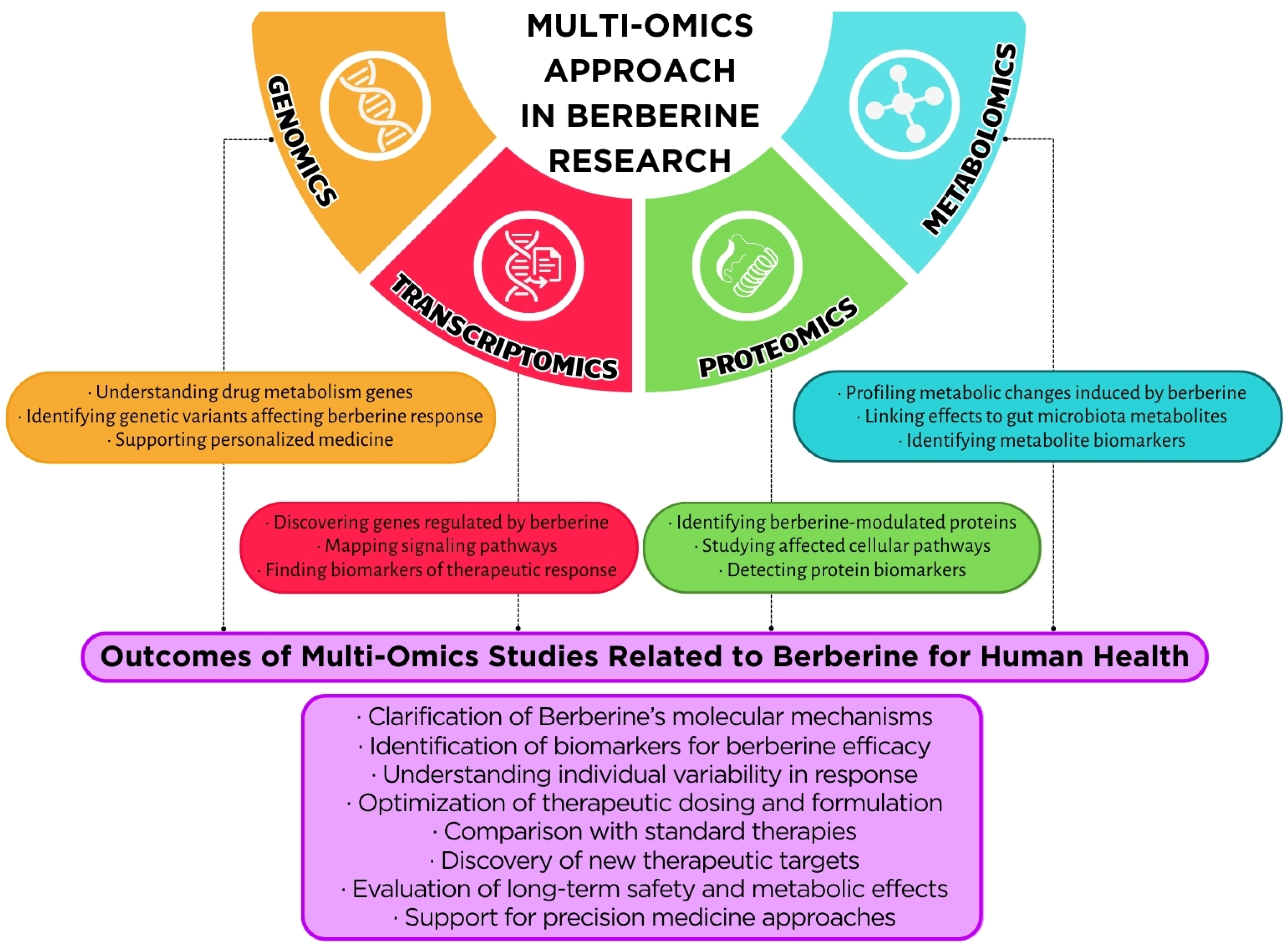
Understanding the multifaceted therapeutic effects of BBR necessitates a systems-level approach that transcends the study of isolated molecular pathways. Integrating multi-omics datasets—particularly metabolomics, transcriptomics, and proteomics—has proven to be a powerful strategy for unraveling the complex biological networks and regulatory circuits modulated by BBR across different tissues and disease contexts.

5.3.1. Holistic Insights Through Multi-Omics Platforms

Multi-omics approaches enable the simultaneous interrogation of metabolic flux, gene expression, and protein abundance, providing a comprehensive understanding of how BBR orchestrates systemic physiological changes. In metabolic diseases such as type 2 diabetes mellitus (T2DM) and obesity, BBR has been shown to coordinate responses at multiple molecular levels, restoring metabolic balance, reducing inflammation, and improving insulin sensitivity [106,107,108].

5.3.2. Berberine in Inflammatory Bowel Disease: A Multi-Omics Paradigm

One of the clearest examples of multi-omics integration in BBR research comes from studies on inflammatory bowel disease (IBD). In a murine model of ulcerative colitis, Yu et al. [109] employed a combination of metagenomics, metabolomics, and transcriptomics to demonstrate that BBR modulated gut microbiota composition—enriching beneficial taxa like Lactobacillus—and restored the expression of tight junction proteins (e.g., ZO-1, occludin), thereby reinforcing intestinal barrier integrity. Concurrently, significant shifts in bile acid metabolism were observed, suggesting that BBR’s efficacy arises from the interplay between microbial, metabolic, and epithelial pathways [109].
Similarly, Yang et al. [110] employed a multi-omics approach, integrating 16S rDNA sequencing, serum metabolomics, and transcriptomics, to investigate the effects of BBR on the intestinal microbiome and serum metabolome in ulcerative colitis. Their findings revealed that BBR ameliorated the community of intestinal microbiota and significantly promoted the abundance of beneficial bacteria such as Muribaculaceae and Akkermansia. Furthermore, BBR modulated inflammation-related metabolites and metabolic pathways in serum, indicating its role in restoring metabolic homeostasis [110].

5.3.3. Transcriptome–Proteome Interplay in Energy Metabolism

Transcriptomic and proteomic analyses have revealed that BBR modulates multiple pathways involved in mitochondrial function and energy metabolism. For instance, integrated omics profiling has shown that BBR upregulates peroxisome proliferator-activated receptor gamma coactivator 1-alpha (PGC-1α) and carnitine palmitoyltransferase 1A (CPT1A), key regulators of fatty acid β-oxidation, while downregulating lipogenic genes such as SREBP-1c and FASN. These changes align with increased mitochondrial biogenesis and oxidative phosphorylation, consistent with improved metabolic phenotypes in BBR-treated models. Additionally, proteomic data have confirmed alterations in enzymes of the tricarboxylic acid (TCA) cycle and glycolysis, supporting the hypothesis that BBR redirects cellular energy metabolism toward improved glucose utilization and decreased lipid storage [106,111].

5.3.4. MAPK and Inflammatory Pathways: Multilayered Regulation

A study employing transcriptomics and phosphoproteomics revealed that BBR significantly suppresses the MAPK signaling cascade—including ERK, JNK, and p38 pathways—thereby attenuating the expression of pro-inflammatory cytokines in chronic atrophic gastritis models. This multilayered regulation at both the transcriptional and post-translational levels suggests that BBR not only dampens inflammation but also reprograms the cellular stress response machinery [112]. Studies indicate that BBR suppresses the activation of these kinases, leading to a decrease in the expression of pro-inflammatory cytokines, including TNF-α, IL-1β, and IL-6 [112]. This modulation occurs through both transcriptional and post-translational mechanisms, suggesting a multilayered regulatory effect on the inflammatory response.
For instance, in models of gastric mucosal inflammation, BBR inhibited the phosphorylation of ERK, JNK, and p38, which was associated with reduced cytokine expression and improved mucosal barrier integrity [112]. Furthermore, BBR has been shown to influence the expression of stress response-related genes, including HO-1 and Nrf2, indicating a role in enhancing cellular defense mechanisms [50].
Although direct studies using combined transcriptomic and phosphoproteomic approaches to evaluate BBR’s effects on the MAPK pathway remain limited, available data support its potential to influence this pathway via modulation of gene expression and kinase activity. Further research employing advanced omics technologies is warranted to comprehensively understand these interactions and confirm the molecular mechanisms underlying BBR’s effects (Figure 4).

6. Safety, Bioavailability, and Future Directions

6.1. Pharmacokinetics, Toxicity, and Side Effects

Berberine has a remarkably wide range of health benefits. These include regulating glucose and lipid metabolism, as mentioned earlier, as well as fighting infections, reducing inflammation, and showing potential for protecting brain and heart health [113]. Although berberine shows excellent promise as a therapeutic, several major obstacles hinder its widespread use in clinical settings. The main challenges are its unfavorable pharmacokinetic profile, which results in very low bioavailability, and the need for a thorough understanding of its long-term safety profile, particularly regarding interactions with other medications and its use in specific populations [114]. Overcoming these limitations through innovative research is crucial to harnessing the full potential of berberine and expanding its established uses.
One major limitation of berberine’s clinical use is its very low absorption rate in the body, which is usually estimated to be less than 1% when taken orally [115]. Moreover, berberine is poorly absorbed across the intestinal epithelium. Its chemical structure, featuring a quaternary ammonium group, contributes to low aqueous solubility and poor passive diffusion through the lipid membranes of the intestinal cells [15]. Only a small part of the berberine that is absorbed enters the portal vein, where it undergoes significant metabolism in the liver before entering systemic circulation. The primary reactions involved are Phase II conjugation reactions, specifically glucuronidation, which forms metabolites such as berberrubine glucuronide, and sulfation. These metabolites often have lower pharmacological activity than the original berberine compound [116]. Berberine and its metabolites are primarily eliminated from the body via renal excretion (urine) and biliary excretion (feces). Its elimination half-life is relatively short, typically ranging from a few to several hours [117]. This necessitates frequent dosing to maintain potentially therapeutic plasma concentrations.
The gut microbiota plays a crucial role in overcoming berberine’s low oral bioavailability (<1%). Cheng et al. [118] confirmed that despite poor solubility and absorption, berberine enters systemic circulation through bidirectional interactions with gut bacteria. Importantly, anaerobic bacteria (e.g., Enterobacter cloacae, Escherichia coli) convert berberine into dihydroberberine via bacterial nitroreductase activity [119], a reduced form that shows 5-fold higher intestinal absorption because of increased lipophilicity and decreased positive charge [119]. After absorption, dihydroberberine quickly re-oxidizes non-enzymatically back to active berberine in intestinal tissues [119], acting as a “stealth transporter” that bypasses P-glycoprotein efflux. This microbial biotransformation greatly boosts systemic berberine levels [119], which explains its strong pharmacological effects despite low plasma concentrations [119]. Meanwhile, berberine alters the gut ecosystem: it encourages beneficial SCFA-producing bacteria (e.g., Lachnospiraceae, Blautia) and suppresses harmful pathogens [118,120]. Interestingly, Blautia species further metabolize berberine into bioactive demethylated compounds (e.g., thalifendine) through acetogenesis [120], aiding therapeutic effects and promoting cross-feeding that increases butyrate production [118,120]. Antibiotic treatment that disrupts gut microbiota reduces both dihydroberberine formation and demethylation pathways, leading to lower systemic berberine levels and weaker glucose-lowering and lipid-regulating effects [118,119].
Berberine is generally regarded as safe for short to medium-term use in healthy adults at typical therapeutic doses (commonly 500–1500 mg per day, administered in divided doses) [121]. It shows relatively low acute toxicity. Research in mice found an LD50 of pure berberine to be 329 mg/kg when given orally and 23 mg/kg when given intraperitoneally [122]. The most commonly reported side effects are related to the digestive system, likely due to the high concentration of the substance in the gut and its antimicrobial properties. Symptoms like diarrhea, constipation, gas, stomach cramps, and nausea are common, especially at higher doses or when treatment first starts. These symptoms often decrease with continued use or a lower dose [123].
Berberine inhibits several CYP enzymes, primarily CYP2D6, CYP2C9, and CYP3A4, which can significantly increase blood levels and the risk of toxicity for drugs that rely on these pathways. This includes antidepressants, statins, and immunosuppressants. Berberine also blocks P-glycoprotein, making it easier for P-gp substrate drugs to be absorbed and affecting their distribution in the body, which can lead to toxicity. Furthermore, berberine’s ability to lower glucose levels can interact with diabetes medications like insulin, sulfonylureas, or metformin, greatly increasing the risk of hypoglycemia [124]. Berberine is contraindicated during pregnancy and breastfeeding due to its ability to cross the placenta and displace bilirubin from albumin, posing a risk of kernicterus in newborns, and because it is excreted in breast milk [125].

6.2. Future Research and Applications

For berberine to become a widely accepted therapeutic agent, its pharmacokinetic limitations must be addressed, and its effectiveness must be thoroughly evaluated across a broader range of conditions.
The development of nanotechnology-based delivery systems is crucial for addressing the clinical limitations of berberine, primarily its low bioavailability due to poor solubility, instability, and susceptibility to efflux. These systems enhance dissolution, protect berberine from degradation, improve absorption (potentially through lymphatic transport), and enable targeted delivery. Effective nanoplatforms include solid lipid nanoparticles, chitosan-based polymeric nanoparticles, liposomes, niosomes, magnetic nanoparticles, silver nanoparticles, and lyotropic liquid crystalline nanoparticles. According to Iqbal et al. [126], the use of these nanocarriers enhances berberine’s uptake by cancer cells and its therapeutic effect against malignancies, including breast, liver, nasopharyngeal, and prostate cancer. Key advantages comprise controlled release kinetics and the potential for synergistic co-administration with agents such as doxorubicin or silver nanoparticles to overcome multidrug resistance (MDR).
There are also various conventional formulations, such as pH-dependent tablets, floating pellets or beads, solid dispersions, and topical hydrogels, developed to enhance dissolution, provide gastric retention, target specific sites (e.g., the colon), or improve permeation, thereby significantly increasing bioavailability and enabling sustained release [127]. While conventional systems offer easier processing and scalability, polymeric systems provide greater control and improved bioavailability but face challenges such as biocompatibility issues, complex manufacturing, scalability problems, and potential toxicity from monomers. Raju et al. [127] also described advanced carriers, including microparticles, microcapsules, microspheres, or dendrimers, that effectively encapsulate berberine, achieving high drug loading and dramatically enhancing pharmacokinetic parameters while improving oral absorption.
As an example, encapsulating berberine in Solid Lipid Nanoparticles significantly increased its plasma levels and bioavailability in diabetic (db/db) mice, resulting in greater reductions in fasting blood glucose, body weight gain, and insulin resistance compared to free alkaloid. The nanoparticles predominantly accumulated in the liver, where they effectively reduced hepatic steatosis by modulating fat metabolism genes (such as by increasing CPT-1 and suppressing SREBP-1c), with a 100 mg/kg dose showing markedly stronger effects than an equivalent free berberine dose. The authors confirmed that the enhanced bioavailability and liver targeting led to superior improvements in glucose tolerance, insulin sensitivity, and protection of islet function in diabetic models [128].
The study by Demet et al. [129] demonstrated that liposomal encapsulation enhances the stability and bioavailability of berberine, highlighting its potential, particularly in its liposomal form, to induce browning (via uncoupling protein 1, UCP1) and modulate lipid metabolism in adipocytes, laying the foundation for novel anti-obesity strategies. In their study, berberine was successfully encapsulated in liposomes, which provided sustained release and significantly improved its physical and chemical stability compared to free berberine, especially protecting it from degradation under light. Both free and liposomal berberine increased the expression of the key browning marker UCP1 in 3T3-L1 adipocytes during both differentiation and maturation processes. Moreover, liposomal berberine (20 µM) significantly reduced triglyceride levels during the differentiation treatment, indicating a potential role in modulating lipid metabolism in early adipocyte development [129].
Researchers have also attempted to enhance berberine’s bioavailability by creating numerous derivatives. Key strategies included chemical modifications (e.g., 9-O-esters, 12-aryl substitutions, carbohydrate conjugates, and hybrids such as metformin-berberine) to enhance absorption and reduce intestinal efflux. The modified compounds demonstrated significantly stronger effects in tests, including enhanced hypoglycemic effects (via the AMPK/PI3K/Akt pathways), lipid-lowering capabilities, anti-inflammatory actions, and cytotoxicity against cancer cells compared to natural berberine. Moreover, studies have confirmed the efficacy of these derivatives using in vitro assays (e.g., glucose consumption in HepG2 cells and insulin sensitivity in adipocytes) and in vivo diabetic and obese animal models [130].
Although berberine exhibits promising therapeutic effects in obesity and metabolic disorders, several critical gaps remain to be addressed for successful clinical translation. Regulatory hurdles stem from variability in compound purity, inconsistencies in commercial formulations, and the absence of standardized clinical protocols guiding berberine usage. Furthermore, much of the current evidence originates from preclinical studies or small-scale clinical trials, necessitating larger, well-controlled human studies to establish robust safety and efficacy profiles. Future research should prioritize longitudinal human investigations into berberine’s effects on the gut microbiome and its metabolic consequences, as well as standardized dosing strategies to optimize therapeutic outcomes. Additionally, human-based multi-omics trials are essential to confirm molecular targets and mechanistic pathways identified in preclinical research, paving the way for precision medicine applications. Addressing these challenges will be pivotal for integrating berberine into evidence-based clinical practice as an adjunctive therapy in obesity and metabolic health management.

7. Conclusions

Berberine demonstrates substantial anti-obesity and metabolic regulatory potential through its multifaceted actions on key biological pathways. By activating AMPK, it promotes lipid oxidation, mitochondrial biogenesis, and glucose homeostasis, while modulation of the gut microbiota contributes to improved metabolic signaling via increased SCFA production and reduced systemic inflammation. Berberine also influences adipokine profiles, favoring insulin sensitivity and reduced inflammatory responses. Insights from metabolomics have expanded our understanding of berberine’s systemic effects, highlighting its roles in bile acid modulation, carnitine-mediated fatty acid transport, and TCA cycle optimization, collectively contributing to reduced adipogenesis and ectopic lipid deposition. However, its clinical application faces significant challenges, including poor oral bioavailability, gastrointestinal side effects, and potential drug interactions related to CYP450 and P-glycoprotein substrates. Innovative delivery systems such as lipid nanoparticles, cyclodextrin complexes, and structurally modified derivatives, including berberine–metformin hybrids, are under investigation to overcome these barriers and enhance therapeutic efficacy. Future research integrating multi-omics approaches in human studies is crucial to validate mechanistic insights and guide personalized therapeutic strategies. With these advancements, berberine holds promise as a complementary, evidence-based option in the management of obesity and metabolic disorders.

Author Contributions

Conceptualization, M.P.; resources, B.Z. and M.P.; writing—original draft preparation, B.Z. and M.P.; visualization, B.Z.; supervision, M.P. All authors have read and agreed to the published version of the manuscript.

Funding

This research received no external funding.

Institutional Review Board Statement

Not applicable.

Informed Consent Statement

Not applicable.

Data Availability Statement

No new data were created or analyzed in this study. Data sharing is not applicable to this article.

Acknowledgments

During the preparation of this manuscript, the authors used Perplexity AI for literature searching. The authors have reviewed and edited the output and take full responsibility for the content of this publication.

Conflicts of Interest

The authors declare no conflicts of interest.

Abbreviations

The following abbreviations are used in this manuscript:
AktProtein kinase B
AMPAdenosine monophosphate
AMPKAMP-activated protein kinase
AOMAnti-obesity medication
ASEAccelerated solvent extraction
ATPAdenosine triphosphate
BABile acid
BBRBerberine
BCAABranched-chain amino acid
BSEPBile salt export pump
C/EBP-αCCAAT/enhancer-binding protein alpha
CIColor Index
CPT1ACarnitine palmitoyltransferase 1A
CRPC-reactive protein
CVDCardiovascular disease
CYP2C9Cytochrome P450 2C9 enzyme
CYP2D6Cytochrome P450 2D6 enzyme
CYP3A4Cytochrome P450 3A4 enzyme
CYP7A1Cholesterol 7α-hydroxylase
DES-UA-MSPDDeep eutectic solvent ultrasound-assisted matrix solid-phase dispersion
ERKExtracellular signal-regulated kinase (subgroup of MAPK)
FASNFatty acid synthase
FXRFarnesoid X receptor
G6PaseGlucose-6-phosphatase
GC-MSGas chromatography-mass spectrometry
GIPGlucose-dependent insulinotropic polypeptide
GLUT4Glucose transporter type 4
GLP-1Glucagon-like peptide-1
HbA1cGlycated hemoglobin
HFDHigh-fat diet
HO-1Heme oxygenase-1
HPβCD2-hydroxypropyl-β-cyclodextrin
HPLC-DADHigh-performance liquid chromatography with a diode array detector
HPLC-ESI-Q-TOF-MS/MSHigh-performance liquid chromatography–electrospray ionization–quadrupole time-of-flight tandem mass spectrometry
HPTLCHigh-performance thin-layer chromatography
IBDInflammatory bowel disease
IKK-βInhibitor of nuclear factor kappa-B kinase subunit beta
IL-1βInterleukin-1 beta
IL-6Interleukin-6
IRS-1Insulin receptor substrate 1
JNKc-Jun N-terminal kinase (subgroup of MAPK)
LC-MS/MSLiquid chromatography–tandem mass spectrometry
LDLLow-density lipoprotein
LDL-CLow-density lipoprotein cholesterol
LDLRLow-density lipoprotein receptor
LPSLipopolysaccharide
LXRαLiver X receptor alpha
MAEMicrowave-assisted extraction
MAPKMitogen-activated protein kinase
MASLDMetabolic dysfunction-associated steatotic liver disease
MASHMetabolic dysfunction-associated steatohepatitis (inflammatory form of MASLD)
MCP-1Monocyte chemoattractant protein-1
MDRMultidrug resistance
mTORMechanistic target of rapamycin
MWMolecular weight
NADESNatural deep eutectic solvent
NAFLDNon-alcoholic fatty liver disease
NCDNon-communicable disease
NF-κBNuclear factor kappa-light-chain-enhancer of activated B cells
NHANESNational Health and Nutrition Examination Survey
NONitric oxide
NRF-1Nuclear respiratory factor 1
Nrf2Nuclear factor erythroid 2-related factor 2
Opa1Optic atrophy 1 (mitochondrial fusion protein)
OTUOperational taxonomic unit
p38p38 mitogen-activated protein kinase (subgroup of MAPK)
P-gpP-glycoprotein
PDAPhoto diode array
PEPCKPhosphoenolpyruvate carboxykinase
PHB2Prohibitin 2 (mitophagy receptor)
PI3KPhosphatidylinositol 3-kinase
PGC-1αPeroxisome proliferator-activated receptor gamma coactivator 1-alpha
PINK1PTEN induced kinase 1 (mitophagy regulator)
PPAR-γPeroxisome proliferator-activated receptor gamma
PPAR-δPeroxisome proliferator-activated receptor delta
PYYPeptide YY
ROSReactive oxygen species
SCFAShort-chain fatty acid
SHPSmall heterodimer partner
SIRT1Sirtuin 1 (NAD+-dependent deacetylase)
SREBP-1cSterol regulatory element-binding protein-1c
T1DMType 1 diabetes mellitus
T2DMType 2 diabetes mellitus
TCATricarboxylic acid cycle (Krebs cycle)
TCTotal cholesterol
TFAMMitochondrial transcription factor A
TGTriglycerides
TGR5Takeda G protein-coupled receptor 5
TMAOTrimethylamine N-oxide
TNF-αTumor necrosis factor alpha
TORC2Target of rapamycin complex 2
UAEUltrasound-assisted extraction
UCP1Uncoupling protein 1
UPLC-Q-TOF-MS/MSUltra-performance liquid chromatography–quadrupole time-of-flight tandem mass spectrometry
WHOWorld Health Organization
ZO-1Zonula occludens-1 (tight-junction protein)

References

  1. Jaacks, L.M.; Vandevijvere, S.; Pan, A.; McGowan, C.J.; Wallace, C.; Imamura, F.; Mozaffarian, D.; Swinburn, B.; Ezzati, M. The Obesity Transition: Stages of the Global Epidemic. Lancet Diabetes Endocrinol. 2019, 7, 231–240. [Google Scholar] [CrossRef] [PubMed]
  2. Popkin, B.M.; Adair, L.S.; Ng, S.W. Global Nutrition Transition and the Pandemic of Obesity in Developing Countries. Nutr. Rev. 2012, 70, 3–21. [Google Scholar] [CrossRef]
  3. Obesity and Overweight. Available online: https://www.who.int/news-room/fact-sheets/detail/obesity-and-overweight (accessed on 8 June 2025).
  4. NCD Risk Factor Collaboration (NCD-RisC). Worldwide Trends in Body-Mass Index, Underweight, Overweight, and Obesity from 1975 to 2016: A Pooled Analysis of 2416 Population-Based Measurement Studies in 128·9 Million Children, Adolescents, and Adults. Lancet 2017, 390, 2627–2642. [Google Scholar] [CrossRef]
  5. Stierman, B.; Afful, J.; Carroll, M.D.; Chen, T.-C.; Davy, O.; Fink, S.; Fryar, C.D.; Gu, Q.; Hales, C.M.; Hughes, J.P.; et al. National Health and Nutrition Examination Survey 2017-March 2020 Prepandemic Data Files-Development of Files and Prevalence Estimates for Selected Health Outcomes. Natl. Health Stat. Rep. 2021, 158, 10–15620. [Google Scholar] [CrossRef]
  6. Available online: https://iris.who.int/bitstream/handle/10665/353747/9789289057738-eng.pdf?sequence=1 (accessed on 8 June 2025).
  7. Poirier, P.; Giles, T.D.; Bray, G.A.; Hong, Y.; Stern, J.S.; Pi-Sunyer, F.X.; Eckel, R.H.; American Heart Association; Obesity Committee of the Council on Nutrition, Physical Activity, and Metabolism. Obesity and Cardiovascular Disease: Pathophysiology, Evaluation, and Effect of Weight Loss: An Update of the 1997 American Heart Association Scientific Statement on Obesity and Heart Disease from the Obesity Committee of the Council on Nutrition, Physical Activity, and Metabolism. Circulation 2006, 113, 898–918. [Google Scholar] [CrossRef]
  8. Zatterale, F.; Longo, M.; Naderi, J.; Raciti, G.A.; Desiderio, A.; Miele, C.; Beguinot, F. Chronic Adipose Tissue Inflammation Linking Obesity to Insulin Resistance and Type 2 Diabetes. Front. Physiol. 2019, 10, 1607. [Google Scholar] [CrossRef]
  9. Wondmkun, Y.T. Obesity, Insulin Resistance, and Type 2 Diabetes: Associations and Therapeutic Implications. Diabetes Metab. Syndr. Obes. 2020, 13, 3611–3616. [Google Scholar] [CrossRef]
  10. Lauby-Secretan, B.; Scoccianti, C.; Loomis, D.; Grosse, Y.; Bianchini, F.; Straif, K.; International Agency for Research on Cancer Handbook Working Group. Body Fatness and Cancer—Viewpoint of the IARC Working Group. N. Engl. J. Med. 2016, 375, 794–798. [Google Scholar] [CrossRef] [PubMed]
  11. Younossi, Z.M.; Golabi, P.; de Avila, L.; Paik, J.M.; Srishord, M.; Fukui, N.; Qiu, Y.; Burns, L.; Afendy, A.; Nader, F. The Global Epidemiology of NAFLD and NASH in Patients with Type 2 Diabetes: A Systematic Review and Meta-Analysis. J. Hepatol. 2019, 71, 793–801. [Google Scholar] [CrossRef]
  12. Luppino, F.S.; de Wit, L.M.; Bouvy, P.F.; Stijnen, T.; Cuijpers, P.; Penninx, B.W.J.H.; Zitman, F.G. Overweight, Obesity, and Depression: A Systematic Review and Meta-Analysis of Longitudinal Studies: A Systematic Review and Meta-Analysis of Longitudinal Studies. Arch. Gen. Psychiatry 2010, 67, 220–229. [Google Scholar] [CrossRef]
  13. Sarwer, D.B.; Polonsky, H.M. The Psychosocial Burden of Obesity. Endocrinol. Metab. Clin. N. Am. 2016, 45, 677–688. [Google Scholar] [CrossRef] [PubMed]
  14. Dehghani, F.; Ali Ahmadi, M.; Hefner, M.; Kudchadkar, G.; Najam, W.; Nateqi, M.; Siddik, M.A.B.; Booe, H.; Dhurandhar, N.V. An Algorithm for the Use of Anti-Obesity Medications. Nutr. Diabetes 2024, 14, 20. [Google Scholar] [CrossRef]
  15. Liu, Y.-T.; Hao, H.-P.; Xie, H.-G.; Lai, L.; Wang, Q.; Liu, C.-X.; Wang, G.-J. Extensive Intestinal First-Pass Elimination and Predominant Hepatic Distribution of Berberine Explain Its Low Plasma Levels in Rats. Drug Metab. Dispos. 2010, 38, 1779–1784. [Google Scholar] [CrossRef] [PubMed]
  16. Kumar, A.; Ekavali; Chopra, K.; Mukherjee, M.; Pottabathini, R.; Dhull, D.K. Current Knowledge and Pharmacological Profile of Berberine: An Update. Eur. J. Pharmacol. 2015, 761, 288–297. [Google Scholar] [CrossRef]
  17. Patel, P. A Bird’s Eye View on a Therapeutically ‘Wonder Molecule’: Berberine. Phytomedicine Plus 2021, 1, 100070. [Google Scholar] [CrossRef]
  18. Ilyas, Z.; Perna, S.; Al-Thawadi, S.; Alalwan, T.A.; Riva, A.; Petrangolini, G.; Gasparri, C.; Infantino, V.; Peroni, G.; Rondanelli, M. The Effect of Berberine on Weight Loss in Order to Prevent Obesity: A Systematic Review. Biomed. Pharmacother. 2020, 127, 110137. [Google Scholar] [CrossRef]
  19. Cheng, X.-X.; Lui, Y.; Hu, Y.-J.; Liu, Y.; Li, L.-W.; Di, Y.-Y.; Xiao, X.-H. Thermal Behavior and Thermodynamic Properties of Berberine Hydrochloride. J. Therm. Anal. Calorim. 2013, 114, 1401–1407. [Google Scholar] [CrossRef]
  20. Battu, S.K.; Repka, M.A.; Maddineni, S.; Chittiboyina, A.G.; Avery, M.A.; Majumdar, S. Physicochemical Characterization of Berberine Chloride: A Perspective in the Development of a Solution Dosage Form for Oral Delivery. AAPS PharmSciTech 2010, 11, 1466–1475. [Google Scholar] [CrossRef]
  21. Available online: https://www.sigmaaldrich.com/PL/pl/product/sigma/b3251 (accessed on 9 June 2025).
  22. Ai, X.; Yu, P.; Peng, L.; Luo, L.; Liu, J.; Li, S.; Lai, X.; Luan, F.; Meng, X. Berberine: A Review of Its Pharmacokinetics Properties and Therapeutic Potentials in Diverse Vascular Diseases. Front. Pharmacol. 2021, 12, 762654. [Google Scholar] [CrossRef]
  23. Qi, Y.; Chen, Y.; Liu, G.; Shen, L. Study on Microwave Extraction of Berberine Coloring Compound and Extracts Stability: Process Optimization by Response Surface Method (RSM). Dye. Pigment. 2025, 237, 112609. [Google Scholar] [CrossRef]
  24. Hazra, K.; Kumar, D.; Debnath, S.; Mondal, S.; Batule, M.; Dutta, S.; Singh, A.; Singh, R.; Mangal, A.K. Dynamicity and Extractability of Hydro-Alcoholic Solvents for Tinospora Cordifolia Stem: An Investigation for Target-Oriented Traditional Drug Discovery Based on Biologically Active Phytocompounds. Vegetos 2024, 38, 931–941. [Google Scholar] [CrossRef]
  25. Singh, S.M.; Tripathy, S.; Srivastav, P.P. Bioactive Compound Extraction from Giloy Leaves and Steam Using Ultrasound: Bioactivity, Antimicrobial, and LC-MS/MS Study. Food Sci. Biotechnol. 2025, 34, 1835–1847. [Google Scholar] [CrossRef]
  26. Chacón-Fuentes, M.; Burgos-Díaz, C.; Opazo-Navarrete, M.; Mercado, A.; Westermeyer, F. Berberine and Palmatine Distribution across Plant Organs in Berberis darwinii: Basis for Selecting Superior-Producing Accessions. Molecules 2025, 30, 1849. [Google Scholar] [CrossRef] [PubMed]
  27. Nguyen, K.V.; Le, N.T.; Dang, V.T.T.; Koshovyi, O.; Raal, A.; Nguyen, H.T. Alkaloid Extraction from Coptis chinensis Franch. Using Ultrasound-Assisted Aqueous Solutions of Surfactants, Organic Acids, Deep Eutectic Solvents, and Supramolecular Deep Eutectic Solvents. Molecules 2025, 30, 1418. [Google Scholar] [CrossRef]
  28. Wei, L.; Ma, W.; Liu, S.; Mi, S.; Shen, Q.; Lu, Q.; Liu, Z. Mahonia bealei (Fort.) Carr. Leaf Extract Modulates the TLR2/MyD88/NF-κB Signaling Pathway to Inhibit PGN-Induced Inflammation in RAW264.7 Cells. J. Ethnopharmacol. 2025, 344, 119510. [Google Scholar] [CrossRef] [PubMed]
  29. Wang, J.; Xu, X.; Huang, X.; Zhai, L.; Li, Z.; Sun, G.; Jiang, R.; Sun, L. Coptis Cream Ethanol Extract Regulates Degranulation Caused by Allergic Reactions through MGPRB3/PLC/TRPV1 Signaling Pathway. J. Ethnopharmacol. 2025, 343, 119473. [Google Scholar] [CrossRef]
  30. Satija, S.; Vyas, M.; Pandey, P.; Garg, M. Taguchi L9 Optimization of Microwave-Assisted Solid-Liquid Duo Heating for Berberine Extraction with HPTLC Analysis. Biomed. Chromatogr. 2025, 39, e70008. [Google Scholar] [CrossRef]
  31. Moravcová, P.; Schröterová, L.; Zíka, J.; Hošťálková, A.; Švec, F.; Šatínský, D. A UHPLC-DAD Method for Quantification of Berberine and Protoberberine Alkaloids in Herbal Food Supplements Based on Berberis aristata Extract and Evaluation of Their Biological Activity. J. Food Compos. Anal. 2025, 139, 107150. [Google Scholar] [CrossRef]
  32. Sarma, L.; Patra, F.; Borah, P.K.; Meena, S.; Duary, R.K. Comparative Modeling of Microwave and Ultrasound Assisted Extraction of Phenolics and Berberine from Coptis teeta Wall. Rhizomes. Sustain. Food Technol. 2025, 3, 570–581. [Google Scholar] [CrossRef]
  33. Didar, Z.; Afshar, A.S.; Nia, M.M.Z. Evaluation of Chemical and Biological Characteristics of Subcritical Water Extract from Helicteres isora L. Fruit and Its Incorporation into Chia Seed Mucilage Film. Iran. J. Chem. Chem. Eng. 2025, 44, 479–491. [Google Scholar] [CrossRef]
  34. Singh, B.; Singh, L.; Bhatt, I.D.; Kandpal, N.D. Tailored NADES Solvents for the Extraction Optimization of Benzylisoquinoline Alkaloids from Thalictrum foliolosum DC.—A Potential Phyto-Nutraceutical Source. Food Chem. 2025, 463, 141016. [Google Scholar] [CrossRef] [PubMed]
  35. Marcinčáková, D.; Hudáková, N.; Miłek, M.; Kolesárová, M.; Dżugan, M.; Cizkova, D.; Legáth, J. Evaluation of the Antioxidant Properties and Biological Effects of a Novel Combined Barberry Root-Propolis Extract on HEK293T Cells. Pharmaceuticals 2024, 18, 27. [Google Scholar] [CrossRef]
  36. Le, N.T.; Le, T.T.; Ho, D.V.; Nguyen, K.V.; Nguyen, H.T. Extraction and Recovery of Bioactive Alkaloids from Phellodendron Amurense Rupr. Using Ultrasound-Assisted Green Solvents and Macroporous Resins. Sustain. Chem. Pharm. 2024, 42, 101809. [Google Scholar] [CrossRef]
  37. Wang, W.; Zhang, Z.-Q.; Zhang, Y.-C.; Wu, Y.-Q.; Yang, Z.; Zheng, Y.-Z.; Lu, J.-H.; Tu, P.-F.; Zeng, K.-W. Cayratia albifolia C.L.Li Exerts Anti-Rheumatoid Arthritis Effect by Inhibiting Macrophage Activation and Neutrophil Extracellular Traps (NETs). Chin. Med. 2024, 19, 42. [Google Scholar] [CrossRef] [PubMed]
  38. Miłek, M.; Dżugan, M.; Pieńkowska, N.; Galiniak, S.; Mołoń, M.; Litwińczuk, W. Ornamental Barberry Twigs as an Underexploited Source of Berberine-Rich Extracts-Preliminary Research. Curr. Issues Mol. Biol. 2024, 46, 13193–13208. [Google Scholar] [CrossRef]
  39. Liu, C.; Gong, F.; Xiong, Z.; Wang, C.; Ran, X.; Ran, J.; Li, R.; Ou, Y.; Xia, Q.; Wei, P.; et al. An Extraction Process Based on the Collaborative Extraction of Coptis chinensis Franch. Phytoconstituents Using a Deep Eutectic Solvent and an Organic Solvent. Separations 2024, 11, 249. [Google Scholar] [CrossRef]
  40. Han, M.; Du, K.; He, X.; Li, H.; Li, J.; Li, X.; Chang, Y. Advancing Green Extraction of Bioactive Compounds Using Deep Eutectic Solvent-Based Ultrasound-Assisted Matrix Solid-Phase Dispersion: Application to UHPLC-PAD Analysis of Alkaloids and Organic Acids in Coptidis rhizoma. Talanta 2024, 274, 125983. [Google Scholar] [CrossRef]
  41. Ivan, I.M.; Olaru, O.T.; Popovici, V.; Chițescu, C.L.; Popescu, L.; Luță, E.A.; Ilie, E.I.; Brașoveanu, L.I.; Hotnog, C.M.; Nițulescu, G.M.; et al. Antioxidant and Cytotoxic Properties of Berberis vulgaris (L.) Stem Bark Dry Extract. Molecules 2024, 29, 2053. [Google Scholar] [CrossRef]
  42. Nakonieczna, S.; Susniak, K.; Bozhadze, A.; Grabarska, A.; Głowniak-Lipa, A.; Głowniak, K.; Kukula-Koch, W. Optimized Centrifugal Partition Chromatography (CPC) Protocol for Isolation of Urease Inhibitors: Magnoflorine and Berberine from Berberis vulgaris Extracts. Separations 2024, 11, 94. [Google Scholar] [CrossRef]
  43. Trong Le, N.; Huyen Thi Chau, N.; Quynh Dinh Nguyen, P.; Thuy Thi Tran, L.; Thanh Phung, H.; Thi Nguyen, H. Green Extraction of Berberine from Coscinium fenestratum (Gaertn.) Colebr. Using Ultrasound-Assisted Aqueous Solutions of Organic Acids, Polyalcohols, and Deep Eutectic Solvents. Sep. Purif. Technol. 2024, 330, 125541. [Google Scholar] [CrossRef]
  44. Erhunse, N.; Kumari, S.; Anmol; Singh, P.; Omoregie, E.S.; Singh, A.P.; Sharma, U.; Sahal, D. Annickia affinis (Exell) Versteegh & Sosef Methanol Stem Bark Extract, Potent Fractions and Isolated Berberine Alkaloid Target Both Blood and Liver Stages of Malaria Parasites. J. Ethnopharmacol. 2024, 319, 117269. [Google Scholar] [CrossRef]
  45. Li, R.; Wu, X.; Jiao, X.; Zhang, X.; Wang, C.; Han, L.; Song, M.; Zhang, Y.; Pan, G.; Zhang, Z. Chemical Profiles, Differentiation, and Quality Evaluation of Radix et Rhizoma Thalictri Foliolosi Based on LC-MS. J. Pharm. Biomed. Anal. 2024, 237, 115747. [Google Scholar] [CrossRef]
  46. Huang, C.; Zhang, Y.; Gong, Z. Berberine Inhibits 3T3-L1 Adipocyte Differentiation through the PPAR-γ Pathway. Biochem. Biophys. Res. Commun. 2006, 348, 571–578. [Google Scholar] [PubMed]
  47. Lee, Y.S.; Kim, W.S.; Kim, K.H.; Yoon, M.J.; Cho, H.J.; Shen, Y.; Ye, J.-M.; Lee, C.H.; Oh, W.K.; Kim, C.T.; et al. Berberine, a Natural Plant Product, Activates AMP-Activated Protein Kinase with Beneficial Metabolic Effects in Diabetic and Insulin-Resistant States. Diabetes 2006, 55, 2256–2264. [Google Scholar] [CrossRef] [PubMed]
  48. Brusq, J.-M.; Ancellin, N.; Grondin, P.; Guillard, R.; Martin, S.; Saintillan, Y.; Issandou, M. Inhibition of Lipid Synthesis through Activation of AMP Kinase: An Additional Mechanism for the Hypolipidemic Effects of Berberine. J. Lipid Res. 2006, 47, 1281–1288. [Google Scholar] [CrossRef]
  49. Kim, W.S.; Lee, Y.S.; Cha, S.H.; Jeong, H.W.; Choe, S.S.; Lee, M.-R.; Oh, G.T.; Park, H.-S.; Lee, K.-U.; Lane, M.D.; et al. Berberine Improves Lipid Dysregulation in Obesity by Controlling Central and Peripheral AMPK Activity. Am. J. Physiol. Endocrinol. Metab. 2009, 296, E812–E819. [Google Scholar] [CrossRef]
  50. Kong, W.; Wei, J.; Abidi, P.; Lin, M.; Inaba, S.; Li, C.; Wang, Y.; Wang, Z.; Si, S.; Pan, H.; et al. Berberine Is a Novel Cholesterol-Lowering Drug Working through a Unique Mechanism Distinct from Statins. Nat. Med. 2004, 10, 1344–1351. [Google Scholar] [CrossRef]
  51. Zhang, Y.; Li, X.; Zou, D.; Liu, W.; Yang, J.; Zhu, N.; Huo, L.; Wang, M.; Hong, J.; Wu, P.; et al. Treatment of Type 2 Diabetes and Dyslipidemia with the Natural Plant Alkaloid Berberine. J. Clin. Endocrinol. Metab. 2008, 93, 2559–2565. [Google Scholar] [CrossRef]
  52. Dong, H.; Zhao, Y.; Zhao, L.; Lu, F. The Effects of Berberine on Blood Lipids: A Systemic Review and Meta-Analysis of Randomised Controlled Trials. Planta Medica 2013, 79, 437–446. [Google Scholar]
  53. Gu, S.; Cao, B.; Sun, R.; Tang, Y.; Paletta, J.L.; Wu, X.; Liu, L.; Zha, W.; Zhao, C.; Li, Y.; et al. A Metabolomic and Pharmacokinetic Study on the Mechanism Underlying the Lipid-Lowering Effect of Orally Administered Berberine. Mol. Biosyst. 2015, 11, 463–474. [Google Scholar] [CrossRef]
  54. Yin, J.; Gao, Z.; Liu, D.; Liu, Z.; Ye, J. Berberine Improves Glucose Metabolism through Induction of Glycolysis. Am. J. Physiol. Endocrinol. Metab. 2008, 294, E148–E156. [Google Scholar] [CrossRef] [PubMed]
  55. Fusco, W.; Lorenzo, M.B.; Cintoni, M.; Porcari, S.; Rinninella, E.; Kaitsas, F.; Lener, E.; Mele, M.C.; Gasbarrini, A.; Collado, M.C.; et al. Short-Chain Fatty-Acid-Producing Bacteria: Key Components of the Human Gut Microbiota. Nutrients 2023, 15, 2211. [Google Scholar] [CrossRef]
  56. Koh, A.; De Vadder, F.; Kovatcheva-Datchary, P.; Bäckhed, F. From Dietary Fiber to Host Physiology: Short-Chain Fatty Acids as Key Bacterial Metabolites. Cell 2016, 165, 1332–1345. [Google Scholar] [CrossRef] [PubMed]
  57. den Besten, G.; van Eunen, K.; Groen, A.K.; Venema, K.; Reijngoud, D.-J.; Bakker, B.M. The Role of Short-Chain Fatty Acids in the Interplay between Diet, Gut Microbiota, and Host Energy Metabolism. J. Lipid Res. 2013, 54, 2325–2340. [Google Scholar] [CrossRef] [PubMed]
  58. Peng, L.; Li, Z.-R.; Green, R.S.; Holzman, I.R.; Lin, J. Butyrate Enhances the Intestinal Barrier by Facilitating Tight-Junction Assembly via Activation of AMP-Activated Protein Kinase in Caco-2 Cells. J. Nutr. 2009, 139, 1619–1625. [Google Scholar]
  59. Pham, N.H.T.; Joglekar, M.V.; Wong, W.K.M.; Nassif, N.T.; Simpson, A.M.; Hardikar, A.A. Short-Chain Fatty Acids and Insulin Sensitivity: A Systematic Review and Meta-Analysis. Nutr. Rev. 2024, 82, 193–209. [Google Scholar] [CrossRef]
  60. Cani, P.D.; Bibiloni, R.; Knauf, C. Changes in Gut Microbiota Control Metabolic Endotoxaemia-Induced Inflammation in High-Fat Diet-Induced Obesity and Diabetes in Mice. Diabetes 2008, 57, 1470–1481. [Google Scholar]
  61. Malesza, I.J.; Malesza, M.; Walkowiak, J.; Mussin, N.; Walkowiak, D.; Aringazina, R.; Bartkowiak-Wieczorek, J.; Mądry, E. High-Fat, Western-Style Diet, Systemic Inflammation, and Gut Microbiota: A Narrative Review. Cells 2021, 10, 3164. [Google Scholar] [CrossRef]
  62. Zhang, X.; Zhao, Y.; Xu, J.; Xue, Z.; Zhang, M.; Pang, X.; Zhang, X.; Zhao, L. Modulation of Gut Microbiota by Berberine and Metformin during the Treatment of High-Fat Diet-Induced Obesity in Rats. Sci. Rep. 2015, 5, 14405. [Google Scholar] [CrossRef]
  63. Zhang, X.; Zhao, Y.; Zhang, M.; Pang, X.; Xu, J.; Kang, C.; Li, M.; Zhang, C.; Zhang, Z.; Zhang, Y.; et al. Structural Changes of Gut Microbiota during Berberine-Mediated Prevention of Obesity and Insulin Resistance in High-Fat Diet-Fed Rats. PLoS ONE 2012, 7, e42529. [Google Scholar] [CrossRef]
  64. Li, J.; Li, J.; Ni, J.; Zhang, C.; Jia, J.; Wu, G.; Sun, H.; Wang, S. Berberine Relieves Metabolic Syndrome in Mice by Inhibiting Liver Inflammation Caused by a High-Fat Diet and Potential Association with Gut Microbiota. Front. Microbiol. 2021, 12, 752512. [Google Scholar] [CrossRef]
  65. Wu, C.; Zhao, Y.; Zhang, Y. Gut Microbiota Specifically Mediates the Anti-Hypercholesterolemic Effect of Berberine (BBR) and Facilitates to Predict BBR’s Cholesterol-Decreasing Therapeutic in Patients. J. Adv. Res. 2022, 37, 197–208. [Google Scholar] [PubMed]
  66. Yang, Y.-N.; Wang, Q.-C.; Xu, W.; Yu, J.; Zhang, H.; Wu, C. The Berberine-Enriched Gut Commensal Blautia Producta Ameliorates High-Fat Diet (HFD)-Induced Hyperlipidemia and Stimulates Liver LDLR Expression. Biomed. Pharmacother. 2022, 155, 113749. [Google Scholar] [CrossRef]
  67. Lyu, Y.; Li, D.; Yuan, X.; Li, Z.; Zhang, J.; Ming, X.; Shaw, P.C.; Zhang, C.; Kong, A.P.S.; Zuo, Z. Effects of Combination Treatment with Metformin and Berberine on Hypoglycemic Activity and Gut Microbiota Modulation in Db/Db Mice. Phytomedicine 2022, 101, 154099. [Google Scholar] [CrossRef]
  68. Zhang, Y.; Proenca, R.; Maffei, M.; Barone, M.; Leopold, L.; Friedman, J.M. Positional Cloning of the Mouse Obese Gene and Its Human Homologue. Nature 1994, 372, 425–432. [Google Scholar] [CrossRef]
  69. Arita, Y.; Kihara, S.; Ouchi, N.; Takahashi, M.; Maeda, K.; Miyagawa, J.-I.; Hotta, K.; Shimomura, I.; Nakamura, T.; Miyaoka, K.; et al. Paradoxical Decrease of an Adipose-Specific Protein, Adiponectin, in Obesity. 1999. Biochem. Biophys. Res. Commun. 2012, 425, 560–564. [Google Scholar] [CrossRef] [PubMed]
  70. Hotamisligil, G.S.; Shargill, N.S.; Spiegelman, B.M. Adipose Expression of Tumor Necrosis Factor-Alpha: Direct Role in Obesity-Linked Insulin Resistance. Science 1993, 259, 87–91. [Google Scholar] [CrossRef]
  71. Wang, M.; Xu, R.; Liu, X.; Zhang, L.; Qiu, S.; Lu, Y.; Zhang, P.; Yan, M.; Zhu, J. A Co-Crystal of Berberine and Ibuprofen Improves Obesity by Inhibiting the Protein Kinase TBK1 and IKKε. Commun. Biol. 2022, 5, 807. [Google Scholar]
  72. Rey, V.; Tamargo-Gómez, I. From Kinases to Diseases: Investigating the Role of AMPK in Human Pathologies. Kinases Phosphatases 2023, 1, 181–205. [Google Scholar]
  73. Zhao, J.V.; Yeung, W.F.; Chan, Y.H.; Vackova, D.; Leung, J.Y.; Ip, D.K.; Zhao, J.; Ho, W.K.; Tse, H.; Schooling, C.M. Effect of Berberine on Cardiovascular-Risk Factors: A Mechanistic Randomised Controlled Trial. Nutrients 2021, 13, 2550. [Google Scholar]
  74. Qiu, Y.; Li, M.; Zhang, Y.; Liu, Y.; Zhao, Y.; Zhang, J.; Jia, Q.; Li, J. Berberine Treatment for Weight Gain in Patients with Schizophrenia by Regulating Leptin Rather than Adiponectin. Asian J. Psychiatr. 2022, 67, 102896. [Google Scholar] [CrossRef] [PubMed]
  75. van Gerwen, J.; Shun-Shion, A.S.; Fazakerley, D.J. Insulin Signalling and GLUT4 Trafficking in Insulin Resistance. Biochem. Soc. Trans. 2023, 51, 1057–1069. [Google Scholar] [CrossRef] [PubMed]
  76. Wang, Z.; Ma, J.; Wu, R.; Kong, Y.; Sun, C. Recent Advances of Long Non-Coding RNAs in Control of Hepatic Gluconeogenesis. Front. Endocrinol. 2023, 14, 1167592. [Google Scholar] [CrossRef]
  77. Cheng, J.; Ma, X.; Yan, G.; Yu, Q.; Huang, Z.; Lin, G.; Li, M.; Guan, F.; Su, Z.; Yan, F.; et al. High Fructose-Induced Skeletal Muscle Insulin Resistance Could Be Alleviated by Berberine via AMPD1 and ADSL. Food Chem. Toxicol. 2023, 175, 113731. [Google Scholar] [CrossRef]
  78. Xu, X.-H.; Hu, Q.; Zhou, L.-S.; Xu, L.-J.; Zou, X.; Lu, F.-E.; Yi, P. Berberine Inhibits Gluconeogenesis in Skeletal Muscles and Adipose Tissues in Streptozotocin-Induced Diabetic Rats via LKB1-AMPK-TORC2 Signaling Pathway. Curr. Med. Sci. 2020, 40, 530–538. [Google Scholar] [CrossRef] [PubMed]
  79. Lu, M.; Wang, Y.; Jiang, Y. Berberine Inhibits Gluconeogenesis in Spontaneous Diabetic Rats by Regulating the AKT/MAPK/NO/cGMP/PKG Signalling Pathway. Mol. Cell. Biochem. 2023, 478, 2013–2027. [Google Scholar]
  80. Ma, J.L.; Ma, J. Berberine Ursodeoxycholate for the Treatment of Type 2 Diabetes: A Randomised Clinical Trial. JAMA Netw. Open 2025, 8, e2462185. [Google Scholar]
  81. Li, Y.; Chen, X.; Chen, Y.; Yu, D.; Jiang, R.; Kou, X.; Sheng, L.; Liu, Y.; Song, Y. Berberine Improves TNF-α-Induced Hepatic Insulin Resistance by Targeting MEKK1/MEK Pathway. Inflammation 2022, 45, 2016–2026. [Google Scholar] [CrossRef]
  82. Su, X.; Yang, D.; Hu, Y.; Yuan, Y.; Song, L. Berberine Suppressed Sarcopenia Insulin Resistance through SIRT1-Mediated Mitophagy. Open Life Sci. 2023, 18, 20220648. [Google Scholar] [CrossRef]
  83. Amssayef, A.; Eddouks, M. Alkaloids as Promising Agents for the Management of Insulin Resistance: A Review. Curr. Pharm. Des. 2023, 29, 3123–3136. [Google Scholar] [CrossRef]
  84. Sun, A.; Yang, H.; Li, T.; Luo, J.; Zhou, L.; Chen, R.; Han, L.; Lin, Y. Molecular Mechanisms, Targets and Clinical Potential of Berberine in Regulating Metabolism: A Review Focussing on Databases and Molecular Docking Studies. Front. Pharmacol. 2024, 15, 1368950. [Google Scholar] [CrossRef]
  85. Georgiev, A.; Granata, C.; Roden, M. The Role of Mitochondria in the Pathophysiology and Treatment of Common Metabolic Diseases in Humans. Am. J. Physiol. Cell Physiol. 2022, 322, C1248–C1259. [Google Scholar] [CrossRef]
  86. Barber, T.M.; Kyrou, I.; Randeva, H.S.; Weickert, M.O. Mechanisms of Insulin Resistance at the Crossroad of Obesity with Associated Metabolic Abnormalities and Cognitive Dysfunction. Int. J. Mol. Sci. 2021, 22, 546. [Google Scholar] [CrossRef]
  87. Xu, J.; Zhang, Y.; Yu, Z.; Guan, Y.; Lv, Y.; Zhang, M.; Zhang, M.; Chen, L.; Lv, X.; Guan, F. Berberine Mitigates Hepatic Insulin Resistance by Enhancing Mitochondrial Architecture via the SIRT1/Opa1 Signalling Pathway. Acta Biochim. Biophys. Sin. 2022, 54, 1464–1475. [Google Scholar] [CrossRef]
  88. Um, J.H.; Lee, K.M.; Kim, Y.Y.; Lee, D.Y.; Kim, E. Berberine Induces Mitophagy through AMPK and Ameliorates Mitochondrial Dysfunction in PINK1-Knockout Mouse Embryonic Fibroblasts. Int. J. Mol. Sci. 2023, 25, 219. [Google Scholar]
  89. Hu, Y.; Chen, X.; Zhao, Q.; Li, G.; Zhang, H.; Ma, Z.; Yu, H.; Zeng, Q.; Zhang, H.; Xu, D. Berberine Improves Cardiac Insufficiency through AMPK/PGC-1α Signalling-Mediated Mitochondrial Homeostasis and Apoptosis in HFpEF Mice. Int. Immunopharmacol. 2025, 155, 114613. [Google Scholar]
  90. Chen, B.; Zhang, J.-P. Bcl-xL Is Required for the Protective Effects of Low-Dose Berberine against Doxorubicin-Induced Cardiotoxicity through Blocking Apoptosis and Activating Mitophagy-Mediated ROS Elimination. Phytomedicine 2022, 101, 154130. [Google Scholar] [CrossRef]
  91. Wang, L.; Tang, X.-Q.; Shi, Y.; Li, H.-M.; Meng, Z.-Y.; Chen, H.; Li, X.-H.; Chen, Y.-C.; Liu, H.; Hong, Y.; et al. Tetrahydroberberrubine Retards Heart Aging in Mice by Promoting PHB2-Mediated Mitophagy. Acta Pharmacol. Sin. 2023, 44, 332–344. [Google Scholar] [CrossRef]
  92. Zhang, Q.; Xiao, X.; Li, M.; Yu, M.; Ping, F.; Zheng, J.; Wang, T. Berberine Moderates Glucose and Lipid Metabolism through Multi-Target Regulation and Promotes Gut Microbiota Profile in Obesity. Sci. Rep. 2020, 10, 924851. [Google Scholar]
  93. Yue, K.; Haokun, Y.; Rong, N.; Xuxiang, Z.; Hongtao, Z.; Xin, N. Progress of the Anti-Obesity of Berberine. arXiv 2025, arXiv:2501.02282. [Google Scholar]
  94. Zhang, Y.; Wang, X.; Wang, Y. Berberine Reduces Lipid Accumulation in Obesity via Mediating PPARδ-Dependent Pathway. Biomed. Pharmacother. 2023, 161, 11600. [Google Scholar]
  95. Nie, X.; Lu, Q.; Yin, Y.; He, Z.; Bai, Y.; Zhu, C. Microbiome and Metabolome Analyses Reveal Significant Alterations of Gut Microbiota and Bile Acid Metabolism in ETEC-Challenged Weaned Piglets by Dietary Berberine Supplementation. Front. Microbiol. 2024, 15, 1428287. [Google Scholar] [CrossRef]
  96. Sun, R.; Yang, N.; Kong, B.; Cao, B.; Feng, D.; Yu, X.; Ge, C.; Huang, J.; Shen, J.; Wang, P.; et al. Orally Administered Berberine Modulates Hepatic Lipid Metabolism by Altering Microbial Bile Acid Metabolism and the Intestinal FXR Signaling Pathway. Mol. Pharmacol. 2017, 91, 110–122. [Google Scholar] [CrossRef]
  97. Zou, Z.-Y.; Hu, Y.-R.; Ma, H.; Feng, M.; Li, X.-G.; Ye, X.-L. Epiberberine Reduces Serum Cholesterol in Diet-Induced Dyslipidemia Syrian Golden Hamsters via Network Pathways Involving Cholesterol Metabolism. Eur. J. Pharmacol. 2016, 774, 1–9. [Google Scholar] [CrossRef]
  98. Eizirik, D.L.; Pasquali, L.; Cnop, M. Pancreatic β-Cells in Type 1 and Type 2 Diabetes Mellitus: Different Pathways to Failure. Nat. Rev. Endocrinol. 2020, 16, 349–362. [Google Scholar] [CrossRef] [PubMed]
  99. Kahn, S.E.; Hull, R.L.; Utzschneider, K.M. Mechanisms Linking Obesity to Insulin Resistance and Type 2 Diabetes. Nature 2006, 444, 840–846. [Google Scholar] [CrossRef]
  100. Newgard, C.B. Interplay between Lipids and Branched-Chain Amino Acids in Development of Insulin Resistance. Cell Metab. 2012, 15, 606–614. [Google Scholar] [CrossRef]
  101. Koves, T.R.; Ussher, J.R.; Noland, R.C.; Slentz, D.; Mosedale, M.; Ilkayeva, O.; Bain, J.; Stevens, R.; Dyck, J.R.B.; Newgard, C.B.; et al. Mitochondrial Overload and Incomplete Fatty Acid Oxidation Contribute to Skeletal Muscle Insulin Resistance. Cell Metab. 2008, 7, 45–56. [Google Scholar] [CrossRef] [PubMed]
  102. Qin, J.; Li, Y.; Cai, Z.; Li, S.; Zhu, J.; Zhang, F.; Liang, S.; Zhang, W.; Guan, Y.; Shen, D.; et al. A Metagenome-Wide Association Study of Gut Microbiota in Type 2 Diabetes. Nature 2012, 490, 55–60. [Google Scholar] [CrossRef]
  103. Sjögren, R.J.O.; Rizo-Roca, D.; Chibalin, A.V.; Chorell, E.; Furrer, R.; Katayama, S.; Harada, J.; Karlsson, H.K.R.; Handschin, C.; Moritz, T.; et al. Branched-Chain Amino Acid Metabolism Is Regulated by ERRα in Primary Human Myotubes and Is Further Impaired by Glucose Loading in Type 2 Diabetes. Diabetologia 2021, 64, 2077–2091. [Google Scholar] [CrossRef]
  104. Kövamees, O.; Shemyakin, A.; Checa, A.; Wheelock, C.E.; Lundberg, J.O.; Östenson, C.-G.; Pernow, J. Arginase Inhibition Improves Microvascular Endothelial Function in Patients with Type 2 Diabetes Mellitus. J. Clin. Endocrinol. Metab. 2016, 101, 3952–3958. [Google Scholar] [CrossRef] [PubMed]
  105. Wu, J.; Yang, K.; Fan, H.; Wei, M.; Xiong, Q. Targeting the Gut Microbiota and Its Metabolites for Type 2 Diabetes Mellitus. Front. Endocrinol. 2023, 14, 1114424. [Google Scholar] [CrossRef]
  106. Guo, R.; Zhang, S.; Li, A.; Zhang, P.; Peng, X.; Lu, X.; Fan, X. Ginsenoside Rb1 and Berberine Synergistically Protect against Type 2 Diabetes Mellitus via GDF15/HAMP Pathway throughout the Liver Lobules: Insights from Spatial Transcriptomics Analysis. Pharmacol. Res. 2025, 215, 107711. [Google Scholar] [CrossRef]
  107. Di, S.; Han, L.; An, X.; Kong, R.; Gao, Z.; Yang, Y.; Wang, X.; Zhang, P.; Ding, Q.; Wu, H.; et al. In Silico Network Pharmacology and in Vivo Analysis of Berberine-Related Mechanisms against Type 2 Diabetes Mellitus and Its Complications. J. Ethnopharmacol. 2021, 276, 114180. [Google Scholar] [CrossRef] [PubMed]
  108. Roy, D.; Ghosh, M.; Rangra, N.K. Herbal Approaches to Diabetes Management: Pharmacological Mechanisms and Omics-Driven Discoveries. Phytother. Res. 2024. [Google Scholar] [CrossRef]
  109. Yu, J.; Zheng, Y.; Liu, C.; Xie, Z.; Liu, Q.; Yang, S.; Tian, Q.; Song, C.; Chen, S. Multi-Omics Reveals the Alleviating Effect of Berberine on Ulcerative Colitis through Modulating the Gut Microbiome and Bile Acid Metabolism in the Gut-Liver Axis. Front. Pharmacol. 2024, 15, 1494210. [Google Scholar] [CrossRef]
  110. Yang, T.; Qin, N.; Liu, F.; Zhao, Y.; Liu, W.; Fan, D. Berberine Regulates Intestinal Microbiome and Metabolism Homeostasis to Treat Ulcerative Colitis. Life Sci. 2024, 338, 122385. [Google Scholar] [CrossRef]
  111. Liu, J.; Luo, X.; Guo, R.; Jing, W.; Lu, H. Cell Metabolomics Reveals Berberine-Inhibited Pancreatic Cancer Cell Viability and Metastasis by Regulating Citrate Metabolism. J. Proteome Res. 2020, 19, 3825–3836. [Google Scholar] [CrossRef]
  112. Li, Y.-H.; Sun, W.; Zhou, B.-J.; Rosenstein, A.; Zhao, J.; Wang, J.; Bian, Z.-X. iTRAQ-Based Pharmacoproteomics Reveals Potential Targets of Berberine, a Promising Therapy for Ulcerative Colitis. Eur. J. Pharmacol. 2019, 850, 167–179. [Google Scholar] [CrossRef]
  113. Neag, M.A.; Mocan, A.; Echeverría, J.; Pop, R.M.; Bocsan, C.I.; Crişan, G.; Buzoianu, A.D. Berberine: Botanical Occurrence, Traditional Uses, Extraction Methods, and Relevance in Cardiovascular, Metabolic, Hepatic, and Renal Disorders. Front. Pharmacol. 2018, 9, 557. [Google Scholar] [CrossRef]
  114. Koppen, L.M.; Whitaker, A.; Rosene, A.; Beckett, R.D. Efficacy of Berberine Alone and in Combination for the Treatment of Hyperlipidemia: A Systematic Review. J. Evid. Based Complement. Altern. Med. 2017, 22, 956–968. [Google Scholar] [CrossRef]
  115. Chen, W.; Miao, Y.-Q.; Fan, D.-J.; Yang, S.-S.; Lin, X.; Meng, L.-K.; Tang, X. Bioavailability Study of Berberine and the Enhancing Effects of TPGS on Intestinal Absorption in Rats. AAPS PharmSciTech 2011, 12, 705–711. [Google Scholar] [CrossRef]
  116. Zuo, F.; Nakamura, N.; Akao, T.; Hattori, M. Pharmacokinetics of Berberine and Its Main Metabolites in Conventional and Pseudo Germ-Free Rats Determined by Liquid Chromatography/Ion Trap Mass Spectrometry. Drug Metab. Dispos. 2006, 34, 2064–2072. [Google Scholar] [CrossRef] [PubMed]
  117. Feng, X.; Wang, K.; Cao, S.; Ding, L.; Qiu, F. Pharmacokinetics and Excretion of Berberine and Its Nine Metabolites in Rats. Front. Pharmacol. 2020, 11, 594852. [Google Scholar] [CrossRef]
  118. Cheng, H.; Liu, J.; Tan, Y.; Feng, W.; Peng, C. Interactions between Gut Microbiota and Berberine, a Necessary Procedure to Understand the Mechanisms of Berberine. J. Pharm. Anal. 2022, 12, 541–555. [Google Scholar] [CrossRef]
  119. Feng, R.; Shou, J.-W.; Zhao, Z.-X.; He, C.-Y.; Ma, C.; Huang, M.; Fu, J.; Tan, X.-S.; Li, X.-Y.; Wen, B.-Y.; et al. Transforming Berberine into Its Intestine-Absorbable Form by the Gut Microbiota. Sci. Rep. 2015, 5, 12155. [Google Scholar] [CrossRef]
  120. Dehau, T.; Cherlet, M.; Croubels, S.; Van De Vliet, M.; Goossens, E.; Van Immerseel, F. Berberine-Microbiota Interplay: Orchestrating Gut Health through Modulation of the Gut Microbiota and Metabolic Transformation into Bioactive Metabolites. Front. Pharmacol. 2023, 14, 1281090. [Google Scholar] [CrossRef]
  121. Derosa, G.; D’Angelo, A.; Bonaventura, A.; Bianchi, L.; Romano, D.; Maffioli, P. Effects of Berberine on Lipid Profile in Subjects with Low Cardiovascular Risk. Expert Opin. Biol. Ther. 2013, 13, 475–482. [Google Scholar] [CrossRef]
  122. Singh, N.; Sharma, B. Toxicological Effects of Berberine and Sanguinarine. Front. Mol. Biosci. 2018, 5, 21. [Google Scholar] [CrossRef]
  123. Lan, J.; Zhao, Y.; Dong, F.; Yan, Z.; Zheng, W.; Fan, J.; Sun, G. Meta-Analysis of the Effect and Safety of Berberine in the Treatment of Type 2 Diabetes Mellitus, Hyperlipemia and Hypertension. J. Ethnopharmacol. 2015, 161, 69–81. [Google Scholar] [CrossRef]
  124. Li, G.; Zhao, M.; Zhao, L. The Drug Interaction Potential of Berberine Hydrochloride When Co-Administered with Simvastatin, Fenofibrate, Gemfibrozil, Metformin, Glimepiride, Nateglinide, Pioglitazone and Sitagliptin in Beagles. Arab. J. Chem. 2022, 15, 103562. [Google Scholar] [CrossRef]
  125. Chan, E. Displacement of Bilirubin from Albumin by Berberine. Neonatology 1993, 63, 201–208. [Google Scholar] [CrossRef]
  126. Javed Iqbal, M.; Quispe, C.; Javed, Z.; Sadia, H.; Qadri, Q.R.; Raza, S.; Salehi, B.; Cruz-Martins, N.; Abdulwanis Mohamed, Z.; Sani Jaafaru, M.; et al. Nanotechnology-Based Strategies for Berberine Delivery System in Cancer Treatment: Pulling Strings to Keep Berberine in Power. Front. Mol. Biosci. 2020, 7, 624494. [Google Scholar] [CrossRef]
  127. Raju, M.; Kulkarni, Y.A.; Wairkar, S. Therapeutic Potential and Recent Delivery Systems of Berberine: A Wonder Molecule. J. Funct. Foods 2019, 61, 103517. [Google Scholar] [CrossRef]
  128. Behl, T.; Singh, S.; Sharma, N.; Zahoor, I.; Albarrati, A.; Albratty, M.; Meraya, A.M.; Najmi, A.; Bungau, S. Expatiating the Pharmacological and Nanotechnological Aspects of the Alkaloidal Drug Berberine: Current and Future Trends. Molecules 2022, 27, 3705. [Google Scholar] [CrossRef]
  129. Demet, E.D.Ö.; Baş, K.U.; Alpaslan, A.; Aslan, D.T.; Reçber, T.; Gülsün, T.; Çelebier, M.; Göktaş, Z. Exploring the Potential of Liposomal Delivery of Naringenin and Berberine for Browning Adipose Tissue and Obesity Management. Eur. J. Lipid Sci. Technol. 2025, e70033. [Google Scholar] [CrossRef]
  130. Habtemariam, S. The Quest to Enhance the Efficacy of Berberine for Type-2 Diabetes and Associated Diseases: Physicochemical Modification Approaches. Biomedicines 2020, 8, 90. [Google Scholar] [CrossRef]
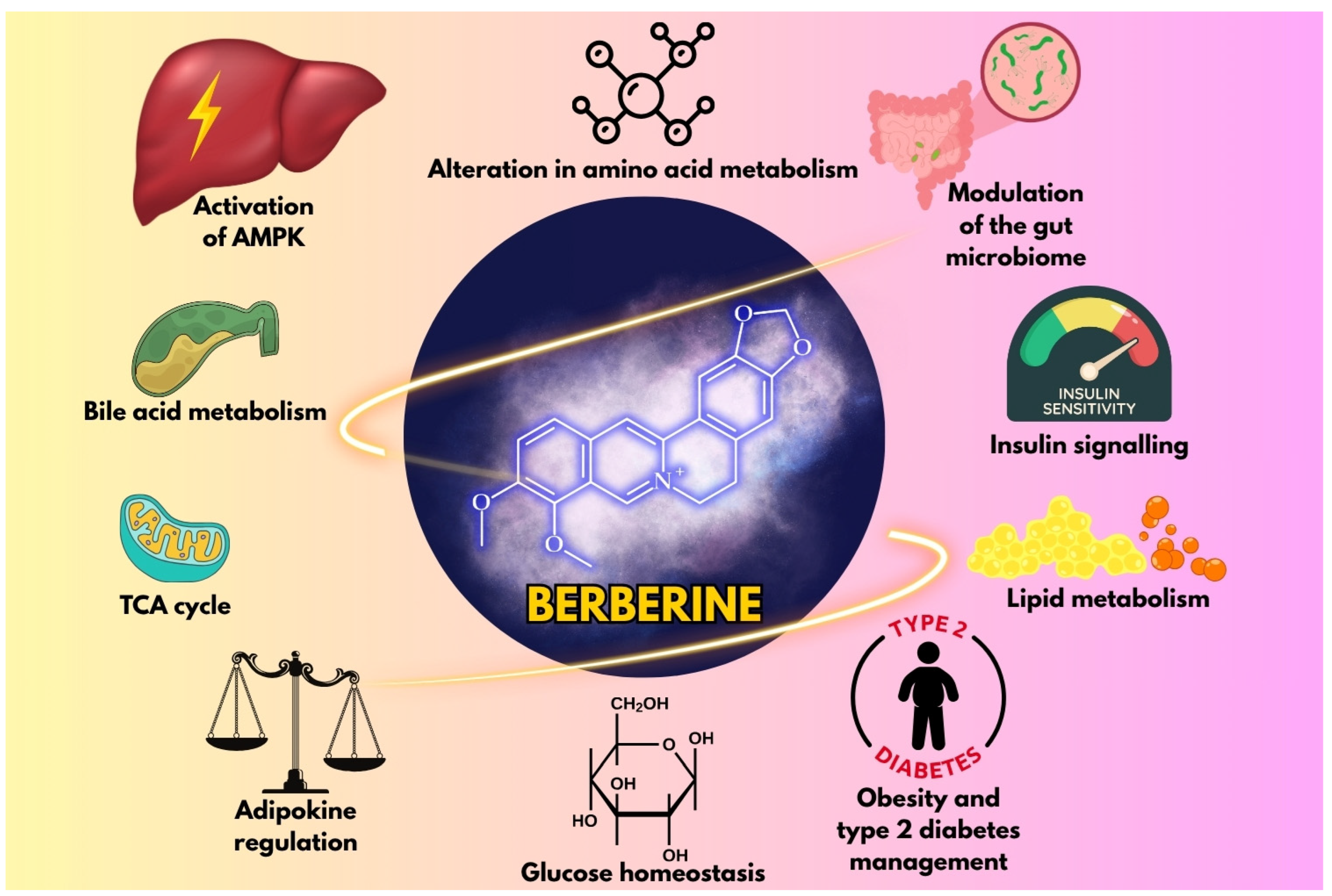
Figure 1. Illustration of the key mechanisms by which berberine influences metabolic health.
Metabolites 15 00467 g001
Figure 2. Chemical structure of berberine.
Metabolites 15 00467 g002
Figure 3. Integrative schematic of berberine’s molecular mechanisms in obesity and diabetes. Berberine activates AMP-activated protein kinase (AMPK), modulates gut microbiota, regulates adipokines, and alters bile acid metabolism, collectively reducing adiposity, improving insulin sensitivity, and decreasing lipid accumulation. Arrows indicate the direction of regulation or effect, with ↑ representing an increase or activation and ↓ representing a decrease or inhibition.
Metabolites 15 00467 g003
Figure 4. Schematic illustrating the multi-omics approach in berberine research. Genomics, transcriptomics, proteomics, and metabolomics data reveal the systemic effects of berberine, enabling the discovery of biomarkers and the development of personalized metabolic therapies.
Metabolites 15 00467 g004
Table 1. Key physicochemical properties of berberine chloride hydrate.
PropertyValue/Description
Molecular formulaC20H17NO4HCl·2H2O
Molecular weight407.65 g/mol
AppearanceYellow crystalline solid
Melting point145.1–146.7 °C
SolubilitypH-dependent: 9.69 mM (pH 7.0, 37 °C);
insoluble in nonpolar solvents [22]
Stability<5% degradation (6 months, pH 1.2–9.0, 40 °C) [21]
FluorescenceAbsorption: 350 nm; emission: 530 nm [19]
Table 2. Distribution of berberine in diverse plant taxa. A compilation of research findings from 2024 to 2025.
Plant TaxaPlant
Family
Plant Part UsedGeographical OriginConcentration (Reported Value)Analytical Method for QuantificationExtraction MethodReference
Phellodendron amurense Rupr.RutaceaeBarkDujiangyan, Sichuan, China0.243 mg/g of extractUV-Vis spectrophotometry
(λmax = 354 nm); UPLC-Q-TOF-MS/MS
Microwave-assisted extraction (ethanol solvent; 350 W; 75 s; material–liquid ratio 1:25 g/mL)[23]
Tinospora cordifolia (Willd.) MiersMenispermaceaeStemsKolkata, West Bengal, IndiaNot reportedHPLC-DADhydro–alcoholic (ethanol–water, 60:40)[24]
T. cordifoliaMenispermaceaeStemsKharagpur, West Bengal, IndiaNot reportedLC-MS/MSUAE (22.5:1 solvent-to-solid ratio, 40 min sonication time, 75% ethanol)[25]
Berberis darwinii Hook.BerberidaceaeRootsTemuco, ChileUp to 26,482.20 µg/g dry weight; mean: ~1437–4427 µg/g across locationsHPLC-DADFreeze-drying of plant tissues, acid extraction, liquid–liquid extraction with chloroform[26]
B. darwiniiStemsTemuco, ChileUp to 6639.58 µg/g dry weight; mean: ~828–2035 µg/g across locations
B. darwiniiSeedsValdivia, ChileUp to 1181.75 µg/g dry weight; mean: ~89–467 µg/g across locations
B. darwiniiLeavesNueva Imperial, ChileUp to 511.02 µg/g dry weight; mean: ~24–118 µg/g across locations
Coptis chinensis Franch.RanunculaceaeRhizomesHue, Vietnam15.2–46.2 mg/g dry weightHPLCUAE (solvents: 96% lactic acid, 40% malic acid, or 88% pyruvic acid (w/w), liquid–solid ratio: 30 mL/g, temperature: 60 °C (lactic acid), 80 °C (malic acid), 75 °C (pyruvic acid), time: 30 min[27]
Mahonia bealei (Fort.) Carr.BerberidaceaeLeavesSichuan Province, ChinaNot reportedUPLC-Q-TOF-MS/MSUltrasonic synergistic high-speed homogeneous extraction (50% ethanol, 1:10 solid–liquid ratio, 20,000 r/min homogenization + 300 W ultrasonication for 30 min)[28]
C. chinensis Franch. (as a part of a multi-herb formula)RanunculaceaeNot specifiedChinaNot reportedHPLC (UV detector)Reflux with 75% ethanol (1:8 ratio)[29]
T. cordifoliaMenispermaceaeStemsNot specified2.49% (w/w)HPTLCMAE (500 W, 3 min, 1:15 ratio, 50 °C, methanol)[30]
B. aristata DC.BerberidaceaeNot specifiedNot specifiedNot specifiedUHPLC-DADUltrasonic bath (10 min, 25 °C, methanol)[31]
C. teeta Wall.RanunculaceaeRhizomesArunachal Pradesh, India212.18 ppm for MAE; 162.96 ppm for UAEHPLC (UV detector)MAE (65% solvent concentration, 310 W power, 30 min extraction time, and 1:39 g/mL solid–liquid ratio); UAE (36% solvent concentration, 160 W ultrasound power, 10 min extraction time, and 1:78 g/mL solid–liquid ratio)[32]
Helicteres isora L.MalvaceaeFruitsNeyshabur, Iran21.37% of the total areaGC-MSSubcritical water extraction (175 °C, 4 mL/min, 120 min, 4 g sample)[33]
Thalictrum foliolosum DC.RanunculaceaeRootsAlmora district, Uttarakhand, India13.14 mg/g dry weightHPLC-PDAUAE using NADES (tartaric acid–glycerol, 1:1 molar ratio with 30% water content, duty cycle 80%, liquid-to-solid ratio of 30 mL/g, time: 14 min)[34]
B. vulgarisBerberidaceaeRootsPoland111.06 mg/gHPTLC70% aqueous ethanol, sonicated (2 cycles of 20 min each, 700 W, 50 °C)[35]
Phellodendron amurense Rupr.RutaceaeBarkThua Thien Hue, Vietnam50.88 mg/gHPLCUAE (46% aqueous malic acid, 80 °C, 33.5 min, 26.8 mL/g solvent-to-solid ratio)[36]
Cayratia albifolia C.L.LiVitaceaeRootsHunan Province, ChinaNot specifiedLC-MS/MSBoiled in 95% ethanol for 1 h[37]
B. koreana Palib.BerberidaceaeTwigsStobierna, Poland0.042–0.067%HPTLCUltrasonic bath (70% ethanol, 50 °C, 2 × 20 min)[38]
Berberis × ottawensis “Superba”0.103%
B. thunbergii DC.0.364–0.676%
C. chinensis Franch.RanunculaceaeRootsChinaUp to 10%HPLCDES-based UAE (choline chloride–urea (1:2 molar ratio) in 50% aqueous DES, 150 W, 60 °C, 15 min)[39]
C. chinensisRanunculaceaeRhizomesSichuan, China39.57–77.12 mg/g dry weightUHPLC-PDADES-UA-MSPD (betaine–acrylic acid (1:4) with 50% water, silica gel as a sorbent in a 1:1 ratio with the sample, 1:62 g/mL solid-to-DES ratio, 200 W of ultrasonication for 6 min)[40]
B. vulgaris L.BerberidaceaeStem barkOratia, Buzau County, Romania78.95 µg/g dry extractHPLC-DADHydro–ethanolic (50% ethanol) reflux extraction[41]
B. vulgaris L.BerberidaceaeRootsPoland (commercial)Not reportedHPLC-ESI-Q-TOF-MS/MSASE with methanol (90 °C, 96 bar, 4 cycles)[42]
B. vulgaris L.BerberidaceaeStemsTbilisi, Georgia
Coscinium fenestratum (Gaertn.) Colebr.MenispermaceaeStems and RootsThua Thien Hue, Vietnam38.23 mg/gHPLC60% lactic acid (w/w), liquid–solid ratio of 17.25 mL/g, 66 °C, 20 min[43]
Annickia affinis (Exell) Versteegh & SosefAnnonaceaeStem barkOvia area, Benin City, Edo State, Nigeria~0.02% w/wLC-ESI-MS/MSDirect methanol extraction (pulverized stem bark shaken in methanol, 120 rpm, 72 h, 25 °C)[44]
Thalictrum spp. (57 batches)RanunculaceaeStems and rootsYunnan and Xizang Provinces, China0.01–12.44 mg/gLC-MS/MS75% methanol, solid-to-liquid ratio of 1:100, 60 min of sonication[45]
MAE—microwave-assisted extraction; UPLC-Q-TOF-MS/MS—ultra-performance liquid chromatography–quadrupole time-of-flight tandem mass spectrometry; UAE—ultrasound-assisted extraction; HPLC-DAD—high-performance liquid chromatography with a diode array detector; LC-MS/MS—liquid chromatography–tandem mass spectrometry; HPTLC—high-performance thin-layer chromatography; GC-MS—gas chromatography–mass spectrometry; PDA—photo diode array; NADES—natural deep eutectic solvent; DES-UA-MSPD—deep eutectic solvent ultrasound-assisted matrix solid-phase dispersion; HPLC-ESI-Q-TOF-MS/MS—high-performance liquid chromatography–electrospray ionization–quadrupole time-of-flight tandem mass spectrometry; ASE—accelerated solvent extraction.
Disclaimer/Publisher’s Note: The statements, opinions and data contained in all publications are solely those of the individual author(s) and contributor(s) and not of MDPI and/or the editor(s). MDPI and/or the editor(s) disclaim responsibility for any injury to people or property resulting from any ideas, methods, instructions or products referred to in the content.