Article

In Vitro Metabolism of Doping Agents (Stanozolol, LGD-4033, Anastrozole, GW1516, Trimetazidine) by Human Seminal Vesicle and Liver Fractions

1
Institute of Biochemistry, Center for Preventive Doping Research, German Sport University Cologne, 50933 Cologne, Germany
2
European Monitoring Center for Emerging Doping Agents (EuMoCEDA), 50933 Cologne, Germany
3
European Monitoring Center for Emerging Doping Agents (EuMoCEDA), 53113 Bonn, Germany
*
Author to whom correspondence should be addressed.
Metabolites 2025, 15(7), 452; https://doi.org/10.3390/metabo15070452
Submission received: 12 June 2025 / Revised: 1 July 2025 / Accepted: 2 July 2025 / Published: 4 July 2025
(This article belongs to the Section Pharmacology and Drug Metabolism)

Abstract

Background: In order to address complex scenarios in anti-doping science, especially in cases where an unintentional exposure of athletes to prohibited substances and a corresponding contamination of doping control samples at the collection event are argued, an understanding of tissue-specific drug metabolism is essential. Hence, in this study, the metabolic capacity of the seminal vesicle using in vitro assays was investigated. Methods: The aim was to assess whether selected doping-relevant substances—stanozolol, LGD-4033, GW1516, trimetazidine, and anastrozole—are metabolised in seminal vesicle cellular fractions (SV-S9) and how that metabolism compares to biotransformations induced by human liver S9 fractions (HL-S9). Liquid chromatography coupled to high-resolution/accurate mass spectrometry (LC HRAM MS) enabled the sensitive detection and identification of metabolites, revealing a limited metabolic activity of SV-S9. Results: For LGD-4033, GW1516, and trimetazidine, minor metabolic transformations were observed, whereas no metabolites of stanozolol or anastrozole were detected. Gene expression analysis using digital polymerase chain reaction (dPCR) confirmed transcripts of CYP2D6, CYP2E1, and CYP2C9 in SV-S9, though no enzymatic activity was detected. Gene expression and enzymatic activity in CYP3A4 and CYP1A2—major hepatic enzymes—were absent in SV-S9. Conclusions: Overall, these pilot study results suggest that the seminal vesicle has only a low capacity for xenobiotic metabolism, which translates into a limited role in the biotransformation of drugs and, hence, the metabolic pattern.

Graphical Abstract

1. Introduction

Improvements in anti-doping analysis are vital for meeting the various challenges of uncovering doping practices in sport. However, these improvements, which commonly include increased sensitivities offered by modern analytical methods, have also created the necessity of distinguishing between intentional drug abuse (that occurred long ago) and contamination scenarios, as low levels of a substance are compatible with both scenarios [1]. One of these contamination scenarios can be referred to as ‘intimate contact drug (metabolites) residuals’, which may occur when drugs have been transferred to the athlete via skin contact, saliva, or seminal fluid, thereby triggering an Adverse Analytical Finding (AAF) in the doping control sample [1]. The metabolism of substances can be of particular interest in the detection of intimate contact contamination, as metabolite profiles may differ depending on the route of administration (oral/saliva or dermal) and the excretion matrix (urine or seminal fluid) and can therefore serve as markers [1]. Since it has been shown that various substances can be transferred into the seminal fluid and cases of AAF due to seminal transfer have been reported, it is now of interest to analyse whether metabolism occurs in the vesicles responsible for the production of seminal fluid and whether this can lead to specific or diagnostic metabolite markers [2,3,4].
Therefore, the use of subcellular fractions in in vitro experiments can be beneficial to gain insights into metabolic activities. In general, liver subcellular fractions are used because xenobiotics are mainly metabolised in the liver by cytochrome P450 (CYP) enzymes and uridine-5′-diphospho-glucuronosyltransferase (UGT) [5]. However, other tissues, such as skin subcellular fractions, can also be used to study the specific metabolism, which in this case is useful for determining subcutaneous and transdermal administration of a drug. To study the (possible) metabolite formation in seminal fluid, obtaining S9 fractions from tissues that contribute significantly to the ejaculate, such as the seminal vesicles or prostate, is a reasonable approach. The seminal vesicle produces a fluid that makes up 50–80% of the ejaculate, and various CYPs have already been detected in the seminal vesicles by real-time polymerase chain reaction (RT-PCR) [6,7,8]. The fluid produced by the seminal vesicle is therefore an important component of the ejaculate and may provide an insight into potential metabolism in the seminal fluid.
The subcellular fraction of the human liver (HL-S9) or seminal vesicle subcellular fraction (SV-S9) are the supernatants obtained from homogenised liver or seminal vesicle tissue after centrifugation at 9000× g [9]. This means that cell debris, nuclei, lysosomes and mitochondria are removed, but microsomal and cytosolic fractions remain. The S9 fraction includes the subcellular fraction of the endoplasmic reticulum, which mainly contains CYPs and UGTs. It also includes cytosolic enzymes such as aldehyde oxidase, xanthine oxidase, sulfotransferases, methyltransferases, N-acetyltransferases and glutathione transferases, which may also be important in the metabolism of certain substances [5].
CYPs are a superfamily of haemoproteins responsible for phase-I metabolism, i.e., the oxidative biotransformation of most xenobiotics, with each subfamily having specificity for certain drugs [10,11]. Therefore, the activity of a particular CYP can be determined by analysing the metabolism of model substances. For example, CYP1A2 is known to catalyse the metabolism of phenacetin to acetaminophen, while CYP3A4 promotes the metabolism of midazolam to hydroxy-midazolam [12,13]. In addition to analysing CYP protein activity in the S9 fraction, the cellular expression of CYP enzymes can also be assessed at the transcriptional level, e.g., by using digital PCR (dPCR).
The aim of this study was to perform in vitro experiments with SV-S9 to investigate its potential metabolic activity towards various doping substances. Using typical doping substances such as stanozolol, LGD-4033 and trimetazidine, as well as substances that have been described in connection with potential seminal fluid transfer scenarios (GW1516) and that could frequently be found in seminal fluid (anastrozole), obtaining a first overview of possible metabolite markers was desirable [3,14,15,16]. Further, assessing metabolic contributions of selected and isolated tissues has been considered warranted, especially in comparison to more holistic approaches such as animal experiments reported previously concerning stanozolol, LGD-4033 and their metabolites in seminal fluid [17].
HL-S9 was used as a control to compare the different metabolite profiles with SV-S9 and as an assay quality control. Liquid chromatography coupled to high-resolution/accurate mass spectrometry (LC-HRAM MS) was used to identify both known and novel metabolites. In addition, SV-S9 and HL-S9 were characterised by testing the activity of relevant CYPs by LC-HRAM MS and the cellular expression of CYPs by dPCR.

2. Materials and Methods

2.1. Chemicals, Kits and Reagents

Acetonitrile (ACN), methanol (MeOH), chloroform and formic acid (FA) were purchased from VWR Chemicals (Langenfeld, Germany). Ultrapure H2O was received from Barnstead™ GenPure™ xCAD Plus from Thermo Scientific (Niederelbert, Germany). Nicotinamide adenine dinucleotide phosphate (NADPH reg.) regeneration system was obtained from Promega (Madison, WI, USA). Ethanol (EtOH), potassium dihydrogen phosphate (KH2PO4), magnesium chloride (MgCl2) and reference material of acetaminophen-d3, anastrozole, phenacetin, propafenone and stanozolol were obtained from Sigma-Aldrich (St. Louis, MO, USA). Reference material for LGD-4033 was obtained from Selleck-Chem (Houston, TX, USA). High-Capacity cDNA Reverse Transcription Kit, pooled human liver S9 fractions (HL-S9, n = 6, 20 mg/mL) and Invitrogen TRIzol Reagent were obtained from Thermo Scientific (Schwerte, Germany). Customised male human seminal vesicle S9 fraction (SV-S9, 0.75 mg/mL, n = 1) was obtained from BioIVT (Brussels, Belgium). The QIAcuity Nanoplate 26 k 96-Well and QIAcuity EG PCR Kit were obtained from QIAGEN GmbH (Hilden, Germany). RNA Clean & Concentrator Kit was obtained from Zymo Research Europe GmbH (Freiburg, Germany). Reference material for S-mephenytoin and amodiaquine was obtained from Biomol (Hamburg, Germany), reference material for Trimetazidine was obtained from LGC Standards (Teddington, UK), and reference material for tolbutamide was purchased from Merck (Darmstadt, Germany). The primers used for dPCR were purchased from Biomers (Ulm, Germany) and are listed in Table 1. A total of six CYP enzymes and three housekeeping genes (HG) were tested, which were previously described elsewhere [18,19].

2.2. Preparation of Incubation Buffer

For the preparation of the phosphate buffer (100 mM), 3.54 g sodium dihydrogen phosphate (NaH2PO4) was dissolved in 250 mL of H2O (solution A), and 1.75 g of potassium dihydrogen phosphate (KH2PO4) was dissolved in 125 mL of H2O (solution B). Solution B was then added to solution A until a pH of 7.4 was reached (solution C). For the final incubation buffer, 0.25 g MgCl2∙6H2O were mixed with 250 mL solution C.

2.3. In Vitro S9 Fraction Metabolic Assays

A substrate cocktail (CYP-cocktail) for an in vitro assay was prepared as follows: 10,000 µM of tolbutamide and S-mephenytoin, 5000 µM of phenacetin and 500 µM of amodiaquine and propafenone were dissolved in ACN: H2O (1:1 (v/v)). Before use, it was diluted 1:10 (v/v) in phosphate buffer.
Substances of interest (anastrozole, GW1516, LGD-4033, stanozolol, and trimetazidine) were diluted in 50 mM phosphate buffer (pH 7.4) containing 5 mM MgCl2 to produce a stock solution (10 µM each).
To test phase-I metabolism, 10 µL of the stock solution of each substance or CYP-cocktail, 25 µL of NADPH reg. (50 mM) and 6 µL of the HL-S9 fraction (20 mg/mL) or 160 µL of the SV-S9 fraction (0.75 mg/mL) were mixed and made up to a total volume of 200 µL with phosphate buffer to obtain the same protein concentration of 0.6 mg/mL in each sample. Samples were incubated at 37 °C for 24 h. Negative control samples were prepared without S9 fraction and without the substance of interest.

2.4. Sample Preparation and Extraction Procedure

Each metabolism sample was quenched by the addition of 600 µL of ice-cold ACN and kept on ice for 20 min. The supernatant was harvested after centrifugation (17,000× g, 10 min) and transferred into a fresh tube. After evaporating using a vacuum centrifuge (70 °C, 90 min), the samples were reconstituted in 100 µL of ACN:H2O (20:80 (v/v)) and diluted before measuring.

2.5. LC-HRAM MS Instrumentation and Analytical Conditions

A Vanquish HPLC system, coupled to a Thermo Scientific Orbitrap Exploris 480 (both from Thermo Scientific, Bremen, Germany), was used as the LC-HRAM MS system. Chromatographic separation was achieved using an EC 4/3 Nucleoshell RP 18 Plus guard column (4 × 3 mm, 5 μm particle size; Macherey–Nagel, Düren, Germany) connected to a Poroshell 120 EC-C18 analytical column (50 × 3.0 mm, 2.7 μm particle size; Agilent, Waldbronn, Germany). For each doping substance, an LC-HRAM MS method was developed. Measurements were performed in both positive (anastrozole, GW1516, stanozolol, trimetazidine) and negative ionisation (LGD-4033) modes, with an ionisation voltage of ±3000 V and a transfer tube temperature of 320 °C. For positive mode measurements, H2O (A) and ACN (B) with 0.1 % FA each were used as mobile phases at a flow rate of 0.4 mL/min. The gradient for this measurement was as follows: 0–11 min B, starting at 0 % B, increased to 90 %, then increased to 100 %, held for 1 min, then equilibrated at 0 % B for 3 min. For measurements in negative mode the mobile phases were 50 mM ammonium acetate buffer with 0.1 % FA (A) and ACN (B). The gradient was as follows: 0–4.5 min B, starting at 20 %, increased to 50 % B, then increased to 100 % in 2.5 min and held for 1 min. The gradient was then decreased to 70 % B and held for 1.5 min. Then equilibrated at 20 % B for 2 min. The full scan was conducted at a resolution of 60,000 full width at half maximum (FWHM), while MS2 experiments (isolation window 1.2 m/z) were performed at 45,000 FWHM in positive mode and 15,000 FWHM in negative mode. The corresponding ion transitions, normalised collision energies (NCE) and retention times are shown in Table 2. Full scan data were acquired over a m/z range of 100–800. Nitrogen, supplied by the CMC nitrogen generator (Eschborn, Germany), was used as the collision gas.
A full scan and data-independent acquisition (DIA) modes were set up for the measurements of the protein activity assay. Positive mode with an ionisation voltage of 3500 V and a transfer tube temperature of 350 °C was used. For the full scan, the resolution was set to 45,000 FWHM, and data were acquired over a m/z range of 100–800. The DIA scan was performed with a resolution of 30,000 FWHM and stepped NCEs of 30 % and 55 %. The isolation window was set at m/z 100. A total of five DIA experiments were performed: Precursor: m/z 100–200 from 0 to 14 min, m/z 200–400 from 5 to 14 min, m/z 200–400 from 5 to 14 min, m/z 400–500 from 6 to 14 min, and m/z 500–600 from 6 to 14 min.

2.6. Cell Culture

Undifferentiated HepaRG™ cells (Biopredic International, Rennes, France) were differentiated in-house in William’s E medium containing 10 % foetal bovine serum, 5 µg/mL human insulin, 5 µg/mL gentamicin sulphate, 0.25 µg/mL amphotericin B (all from PAN-Biotech, Aidenbach, Germany), 5 × 10⁻⁵ M hydrocortisone hemisuccinate (Sigma-Aldrich), 2 mM Glutagrow (Corning, NY, USA) and 2 % DMSO. One million cells were used directly for RNA extraction.

2.7. RNA Extraction

Total RNA was extracted from HepaRG™, SV-S9 and HL-S9 fractions using TRIzol/chloroform extraction, and purification was performed using an RNA extraction and purification kit from Zymo Research. For extraction of SV-S9 and HL-S9, 900 µL of TRIzol was mixed with 100 µL of the sample and incubated for 15 min at room temperature (RT). For phase separation, 200 µL of chloroform was added, mixed and incubated for 3 min at RT. The differentiated HepaRG™ cells were incubated with 500 µL of TRIzol, then mixed with 80 µL of chloroform and incubated for 3 min at RT.
The samples were then centrifuged at 12,000× g at 4 °C for 15 min. The upper, aqueous phase (RNA) was transferred to a new tube, and an equal volume of ethanol (95–100 %) was added. RNA was purified according to the RNA kit instructions, including the DNAse treatment. In brief, the sample was transferred to the Zymo-Spin IC column in a collection tube and centrifuged for 30 s at 12,000× g, RT. For DNAse treatment, 400 µL of wash buffer was added to the column and centrifuged (30 s, 12,000× g, RT). The DNAse reaction mix was prepared by mixing 5 µL DNAse I with 35 µL DNAse digestion buffer. This mixture was added directly to the column matrix and incubated at RT for 15 min. Afterwards 400 µL of RNA prep buffer was added to the column and centrifuged (30 s, 12,000× g, RT). Then, 700 µL RNA wash buffer was added to the column and centrifuged (30 s, 12,000× g, RT). The column was then loaded with 400 µL of RNA Wash Buffer and centrifuged at 12,000× g for 1 min to ensure complete removal of the Wash Buffer. For elution of the RNA, 15 µL of RNAse/DNAse-free H2O was added to the column matrix and centrifuged (30 s, 12,000× g, RT). The quantity and purity of the extracted RNA was tested using a NanoDrop One device (Thermo Fisher Scientific, Waltham, MA, USA). The RNA was stored at −20 °C until further use.

2.8. cDNA Reverse Transcription

Reverse transcription was performed using the High Capacity cDNA Reverse Transcription Kit. The master mix was prepared according to the instructions. Briefly, 8 µL of 10× reverse transcription buffer, 3.2 µL of 25× dNTP mix (100 mM), 8 µL of MultiScribe™ reverse transcriptase, and 16.8 µL of nuclease-free H2O were prepared on ice. Then 10 µL of the master mix was mixed with 10 µL of RNA sample (HL-S9 113.4 ng/µL (test 1) and 233.6 ng/µL (test 2); SV-S9 153 ng/µL (test 1) and 76.3 ng/µL (test 2); HepaRG™ 200 ng/µL) in a PCR plate and loaded into the thermal cycler (SensoQuest, Göttingen, Germany). The thermal cycler run consisted of 4 steps. The first step was at 25 °C for 10 min, followed by 37 °C for 120 min. The temperature was then increased to 85 °C for 5 min. For the final step, the temperature was cooled to 4 °C and held at this temperature until sample collection. The cDNA was stored at −20 °C until further use.

2.9. Digital PCR for Detecting Gene Expression

The dPCR was performed using the QIAcuity EG PCR Kit. In short, the reaction mix was prepared by mixing 4 µL of 3× EvaGreen PCR Master Mix, 3.6 µL of RNase-free H2O and 3.2 µL of a primer mix (1.5 µM) containing forward and reverse primers of a CYP or HKG (CYP2B6, CYP2D6, CYP3A4, CYP1A2, CYP2C9, CYP2E1, YHWAZ, UBC, TBP). The reaction mixture containing one primer pair was added to each well of a standard PCR plate. The HL-S9 cDNA was diluted at a 1:5 ratio, and the SV-S9 cDNA was diluted at a 1:2 ratio before adding 1.2 µL in triplicate to the respective wells of the PCR plate. The content of each well from the standard PCR plate was transferred to the wells of the QIAcuity Nanoplate 26 k 8-well (Qiagen, Hilden, Germany). After sealing, the plate was processed and analysed with the QIAcuity One digital PCR system.
The thermal cycling programme consisted of 2 min at 95 °C, 35 cycles of 15 s at 95 °C for denaturation and 15 s at 60 °C for annealing, and a further 15 s at 72 °C for extension. This was followed by cooling at 40 °C for 5 min. Absolute copy number was calculated using Poisson distribution and automatic thresholding using QIAcuity software (V.2.5.0.1 and V.3.1.0.0). A negative control without template (non-template control, NTC, n = 3, see supplement) was included to validate the specific PCR reaction for each sample primer set. As a positive control, differentiated HepaRG™ cells (n = 3) were tested.

3. Results

3.1. In Vitro Assay for Doping Substances

The in vitro tests were performed three times (three experiments) with the respective doping substances and measured using targeted LC-HRAM MS methods. Well-established metabolites were identified, and the specific transitions were integrated into LC-HRAM MS methods (see Table 2). The structures of these metabolites can be found in the Supplementary Materials (Figures S6–S10). The data obtained were compared to human (GW1516, anastrozole) or animal (LGD-4033) seminal fluid samples described elsewhere [3,17].
The in vitro tests with LGD-4033 yielded several metabolites when incubating the anabolic agent with HL-S9 and SV-S9 fractions (see Table 2, Figure 1 and Figure S6). The hydroxylated metabolite L-M5 was most abundant in the HL-S9 sample. Metabolite L-M3 was the most prominent metabolite in the SV-S9 fraction and showed higher intensities than in the HL-S9 fraction in two out of three experiments. Metabolite L-M5 and metabolite L-M6 were also detected in the SV-S9 fraction in two out of three experiments, but with substantially lower intensities compared to the HL-S9 fraction.
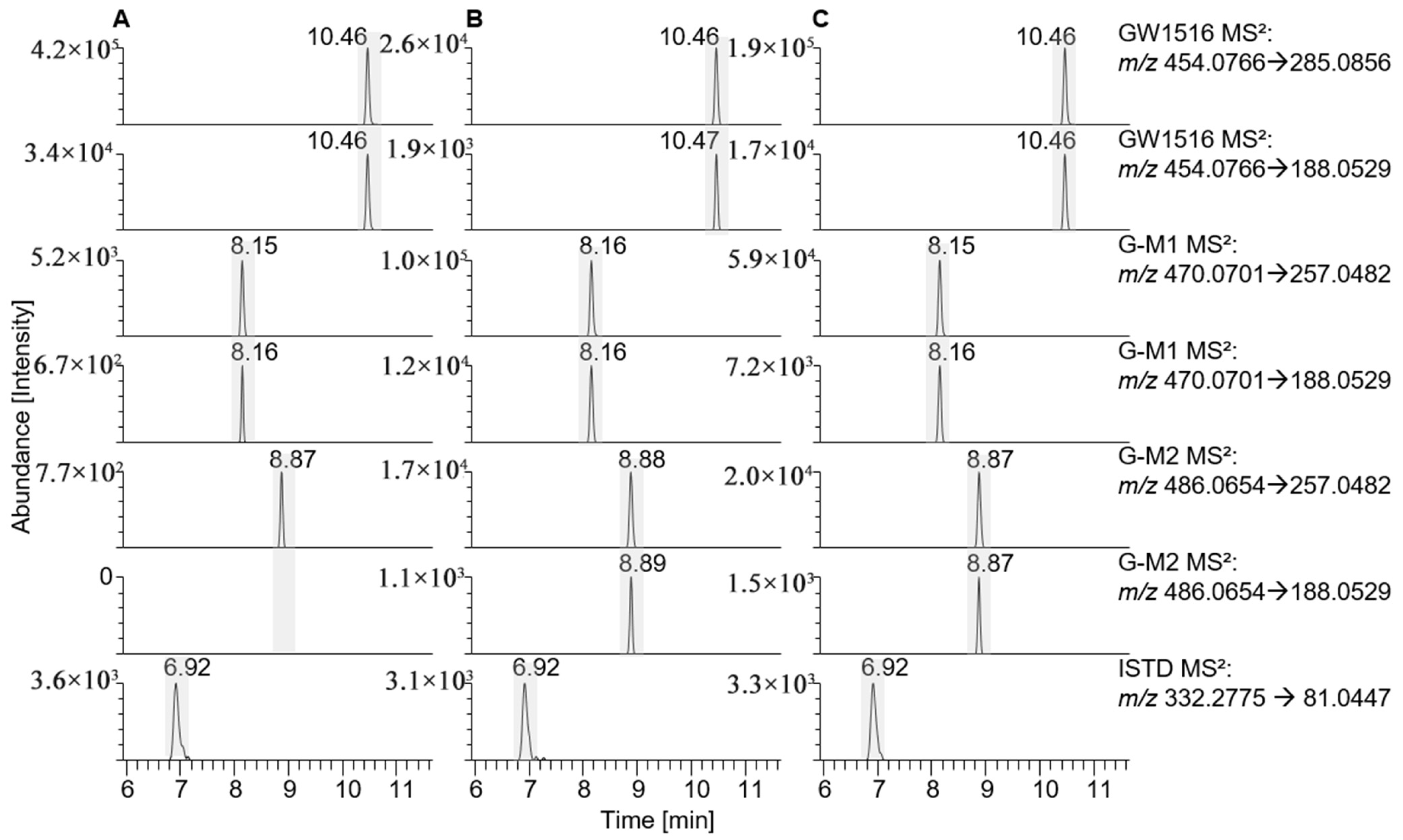
GW1516 metabolites G-M1-a and G-M2 were observed in samples after incubation with HL-S9 and SV-S9. Small amounts of the metabolite M1-a were present in the enzyme blank, which could be explained by non-enzymatic oxidation, but the intensity was low compared to those in the HL-S9 and SV-S9 fractions (see Figure 2). No M1-b metabolite was found in the SV-S9 and HL-S9 but was detected in vivo [21] (see Table 2).
For trimetazidine, only one metabolite was found in the HL-S9 and SV-S9 fractions, which met the identification criteria applicable to retention time and precursor/product ion pairs (see Table 2). The observed intensities of the metabolite suggest modest conversion when compared to the signal abundance of the intact compound (see Supplemental Figure S1).
No anastrozole metabolites were found in the SV-S9 fraction, and only one anastrozole metabolite was identified at low intensity in HL-S9 (see Table 2, Supplemental Figure S2).
The in vitro experiment showed typical metabolites for stanozolol in the HL-S9 fraction but no metabolites in the SV-S9 fraction (see Table 2). Interestingly, the intensities of the parent substance stanozolol were considerably lower in SV-S9 fractions than in the enzyme blank and in the HL-S9 sample after incubation for 24 h, which could indicate that stanozolol is metabolised in these samples (see Supplemental Figure S3). However, no typical metabolites described in the literature were identified, and also no other metabolites related to stanozolol could be found in positive and negative DIA scans using LC-HRAM MS. Further investigations indicated that stanozolol was bound to proteins and could be extracted from the precipitation pellet, without any metabolites present in this fraction.

3.2. In Vitro Protein Activity of CYP Enzymes

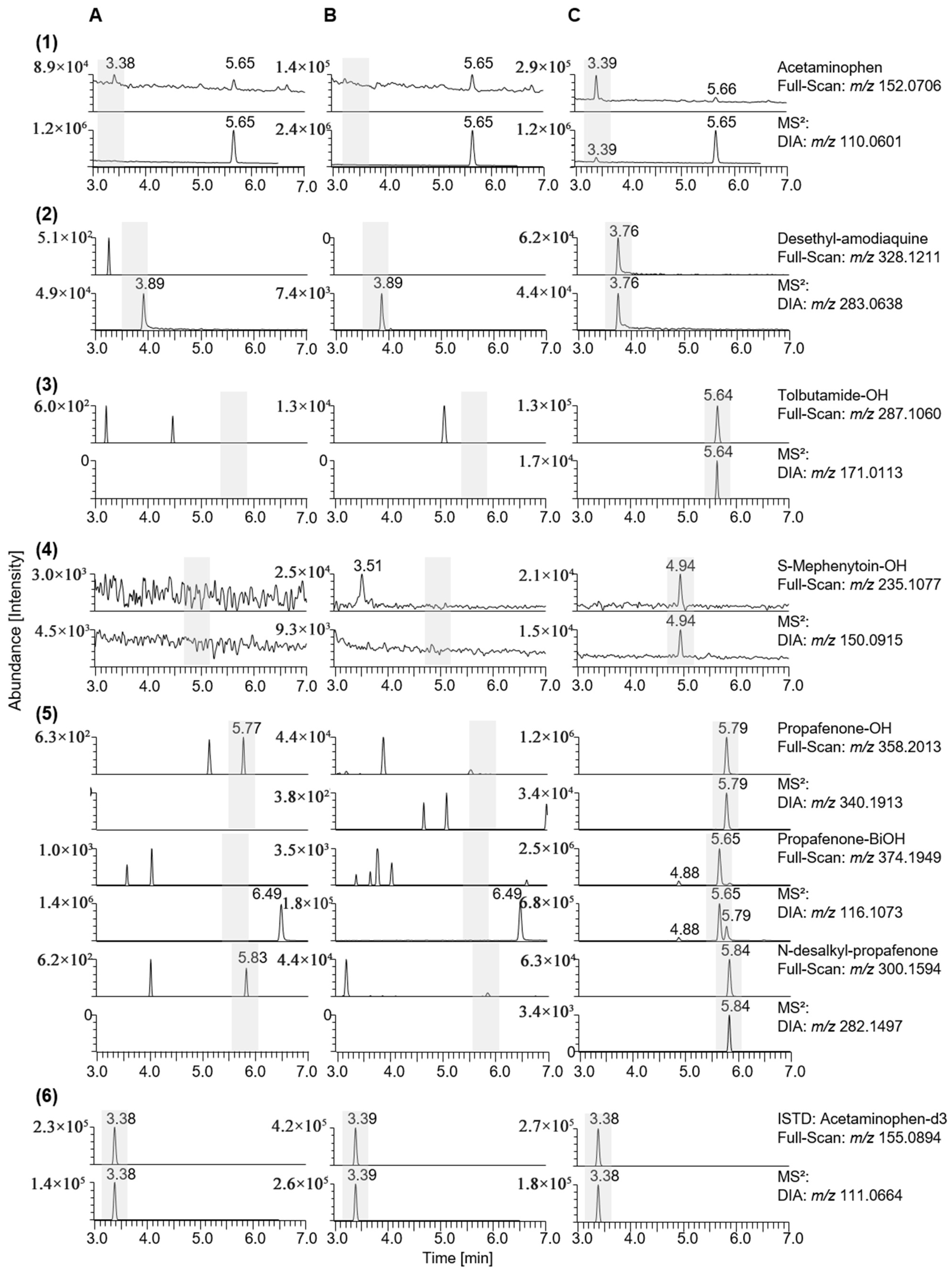
To elucidate and compare the metabolic capacities of the seminal vesicles and the liver, a protein activity assay was conducted to analyse the characteristic transformation of typical substrates, as already described earlier [19,29]. Therefore, an in vitro test was performed twice to compare the SV-S9 and HL-S9 fractions. In contrast to the protein assay cocktails described previously, midazolam could not be used as a marker for CYP3A4 because the SV-S9 specimen was contaminated with both midazolam and its metabolite [19]. The assay was conducted using LC-HRAM MS in full-scan and DIA mode to detect not only the target metabolites described in the literature [29,30], but also other possible metabolic reactions. The mass spectrometric characteristics of the substances used in the CYP cocktail and their respective metabolites are listed in Table 3. The structures of these metabolites can be found in the Supplementary Materials (Figure S11). Overall, metabolites are formed exclusively in the HL-S9 enzyme-substrate complex (Figure 3C) and no metabolites in the incubation mix without S9 fraction (Figure 3A), indicating that the incubation test is working.
In detail, the CYP1A2 isoform catalyses the metabolic reaction of phenacetin, which leads to the formation of acetaminophen (paracetamol) [31], which could only be found in the HL-S9 incubated sample (see Figure 3((1)C)). No other metabolites of phenacetin were detected. Amodiaquine is metabolised by CYP2C8 to desethyl-amodiaquine [32], which can also only be detected in the sample incubated with HL-S9 (see Figure 3((2)C)). Other metabolites of amodiaquine could not be detected in any incubation. CYP2C9 catalyses the hydroxylation of tolbutamide [33], which was detected in the sample incubated with HL-S9, and no further metabolites could be detected (see Figure 3((3)C)). S-Mephenytoin is metabolised by CYP2C19 to form hydroxy-mephenytoin [34], which can only be observed in the sample incubated with HL-S9 (see Figure 3((3)C)). No further metabolites were found here either. Propafenone is metabolised by CYP2D6 to hydroxy-propafenone, as described in the literature [35], but the HL-S9 sample shows a metabolite with m/z 374.1949 in addition to the hydroxy metabolite, which could indicate twofold-hydroxylation. This metabolite is clearly more intense in the chromatogram than the hydroxy metabolite, and they share the production m/z 116.1069 (see Figure 3((5)C)). Propafenone is also dealkylated by CYP3A4 and CYP1A2 [35], which can be observed in the sample incubated with HL-S9.

3.3. Gene Expression of Different CYPs

The dPCR was used to probe for gene expressions of CYPs in HL-S9 and SV-S9, performed twice at different time points. While two different HL-S9 donor pools existed, which may explain the observed deviations in RNA concentration and gene expression between the different experiments for the HL-S9 fractions, material from only one donor was available for the SV-S9. In the first experiment (test 1), RNA concentrations of 226.8 ng/µL HL-S9 and 15.3 ng/µL SV-S9 were quantified, while in the second experiment (test 2), concentrations of 1168.25 ng/µL HL-S9 and 7.63 ng/µL SV-S9 were measured. Purity determined by A260/A280 and A260/A230 ratios gave acceptable values for the HL-S9 sample (test 1: 2.00/1.91; test 2: 2.06/2.07) and lower values for the SV-S9 sample (test 1: 1.78/1.65; test 2: 1.65/0.92), attributable to some degree of contamination. For example, increased absorbance at 230 nm in RNA samples may be due to contamination by guanidine thiocyanate, which absorbs strongly at 220–230 nm and is present at very high concentrations in the extraction reagent (TRIzol) [36]. Further clean-up of the SV-S9 RNA did not improve the A260/A230 but resulted in loss of RNA and was therefore not pursued.
The dPCR experiments were performed with primers for the detection of CYP2B6, CYP2D6, CYP3A4, CYP1A2, CYP2C9, CYP2E1 and the HKGs UBC, TBP and YHWAZ (see Table 1), which have been used for quantitative PCR before [18,19]. Due to the very limited information about the CYP expression in the human seminal vesicle [8], HKGs were included as additional dPCR controls for the SV-S9 fraction but were not used as a normalisation variable for quantification. Here, the low RNA concentration and potential contamination would not allow for a reliable quantification. The presence of residual genomic DNA by direct addition of a portion of the isolated RNA to the dPCR was not tested due to the limited sample volume of the SV-S9 fraction. Therefore, it cannot be excluded that genomic DNA could bias the results, but thorough DNAse treatment was performed during RNA purification to ensure that no genomic DNA was present in the sample. After an initial dPCR test with the CYP2B6 primer pair, the concentration of HL-S9 was increased and the experiment repeated (see Supplemental Figures S6 and S7). All subsequent dPCR measurements were then performed with the adjusted concentration as described in the methods section. The calculated mean concentration of undiluted samples of CYPs in HL-S9, SV-S9 and HepaRG™ in two experiments (test 1, test 2) are shown in Figure 4. Due to the different RNA input concentrations used for HL-S9, SV-S9 and HepaRG™, the expression values [cp/µL] cannot be compared. However, both experiments show that CYP2B6, CYP2D6, CYP2E1, and CYP2D9 are expressed in SV-S9. CYP2B6, CYP2D6, CYP2E1, CYP2C9, CYP3A4, and CYP1A2 were found in HL-S9. The SV-S9 and HL-S9 were also tested for HKGs, showing low values in HL-S9 and SV-S9, except for UBC and YHWAZ in SV-S9 in the first test (see Figure 4). Detailed information on scatter plots, fluorescence intensity and thresholds for all experiments can be found in the Supplementary Materials.

4. Discussion

4.1. In Vitro Assay for Doping Substances

The protein concentration (0.6 mg/mL) of the HL-S9 fraction used in this study was adjusted to the concentration of the SV-S9 fraction in order to operate the assays under comparable conditions. As these protein concentrations were lower than those employed in earlier studies [37], the herein presented results of the HL-S9 incubation experiments are not immediately comparable to other in vitro studies.
The metabolism of LGD-4033 has been studied in great detail, and the occurrence of this substance in doping control samples has increased in recent years, possibly due to highly sensitive analytical test methods and/or contamination scenarios [15]. While it has been demonstrated that LGD-4033 and its metabolites can occur in seminal fluid, it remains to be clarified whether they are formed in the seminal vesicle [17]. Since no metabolites were detected in the enzyme blank, the presence of three metabolites identified in two out of three SV-S9 experiments suggests some metabolic activity of the SV-S9 fraction. Analytical results obtained from samples prepared after incubation of LGD-4033 in SV-S9 or HL-S9 and from an in vivo experiment, described previously, differ in their patterns of main metabolites, indicating different enzymatic activities and processes [17]. Of note, no metabolite exclusive to SV-S9 was observed.
Stanozolol has been widely abused in sports until today, although modern mass spectrometry techniques allow for highly sensitive detection of stanozolol and its metabolites. In addition, it is a good model substance because of its well-studied metabolism in the anti-doping context [14,26,38,39]. Several metabolites were found in the HL-S9 fraction, indicating the principal functionality of the assay, but no metabolites of stanozolol were found in SV-S9.
In a previous study, a number of human seminal fluid samples were found to contain anastrozole and its metabolites [3]. This suggests that anastrozole may be a potential contaminant in urine samples of female athletes after unprotected intimate contact. However, only one phase-I metabolite of anastrozole could be detected in the HL-S9 fraction and none in the SV-S9 fraction. As the intensity of anastrozole in SV-S9 is similar to the enzyme blank, no extensive metabolism is expected. Therefore, these tests could not provide any further evidence/markers for the seminal fluid contamination scenario.
GW1516 has also been detected in a seminal fluid sample in a previous study, in which a case scenario is described where a professional athlete tested positive for GW1516 metabolites and denied intentional use. The athlete’s partner, who confirmed the use of GW1516, was identified as a possible source of exposure through intimate contact [2,4]. As metabolites of GW1516 were found in the SV-S9 fraction (Figure 2), it is possible that GW1516 is further metabolised in the seminal vesicles.

4.2. In Vitro Protein Activity of Different CYPs

The absence of metabolites typically formed by common CYPs in the SV-S9-incubated samples indicated very low concentrations or no presence of the investigated CYPs, translating to very low or no metabolic activity in the seminal vesicle, at least concerning the assessed typical/frequent hepatic CYPs.

4.3. Gene Expression of Different CYPs

A limitation of this experiment, apart from the availability of only one SV-S9 donor, was the very low RNA concentration of the SV-S9 sample. This negatively affected the number of positive partitions in the dPCR, which had to be considered when comparing the experiments (see Supplemental Figures S6–S16), where the same CYPs were found in both SV-S9 experiments, although differing in the observed concentrations.
CYP2D6 expression was detected in the SV-S9 fraction, although no activity was observed on a protein level in the metabolism test. CYP2D6 contributes to the metabolisation of many prescribed drugs and has been detected in various tissues such as the liver, brain, intestinal tissue, lymphoid cells and also seminal vesicles [8,40,41,42]. But CYP2D6 is also a highly polymorphic gene, and variations in the CYP genes can influence the expression and function of the enzymes [10,40,41,43]. For example, one study showed that minor changes in the structure of the active site of CYP2D6, such as the substitution of a single amino acid residue (F483) in CYP2D6, are sufficient to convert the enzyme to a steroidal 15α-hydroxylase, thus presenting a function that had not previously prevailed and thereby affecting the substrate specificity of the enzyme [44,45]. As the samples were not genotyped, it cannot be excluded that CYP2D6 has a different function and expression level in the SV-S9 fraction than in the HL-S9 fraction. In addition, SV-S9 is from a single donor and HL-S9 is from pooled donors (n = 6), which also influences the results with regard to the described polymorphic character of CYP2D6.
CYP2E1 is mainly found in the liver and plays an important role in metabolising potential toxic substrates (such as ethanol) to even more toxic products [46,47]. CYP2E1 was detected in HL-S9 and also in SV-S9. CYP2E1 has previously been reported to be present in the seminal vesicle and rat testis, but not in the prostate [8,45,48]. For some CYPs, such as CYP2E1, it is also important to note that mRNA levels do not necessarily correlate with enzyme activity, as studies have shown pre-transcriptional, pre-translational, translational and post-translational regulation of CYP2E1 expression [10,49,50]. In fact, one study showed high mRNA levels of CYP2E1 (most abundant together with CYP3A4), but no correlation with protein activity [43]. Therefore, in this case, a high gene expression level of this CYP enzyme does not necessarily correspond to a metabolic activity.
CYP2C9 is one of the most abundantly expressed CYP2C isoforms and the third most important CYP in the liver after CYP3A4 and CYP2D6 [51,52]. The gene expression detected by dPCR in all samples is relatively high. This is the first time that CYP2C9 has been detected in the seminal vesicles, which is in contrast to a previous study [8]. CYP2C9 was found previously in the liver, kidney, testis, adrenal gland, ovary and duodenum [53]. The lack of CYP2C9 protein activity in seminal vesicles despite the presence of mRNA can be explained by a study showing no correlation between mRNA levels and protein activity, with polymorphic variants also possibly contributing to lower activity [43].
CYP3A4 is the major human enzyme for the metabolism of xenobiotics and the most abundantly expressed CYP in the human liver [54,55]. Midazolam, the commonly used model substance, could not be used because impurities of midazolam and hydroxy-midazolam were found in the SV-S9. CYP3A4 activity was only observed in HL-S9 in this study, corroborated by the formation of the metabolite N-desalkyl-propafenone [35,56]. No metabolites of propafenone were detected in SV-S9. The SV-S9 manufacturer’s CYP3A4 assay (using midazolam and testosterone) also showed no metabolic activity. Consistent with this, no CYP3A4 mRNA expression was detected in SV-S9.
The CYP1A subfamily is one of the most abundant CYP families and consists of two members, 1A1 and 1A2, which are mainly found in the human liver [12,57]. Therefore, it is plausible that CYP1A2 could not be found in SV-S9, as no protein activity and no signal in dPCR were found. In contrast, previous studies reported on CYP1A2 mRNA in the seminal vesicle and in the prostate [8,45]. As CYP1A2 was only present in small quantities in the positive control (HepaRG™) in this study, it is, of course, possible that the quantities in SV-S9 are below the detection limit.
To normalise raw values obtained from qPCR, housekeeping genes (HKG) are commonly used as internal standards [58]. Since dPCR can be used to measure absolute transcript levels, results do not need to be expressed relative to HKGs, but for quantification it is recommended to use HKGs for normalisation [59]. In this qualitative study, however, probing for the presence of HKGs such as UBC, TBP and YWHAZ in the S9 fractions and verifying the PCR assay’s functionality was considered relevant.
UBC was detected in SV-S9 but not in HL-S9. This is consistent with the literature, as ubiquitin is present in human seminal plasma and plays an important role in regulating sperm quality and male fertility [60,61].
TBP was not detected in SV-S9 and HL-S9. TBP is a suitable HKG for qPCR and is recommended for normalisation because of the high stability of the gene [58,62]. However, it also shows a relatively low expression level compared to other HKGs such as glyceraldehyde-3-phosphate dehydrogenase (GAPDH), beta-actin (ACTB), and 18S ribosomal RNA (18S) [58,62].
The chaperone protein YWHAZ has also previously been identified as a suitable reference gene [58,63] and was detected in HL-S9 and SV-S9 in low levels, which is in line with the literature [58], so that overall the functionality of the employed dPCR was confirmed.

5. Conclusions

Using in vitro metabolism assays and gene expression analysis, this study provides a comprehensive assessment of the metabolic and transcriptional activity of the seminal vesicle in comparison to the liver, with a particular focus on the metabolism of doping agents. The results indicate that, although certain metabolic transformations occur in the SV-S9 fraction, the overall metabolic potential remains substantially lower than that of the HL-S9 fraction. Modest metabolism of LGD-4033, GW1516 and trimetazidine was observed in SV-S9, while no metabolic transformation of stanozolol or anastrozole was detected.
Metabolic markers exclusive to seminal vesicle metabolism would offer distinct target analytes to identify ejaculate-borne contaminations in female athletes’ doping control urine samples, supporting result interpretation and decision-making processes in cases of AAFs. The obtained data suggest that the metabolic contributions from seminal vesicles are minimal and no specific biotransformation products were identified. However, other techniques have been proposed to provide analytical insight into this specific contamination scenario, such as monitoring specific seminal fluid proteins in the athlete’s urine or complementary hair testing [4,64]. Hair testing in particular has led to successful outcomes for athletes in tribunals in recent years and could be considered as a method of confirming unintentional ingestion [65].
A major limitation of this study was that SV-S9 was available from a single donor only. Future studies investigating inter-individual variability, potential unidentified enzymatic pathways and the effect of genetic polymorphisms on CYP expression in the seminal vesicle appear warranted. Furthermore, our approach only identifies metabolites that can be detected in vitro using SV-S9. As previous studies have shown that in vitro and in vivo systems can produce diverging results, it is important to highlight the limitations of using only an in vitro model and emphasise the need for further research [66,67].
The presented findings are consistent with previous studies and confirm that the seminal vesicle plays a negligible role in the metabolism of xenobiotics, including doping agents. Importantly, with the lack of unique metabolic markers in seminal fluid, the differentiation between contamination and intentional drug administration in anti-doping investigations remains challenging.

Supplementary Materials

The following supporting information can be downloaded at: https://www.mdpi.com/article/10.3390/metabo15070452/s1, Figure S1: Extracted ion chromatograms of trimetazidine (top test 1, middle test 2, bottom test 3) (A) incubated without enzyme, (B) incubated with SV-S9 and (C) with HL-S9 showing the presence of one metabolite in SV-S9 and HL-S9. Figure S2: Extracted ion chromatograms of anastrozole (top test 1, middle test 2, bottom test 3) (A) incubated without enzyme, (B) incubated with SV-S9 and (C) with HL-S9 showing the presence of one metabolite only in HL-S9. Figure S3: Extracted ion chromatograms of stanozolol (top test 2, bottom test 3) (A) incubated without enzyme, (B) incubated with SV-S9 and (C) with HL-S9 showing the presence of several metabolites only in HL-S9. Figure S4: Extracted ion chromatograms of GW1516 (top: test 1, bottom test: 2) (A) incubated without enzyme, (B) incubated with SV-S9 and (C) with HL-S9 showing the presence of several metabolites only in HL-S9. Figure S5: Extracted ion chromatograms of LGD-4033 (top: test 1, bottom test: 2) (A) incubated without enzyme, (B) with SV-S9 and (C) with HL-S9 showing the presence of several metabolites only in HL-S9. Figure S6: Structure of LGD-4033 and the tentative identified metabolites in vitro using HL-S9 (shown in blue) and SV-S9 (shown in grey) are displayed. Figure S7: Structure of GW1516 and the tentative identified metabolites in vitro using HL-S9 (shown in blue) and SV-S9 (shown in grey) are displayed. Figure S8: Structure of anastrozole and the tentative identified metabolite in vitro using HL-S9 (shown in blue) are displayed. Figure S9: Structure of trimetazidine and the tentative identified metabolite in vitro using HL-S9 (shown in blue) and SV-S9 (shown in grey) are displayed. Figure S10: Structure of stanozolol and the tentative identified metabolites in vitro using HL-S9 (shown in blue) are displayed. Figure S11: Structures of CYP substrates and the tentative identified metabolites in vitro using HL-S9 (shown in blue) are displayed. Figure S12: 1-D scatter plot (test 1) of HL-S9, SV-S9, NTC and PC samples for dPCR using CYP2B6 primers. Figure S13: 1-D scatter plot (test 1) of a repeat measurement of HL-S9 samples with increased concentration compared to the previous measurement for dPCR using CYP2B6 primers. The adjusted concentration of HL-S9 was used for further measurements. Figure S14: 1-D scatter plot (test 2) of HL-S9, SV-S9, NTC and PC samples for dPCR using CYP2B6 primers. Figure S15: 1-D scatter plots (test 1, test 2) of HL-S9, SV-S9, NTC and PC samples for dPCR using CYP2D6 primers. Figure S16: 1-D scatter plots (test 1, test 2) of HL-S9, SV-S9, NTC and PC samples for dPCR using CYP2C9 primers. Figure S17: 1-D scatter plots (test 1, test 2) of HL-S9, SV-S9, NTC and PC samples for dPCR using CYP1A2 primers. Figure S18: 1-D scatter plots (test 1, test 2) of HL-S9, SV-S9, NTC and PC samples for dPCR using CYP2E1 primers. Figure S19: 1-D scatter plots (test 1, test 2) of HL-S9, SV-S9, NTC and PC samples for dPCR using CYP3A4 primers. Figure S20: 1-D scatter plots (test 1, test 2) of HL-S9, SV-S9, NTC and PC samples for dPCR using TBP primers. Figure S21: 1-D scatter plots (test 1, test 2) of HL-S9, SV-S9, NTC and PC samples for dPCR using UBC primers. Figure S22: 1-D scatter plots (test 1, test 2) of HL-S9, SV-S9, NTC and PC samples for dPCR using YWHAZ primers.

Author Contributions

Conceptualization, J.S., A.T., N.N. and M.T.; methodology, J.S. and I.P.; formal analysis, J.S. and I.P.; resources, N.N. and M.T.; data curation, J.S.; writing—original draft preparation, J.S.; writing—review and editing, N.N., A.T. and M.T.; supervision, M.T.; project administration, A.T.; funding acquisition, M.T. All authors have read and agreed to the published version of the manuscript.

Funding

The study was funded by the Federal Ministry of the Interior, Building and Community of the Federal Republic of Germany (Berlin, Germany), the Ingeborg Gross Foundation (Hamburg, Germany) and the Manfred–Donike Institute for Doping Analysis (Cologne, Germany).

Institutional Review Board Statement

Not applicable.

Informed Consent Statement

Not applicable.

Data Availability Statement

Data is contained within the article or Supplementary Materials.

Acknowledgments

We would like to thank Christiane Guguen-Guillouzo, Philippe Gripon, and Christian Trepo (Rennes, France) for the kind contribution of undifferentiated HepaRG™ cells.

Conflicts of Interest

The authors declare no conflicts of interest.

Abbreviations

The following abbreviations are used in this manuscript:
AAFAdverse analytical finding
LCLiquid chromatography
HRAM MSHigh-resolution/accurate mass spectrometry
dPCRdigital polymerase chain reaction

References

  1. Thevis, M.; Kuuranne, T.; Fedoruk, M.; Geyer, H. Sports drug testing and the athletes’ exposome. Drug Test. Anal. 2021, 13, 1814–1821. [Google Scholar] [CrossRef] [PubMed]
  2. Breuer, J.; Garzinsky, A.; Thomas, A.; Nieschlag, E.; Kliesch, S.; Fedoruk, M.; Geyer, H.; Thevis, M. Complementary information concerning the suspected interindividual transmission of gw1516, a substance prohibited in sport, through intimate contact: A case report. Forensic Toxicol. 2024, 2, 248–254. [Google Scholar] [CrossRef] [PubMed]
  3. Breuer, J.; Garzinsky, A.-M.; Thomas, A.; Kliesch, S.; Nieschlag, E.; Wenzel, F.; Georgas, E.; Geyer, H.; Thevis, M. Investigations into the concentrations and metabolite profiles of doping agents and antidepressants in human seminal fluid using liquid chromatography-mass spectrometry. Drug Metab. Dispos. 2024, 52, 1313–1322. [Google Scholar] [CrossRef] [PubMed]
  4. Kintz, P. Drug transfer during intimate moments: A key issue in doping control that can be documented by hair tests of the athlete and the partner. Med. Sci. Law. 2024, 64, 72–76. [Google Scholar] [CrossRef]
  5. Richardson, S.J.; Bai, A.; Kulkarni, A.A.; Moghaddam, M.F. Efficiency in drug discovery: Liver s9 fraction assay as a screen for metabolic stability. Drug Metab. Lett. 2016, 10, 83–90. [Google Scholar] [CrossRef]
  6. Cao, Y.J.; Hendrix, C.W. Male genital tract pharmacology: Developments in quantitative methods to better understand a complex peripheral compartment. Clin. Pharmacol. Ther. 2008, 83, 401–412. [Google Scholar] [CrossRef]
  7. Ndovi, T.T.; Parsons, T.; Choi, L.; Caffo, B.; Rohde, C.; Hendrix, C.W. A new method to estimate quantitatively seminal vesicle and prostate gland contributions to ejaculate. Br. J. Clin. Pharmacol. 2007, 63, 404–420. [Google Scholar] [CrossRef]
  8. Bylund, J.; Finnström, N.; Oliw, E.H. Gene expression of a novel cytochrome P450 of the cyp4f subfamily in human seminal vesicles. Biochem. Biophys. Res. Commun. 1999, 261, 169–174. [Google Scholar] [CrossRef]
  9. Wang, X.; He, B.; Shi, J.; Li, Q.; Zhu, H.-J. Comparative proteomics analysis of human liver microsomes and s9 fractions. Drug Metab. Dispos. 2020, 48, 31–40. [Google Scholar] [CrossRef]
  10. Zanger, U.M.; Schwab, M. Cytochrome P450 enzymes in drug metabolism: Regulation of gene expression, enzyme activities, and impact of genetic variation. Pharmacol. Ther. 2013, 138, 103–141. [Google Scholar] [CrossRef]
  11. Bernhardt, R. Cytochromes P450 as versatile biocatalysts. J. Biotechnol. 2006, 124, 128–145. [Google Scholar] [CrossRef] [PubMed]
  12. Huang, Q.; Deshmukh, R.S.; Ericksen, S.S.; Tu, Y.; Szklarz, G.D. Preferred binding orientations of phenacetin in cyp1a1 and cyp1a2 are associated with isoform-selective metabolism. Drug Metab. Dispos. 2012, 40, 2324–2331. [Google Scholar] [CrossRef] [PubMed]
  13. Sevrioukova, I.F.; Poulos, T.L. Structural basis for regiospecific midazolam oxidation by human cytochrome P450 3a4. Proc. Natl. Acad. Sci. USA 2017, 114, 486–491. [Google Scholar] [CrossRef] [PubMed]
  14. Schänzer, W.; Opfermann, G.; Donike, M. Metabolism of stanozolol: Identification and synthesis of urinary metabolites. J. Steroid Biochem. 1990, 36, 153–174. [Google Scholar] [CrossRef]
  15. Wagener, F.; Guddat, S.; Görgens, C.; Angelis, Y.S.; Petrou, M.; Lagojda, A.; Kühne, D.; Thevis, M. Investigations into the elimination profiles and metabolite ratios of micro-dosed selective androgen receptor modulator lgd-4033 for doping control purposes. Anal. Bioanal. Chem. 2022, 414, 1151–1162. [Google Scholar] [CrossRef]
  16. Sigmund, G.; Koch, A.; Orlovius, A.K.; Guddat, S.; Thomas, A.; Schänzer, W.; Thevis, M. Doping control analysis of trimetazidine and characterization of major metabolites using mass spectrometric approaches. Drug Test. Anal. 2014, 6, 1197–1205. [Google Scholar] [CrossRef]
  17. Breuer, J.; Thomas, A.; Delahaut, P.; Schanzer, W.; Geyer, H.; Thevis, M. Investigations into the concentration and metabolite profiles of stanozolol and lgd-4033 in blood plasma and seminal fluid using liquid chromatography high-resolution mass spectrometry. Anal. Bioanal. Chem. 2023, 415, 669–681. [Google Scholar] [CrossRef]
  18. Ceelen, L.; De Spiegelaere, W.; David, M.; De Craene, J.; Vinken, M.; Vanhaecke, T.; Rogiers, V. Critical selection of reliable reference genes for gene expression study in the heparg cell line. Biochem. Pharmacol. 2011, 81, 1255–1261. [Google Scholar] [CrossRef]
  19. Luckert, C.; Schulz, C.; Lehmann, N.; Thomas, M.; Hofmann, U.; Hammad, S.; Hengstler, J.G.; Braeuning, A.; Lampen, A.; Hessel, S. Comparative analysis of 3D culture methods on human hepg2 cells. Arch. Toxicol. 2017, 91, 393–406. [Google Scholar] [CrossRef]
  20. Kamdem, L.K.; Liu, Y.; Stearns, V.; Kadlubar, S.A.; Ramirez, J.; Jeter, S.; Shahverdi, K.; Ward, B.A.; Ogburn, E.; Ratain, M.J. In vitro and in vivo oxidative metabolism and glucuronidation of anastrozole. Br. J. Clin. Pharmacol. 2010, 70, 854–869. [Google Scholar] [CrossRef]
  21. Möller, I.; Thomas, A.; Beuck, S.; Rodchenkov, G.; Bornatsch, W.; Geyer, H.; Schänzer, W.; Thevis, M. In-vitro metabolism of gw1516 and implementation of its major metabolites in routine doping controls. In Cologne Workshop on Dope Analysis; Sportverlag Strauß: Hellenthal, Germany, 2010; pp. 17–26. [Google Scholar]
  22. Thevis, M.; Möller, I.; Thomas, A.; Beuck, S.; Rodchenkov, G.; Bornatsch, W.; Geyer, H.; Schänzer, W. Characterization of two major urinary metabolites of the pparδ-agonist gw1516 and implementation of the drug in routine doping controls. Anal. Bioanal. Chem. 2010, 396, 2479–2491. [Google Scholar] [CrossRef] [PubMed]
  23. Fragkaki, A.G.; Sakellariou, P.; Kiousi, P.; Kioukia-Fougia, N.; Tsivou, M.; Petrou, M.; Angelis, Y. Human in vivo metabolism study of lgd-4033. Drug Test. Anal. 2018, 10, 1635–1645. [Google Scholar] [CrossRef]
  24. Geldof, L.; Pozo, O.J.; Lootens, L.; Morthier, W.; Van Eenoo, P.; Deventer, K. In vitro metabolism study of a black market product containing sarm lgd-4033. Drug Test. Anal. 2017, 9, 168–178. [Google Scholar] [CrossRef] [PubMed]
  25. Thevis, M.; Lagojda, A.; Kuehne, D.; Thomas, A.; Dib, J.; Hansson, A.; Hedeland, M.; Bondesson, U.; Wigger, T.; Karst, U. Characterization of a non-approved selective androgen receptor modulator drug candidate sold via the internet and identification of in vitro generated phase-i metabolites for human sports drug testing. Rapid Commun. Mass. Spectrom. 2015, 29, 991–999. [Google Scholar] [CrossRef]
  26. Mazzarino, M.; de la Torre, X.; Fiacco, I.; Botre, F. Drug-drug interaction and doping, part 2: An in vitro study on the effect of non-prohibited drugs on the phase i metabolic profile of stanozolol. Drug Test. Anal. 2014, 6, 969–977. [Google Scholar] [CrossRef]
  27. Pozo, O.J.; Van Eenoo, P.; Deventer, K.; Lootens, L.; Grimalt, S.; Sancho, J.V.; Hernandez, F.; Meuleman, P.; Leroux-Roels, G.; Delbeke, F.T. Detection and structural investigation of metabolites of stanozolol in human urine by liquid chromatography tandem mass spectrometry. Steroids 2009, 74, 837–852. [Google Scholar] [CrossRef]
  28. Nalakath, J.; Pt, R.; Kadry, A.; Palathinkal, A.B.; Praseen, O.K.; Nelliyott, I.; Hebel, C.; Selvapalam, N.; Nagarajan, E.R. Comprehensive metabolite profiling of trimetazidine in camels using high-resolution accurate mass spectrometry: Implications for doping control. Rapid Commun. Mass. Spectrom. 2023, 37, e9626. [Google Scholar] [CrossRef]
  29. Feidt, D.M.; Klein, K.; Hofmann, U.; Riedmaier, S.; Knobeloch, D.; Thasler, W.E.; Weiss, T.S.; Schwab, M.; Zanger, U.M. Profiling induction of cytochrome P450 enzyme activity by statins using a new liquid chromatography-tandem mass spectrometry cocktail assay in human hepatocytes. Drug Metab. Dispos. 2010, 38, 1589–1597. [Google Scholar] [CrossRef]
  30. Walsky, R.L.; Obach, R.S. Validated assays for human cytochrome P450 activities. Drug Metab. Dispos. 2004, 32, 647–660. [Google Scholar] [CrossRef]
  31. Taxak, N.; Prasad, K.C.; Bharatam, P.V. Mechanistic insights into the bioactivation of phenacetin to reactive metabolites: A dft study. Comput. Theor. Chem. 2013, 1007, 48–56. [Google Scholar] [CrossRef]
  32. Zhang, Y.; Vermeulen, N.P.; Commandeur, J.N. Characterization of human cytochrome P450 mediated bioactivation of amodiaquine and its major metabolite n-desethylamodiaquine. Br. J. Clin. Pharmacol. 2017, 83, 572–583. [Google Scholar] [CrossRef] [PubMed]
  33. Lee, C.R.; Pieper, J.A.; Frye, R.F.; Hinderliter, A.L.; Blaisdell, J.A.; Goldstein, J.A. Tolbutamide, flurbiprofen, and losartan as probes of cyp2c9 activity in humans. J. Clin. Pharmacol. 2003, 43, 84–91. [Google Scholar] [CrossRef] [PubMed]
  34. Vijayabhaskar, V.; Srivastava, P.; Rajagopal, S. Breaking the sensitivity limitations of cytochrome P450 oxidation product: Dansyl chloride derivatisation of 4-oh mephenytoin, a cyp2c19 metabolite and its application to in vitro cyp inhibition assay. J. Chromatogr. B Analyt Technol. Biomed. Life Sci. 2015, 989, 27–36. [Google Scholar] [CrossRef] [PubMed]
  35. Andy, A.; Kallem, R.R.; Mullangi, R.; Andy, D.; Seshagiri Rao, J. Highly sensitive uhplc-ms/ms method for the simultaneous estimation of propafenone and its metabolites 5-hydroxypropafenone and n-depropylpropafenone on human dried blood spots technique and application to a pharmacokinetic study. J. Pharm. Biomed. Anal. 2017, 142, 328–336. [Google Scholar] [CrossRef]
  36. Von Ahlfen, S.; Schlumpberger, M. Effects of low A260/A230 ratios in rna preparations on downstream applications. Qiagen Gene Expr. Newsl. 2010, 15, 6–7. [Google Scholar]
  37. Kuuranne, T.; Leinonen, A.; Thevis, M.; Schänzer, W.; Pystynen, K.; Kostiainen, R. Metabolism of ‘new’anabolic steroids: Development of in vitro methodology in metabolite production and analytical techniques. Recent. Adv. Doping Anal. 2006, 14, 161–168. [Google Scholar]
  38. Göschl, L.; Gmeiner, G.; Gärtner, P.; Stadler, G.; Enev, V.; Thevis, M.; Schänzer, W.; Guddat, S.; Forsdahl, G. Stanozolol-n-glucuronide metabolites in human urine samples as suitable targets in terms of routine anti-doping analysis. Drug Test. Anal. 2021, 13, 1668–1677. [Google Scholar] [CrossRef]
  39. Schänzer, W.; Guddat, S.; Thomas, A.; Opfermann, G.; Geyer, H.; Thevis, M. Expanding analytical possibilities concerning the detection of stanozolol misuse by means of high resolution/high accuracy mass spectrometric detection of stanozolol glucuronides in human sports drug testing. Drug Test. Anal. 2013, 5, 810–818. [Google Scholar] [CrossRef]
  40. Wang, W.Y.; Lin, L.; Boone, E.C.; Stevens, J.; Gaedigk, A. Cyp2d6 copy number determination using digital pcr. Front. Pharmacol. 2024, 15, 1429286. [Google Scholar] [CrossRef]
  41. Hekim, N.; Gure, M.A.; Metin Mahmutoglu, A.; Gunes, S.; Asci, R.; Henkel, R. Snp’s in xenobiotic metabolism and male infertility. Xenobiotica 2020, 50, 363–370. [Google Scholar] [CrossRef]
  42. Taylor, C.; Crosby, I.; Yip, V.; Maguire, P.; Pirmohamed, M.; Turner, R.M. A review of the important role of cyp2d6 in pharmacogenomics. Genes 2020, 11, 1295. [Google Scholar] [CrossRef] [PubMed]
  43. Rodríguez-Antona, C.; Donato, M.T.; Pareja, E.; Gómez-Lechón, M.-J.; Castell, J.V. Cytochrome p-450 mrna expression in human liver and its relationship with enzyme activity. Arch. Biochem. Biophys. 2001, 393, 308–315. [Google Scholar] [CrossRef] [PubMed]
  44. Smith, G.; Modi, S.; Pillai, I.; Lian, L.-Y.; Sutcliffe, M.J.; Pritchard, M.P.; Friedberg, T.; Roberts, G.C.; Wolf, C.R. Determinants of the substrate specificity of human cytochrome p-450 cyp2d6: Design and construction of a mutant with testosterone hydroxylase activity. Biochem. J. 1998, 331, 783–792. [Google Scholar] [CrossRef] [PubMed]
  45. Finnstrom, N.; Bjelfman, C.; Soderstrom, T.G.; Smith, G.; Egevad, L.; Norlen, B.J.; Wolf, C.R.; Rane, A. Detection of cytochrome P450 mrna transcripts in prostate samples by rt-pcr. Eur. J. Clin. Investig. 2001, 31, 880–886. [Google Scholar] [CrossRef]
  46. Lu, Y.; Cederbaum, A.I. Cyp2e1 and oxidative liver injury by alcohol. Free Radic. Biol. Med. 2008, 44, 723–738. [Google Scholar] [CrossRef]
  47. Tanaka, E.; Terada, M.; Misawa, S. Cytochrome P450 2e1: Its clinical and toxicological role. J. Clin. Pharm. Ther. 2000, 25, 165–175. [Google Scholar] [CrossRef]
  48. Shayakhmetova, G.M.; Bondarenko, L.B.; Kovalenko, V.M.; Ruschak, V.V. Cyp2e1 testis expression and alcohol-mediated changes of rat spermatogenesis indices and type i collagen. Arh. Hig. Rada Toksikol. 2013, 64, 51–60. [Google Scholar] [CrossRef]
  49. Powell, H.; Kitteringham, N.R.; Pirmohamed, M.; Smith, D.A.; Park, B.K. Expression of cytochrome p4502e1 in human liver: Assessment by mrna, genotype and phenotype. Pharmacogenetics 1998, 8, 411–421. [Google Scholar] [CrossRef]
  50. Kocarek, T.A.; Zangar, R.C.; Novak, R.F. Post-transcriptional regulation of rat cyp2e1 expression: Role of cyp2e1 mrna untranslated regions in control of translational efficiency and message stability. Arch. Biochem. Biophys. 2000, 376, 180–190. [Google Scholar] [CrossRef]
  51. Daly, A.K.; Rettie, A.E.; Fowler, D.M.; Miners, J.O. Pharmacogenomics of cyp2c9: Functional and clinical considerations. J. Pers. Med. 2017, 8, 1. [Google Scholar] [CrossRef]
  52. Sangkuhl, K.; Claudio-Campos, K.; Cavallari, L.H.; Agundez, J.A.G.; Whirl-Carrillo, M.; Duconge, J.; Del Tredici, A.L.; Wadelius, M.; Rodrigues Botton, M.; Woodahl, E.L.; et al. Pharmvar genefocus: Cyp2c9. Clin. Pharmacol. Ther. 2021, 110, 662–676. [Google Scholar] [CrossRef] [PubMed]
  53. Klose, T.S.; Blaisdell, J.A.; Goldstein, J.A. Gene structure of cyp2c8 and extrahepatic distribution of the human cyp2cs. J. Biochem. Mol. Toxicol. 1999, 13, 289–295. [Google Scholar] [CrossRef]
  54. Wolbold, R.; Klein, K.; Burk, O.; Nüssler, A.K.; Neuhaus, P.; Eichelbaum, M.; Schwab, M.; Zanger, U.M. Sex is a major determinant of cyp3a4 expression in human liver. Hepatology 2003, 38, 978–988. [Google Scholar] [CrossRef] [PubMed]
  55. Liu, J.; Lu, Y.F.; Corton, J.C.; Klaassen, C.D. Expression of cytochrome P450 isozyme transcripts and activities in human livers. Xenobiotica 2021, 51, 279–286. [Google Scholar] [CrossRef]
  56. Afshar, M.; Thormann, W. Validated capillary electrophoresis assay for the simultaneous enantioselective determination of propafenone and its major metabolites in biological samples. Electrophoresis 2006, 27, 1517–1525. [Google Scholar] [CrossRef]
  57. Fekete, F.; Mangó, K.; Minus, A.; Tóth, K.; Monostory, K. Cyp1a2 mrna expression rather than genetic variants indicate hepatic cyp1a2 activity. Pharmaceutics 2022, 14, 532. [Google Scholar] [CrossRef]
  58. Kim, S.; Park, J.Y.; Lee, H.W.; Bae, S.U.; Kim, K.E.; Byun, S.J.; Seo, I. Ywhaz and tbp are potential reference gene candidates for qpcr analysis of response to radiation therapy in colorectal cancer. Sci. Rep. 2023, 13, 12902. [Google Scholar] [CrossRef]
  59. Kaitu’u-Lino, T.J.; Hastie, R.; Cannon, P.; Lee, S.; Stock, O.; Hannan, N.J.; Hiscock, R.; Tong, S. Stability of absolute copy number of housekeeping genes in preeclamptic and normal placentas, as measured by digital pcr. Placenta 2014, 35, 1106–1109. [Google Scholar] [CrossRef]
  60. Lippert, T.H.; Seeger, H.; Schieferstein, G.; Voelter, W. Immunoreactive ubiquitin in human seminal plasma. J. Androl. 1993, 14, 130–131. [Google Scholar] [CrossRef]
  61. Kumar, N.; Singh, N.K. Emerging role of novel seminal plasma bio-markers in male infertility: A review. Eur. J. Obstet. Gynecol. Reprod. Biol. 2020, 253, 170–179. [Google Scholar] [CrossRef]
  62. Fu, L.-Y.; Jia, H.-L.; Dong, Q.-Z.; Wu, J.-C.; Zhao, Y.; Zhou, H.-J.; Ren, N.; Ye, Q.-H.; Qin, L.-X. Suitable reference genes for real-time pcr in human hbv-related hepatocellular carcinoma with different clinical prognoses. BMC Cancer 2009, 9, 1–11. [Google Scholar] [CrossRef] [PubMed]
  63. Bruce, K.D.; Sihota, K.K.; Byrne, C.D.; Cagampang, F.R. The housekeeping gene ywhaz remains stable in a model of developmentally primed non-alcoholic fatty liver disease. Liver Int. 2012, 32, 1315–1321. [Google Scholar] [CrossRef] [PubMed]
  64. Breuer, J.; Thomas, A.; Geyer, H.; Thevis, M. Probing for the presence of semenogelin in human urine by immunological and chromatographic-mass spectrometric methods in the context of sports drug testing. Anal. Sci. Adv. 2022, 3, 21–28. [Google Scholar] [CrossRef] [PubMed]
  65. Kintz, P. Reply to the paper of breuer et al.: Complementary information concerning the suspected interindividual transmission of gw1516, a substance prohibited in sport, through intimate contact—A case report. Forensic Toxicol. 2024, 43, 176–178. [Google Scholar] [CrossRef]
  66. Tang, Y.; Xu, L.; Zhao, J.; Qian, X.; Qiang, H.; Xiang, P.; Yan, H. Metabolic profile of etomidate and its three analogs in zebrafish, human liver microsomes, human urine and hair samples using uhplc-q exactive orbitrap-hrms. Drug Test. Anal. 2025. [Google Scholar] [CrossRef]
  67. Wagener, F.; Naumann, N.; Göldner, V.; Görgens, C.; Guddat, S.; Karst, U.; Thevis, M. Comparison of in vitro approaches for predicting the metabolism of the selective androgen receptor modulator rad140. Anal. Bioanal. Chem. 2023, 415, 5657–5669. [Google Scholar] [CrossRef]
Figure 1. Extracted ion chromatograms of LGD-4033 (A) incubated without enzyme, (B) incubated with seminal vesicle (SV)-S9, and (C) incubated with human liver (HL)-S9, showing the presence of several metabolites in SV-S9 and HL-S9 and the internal standard (ISTD).
Metabolites 15 00452 g001
Figure 2. Extracted ion chromatograms of GW1516 (A) incubated without enzyme, (B) incubated with seminal vesicle (SV)-S9, and (C) incubated with human liver (HL)-S9 showing the presence of several metabolites in enzyme blank, SV-S9 and HL-S9 and the internal standard (ISTD).
Metabolites 15 00452 g002
Figure 3. Full-MS-extracted ion chromatograms and MS2 extracted ion chromatograms of the three samples incubated with the CYP cocktail (A) without enzyme, (B) with seminal vesicle (SV)-S9, and (C) with human liver (HL)-S9. They show the presence of (1) phenacetine, (2) amodiaquine, (3) tolbutamide, (4) S-mephenytoin, and (5) propafenone, metabolites only in HL-S9. Part (6) shows the spiked internal standard (ISTD) in the three samples.
Metabolites 15 00452 g003
Figure 4. RNA concentration and mean concentration of undiluted sample [cp/µL] of CYPs and housekeeping genes (TBP, YHWAZ, UBC) in two experiments (test 1, test 2) in (A) human liver (HL)-S9, (B) seminal vesicle SV-S9, and (C) positive control (HepaRG). The blue–white–grey spectrum indicates the high/low/no expression level for each gene; The dPCR samples were measured in triplicate (n = 3) and the coefficient of variation (CV) is indicated below the sample.
Metabolites 15 00452 g004
Table 1. Forward (for) and reverse (rev) primers used for dPCR.
PrimerSequence (Forward)Sequence (Reverse)
CYP1A25′-ctc ctc ctt ctt gcc ctt ca-3′5′-gta gaa gcc att cag cgt tgt g-3′
CYP2B65′-ttc cta ctg ctt ccg tct atc aaa-3′5′-gtg cag aat ccc aca gct ca-3′
CYP2C95′-aag gag atc cgg cgt ttc tc-3′5′-cgg tcc tca atg ctc ctc ttc-3′
CYP2D65′-gac cag aga tgg gtg acc ag-3′5′-cga tgt cac ggg atg tca ta-3′
CYP2E15′-cat gag att cag cgg ttc atc-3′5′-ggt gtc tcg ggt tgc ttc a-3′
CYP3A45′-tca gcc tgg tgc tcc tct atc tat-3′5′-aag ccc tta tgg tag gac aaa ata ttt-3′
TBP5′-gag agt tct ggg att gta ccg-3′5′-atc ctc atg att acc gca gc-3′
UBC5′-gcc tta gaa ccc cag tat cag-3′5′-aag aaa acc agt gcc cta gag-3′
YHWAZ5′-atg caa cca aca cat cct atc-3′5′-gca tta tta gcg tgc tgt ctt-3′
Table 2. Mass spectrometric characteristics of anastrozole, GW1516, LGD-4033, stanozolol, and trimetazidine.
AnalyteExact Mass [m/z]FormulaRetention Time [min]NCE [%]Product Ions [m/z]Formation In Vitro/In VivoLiterature
Anastrozole294.1713C17H19N57.1330225.1386
266.1652
A-M3310.1662C17H19N5O5.8430241.1335HL-S9[3,20]
214.1226
GW1516454.0766C21H19O3NF3S210.4450257.0482
188.0529
G-M1-a470.0701C21H19O4NF3S28.1050274.0508HL-S9,
SV-S9,
in vivo
[21,22]
257.0482
G-M1-b8.9850274.0508in vivo[21]
257.0482
G-M2486.0654C21H19O5NF3S28.8350272.0346HL-S9,
SV-S9,
in vivo
[21,22]
257.0482
LGD-4033337.0781C14H11F6N2O-7.0530267.0751
170.0225
L-M17.18267.0751HL-S9,
in vivo
[15,23,24]
239.0438
L-M3351.0574C14H9F6N2O2-5.8230237.0645SV-S9,
HL-S9,
in vivo
[15,24,25]
253.0219
L-M4353.073C14H11F6N2O2-6.2430255.0751HL-S9,
in vivo
[15,23,24]
199.0492
L-M5355.0887C14H13F6N2O2-5.5930285.0856HL-S9,
SV-S9,
in vivo
[15,23,24]
257.0907
L-M6369.0679C14H11F6N2O3-5.3630281.0543HL-S9,
SV-S9,
in vivo
[15,23,24,25]
170.0212
L-M7385.0628C14H11F6N2O4-4.6830225.0645HL-S9[15,23]
227.0427
Stanozolol329.2598C21H33N2O+6.926581.0447-
107.0855
S-M3-a345.2537C21H33N2O2+4.96581.0447HL-S9[14,26,27]
95.0855
S-M3-b5.0181.0447HL-S9
95.0604
S-M3-c5.5881.0447HL-S9
95.0604
S-M3-d5.7481.0447HL-S9
95.0604
S-M3-e6.0897.0396HL-S9
121.1012
S-M3-f6.27145.076HL-S9[14,26]
95.0855
S-M4-a361.2486C21H33N2O3+4.46581.0447HL-S9[14,27]
95.0604
S-M4-b4.5281.0447HL-S9
361.2486
S-M4-c4.8981.0447HL-S9
361.2486
S-M4-d5.0597.0396HL-S9
95.0855
S-M4-e5.36145.076HL-S9[14]
95.0604
S-M5343.238C21H31N2O2+5.686581.0447HL-S9[26,27]
257.2012
S-M6359.2329C21H31N2O3+5.056597.0396HL-S9[26,27]
273.1961
Trimetazidine267.1703C14H22N2O35.0630181.0858
166.0629
T-M1283.1649C14H23N2O44.5030181.0858HL-S9,
SV-S9
[16,28]
166.0629
Paroxetine-d6336.1876 6.674576.0995
S-24381.0868C18H13O3N2F4-7.0030241.0594
Stanozolol-d3332.2275 6.976581.047
Table 3. Mass spectrometric characteristics of the substances used in the CYP cocktail (bold) and their respective metabolites.
AnalyteFormulaRetention Time [min] [m/z]Formation In Vitro
PhenacetinC10H13NO5.65Full-Scan180.1019-
MS2138.0915
Acetaminophen
(CYP1A2)
C8H9NO23.39Full-Scan152.0706HL-S9
MS2110.0601
AmodiaquineC20H22ClN3O3.89Full-Scan356.1524-
MS2283.0638
desethyl-amodiaquine
(CYP2C8)
C18ClN3O3.76Full-Scan328.1211HL-S9
MS2283.0638
S-mephenytoinC12H14N2O26.17Full-Scan219.1128-
MS2134.0967
OH-mephenytoin
(CYP2C19)
C12H14N2O34.94Full-Scan235.1077HL-S9
MS2150.0915
TolbutamideC12H20N2O3S7.44Full-Scan271.1110-
MS2155.0165
OH-tolbutamide
(CYP2C9)
C12H20N2O4S5.64Full-Scan287.1060HL-S9
MS2171.0113
PropafenoneC21H27NO36.49Full-Scan342.2063-
MS2116.1073
OH-propafenone
(CYP2D6)
C21H27NO45.79Full-Scan358.2013HL-S9
MS2340.1913
Bi-OH-propafenoneC21H27NO55.65Full-Scan374.1949HL-S9
MS2116.1069
N-desalkyl-propafenone
(CYP3A4/CYP1A2)
C18H22NO35.82Full-Scan300.1594HL-S9
MS2282.1497
Acetaminophen-d3-3.38Full-Scan155.0894-
MS2111.0664
Disclaimer/Publisher’s Note: The statements, opinions and data contained in all publications are solely those of the individual author(s) and contributor(s) and not of MDPI and/or the editor(s). MDPI and/or the editor(s) disclaim responsibility for any injury to people or property resulting from any ideas, methods, instructions or products referred to in the content.