Article

Effects of Exercise on Gut Microbiome and Serum Metabolomics in Post-Traumatic Osteoarthritis Rats

1
Department of Rehabilitation, Tongji Hospital, Tongji Medical College, Huazhong University of Science and Technology, Wuhan 430030, China
2
Department of Rehabilitation, The First Affiliated Hospital of Chongqing Medical University, Chongqing 400015, China
3
Clinical Innovation & Research Center (CIRC), Shenzhen Hospital, Southern Medical University, Shenzhen 518100, China
*
Authors to whom correspondence should be addressed.
These authors contributed equally to this work.
Metabolites 2025, 15(5), 341; https://doi.org/10.3390/metabo15050341
Submission received: 3 April 2025 / Revised: 17 May 2025 / Accepted: 19 May 2025 / Published: 20 May 2025
(This article belongs to the Special Issue Dysbiosis and Metabolic Disorders of the Microbiota)

Abstract

:
Objective: The aim of this work is to investigate the impact of exercise on gut microbiome composition, serum metabolites, and their correlation with osteoarthritis (OA) severity. Methods: Thirty-six Sprague-Dawley (SD) rats were randomly divided into four groups: Sham rats without treadmill walking (Sham/Sed group, n = 9), Sham rats with treadmill walking 2 months (Sham/TW2M group, n = 9), PTOA rats without treadmill walking (PTOA/Sed group, n = 9), and PTOA rats with treadmill walking 2 months (PTOA/TW2M group, n = 9). The PTOA model was induced by transection of the anterior cruciate ligament (ACLT) and destabilization of the medial meniscus (DMM). Histological evaluation and micro-CT analysis were performed to observe the pathological changes in cartilage and subchondral bone, respectively. Additionally, we conducted 16S rDNA sequencing of fecal samples and untargeted metabolomic analysis using liquid chromatography–mass spectrometry (LC–MS) of serum samples to detect the alteration of gut microbiota composition and metabolites. Results: Exercise effectively mitigated OA-related pathological changes, including articular cartilage degeneration and subchondral bone loss. Moreover, 16S rDNA sequencing analysis of gut microbiome revealed a decreased abundance of Bacteroidetes (p < 0.01), Bacteroidia (p < 0.01), Rikenellaceae (p < 0.01), [Paraprevotellaceae] (p < 0.01), and Paraprevotella (p < 0.01) but an increase in Firmicutes (p < 0.01) in PTOA/TW2M group rats compared with PTOA/Sed group as a response to exercise. In addition, the results of metabolomics analysis showed that exercise treatment contributed to the upregulation of Daidzein and Anthranilic acid and downregulation of 1-Palmitoyllysophosphatidylcholine. Moreover, the correlation analysis showed that Rikenellaceae significantly positively correlated with both OARSI (r = 0.81, p < 0.01) and Mankin score (r = 0.83, p < 0.01) and negatively correlated with the serum level of Anthranilic acid (r = −0.56, p < 0.01) and Daidzein (r = −0.46, p < 0.01). Conclusions: Exercise can effectively mitigate OA through slowing down articular cartilage degeneration and subchondral bone loss, modulating gut microbiota composition, and increasing beneficial metabolites.

1. Introduction

Osteoarthritis (OA) is a prevalent joint disorder characterized by extensive cartilage degradation, subchondral bone sclerosis, osteophyte development, and alterations in various other joint components [1]. As estimated, OA cases increased from 247.51 million in 1990 to 527.81 million in 2019 due to population growth and aging, resulting in a substantial economic burden [2]. It represents the primary cause of global disability among the aging population, leading to joint pain, stiffness, swelling, restricted mobility, physical dysfunction, and diminished quality of life [3]. It is now widely recognized that low-grade chronic inflammation, associated with various OA-relevant risk factors including age, diet, trauma, and obesity, is crucial in the advancement of OA [4]. Post-traumatic osteoarthritis (PTOA) is one phenotype of OA, triggered by joint injury. DMM and ACLT surgical models, either independently or together, are efficient and consistent techniques to induce OA, which can imitate the human pathogenesis of post-traumatic phenotypes [5]. It has been suggested that perpetuating an inflammatory response to joint trauma plays an important role in the onset and progression of PTOA [6]. One possible driver of this inflammation, as observed in a range of other chronic conditions, is disruption of the gut microbiome [7].
The gut microbiome, comprising approximately 35,000 bacterial species, colonizes the host’s intestinal tract, of which the most dominant phyla are Firmicutes and Bacteroidetes [8]. Gut microbiome impacts the host immune system and regulates metabolic balance through microbial metabolites and bacterial structural components [9]. Gut microbiome dysbiosis denotes an imbalance in the composition and function of intestinal microbes, resulting in dysregulation of the metabolism and disturbance of inflammation and immune reactions [10]. This review summarizes relevant animal and human studies relating to gut microbiome and OA to explore the potential gut–joint axis in OA [11]. Ulici et al. reported that specific pathogen-free (SPF) mice exhibited higher articular cartilage structure scores compared to germ-free (GF) mice following DMM surgery, which suggests the contribution of gut microbiome to the development of injury-induced OA [12]. Guan et al. showed that antibiotic-induced gut microbiota dysbiosis could reduce the serum level of lipopolysaccharide (LPS) and suppress the inflammatory response to alleviate the progression of OA mice induced by DMM surgery [13]. In addition, Boer et al. indicated that the abundance of Streptococcus positively correlated with knee pain in patients with OA based on the largest cohort study [14]. Together, these findings support the fact that there is a strong relationship between gut microbiome and OA pathogenesis. Specifically, dysbiosis of the gut microbiome triggered increased systemic and local levels of microbe-associated pro-inflammatory metabolites, including LPS, peptidoglycan (PGN), and free fatty acids (FFAs) [15]. These key molecules transmitted signals from the gut to systemic tissues, leading to low-grade inflammation, metabolic endotoxemia, macrophage infiltration, and joint damage, which mechanistically contributed to the etiopathology of OA [16].
Currently, with the development of metabolomics technology, focusing on altered metabolites induced by variations in gut microbiome offers a fresh perspective for understanding and treating OA [17]. Rushing et al. found that OA subjects had significant disruptions in microbial metabolites such as propionic acid, indoles, and other tryptophan metabolites [18]. Therefore, manipulating the metabolic activity of gut microbiota may provide a potential approach for OA management. This strategy may include dietary supplementation with probiotics or prebiotics, fecal microbiota transplantation, and exercise [19,20,21]. Recent studies showed that moderate treadmill-walking exercise can notably mitigate joint damage and inflammation in OA rats [22]. However, the specific mechanism remains elusive and needs to be further explored. Furthermore, the impact of exercise on gut microbiome compositions and serum metabolites in the pathogenesis of OA necessitates comprehensive elucidation.
Based on this, we hypothesize that exercise may suppress articular cartilage damage and subchondral bone loss, with this effect possibly being mediated by the manipulation of gut microbiome and corresponding metabolites. To investigate this, we conducted 16S rRNA sequencing on fecal samples and untargeted metabolomics analysis on serum samples collected from rats in each group. The objective was to evaluate the impact of exercise on gut microbiome composition of PTOA rats induced by DMM + ACLT surgery, along with the abundance of serum metabolites, and their correlation with OA severity. These results may provide a theoretical foundation for understanding the therapeutic effects of exercise on OA and support its wider application.

2. Materials and Methods
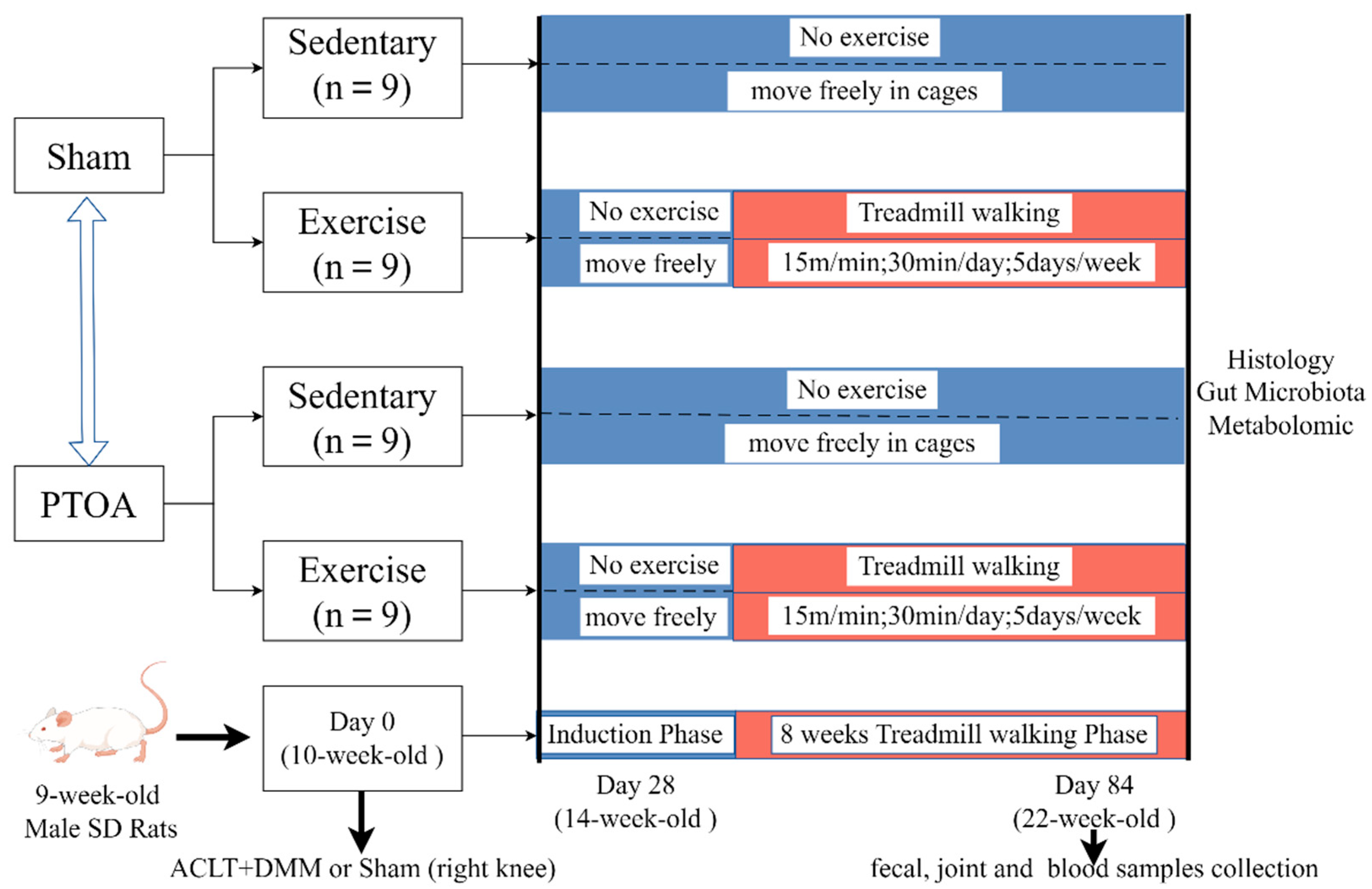
2.1. Animals

Thirty-six 9-week-old male SD rats weighing 180–250 g were obtained from the Animal Center of Tongji Hospital. All animal experiments complied with current ethical regulations and were approved by the Committee on the Ethics of Animal Experiments of Tongji Hospital, Tongji Medical College (permission number: TJH-202007010). We adhered to the ARRIVE guidelines [23] and included the ARRIVE checklist as Supplementary Materials. The SD rats were individually housed in per standard ventilated facility filled with irradiated sawdust at constant temperature (22 ± 1 °C), humidity (45%), and a regular 12 h light/dark cycle. All animals could move freely in the cages and had unrestricted access to sterilized food and water.

2.2. Study Design and Exercise Protocols

Before the start of the experiment, all animals were randomly assigned to four groups: Sham rats without treadmill walking (namely, Sham/Sed group, n = 9), Sham rats with treadmill walking 2 months (namely, Sham/TW2M group, n = 9), PTOA rats without treadmill walking (namely, PTOA/Sed group, n = 9), and PTOA rats with treadmill walking 2 months (namely, PTOA/TW2M group, n = 9). PTOA group rats underwent surgical procedures after anesthesia to induce OA models of ACLT and DMM as previously described [21].
The sample size for per group was determined using the Proc Power procedure in SAS 21 (version 9.4) with a significance level (α) at 0.05 and a power of 80%. A minimum sample size of five rats per group is based on prior publication [24] to detect a pre-specified effect size. Therefore, our sample size met the requirement and was sufficient to observe meaningful distinctions in the histological score of the joint. Exercise training was performed at 4 weeks after joint surgery on a flatbed treadmill at a rate of 15 m/min for 30 min/d, 5 days/week. The body weight of rats was measured once per week. The animals were euthanized at 22 weeks of age. Prior to euthanasia, fresh fecal samples and blood samples were collected for analysis. The protocols employed in this study are illustrated in Figure 1.

2.3. Micro-Computed Tomography (Micro-CT) Analysis of Subchondral Bone

Following euthanasia, the knee joints of all rats were scanned using a micro-CT system (micro-CT 50 Scanco Medical, Bassersdorf, Switzerland) with 10.5 µm voxel size, 100 kV voltage, and 98 µA current. Reconstructed images and data were processed with three-dimensional built-in software. We analyzed parameters including bone volume/tissue volume fraction (BV/TV), trabecular thickness (Tb.Th), trabecular number (Tb.N), and trabecular separation (Tb.Sp) as previously described [25].

2.4. Histological Analysis of Articular Cartilage

Following micro-CT scanning, knee joints were fixed in a 4% paraformaldehyde solution for 48 h at room temperature, decalcified in 10% EDTA solution at 4 °C for 4 weeks, and dehydrated with graded ethanol solutions. The joints were embedded in paraffin in the sagittal plane, and 10 to 20 slides with 4 µm sections were prepared for histological assessments. Subsequently, articular cartilage degeneration of the medial tibial plateau (MTP) was scored by the Osteoarthritis Research Society International (OARSI) scoring system (0–6) [26] and modified Mankin scoring system (0–14) [27], which involve evaluation of chondrocyte loss, articular cartilage structure change, and matrix fibrillation/loss. Three independent, experienced researchers will perform scoring in a blinded way and calculate the mean value for each slide. The mean score of ten randomly selected slides was computed to represent a single sample.

2.5. DNA Extraction, 16S rRNA Amplification, and Illumina Miseq Sequencing

Total bacterial genomic DNA was extracted from each rat fecal sample (200 mg) using the Fast DNA SPIN extraction kits (MP Biomedicals, Santa Ana, CA, USA) in accordance with the manufacturer’s instructions. The concentration of extracted DNAs was determined by a NanoDrop ND-1000 spectrophotometer (Thermo Fisher Scientific, Waltham, MA, USA). PCR amplification targeted the V3–V4 region of the bacterial 16S rRNA genes with universal primers [28]. PCR products were purified by Agencourt AMPure Beads (Beckman Coulter, Brea, CA, USA) and quantified using the PicoGreen dsDNA Assay Kit (Invitrogen, Waltham, CA, USA). Quantitative PCR products were prepared for sequencing on Illumina MiSeq by Shanghai Personal Biotechnology Co., Ltd. (Shanghai, China).

2.6. Bioinformatics Analysis of Sequencing Data

The optimized sequences were obtained through quality control, filtering, and splicing of raw reads. Sequences with  ≥97% similarity were grouped into the same operational taxonomic units (OTUs) by Usearch version 2.3.4. The dereplication was performed using DADA2 to obtain feature sequences. Then, the taxonomic classification and annotation of OTUs were executed using Quantitative Insights into Microbial Ecology 2 (QIIME2) software and the Greengenes Database [29]. Bacterial taxonomy was determined using relative abundance. A Venn diagram was created to compare the OTUs of the groups. The alpha diversity indices (observed OTUs, Chao1, Shannon, and Simpson) were calculated by QIIME2. Beta diversity was performed based on Weighted UniFrac distance using mothur. The comparison of relative abundances of bacterial taxa of different groups was conducted using the R package (version 3.5.3).

2.7. Untargeted Metabolomic Profiling of Serum Samples

Approximately 100 μL of serum was thawed at 4 °C and precipitated with 400 µL ice-cold methanol/acetonitrile (1:1, v/v; including 10 μL DL-o-Chlorophenyl alanine (2.9 mg/mL) as internal standard); 200 µL supernatants were transferred into a new tube and evaporated to dryness after vortexing and centrifugation. To evaluate the stability and repeatability of the liquid chromatograph mass spectrometer (LC–MS) system, quality control (QC) samples were created by combining 10 μL from each sample. The metabolomics profiling was conducted with ultra-high-performance liquid chromatography (1290 Infinity LC, Agilent Technologies, Santa Clara, CA, USA) coupled with a quadrupole time-of-flight system (AB Sciex TripleTOF 6600, AB SCIEX, Framingham, MA, USA) at Shanghai Applied Protein Technology Co., Ltd. (Shanghai, China). [30]. The separation of prepared samples was injected on an ACQUIY UPLC BEH column (2.1 × 100 mm, 1.7 µm, Waters, Milford, MA, USA) with a flow rate of 0.5 mL/min at 25 °C. The mobile phase was made up of water containing 25 mM ammonium acetate and 25 mM ammonium hydroxide (A) and acetonitrile (B). The gradient elution was performed as reported by Zhang et al. [31]. After separation, mass data acquisition was carried out on an AB Triple TOF 6600 Mass Spectrometer (AB SCIEX, Framingham, MA, USA) equipped with a dual electrospray ionization source (ESI) operating in positive and negative ion modes. The ESI source parameters were set in accordance with the description by Zhang et al. [31]. In the mode of MS acquisition, TOF MS scan time was set at 0.20 s/spectra, and the data were acquired over the m/z range 60–1000 Da. In the mode of auto MS/MS acquisition, product ion scan accumulation time was 0.05 s/spectra, and the data were collected from 25 to 1000 m/z. The detailed parameters were outlined by Zhang et al. [31].

2.8. Statistical Analysis of Metabolite Profiles

The raw mass spectrometry data (wiff.scan files) were converted to MzXML format by Proteo Wizard software (version 3.0.9134), and then, the nonlinear alignment, automatic integration, and extraction were carried out within the XCMS program (version 3.5.1). Metabolites were identified by comparing their mass spectra with an established in-house database according to available authentic standards. After normalizing total peak intensity, the data were analyzed using SIMCA-P (version 14.1) for multivariate analysis, including orthogonal partial least-squares discriminant analysis (OPLS-DA). The robustness of the OPLS-DA model was assessed by sevenfold cross-validation and response permutation testing. The variable importance for projection (VIP) value from the OPLS-DA model was calculated to show its significance in classification. With VIP value > 1 and p values < 0.05, candidate metabolites were regarded as potential biomarkers [32].

2.9. Statistical Analysis

All experiments were repeated independently three times, with data presented as means  ±  standard deviation (SD). For parametric tests, data comparisons among multiple groups were conducted using one-way analysis of variance (one ANOVA) followed by Tukey’s post hoc test, while comparisons between two groups were conducted using Student’s t-test. Furthermore, the Kruskal–Wallis H test was performed to analyze the nonparametric data (histological scores). GraphPad Prism software version 7.0 was utilized for data analysis and graph production. p  <  0.05 was considered statistically significant. In the R software platform (version 3.5.3), the stats package was applied to acquire Spearman’s correlation coefficient to analyze the correlation between some significantly changed metabolites, gut microbiome, and relevant physiological parameters of OA. The correlation threshold was chosen as p < 0.05.

3. Results

3.1. Exercise Attenuates PTOA-Relevant Phenotypes of Cartilage–Subchondral Bone Unit

Rats’ body weights were recorded weekly to exclude the factor of obesity and overloading on OA severity. As shown in Figure 2a, at the start of the experiment, body weight was similar among the four groups. Throughout the experiment, body weight increased significantly across all groups, with no notable differences at the end of the experiment.
We used H&E, Safranin O-Fast, and Toluidine blue histological staining to evaluate articular cartilage degeneration. As shown in Figure 2b, the cartilage degeneration observed in the PTOA/Sed group was characterized by vertical fissures extending to the deep zone, decreased chondrocytes, and deficiency in cartilage matrix staining, which were consistent with higher OARSI score and modified Mankin score compared with Sham/Sed group (Figure 2c). Micro-CT observations revealed subchondral bone lesions in the PTOA/Sed group, characterized by bone loss, subchondral perforations, and osteophyte mineralization (Figure 2d). Moreover, the parameters of subchondral trabecular bone, including BV/TV and Tb.Th, were significantly lower than the Sham/Sed group (Figure 2e). These alterations highlight the disrupted integrity of the cartilage–subchondral bone unit in the PTOA animal model. However, the cartilage degeneration in the PTOA/TW2M group was mitigated as a response to 8-week treadmill walking compared to the PTOA/Sed group (Figure 2b,c). Simultaneously, the feedback of subchondral bone characterized by the reduced bone loss and higher BV/TV was also detected in the exercised animals (Figure 2d,e). Collectively, these findings validate the hypothesis that exercise prevents the progression of PTOA-relevant cartilage degeneration and subchondral bone loss.

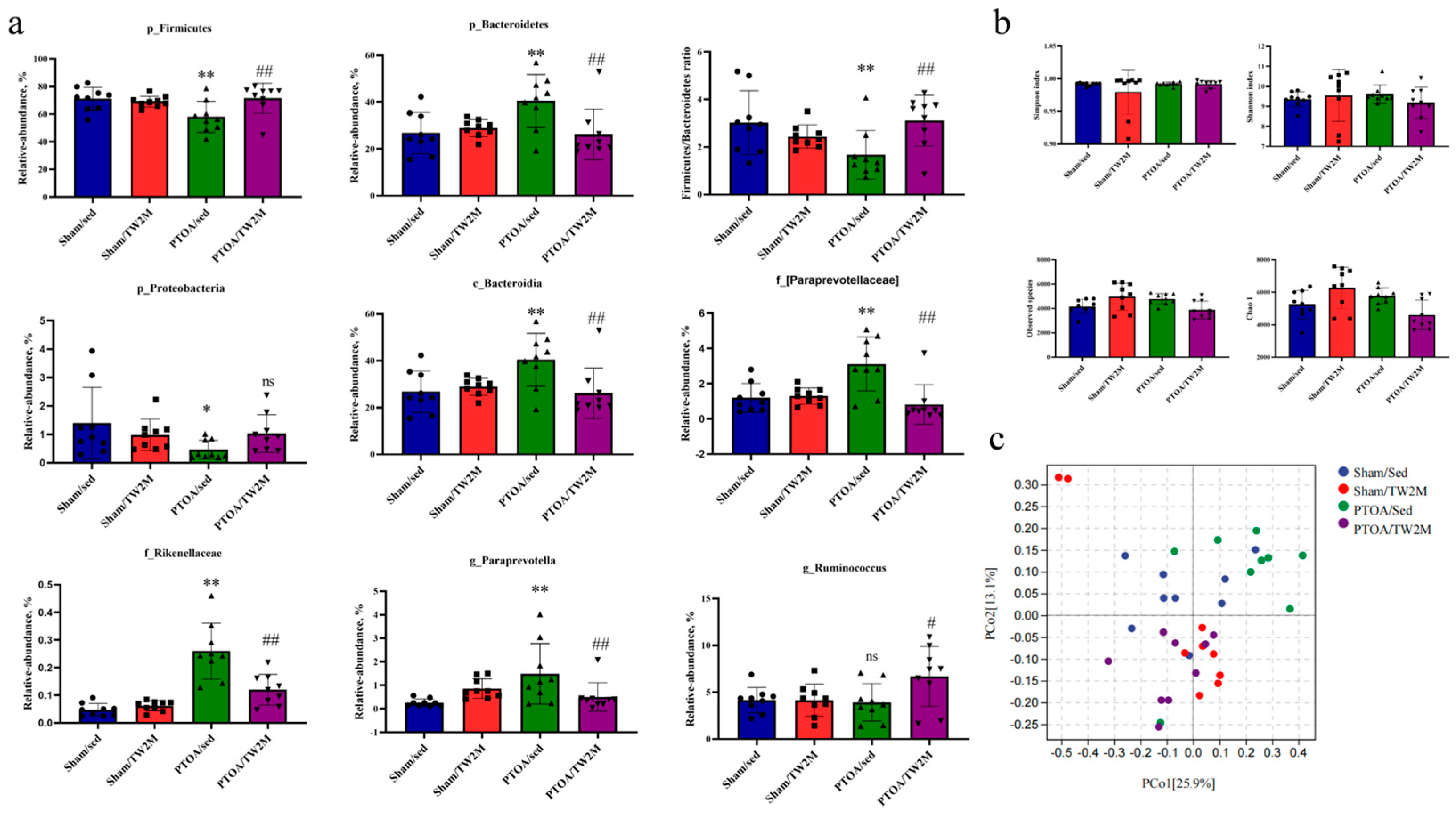
3.2. Exercise Modifies the PTOA-Relevant Gut Dysbiosis

To reveal the influence of joint injury and treadmill walking on gut microbiome, 16S rRNA gene sequencing was performed targeting on V3–V4 region of 36 fecal samples from Sham/Sed, Sham/TW2M, PTOA/Sed, and PTOA/TW2M groups to assess the microbiome composition and diversity. Based on 97% sequence similarity, we identified 78,915 OTUs; 2258 of which existed across all groups were consequently designated as core OTUs. The core OTUs accounted for 2.86% of all OTUs, whereas 17,268, 21,193, 22,863, and 17,591 OTUs were uniquely identified in the Sham/Sed, Sham/TW2M, PTOA/Sed, and PTOA/TW2M groups, respectively (Figure 3a). The OTUs were categorized into 19 bacterial phyla, 33 classes, 78 orders, 141 families, 290 genera, and 407 species. Based on the sequence read counts, the top 19 phyla (Figure 3b,d) and top 20 genera (Figure 3c,e) in relative abundance of the fecal microbiome are presented. The most dominant phyla were Firmicutes and Bacteroidetes across all four groups, followed by Proteobacteria and Tenericutes, which collectively comprised 99.50%, 99.26%, 99.22%, and 99.09% of the reads in Sham/Sed, Sham/TW2M, PTOA/Sed, and PTOA/TW2M, respectively. At the genus level, Lactobacillus was the most prevalent in the four groups, followed by Oscillospira, Ruminococcus, Prevotella, Turicibacter, and Blautia. These six genera accounted for 25.35%, 26.41%, 18.97%, and 26.70% of the sequences of the Sham/Sed, Sham/TW2M, PTOA/Sed, and PTOA/TW2M groups.
Differences of fecal microbiome at the phylum, class, family, and genus levels were identified among four groups (Figure 4a). At the phylum level, compared with Sham/Sed group rats, the relative abundance of Firmicutes and Proteobacteria was markedly decreased, and the relative abundance of Bacteroidetes was markedly increased in PTOA/Sed group, as well as the decreased Firmicutes/Bacteroidetes ratio, indicating PTOA-relevant gut dysbiosis. However, PTOA/TW2M group rats exhibited significantly increased Firmicutes and Firmicutes/Bacteroidetes ratio but significantly decreased Bacteroidetes compared with PTOA/Sed group rats as a response to exercise. At the class level, the relative abundance of Bacteroidia was increased in the PTOA/Sed group compared to the Sham/Sed group, while the increased alterations were attenuated in the PTOA/TW2M group. Moreover, the relative abundances of the family [Paraprevotellaceae] and Rikenellaceae were the highest in the PTOA/Sed group, while a remarkable decrease was observed in the PTOA/TW2M group. At the genus level, PTOA/Sed group presented a higher abundance of Paraprevotella compared to the Sham/Sed group rats. Interestingly, Paraprevotella remarkably decreased in the PTOA/TW2M group. Additionally, genus Ruminococcus was highly enriched in the PTOA/TW2M group. In light of the significant alterations in the composition of the rats’ gut microbiome due to experimental injury and exercise, we evaluated the alpha and beta diversity of the fecal microbiota. The Sham/TW2M group had the highest Chao1 and Observed species, with significant differences in the Shannon and Simpson indices across the four groups (Figure 4b). Through principal coordinate analysis (PCoA) based on Weighted UniFrac distance, we found that the gut microbiota of rats was less dispersed in the Sham/Sed, PTOA/Sed, and PTOA/TW2M groups, but there was obvious segregation in the Sham/TW2M group (Figure 4c). Collectively, these findings indicate that joint injury and exercise both alter the composition and diversity of the intestinal microbiome. Moreover, the microbiome may act as a possible connection between OA and exercise, attributable to the ameliorative effects of exercise on OA-associated microbial alterations.

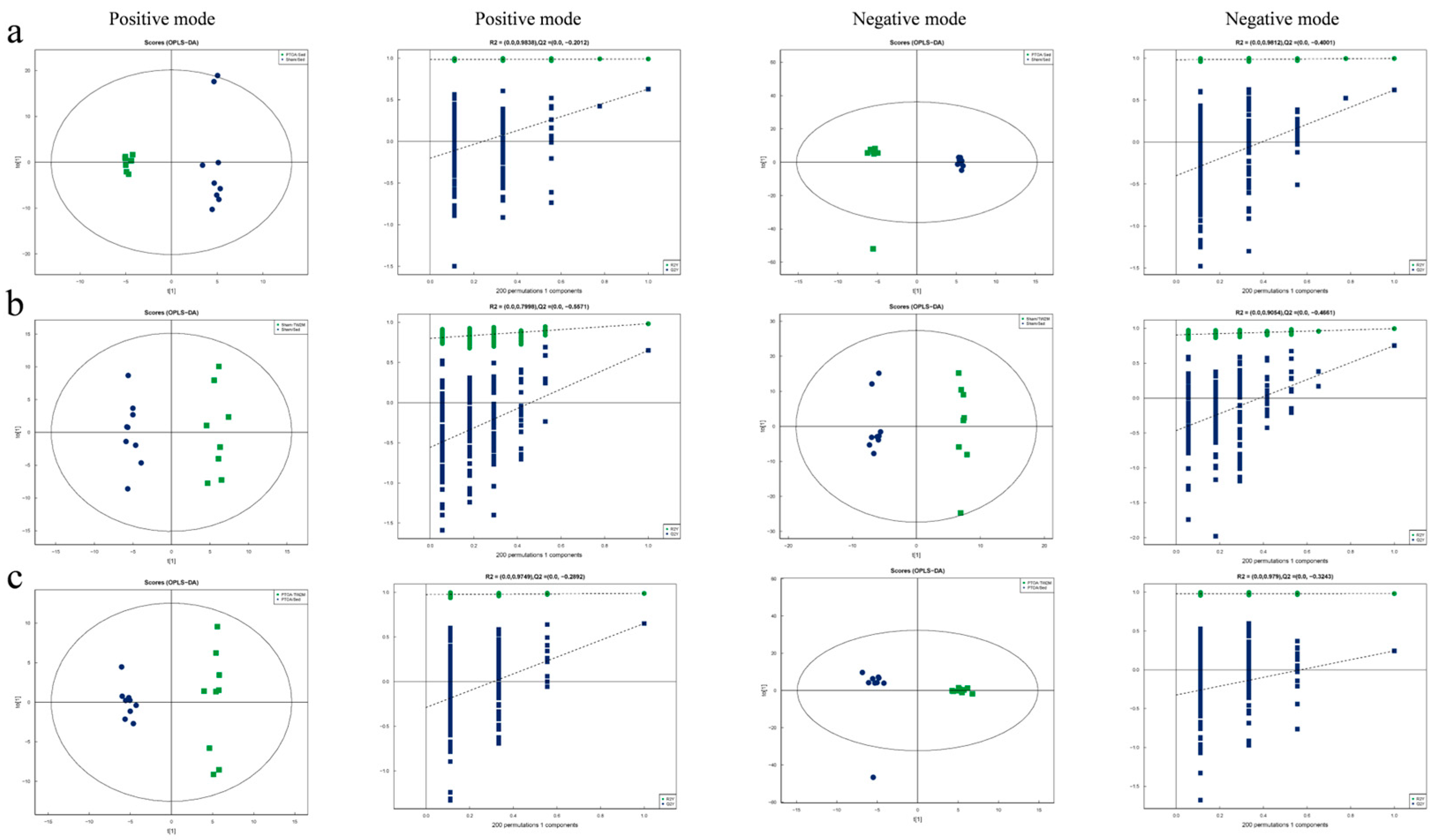
3.3. Exercise Modifies the PTOA-Relevant Serum Metabolic Profiles

As the microbe–host bridge, many metabolites of the gut microbiome could influence host physiology by entering the bloodstream. Therefore, the serum samples from Sham/Sed, Sham/TW2M, PTOA/Sed, and PTOA/TW2M groups were analyzed to explore the serum metabolic profiling using LC–MS. In total, 198 and 143 peak features were identified in positive and negative ion modes, respectively. Subsequently, OPLS-DA was conducted to optimize the separation and identification of metabolites. The validation plots were obtained using 200 permutation tests. The OPLS-DA score plot revealed a distinct separation between the Sham/Sed and PTOA/Sed groups in positive mode (R2 = 0.9838, Q2 = −0.2012) and negative mode (R2 = 0.9812, Q2 = −0.4001). R2 indicates the goodness of the fit, while Q2 reflects the prediction ability of the model. The models effectively divide the samples into two categories, indicating strong reliability and predictive capability (Figure 5a).
According to score plots (Figure 5b), there was a noticeable separation trend between the Sham/TW2M and Sham/Sed groups in both positive ion mode (R2 = 0.7998, Q2 = −0.5571) and negative ion mode (R2 = 0.9054, Q2 = −0.4661). Moreover, in both positive (R2 = 0.9749, Q2 = −0.2892) and negative ion modes (R2 = 0.979, Q2 = −0.3243), clear distinctions between PTOA/TW2M and PTOA/Sed groups (Figure 5c) were observed, indicating that joint injury and exercise cause changes in metabolites. To determine the variables responsible for this separation, the VIP parameter was employed. From the OPLS-DA model, VIP indicated which metabolites played a crucial role in differentiating rat samples. Metabolites with VIP score >1 were selected as potential candidates. Significant differences in variables were identified with p value threshold of 0.05. The distinguished different metabolites are displayed in Table 1 and Table 2. In addition, potential metabolites are shown in a histogram according to the fold change parameter. When compared with the Sham/Sed group, five metabolites were up-regulated and nine metabolites were down-regulated in positive mode, as well as four metabolites were down-regulated in the PTOA/Sed group (Figure 6a). Furthermore, compared to the Sham/Sed group, the Sham/TW2M group exhibited four up-regulated and four down-regulated metabolites in positive mode, as well as three up-regulated and one down-regulated metabolites in negative mode (Figure 6b). Interestingly, compared to the PTOA/Sed group, the PTOA/TW2M group showed an up-regulation of six metabolites and a down-regulation of four metabolites in positive mode, with six metabolites up-regulated in negative mode (Figure 6c). It was also observed that joint injury and exercise cause changes in serum metabolic profiles among the four groups.

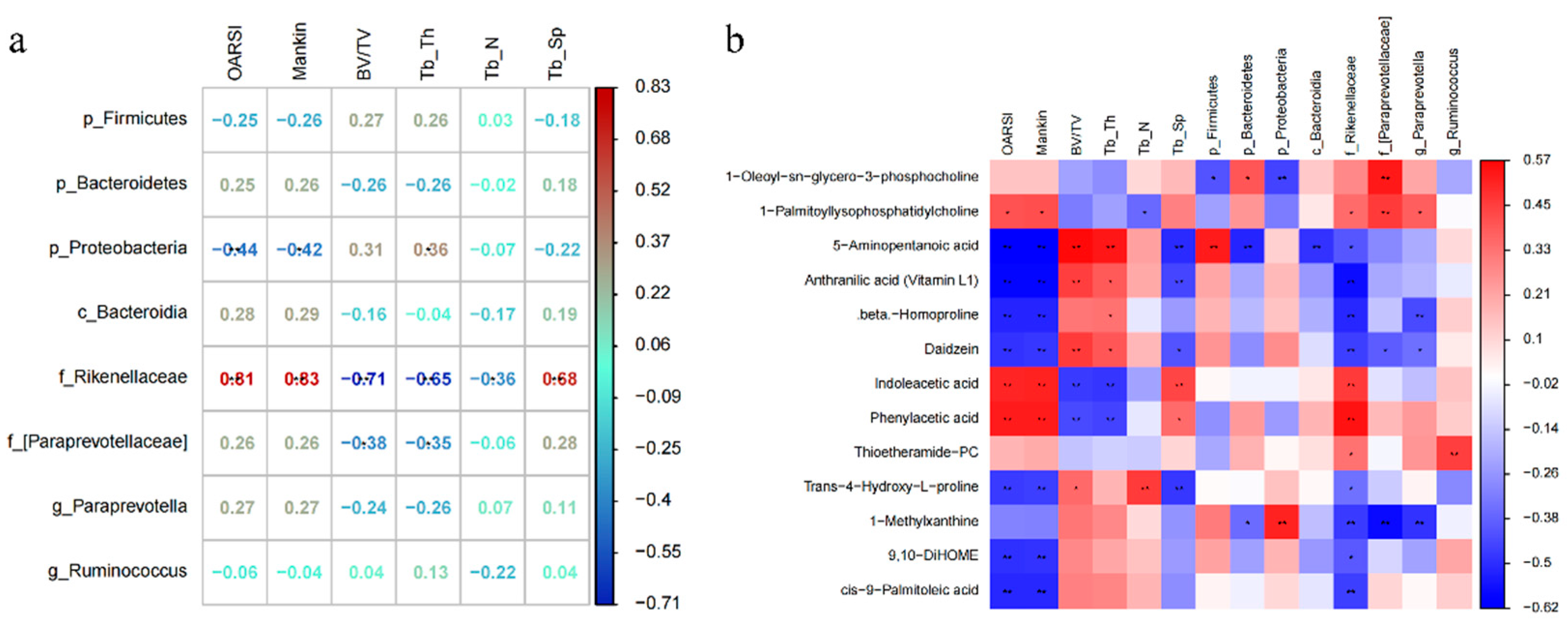
3.4. Exercise-Induced Changes in Serum Metabolism Are Related to the Integrity of Cartilage–Subchondral Bone Unit and Gut Microbiome

Given that the effects of exercise on articular cartilage, subchondral bone unit, gut microbiome, and serum metabolites were defined, respectively, we further investigate the relationship between these impacts through correlation analysis.
As depicted in Figure 7a, there are identified correlations between joint structural features, such as OARSI score and micro-CT data, and PTOA-relevant microorganisms, including phylum Firmicutes, phylum Bacteroidetes, phylum Proteobacteria, class Bacteroidia, family Rikenellaceae, family [Paraprevotellaceae] and genus Paraprevotella, and genus Ruminococcus. Interestingly, family Rikenellaceae significantly positively correlated with OARSI score (r = 0.81, p < 0.01), Mankin score (r = 0.83, p < 0.01), and Tb.Sp (r = 0.68, p < 0.01) and significantly negatively correlated with BV/TV (r = −0.71, p < 0.01) and Tb.Th (r = −0.65, p < 0.01), indicating its potential participation in PTOA-relevant cartilage modifications. However, phylum Proteobacteria exhibited stronger negative correlations with OARSI, Mankin, and subchondral bone loss compared to other PTOA-relevant microorganisms. These findings imply a robust connection between structural phenotypes and microorganisms associated with PTOA, where the exercise-responsive gut microbiome family Rikenellaceae may partially contribute to the effects of exercise on cartilage-subchondral bone unit.
Moreover, as shown in Figure 7b, significant correlations were observed between many altered metabolites, OARSI score, Mankin score, micro-CT data, and the relative abundance of PTOA-relevant microbiome. (r > 0.45, p < 0.05). For example, 5-Aminopentanoic acid, Anthranilic acid (Vitamin L1), beta-Homoproline, Daidzein, Trans-4-Hydroxy-L-proline, 9,10-DiHOME, and Cis-9-Palmitoleic acid significantly negatively correlated with OARSI and Mankin score, whereas Indoleacetic acid and Phenylacetic acid significantly positively correlated with OARSI and Mankin score. In addition, 5-Aminopentanoic acid, Anthranilic acid (Vitamin L1), and Daidzein had significant positive correlations with BV/TV and Tb.Th, whereas Indoleacetic acid and Phenylacetic acid significantly negatively correlated with BV/TV and Tb.Th. Moreover, 1-Oleoyl-sn-glycero-3-phosphocholine and 1-Palmitoyllysophosphatidylcholine positively correlated with the family [Paraprevotellaceae]. 5-Aminopentanoic acid positively correlated with phylum Firmicutes and negatively correlated with phylum Bacteroidetes and class Bacteroidia. Anthranilic acid (Vitamin L1), beta-Homoproline, Daidzein, cis-9-Palmitoleic acid, and 1-Methylxanthine were negatively correlated with family Rikenellaceae, whereas Indoleacetic acid and Phenylacetic acid were positively correlated with family Rikenellaceae. Thioetheramide−PC positively correlated with genus Ruminococcus. 1-Methylxanthine positively correlated with phylum Proteobacteria and negatively correlated with family [Paraprevotellaceae] and genus Paraprevotella. These findings indicated that exercise might reduce the relative abundance of family Rikenellaceae, which resulted in an increase in anti-inflammatory metabolites and a decrease in inflammatory metabolites, as well as subsequent structural changes in PTOA rats.

4. Discussion

In this study, we revealed a possible mechanism of exercise and OA development by the modulation of gut microbiome and metabolites. Our study demonstrates that exercise significantly alleviates cartilage degeneration and subchondral bone loss, potentially through regulating an imbalanced gut microbiome and disturbed serum metabolome (Figure S1). Despite several animal experiments that have demonstrated the therapeutic role of exercise in preventing joint inflammation and cartilage degradation [21,25], the underlying regulatory mechanisms focused on gut microbiome and metabolites are still not well understood.
To delve deeper into this, we employed 16S rRNA gene sequencing techniques to analyze the composition and diversity of gut microbiota in response to joint injury and exercise across different groups. It has been shown that Firmicutes and Bacteroidetes are dominant phyla of gut microbiota. In our study, compared with the Sham/Sed group, PTOA/Sed group rats displayed a significant decrease in Firmicutes and an increase in Bacteroidetes, while exercise reversed the aberrant gut microbiota composition. Our findings are corroborated by Jiang et al., who found that collagen-induced arthritis rats exhibited a marked reduction in the relative abundance of Firmicutes and an increase in Bacteroidetes abundance [33]. Moreover, the decreased Firmicutes/Bacteroidetes ratio is relevant to mild inflammatory reaction in high-fat diet rats [34]. Therefore, in our study, the increased Firmicutes/Bacteroidetes ratio may interpret the positive anti-inflammatory effect of exercise in OA. As a common intestinal Gram-negative bacterium, the family Rikenellaceae was thought to be involved in bone metabolism, with a negative impact on bone resorption and bone density [35]. Several studies have found that an abundance of Rikenellaceae may lead to the secretion of proinflammatory cytokines [36]. Prinz et al. have shown that the presence of Rikenellaceae is associated with worse OA outcomes, which is consistent with our findings [37]. Our investigation demonstrated that the relative abundance of Rikenellaceae was upregulated in PTOA rats and positively correlated with the severity of PTOA-related structural changes and actively responded to exercise treatment. Moreover, the PTOA/Sed group showed an increase in the abundance of genus Paraprevotella, which was suppressed by exercise in the PTOA/TW2M group. Our results are supported by Tand et al., who observed that Paraprevotella was abundant in the gut microbiota of individuals with depression and associated with OA severity [38]. This finding provides evidence that the protective effect of exercise on OA may be realized by mitigating the presence of pathogenic bacteria, particularly Rikenellaceae and Paraprevotella, in PTOA rats.
Furthermore, non-targeted LC–MS metabolomics analysis was employed to explore the effects of exercise on serum metabolite profiles of PTOA rats. The results showed that metabolites including Daidzein, Anthranilic acid, and L-Anserine were downregulated, and Indoleacetic acid, Phenylacetic acid, and 1-Palmitoyllysophosphatidylcholine were upregulated in PTOA/Sed group compared to Sham/Sed group. These metabolites are involved in tryptophan metabolism, histidine metabolism, beta-alanine metabolism, phenylalanine metabolism, and amino acid biosynthesis. Additionally, Anthranilic acid and Daidzein were negatively correlated with OARSI score and family Rikenellaceae, and they responded actively to exercise treatment.
Anthranilic acid has been found to have potential for anti-inflammatory and anti-cancer activity, and it serves as a crucial agent participating in tryptophan metabolism [39]. There is increasing evidence that has shown that altered tryptophan metabolism has been associated with several disorders, such as erosive hand osteoarthritis and rheumatoid arthritis [40,41]. Daidzein has been reported for effective prevention and treatment of diseases because of its outstanding antioxidant and anti-inflammatory roles [42]. Interestingly, a study [43] indicated that daidzein protected against monosodium iodoacetate-induced OA via reducing the levels of TNF-α, IL-1β, and MMP13 in serum and joint tissue samples and improving cartilage surface fibrillation, which is in line with our discoveries. Our data support that the beneficial effect of exercise in blocking OA pathological progress may be achieved by upregulating the serum level of Anthranilic acid and Daidzein.
Phenyllactic acid is involved in phenylalanine metabolism, which is elevated in OA synovial fluid [44]. Previous studies have shown that increased phenylalanine concentration may be linked to the progression of OA and considered as a pivotal indicator in detecting OA [45,46]. Our results are in agreement with the evidence presented in this study. 1-Palmitoyllysophosphatidylcholine is one of the most prominent lysophosphatidylcholine species, which promotes endothelial pro-inflammatory and pro-adhesive chemokine IL-8 synthesis [47]. The study reported that lysophosphatidylcholine overproduction was positively associated with circulating TNF-α and IL-1β levels [48]. We observed that the level of 1-Palmitoyllysophosphatidylcholine was higher in the PTOA/Sed group compared to the Sham/Sed group. This is consistent with Zhang et al.’s report, in which the concentration of lysophosphatidylcholine was increased in patients with knee OA [49]. The elevated level of 1-Palmitoyllysophosphatidylcholine was decreased as a response to exercise, implying the beneficial anti-inflammatory effect of exercise on OA. These findings provide evidence that the modifications in metabolites are strongly associated with gut microbiota and joint degeneration.

5. Limitations

Although our study has elucidated how exercise, through the modulation of gut microbiota and serum metabolites, acts in the context of OA, several limitations should be acknowledged. First, 16S rRNA sequencing technology utilized in our study is predominantly restricted to the taxonomic identification of bacteria, which is incapable of identifying specific microbial species and strains. Further research can employ a whole-genome shotgun metagenomic sequencing approach to generate robust estimates and detailed functional annotations of microbial communities to improve the gut microbiota–joint axis in OA. Second, our sample size was comparatively small, and the findings require further validation in a larger sample size study to strengthen the robustness of the conclusions. Meanwhile, the lack of cross-sectional studies limits the ability to establish causal connections between gut microbiome dysbiosis and OA progression. The direct relationship between joint degeneration and the identified gut microbiota, specifically Firmicutes and Rikenellaceae, as well as the putative key metabolites, Daidzein and Anthranilic acid, requires additional experimental verification. In addition, some potential unmeasured confounders, including circadian rhythm and stress levels, were not well controlled, which may influence gut microbiota and metabolite profiles. Moreover, there is a significant disparity in microbiota composition between humans and animals, as evidenced by the fact that 85% of the bacterial genera found in the animal gastrointestinal tract are absent in the human gut [50]. The genetic background of different animal models also affects microbiota composition. These factors present challenges in the translational relevance of animal models for human applications and indicate that an exact correspondence cannot be achieved between animal and human models, or even among different animal models. Finally, PTOA, metabolic syndrome-associated OA, and age-related OA have distinct causes, mechanisms, and clinical presentations, which may lead to varied responses to exercise interventions. Our study exclusively focuses on the effects of exercise on PTOA due to limitations in sample size and the heterogeneity of the animal model.

6. Conclusions and Future Perspectives

In summary, our study demonstrates that exercise can serve as a therapeutic agent to mitigate OA through slowing down articular cartilage degeneration and subchondral bone loss, modulating gut microbiota composition, and increasing beneficial metabolites. Moreover, our study highlights that the benefits of exercise on joints might be related to the identified gut microbiota, particularly Firmicutes and Rikenellaceae, along with the possible key metabolites, Daidzein and Anthranilic acid. The recognized gut microbiota and essential metabolites may be a potential target for exercise intervention, offering clinical potential implications for OA therapy. In addition, these findings provide fresh insights into uncovering the mechanism of exercise treatment for OA.
Furthermore, preclinical animal models only partially mimic the complexity of the human microbiome; there is a need for more high-quality clinical studies or large sample size cross-sectional studies to identify the causal links between gut microbiota and OA progression in future research. Collectively, the innovative concept along with advanced research provides novel direction to develop a therapeutic approach for OA by targeting key metabolites or gut microbiota.

Supplementary Materials

The following supporting information can be downloaded at: https://www.mdpi.com/article/10.3390/metabo15050341/s1, Figure S1: Schematic diagram of the potential protective mechanism and effects of exercise in the development of osteoarthritis by altering the compositions of gut microbiome and serum metabolites. Exercise can decrease the abundance of family Rikenellaceae, increases the production of Anthranilic acid and Daidzein, and prevent cartilage degeneration and the loss of subchondral bone of osteoarthritis joint.

Author Contributions

X.H. and X.S. performed and designed this study and wrote the manuscript; Y.Z., W.H., R.C., C.P., J.L. and X.D. analyzed the data and prepared the figures; J.Z. and T.X. evaluated and defined the last version of the manuscript. All authors have read and agreed to the published version of the manuscript.

Funding

This study was supported by the National Natural Science Foundation of China (Grant No. 82272610).

Institutional Review Board Statement

All animal experiments were approved by the Committee on the Ethics of Animal Experiments of Tongji Hospital, Tongji Medical College (permission number: TJH-202007010, approval date 10 July 2020).

Informed Consent Statement

Not applicable.

Data Availability Statement

The data presented in this study are available on request from the corresponding author. The raw data were deposited in the NCBI Sequence Read Archive (SRA) (accession numbers for NCBI: BioProject: PRJNA1240216 for 16S rRNA sequencing).

Acknowledgments

The authors also acknowledge the technical assistance of Shanghai Applied Protein Technology Co., Ltd.

Conflicts of Interest

The authors declare no conflict of interest.

Abbreviations

OA, osteoarthritis; PTOA, post-traumatic osteoarthritis; SD, Sprague-Dawley; Sham/Sed, Sham/sedentary; Sham/TW2M, Sham/treadmill walking 2 months; PTOA/Sed, PTOA/sedentary; PTOA/TW2M, PTOA/treadmill walking 2 months; DMM, destabilization of the medial meniscus; ACLT, transection of the anterior cruciate ligament; LPS, lipopolysaccharide; PGN, peptidoglycan; FFAs, free fatty acids; SPF, specific pathogen-free; BV/TV, bone volume/tissue volume fraction; Tb.N, trabecular number; Tb.Sp, trabecular separation; Tb.Th, trabecular thickness; EDTA, ethylenediaminetetraacetic acid; MTP, medial tibial plateau; OARSI, Osteoarthritis Research Society International; ELISA, enzyme-linked immunosorbent assay; OTUs, operational taxonomic units; Chao1, Chao richness estimator; QC, quality control; LC–MS, liquid chromatography–mass spectrometry; OPLS-DA, orthogonal partial least-squares discriminant analysis; VIP, variable importance for projection; PCoA, principal coordinate analysis.

References

  1. Kraus, V.B.; Blanco, F.J.; Englund, M.; Karsdal, M.A.; Lohmander, L.S. Call for standardized definitions of osteoarthritis and risk stratification for clinical trials and clinical use. Osteoarthr. Cartil. 2015, 23, 1233–1241. [Google Scholar] [CrossRef] [PubMed]
  2. Long, H.; Liu, Q.; Yin, H.; Wang, K.; Diao, N.; Zhang, Y.; Lin, J.; Guo, A. Prevalence Trends of Site-Specific Osteoarthritis from 1990 to 2019: Findings from the Global Burden of Disease Study 2019. Arthritis Rheumatol. 2022, 74, 1172–1183. [Google Scholar] [CrossRef] [PubMed]
  3. Katz, J.N.; Arant, K.R.; Loeser, R.F. Diagnosis and Treatment of Hip and Knee Osteoarthritis: A Review. J. Am. Med. Assoc. 2021, 325, 568–578. [Google Scholar] [CrossRef] [PubMed]
  4. Robinson, W.H.; Lepus, C.M.; Wang, Q.; Raghu, H.; Mao, R.; Lindstrom, T.M.; Sokolove, J. Low-grade inflammation as a key mediator of the pathogenesis of osteoarthritis. Nat. Rev. Rheumatol. 2016, 12, 580–592. [Google Scholar] [CrossRef]
  5. Chapman, J.H.; Ghosh, D.; Attari, S.; Ude, C.C.; Laurencin, C.T. Animal Models of Osteoarthritis: Updated Models and Outcome Measures 2016–2023. Regen. Eng. Transl. Med. 2024, 10, 127–146. [Google Scholar] [CrossRef]
  6. Lieberthal, J.; Sambamurthy, N.; Scanzello, C. Inflammation in joint injury and post-traumatic osteoarthritis. Osteoarthr. Cartil. 2015, 23, 1825–1834. [Google Scholar] [CrossRef]
  7. Buford, T.W. (Dis)Trust your gut: The gut microbiome in age-related inflammation, health, and disease. Microbiome 2017, 5, 80. [Google Scholar] [CrossRef]
  8. Rinninella, E.; Raoul, P.; Cintoni, M.; Franceschi, F.; Miggiano, G.A.D.; Gasbarrini, A.; Mele, M.C. What is the Healthy Gut Microbiota Composition? A Changing Ecosystem across Age, Environment, Diet, and Diseases. Microorganisms 2019, 7, 14. [Google Scholar] [CrossRef]
  9. Vallianou, N.; Stratigou, T.; Christodoulatos, G.S.; Dalamaga, M. Understanding the Role of the Gut Microbiome and Microbial Metabolites in Obesity and Obesity-Associated Metabolic Disorders: Current Evidence and Perspectives. Curr. Obes. Rep. 2019, 8, 317–332. [Google Scholar] [CrossRef]
  10. Lobionda, S.; Sittipo, P.; Kwon, H.Y.; Lee, Y.K. The Role of Gut Microbiota in Intestinal Inflammation with Respect to Diet and Extrinsic Stressors. Microorganisms 2019, 7, 271. [Google Scholar] [CrossRef]
  11. Liu, S.; Li, G.; Xu, H.; Wang, Q.; Wei, Y.; Yang, Q.; Xiong, A.; Yu, F.; Weng, J.; Zeng, H. “Cross-talk” between gut microbiome dysbiosis and osteoarthritis progression: A systematic review. Front. Immunol. 2023, 14, 1150572. [Google Scholar]
  12. Ulici, V.; Kelley, K.; Azcarate-Peril, M.; Cleveland, R.; Sartor, R.; Schwartz, T.; Loeser, R. Osteoarthritis induced by destabilization of the medial meniscus is reduced in germ-free mice. Osteoarthr. Cartil. 2018, 26, 1098–1109. [Google Scholar] [CrossRef] [PubMed]
  13. Guan, Z.; Jia, J.; Zhang, C.; Sun, T.; Zhang, W.; Yuan, W.; Leng, H.; Song, C. Gut microbiome dysbiosis alleviates the progression of osteoarthritis in mice. Clin. Sci. 2020, 134, 3159–3174. [Google Scholar] [CrossRef] [PubMed]
  14. Boer, C.G.; Radjabzadeh, D.; Medina-Gomez, C.; Garmaeva, S.; Schiphof, D.; Arp, P.; Koet, T.; Kurilshikov, A.; Fu, J.; Ikram, M.A.; et al. Intestinal microbiome composition and its relation to joint pain and inflammation. Nat. Commun. 2019, 10, 4881. [Google Scholar] [CrossRef]
  15. Liu, Y.; Ding, W.; Wang, H.; Dai, L.; Zong, W.; Wang, Y.; Bi, J.; Han, W.; Dong, G. Gut microbiota and obesity-associated osteoarthritis. Osteoarthr. Cartil. 2019, 27, 1257–1265. [Google Scholar] [CrossRef]
  16. Hao, X.; Shang, X.; Liu, J.; Chi, R.; Zhang, J.; Xu, T. The gut microbiota in osteoarthritis: Where do we stand and what can we do? Arthritis Res. Ther. 2021, 23, 42. [Google Scholar] [CrossRef]
  17. Fu, L.; Duan, H.; Cai, Y.; Chen, X.; Zou, B.; Yuan, L.; Liu, G. Moxibustion ameliorates osteoarthritis by regulating gut microbiota via impacting cAMP-related signaling pathway. Biomed. Pharmacother 2024, 170, 116031. [Google Scholar] [CrossRef]
  18. Rushing, B.; McRitchie, S.; Arbeeva, L.; Nelson, A.; Azcarate-Peril, M.; Li, Y.-Y.; Qian, Y.; Pathmasiri, W.; Sumner, S.; Loeser, R. Fecal metabolomics reveals products of dysregulated proteolysis and altered microbial metabolism in obesity-related osteoarthritis. Osteoarthr. Cartil. 2022, 30, 81–91. [Google Scholar] [CrossRef]
  19. Schott, E.M.; Farnsworth, C.W.; Grier, A.; Lillis, J.A.; Soniwala, S.; Dadourian, G.H.; Bell, R.D.; Doolittle, M.L.; Villani, D.A.; Awad, H.; et al. Targeting the gut microbiome to treat the osteoarthritis of obesity. J. Clin. Investig. 2018, 3, e95997. [Google Scholar] [CrossRef]
  20. Huang, Z.; Chen, J.; Li, B.; Zeng, B.; Chou, C.-H.; Zheng, X.; Xie, J.; Li, H.; Hao, Y.; Chen, G.; et al. Faecal microbiota transplantation from metabolically compromised human donors accelerates osteoarthritis in mice. Ann. Rheum. Dis. 2020, 79, 646–656. [Google Scholar] [CrossRef]
  21. Hao, X.; Zhang, J.; Shang, X.; Sun, K.; Zhou, J.; Liu, J.; Chi, R.; Xu, T. Exercise modifies the disease-relevant gut microbial shifts in post-traumatic osteoarthritis rats. Bone Jt. Res. 2022, 11, 214–225. [Google Scholar] [CrossRef] [PubMed]
  22. Deng, X.; Xu, H.; Hao, X.; Liu, J.; Shang, X.; Xu, T. Effect of moderate exercise on osteoarthritis. EFORT Open Rev. 2023, 8, 148–161. [Google Scholar] [CrossRef] [PubMed]
  23. Percie du Sert, N.; Hurst, V.; Ahluwalia, A.; Alam, S.; Avey, M.T.; Baker, M.; Browne, W.J.; Clark, A.; Cuthill, I.C.; Dirnagl, U.; et al. The ARRIVE guidelines 2.0: Updated guidelines for reporting animal research. PLoS Biol. 2020, 18, e3000410. [Google Scholar]
  24. Hao, X.; Wang, S.; Zhang, J.; Xu, T. Effects of body weight-supported treadmill training on cartilage-subchondral bone unit in the rat model of posttraumatic osteoarthritis. J. Orthop. Res. 2020, 39, 1227–1235. [Google Scholar] [CrossRef]
  25. Shang, X.; Hao, X.; Hou, W.; Liu, J.; Chi, R.; Deng, X.; Pan, C.; Xu, T. Exercise-induced modulation of myokine irisin on muscle-bone unit in the rat model of post-traumatic osteoarthritis. J. Orthop. Surg. Res. 2024, 19, 49. [Google Scholar] [CrossRef]
  26. Glasson, S.S.; Chambers, M.G.; Van Den Berg, W.B.; Little, C.B. The OARSI histopathology initiative—Recommendations for histological assessments of osteoarthritis in the mouse. Osteoarthr. Cartil. 2010, 18 (Suppl. 3), S17–S23. [Google Scholar] [CrossRef]
  27. Mankin, H.J.; Dorfman, H.; Lippiello, L.; Zarins, A. Biochemical and Metabolic Abnormalities in Articular Cartilage from Osteo-Arthritic Human Hips: II. Correlation of Morphology with Biochemical and Metabolic Data. J. Bone Jt. Surg. 1971, 53, 523–537. [Google Scholar] [CrossRef]
  28. Wang, Y.; Tang, J.; Lv, Q.; Tan, Y.; Dong, X.; Liu, H.; Zhao, N.; He, Z.; Kou, Y.; Tan, Y.; et al. Establishment and resilience of transplanted gut microbiota in aged mice. iScience 2022, 25, 103654. [Google Scholar] [CrossRef]
  29. DeSantis, T.Z.; Hugenholtz, P.; Larsen, N.; Rojas, M.; Brodie, E.L.; Keller, K.; Huber, T.; Dalevi, D.; Hu, P.; Andersen, G.L. Greengenes, a Chimera-Checked 16S rRNA Gene Database and Workbench Compatible with ARB. Appl. Env. Microbiol. 2006, 72, 5069–5072. [Google Scholar] [CrossRef]
  30. Ma, N.; Yang, Y.; Liu, X.; Li, S.; Qin, Z.; Li, J. Plasma metabonomics and proteomics studies on the anti-thrombosis mechanism of aspirin eugenol ester in rat tail thrombosis model. J. Proteom. 2020, 215, 103631. [Google Scholar] [CrossRef] [PubMed]
  31. Zhang, X.; Zheng, Y.; Liu, Z.; Su, M.; Wu, Z.; Zhang, H.; Zhang, C.; Xu, X. Insights into characteristic metabolites and potential bioactive peptides profiles of fresh cheese fermented with three novel probiotics based metabolomics and peptidomics. Food Chem. X 2024, 21, 101147. [Google Scholar] [CrossRef] [PubMed]
  32. He, Z.; Liu, Z.; Gong, L. Systematic evaluation of sample preparation strategy for GC-MS-based plasma metabolomics and its application in osteoarthritis. Anal. Biochem. 2021, 621, 114153. [Google Scholar] [CrossRef] [PubMed]
  33. Jiang, Z.-M.; Zeng, S.-L.; Huang, T.-Q.; Lin, Y.; Wang, F.-F.; Gao, X.-J.; Li, J.; Li, P.; Liu, E.-H. Sinomenine ameliorates rheumatoid arthritis by modulating tryptophan metabolism and activating aryl hydrocarbon receptor via gut microbiota regulation. Sci. Bull. 2023, 68, 1540–1555. [Google Scholar] [CrossRef]
  34. Kim, J.Y.; Kwon, Y.M.; Kim, I.S.; Kim, J.A.; Yu, D.Y.; Adhikari, B.; Lee, S.S.; Choi, I.S.; Cho, K.K. Effects of the Brown Seaweed Laminaria japonica Supplementation on Serum Concentrations of IgG, Triglycerides, and Cholesterol, and Intestinal Microbiota Composition in Rats. Front. Nutr. 2018, 5, 23. [Google Scholar] [CrossRef]
  35. Ozaki, D.; Kubota, R.; Maeno, T.; Abdelhakim, M.; Hitosugi, N. Association between gut microbiota, bone metabolism, and fracture risk in postmenopausal Japanese women. Osteoporos. Int. 2021, 32, 145–156. [Google Scholar] [CrossRef]
  36. Daniel, H.; Gholami, A.M.; Berry, D.; Desmarchelier, C.; Hahne, H.; Loh, G.; Mondot, S.; Lepage, P.; Rothballer, M.; Walker, A.; et al. High-fat diet alters gut microbiota physiology in mice. ISME J. 2013, 8, 295–308. [Google Scholar] [CrossRef]
  37. Prinz, E.; Schlupp, L.; Dyson, G.; Barrett, M.; Szymczak, A.; Velasco, C.; Izda, V.; Dunn, C.M.; A Jeffries, M. OA susceptibility in mice is partially mediated by the gut microbiome, is transferrable via microbiome transplantation and is associated with immunophenotype changes. Ann. Rheum. Dis. 2024, 83, 382–393. [Google Scholar] [CrossRef]
  38. Tang, J.; Song, X.; Zhao, M.; Chen, H.; Wang, Y.; Zhao, B.; Yu, S.; Ma, T.; Gao, L. Oral administration of live combined Bacillus subtilis and Enterococcus faecium alleviates colonic oxidative stress and inflammation in osteoarthritic rats by improving fecal microbiome metabolism and enhancing the colonic barrier. Front. Microbiol. 2022, 13, 1005842. [Google Scholar] [CrossRef]
  39. Şenol, H.; Çağman, Z.; Katmerlikaya, T.G.; Tokalı, F.S. New Anthranilic Acid Hydrazones as Fenamate Isosteres: Synthesis, Characterization, Molecular Docking, Dynamics & in Silico ADME, in Vitro Anti-Inflammatory and Anticancer Activity Studies. Chem. Biodivers. 2023, 20, e202300773. [Google Scholar]
  40. Rosser, E.C.; Piper, C.J.; Matei, D.E.; Blair, P.A.; Rendeiro, A.F.; Orford, M.; Alber, D.G.; Krausgruber, T.; Catalan, D.; Klein, N.; et al. Microbiota-Derived Metabolites Suppress Arthritis by Amplifying Aryl-Hydrocarbon Receptor Activation in Regulatory B Cells. Cell Metab. 2020, 31, 837–851. [Google Scholar] [CrossRef]
  41. Binvignat, M.; Emond, P.; Mifsud, F.; Miao, B.; Courties, A.; Lefèvre, A.; Maheu, E.; Crema, M.D.; Klatzmann, D.; Kloppenburg, M.; et al. Serum tryptophan metabolites are associated with erosive hand osteoarthritis and pain: Results from the DIGICOD cohort. Osteoarthr. Cartil. 2023, 31, 1132–1143. [Google Scholar] [CrossRef] [PubMed]
  42. Kassab, R.B.; Elhenawy, A.A.; Theyab, A.; Hawsawi, Y.M.; Al-Amer, O.M.; Oyouni, A.A.A.; Habotta, O.A.; Althagafi, H.A.; Alharthi, F.; Lokman, M.S.; et al. Modulation of inflammatory, oxidative, and apoptotic stresses mediates the renoprotective effect of daidzein against glycerol-induced acute kidney injury in rats. Environ. Sci. Pollut. Res. 2023, 30, 119016–119033. [Google Scholar] [CrossRef] [PubMed]
  43. Gundogdu, G.; Miloglu, F.D.; Gundogdu, K.; Tasci, S.Y.; Albayrak, M.; Demirci, T.; Cetin, M. Investigation of the efficacy of daidzein in experimental knee osteoarthritis-induced with monosodium iodoacetate in rats. Clin. Rheumatol. 2020, 39, 2399–2408. [Google Scholar] [CrossRef]
  44. Kang, K.Y.; Lee, S.H.; Jung, S.M.; Park, S.-H.; Jung, B.-H.; Ju, J.H. Downregulation of Tryptophan-related Metabolomic Profile in Rheumatoid Arthritis Synovial Fluid. J. Rheumatol. 2015, 42, 2003–2011. [Google Scholar] [CrossRef]
  45. Ekeuku, S.O.; Tan, J.-K.; Al-Saadi, H.M.; Ahmad, F.; Suhana, M.R.E.; Arlamsyah, A.M.; Sidik, F.Z.J.; Hamid, J.A.; Ima-Nirwana, S.; Chin, K.-Y. Serum Metabolomic Alteration in Rats with Osteoarthritis Treated with Palm Tocotrienol-Rich Fraction Alone or in Combination with Glucosamine Sulphate. Life 2023, 13, 2343. [Google Scholar] [CrossRef]
  46. Zhai, G.; Sun, X.; Randell, E.W.; Liu, M.; Wang, N.; Tolstykh, I.; Rahman, P.; Torner, J.; Lewis, C.E.; Nevitt, M.C.; et al. Phenylalanine Is a Novel Marker for Radiographic Knee Osteoarthritis Progression: The MOST Study. J. Rheumatol. 2021, 48, 123–128. [Google Scholar] [CrossRef]
  47. Riederer, M.; Lechleitner, M.; Hrzenjak, A.; Koefeler, H.; Desoye, G.; Heinemann, A.; Frank, S. Endothelial lipase (EL) and EL-generated lysophosphatidylcholines promote IL-8 expression in endothelial cells. Atherosclerosis 2011, 214, 338–344. [Google Scholar] [CrossRef]
  48. Liu, M.; Zhang, H.; Xie, Z.; Huang, Y.; Sun, G.; Qi, D.; Furey, A.; Randell, E.W.; Rahman, P.; Zhai, G. Glutathione, polyamine, and lysophosphatidylcholine synthesis pathways are associated with circulating pro-inflammatory cytokines. Metabolomics 2022, 18, 76. [Google Scholar] [CrossRef]
  49. Zhang, W.; Sun, G.; Aitken, D.; Likhodii, S.; Liu, M.; Martin, G.; Furey, A.; Randell, E.; Rahman, P.; Jones, G.; et al. Lysophosphatidylcholines to phosphatidylcholines ratio predicts advanced knee osteoarthritis. Rheumatology 2016, 55, 1566–1574. [Google Scholar] [CrossRef]
  50. Nguyen, T.L.; Vieira-Silva, S.; Liston, A.; Raes, J. How informative is the mouse for human gut microbiota research? Dis. Model Mech. 2015, 8, 1–16. [Google Scholar] [CrossRef]
Figure 1. Schematic diagram showing procedures used in the animal model experiment.
Metabolites 15 00341 g001
Figure 2. Exercise attenuates PTOA-relevant phenotypes of cartilage–subchondral bone unit. (a) The body weight of rats. (b,c) The representative images of H&E, Safranin O-Fast, and Toluidine blue staining (scale bar = 200 μm, black) in a sagittal plane, OARSI, and modified Mankin score of the Sham/Sed, Sham/TW2M, PTOA/Sed, and PTOA/TW2M groups. (d) Micro-CT representative images of the tibial subchondral bone of each group (scale bar = 1 mm, white). Subchondral perforations (white arrow head) and osteophyte mineralization (red arrow head) were confirmed. (e) The histograms represent the parameters of tibial trabecular bone, trabecular number (Tb.N), volume/tissue volume (BV/TV), trabecular thickness (Tb.Th), and trabecular separation (Tb.Sp). Data are presented as the mean ± SD, n = 9. ** p < 0.01, *** p < 0.001, PTOA/Sed versus Sham/Sed, one-way ANOVA; # p < 0.05, ## p < 0.01, PTOA/Sed versus PTOA/TW2M, one-way ANOVA.
Metabolites 15 00341 g002
Figure 3. The composition of the gut microbiome in the exercised PTOA animals. (a) A Venn diagram was generated to compare OTUs among four groups and to describe OTUs that were specific to the four groups. (be) Taxonomic profiles of the fecal bacteria through 16S rRNA gene sequencing show the relative abundance of the top 19 phyla (b,d) and the top 20 genera (c,e) from four groups.
Metabolites 15 00341 g003
Figure 4. The altered abundance and diversity of the gut microbiome in the exercised PTOA animals. (a) Comparison of the phylum, class, family, and genus of fecal gut microbiome composition of four groups. (b) Comparison of alpha diversity including Shannon index, Simpson index, Chao1, and Observed species. (c) Principal coordinate analysis based on Weighted UniFrac distance. Data are presented as the mean ± SD, n = 9. * p < 0.05, ** p < 0.01, PTOA/Sed versus Sham/Sed, one-way ANOVA; # p < 0.05, ## p < 0.01, PTOA/Sed versus PTOA/TW2M, one-way ANOVA; ns: not significant.
Metabolites 15 00341 g004
Figure 5. The metabolomic analysis in the exercised PTOA animals. The OPLS-DA scatter plots and validation of the OPLS-DA model via a permutation test in positive mode and negative mode: (a) PTOA/Sed versus Sham/Sed, (b) Sham/TW2M versus Sham/Sed, and (c) PTOA/TW2M versus PTOA/Sed.
Metabolites 15 00341 g005
Figure 6. The changed metabolites obtained from serum in the exercised PTOA animals. Histogram of significantly changed metabolites among different groups in positive mode and negative mode: (a) PTOA/Sed versus Sham/Sed, (b) Sham/TW2M versus Sham/Sed, and (c) PTOA/TW2M versus PTOA/Sed.
Metabolites 15 00341 g006
Figure 7. Exercise-induced changes in serum metabolism are related to the integrity of the cartilage–subchondral bone unit and gut microbiome. (a) Heat map of Spearman correlations between the abundance of 8 PTOA-relevant gut microbiome and OARSI and Mankin score, as well as subchondral bone micro-CT parameters. The color bar with numbers indicates the correlation coefficient. * p < 0.05, ** p < 0.01. (b) Heat map of Spearman correlations between 13 metabolites with important functions and significant differences and OARSI and Mankin score and subchondral bone micro-CT parameters, as well as 8 PTOA-relevant gut microbiome. * p < 0.05, ** p < 0.01.
Metabolites 15 00341 g007
Table 1. Significantly different metabolites among the four groups based on OPLS-DA in positive ion mode.
Comparative GroupMetabolitesVIPFold Changep-Value
PTOA/Sed versus Sham/Sedbeta-Homoproline2.4009258610.5764638270.001
Daidzein1.9396214770.325999060.001
Phenylacetic acid1.6919657531.2531894160.002
5-Aminopentanoic acid1.5156181940.5392276090.003
Anthranilic acid (Vitamin L1)6.1143843250.5792888970.003
L-Anserine3.4485079040.4852249030.019
1-Palmitoyllysophosphatidylcholine3.2412820541.1502087860.026
Trimethylamine N-oxide2.2380673820.6744059480.036
L-Methionine1.4265228310.7473399170.036
7-Oxocholesterol1.1099134270.7277771460.040
Thioetheramide-PC16.142398431.4222101890.041
Isobutyrylglycine1.4297487531.6324787270.043
Indoleacetic acid1.641165891.4867308410.044
Thr-Met1.1980557670.3673484090.046
Sham/TW2M versus Sham/SedCaproic acid2.3225108913.1472972160.001
Trans-4-Hydroxy-L-proline1.7225526988.1294224260.001
3-Hydroxyisovaleric acid1.17783710.1273663180.001
1-Myristoyl-sn-glycero-3-phosphocholine5.2213060550.6737259350.005
Phe-Tyr1.0711133182.5051138410.005
D-Mannose1.5521970471.3961766040.018
Indoleacetic acid1.1552730740.6614804920.027
beta-Homoproline1.7801797940.6998081170.035
PTOA/TW2M versus PTOA/SedDaidzein1.5674541342.6953360310.001
L-Carnosine1.3720579132.3610962520.003
L-Anserine6.3733548593.9396180390.005
Isobutyrylglycine1.5780196040.5011909580.012
Sphinganine1.0180519661.6028140190.013
Trimethylamine N-oxide1.988581321.487555490.015
Anthranilic acid (Vitamin L1)3.9147773181.4493574960.016
1-Oleoyl-sn-glycero-3-phosphocholine12.96335250.8516237970.016
3-Methylhistidine1.588954380.8340931770.017
1-Palmitoyllysophosphatidylcholine3.3872677010.8527080070.033
Table 2. Significantly different metabolites among the four groups based on OPLS-DA in negative ion mode.
Comparative GroupMetabolitesVIPFold Changep-Value
PTOA/Sed versus Sham/Sed1-Oleoyl-L-.alpha.-lysophosphatidic acid2.2784667830.550201540.001
Coumestrol2.7047984420.3049594520.002
cis-9-Palmitoleic acid6.5697650190.5351451290.018
9,10-DiHOME1.6106986430.285589910.028
Sham/TW2M versus Sham/Sed3-Methoxy-4-hydroxyphenylglycol sulfate1.5619002331.7958191770.004
1-Oleoyl-L-.alpha.-lysophosphatidic acid2.0871002030.5854882840.009
Alpha-D-glucose1.0624563921.5804777160.012
D-Threitol1.5601578631.3616879680.042
PTOA/TW2M versus PTOA/SedCoumestrol2.7816645033.253406150.002
Glyceric acid1.5155361251.7564413640.005
D-Ornithine2.6268682221.4043552610.023
Palmitic acid16.402539111.4586335940.024
1-Methylxanthine1.5580130913.3276961760.029
3-Methoxy-4-hydroxyphenylglycol sulfate1.0115347761.4468360560.036
Disclaimer/Publisher’s Note: The statements, opinions and data contained in all publications are solely those of the individual author(s) and contributor(s) and not of MDPI and/or the editor(s). MDPI and/or the editor(s) disclaim responsibility for any injury to people or property resulting from any ideas, methods, instructions or products referred to in the content.

