Article

Antifouling Lipids from Marine Fungi of the Beibu Gulf

1
Institute of Marine Drugs, Guangxi Key Laboratory of Marine Drugs, Guangxi University of Chinese Medicine, Nanning 530200, China
2
University Engineering Research Center of High-Efficient Utilization of Marine Traditional Chinese Medicine Resources, Guangxi, Nanning 530200, China
3
College of Agriculture, Guangxi University, Nanning 530004, China
*
Authors to whom correspondence should be addressed.
Metabolites 2025, 15(11), 721; https://doi.org/10.3390/metabo15110721
Submission received: 23 October 2025 / Revised: 1 November 2025 / Accepted: 4 November 2025 / Published: 5 November 2025
(This article belongs to the Special Issue Bioactive Secondary Metabolites from Marine Macro- and Microorganisms)

Abstract

Background: The search for environmentally friendly antifouling agents has led to an increased focus on marine natural products. Methods: This study investigated the antifouling potential of lipid fractions extracted from ten marine fungal strains isolated from the Beibu Gulf, China. The lipids were evaluated through a multi-level bioassay approach, including the inhibition of microfouling (against four fouling bacteria: Marinobacterium jannaschii, Vibrio pelagius, Vibrio rotiferianus, and Alteromonas macleodii), the prevention of macrofouling (inhibition of barnacle Amphibalanus reticulatus cyprid settlement), and long-term (90-day) marine field trials. Results: Eight lipid fractions demonstrated inhibitory effects against at least one bacterial strain. Five lipids significantly inhibited barnacle cyprid settlement, with half-maximal effective concentration (EC50) values ranging from 0.21 to 1.81 µg/mL and exhibited low toxicity (half-maximal lethal concentration (LC50) > 50 µg/mL). Notably, four lipid fractions maintained potent antifouling efficacy (>70% inhibition) throughout the 90-day field exposure. Chemical characterization via gas chromatography–mass spectrometry (GC–MS) revealed that the bioactive fractions were predominantly composed of fatty acids and their derivatives. Major identified compounds included palmitic acid, methyl palmitate, linoleic acid, dodecyl-9-ynyl chloroacetate, cis-13-octadecenoic acid, oleic acid, methyl 11,14-octadecadienoate, and (E)-9-octadecenoic acid methyl ester. Conclusions: This work represents the first comprehensive investigation of marine fungal lipids from the Beibu Gulf with multi-target antifouling properties, providing a theoretical foundation and practical candidate compounds for developing eco-friendly antifouling coatings.

Graphical Abstract

1. Introduction

Marine biofouling, refers to the settlement of fouling organisms on submerged surfaces, poses significant economic and ecological threats to maritime industries and marine ecosystems [1,2]. The fouling process initiates with the adsorption of microorganisms and the subsequent formation of biofilm, a complex matrix of proteins and polysaccharides, on submerged surfaces. This biofilm facilitates the attachment of microalgae and invertebrate larvae within hours to days, culminating in a mature and stable fouling community within 2–3 weeks [3]. The resulting increase in hull roughness and drag leads to an estimated 8–15% rise in fuel consumption for commercial vessels, contributing to annual global economic losses surpassing $150 billion and significantly increasing greenhouse gas emissions [4,5]. Historically, heavy metal-based coatings, particularly those containing tributyltin (TBT), were widely used for their cost-effectiveness and efficacy. However, the persistence and toxicity of TBT in the marine environment led to severe ecological consequences, including bioaccumulation in seafood and impacts on non-target organisms, prompting a global ban by the International Maritime Organization (IMO) in 2008 [6,7]. This regulatory shift has driven urgent demand for non-toxic, biodegradable, and eco-friendly antifouling alternatives [8,9].
Marine organisms represent a prolific source of structurally diverse bioactive secondary metabolites with potential applications in antifouling technology [10]. As key participants in marine biogeochemical cycles, fungi are widely distributed throughout the ocean, occurring from the surface waters to sediments thousands of meters deep, and exert influence on other marine organisms [11]. Marine fungi have also gained increasing attention for their metabolic versatility and the ecological promise of their bioactive compounds [12]. For example, the marine fungus Aspergillus westerdijkiae DFFSCS013, isolated from sediments in the South China Sea at a depth exceeding 2000 m, with alkaloids as its major metabolites, has been shown to inhibit fouling bacteria as well as the fouling organism Bugula neritina. Furthermore, field experiments have demonstrated its long-lasting anti-fouling efficacy [13].
Natural products derived from marine sources offer distinct advantages, such as environmental compatibility, low ecotoxicity, and biodegradability [14]. Various compound classes, including fatty acids, phenolic derivatives, diphenyl ethers, and alkaloids, have demonstrated promising antifouling activity [15]. For instance, bacterially derived fatty acids like 2-hydroxytetradecanoic acid and cis-9-oleic acid from Shewanella oneidensis showed activity against fouling bacteria [16]. Similarly, synthetic n-alkyl 2-furoates incorporated into coatings exhibited strong antifouling performance [17]. Despite these advances, comprehensive evaluations of lipid components, particularly from marine fungi, against multi-scale fouling (encompassing microfouling, macrofouling, and long-term field performance) remain scarce. As our ongoing search for bioactive natural metabolites from marine fungi, ten fungal lipid fractions were obtained and determined for their antifouling effects. This study presents the first comprehensive screening of marine fungal lipids for their multifaceted antifouling performance, including antibacterial assays, barnacle cyprid settlement inhibition tests, and extended marine field trials. Furthermore, the principal chemical constituents of the most active fractions were analyzed using GC–MS. Our findings highlight the potential of marine fungal lipids as a sustainable source for developing natural antifouling agents.

2. Materials and Methods

2.1. Fungal Material and Lipid Extraction

Ten marine fungal strains were isolated from various substrates, including seawater, salt field sediments, sponges, and corals, collected from the Beibu Gulf, China (Table 1, Figure 1). The strains were identified by its morphological features (Figure 2) and the sequence analysis of the internally transcribed spacer (ITS) region of the rRNA gene (Table S1). All the strains were preserved at the Institute of Marine Medicine, Guangxi University of Chinese Medicine. After activation on potato dextrose agar (PDA) plates, each strain was inoculated into 1 L Erlenmeyer flasks containing 400 mL of fermentation medium (mannitol 2%, yeast extract 0.3%, MgSO4 0.03%, sodium glutamate 1%, glucose 1%, KH2PO4 0.05%, maltose 2%, sea salt 3%) and incubated at 28 °C for 30 days under static conditions. The entire fermentation broth of each strain was extracted three times with an equal volume of petroleum ether (60–90 °C). The combined organic extracts were concentrated in vacuo at 40 °C to obtain the lipid fractions, which were stored at 4 °C until further use.

2.2. Anti-Fouling Bacterial Activity Assay

The inhibitory activities of lipid components against fouling bacteria were determined by the filter paper method [18]. The fouling bacteria Marinobacterium jannaschii (MJ), Vibrio Pelagius (VP), V. rotiferianus (VR) and Alteromonas macleodii (AM) were isolated from polyvinyl chloride (PVC) panels settled by marine fouling organisms, kindly donated by Dr. Zhang Xiaoyong, South China Agricultural University. Each bacterial strain was cultured in LB medium at 30 °C with shaking at 180 rpm until the optical density at 600 nm (OD600) reached 0.6–0.8 (logarithmic phase). A 100 μL aliquot of the bacterial suspension was spread evenly onto LB agar plates. Sterile 6 mm diameter filter paper discs were placed on the inoculated agar surfaces. A 5 μL volume of each lipid fraction, dissolved in dimethyl sulfoxide (DMSO) at a concentration of 10 mg/mL, was applied to the discs. Discs containing only DMSO served as the negative control, penicillin and chloramphenicol (10 μg/disc) were used as the positive control. All assays were performed in triplicate. The plates were incubated at 37 °C for 24 h, after which the diameters of the inhibition zones (including the disc) were measured in millimeters.

2.3. Anti-Barnacle Cyprid Larval Settlement Assay

The anti-larval settlement activity was assessed against cyprid larvae of the barnacle Amphibalanus reticulatus according to a previously described method [19] with minor modifications. Adult barnacles were collected from the coastal waters of Fangchenggang, Guangxi, and identified by Dr. Liu Xinming (Institute of Marine Medicine, Guangxi University of Chinese Medicine). Adults were maintained in filtered, aged seawater and fed the diatom Phaeodactylum tricornutum. Nauplii larvae were collected using light attraction and reared to the cyprid stage on a diet of P. tricornutum over 5–7 days. For the bioassay, 995 μL of filtered seawater, containing 10–15 actively swimming cyprids, was added to each well of a 24-well plate. Then, 5 μL of the test sample (dissolved in DMSO) was added to achieve the desired final concentration. The solution was gently mixed, and the plates were incubated at room temperature (25 °C) under a natural light cycle for 24 h. Each concentration was tested in triplicate. DMSO (0.5% v/v) served as the negative control, and the commercial antifoulant SeaNine-211 was used as the positive control. After incubation, the number of settled (attached) and dead (non-motile) cyprids in each well was counted under a stereomicroscope. The EC50 (concentration for 50% settlement inhibition) and LC50 (concentration causing 50% mortality) values were calculated using probit analysis.

2.4. Antifouling Marine Field Trial

Marine field trials were conducted from March to June 2024 in a natural harbor environment in the Beibu Gulf. PVC panels (10 cm × 10 cm) were used as substrates. Approximately 5 mL of a commercial priming paint was applied to each panel and allowed to dry. For the test groups, 400 mg of each liquid lipid fraction was thoroughly mixed with the priming paint before application to the panels. Panels coated with priming paint containing cuprous oxide served as the positive control, while panels with priming paint alone served as the blank control. All panels were submerged in seawater at a depth of approximately 1 m. Fouling progression was monitored and recorded at 15, 30, 45, 60, and 90 days. The percentage of surface area covered by fouling organisms (e.g., bryozoans, tube worms, algae) was visually estimated for each panel [20]. The antifouling efficacy was calculated based on the reduction in fouling coverage compared to the blank control.

2.5. GC-MS Analysis

The chemical composition of the selected lipid fractions was analyzed by GC–MS. A 20 μL aliquot of each lipid sample was dissolved in 980 μL of hexane. Subsequently, 50 μL of 2 N potassium hydroxide in methanol (prepared by dissolving 11.2 g of KOH in 100 mL of methanol) was added for derivatization. The mixture was vortexed, sonicated for 10 min, and then centrifuged at 12,000 rpm for 10 min. The supernatant was carefully transferred to a GC vial for analysis. GC–MS analysis was performed using an Agilent 7890B GC system coupled with an Agilent 5977A MSD (Agilent Technologies, Santa Clara, CA, USA). Separation was achieved on an HP-5MS capillary column (30 m × 250 μm × 0.25 μm). High-purity helium was used as the carrier gas at a constant flow rate of 1.0 mL/min. The injection volume was 1 μL in splitless mode, with the injector temperature set at 250 °C. The oven temperature program was as follows: initial temperature 100 °C (held for 2 min), ramped to 180 °C at 3 °C/min (held for 1 min), then ramped to 250 °C at 10 °C/min (held for 5 min). The total run time was 35.33 min. The mass spectrometer was operated in electron ionization (EI) mode at 70 eV. The ion source temperature was 230 °C, and the quadrupole temperature was 150 °C. The mass scan range was m/z 50–550. Solvent delay was set at 3 min. Compound identification was tentatively assigned by comparing the mass spectra with the NIST 14 and Wiley database.

2.6. Statistical Analysis

All laboratory bioassays were conducted with at least three independent replicates. Data are presented as mean ± standard deviation (SD). The EC50 and LC50 values for the barnacle assay were calculated using probit regression analysis in SPSS software (version 26.0, IBM Corp., Armonk, NY, USA).

2.7. Ethics Statement

The barnacles (Amphibalanus reticulatus) used in this study are invertebrates and are not subject to ethical approval requirements according to national and institutional guidelines. All efforts were made to minimize suffering during collection and maintenance.

3. Results

3.1. Anti-Fouling Bacterial Activity

The antibacterial activities of the ten fungal lipid fractions against four fouling bacterial strains are summarized in Table 2. Eight out of the ten lipid fractions (80%) exhibited inhibitory activity against at least one bacterial strain. The lipid fraction from strain GXIMD00545 (Curvularia lunata) showed the broadest spectrum of activity, inhibiting all four indicator bacteria, with inhibition zones ranging from 7.15 ± 0.33 mm (against MJ) to 8.08 ± 0.37 mm (against VR). The lipid from GXIMD00543 (Aspergillus carneus) was active against three strains (VR, MJ, AM). Strains GXIMD00519 and GXIMD00548 were active against two bacterial strains each.

3.2. Anti-Barnacle Cyprid Larval Settlement Activity

The results of the barnacle cyprid settlement inhibition assay are presented in Table 3. Seven lipid fractions exhibited anti-settlement activity. Four lipid fractions, GXIMD00527, GXIMD00548, GXIMD00519, and GXIMD00541, showed particularly high potency, with EC50 values of 0.23, 0.21, 0.59, and 1.81 μg/mL, respectively. Importantly, these fractions demonstrated low toxicity, with LC50 values exceeding 50 μg/mL, resulting in high therapeutic ratios (LC50/EC50). The activity of these three fractions was superior to the positive control, SeaNine-211 (EC50 = 1.95 μg/mL). The lipid fractions from GXIMD00547 and GXIMD00543 also showed strong activity, with EC50 values of 3.89 and 5.50 μg/mL, respectively, which were comparable to or better than the positive control.

3.3. Antifouling Marine Field Trial

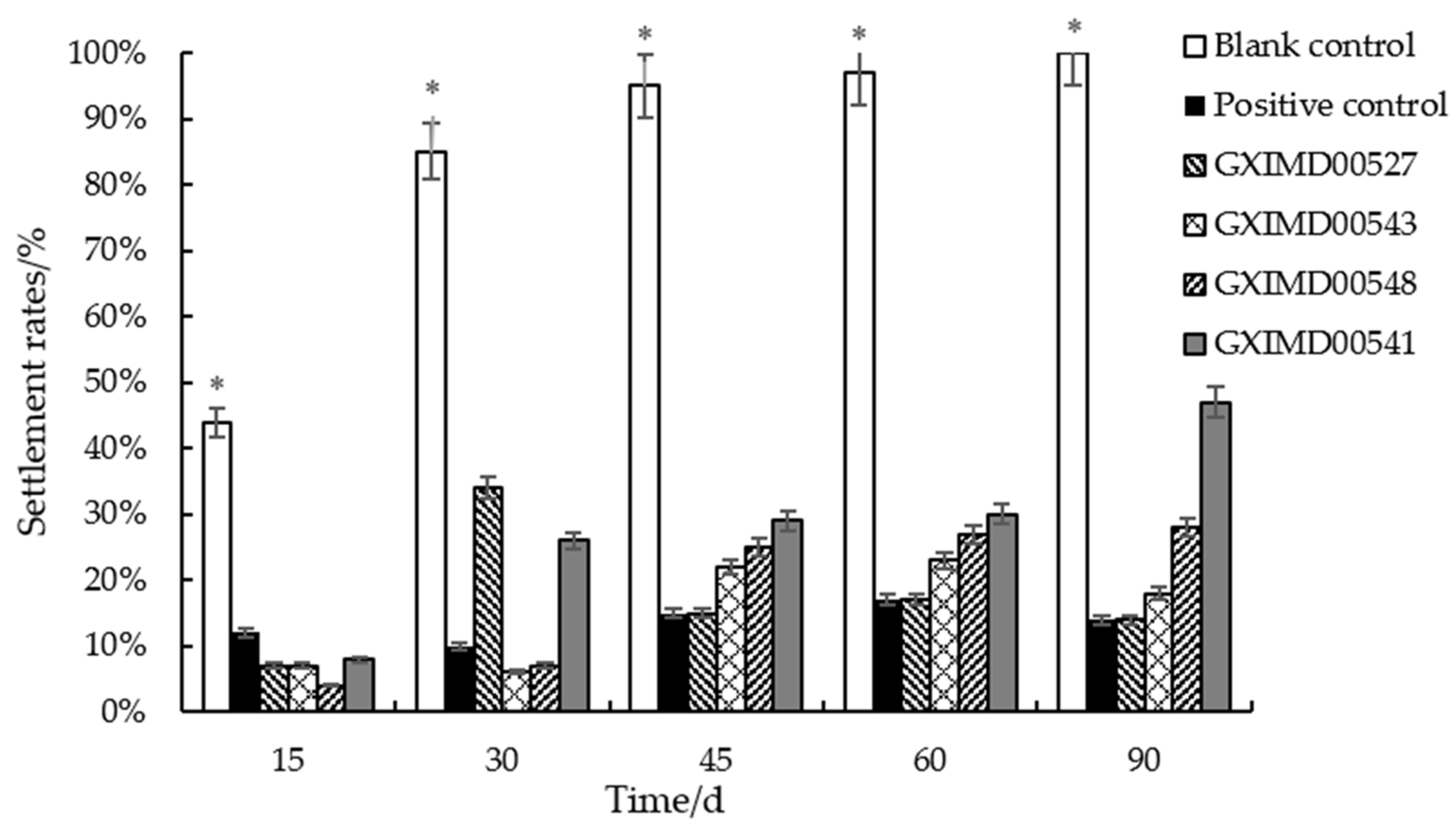
The results of the 90-day marine field trial are summarized in Table 4 and illustrated in Figure 3 and Table S2. The blank control panels (priming paint only) were rapidly colonized, showing the first signs of bryozoans and tube worms within 15 days and reaching 100% fouling coverage by day 90. In contrast, the positive control panels (cuprous oxide coating) showed significantly reduced fouling, with a final coverage of 14% at 90 days. The lipid fractions demonstrated varying degrees of long-term efficacy. The coatings containing GXIMD00527 and GXIMD00547 lipids exhibited excellent and sustained antifouling performance, with fouling coverage remaining below 25% throughout the entire trial period, ultimately reaching 14% and 18% at day 90, respectively. This performance was comparable to the cuprous oxide positive control. The lipid fractions GXIMD00548 and GXIMD00541 also showed significant activity, with final fouling coverages of 28% and 47%, respectively, which were substantially lower than the blank control.

3.4. Chemical Composition of Selected Lipids Fractions

To identify the compounds responsible for the observed antifouling activity, the three most promising lipid fractions (GXIMD00527, GXIMD00548, and GXIMD00543) were subjected to GC–MS analysis. The relative contents of the identified lipids are listed in Table 5. The lipid fraction from GXIMD00527 was complex, containing 14 identified compounds accounting for 99.46% of the total volatile content. It was dominated by dodecyl-9-alkynyl chloroacetate (34.80%), palmitic acid (28.35%), and cis-13-octadecenoic acid (15.11%) (Figure 4). In contrast, GXIMD00548 contained seven identified compounds (95.56% of total volatiles), with linoleic acid being overwhelmingly predominant (67.48%), followed by palmitic acid (19.59%). The GXIMD00543 fraction contained 15 identified compounds (99.60% of total volatiles), with methyl 11,14-octadecadienoate (28.17%), methyl palmitate (19.35%), and dodecyl-9-alkynyl chloroacetate (17.19%) as the major constituents. Notably, palmitic acid and methyl palmitate were present in all three bioactive fractions. Dodecyl-9-alkynyl chloroacetate was a major component in both GXIMD00527 and GXIMD00543.

4. Discussion

The search for sustainable and eco-friendly antifouling solutions has intensified following the global ban on TBT-based coatings. Marine fungi, with their vast and largely untapped metabolic diversity, represent a promising source of novel bioactive compounds [12,21]. In this study, we demonstrated that lipid fractions from marine fungi isolated from the Beibu Gulf possess significant and multi-faceted antifouling activity.
The antibacterial screening revealed that 80% of the tested lipid fractions were active against at least one of the four common marine fouling bacteria, indicating a broad-spectrum potential for preventing the initial stages of biofilm formation, which is critical for the subsequent recruitment of macrofoulers [3]. The lipid from Curvularia lunata (GXIMD00545) was particularly notable for its activity against all four test strains.
More impressively, in the anti-settlement assay against barnacle cyprids, several lipid fractions exhibited remarkable potency and selectivity. The EC50 values for GXIMD00527 (0.23 μg/mL), GXIMD00548 (0.21 μg/mL), and GXIMD00519 (0.59 μg/mL) were lower than that of the commercial standard SeaNine-211 (1.95 μg/mL), highlighting their exceptional efficacy. Furthermore, the lack of observed toxicity (LC50 > 50 μg/mL) at the effective concentrations is a crucial advantage for environmentally benign antifouling agents, as it suggests a non-biocidal mode of action, possibly through the inhibition of larval settlement signaling pathways or temporary sensory manipulation [22]. SeaNine 211, as one of the most widely used small-molecule antifoulants, is generally considered to have a favorable safety profile. However, compared to the lipid fractions mentioned above, it has a relatively narrow LC50 range (33.93 μg/mL). Literature reports indicate that SeaNine 211 can exert long-term effects on marine phytoplankton communities, exhibit immunotoxicity to the hemocytes of edible bivalve mollusks, and potentially cause adverse effects on sea urchins, suggesting a potential risk of global environmental pollution [23,24].
The long-term marine field trial provided the most compelling evidence for the practical application of these lipid fractions. The sustained efficacy of GXIMD00527 and GXIMD00547 over 90 days, with fouling coverage remaining comparable to the cuprous oxide control, underscores their potential as active ingredients in coatings. The fluctuating settlement rate observed for GXIMD00527 (e.g., 34% at 30 days dropping to 15% at 45 days) could be related to dynamic environmental factors, leaching rates, or the succession of fouling communities, which is a common phenomenon in field tests [20].
The GC–MS analysis of the most active fractions (GXIMD00527, GXIMD00548, GXIMD00543) aimed to identify the key metabolites responsible for the observed activities. The consistent presence of palmitic acid and methyl palmitate in all three fractions is significant. Previous studies have reported the antifouling properties of fatty acids. For instance, Gao et al. [25] identified palmitic acid, myristic acid, and octadecanoic acid as the active principles in antifouling bacteria. Fatty acids can disrupt fouling bacterial quorum sensing and interfere with the settlement and metamorphosis of invertebrate larvae [16,26]. The high concentration of linoleic acid in GXIMD00548 (67.48%) suggests it may be a key active component in this specific fraction, contributing to its strong anti-settlement activity. Compound dodecyl-9-alkynyl chloroacetate was a major component in both GXIMD00527 (34.80%) and GXIMD00543 (17.19%). This compound, containing both an alkyne and a chloroacetate functional group, is a relatively unusual natural product and may contribute significantly to the potent and broad-spectrum activity, especially of GXIMD00527. Its mode of action warrants further investigation. The variation in composition among the active fractions suggests that antifouling efficacy may not be attributable to a single compound but could result from synergistic interactions within the lipid mixture [27]. This synergy could potentially broaden the spectrum of activity and delay the development of resistance in fouling organisms.
While natural products offer great promise, challenges such as limited natural abundance, compound stability, and cost-effective production remain [28]. The high relative content of the identified active compounds (e.g., palmitic acid, dodecyl-9-alkynyl chloroacetate) in these fungal lipids is advantageous. Future work should focus on the sustainable production of these compounds, possibly through optimized fermentation of the fungal strains or synthesis of the most active analogues, to ensure a viable supply for coating formulations.

5. Conclusions

This study provides comprehensive evidence for the antifouling potential of lipid fractions derived from marine fungi of the Beibu Gulf, China. Through a multi-tiered evaluation strategy, we identified several lipid fractions, particularly from strains GXIMD00527, GXIMD00547, and GXIMD00548, that exhibit strong activity against both microfouling (bacteria) and macrofouling (barnacle settlement), while maintaining efficacy in long-term field trials. GC–MS analysis indicated that fatty acids such as palmitic acid and their esters, along with unusual compounds like dodecyl-9-alkynyl chloroacetate, are likely key contributors to this activity.
These findings not only validate the role of marine fungi as a valuable resource for green antifouling technologies but also identify specific candidate compounds and producing strains for further development. Future research will focus on elucidating the precise molecular mechanisms of action of these lipid components, optimizing their production, and incorporating them into practical, durable, and environmentally friendly coating systems.

Supplementary Materials

The following supporting information can be downloaded at: https://www.mdpi.com/article/10.3390/metabo15110721/s1. Table S1: The internal transcribed spacers (ITS) sequence of fungal strains; Table S2: Biofouling settlement on PVC panels of selected lipid fractions during the 90-day marine field trial.

Author Contributions

Conceptualization, Y.L. and X.X.; methodology, X.L. and Z.S.; formal analysis and investigation, M.Q., W.J., H.H. and L.L.; Writing—original draft preparation, M.Q.; Writing—review and editing, X.X.; Supervision, C.G. and X.X.; Funding acquisition, X.X. All authors have read and agreed to the published version of the manuscript.

Funding

This research was funded by National Natural Science Foundation of China (42066006), Guangxi Natural Science Foundation (2024GXNSFAA010403), High-Level Talent Training Project Foundation of Guangxi University of Chinese Medicine (202407, 2022C011), and Guangxi First-class Discipline: Chinese Materia Medica (Scientific Research of Guangxi Education Department [2022] No. 1).

Institutional Review Board Statement

Not applicable.

Informed Consent Statement

Not applicable.

Data Availability Statement

The original data presented in the study are included in the article/Supplementary Materials. Further inquiries can be directed to the corresponding author.

Conflicts of Interest

The authors declare no conflicts of interest.

References

  1. Chen, C.; Huang, S.Y.; Wang, S.K.; Jiang, B.; Li, H.F.; Wang, K.L. Antifouling natural products from marine fungi. Mycosystema 2021, 40, 1241–1258. [Google Scholar]
  2. Yan, T.; Li, Z.F.; Hu, L.F.; Li, X.; Cao, W.H.; Luo, W.J.; Cheng, Z.Q. A review on the balanomorph barnacles in the coastal waters of China. Acta Ecol. Sin. 2012, 32, 5230–5241. [Google Scholar] [CrossRef]
  3. Liu, X.B.; Zou, L.; Li, B.Q.; Martino, P.D.; Rutschof, D.; Yang, J.L.; Maki, J.; Liu, W.; Gu, J.D. Chemical signaling in biofilm-mediated biofouling. Nat. Chem. Biol. 2024, 20, 1406–1419. [Google Scholar] [CrossRef]
  4. Schultz, M.P.; Bendick, J.A.; Holm, E.R.; Hertel, W.M. Economic impact of biofouling on a naval surface ship. Biofouling 2011, 27, 87–98. [Google Scholar] [CrossRef]
  5. Demirel, Y.K.; Uzun, D.; Zhang, Y.; Fang, H.C.; Day, A.H.; Turan, O. Effect of barnacle fouling on ship resistance and powering. Biofouling 2017, 33, 819–834. [Google Scholar] [CrossRef] [PubMed]
  6. He, X.Y.; Bai, X.Q.; Yuan, C.Q.; Ren, K. Research progress of nanomaterial-based coatings for marine antifouling applications. Chin. Surf. Eng. 2023, 36, 37–51. [Google Scholar]
  7. Fernandes, J.A.; Santos, L.; Vance, T.; Fileman, T.W.; Smith, D.; Bishop, J.D.D.; Viard, F.; Queirós, A.M.; Merino, G.; Buisman, E.; et al. Costs and benefits to European shipping of ballast-water and hull-fouling treatment: Impacts of native and non-indigenous species. Mar. Policy 2016, 64, 148–155. [Google Scholar] [CrossRef]
  8. Szeto, W.; Leung, M.K.H.; Leung, D.Y.C. Recent developments of titanium dioxide materials for aquatic antifouling application. J. Mar. Sci. Technol. 2021, 26, 301–321. [Google Scholar] [CrossRef]
  9. Jin, H.C.; Tian, L.M.; Bing, W.; Zhao, J.; Ren, L. Bioinspired marine antifouling coatings: Status, prospects, and future. Prog. Mater. Sci. 2022, 124, 100889. [Google Scholar] [CrossRef]
  10. Miftah, J.A.; Hanif, N.; Tan, L.T.; Muljani, S.; Mubarik, N.R. The next generation of integrated eco-friendly anti-biofouling and anti-biocorrosion marine natural products. Chem. Biodivers. 2025, e02263. [Google Scholar] [CrossRef]
  11. Varrella, S.; Barone, G.; Corinaldesi, C.; Giorgetti, A.; Nomaki, H.; Nunoura, T.; Rastelli, E.; Tangherlini, M.; Danovaro, R.; Dell’Anno, A. Fungal abundance and diversity in the Mariana Trench, the deepest ecosystem on earth. J. Fungi 2024, 16, 73. [Google Scholar] [CrossRef]
  12. Ghattavi, S.; Homaei, A.; Fernandes, P. Marine natural products for biofouling elimination in marine environments. Biocatal. Agric. Biotechnol. 2024, 61, 103385. [Google Scholar] [CrossRef]
  13. Zhang, X.Y.; Xu, X.Y.; Peng, J.; Ma, C.F.; Nong, X.H.; Bao, J.; Zhang, G.Z.; Qi, S.H. Antifouling potentials of eight deep-sea-derived fungi from the South China Sea. J. Ind. Microbiol. Biotechnol. 2014, 41, 741–748. [Google Scholar] [CrossRef]
  14. Liu, L.L.; Wu, C.H.; Qian, P.Y. Marine natural products as antifouling molecules—A mini-review (2014–2020). Biofouling 2020, 36, 1210–1226. [Google Scholar] [CrossRef] [PubMed]
  15. Morgan, R.N.; Ali, A.A.; Alshahrani, M.Y.; Aboshanab, K.M. New insights on biological activities, chemical compositions, and classifications of marine actinomycetes antifouling agents. Microorganisms 2023, 11, 2444. [Google Scholar] [CrossRef]
  16. Bhattarai, H.D.; Ganti, V.S.; Paudel, B.; Lee, Y.; Lee, H.K.; Hong, Y.; Shin, H.W. Isolation of antifouling compounds from the marine bacterium, Shewanella oneidensis SCH0402. World J. Microbiol. Biotechnol. 2007, 23, 243–249. [Google Scholar] [CrossRef]
  17. Escobar, A.; Pérez, M.; Sathicq, A.; García, M.; Paola, A.; Romanelli, G.; Blustein, G. Alkyl 2-furoates obtained by green chemistry procedures as suitable new antifoulants for marine protective coatings. J. Coat. Technol. Res. 2019, 16, 159–166. [Google Scholar] [CrossRef]
  18. Chu, C.I.; Ouyang, D.J.; Chen, R.K.; Chen, Q.V.; Liang, H.Y.; Li, Q.S.; Jin, L. Study on the antibacterial activity of the extract of Melscope pielcifolia. Shandong Chem. Ind. 2024, 53, 30–32, 36. [Google Scholar]
  19. Takamura, H.; Yorisue, T.; Tanaka, K.; Kadota, I. Antifouling activity of xylemin, its structural analogs, and related polyamines. Chem. Biodivers. 2025, 22, e202403213. [Google Scholar] [CrossRef] [PubMed]
  20. Yan, T.; Han, S.S.; Wang, J.J.; Lin, H.S.; Cao, W.H. An overview of fouling ascidians. Acta Ecol. Sin. 2017, 37, 6647–6655. [Google Scholar] [CrossRef]
  21. Qiu, Q.; Gu, Y.; Ren, Y.; Ding, H.; Hu, C.; Wu, D.; Mou, J.; Wu, Z.; Dai, D. R Research progress on eco-friendly natural antifouling agents and their antifouling mechanisms. Chem. Eng. J. 2024, 495, 153638. [Google Scholar] [CrossRef]
  22. Dafforn, K.A.; Lewis, J.A.; Johnston, E.L. Antifouling strategies: History and regulation, ecological impacts and mitigation. Mar. Pollut. Bull. 2011, 62, 453–465. [Google Scholar] [CrossRef]
  23. Chen, L.; Lam, J.C.W. SeaNine 211 as antifouling biocide: A coastal pollutant of emerging concern. J. Environ. Sci. 2017, 61, 68–79. [Google Scholar] [CrossRef]
  24. Konstantinou, I.K.; Albanis, T.A. Worldwide occurrence and effects of antifouling paint booster biocides in the aquatic environment: A review. Environ. Int. 2004, 30, 235–248. [Google Scholar] [CrossRef]
  25. Gao, M.; Wang, K.; Su, R.G.; Li, X.Z.; Lu, W. Antifouling potential of bacteria isolated from a marine biofilm. J. Ocean Univ. China 2014, 13, 799–804. [Google Scholar] [CrossRef]
  26. Martín-Rodríguez, A.J.; Reyes, F.; Martín, J.; Pérez-Yépez, J.; León-Barrios, M.; Couttolenc, A.; Espinoza, C.; Trigos, A.; Martín, V.S.; Norte, M.; et al. Inhibition of bacterial quorum sensing by extracts from aquatic fungi: First report from marine endophytes. Mar. Drugs 2014, 12, 5503–5526. [Google Scholar] [CrossRef] [PubMed]
  27. Chen, L.; Qian, P.Y. Review on molecular mechanisms of antifouling compounds: An update since 2012. Mar. Drugs 2017, 15, 264. [Google Scholar] [CrossRef]
  28. Hao, R.; Li, T.; Zhou, X.; Guo, W.; Liu, J.; Guo, L.; Wang, Y. Strategies and challenges in marine antifouling coatings: Current innovations and future outlook. J. Coat. Technol. Res. 2025. [Google Scholar] [CrossRef]
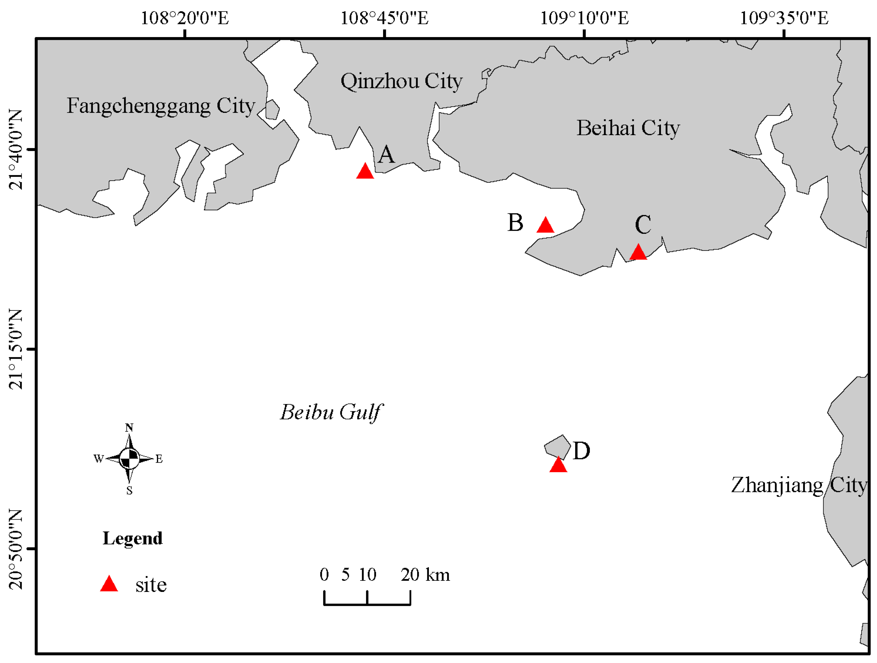
Figure 1. Map of Beibu gulf and the location of the sampling sites (A: Qinzhou seawater; B: Beihai seawater; C: Beihai Zhulin salt field; D: Weizhou Island).
Metabolites 15 00721 g001
Figure 2. The colony morphology images of 10 strains marine fungi.
Metabolites 15 00721 g002
Figure 3. Biofouling settlement rates on PVC panels coated with blank control, positive control, and selected lipid fractions (GXIMD00527, GXIMD00543, GXIMD00548, GXIMD00541) over a 90-day marine field trial. * showed that there was a significant difference between the experimental group and the other groups (p < 0.05).
Metabolites 15 00721 g003
Figure 4. The main chemical structures of lipids (relative contents more than 10%).
Metabolites 15 00721 g004
Table 1. The origin and species of the ten marine fungal strains.
No.Strain NumberSpeciesSource
1GXIMD00527Fusarium incarnatumBeihai Zhulin salt field
2GXIMD00543Aspergillus carneusSponge, Weizhou Island, Beihai
3GXIMD00545Curvularia lunataBeihai seawater
4GXIMD00533Cladosporium cladosporioidesBeihai Zhulin salt field
5GXIMD00547Trichoderma brevicompactumQinzhou seawater
6GXIMD00548Aspergillus iizukaeBeihai Zhulin salt field
7GXIMD00519Aspergillus carneusCoral, Weizhou Island, Beihai
8GXIMD00544Aspergillus fumigatusBeihai Zhulin salt field
9GXIMD00541Fusarium chlamydosporumBeihai Zhulin salt field
10GXIMD00502Aspergillus nigerCoral, Weizhou Island, Beihai
Table 2. Antibacterial activity of ten marine fungal lipid fractions (inhibition zone diameter in mm) a.
Fungal Lipid FractionStrain NumberVRVPMJAM
1GXIMD005198.19 ± 0.75b6.90 ± 0.54
2GXIMD005417.00 ± 0.22
3GXIMD005458.08 ± 0.377.21 ± 0.117.15 ± 0.337.99 ± 0.30
4GXIMD005026.47 ± 0.23
5GXIMD00547
6GXIMD005437.58 ± 0.717.73 ± 0.216.66 ± 0.38
7GXIMD005447.62 ± 0.22
8GXIMD005276.84 ± 0.02
9GXIMD005487.24 ± 0.126.34 ± 0.06
10GXIMD00533
Positive controlPenicillin18.16 ± 0.1112.45 ± 0.2217.18 ± 0.049.47 ± 0.07
Chloramphenicol14.88 ± 0.0516.23 ± 0.14
a Values represent mean ± standard deviation (n = 3). b — indicates no observable inhibition zone.
Table 3. Anti-settlement activity (EC50) and toxicity (LC50) of marine fungal lipid fractions against barnacle cyprids.
Strain NumberEC50 (μg/mL) aLC50 (μg/mL) a
GXIMD005435.50 ± 0.25>50
GXIMD005411.81 ± 0.23>50
GXIMD0053313.92 ± 0.41>50
GXIMD005270.23 ± 0.13>50
GXIMD005480.21 ± 0.13>50
GXIMD005190.59 ± 0.2750
GXIMD005473.89 ± 0.32>50
seaNine-2111.95 ± 0.0533.93 ± 2.06
a Values represent mean ± standard deviation (n = 3), calculated by probit analysis.
Table 4. Biofouling settlement rates (%) on PVC panels coated with selected lipid fractions during the 90-day marine field trial.
Sample NumberSettlement Rates (%)
15 d30 d45 d60 d90 d
GXIMD0054376222318
GXIMD00541826293047
GXIMD005332748676849
GXIMD00527743151720
GXIMD0054847252728
GXIMD00519915231972
GXIMD005471017556057
Positive control1210151714
Blank control44859597100
Table 5. Chemical composition and relative content (%) of volatile components in selected lipid from strains GXIMD00527, GXIMD00548, and GXIMD00543.
CompoundMolecular FormulaRelative Content (%)
GXIMD00527GXIMD00548GXIMD00543
12,4-Di-tert-butylphenolC14H22Oa1.470.31
2Methyl palmitateC17H34O22.732.3319.35
3Palmitic acidC16H32O228.3519.597.9
4Methyl 9,10-octadecadienoateC19H34O22.65
5Methyl 9-octadecenoateC19H36O21.381.77
6Methyl 8-methyl nonanoateC11H22O21.27
7Linoleic acidC18H32O267.48
82,5-Di-tert-butylphenolC14H22O1.12
9Ethyl palmitateC18H36O20.6
10Methyl linoleateC19H34O21.7
11Methyl stearateC19H38O21.433.67
12Dodecyl-9-alkynyl chloroacetateC14H23ClO234.817.19
13cis-13-Octadecenoic acidC18H34O215.11
14Stearic acidC18H36O28.960.86
15Butyl palmitateC20H40O21.50.72
162-Chloroethyl linoleateC20H35ClO20.72
172,2,2-TrifluoroethanolC20H35F3O20.52
18Butyl octadecanoateC22H44O20.54
19Oleic acidC18H34O23.66
20Methyl 12-methyltridecanoateC15H30O20.76
21Methyl pentadecanoateC16H32O20.31
22Methyl 11,14-octadetadienoateC19H34O228.17
23(E)-9-Octadecenoic acid methyl esterC19H36O214.5
24PhenazocineC22H27NO1.02
25Ethyl linoleateC20H36O20.77
262-Hydroxy-1-(hydroxymethyl)ethyl ester cetanoateC19H38O40.41
Total identified 99.4695.5699.60
a —: Not detected.
Disclaimer/Publisher’s Note: The statements, opinions and data contained in all publications are solely those of the individual author(s) and contributor(s) and not of MDPI and/or the editor(s). MDPI and/or the editor(s) disclaim responsibility for any injury to people or property resulting from any ideas, methods, instructions or products referred to in the content.