Article

Research on Movement Intention Recognition Based on CNN-LSTM

1
School of Mechanical Engineering, Yanshan University, Qinhuangdao 066000, China
2
College of Electronic Information and Automation, Tianjin University of Science and Technology, Tianjin 300457, China
3
Erzhong (Deyang) Heavy Equipment Co., Ltd., Deyang 618000, China
*
Author to whom correspondence should be addressed.
Electronics 2026, 15(4), 797; https://doi.org/10.3390/electronics15040797
Submission received: 8 January 2026 / Revised: 6 February 2026 / Accepted: 9 February 2026 / Published: 12 February 2026

Abstract

Existing methods for recognizing motion intent in lower limb rehabilitation robots focus on spatial feature extraction while neglecting movement continuity, thus failing to extract temporal features. This paper proposes a movement intention recognition model based on a CNN-LSTM parallel dual-stream spatio-temporal neural network, taking surface electromyography (sEMG) signals as the core data. This model concurrently extracts temporal and spatial features from sEMG signals, integrating dual-dimensional information to comprehensively explore deep signal characteristics. By overcoming the limitations of traditional single-feature extraction, it significantly enhances recognition accuracy. Experimental results from movement intention recognition studies involving multiple subjects demonstrate an average recognition accuracy of 97%, providing reliable technical support for precise intent recognition and human–robot collaborative control in lower limb rehabilitation robots.

1. Introduction

The accelerating global trend of ageing has led to a surge in rehabilitation demand among patients with lower limb motor dysfunction [1]. Traditional rehabilitation training relies on passive interventions dominated by therapists’ experience, presenting issues such as high labour costs and insufficient personalisation, making it difficult to meet patients’ urgent need for precise, active rehabilitation [2]. In contrast, rehabilitation training assisted by lower limb rehabilitation robots provides objective data feedback to rehabilitation physicians, offering a reference basis for formulating and adjusting rehabilitation programmes [3,4,5,6]. This approach can meet the personalised rehabilitation needs of patients with lower limb dysfunction, thereby attracting widespread attention [7]. Although commercial lower limb rehabilitation robots such as HAL [8] and Lokomat [9] are available on the market, most products still rely on passive control, exhibiting significant limitations in recognising movement intent [10].
Human movement intention recognition, as a core technology in fields such as human–computer interaction, intelligent monitoring, rehabilitation medicine, and virtual reality, enables the accurate prediction or identification of an individual’s movement intentions through the analysis of human motion data. This provides a basis for interaction within intelligent systems, facilitating more natural and efficient human–machine collaboration [11]. Surface Electromyography (sEMG) signals, as direct bioelectrical representations of muscular activity, contain rich movement intention information. They offer millisecond-level predictive advantages, capturing movement intentions 30–150 ms before limb actions occur [12,13,14]. Human movement intent recognition extends beyond rehabilitation medicine, finding extensive application in military, industrial manufacturing, and aerospace domains [15]. Nevertheless, technical bottlenecks and the complexity of real-world scenarios remain critical challenges requiring resolution. Optimising human–machine interaction robustness and scenario adaptability through deep learning constitutes the core challenge in advancing this technology’s practical implementation [16]. Su et al. [17] fed the first 45 frames of IMU data from the swing phase into a convolutional neural network (CNN) to identify 13 movement patterns; Sheng et al. [18] extracted the slope of knee joint angle extremes to compensate for statistical feature instability, combining geometric and physical characteristics to classify 13 movement patterns; Kim et al. [19] proposed a unified pattern-intent estimation method based on deep temporal convolutional networks, mapping five distinct movement patterns onto a continuous gradient domain for regression learning and gradient estimation; Tang et al. [20] employed a 250 ms window length and 10 ms sliding increment for data segmentation and feature extraction, training and recognising six distinct movements across datasets of healthy subjects and hip amputees. Xie et al. [21] proposed a bidirectional convolutional gated recurrent neural network with attention mechanisms for gesture recognition, enabling real-time control. An average accuracy of 88.7% was achieved on the Ninapro benchmark database. Reference [22] proposed the SSO-CNN-BiLSTM-HAM deep learning model to investigate the risk mechanism of ligament fatigue failure. It was validated that the deep learning model, capable of simultaneously capturing spatial correlation and temporal dynamics, exhibits superior performance over conventional methods in the analysis of biological and mechanical systems. This provides direct evidence for the rationality of integrating CNN and LSTM for bioelectrical signal decoding.
Anselmino et al. [23] developed an sEMG-based gait initiation and direction prediction algorithm, employing two support vector machines to predict stride initiation and gait direction, respectively, thereby enhancing the control performance and adaptability of lower limb rehabilitation robots. However, limitations exist in terms of model complexity and adaptability to variations in modalities and sensor channel counts. Zhang et al. [24] proposed a deep learning-based multimodal information fusion framework. By organically integrating CNN, recurrent neural networks (RNN), and graph neural networks (GNN), they significantly enhanced the accuracy and generalisation capability of lower limb gait pattern recognition. Nevertheless, issues such as slow response times persist during transitional states involving rapid gait pattern switching. Luo et al. [25] proposed a low-cost, efficient sEMG-based gait sub-phase recognition system. Utilising a combination of Long-Short-Term-Memory (LSTM) and Multilayer Perceptron (MLP) networks, this system achieved an average test recognition accuracy exceeding 90%.
Most existing methods for movement intention recognition adopt single-branch network structures such as single LSTM, CNN, SVM, and MLP, which fail to achieve comprehensive feature characterization of sEMG signals and only consider partial characteristics. This leads to poor model adaptability and low recognition accuracy. Such methods also have limitations in modeling motion continuity and the joint extraction of spatial-temporal features, which severely restricts their clinical applications in rehabilitation robots. Single-branch models that merely focus on either spatial or temporal features alone cannot capture the dynamic temporal dependencies during the continuous activation of muscles. For example, Wang et al. [26] proposed a single CNN model to estimate multi-joint angles of the lower limb using multi-channel sEMG signals. Although the model achieved relatively low root mean square error (RMSE) in the hip and knee joints, it completely neglected the temporal continuity of sEMG signals. For the ankle joint, which exhibits more frequent dynamic variations during movement, the RMSE of the model rose significantly because it failed to track the sequential activation patterns of synergistic muscle groups. Models relying on single-modal feature extraction or simple serial fusion are incapable of adaptively balancing spatial and temporal information. Bian et al. [27] developed a stacked LSTM network to predict gait intention using only kinematic signals. Although this LSTM model outperformed the feedforward MLP, it lacked an effective spatial feature fusion mechanism. When joint angular velocity increased, the prediction accuracy of the model dropped drastically, as it could not combine the spatial correlation of muscle activation with temporal dynamics. In addition, the traditional serial architecture employs simple feature concatenation, which cannot dynamically adjust the weights of spatial and temporal features according to motion characteristics, leading to information loss in the feature transmission process.
To address this issue, this paper proposes a parallel dual-stream spatial-temporal neural network model for movement intention recognition based on CNN-LSTM. This model performs coordinated extraction of spatial features and temporal features from sEMG signals, respectively, effectively excavating deep spatial-temporal correlation information in the signals, and ultimately achieving an average recognition accuracy of over 97% for motor intention. In addition, most existing studies only collect data from healthy subjects (e.g., [21,25]), which is difficult to meet the requirements of clinical application scenarios of rehabilitation robots. In the process of dataset construction, this paper specifically included 1 subject with lower limb dysfunction, significantly improving the clinical relevance between the dataset and the application of rehabilitation robots. Meanwhile, through a standardized data collection process, the variability of sEMG signals caused by individual movement differences is reduced, and the signal quality and consistency are improved. In the dataset division stage, a subject-based stratified division method is adopted, which effectively avoids data distribution bias and ensures the objectivity and credibility of the evaluation of recognition accuracy.
Innovations in this paper:
(1)
Task-Adaptive Parallel Dual-Stream Network Architecture
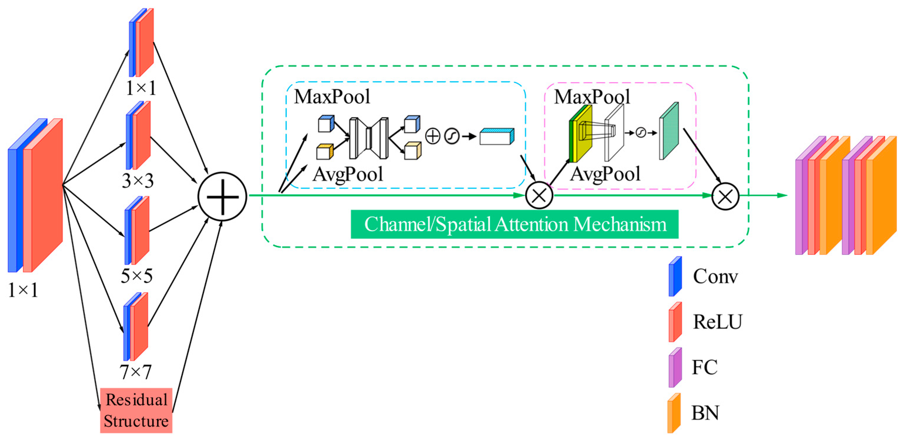
The proposed model innovatively designs two parallel branches with differentiated functions: the multi-scale spatial branch integrates channel/spatial attention mechanism and residual structure, which is specifically used to extract inter-channel correlation features of sEMG signals; the temporal branch combines an LSTM network with 1D convolution, focusing on capturing the long-term sequential dependencies of signals. This design overcomes the limitation that single-branch models cannot simultaneously optimize the extraction of spatial and temporal features.
(2)
Efficient Spatiotemporal Feature Fusion Mechanism
This study designs a dedicated decoding fusion module to integrate dual-stream features, instead of using simple feature concatenation. Based on the characteristic patterns of lower limb motion, this module performs weighted allocation on spatial topological information and temporal dynamic information, thereby enhancing the model’s discriminative capability for similar actions.
(3)
Residual-Attention Hybrid Structure Optimized for sEMG Signals
The residual structure adopts the sequential order of Batch Normalization–Rectified Linear Unit–Convolution; the multi-scale attention mechanism can adaptively emphasize the critical frequency bands and muscle channel features of sEMG signals, which improves feature extraction efficiency while mitigating the vanishing gradient problem.

2. Establishment of a System for sEMG Signal Acquisition and Preprocessing

sEMG constitutes a bioelectric signal generated by muscle contraction in the human body, reflecting the activity state of muscle groups. By analysing the temporal and spatial information embedded within surface electromyographic signals, relevant movement information is decoded to infer an individual’s subjective motor intention. This section focuses on acquiring surface electromyographic signals corresponding to different rehabilitation movements to obtain raw electromyographic signals, followed by preprocessing to enhance signal quality and signal-to-noise ratio. The processed sEMG undergoes rearrangement via permutation algorithms and segmentation of data segments using sliding windows. The segmented sEMG amplitudes are then mapped to generate greyscale images. To improve the generalization ability of the model, this paper adopts time-axis flipping to perform data augmentation operations. On the premise of ensuring signal authenticity, it provides sufficient data diversity for model training. These greyscale images serve as input samples for neural network models, where feature extraction is performed. Feature decoupling enables movement intention recognition, providing a basis for robotic systems to comprehend patient movement intentions and thereby enhance human–machine coordination.

2.1. sEMG Signal Acquisition

2.1.1. sEMG Signal Acquisition System

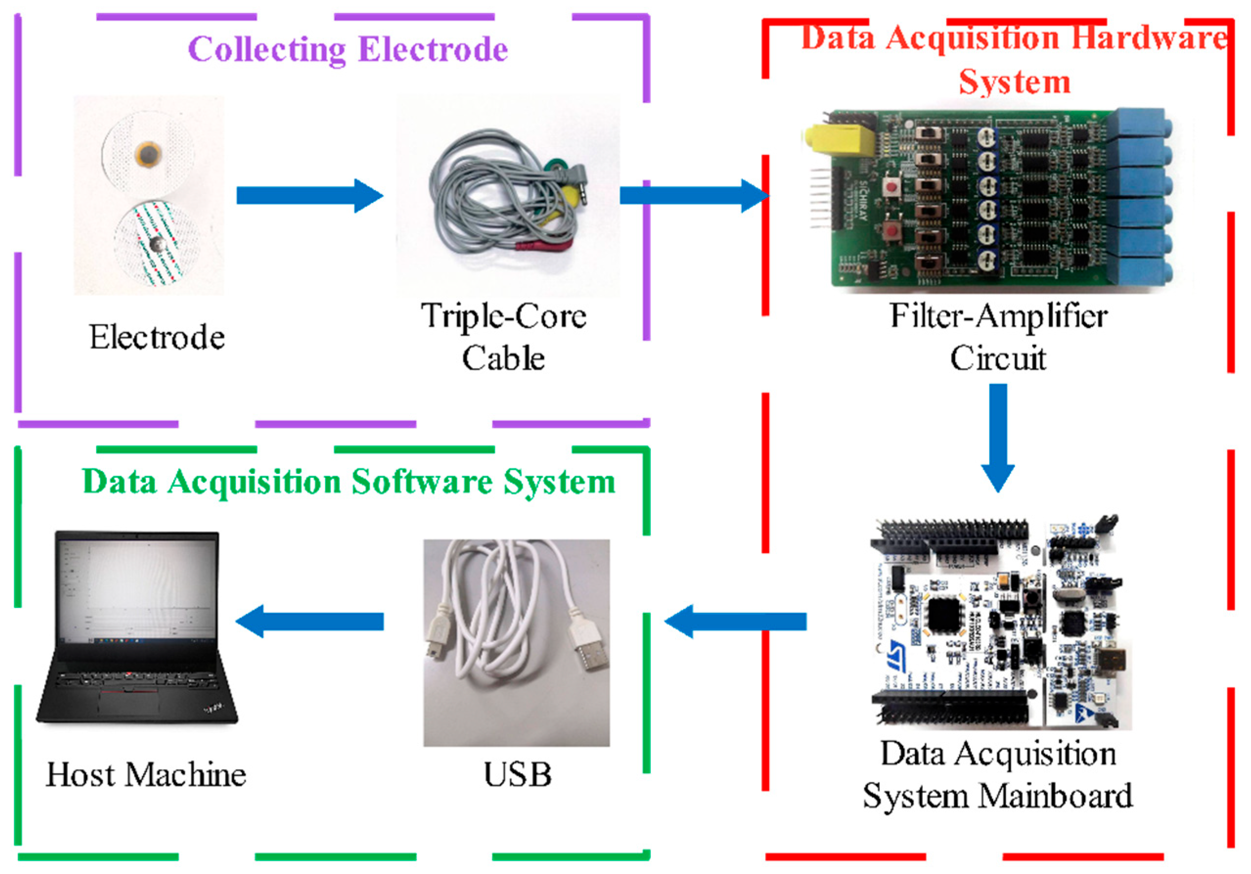
The sEMG signal acquisition and preprocessing workflow is illustrated in Figure 1, primarily comprising acquisition electrodes, hardware acquisition systems, and software acquisition systems. Electromyographic signal acquisition techniques can be categorised into two main approaches based on invasiveness: needle electrode acquisition and surface patch electrode acquisition. Surface electrode acquisition can be further subdivided into dry electrode and wet electrode types based on the characteristics of the contact medium. In contrast, wet electrodes achieve skin coupling via conductive gel media, whose adhesive strength effectively suppresses motion artefacts. Although collected signals may still exhibit issues such as baseline drift and mains interference, mature signal processing techniques—including wavelet transforms and adaptive filtering—can effectively extract signal components within the desired frequency bands.
The reliability of sEMG signal acquisition directly depends on the impedance characteristics of the electrode-skin interface. In this study, wet electrodes were employed to capture bioelectrical signals with the assistance of conductive gel. To optimize the quality of signal acquisition, a standardized skin preprocessing protocol was implemented prior to electrode placement: first, hair in the target acquisition region was removed using medical-grade hair removal tools; the skin surface was then wiped with 75% ethanol solution to remove sweat and grease, and the wiping was performed no less than 3 times. Electrode attachment was conducted only after the volatile components in ethanol had completely evaporated. This procedure can substantially reduce the contact impedance.
The optimal design of a sEMG signal acquisition system should be based on the bioelectric generation mechanism and signal characteristics of sEMG. This system adopts bipolar differential measurement technology, whose inherent common-mode noise rejection characteristic can not only significantly improve the anti-interference ability during data transmission, but also effectively suppress signal distortion caused by electrode displacement, thereby optimizing signal quality and enhancing the signal-to-noise ratio (SNR).
According to the conduction characteristics of bioelectrical signals, there is a significant correlation between the spacing of dual working electrodes and signal fidelity. Adopting a spacing that is an integer multiple of 10 mm can effectively optimize the signal acquisition performance, and a spacing of 20 mm is generally employed to improve the SNR. In terms of the electrode layout strategy, working electrodes shall be placed in the central region of the muscle belly of the target muscle, and the line connecting the two working electrodes must be strictly parallel to the orientation of muscle fibers. This arrangement enables the maximum capture of action potentials generated by muscle contraction. In accordance with the principle for selecting reference points of bioelectrical signals, the reference electrode is positioned in a low myoelectric activity area adjacent to the joint. Crosstalk from cross-muscle signals is reduced by means of physical isolation. The proposed electrode layout scheme can provide high-quality raw data for subsequent signal processing. The precautions for electrode placement are summarized in Table 1.
The six-channel signal acquisition device employed in the experiment comprises a signal conditioning module and a main control unit, integrating signal amplification, envelope extraction, and filtering functions. Five active channels are used to acquire sEMG signals from five preselected muscle groups, with one dedicated reference channel exclusively for noise suppression. The dedicated reference channel does not participate in muscle signal extraction; it only serves as a common-mode reference terminal to improve the system’s Common-Mode Rejection Ratio (CMRR)and suppress ambient noise Its front-end differential amplifier circuit achieves a maximum signal gain of 330 times. The front-end amplification adopts a three-op-amp differential topology based on an instrumentation amplifier, with 20-fold fixed gain from the differential input stage and 1–16.5-fold adjustable gain from the programmable gain amplifier (PGA) for dynamic adaptation to sEMG signals. Key performance parameters are as follows: CMRR ≥ 110 dB at 50 Hz, input impedance ≥ 10 MΩ, and equivalent input noise ≤ 0.8 μVrms in the 20–500 Hz band. For dynamic conditions, the symmetric layout of the differential input stage combined with wet electrode coupling suppresses motion artifacts, while a 5 Hz high-pass filter further eliminates low-frequency interference. The high input impedance minimizes signal attenuation from high electrode-skin impedance, and the input bias current reduces polarization effects, ensuring signal integrity even with prolonged movement or electrode loosening. The conditioned analogue signals undergo analogue-to-digital conversion via the STM32F103 microcontroller’s built-in ADC, then are transmitted through a high-speed serial interface to the host computer for subsequent analysis and processing. The hardware system operates on a single 5 V power supply, utilising the USB interface for both power delivery and data transmission. Based on a programmable microcontroller architecture, it supports software configuration of sampling frequency and communication baud rate. An ADC conversion mechanism triggered by a timer is employed, with a 1000 Hz sampling frequency set to satisfy the Nyquist sampling theorem. An asynchronous serial communication interface operating at a baud rate of 115,200 bps is configured. The ADC quantisation value corresponding to the 3.3 V reference voltage serves as the frame verification flag. This is combined with hardware pin status monitoring to ensure synchronisation and reliability of data transmission timing.
The STM32F103’s integrated 12-bit ADC is configured with an external precision voltage reference to ensure reliable and stable analog-to-digital conversion of weak sEMG signals, with key performance parameters relevant to sEMG acquisition systematically characterised as follows: limited by inherent system noise, the ADC exhibits an ENOB of 9.2 bits—derived from a signal-to-noise and distortion ratio (SINAD) of 57.8 dB—corresponding to a voltage step of ~0.8 mV, which is sufficiently fine to capture sEMG signals within the typical amplitude range of 10–1000 μV without significant quantisation loss; synchronised with a 12 MHz crystal oscillator, the ADC yields a conversion time of 1 μs per channel, resulting in jitter-induced noise of ≤0.05 μVrms—negligible relative to the ADC’s inherent noise and the amplitude of sEMG signals; the external precision reference demonstrates long-term stability of ±0.01% per year and temperature stability of ≤5 ppm/°C, and over the experimental temperature range of 20–25 °C, its voltage drift induces a quantisation error variation of ≤0.03 LSB—an effect sufficiently minor to not compromise signal consistency.
To address conducted interference and ensure electrical safety in clinical and preclinical application scenarios, this acquisition system incorporates targeted hardware design strategies. A USB isolation module is integrated between the USB interface and the main control unit to cut off the direct electrical connection between the host computer and the patient-side circuit, which not only eliminates interference induced by galvanic coupling but also mitigates the risk of electric shock caused by ground potential differences. A ferrite bead filter is integrated into the USB power line to attenuate high-frequency conducted interference from the host computer; a common-mode choke is incorporated into the front-end differential amplifier circuit to further suppress power line-induced interference, thereby ensuring the purity of weak sEMG signals. In addition, over-current protection and an over-voltage clamping circuit are configured in the USB power path to prevent excessive current and voltage from reaching the patient-side circuit.
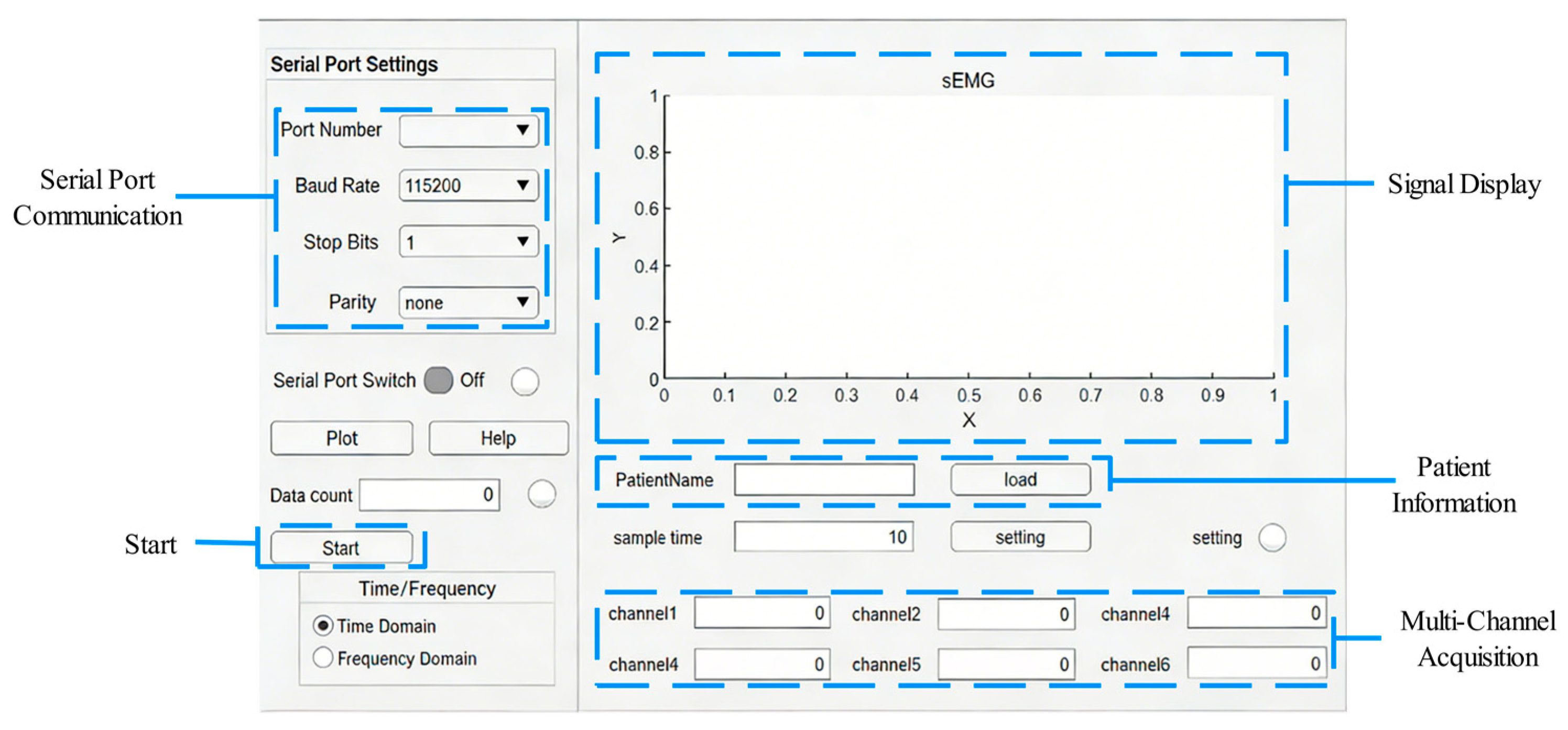
The signal acquisition system’s host computer interface is illustrated in Figure 2. This platform is constructed using the MATLAB (R2022b)App Designer development environment, integrating multi-stream data communication and processing functional modules. Employing a layered architecture design, its core functionalities comprise: a data transmission interface based on serial port communication protocols, signal integrity verification algorithms, and multi-channel data storage and pre-processing units. This enables efficient control of the sEMG hardware acquisition system and optimised data stream management. At the operational control level, the system initiates handshake protocol communication via the GUI interface, effectively preventing abnormal responses from the rehabilitation robots due to patient misoperation. This ensures patients operate only when fully prepared, avoiding erroneous robot responses caused by incorrect input and thereby reducing the risk of unnecessary injury.

2.1.2. Determination of Muscle Group Locations for Acquisition

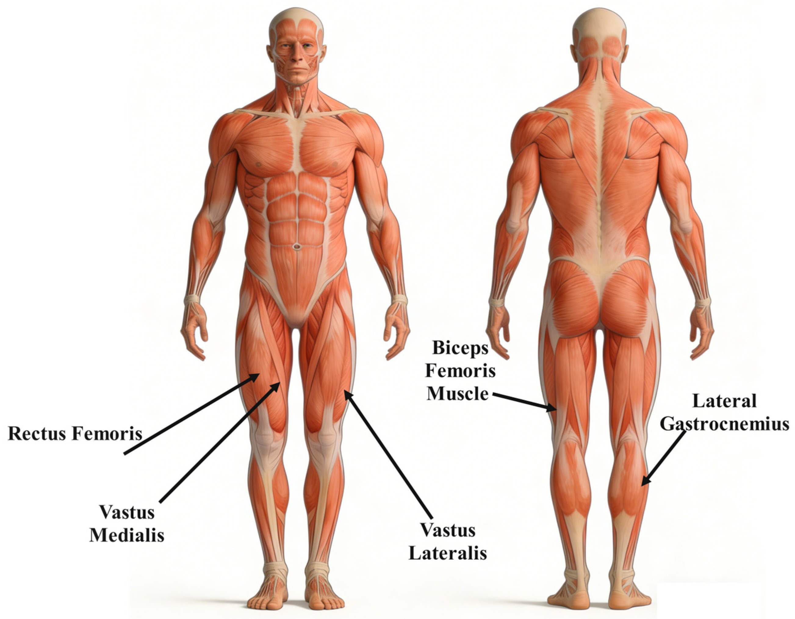
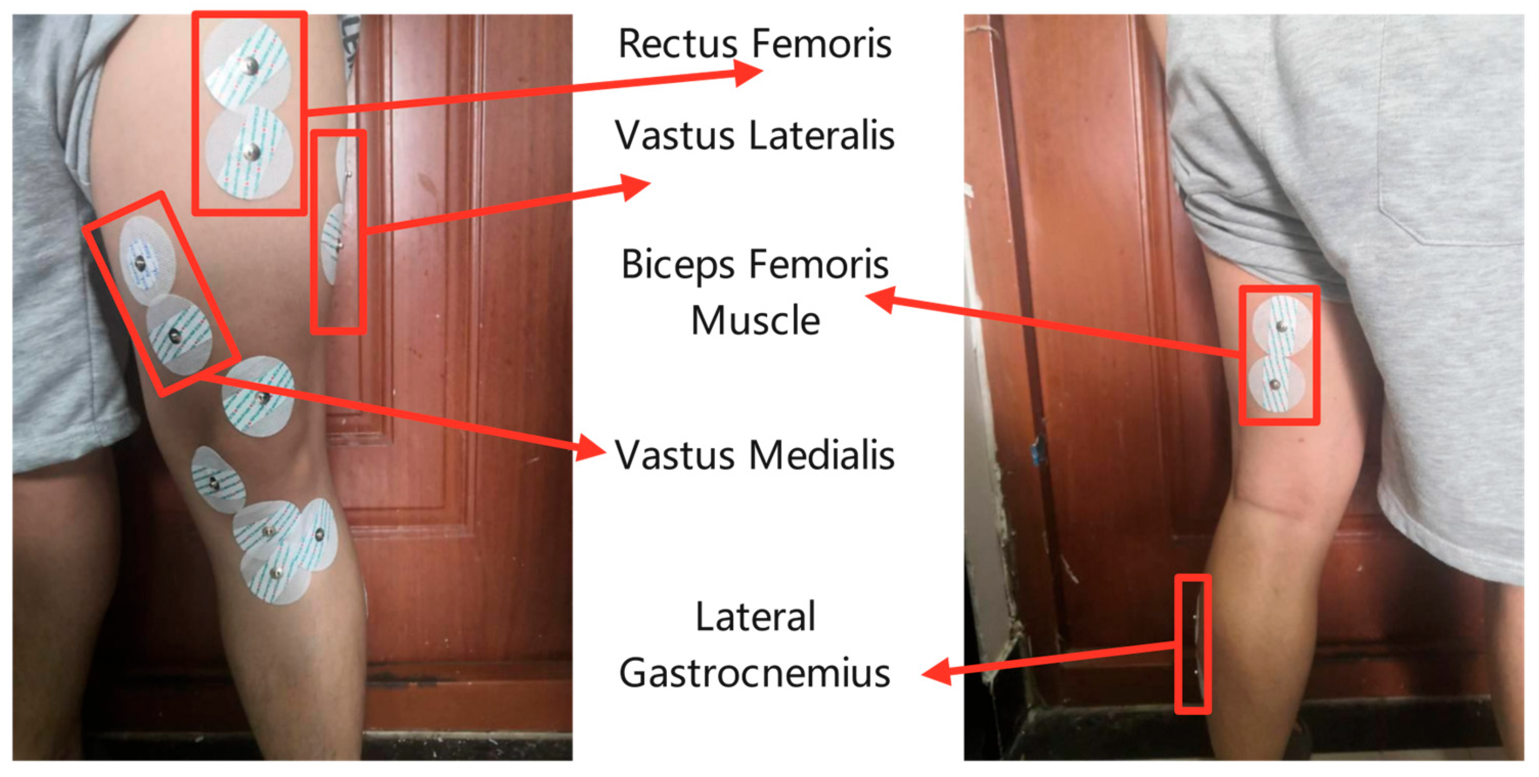
The theory of synergistic action emphasises that the central nervous system’s mechanism for motor control does not function through independent regulation of individual muscles, but rather through holistic coordination using neuromuscular synergistic modules as functional units [28]. This experiment investigates the movement characteristics of the human lower limbs within the sagittal plane, specifically encompassing two active movement patterns—flexion and extension—alongside the resting state, defined as the absence of active electromyographic activity. Preliminary studies validated that muscle group selection requires an optimal balance between signal representativeness and optimisation of system redundancy. Five key muscle groups—the Rectus Femoris, Vastus Medialis, Vastus Lateralis, Biceps Femoris Muscle, and Lateral Gastrocnemius—were pre-selected as sEMG recording sites. The pre-selected muscle groups for electromyographic signal acquisition are illustrated in Figure 3.

2.1.3. sEMG Signal Acquisition Experiment

Prior to the experiment, standardised procedures must be completed: record the subject’s gender, lower limb test side, height, weight, and other basic physiological parameters; explain the experimental process and precautions. When applying electrodes, first wipe the target muscle area with an alcohol swab. Once the alcohol has evaporated, remove hair to reduce contact impedance. Then affix the bipolar Ag/AgCl electrodes to the pre-determined sites, with reference electrodes placed at joints with low muscle activity. See Figure 4 for details.
In this experiment, there are two main rehabilitation movements: lower limb flexion and extension. A metronome was used to standardize the movement speed, ensuring that one flexion-extension cycle was completed every 2 s to avoid signal variability caused by speed differences. Each subject performed 10 consecutive flexion-extension cycles in a single test, with a 60 s rest after each cycle to prevent muscle fatigue. All subjects completed multiple tests in total, and the interval between two consecutive tests was no less than 24 h.
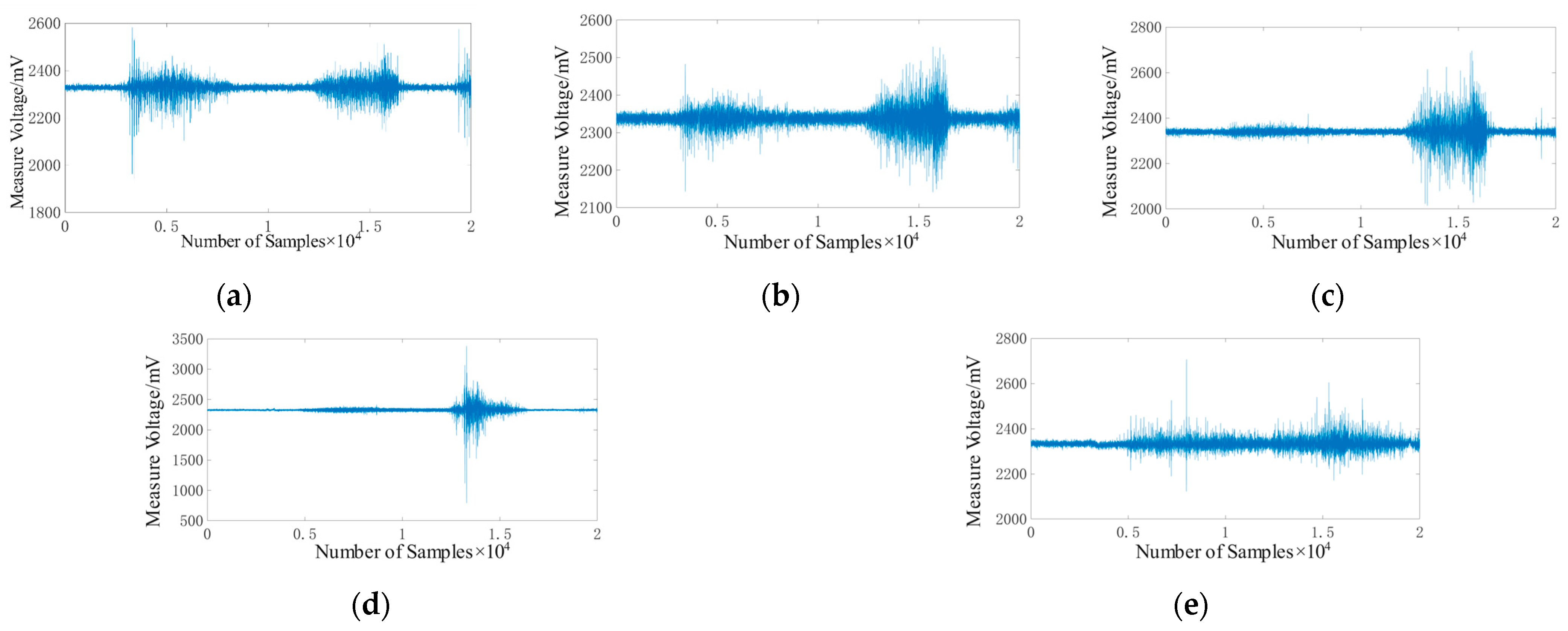
Prior to commencing movements, the patient should lie supine on the rehabilitation robots with lower limb muscles relaxed. After connecting the equipment, launch the host computer software and set the sampling frequency to 1000 Hz and the baud rate to 115,200 bps. Click “Start” to commence electromyographic signal acquisition. To enhance data authenticity, multiple acquisitions should be performed to obtain several sets of valid data, facilitating problem analysis and resolution. Collected electromyographic data should be promptly saved for subsequent signal processing. Figure 5 shows segments of electromyographic signals recorded from five muscle groups during exercise.
The surface electromyographic signal amplitudes for each muscle group exhibit periodic variations, as demonstrated by the aforementioned five sets of sEMG data. A distinct difference is observed between the surface sEMG signal amplitudes during active movements and at rest. Muscle group activity levels exhibit marked differences across various movements, enabling straightforward differentiation between rehabilitation exercises based on EMG signal amplitude. This confirms the appropriate selection of muscle groups, laying the groundwork for establishing an efficient motor intent decoding model.

2.2. Construction of the Pre-Treatment System

2.2.1. Filtering and Noise Reduction Processing of sEMG Signals

Surface electromyography signals are inherently weak and susceptible to interference during acquisition from factors such as inherent noise, mains interference, baseline drift, and crosstalk [29], resulting in a low signal-to-noise ratio. It is necessary to first analyse the characteristics of various noise sources, then apply targeted filtering techniques to reduce noise and enhance signal quality, thereby approximating the true signal.
First, the causes and characteristics of various types of noise are systematically analyzed, and targeted noise reduction strategies are formulated accordingly. There are four common types of noise contaminating sEMG signals, as detailed below:
(1)
Inherent noise: This type of noise originates from the intrinsic noise generated by electronic components in the signal acquisition equipment. It can be reduced by selecting high-performance components, but it cannot be completely eliminated, making inherent noise unavoidable.
(2)
Power-line noise: It mainly arises from the electromagnetic radiation emitted by acquisition equipment connected to the power supply and the host computer. The fundamental frequency of the power grid in China is 50 Hz, while the effective information of sEMG is mainly distributed in 20–500 Hz, resulting in severe aliasing between the interference and the effective frequency band. Characterized by strong periodicity and stable amplitude, power-line noise can be processed using digital filtering techniques such as notch filtering.
(3)
Baseline drift: During sEMG acquisition, baseline drift or deviation from the zero level occurs due to two main factors: changes in internal biopotentials (e.g., those caused by heartbeat and respiration), and electrode displacement induced by muscle contraction, which alters the contact impedance between electrodes and skin. Digital filters can be employed to eliminate the DC component in the signal.
(4)
Crosstalk Phenomenon: It mainly originates from electrocardiogram (ECG) signals and electrical signals of adjacent muscle groups. ECG interference presents a characteristic QRS complex with a frequency band ranging from 0.5 to 100 Hz, whereas cross-muscle group interference possesses similar frequency-domain characteristics to the target signal, thus necessitating the combination of time-frequency analysis and spatial filtering techniques for signal separation.
Following the analysis of potential noises and interferences in sEMG signals, this section conducts filtering and denoising on the aforementioned noises to improve the SNR and enhance signal quality and reliability. Inherent noise, generated by electronic components, cannot be completely eliminated. Three types of noises can be filtered and denoised via algorithms, namely power-line interference, baseline drift and crosstalk. Since filters mainly operate on the frequency domain and considering that subsequent research will employ the frequency information of the signals, the frequency band with the most abundant signal information shall be retained to the greatest extent possible.
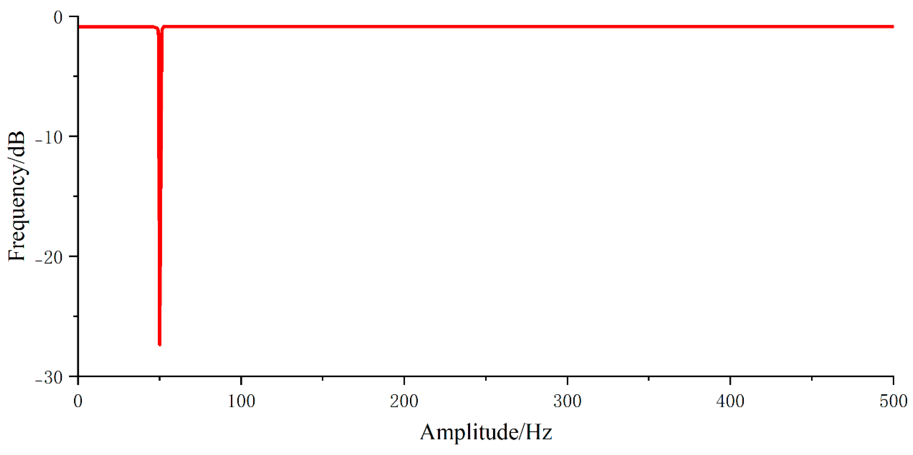
Aiming at the problem of 50 Hz power-line interference in sEMG signals, existing studies commonly adopt notch filters for frequency-domain suppression [30,31,32,33]. A notch filter is a type of band-stop filter with an extremely narrow stopband. Based on the narrow band-stop filtering characteristic, this study preferentially selects the Infinite Impulse Response (IIR)digital filter to construct a power-line notch filter. The IIR notch filter can achieve a steep roll-off at 50 Hz, enabling accurate suppression of power-line interference with low computational complexity, and avoids the influence of temporal feature distortion on the outcomes of motion intention recognition.
Adhering to the principle of minimum order selection within the order-selection criteria, a second-order filter was chosen to reduce computational complexity and enhance filtering efficiency. The amplitude-frequency response of the notch filter is illustrated in Figure 6.
Regarding baseline drift and high-frequency crosstalk issues, a comparative analysis of three approaches—Gaussian filtering [34], Variational Modal Decomposition (VMD) [35], and Butterworth bandpass filtering [36]—revealed certain limitations in the first two: The computational complexity of Gaussian filtering’s convolution operation increases exponentially with window size, while the VMD method requires iterative optimisation of the penalty factor and the preset modal number K.
To conduct a detailed comparison of the advantages and disadvantages of the above three filtering methods, this paper carries out a quantitative comparative study on each of them under the same experimental conditions. The detailed results are presented in Table 2.
As can be seen from the results in Table 2: In terms of SNR, the Butterworth filter achieves the highest average value, which is 2.4 dB and 1.5 dB higher than that of VMD and Gaussian filtering, respectively. This is because the Butterworth filter can precisely match the effective 20–500 Hz frequency band of sEMG signals. In contrast, Gaussian filtering, as a spatial-domain smoothing method, fails to sufficiently suppress frequency-specific noise, and VMD is prone to modal mixing, which results in incomplete noise separation. In terms of model classification accuracy, the signal processing scheme based on the Butterworth filter achieves the highest classification accuracy of 97.75%, which is 1.63 percentage points and 1.00 percentage point higher than that of VMD and Gaussian filtering, respectively. The lower classification accuracy of Gaussian filtering stems from its inherent spatial-domain smoothing effect that blurs the subtle temporal-spatial features of sEMG signals, whereas the flat passband response of the Butterworth filter can completely preserve such detailed information.
Meanwhile, the signals in sEMG that best characterize motion information are primarily concentrated in the 20–500 Hz frequency band. For these reasons, a Butterworth band-pass filter is employed to mitigate baseline drift, and its transfer function is given by
Y ( s ) X ( s ) = b ( 1 ) + b ( 2 ) s 1 + + b ( n + 1 ) s n 1 + a ( 2 ) s 1 + a ( n + 1 ) s n ,
where Y ( s ) is the output signal, X ( s ) is the input signal, and n is the filter order.
Simplifying Equation (1) yields:
Y ( s ) = X ( s ) b ( 1 ) + b ( 2 ) s 1 + + b ( n + 1 ) s n Y ( s ) a ( 2 ) s 1 + a ( n + 1 ) s n
From Equation (2), it follows that the order is a crucial parameter affecting filter performance. Subsequently, the order of the Butterworth filter is determined by the magnitude of the sSNR. A higher SNR value indicates superior noise reduction. The SNR formula is given by Equation (3):
SNR = 10 lg i = 1 N v i 2 i = 1 N v i v 1 2 ,
where v i is an ideal signal and v 1 is the output signal.
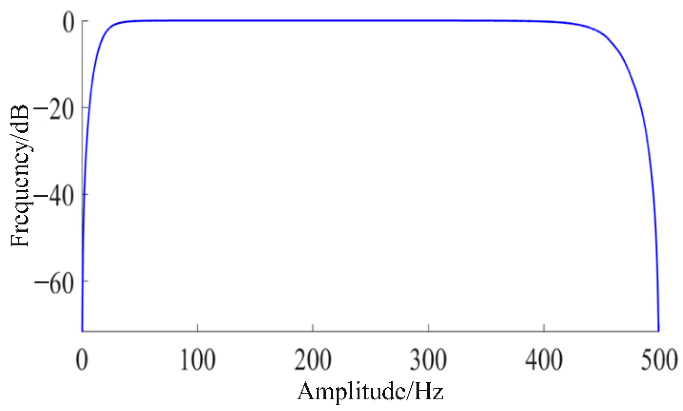
The signal-to-noise ratio is calculated based on the existing surface electromyography signals. A fourth-order Butterworth filter is employed, with its amplitude-frequency response plot shown in Figure 7.
To verify the effectiveness of the noise reduction strategy and the stability of the acquisition chain, we conducted experimental measurements of key performance indicators:
① Noise level: The RMS noise of the acquisition chain (short-circuited input) was measured as 0.32–0.41 μVrms in the 20–500 Hz band, consistent with the inherent noise characteristics of the selected components;
② SNR: After filtering, the average SNR of sEMG signals from ten subjects was 58.7 ± 1.2 dB, indicating effective suppression of mains interference and crosstalk;
③ ADC ENOB: Calculated from the SINAD of a 1 kHz input signal, the ADC’s ENOB was 9.2 bits, meeting the requirements for weak sEMG quantisation;
④ Frequency response: The acquisition chain maintained a flat response (±0.5 dB) in the 20–500 Hz band, with attenuation >20 dB outside this range, confirming effective retention of valid signal components.

2.2.2. Normalisation of sEMG Signals

To improve the accuracy of motor intention classification, standardization of sEMG signal data is a critical step. This study adopts a subject-level Min-Max normalization method, which performs linear transformation on the data based on the maximum and minimum values of the entire sEMG dataset for each individual subject to achieve feature standardization. This approach ensures consistent amplitude scaling across all windows of the same subject, avoids the problem of intra-subject feature inconsistency caused by window-level normalization, and preserves the original distribution of the data during the normalization process.
The calculation formula is as shown in Equation (4):
x = x x min x max x min ,
where x is normalised data, x m i n is the minimum value in the filtered data, x m a x is the maximum value in the filtered data and x is filtered data.

2.2.3. Sliding Window Data Segment Partitioning

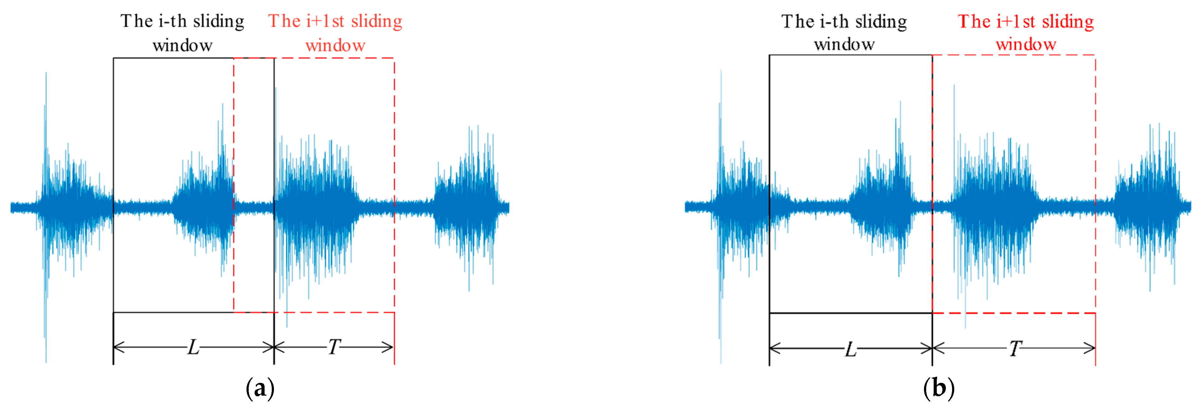
Due to the uncertainty in action duration and the high-frequency variations in acquisition equipment, the entire segment of electromyographic signal data is segmented into several subsets, with each subset mapped to a greyscale image. As illustrated in Figure 8, In overlapping windows, the length of each window shift is less than the window’s full length; in non-overlapping windows, the length of each window shift equals the window’s full length.
The window length L and sliding step size T require balancing real-time performance with feature representation capability (excessively large/small windows or step sizes adversely affect system performance). Considering that action potentials in electromyographic signals precede limb movements, a window length of 200 ms and sliding step size of 50 ms were selected. The segmented data provides high-quality samples for subsequent feature extraction and classification. With the current window length and step size fixed, the end-to-end latency measured on the existing experimental platform is 89 ± 3.2 ms, which is significantly lower than the 100 ms safety threshold for human–robot interaction latency of rehabilitation robots [37]. This ensures no risk of response latency during active training and has no adverse effect on prediction accuracy.
Meanwhile, the LSTM-based temporal branch can preserve historical contextual information of signals spanning up to 200 ms. During movement transitions in patients, it effectively distinguishes transient transition signals from steady-state movement signals, avoiding misclassification caused by short-term signal fluctuations. A sliding step of 50 ms combined with a 200 ms signal window results in an overlap ratio of 25%, which guarantees the continuity of signal sampling during action transitions, fully maintains the continuity of temporal features, and prevents information loss at transition boundaries.
First, the absolute value of the EMG data is calculated, followed by the mean of these absolute values, as shown in Equation (5):
M = 1 N i = 1 N X i ,
where M is the mean value of data within the window, X i is the value of the i-th sampling point within the window and N is the number of sampling points within the window.
Finally, set the starting point threshold TH1 and the ending point threshold TH2, respectively. The determination of the starting point is as shown in Equation (6):
T H 1 < M i T H 2 > M j
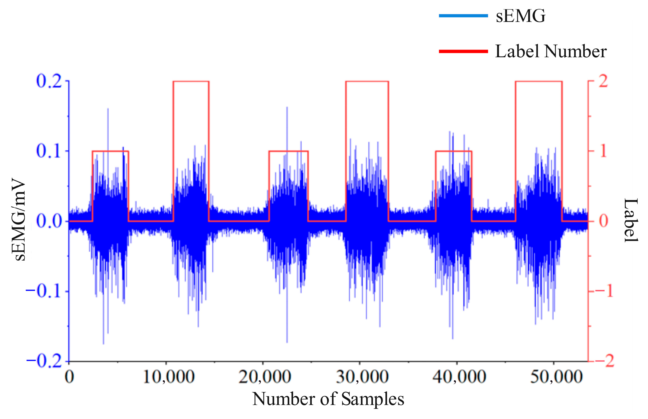
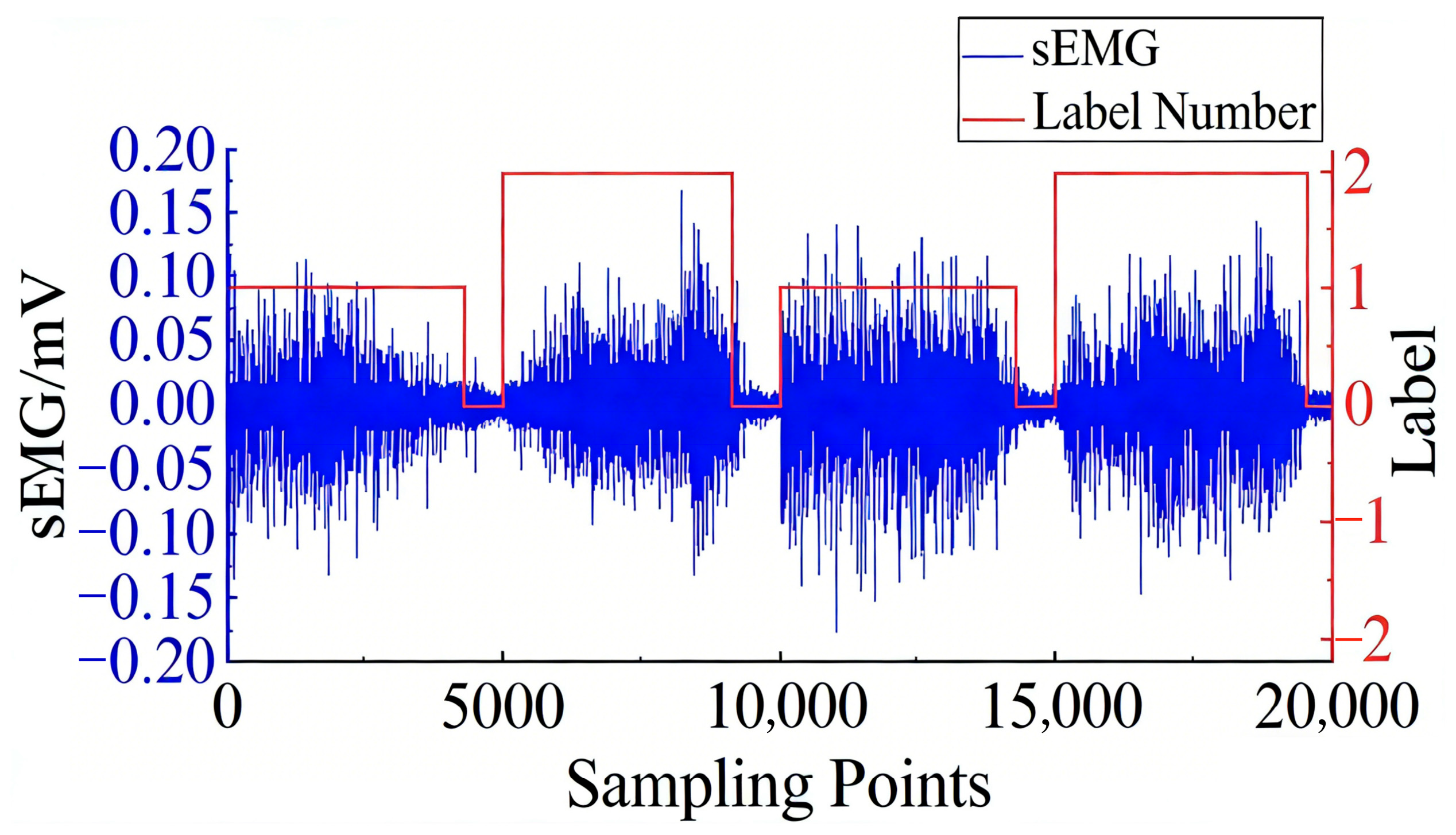
When M j exceeds T H 1 , this marks the onset of the movement, signifying the commencement of the action; when M j falls below T H 2 , this denotes the conclusion of the movement, indicating its termination. Generally, TH1 is set to three times the root mean square (RMS) value of sEMG signals during the subject’s resting state, and TH2 is set to twice the RMS value of sEMG signals in the resting state [38]. The detection of active segments in surface electromyography signals is illustrated in Figure 9. The figure demonstrates that this method can identify the boundary points between each active and inactive segment with high recognition accuracy. Here, label 0 denotes an inactive segment, label 1 indicates a flexion action, and label 2 signifies an extension action.

2.2.4. Visualisation of sEMG Signals

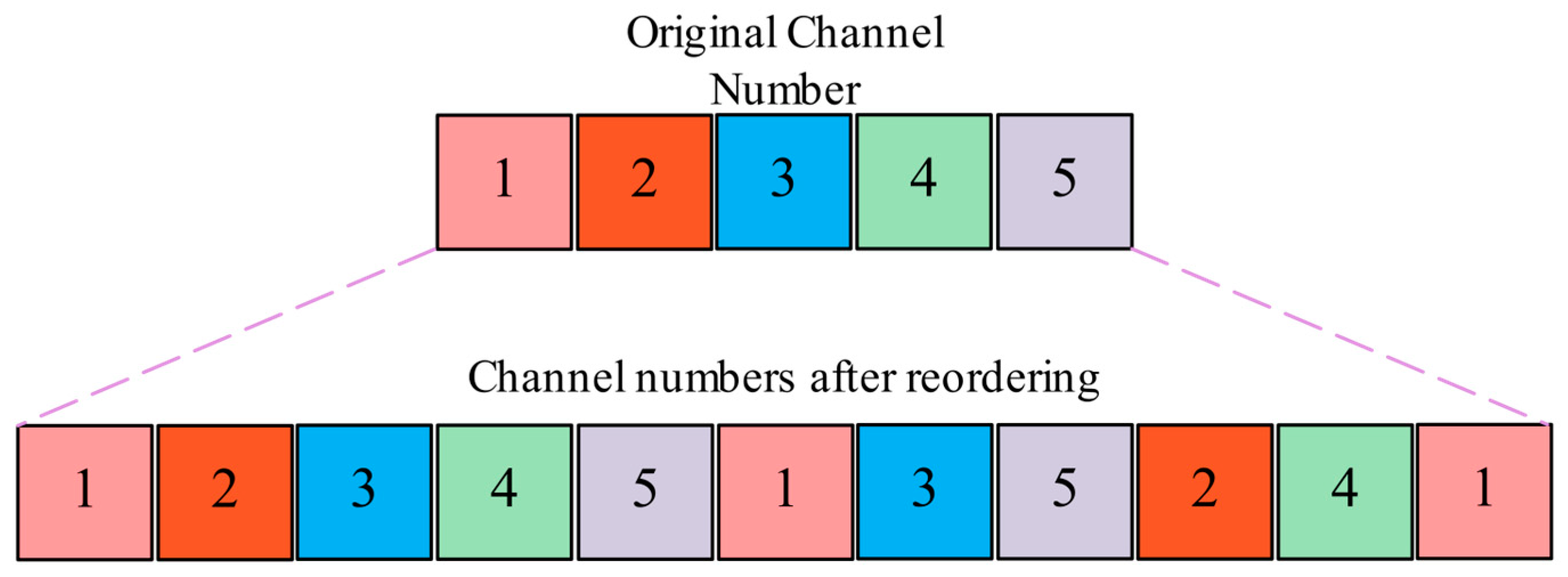
Simultaneously, by reordering the five channels, any two channels can be arranged adjacently, thereby enhancing the correlation between channels. This approach is based on the channel reordering algorithm proposed by Wenchao Jiang et al. [39]. For instance, consider an original image of size T × D, where T denotes the number of sampling points in sEMG signals and the image length in sEMG images, while D represents the number of channels in sEMG signals and the image width in sEMG images. The original channels are rearranged through the algorithm to generate a new channel order, as illustrated in Figure 10. This ensures any two channels are adjacent, facilitating the model’s learning of hidden, more abstract information between channels.
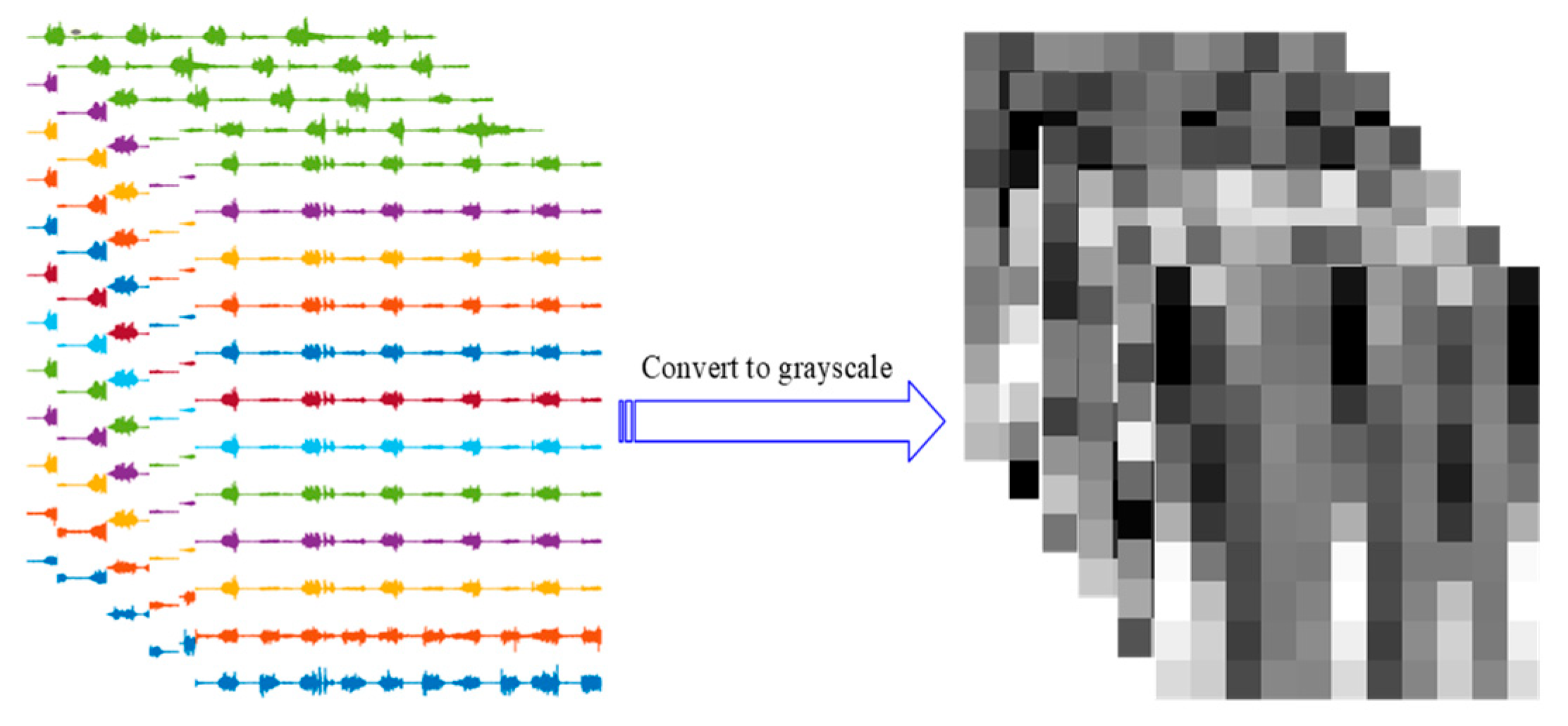
Consider a sample of D-channel sEMG signals. Using a sliding window of length T, the sample is partitioned into multiple subsets, each of length T and width D channels. The amplitude of the signal within each subset must be mapped to an sEMG greyscale image, which can be represented as RT × D. Prior to this, the amplitude of each channel is mapped to the range of 0–1. As the grayscale value range is 0–255, multiplying each channel amplitude by 255 maps it within this grayscale range, where 0 corresponds to black and 255 to white. The grayscale conversion diagram is illustrated in Figure 11.
The final image size is determined by the sliding window length and the number of channels. With a sampling frequency of 1000 Hz and a window length of 200 ms, each window contains 200 sampling points. Given the number of channels D = 5, the final grayscale image size is 200 × 5 pixels. No additional interpolation is adopted. The normalized sEMG signal amplitudes are directly mapped to grayscale values via linear scaling, ensuring the temporal continuity of the original signal and the authenticity of its amplitude. The segment size of the sliding window is kept unchanged, guaranteeing that all data subsets have a uniform length of 200 sampling points. Channel rearrangement only adjusts the order of the 5 channels without altering the number of channels, so the image size remains consistent across all samples. The processed grayscale images are fed into the CNN branch of the two-stream model.

3. Construction of the CNN-LSTM Parallel Dual-Stream Spatio-Temporal Neural Network Model

Based on structured grayscale images of sEMG, this chapter proposes a CNN-LSTM parallel dual-stream spatio-temporal neural network for motor intention decoding. First, the basic structures of core neural network components are elaborated, and their roles in feature extraction are specified. Subsequently, by incorporating the spatiotemporal characteristics of sEMG signals, an intention recognition model with functionally distinct parallel branches and key modules is constructed. Finally, quantitative evaluation metrics are defined to comprehensively assess the model performance. The design philosophy and evaluation framework of the model will be detailed in the following sections.

3.1. Basic Structure of Neural Networks

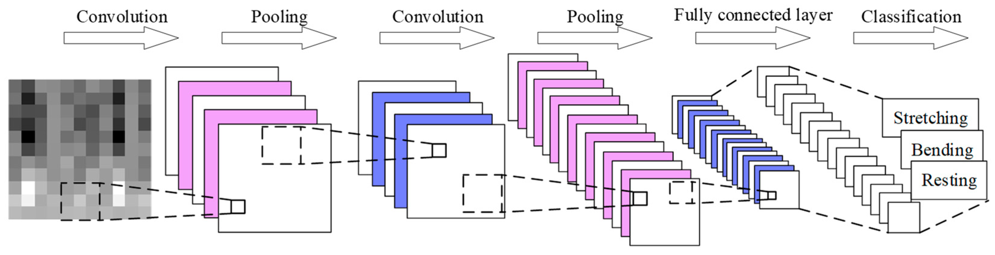
CNN represents a classic deep learning model. Owing to their simple architecture, minimal parameters, ease of training, and inherent properties of local connectivity and weight sharing, they find extensive application in natural language processing and image recognition domains. Given their advantages in image recognition, one-dimensional multi-channel electromyographic signals can be mapped into two-dimensional images for input to a CNN. Through the collaborative action of convolutional layers, pooling layers, and fully connected layers, these networks achieve recognition of movement intentions. A schematic diagram of the CNN architecture is shown in Figure 12.
The input layer receives normalised raw data. The convolutional layer extracts simple features such as edges and local temporal patterns through learnable convolutional kernels. The activation function layer introduces nonlinearity to fit complex relationships. The pooling layer reduces dimensionality while preserving key features and enhancing robustness. The fully connected layer integrates flattened local features into global features, whilst the output layer generates final results via activation functions such as Softmax or Linear, tailored to classification or regression tasks. This structural design enables efficient extraction of critical domain patterns and successful task completion in applications such as sEMG processing and robotic vision within mechanical engineering/automation fields.
The following is a detailed introduction to each level:
(1)
Convolutional Layer
As a core component of deep neural networks, the convolutional layer achieves feature extraction via the parameter sharing mechanism. It is configured with several convolution kernels of identical size. Each kernel performs sliding dot product operations in the spatial domain of the input data. Different kernels capture multimodal features from the input feature maps through distinct weight parameters, thereby constructing a multi-dimensional feature space. This parameter sharing scheme reduces model complexity while effectively preserving the translation invariance of features. The operation of the convolutional layer can be expressed by Equation (7):
o m n = Conv x = i = 1 I j = 1 J w i j x m + i , n + j + b 1 m M , 1 n N ,
where x m + i , n + j is the pixel value of the image at the row and column of the convolution kernel, o m n is the output of the pixel value at the row and column in the feature map, w i j is the value at the row and column of the convolution kernel in this layer, I , J respectively represent the length and width of the convolution kernel, b is the bias term of the convolution operation and M , N are outputting the length and width of the feature map.
(2)
Pooling Layer
The pooling layer performs downsampling in the spatial domain, and its operating mechanism is independent of the channel dimension. By adopting a local receptive field aggregation strategy, it achieves feature compression through max pooling or average pooling, thereby significantly reducing the spatial resolution of feature maps. This operation not only decreases the subsequent computational cost but also enhances the model’s robustness to local deformations and prevents overfitting. The formulations of max pooling and average pooling are given by Equation (8) and Equation (9), respectively:
o m i j = MAXP x = max m = 1 , , H max n = 1 , , W x s × i + m , s × i + n ,
o a i j = AVEP x = 1 H × W m = 1 H n = 1 W x s × i + m , s × i + n ,
where o m i j and o a i j denote the pixel values at the i-th row, j-th column in the output feature maps of max pooling and average pooling, respectively. x is the output the length and width of the feature map, H and W denote the height and width of the pooling region, respectively. m and n represent the position indices of the pooling operation. s is the sliding step size.
(3)
Activation Function
The activation function is a key module that breaks the linear constraints of the model, and it is usually employed after the convolution operation. Given the linear nature of convolution operations, the information transfer between network layers only forms linear correlations. Even with an increase in network depth, the model’s nonlinear expression capability cannot be improved. The introduction of a nonlinear activation function effectively solves the problem of nonlinear mapping. This nonlinear transformation enhances the function approximation ability of convolutional neural networks. The expression of the typical rectified linear unit (ReLU) function is presented in Equation (10) as follows:
f x = x x 0 0 x < 0 .
It can be observed from the equation that the derivative of the ReLU function is zero in the negative region. Once the neuron activation value becomes negative, the gradient will vanish, whereas it remains unchanged for positive values. This operation is referred to as one-sided suppression. Therefore, the ReLU function offers the advantages of high computational efficiency and enables fast convergence of the network.

3.2. Building an Intent Recognition Model

For lower limb movement intention recognition, a spatiotemporal neural network combining CNN and LSTM with dual parallel branches for spatial and temporal processing was designed. This incorporates attention mechanisms and feature fusion modules, leveraging the temporal sequentiality and multi-channel spatiality of electromyographic signals. The model extracts spatiotemporal features through its dual branches, which are then comprehensively integrated via a spatiotemporal feature fusion decoding module to enhance classification performance. The specific structure is shown in Figure 13.
sEMG signals inherently contain two types of complementary information: spatial features and temporal features. Standalone CNN or LSTM networks adopt a unified feature extraction path, which cannot simultaneously optimize the heterogeneous parameters for spatiotemporal feature extraction. This tends to result in one type of feature being suppressed by the other. In contrast, sequential architectures suffer from information loss during sequential processing. Features obtained through preprocessing may lose critical details in subsequent temporal processing, and vice versa. The parallel dual-stream architecture decouples spatial and temporal feature extraction into two functionally distinct branches. Each branch is optimized individually for its target feature type, which completely eliminates mutual interference between features and ensures the integrity of both types of information.
The multi-scale spatial feature extraction branch is designed to mine the inter-channel correlation features of sEMG signals. This branch consists of four convolutional layers, a Convolutional Block Attention Module (CBAM) integrating channel and spatial attention, one residual structure, and two fully connected (FC) layers. The CBAM is embedded after the multi-scale convolutional feature concatenation layer and before the residual convolutional layer. It can adaptively enhance the weights of key muscle channels and local spatial features in sEMG signals, and effectively suppress redundant information such as noise from non-critical channels/regions. The feature enhancement process of CBAM follows the sequential execution logic of channel attention (CA) and spatial attention (SA).
F = S A ( C A ( F ) ) ,
where F denotes the input feature map, CA(⋅) represents the channel attention submodule, SA(⋅) represents the spatial attention submodule, and F′ denotes the attention-enhanced feature map.
(1)
CA Submodule
It aggregates spatial information to focus on the importance of different muscle channels, and compresses the spatial dimension by adopting Global Average Pooling (GAP) and Global Max Pooling (GMP):
F a v g c = 1 H W i = 1 H j = 1 W F i , j , c ,
F m a x c = m a x i = 1 . . H , j = 1 . . W F ( i , j , c ) .
The pooled results are fed into a shared two-layer MLP to implement feature fusion:
C A F = σ M l p F a v g c ; F m a x c = σ ( W 2 ( δ W 1 F a v g c + W 1 ( F a v g c ) ) ) ,
where σ denotes the Sigmoid activation function, δ denotes the ReLU activation function, and W1, W2 represent the weight matrices.
The attention weight vector is multiplied element-wise with the input feature map:
F C A i , j , c = F i , j , c C A w e i g h t ( c ) .
(2)
SA Submodule
It aggregates channel information to focus on local spatial regions containing discriminative sEMG features, and compresses the channel dimension using channel-wise average pooling and channel-wise max pooling:
F a v g s = 1 C c = 1 C F C A i , j , c ,
F m a x s = m a x c = 1 . . C F C A i , j , c .
The two pooled feature maps are concatenated to accomplish spatial feature fusion:
S A F C A = σ C o n v F a v g s ; F m a x s ,
where Conv(·) is the convolution operation with same padding, which outputs the spatial attention map with a size of H×W.
The spatial attention map is multiplied element-wise with F C A to obtain the final enhanced feature map:
F = F C A S A m a p
The multi-scale spatial feature extraction branch is used to extract spatial features from multi-channel sEMG signals for the representation of lower limb movements, as illustrated in Figure 14. It consists of a multi-scale spatial feature extraction module, channel/spatial attention mechanism, residual structure, and FC layer. First, the multi-scale spatial feature extraction branch extracts local spatial features of the signals using multiple sets of convolution kernels with different sizes, which can enlarge the receptive field of the model and improve its ability to recognize movements. Specifically, four sets of convolution kernels with different sizes—1 × 1, 3 × 3, 5 × 5, and 7 × 7—are adopted to expand the scope of feature extraction. Meanwhile, to reduce computational cost and improve the recognition efficiency of the model, it is necessary to decrease the number of parameters of the convolution kernels. Considering that the number of parameters is mainly determined by their geometric size, and according to the research by Christian Szegedy, large convolution kernels can be decomposed into multiple small ones without affecting the effect of feature extraction. That is, a 5 × 5 convolution kernel can be replaced by two serially connected 3 × 3 convolution kernels, and a 7 × 7 convolution kernel can be replaced by three serially connected 3 × 3 convolution kernels, thereby achieving the goal of reducing parameters. Next, the concatenation operation is employed to perform an initial fusion of the four sets of extracted spatial features.
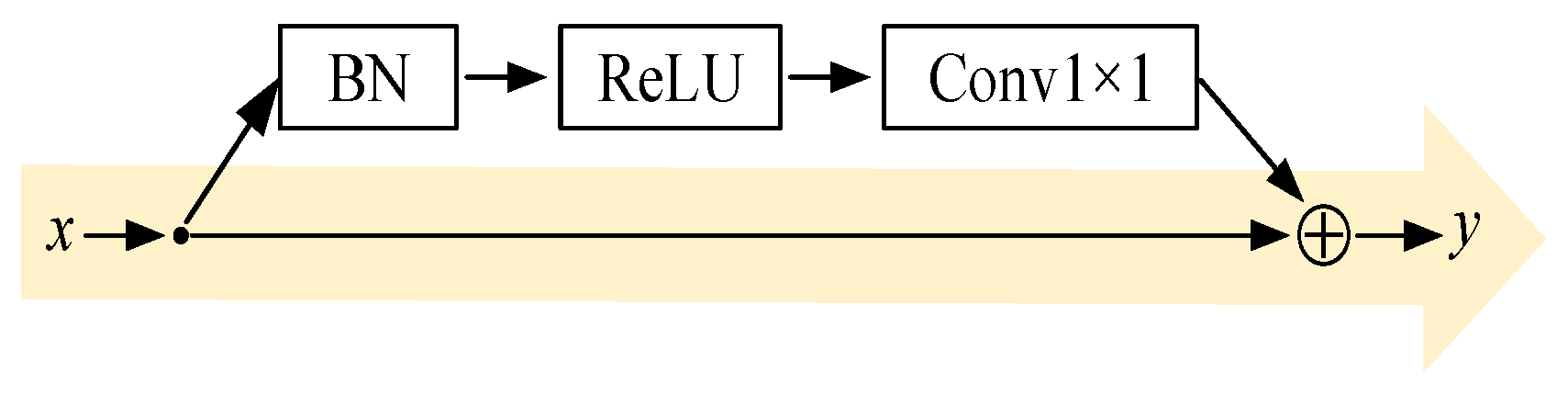
Employing residual structures mitigates issues such as gradient vanishing and model overfitting during deep neural network training, thereby enhancing model robustness. Residual structures comprise convolutional layers, batch normalisation (BN) layers, and ReLU layers. Existing research indicates that BN layers fail to deliver their full benefits when not combined with ReLU layers. When arranged in the sequence BN layer followed by ReLU layer and positioned before the convolutional layer, the model achieves favourable results [40], with the specific architecture illustrated in Figure 15. The residual structure can be expressed by Equation (20):
y = F x , W i + x
where y is module output, x is module input, x —weight of the i-th layer and F is convolution operation.
The convolutional layer is composed of 1 × 1 convolution kernels. The output feature dimension of the residual structure is consistent with that of the multi-scale spatial features, and is added to the preliminarily fused features for further fusion to obtain Feature C, which avoids information loss caused by the increase in model depth. Subsequently, two FC layers are adopted to further learn the correlation among the features of different regions. The FC layers can integrate the input features into a high-dimensional vector, enabling the model to capture the global relationships between different features. To mitigate overfitting, a Dropout layer with a dropout ratio of 50% [41], BN, and ReLU are deployed after each fully connected layer. This configuration reduces internal covariate shift, thereby accelerating network convergence and alleviating the vanishing gradient problem.
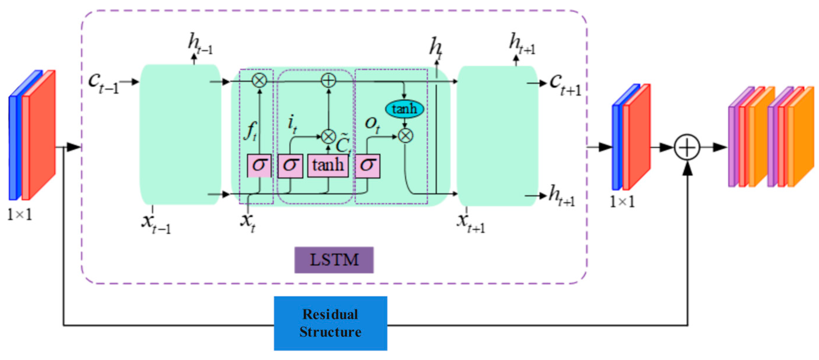
The temporal feature extraction branch is employed to extract temporal features from multi-channel sEMG signals for the recognition of lower limb movement actions, as shown in Figure 16. The temporal feature extraction module is composed of 2D convolutional layers, LSTM layers, 1D convolutional layers, and FC layers. It takes an LSTM as the core layer to enhance the model’s ability to learn long-term temporal dependencies of time-series data, and the output dimension of this branch is consistent with that of the spatial feature extraction branch. Specifically, the temporal feature extraction branch extracts temporal features of sEMG signals via LSTM, which further strengthens the model’s capability to capture long-term temporal dependencies in time-series data. The same residual structure as that in the spatial feature extraction branch is introduced after the LSTM layer to alleviate issues such as the vanishing gradient in LSTM and model overfitting. Subsequently, two fully connected layers are connected, whose structure is identical to that of the FC layers in the multi-scale spatial feature extraction branch.
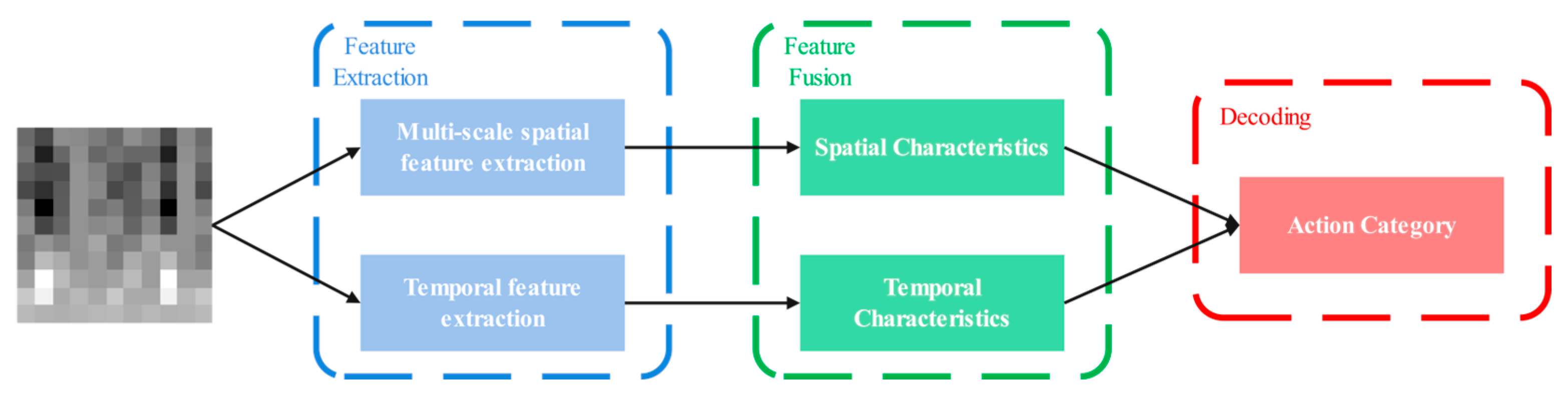
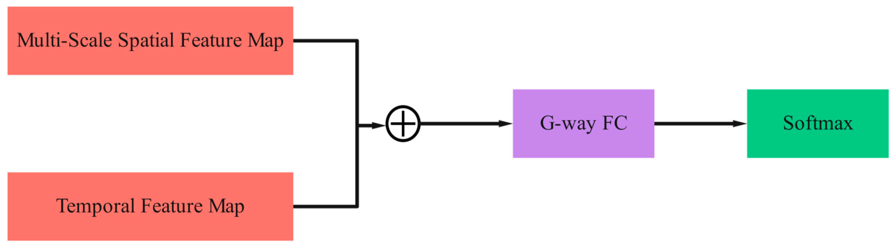
The Spatial-Temporal Feature Fusion and Decoding Module extracts features from spatial and temporal dimensions, obtains feature maps from two different perspectives, and then performs analysis, processing and fusion on them to form a comprehensive feature map, as illustrated in Figure 17. Compared with the original feature maps, the fused feature maps exhibit a significant improvement in feature representation capability, which helps to mine the essential characteristics of the target more accurately and further improve the recognition accuracy. The outputs of spatial features and temporal features are fused by means of concatenation along the channel dimension. Finally, a fully connected layer activated by Softmax is adopted to obtain the classification results. Specifically, a label vector G with a length equal to the number of lower limb movement classes (i.e., G = 3) is first generated through the fully connected layer, and then the Softmax function is used to determine the class to which the label vector G belongs.

3.3. Model Performance Evaluation Metrics

(1)
Accuracy
The proportion of correctly classified items relative to the total sample size. The calculation formula is as per Equation (21):
A c c u r a c y = T P + T N T P + T N + F P + F N ,
where TP is Number of correctly predicted positive samples, TN is Number of correctly predicted negative samples, FP is Number of incorrectly predicted negative samples and FN is Number of incorrectly predicted positive samples.
(2)
Precision
The proportion of correctly classified positive samples to the total number of samples classified as positive by the classifier. The calculation formula is as shown in Equation (22):
P r e c i s i o n = T P T P + F P .
(3)
Recall
The proportion of samples that are actually positive but are predicted as positive. The calculation formula is as shown in Equation (23):
R e c a l l = T P T P + F N .
(4)
F1 score
The harmonic mean of precision and recall. The calculation formula is as shown in Equation (24):
F 1 = 2 P r e c i s i o n R e c a l l P r e c i s i o n + R e c a l l .

4. Movement Intention Recognition Trials and Results Analysis

To verify the effectiveness, generalization capability of the proposed model and the role of each component, this chapter conducts systematic experiments from three aspects: basic intent recognition performance, analysis of core components, and comparative experiment analysis. First, the dataset partitioning rules and experimental settings are clarified, followed by an analysis of the recognition accuracy and detailed evaluation metrics across different subjects. Furthermore, ablation experiments are performed to quantify the contribution of each key component. Finally, comparative experiments are conducted between the proposed model and existing mainstream benchmark models, and the generalization ability of the model is verified via testing on subjects not involved in training and leave-one-out cross-validation (LOOCV). The subsequent sections will present the details of the experimental design and specific results.

4.1. Motor Intent Recognition Trial

Multi-subject sEMG signals were collected during the rehabilitation training of the subjects, and the acquired sEMG dataset was divided into a training set and a validation set. All samples from the same subject were uniformly assigned to either the training set or the validation set to eliminate statistical bias caused by the inter-sample correlation within individual subjects. Specifically, 8 out of 10 subjects were randomly selected as the training group, with the remaining 2 serving as the validation group.
To prevent overfitting and optimize training efficiency, an early stopping mechanism is adopted during the model training process: the cross-entropy loss of the validation set is selected as the monitoring metric, with a patience value set to 5 and the model corresponding to the minimum validation loss during the entire training process is retained to ensure optimal generalization performance. The hyperparameter settings for the CNN-LSTM parallel dual-stream spatio-temporal neural network model are presented in Table 3.

4.2. Analysis of Experimental Results

4.2.1. Motion Intention Recognition Experiments

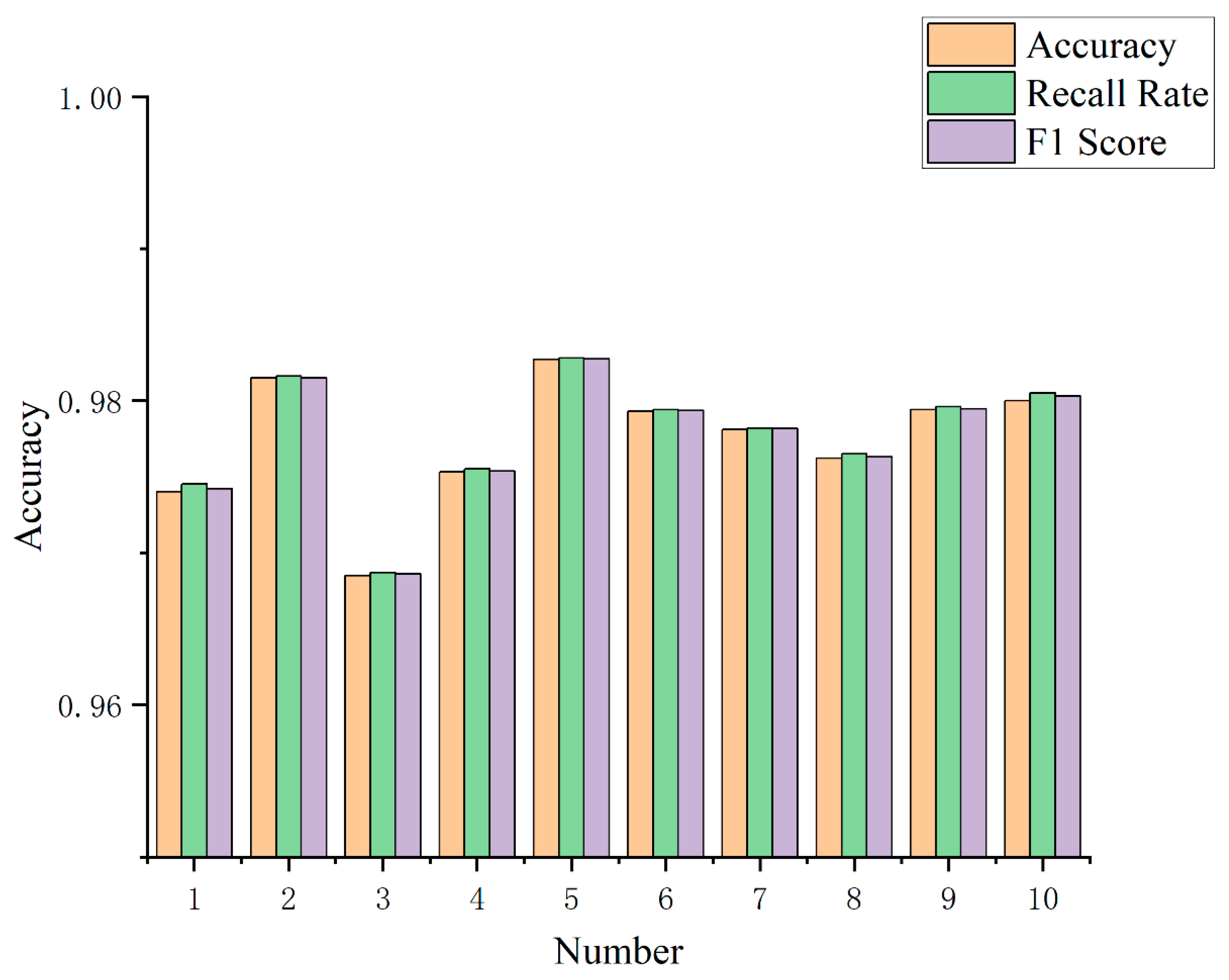
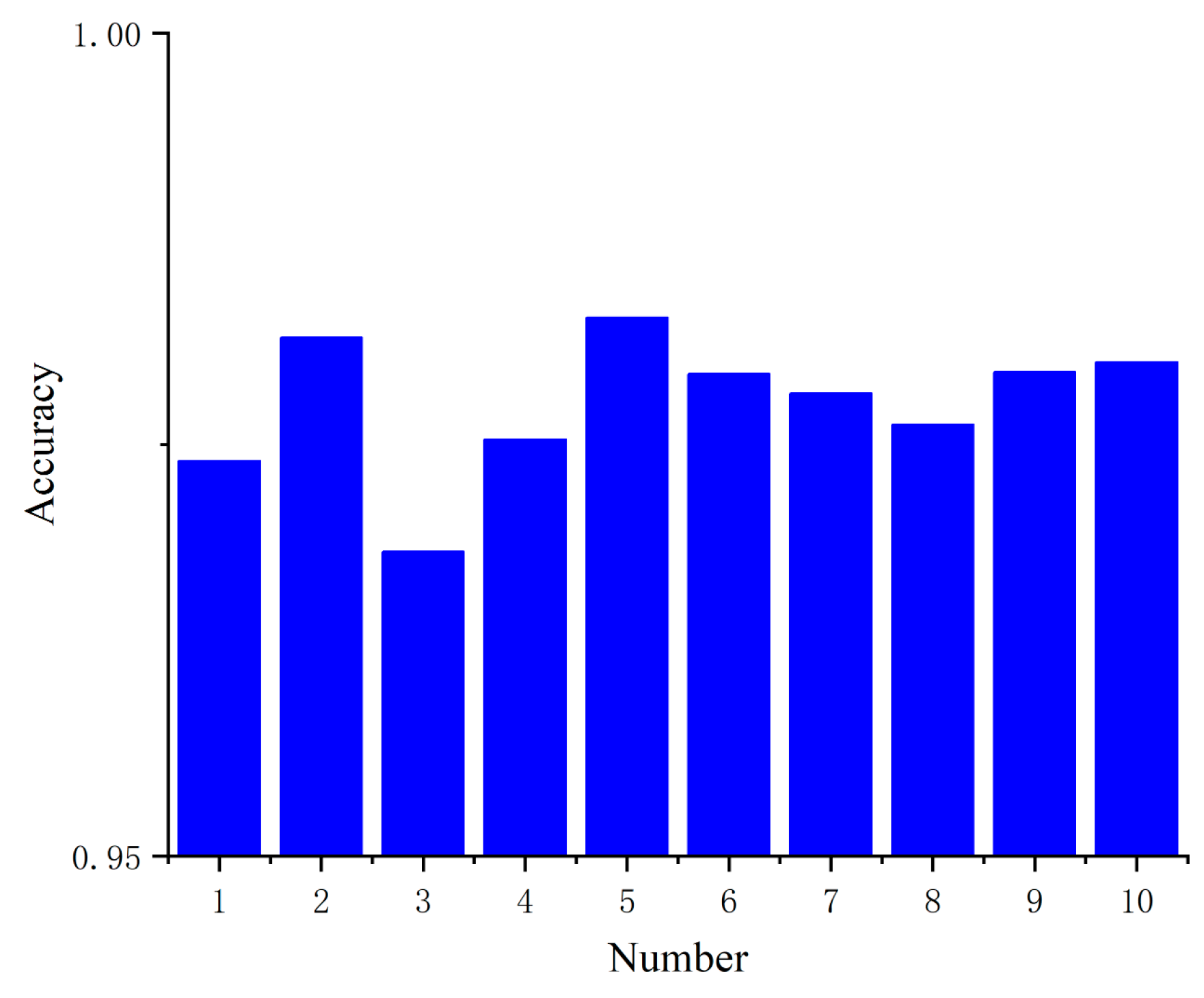
The relevant physical information of the 10 subjects participating in the experiment is shown in Table 4. The raw data were processed to generate grayscale images, which were used as experimental samples. Motion intention recognition was performed on the data from the 10 subjects using the proposed CNN-LSTM parallel dual-stream spatio-temporal neural network model. Figure 18 presents the recognition accuracy of each subject, where the recognition accuracy refers to the average recognition accuracy across the three types of movements.
As shown in the figure, the recognition accuracy ranges between 96.85% and 98.27%. Subject 5 achieved the highest recognition accuracy, while Subject 3 recorded the lowest. The average recognition accuracy across all subjects was 97.75%. This demonstrates that even with the same recognition model, the accuracy varies significantly when applied to different subjects. Subsequently, we analyse the impact of individual variability on recognition accuracy within the CNN-LSTM parallel dual-stream spatio-temporal neural network model. Figure 19 presents the classification precision, recall, and F1 score for the ten subjects’ experimental data processed by this model. These metrics represent the average values across each subject’s three action recognition results.
The results in the Table 5 generally indicate that the CNN-LSTM parallel dual-stream spatio-temporal neural network model proposed in this paper exhibits certain biases in individual variability. Although recognition accuracy varies slightly across different subjects, the differences remain relatively minor. For instance, the maximum discrepancy in recognition accuracy between Subjects 3 and 5 was 1.42%. Due to the differences in subjects’ muscle anatomical structures and motor habits, the feature distribution of their sEMG signals exhibits obvious individual discrepancies. Additionally, differing levels of skin oil secretion among individuals, coupled with electrode pad loosening during prolonged data acquisition, introduced inherent biases in the sEMG signals. Consequently, these factors contributed to discrepancies in the recognition outcomes.
These individual differences lead to slight accuracy fluctuations, but the impact can be mitigated by the following measures: To address the causes of the aforementioned errors, for new subjects, only a small amount of labeled data is collected to fine-tune the pre-trained model. The bottom feature extraction layers of the model are frozen, and only the parameters of the top FC layers and the attention mechanism are updated. This method enables the model to quickly adapt to the individualized characteristics of subjects [42] while significantly reducing the demand for large-scale labeled data from subjects. Meanwhile, the experimental procedure for data collection is standardized. During data acquisition, real-time monitoring is performed to check whether the electrode pads become loose; if loosening occurs, the pads are reattached and secured promptly.
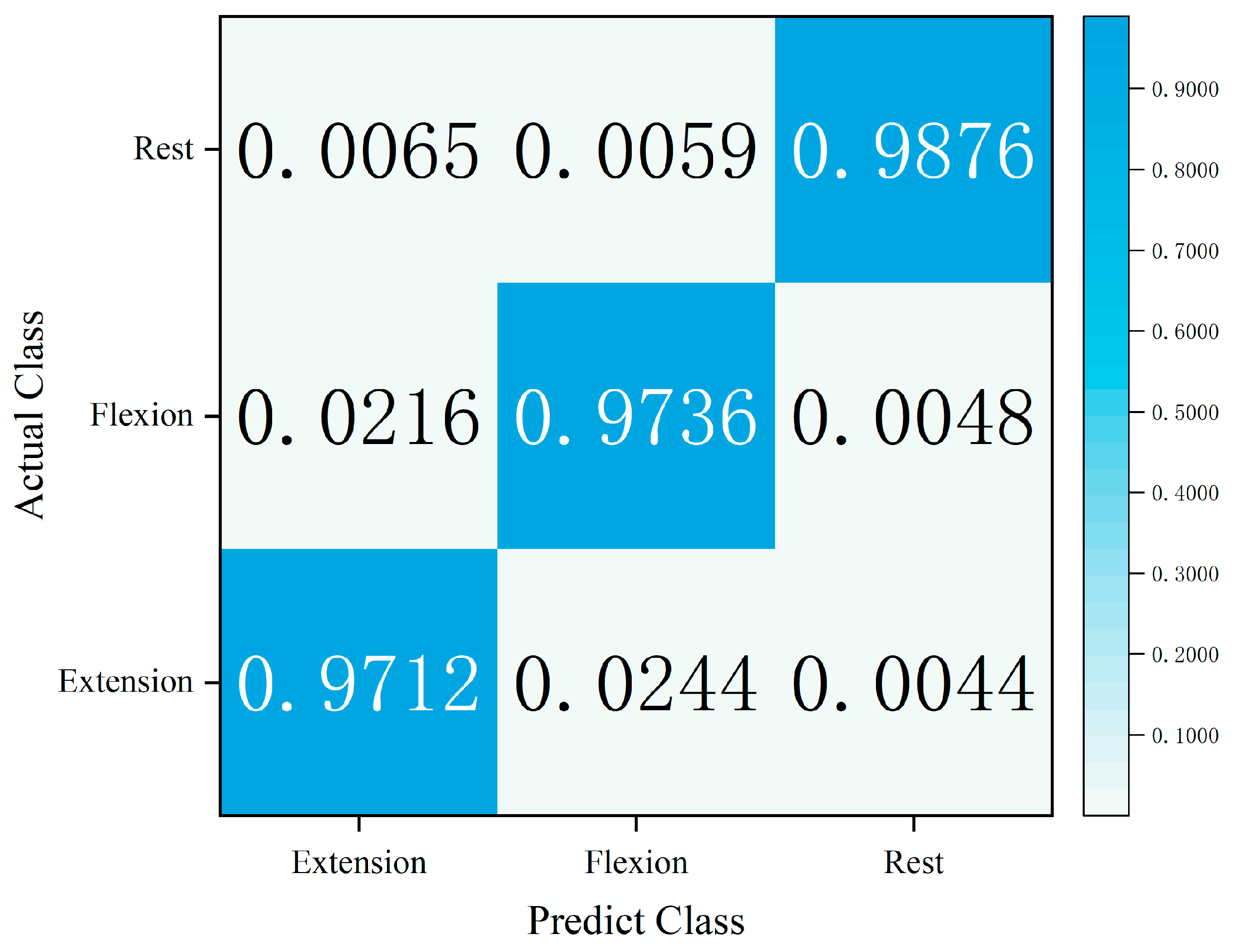
A systematic evaluation of classification performance was conducted using a confusion matrix: first, a confusion matrix was constructed based on three-class labels, calculating precision, recall, and F1-score metrics for the three action categories—extension, flexion, and rest; second, the model’s recognition efficacy for different action patterns was quantitatively analysed through the distribution characteristics of matrix elements; finally, the misclassification rates between action categories were statistically analysed to investigate feature confusion patterns. This experiment employed a five-fold cross-validation strategy, integrating the experimental data from ten subjects to construct the confusion matrix. This approach ensures statistical significance of evaluation results while effectively reflecting the model’s generalisation performance at the population level, and identifies pairwise misclassification scenarios among the three movement actions. The confusion matrix is presented in Figure 20.
By integrating all experimental data from ten subjects, a confusion matrix was constructed where diagonal entries represent accuracy rates. This effectively validated the classification performance of the CNN-LSTM parallel dual-stream spatio-temporal neural network model for three action patterns: extension, flexion, and rest. The highest recognition accuracy was achieved for the resting state at 98.76%, followed by flexion at 97.36%, while the recognition performance for extension was relatively weaker at 97.21%. Action misclassification primarily occurred between extension and flexion: 84.72% of misclassified extension samples were erroneously categorised as flexion, while 77.14% of misclassified flexion samples were misjudged as extension. This bidirectional confusion can be attributed to the following factors. For the same subject, the sEMG signals of flexion and extension movements show similar dynamic variation trends, resulting in overlapping temporal dependency features captured by the LSTM branch. The two types of movements rely on the coordinated activation of the same set of muscle groups, with only subtle differences in activation intensity, leading to highly similar spatial feature maps extracted by the CNN branch. To alleviate the confusion between extension and flexion movements, the corresponding solutions are proposed as follows: highlight the subtle differences in the rising and falling dynamic characteristics of the signals between flexion and extension; and take the weight difference between the Rectus Femoris and biceps femoris as a new spatial feature. The evaluation metrics for individual labels are subsequently calculated, as illustrated in Figure 21.
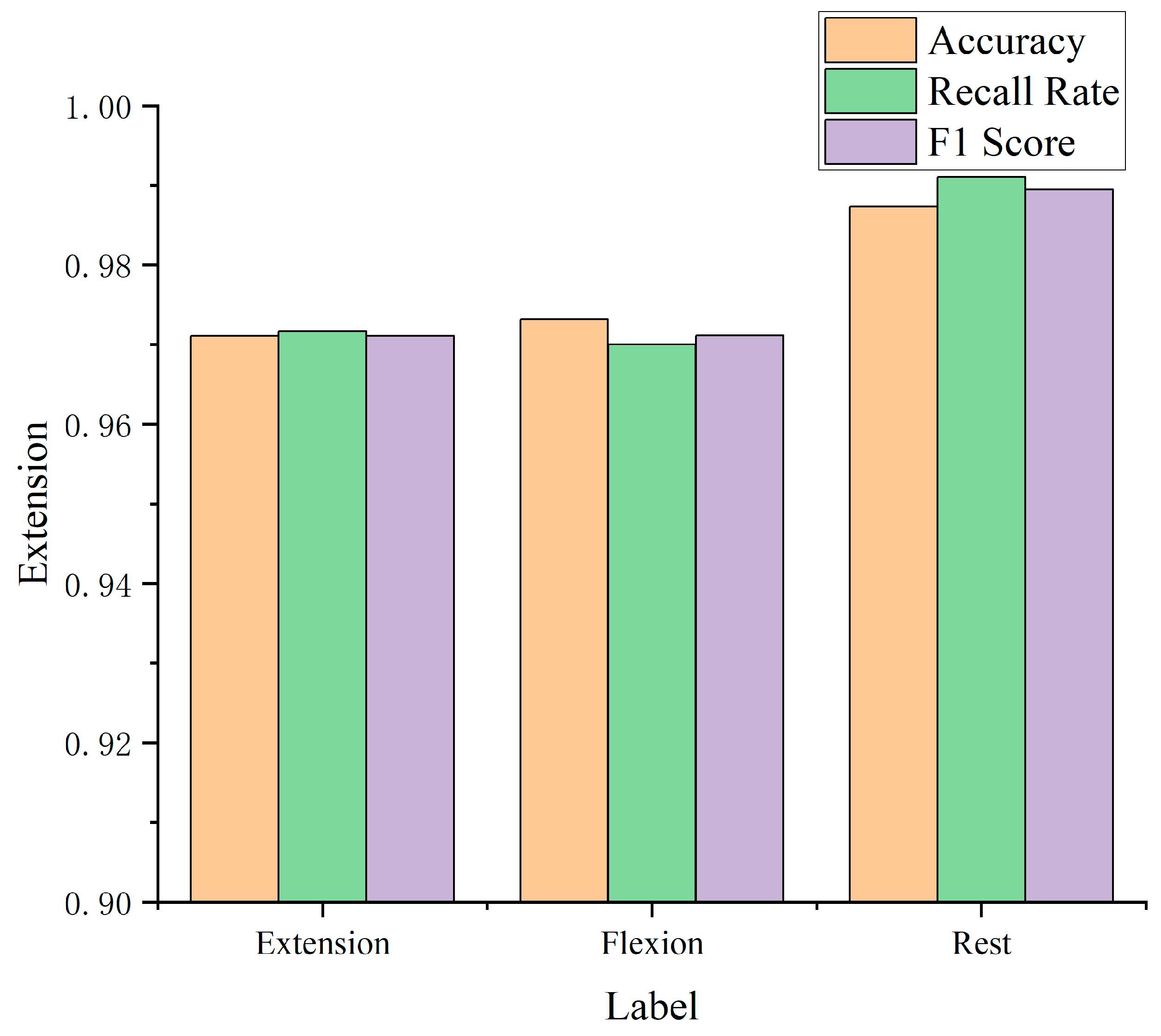
As shown in Figure 21, the CNN-LSTM parallel dual-stream spatio-temporal neural network model proposed herein demonstrates optimal recognition performance for the resting state. In contrast, the recognition accuracy for extension and flexion movements exhibits certain discrepancies compared to that of the resting state. Specifically, the precision, recall, and F1-score for the resting state all exceed 98.5%, while those for the extension and flexion actions hover around 97%.

4.2.2. Core Component Ablation Experiments

To quantify the contribution of core components—including the spatial branch, temporal branch, CBAM attention mechanism, channel rearrangement, and dedicated fusion module—to model performance, this study designed systematic ablation experiments. All experiments were performed under identical conditions to ensure the fairness of comparison. Six model variants were constructed by removing or replacing individual components of the proposed complete model. The configurations and performance metrics of each variant are summarized in Table 6. The data represent the mean experimental results across 10 subjects, including patient participants.
The accuracies of the single spatial-branch model (M3) and single temporal-branch model (M4) are 89.30% and 90.70%, respectively, representing decreases of 8.45% and 7.05% compared with the complete model. This result confirms that the parallel dual-stream design can effectively fuse complementary spatial and temporal features and overcome the limitations of single-branch models in feature extraction. The performance of the pure temporal branch is slightly superior to that of the pure spatial branch, indicating that the long-term temporal dependencies of sEMG signals are critical for motion intention recognition, while the spatial branch can supplement channel correlation features to further improve the model’s recognition accuracy. After removing the CBAM attention mechanism (M2), the model accuracy decreases by 2.57%. This verifies that the channel-spatial dual attention mechanism can adaptively enhance critical muscle channels and local spatial features while suppressing redundant noise; in particular, it exerts a significant effect on distinguishing similar movements such as flexion and extension by highlighting discriminative features. When the channel rearrangement strategy is not adopted (M1), the model accuracy drops by 1.52%. The channel rearrangement algorithm ensures the adjacency of any two channels, facilitating the model in learning correlation features between channels, which are essential for capturing the coordinated activation patterns of multiple muscle groups. When the dedicated weighted fusion module is replaced with simple feature concatenation (M5), the model accuracy decreases by 3.13%. The 1 × 1 convolution in the dedicated fusion module can dynamically adjust the weights of spatial and temporal features according to task characteristics, achieving more efficient feature fusion compared with simple feature concatenation.

4.2.3. Comparative Experiments

To verify the accuracy of the motion intention recognition model proposed in this paper, this study conducted comparative experiments with three traditional models: MLP, single CNN, and single LSTM. Under the same data and hardware conditions, the accuracy of MLP reached 85.6%, single CNN was 89.3%, and single LSTM was 90.7%, while the proposed model outperformed them by at least 6.8%. The above comparative experiments demonstrate that the CNN-LSTM parallel dual-stream spatio-temporal neural network proposed in this paper has significant advantages over existing mainstream benchmark models.
In addition to the original 10 subjects, one new subject who had not participated in this experiment was additionally recruited. A pre-trained model trained on the original 8 subjects was used to directly test the sEMG data of the new subject, without any retraining or fine-tuning, so as to evaluate the model’s generalization ability for completely unseen subjects. The sEMG signals collected from the Rectus Femoris during the subject’s flexion and extension movements, as well as the corresponding intention recognition results, are shown in the figure.
The recognition accuracy of the model for the new healthy subject is 96.83%. The surface electromyography signals and motion intention recognition results of the new subjects are shown in Figure 22. The model can accurately identify the motion intention of completely unseen subjects, verifying its practical generalization value. Extensive retraining is not required for new users, and this property is of critical importance for the clinical deployment of rehabilitation robots.

5. Conclusions

Aiming at the problems of traditional motion intention recognition methods for lower limb rehabilitation robots—such as poor adaptability and limited accuracy caused by overemphasizing spatial feature extraction while ignoring motion continuity—this paper conducts a systematic study on sEMG signal decoding and lower limb motion intention recognition. A complete solution covering signal acquisition and preprocessing, model construction, and experimental verification is proposed, which is suitable for patients with mild to moderate lower limb motor dysfunction. Compared with existing studies, the main research findings and core mechanism contributions of this paper are as follows:
  • Based on the synergistic movement theory, five key muscle groups are selected to ensure the representativeness of sEMG signals. The proposed preprocessing pipeline includes Butterworth band-pass filtering, normalization, and 200 ms overlapping sliding window segmentation, which preserves the spatial-temporal details of sEMG signals. Compared with existing preprocessing methods using global normalization or fixed window size, this design can adapt to individual differences in signal amplitude and motion dynamics, laying a foundation for high-precision feature extraction.
  • The residual structure adopts the sequence of BN-ReLU-convolution, and the multi-scale attention mechanism highlights the key frequency bands and muscle channels of sEMG signals. This design not only alleviates the vanishing gradient problem in deep network training but also improves the efficiency of feature extraction.
  • The unique spatial-temporal feature extraction mechanism of the proposed CNN-LSTM parallel dual-stream spatio-temporal neural network breaks through the limitations of single-stream/serial architectures and achieves a breakthrough in sEMG signal feature decoding:
    (1)
    Advantages of the spatial branch: The multi-scale convolution + channel/spatial attention + residual structure is tailored for the multi-channel spatial characteristics of sEMG signals, capturing inter-channel correlations and local spatial distributions, while the attention mechanism adaptively enhances the features of critical muscle channels.
    (2)
    Advantages of the temporal branch: The LSTM + 1D convolution + residual structure can effectively model the long-term dependencies of sEMG signals and compress redundant temporal information. Different from single LSTM models, which struggle to extract local temporal features, this design balances long-term dependency capture and dynamic trend retention.
    (3)
    Advantages of the fusion mechanism: The dedicated spatial-temporal fusion module realizes weighted allocation of spatial topological information and temporal dynamic information, significantly improving the discriminative ability for similar movements. This is also the core reason why the model achieves an average recognition accuracy of 97.75%.
  • In future work, the sample size will be expanded to include patients of different ages and disease severity; the feature fusion strategy will be optimized to further reduce the confusion rate of flexion and extension movements; multi-modal information fusion will be explored to improve the model’s robustness in complex rehabilitation environments.

Author Contributions

Conceptualization, J.H. and H.L.; methodology, J.H. and H.L.; validation, S.L., J.W. and K.L.; data curation, S.L.; writing—original draft preparation, J.H. and X.M.; writing—review and editing, J.H. and H.L.; supervision, X.S.; project administration, H.L.; funding acquisition, X.S. All authors have read and agreed to the published version of the manuscript.

Funding

YanShan University University-Enterprise Co-operation “Learning, Research, Production and Utilisation” Innovative Talent Cultivation and Practical Application of Technical Service Contracts (Grant No. x2025204), YanShan University Cultivation of Innovative Talents and Innovative Capabilities through University-Enterprise “Industry-Academia-Research-Use” Programme (Grant No. x2024154).

Institutional Review Board Statement

This study uses non-invasive patch collectors to acquire surface electromyographic signals from the human body. The acquisition process is gentle and non-invasive, and did not cause any adverse effects on all subjects in terms of physiology, psychology, society, or reputation. The collected electromyographic signals are only used for the purpose of thesis research and do not involve any commercial development, profit-making activities, or third-party data transfer. At the same time, all subject information has undergone strict and complete anonymization processing, with all personally associated information such as names, contact information, and identity identifiers removed. This ensures that the data cannot be restored or identified to specific individuals through any technical means, complying with the irreversible standard of data anonymization. According to Article 32 of the “Measures for the Ethical Review of Life Science and Medical Research Involving Humans” (Guo Wei Ke Jiao Fa [2023] No. 4) issued in 2023: “Life science and medical research involving humans using human information data or biological samples that does not cause harm to the human body and does not involve sensitive personal information or commercial interests may be exempt from ethical review.” This study is fully in line with the requirements of this clause: 1. No harm to the human body: The non-invasive acquisition method has no risk of trauma, and the actual acquisition process did not have any adverse effects on the subjects, meeting the “non-maleficence” principle; 2. No involvement of sensitive personal information: Although electromyographic signals are data related to biometrics, after complete anonymization, they have lost personal directionality and no longer meet the criteria for identifying sensitive personal information in the “Personal Information Protection Law”, so there is no risk of information leakage or abuse; 3. No involvement of commercial interests: The data is only used for academic research and thesis writing, without any commercial cooperation, profit-making purposes, or data trading activities. In conclusion, this study strictly abides by the requirements of relevant laws and regulations and fully meets the conditions for exemption from ethical review as stipulated in Article 32 of the “Measures for the Ethical Review of Life Science and Medical Research Involving Humans”.

Informed Consent Statement

Informed consent for participation was obtained from all subjects involved in the study.

Data Availability Statement

The raw data supporting the conclusions of this article will be made available by the authors on request.

Conflicts of Interest

Author Kaiyuan Li was employed by the company Erzhong (Deyang) Heavy Equipment Co., Ltd., Deyang, China. The remaining authors declare that the research was conducted in the absence of any commercial or financial relationships that could be construed as a potential conflict of interest.

Abbreviations

The following abbreviations are used in this manuscript:
sEMGSurface Electromyography
CNNConvolutional Neural Network
RNNRecurrent Neural Networks
GNNGraph Neural Networks
LSTMLong-Short-Term-Memory
MLPMultilayer Perceptron
RMSERoot Mean Square Error
SNRSignal-to-Noise Ratio
CMRRCommon-Mode Rejection Ratio
PGAProgrammable Gain Amplifier
SINADSignal-to-Noise and Distortion
ECGElectrocardiogram
IIRInfinite Impulse Response
VMDVariational Modal Decomposition
RMSRoot- Mean Square
CBAMConvolutional Block Attention Module
BNBatch Normalisation
FCFully Connected Layers
CAChannel Attention
SASpatial Attention
GAPGlobal Average Pooling
GMPGlobal Max Pooling
ReLURectified Linear Unit
LOOCVleave-one-out cross-validation

References

  1. Rubin, N.; Hinson, R.; Saul, K.; Hu, X.; Huang, H. Ankle Torque Estimation with Motor Unit Discharges in Residual Muscles Following Lower-Limb Amputation. IEEE Trans. Neural Syst. Rehabil. Eng. 2023, 31, 4821–4830. [Google Scholar] [CrossRef] [PubMed]
  2. Su, D.; Hu, Z.; Wu, J.; Shang, P.; Luo, Z. Review of adaptive control for stroke lower limb exoskeleton rehabilitation robot based on motion intention recognition. Front. Neurorobot. 2023, 17, 1186175. [Google Scholar] [CrossRef] [PubMed]
  3. Zhou, J.; Sun, Y.; Song, R.; Wei, Z. Dynamic movement primitives modulation-based compliance control for a new sitting/lying lower limb rehabilitation robot. IEEE Access 2024, 12, 44125–44134. [Google Scholar] [CrossRef]
  4. Zhang, Y.; Zhao, W.; Wan, C.; Wu, X.; Huang, J.; Wang, X.; Huang, G.; Ding, W.; Chen, Y.; Yang, J. Exoskeleton rehabilitation robot training for balance and lower limb function in sub-acute stroke patients: A pilot, randomized controlled trial. J. Neuroeng. Rehabil. 2024, 21, 98. [Google Scholar] [CrossRef]
  5. Alashram, A.R. Effectiveness of combined robotics and virtual reality on lower limb functional ability in stroke survivors: A systematic review of randomized controlled trials. Neurol. Sci. 2024, 45, 4721–4739. [Google Scholar] [CrossRef]
  6. Chen, Z.; Guo, Q.; Yan, Y.; Shi, Y. Model identification and adaptive control of lower limb exoskeleton based on neighborhood field optimization. Mechatronics 2022, 81, 102699. [Google Scholar] [CrossRef]
  7. Desplenter, T.; Zhou, Y.; Edmonds, B.P.; Lidka, M.; Goldman, A.; Trejos, A.L. Rehabilitative and assistive wearable mechatronic upper-limb devices: A review. J. Rehabil. Assist. Technol. Eng. 2020, 7, 2055668320917870. [Google Scholar] [CrossRef]
  8. Dupont, P.E.; Nelson, B.J.; Goldfarb, M.; Hannaford, B.; Menciassi, A.; O’mAlley, M.K.; Simaan, N.; Valdastri, P.; Yang, G.-Z. A decade retrospective of medical robotics research from 2010 to 2020. Sci. Robot. 2021, 6, eabi8017. [Google Scholar] [CrossRef]
  9. van Kammen, K.; Boonstra, A.M.; van der Woude, L.H.V.; Visscher, C.; Reinders-Messelink, H.A.; Den Otter, R. Lokomat guided gait in hemiparetic stroke patients: The effects of training parameters on muscle activity and temporal symmetry. Disabil. Rehabil. 2020, 42, 2977–2985. [Google Scholar] [CrossRef]
  10. Berman, J.; Hinson, R.; Lee, I.-C.; Huang, H. Harnessing machine learning and physiological knowledge for a novel EMG-based neural-machine interface. IEEE Trans. Biomed. Eng. 2022, 70, 1125–1136. [Google Scholar] [CrossRef]
  11. Wei, W.; Tan, F.; Zhang, H.; Mao, H.; Fu, M.; Samuel, O.W.; Li, G. Surface electromyogram, kinematic, and kinetic dataset of lower limb walking for movement intent recognition. Sci. Data 2023, 10, 358. [Google Scholar] [CrossRef] [PubMed]
  12. Ait Yous, M.; Agounad, S.; Elbaz, S. Detection, identification and removing of artifacts from semg signals: Current studies and future challenges. Comput. Biol. Med. 2025, 186, 109651. [Google Scholar] [CrossRef] [PubMed]
  13. Xiao, F.; Mu, J.; He, L.; Wang, Y. How to use one surface electromyography sensor to recognize six hand movements for a mechanical hand in real time: A method based on Morse code. Med. Biol. Eng. Comput. 2024, 62, 2825–2838. [Google Scholar] [CrossRef] [PubMed]
  14. Wang, X.; Zhang, C.; Yu, Z.; Deng, C. Decoding of lower limb continuous movement intention from multi-channels EMGand design of adaptive exoskeleton controller. Biomed. Signal Process. Control 2024, 94, 106245. [Google Scholar] [CrossRef]
  15. Zheng, C.; Li, W.; Shi, Y.; Wei, S.; Liu, K.; Cheng, J.; Ji, L.; Lu, Y. Stretchable self-adhesive and self-powered smart bandage for motion perception and motion intention recognition. Nano Energy 2023, 109, 108245. [Google Scholar] [CrossRef]
  16. Xu, M.; Chen, X.; Ruan, Y.; Zhang, X. Cross-user electromyography pattern recognition based on a novel spatial-temporal graph convolutional network. IEEE Trans. Neural Syst. Rehabil. Eng. 2023, 32, 72–82. [Google Scholar] [CrossRef]
  17. Su, B.Y.; Wang, J.; Liu, S.Q.; Sheng, M.; Jiang, J.; Xiang, K. A CNN-based method for intent recognition using inertial measurement units and intelligent lower limb prosthesis. IEEE Trans. Neural Syst. Rehabil. Eng. 2019, 27, 1032–1042. [Google Scholar] [CrossRef]
  18. Sheng, M.; Xia, A.Q.; Wang, K.L.; Cha, H.; Wu, H.; Su, B. Movement intention recognition of intelligent lower limb prosthesis based on the fusion of geometric and physical features. Control Decis. 2022, 37, 953–961. [Google Scholar]
  19. Kim, H.; Lee, D.; Maldonado-Contreras, J.Y.; Zhou, S.; Herrin, K.R.; Young, A.J. Mode-unified intent estimation of a robotic prosthesis using deep-learning. IEEE Robot. Autom. Lett. 2025, 10, 3206–3213. [Google Scholar] [CrossRef]
  20. Tang, Y.; Chen, Y.X.; Yu, H.L.; Shi, P. Research and verification of a motion intention recognition method for lower limb prosthesis. Inform. Control 2023, 52, 598–606. [Google Scholar]
  21. Xie, B.; Meng, J.; Li, B.; Harland, A. Biosignal-based transferable attention Bi-ConvGRU deep network for hand-gesture recognition towards online upper-limb prosthesis control. Comput. Methods Programs Biomed. 2022, 224, 106999. [Google Scholar] [CrossRef]
  22. Xu, D.; Zhou, H.; Jie, T.; Zhou, Z.; Yuan, Y.; Jemni, M.; Quan, W.; Gao, Z.; Xiang, L.; Gusztav, F.; et al. Data-driven deep learning for predicting ligament fatigue failure risk mechanisms. Int. J. Mech. Sci. 2025, 301, 110519. [Google Scholar] [CrossRef]
  23. Anselmino, E.; Mazzoni, A.; Micera, S. EMG-based prediction of step direction for a better control of lower limb wearable devices. Comput. Methods Programs Biomed. 2024, 254, 108305. [Google Scholar] [CrossRef] [PubMed]
  24. Zhang, C.; Yu, Z.; Wang, X.; Chen, Z.-J.; Deng, C.; Xie, S.Q. Exploration of deep learning-driven multimodal information fusion frameworks and their application in lower limb motion recognition. Biomed. Signal Process. Control 2024, 96, 106551. [Google Scholar] [CrossRef]
  25. Luo, R.; Sun, S.; Zhang, X.; Tang, Z.; Wang, W. A low-cost end-to-end sEMG-based gait sub-phase recognition system. IEEE Trans. Neural Syst. Rehabil. Eng. 2019, 28, 267–276. [Google Scholar] [CrossRef] [PubMed]
  26. Wang, G.; Liu, Y.; Shen, Y.; Chen, Y.; Liu, K.; Sun, Z. sEMG-based multi-joints motion estimation of lower limb utilizing deep convolutional neural network. In Proceedings of the 2022 IEEE International Conference on Real-Time Computing and Robotics (RCAR), Guiyang, China, 17–22 July 2022; pp. 13–18. [Google Scholar]
  27. Bian, Q.; Castellani, M.; Shepherd, D.; Duan, J.; Ding, Z. Gait intention prediction using a lower-limb musculoskeletal model and long short-term memory neural networks. IEEE Trans. Neural Syst. Rehabil. Eng. 2024, 32, 822–830. [Google Scholar] [CrossRef]
  28. Portilla-Jiménez, M.; Hong, Y.N.G.; Kukkar, K.K.; Park, H.-S.; Li, S.; Roh, J. Generalizability of neuromuscular coordination in the human upper extremity after stroke and its implications in neurorehabilitation. J. Neuroeng. Rehabil. 2025, 22, 213. [Google Scholar] [CrossRef]
  29. Wang, Z.; Yao, J.; Xu, M.; Jiang, M.; Su, J. Transformer-based network with temporal depthwise convolutions for sEMG recognition. Pattern Recognit. 2024, 145, 109967. [Google Scholar] [CrossRef]
  30. Bao, T.; Zhao, Y.; Zaidi, S.A.R.; Xie, S.; Yang, P.; Zhang, Z. A deep Kalman filter network for hand kinematics estimationusing sEMG. Pattern Recognit. Lett. 2021, 143, 88–94. [Google Scholar] [CrossRef]
  31. Kibushi, B.; Okada, J. Auditory sEMG biofeedback for reducing muscle co-contraction duringpedaling. Physiol. Rep. 2022, 10, e15288. [Google Scholar] [CrossRef]
  32. Nazari, F.; Mohajer, N.; Nahavandi, D.; Khosravi, A. Comparison of gait phase detection using traditional machine learning and deep learning techniques. In Proceedings of the 2022 IEEE International Conference on Systems, Man, and Cybernetics (SMC), Prague, Czech Republic, 9–12 October 2022; pp. 403–408. [Google Scholar]
  33. Fu, Z.; Zhang, H.; Zhao, J.; Li, N.; Zheng, F. A modified 2-D notch filter based on image segmentation for RFI mitigation in synthetic aperture radar. Remote Sens. 2023, 15, 846. [Google Scholar] [CrossRef]
  34. He, J.; Wang, G.; Yu, H.; Liu, J.; Peng, B. Generalized minimum error entropy Kalman filter for non-Gaussian noise. ISA Trans. 2023, 136, 663–675. [Google Scholar] [CrossRef]
  35. Ma, S.; Lv, B.; Lin, C.; Sheng, X.; Zhu, X. EMG signal filtering based on variational mode decomposition and sub-bandthresholding. IEEE J. Biomed. Health Inform. 2021, 25, 47–58. [Google Scholar] [CrossRef]
  36. Xu, L.; Shang, K.; Zhang, X.; Zheng, C.; Pan, L. Multi-Scale Feature Fusion-Based Real-Time Anomaly Detection in Industrial Control Systems. Electronics 2025, 14, 1645. [Google Scholar] [CrossRef]
  37. ISO/TS 15066:2016; Robots and Robotic Devices—Collaborative Robots. International Organization for Standardization: Geneva, Switzerland, 2016.
  38. Ting, E.L.W.; Chai, A.; Chin, L.P. A review on EMG signal classification and applications. Int. J. Signal Process. Syst. 2022, 10, 1–6. [Google Scholar] [CrossRef]
  39. Jiang, W.; Yin, Z. Human activity recognition using wearable sensors by deep convolutional neural networks. In Proceedings of the 23rd ACM International Conference on Multimedia, Brisbane, Australia, 26–30 October 2015; pp. 1307–1310. [Google Scholar]
  40. Mestre, J.I.; Barrachina, S.; Quezada, D.; Dolz, M.F. Deep learning inference optimisation for IoT: Conv2D-ReLU-BN layer fusion and quantisation. J. Supercomput. 2025, 81, 621. [Google Scholar] [CrossRef]
  41. Srivastava, N.; Hinton, G.; Krizhevsky, A.; Sutskever, I.; Salakhutdinov, R. Dropout: A simple way to prevent neural networks from overfitting. J. Mach. Learn. Res. 2014, 15, 1929–1958. [Google Scholar]
  42. Tsinganos, P.; Cornelis, J.; Cornelis, B.; Jansen, B.; Skodras, A. Transfer learning in semg-based gesture recognition. In Proceedings of the 2021 12th International Conference on Information, Intelligence, Systems & Applications (IISA), Chania Crete, Greece, 12–14 July 2021; pp. 1–7. [Google Scholar]
Figure 1. sEMG signal acquisition process.
Electronics 15 00797 g001
Figure 2. Host Computer Interface.
Electronics 15 00797 g002
Figure 3. Pre-selected lower limb muscle groups.
Electronics 15 00797 g003
Figure 4. Schematic Diagram of Electrode Attachment.
Electronics 15 00797 g004
Figure 5. Electromyographic signal segments from five muscle groups. (a) Primitive Electromyographic Signals of the Rectus Femoris Muscle; (b) Primitive Electromyographic Signals of The Vastus Medialis; (c) Primitive Electromyographic Signals of The Vastus Lateralis; (d) Primitive Electromyographic Signals of The Lateral Gastrocnemius; (e) Primitive Electromyographic Signals of The Biceps Femoris Muscle.
Electronics 15 00797 g005
Figure 6. Amplitude-frequency response diagram of the band-stop filter.
Electronics 15 00797 g006
Figure 8. Schematic Diagram of a Sliding Window. (a) Overlapping Sliding Window; (b) Non-overlapping Sliding Window.
Electronics 15 00797 g008
Figure 10. Schematic Diagram of Channel Rearrangement.
Electronics 15 00797 g010
Figure 11. Schematic Diagram of Grayscale Image Conversion.
Electronics 15 00797 g011
Figure 7. Amplitude-frequency response diagram of a Butterworth filter.
Electronics 15 00797 g007
Figure 9. Detection of Active Segments in Surface Electromyographic Signals.
Electronics 15 00797 g009
Figure 12. Schematic Diagram of CNN Architecture.
Electronics 15 00797 g012
Figure 13. Motor Intent Recognition Model.
Electronics 15 00797 g013
Figure 14. Multi-scale Spatial Feature Extraction Branch.
Electronics 15 00797 g014
Figure 15. Residual Structure.
Electronics 15 00797 g015
Figure 16. Temporal Feature Extraction Branch.
Electronics 15 00797 g016
Figure 17. Spatio-temporal Feature Fusion Decoding Module.
Electronics 15 00797 g017
Figure 18. Recognition accuracy for each subject.
Electronics 15 00797 g018
Figure 19. Evaluation Metrics for Each Subject’s Experiment.
Electronics 15 00797 g019
Figure 20. Confusion Matrix.
Electronics 15 00797 g020
Figure 21. Evaluation Metrics for Each Action.
Electronics 15 00797 g021
Figure 22. sEMG and Motion Intention Recognition Result Figure.
Electronics 15 00797 g022
Table 1. Summary of precautions for electrode placement.
Electrode PlacementMatters Needing Attention
Before placementSkin preprocessingRemove the hair, wipe the sweat and oil from the skin with alcohol, and wait for the alcohol to evaporate.
Being placedPosition of dual working electrodesIn the middle of the muscle belly and consistent with the direction of the muscle fibers
Distance between two working electrodesMultiples of 10 mm, usually 20 mm is taken.
Position for attaching the reference electrodeJoints with low muscle activity
Table 2. Quantitative Comparison of Different Filtering Methods.
Filtering MethodParameter ConfigurationAverage SNR (dB)Classification Accuracy (%)
Butterworth Bandpass4th order, 20–500 Hz58.7 ± 1.297.75 ± 0.52
VMDPenalty factor, α = 2000, preset modal number K = 456.3 ± 1.596.12 ± 0.68
Gaussian FilteringWindow size = 15, σ = 2.557.2 ± 1.496.75 ± 0.59
Table 3. Hyperparameter Settings for the CNN-LSTM Parallel Dual-Stream Spatio-Temporal Neural Network Model.
Parameter NameParameter Settings
Training batch size64
Number of Iterations32
Initial learning rate0.001
Dropout50%
Loss functionCross-Entropy Loss Function
OptimizerAdam
Lstm hidden layer dimensions64
Table 4. Physical Data of Subjects.
NumberGenderAgeWeight (kg)Whether One Is Ill
1Male2572
2Male2378Y
3Male2856
4Female2449
5Female2453
6Male4368
7Male3284
8Female4954
9Male3476
10Male2967
Table 5. Experimental Data Analysis.
AccuracyRecall RateF1 Score
Rest
(Max value)
98.76%99.29%98.96%
Extension
(Min value)
97.13%97.02%97.16%
Mean97.75%97.79%97.79%
Table 6. Summary of Model Variant Designs and Performance Parameters.
Model VariantsChanged ContentAccuracy
M0A complete CNN-LSTM parallel dual-stream model incorporating channel shuffling, CBAM attention mechanism, and a dedicated spatio-temporal fusion module.97.75 ± 0.52
M1Remove the channel rearrangement step and retain the original channel order.96.23 ± 0.65
M2Remove the CBAM in the spatial branch.95.18 ± 0.78
M3Close the time branch and only use the multi-scale spatial branch to complete the classification task.89.30 ± 1.24
M4Close the spatial branch and only use the LSTM-based temporal branch to complete the classification task.90.70 ± 1.05
M5The dedicated weighted fusion module is replaced with direct feature concatenation, and 1 × 1 convolution is not used for weight adjustment.94.62 ± 0.83
Disclaimer/Publisher’s Note: The statements, opinions and data contained in all publications are solely those of the individual author(s) and contributor(s) and not of MDPI and/or the editor(s). MDPI and/or the editor(s) disclaim responsibility for any injury to people or property resulting from any ideas, methods, instructions or products referred to in the content.