



Computer-Aided Diagnosis for Early Signs of Skin Diseases Using Multi Types Feature Fusion Based on a Hybrid Deep Learning Model



Abstract
1. Introduction
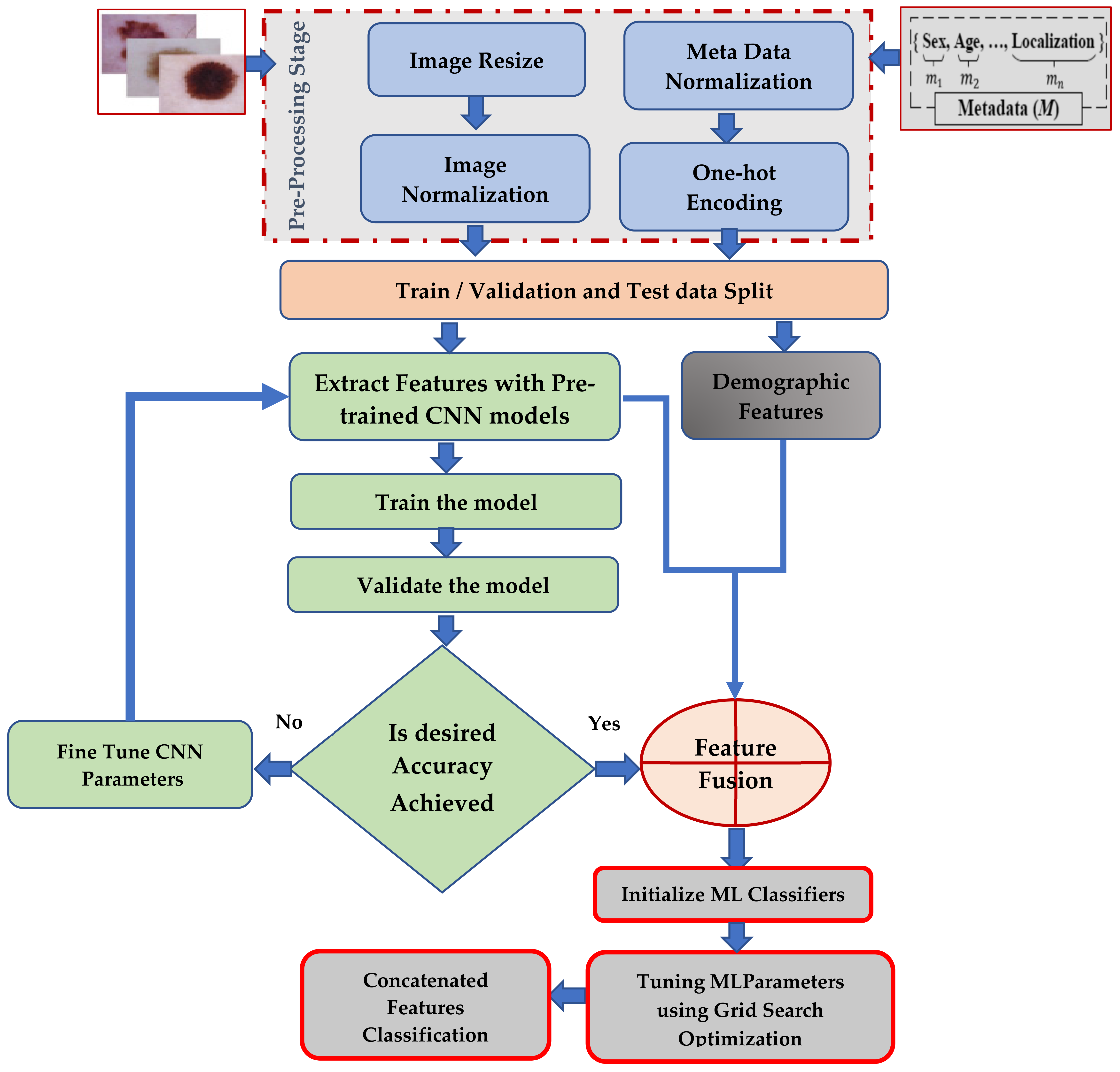
- A multi-type feature fusion approach based on image and meta-data features is developed for multi-type skin lesion detection with heterogeneous ensemble classifiers. We utilized six convolutional deep learning models, including VGG19, ResNet50, InceptionV3, InceptionResnet, Xception, and DenseNet201, which were pre-trained on the ImageNet dataset. We used transfer learning to train a public skin lesion dataset containing more than 10,000 dermoscopic images.
- The proposed approach automatically extracts features from the processed images, avoiding complex manual feature extraction processes.
- After extracting features, the patient’s metadata are fused with the extracted features to diagnose different skin lesions using machine learning classifiers.
- The experimental results show that the proposed approach achieved promising results for the diagnosis of different skin diseases.
2. Related Work
3. Materials and Methods
3.1. Dataset Description
3.2. Framework Architecture and Model Training
3.3. Stages of the Proposed Ensemble Approach
3.4. Transfer Learning (TR)
3.5. Convolution Deep Learning Model for Feature Extraction Process
4. Results
4.1. Implementation Details
4.2. Evaluation Metrics
4.3. Results
4.3.1. Classification Results on HAM10000 Dataset without Metadata
4.3.2. Classification Result of Combined Classifier
4.3.3. Comparative Study with the State-of-the-Art Systems
5. Conclusions
Author Contributions
Funding
Data Availability Statement
Acknowledgments
Conflicts of Interest
References
- Shanthi, T.; Sabeenian, R.S.; Anand, R. Automatic Diagnosis of Skin Diseases Using Convolution Neural Network. Microprocess. Microsyst. 2020, 76, 103074. [Google Scholar] [CrossRef] [Scilit]
- American Cancer Society. Key Statistics for Melanoma Skin Cancer. 2022. Available online: https://www.cancer.org/cancer/melanoma-skin-cancer/about/key-statistics.html (accessed on 27 October 2022).
- Wei, L.-S.; Gan, Q.; Ji, T. Skin Disease Recognition Method Based on Image Color and Texture Features. Comput. Math. Methods Med. 2018, 2018, 8145713. [Google Scholar] [CrossRef] [Scilit] [PubMed]
- Amarathunga, A.A.L.C.; Ellawala, E.P.W.C.; Abeysekara, G.N.; Amalraj, C.R.J. Expert System for Diagnosis Of Skin Diseases. Int. J. Sci. Technol. Res. 2015, 4, 174–178. [Google Scholar]
- Bajwa, M.N.; Muta, K.; Malik, M.I.; Siddiqui, S.A.; Braun, S.A.; Homey, B.; Dengel, A.; Ahmed, S. Computer-Aided Diagnosis of Skin Diseases Using Deep Neural Networks. Appl. Sci. 2020, 10, 2488. [Google Scholar] [CrossRef] [Scilit]
- Monisha, M.; Suresh, A.; Rashmi, M.R. Artificial Intelligence Based Skin Classification Using GMM. J. Med. Syst. 2019, 43, 3. [Google Scholar] [CrossRef] [Scilit]
- Kassem, M.A.; Hosny, K.M.; Damaševičius, R.; Eltoukhy, M.M. Machine Learning and Deep Learning Methods for Skin Lesion Classification and Diagnosis: A Systematic Review. Diagnostics 2021, 11, 1390. [Google Scholar] [CrossRef] [Scilit]
- LeCun, Y.; Bengio, Y.; Hinton, G. Deep learning. Nature 2015, 521, 444. [Google Scholar] [CrossRef] [Scilit]
- Hosny, K.M.; Kassem, M.A.; Fouad, M.M. Classification of Skin Lesions into Seven Classes Using Transfer Learning with AlexNet. J. Digit Imaging 2020, 33, 1325–1334. [Google Scholar] [CrossRef] [Scilit]
- Wu, H.; Yin, H.; Chen, H.; Sun, M.; Liu, X.; Yu, Y.; Tang, Y.; Long, H.; Zhang, B.; Zhang, J.; et al. A Deep Learning, Image-Based Approach for Automated Diagnosis for Inflammatory Skin Diseases. Ann. Transl. Med. 2020, 8, 581. [Google Scholar] [CrossRef] [Scilit]
- Khan, M.A.; Akram, T.; Sharif, M.; Kadry, S.; Nam, Y. Computer decision support system for skin cancer localization and classification. Comput. Mater. Contin. 2021, 68, 1041–1064. [Google Scholar]
- Alsaade, F.W.; Aldhyani, T.H.H.; Al-Adhaileh, M.H. Developing a Recognition System for Diagnosing Melanoma Skin Lesions Using Artificial Intelligence Algorithms. Comput. Math. Methods Med. 2021, 2021, 9998379. [Google Scholar] [CrossRef] [Scilit]
- Ali, S.; Miah, S.; Haque, J.; Rahman, M.; Islam, K. An enhanced technique of skin cancer classification using deep convolutional neural network with transfer learning models. Mach. Learn. Appl. 2021, 5, 100036. [Google Scholar] [CrossRef] [Scilit]
- Ameri, A. A deep learning approach to skin cancer detection in dermoscopy images. J. Biomed. Phys. Eng. 2020, 10, 801–806. [Google Scholar] [CrossRef] [Scilit]
- Manne; Ravi; Kantheti, S.; Kantheti, S. Classification of Skin cancer using deep learning, Convolutional Neural Networks-Opportunities and vulnerabilities-A systematic Review. Int. J. Mod. Trends Sci. Technol. 2020, 6, 2455–3778. [Google Scholar]
- Rajput, G.; Agrawal, S.; Raut, G.; Vishvakarma, S.K. An accurate and noninvasive skin cancer screening based on imaging technique. Int. J. Imaging Syst. Technol. 2022, 32, 354–368. [Google Scholar] [CrossRef] [Scilit]
- Raza, R.; Zulfiqar, F.; Tariq, S.; Anwar, G.B.; Sargano, A.B.; Habib, Z. Melanoma Classification from Dermoscopy Images Using Ensemble of Convolutional Neural Networks. Mathematics 2022, 10, 26. [Google Scholar] [CrossRef] [Scilit]
- Gouda, W.; Sama, N.U.; Al-Waakid, G.; Humayun, M.; Jhanjhi, N.Z. Detection of Skin Cancer Based on Skin Lesion Images Using Deep Learning. Healthcare 2022, 10, 1183. [Google Scholar] [CrossRef] [Scilit]
- Rehman, M.Z.U.; Ahmed, F.; Alsuhibany, S.A.; Jamal, S.S.; Ali, M.Z.; Ahmad, J. Classification of Skin Cancer Lesions Using Explainable Deep Learning. Sensors 2022, 22, 6915. [Google Scholar] [CrossRef] [Scilit]
- Aldhyani, T.H.H.; Verma, A.; Al-Adhaileh, M.H.; Koundal, D. Multi-Class Skin Lesion Classification Using a Lightweight Dynamic Kernel Deep-Learning-Based Convolutional Neural Network. Diagnostics 2022, 12, 2048. [Google Scholar] [CrossRef] [Scilit]
- Kousis, I.; Perikos, I.; Hatzilygeroudis, I.; Virvou, M. Deep Learning Methods for Accurate Skin Cancer Recognition and Mobile Application. Electronics 2022, 11, 1294. [Google Scholar] [CrossRef] [Scilit]
- Hasan, K.; Elahi, T.E.; Alam, A.; Jawad, T.; Martí, R. DermoExpert: Skin lesion classification using a hybrid convolutional neural network through segmentation, transfer learning, and augmentation. Inform. Med. Unlocked 2022, 28, 100819. [Google Scholar] [CrossRef] [Scilit]
- Tschandl, P.; Rosendahl, C.; Kittler, H. The HAM10000 Dataset, a Large Collection of Multi-Source Dermatoscopic Images of Common Pigmented Skin Lesions. Sci. Data 2018, 5, 180161. [Google Scholar] [CrossRef] [Scilit] [PubMed]
- Baker, A.; Nermeen; Zengeler, N.; Handmann, U. A Transfer Learning Evaluation of Deep Neural Networks for Image Classification. Mach. Learn. Knowl. Extr. 2022, 4, 22–41. [Google Scholar] [CrossRef] [Scilit]
- Szegedy, C.; Vanhoucke, V.; Ioffe, S.; Shlens, J.; Wojna, Z. Rethinking the Inception Architecture for Computer Vision. In Proceedings of the 2016 IEEE Conference on Computer Vision and Pattern Recognition (CVPR), Las Vegas, NV, USA, 27–30 June 2016; pp. 2818–2826. [Google Scholar]
- He, K.; Zhang, X.; Ren, S.; Sun, J. Deep Residual Learning for Image Recognition. In Proceedings of the 2016 IEEE Conference on Computer Vision and Pattern Recognition (CVPR), Las Vegas, NV, USA, 27–30 June 2016; pp. 770–778. [Google Scholar] [CrossRef] [Scilit]
- Simonyan, K.; Zisserman, A. Very deep convolutional networks for large-scale image recognition. arXiv 2014, arXiv:1409.1556. [Google Scholar]
- Chollet, F. Xception: Deep Learning with Depthwise Separable Convolutions. In Proceedings of the 2017 IEEE Conference on Computer Vision and Pattern Recognition (CVPR), Honolulu, HI, USA, 21–26 July 2017; pp. 1800–1807. [Google Scholar]
- Huang, G.; Liu, Z.; van der Maaten, L.; Weinberger, K.Q. Densely connected convolutional networks. In Proceedings of the IEEE Conference Computer Vision and Pattern Recognition, Honolulu, HI, USA, 21–26 July 2017; pp. 4700–4708. Available online: https://arxiv.org/abs/1608.06993 (accessed on 14 October 2022).
- Szegedy, C.; Ioffe, S.; Vanhoucke, V.; Alemi, A.A. Inception-v4, Inception-Resnet and the Impact of Residual Connections on Learning. In Proceedings of the Thirty-First AAAI Conference on Artificial Intelligence, San Francisco, CA, USA, 4–9 February 2017; pp. 4278–4284. [Google Scholar]
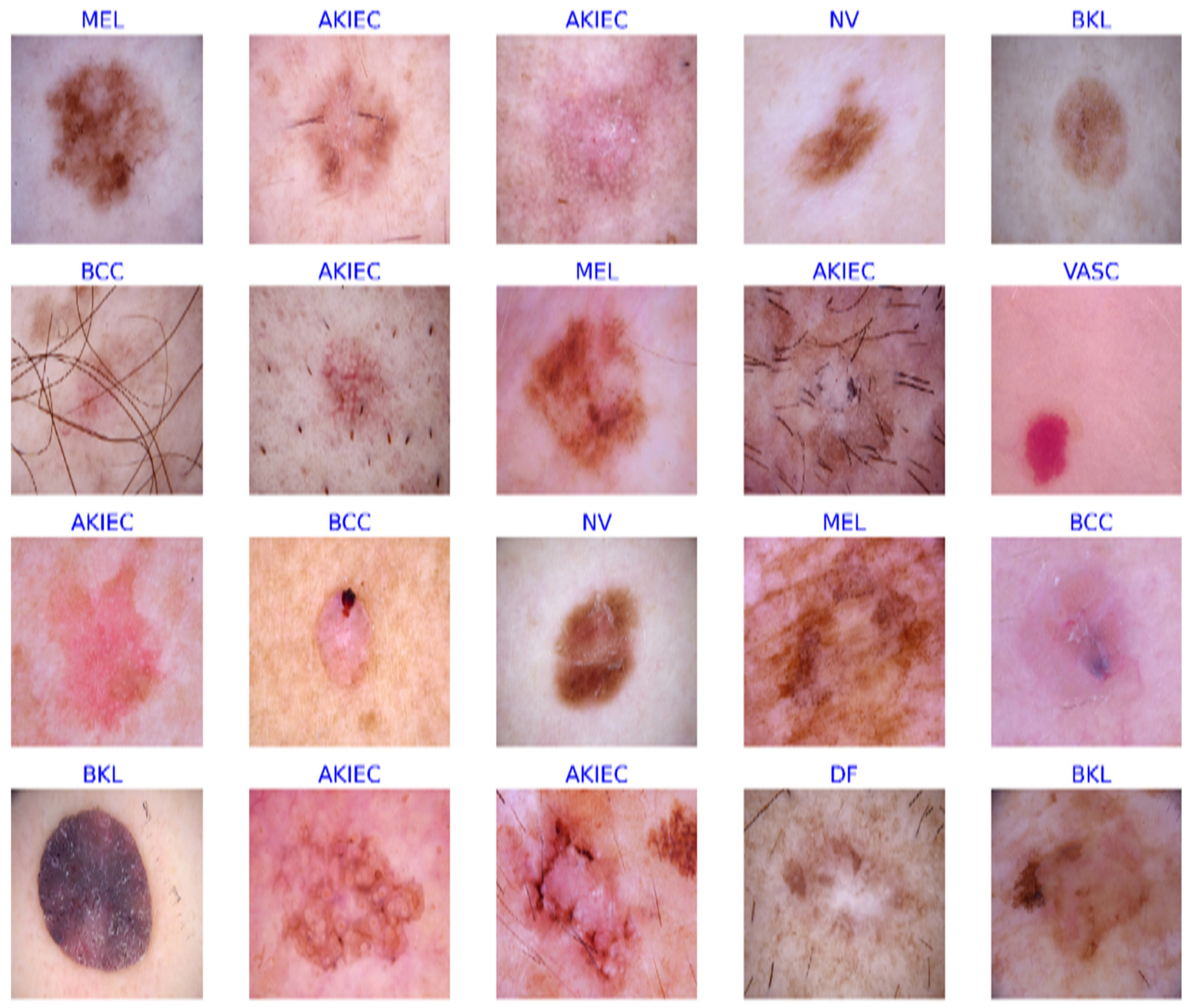



| Class | Training Set | Testing Set | Validation Set | Total |
|---|---|---|---|---|
| Actinic Keratoses (Solar Keratoses) (akeic) | 222 | 92 | 13 | 327 |
| Basal cell carcinoma (BCC) | 339 | 150 | 25 | 514 |
| Benign keratosis (BKL) | 770 | 294 | 49 | 1113 |
| Dermatofibroma (DF) | 68 | 40 | 7 | 115 |
| Melanoma (MEL) | 4340 | 2027 | 338 | 6705 |
| Melanocytic nevi (NV) | 680 | 353 | 66 | 1099 |
| Vascular skin lesions (VASC) | 90 | 49 | 3 | 142 |
| Total | 6509 | 3005 | 501 | 10,015 |
| Model | Number of Parameters | Number of Layers | Input Image Size | Kernel Size |
|---|---|---|---|---|
| Xception | 30,375,912 | 71 | 71 × 71 | 3 × 3 |
| Resnet50 | 27,857,668 | 50 | 71 × 71 | 7 × 7 |
| DenseNet201 | 22,331,392 | 121 | 71 × 71 | 5 × 5 |
| InceptionV3 | 21,817,127 | 48 | 75 × 75 | 3 × 3 |
| VGG19 | 20,027,975 | 19 | 75 × 75 | 3 × 3 |
| InceptionResNetV2 | 54,347,495 | 164 | 71 × 71 | 3 × 3 |
| Hyper-Parameters | Value |
|---|---|
| Number of epoch | 100 |
| Batch size | 64 |
| Pooling | Global average pooling |
| Optimizer | Adam |
| Initial learning rate | 1 × 10⁻⁴ |
| Dropout | 0.5 |
| Patience | 10 |
| Loss function | Categorical-cross entrop |
| Model | SEN (%) | SPE (%) | PRE (%) | DSC (%) | ACC (%) | Training Time (s) | Testing Time (s) |
|---|---|---|---|---|---|---|---|
| Xception | 92.29 | 98.68 | 93.69 | 92.96 | 96.83 | 328.1691 | 12.1131 |
| Resnet50 | 93.23 | 99.02 | 95.49 | 94.44 | 96.93 | 353.5990 | 11.6892 |
| DenseNet201 | 92.76 | 98.88 | 97.45 | 94.74 | 97.16 | 594.0691 | 16.3945 |
| InceptionV3 | 91.52 | 98.43 | 93.44 | 92.46 | 96.62 | 376.2660 | 12.8734 |
| VGG19 | 92.96 | 99.09 | 91.31 | 92.11 | 96.97 | 411.8653 | 11.1327 |
| InceptionResnet | 92.79 | 98.87 | 94.87 | 93.28 | 96.65 | 746.5525 | 26.3085 |
| Model | SEN(%) | SPE(%) | PRE(%) | DSC(%) | ACC(%) | Test Time (s) |
|---|---|---|---|---|---|---|
| VGG19 + RF | 93.39 | 99.72 | 97.80 | 95.39 | 99.73 | 9.5132 |
| VGG19 + LR | 97.90 | 99.95 | 98.79 | 98.12 | 99.92 | 9.4659 |
| VGG19 + SVM | 90.74 | 98.87 | 94.81 | 92.57 | 99.19 | 10.6649 |
| Model | SEN(%) | SPE(%) | PRE(%) | DSC(%) | ACC(%) | Test Time (s) |
|---|---|---|---|---|---|---|
| InceptionV3 + RF | 92.72 | 98.73 | 95.61 | 94.08 | 99.01 | 9.8076 |
| InceptionV3 + LR | 99.49 | 99.83 | 99.17 | 99.24 | 99.90 | 9.7577 |
| InceptionV3 + SVM | 91.14 | 98.37 | 95.60 | 93.26 | 98.84 | 11.0787 |
| Model | SEN(%) | SPE(%) | PRE(%) | DSC(%) | ACC(%) | Test Time (s) |
|---|---|---|---|---|---|---|
| Resent50 + RF | 93.15 | 99.59 | 97.18 | 94.68 | 99.44 | 10.6397 |
| Resent50 + LR | 96.83 | 99.77 | 98.01 | 97.16 | 99.72 | 10.5896 |
| Resent50 + SVM | 90.45 | 98.96 | 95.47 | 92.31 | 98.99 | 11.8022 |
| Model | SEN(%) | SPE(%) | PRE(%) | DSC(%) | ACC(%) | Test Time (s) |
|---|---|---|---|---|---|---|
| DenseNet201 + RF | 92.50 | 99.68 | 97.66 | 94.58 | 99.49 | 9.8076 |
| DenseNet201 + LR | 95.08 | 99.81 | 98.23 | 96.99 | 99.71 | 9.7577 |
| DenseNet201 + SVM | 91.55 | 99.22 | 96.49 | 93.37 | 99.21 | 11.0787 |
| Model | SEN(%) | SPE(%) | PRE(%) | DSC(%) | ACC(%) | Test Time (s) |
|---|---|---|---|---|---|---|
| Xception + RF | 94.09 | 99.41 | 97.62 | 95.54 | 99.93 | 10.1391 |
| Xception + LR | 98.16 | 99.96 | 99.24 | 98.78 | 99.36 | 10.0877 |
| Xception + SVM | 91.48 | 98.82 | 97.01 | 93.90 | 99.09 | 11.3043 |
| Model | SEN(%) | SPE(%) | PRE(%) | DSC(%) | ACC(%) | Test Time (s) |
|---|---|---|---|---|---|---|
| InceptionResnetV2 + RF | 93.25 | 99.51 | 97.43 | 95.26 | 99.34 | 19.9461 |
| InceptionResnetV2 + LR | 98.84 | 99.97 | 99.59 | 99.16 | 99.94 | 21.1139 |
| InceptionResnetV2 + SVM | 92.21 | 98.88 | 96.85 | 94.05 | 98.99 | 21.5670 |
| Work | Year | Dataset | Method | ACC | SEN | SPE |
|---|---|---|---|---|---|---|
| Prposed Syatem | 2022 | HAM10000 | DenseNet201 + logistic regression (LR) | 99.94% | 98.84% | 99.97% |
| Bajwa et al. [5] | 2020 | DermNet and ISIC datasets | 92.4% for DermNet, 93%, for ISIC | |||
| Ameri [14] | 2020 | HAM10000 | 84% | 81% | 88% | |
| Manne et al. [15] | 2020 | HAM10000 and PH2 | 98.16%, And 96% | |||
| Khan et al. [11] | 2021 | HAM10000, ISBI2018, and ISBI2019 | 95.8%, 97.1%, and 85.35%, | |||
| Alsaade et al. [12] | 2021 | HP2 and ISIC 2018 | (97.50%), (98.35%) | |||
| Ali et al. [13] | 2021 | HAM10000 | 91.93% | |||
| Rajput et al. [16] | 2022 | HAM10000 | 98.20% | 98.20% | 98.20% | |
| Raza et al. [17] | 2022 | Figshare | 97.93% | 97.83% | 97.50% | |
| Gouda et al. [18] | 2022 | ISIC 2018 | 85.8% |
Publisher’s Note: MDPI stays neutral with regard to jurisdictional claims in published maps and institutional affiliations. |
© 2022 by the authors. Licensee MDPI, Basel, Switzerland. This article is an open access article distributed under the terms and conditions of the Creative Commons Attribution (CC BY) license (https://creativecommons.org/licenses/by/4.0/).
Share and Cite
Almuayqil, S.N.; Abd El-Ghany, S.; Elmogy, M. Computer-Aided Diagnosis for Early Signs of Skin Diseases Using Multi Types Feature Fusion Based on a Hybrid Deep Learning Model. Electronics 2022, 11, 4009. https://doi.org/10.3390/electronics11234009
Almuayqil SN, Abd El-Ghany S, Elmogy M. Computer-Aided Diagnosis for Early Signs of Skin Diseases Using Multi Types Feature Fusion Based on a Hybrid Deep Learning Model. Electronics. 2022; 11(23):4009. https://doi.org/10.3390/electronics11234009
Chicago/Turabian StyleAlmuayqil, Saleh Naif, Sameh Abd El-Ghany, and Mohammed Elmogy. 2022. "Computer-Aided Diagnosis for Early Signs of Skin Diseases Using Multi Types Feature Fusion Based on a Hybrid Deep Learning Model" Electronics 11, no. 23: 4009. https://doi.org/10.3390/electronics11234009
APA StyleAlmuayqil, S. N., Abd El-Ghany, S., & Elmogy, M. (2022). Computer-Aided Diagnosis for Early Signs of Skin Diseases Using Multi Types Feature Fusion Based on a Hybrid Deep Learning Model. Electronics, 11(23), 4009. https://doi.org/10.3390/electronics11234009


