Review

Advancement of All-Trans Retinoic Acid Delivery Systems in Dermatological Application

by
Sharifah Shakirah Syed Omar
and
Hazrina Hadi
*
Dermatopharmaceutics Research Group, Faculty of Pharmacy, International Islamic University Malaysia, Kuantan 25200, Pahang, Malaysia
*
Author to whom correspondence should be addressed.
Cosmetics 2022, 9(6), 140; https://doi.org/10.3390/cosmetics9060140
Submission received: 14 November 2022 / Revised: 7 December 2022 / Accepted: 8 December 2022 / Published: 11 December 2022
(This article belongs to the Special Issue Formulations and Delivery Systems to the Skin)

Abstract

:
Dermatological conditions, such as acne, photoaging, psoriasis, and wounds, have been treated topically with all-trans retinoic acid (ATRA) for many years owing to its anti-inflammatory activity, comedolytic effect, and ability to increase collagen production. However, common side effects of ATRA known as the retinoid reaction can occur. These side effects are countered by ATRA encapsulation in solid lipid nanoparticles (SLN), nanostructured lipid carriers (NLCs), and liposomes. Liposomes used to encapsulate ATRA include niosomes, ethosomes, and transfersomes. Side effects involving inflammatory reactions, such as irritation, redness, and erythema, were diminished using these approaches. The use of such carriers enhanced the efficacy of ATRA by enhancing its permeation into skin. These formulations have been compared in terms of improving the activity of ATRA and the ability to relieve the side effects. Further research into different delivery systems for ATRA using various formulations will improve the future of topical ATRA delivery.

1. Introduction

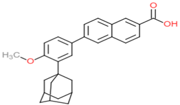
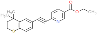
Retinoids are a group of chemicals derived from vitamin A and are structurally and functionally similar to vitamin A. Vitamin A (retinol) consists of a cyclic six-carbon ring attached to a polyene hydrophobic tail containing four isoprene units, which forms a head-to-tail structure, as shown in Figure 1. The conjugated carbon–carbon double bonds in the polyene tail make retinoids light-sensitive [1,2]. Retinoids are highly fat-soluble substances with a low molecular weight of approximately 300 Da. The term retinoids not only refer to the natural derivatives of vitamin A but also to synthetic analogues. Examples of natural analogues include retinol, retinal, and retinoic acid (RA), whereas synthetic analogues include etretinate, acitretin, and tazarotene. Retinoids can be grouped into three generations based on their properties; the first generation consists of natural retinoids, such as retinal, tretinoin, and isotretinoin, and the second generation consists of synthetic analogues, such as etretinate and acitretin, although etretinate is no longer widely available. The third generation consists of a group of polyaromatic synthetic retinoids with a cyclized polyene side chain and includes adapalene and tazarotene [3]. The cyclization of the polyene tail leads to selective activity [4,5]. The three generations of retinoids are as shown in Table 1. More recently developed retinoids include seletinoid G and trifarotene [6,7].
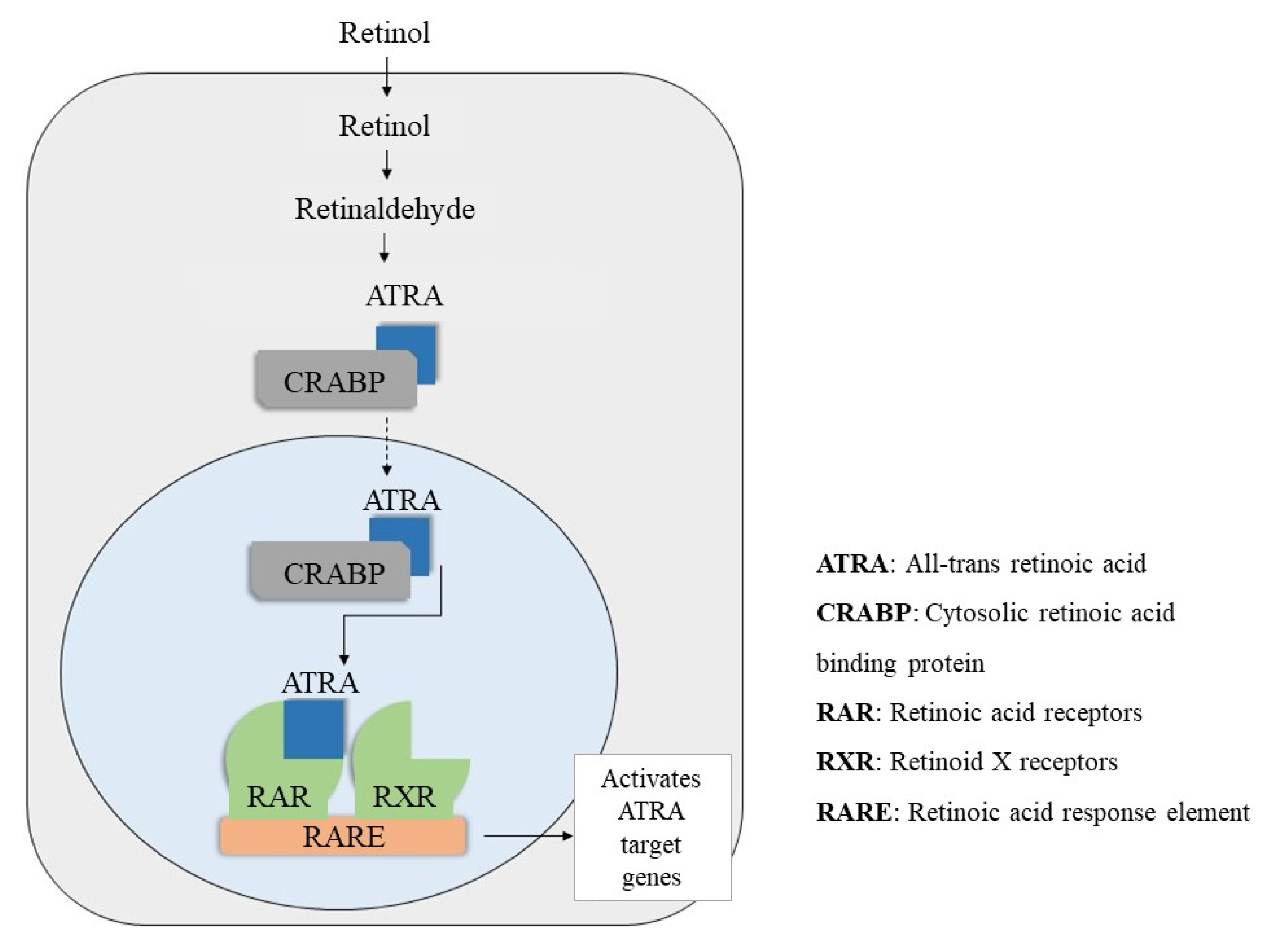
There are several retinol-binding proteins that regulate the metabolism of retinols and retinoic acid. These proteins act as carrier, which will activate receptors in the nucleus. CRBP and cytosolic retinoic acid binding protein (CRABP) both have two subtypes CRBP Ⅰ, CRBP Ⅱ, and CRABP Ⅰ and CRABP Ⅱ. Retinoids carried by carrier proteins interact with retinoid nuclear receptors (RNRs), which consist of two groups: the retinoic acid receptors (RARs) that recognize ATRA and 9-cis retinoic acid, and retinoid X receptors (RXRs) that recognize 9-cis retinoic acid only. Both groups have three isotypes—α, β, and γ—and the RAR mediates RA signalling and RXR activates other signalling pathways with other receptors, such as liver X receptors, farnesoid X receptors, and peroxisome proliferator-activated receptors. The binding of a ligand leads to dimerization of the RAR with RXR forming a heterodimer that binds to the retinoic acid response element to initiate gene transcription in the promoter region of target genes, as depicted in Figure 2 [2]. ATRA, as the most active form of vitamin A, has been used and studied in the treatment of many diseases. The use of ATRA to improve and treat different skin conditions and diseases has been researched widely with multiple different formulations developed to enhance its efficacy and reduce its toxicity. This review will describe the different dermatological applications, actions, and the different formulations used to deliver ATRA to the skin.

2. Retinoic Acid Application to Skin

2.1. Photodamage

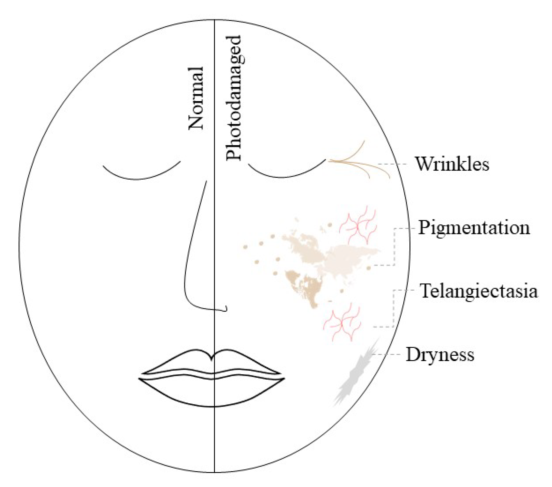
Photoaged skin is characterized by fine and coarse wrinkles, roughness, dryness, dyschromia, inelasticity, pigmentations, and telangiectasia, as depicted in Figure 3. Ultraviolet type B (UVB) is absorbed in the epidermis and can cause non-orderly maturation of keratinocytes and atypia. Aging dermis with sun protection shows a decrease in all cell types and the extracellular matrix content (collagen, elastin, glycosaminoglycan (GAG), fibroblasts, and Langerhans cells), whereas photoaged skin has the same features but also has enhanced effects of chronic inflammation shown by a lack of fibroblast and Langerhans cells surrounded by inflammatory infiltrate. Glycoproteins and GAG are also increased in photoaged skin resulting in the inability of the GAG to regulate hydration, making the skin dry and leathery [5].
Photodamage by UVR can be direct through the absorption of mostly UVB, with a small amount of ultraviolet type A (UVA), by DNA, which can cause permanent damage to mitochondrial DNA and causes oxidative stress across the entire cell. Indirect damage by UVR often occurs because of the production of reactive oxygen species (ROS) and free radicals that can easily cause oxidative damage to aged skin because of the decrease in cellular function [5]. The pathogenesis of photodamage causes negative impacts including acanthosis, which is caused by inflammatory cytokines that increase the proliferation of basal keratinocytes. GAG synthesis is increased, which results in GAG being deposited in abnormal elastotic material causing the skin to become thick, coarse, leathery, and dry because the skin is unable to bind water. Apoptosis of basal cells and keratinocytes by ROS and UVB absorption also occurs. Moreover, UVR reduces the number of retinoid receptors, causing a retinoid deficiency in the skin [8,9].
UVR can cause destruction of the dermal matrix protein, which is essential for the structure and strength of the dermis. Furthermore, collagen gene expression is inhibited, thus decreasing the synthesis of collagen [10]. Collagen depletion is caused by an increase in production of matrix metalloproteinase (MMP), which not only degrades collagen, but also elastin, in the epidermis and dermal matrix [11,12]. Another contributing factor to the degradation of collagen and elastin is the production of free radicals. The inflammatory response because of UVR exposure also causes matrix destruction by the release of inflammatory mediators and angiogenesis that allows inflammatory markers to escape and recruit inflammatory cells through hyperpermeable vessels. As a result, the skin loses its elasticity and wrinkles are formed. Dyschromia or dyspigmentation also occurs, which induces a tanning response signal, thus increasing tyrosinase activity. Additionally, hyperplasia of melanocytes takes place because of UVR exposure [8].
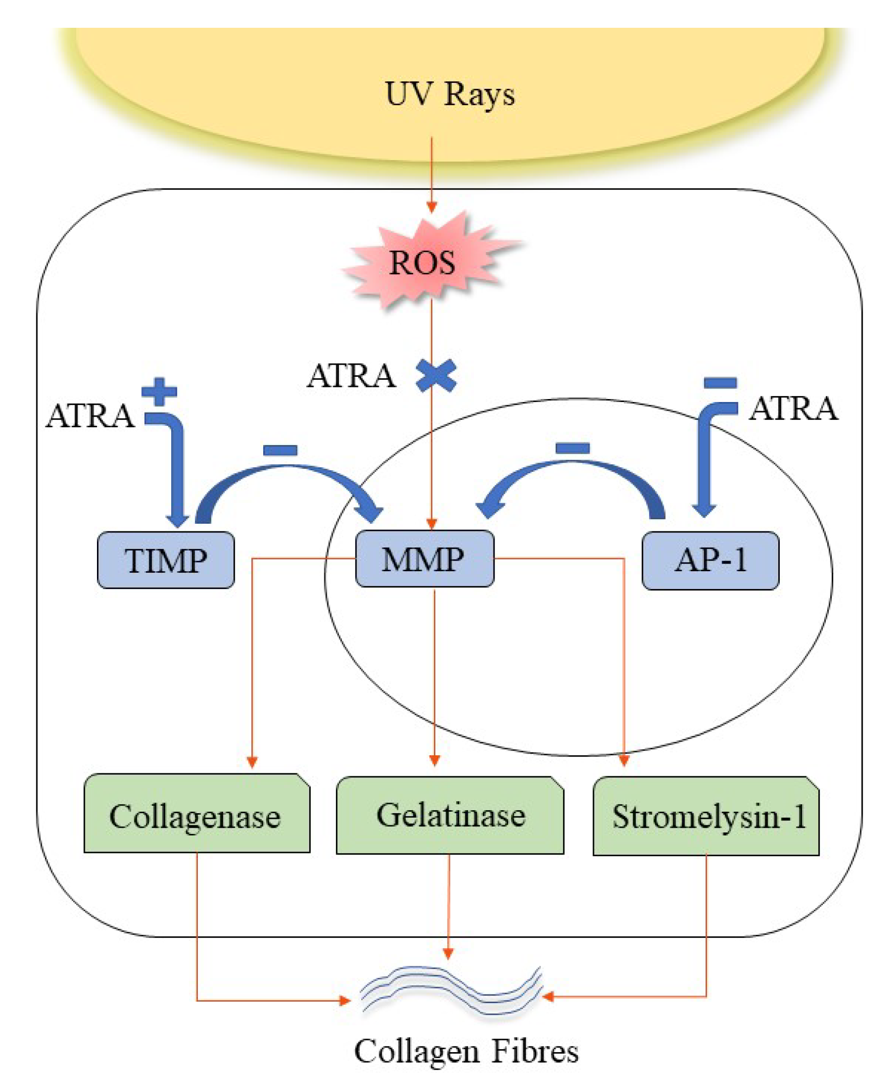
Topical retinoids can improve skin elasticity and reduce wrinkling by inhibiting the transcription of MMP via inhibition of the binding of UV activator protein 1 (AP-1) to DNA. Increased production of tissue inhibitor metalloproteinase (TIMP) also inhibits MMP, thus reversing the destruction and enhancing the production of collagen and elastic fibres, as shown in Figure 4 [8,13]. Genes related to MMP, such as collagenase, gelatinase, and stromelysin-1, are regulatory targets of retinoids, which act as antagonists, resulting in the suppression of the MMP genes [14]. A study on rodents found that ATRA, acting through RAR receptors, increased the collagen content and decreased UV-induced collagen damage in photoaged skin. ATRA and RAR agonists increased the levels of type 1 procollagen protein and reduced the levels of MMP-3 and MMP-13 (the MMP genes responsible for collagenolysis in rodents). These results indicated the possibility of ATRA reducing the levels of MMP-3 and MMP-1, which are responsible for collagen degradation in human skin through RAR receptors. UV-induced C-jun expression, which causes inhibition of procollagen synthesis, can also be inhibited by retinoic acid [11].
These mechanisms lead to the accumulation of extracellular matrix, thus restoring the skin structure along with improvements in skin elasticity, laxity, and wrinkling [11,15]. Skin texture can also be improved by inducing epidermal hyperplasia through heparin-binding epidermal-like growth factor activation and increase mucin deposition by an increase in CD44 and upregulation of hyaluronate polymerizing enzymes [8,11]. The chronic inflammatory response can be reduced because retinoids can reduce the production of proinflammatory cytokines. Retinoids also directly inhibit tyrosinase enzymes involved in the melanogenesis pathway, which in turn reduces the production of melanin and prevents further pigmentation. Dyspigmentation is further reduced by decreasing melanosome transfer to keratinocytes and increasing cell turnover, which results in the shedding of melanin-containing keratinocytes [5,8].

2.2. Acne

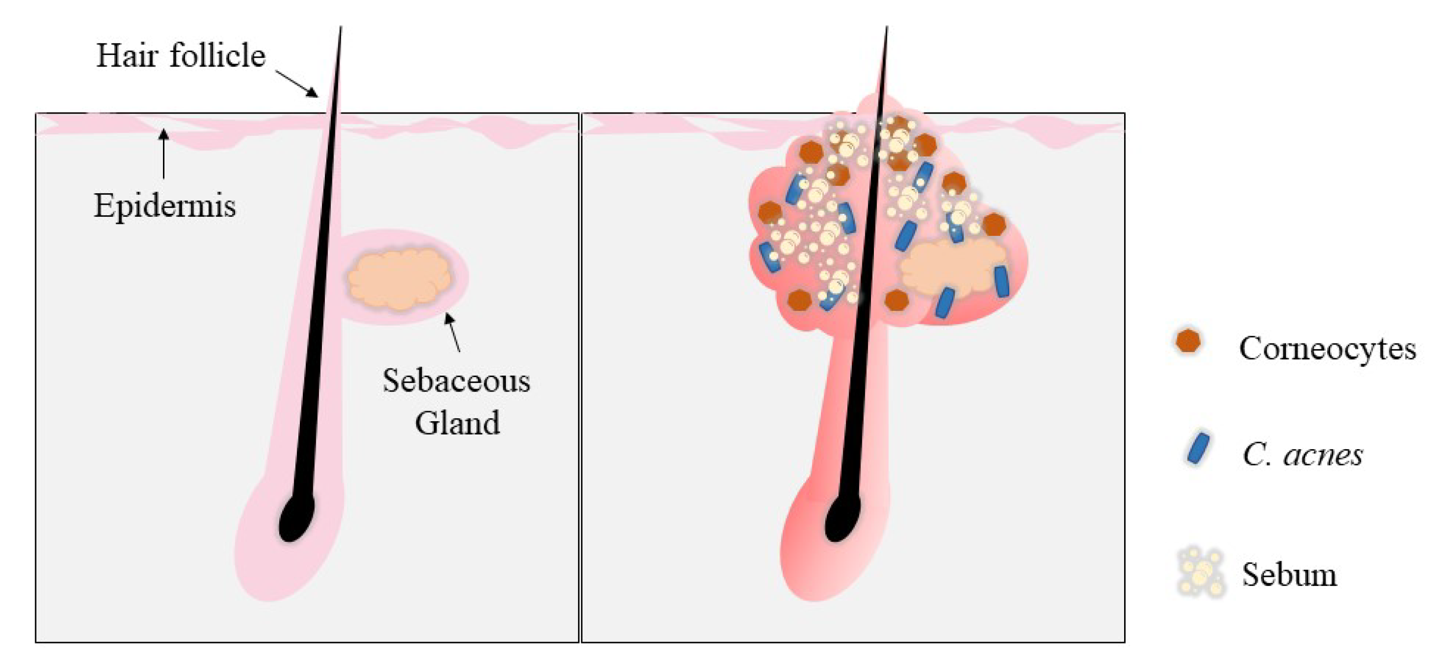
Acne can occur in all age groups, but it is especially common in teenagers. Acne can reduce patients’ quality of life along with an increased risk of anxiety and depression because of its psychological consequences [16]. Acne can be influenced by internal factors, such as medications, diet, and hormones, and external factors from the environment or cosmetics [17]. Acne is a condition that occurs as a result of hair follicles being clogged with sebum and dead skin cells. An increase in sebum production, also known as seborrhoea, can interfere with the skin’s natural desquamation process. Excess sebum binds dead skin cells to the surface, preventing the cells from shedding. Excess sebum that builds up within the sebaceous glands causes inflammation and becomes an ideal site for bacteria, specifically Cuticubacterium acnes, to breed and cause further inflammation [17,18]. The blockage of hair follicles by dead skin cells is caused by a process known as hyperkeratosis. Hyperkeratosis occurs when the external layers of the skin are abnormally thickened as a result of insufficient desquamation or shedding of dead skin cells, as well as excessive production of new skin cells or corneocytes. These cells then block the sebaceous glands by forming a plug [19,20]. The pathogenesis of this process is shown in Figure 5.
Androgen hormones that are present in both men and women can also exacerbate acne by increasing sebum production and follicular development [21]. There are two types of acne lesions: noninflammatory and inflammatory lesions. Blackheads, or open comedones, are noninflammatory lesions that form because of an enlargement of the follicular plug. Whiteheads, or closed comedones, are similar except the pore remains closed. Inflammatory lesions include papules, pustules, and nodules [20]. Papules are swollen and inflamed closed comedones, whereas pustules are comedones that are inflamed to the point of rupturing into the skin. Nodules are acne lesions that have ruptured into the skin and are severely inflamed with a large, tender, and swollen appearance [19].
ATRA has been found to be one of the retinoids with the highest efficacy against acne compared with more recently developed retinoids, such as adapalene [22,23]. Retinoids act directly on the primary lesions, also known as microcomedones, through a comedolytic action. Retinoids act on keratinocytes by binding to nuclear RARs, which are abundant in human skin, especially the RARɣ [24]. This interaction allows the normalization of desquamation through regulating the differentiation and proliferation of keratinocytes, thus clearing the existing follicular plug and preventing new follicular plugging [20,25]. The anti-inflammatory actions take place by retinoids blocking inflammatory pathways involving toll-like receptors, leukocyte migration, and the AP-1 pathway, which inhibits inflammation by reducing the release of cytokines and nitric oxide [26,27]. As well as targeting acne, retinoids can also improve secondary acne lesions, in particular acne scarring and pigmentation. Scarring is improved by the upregulation of procollagen genes and pigmentation is reduced by the acceleration of epidermal turnover and inhibition of melanosome transfer [27]. ATRA can be used to manage inflammatory acne, non-inflammatory acne, and as a maintenance or preventive therapy [28].

2.3. Psoriasis

Psoriasis is an immune-mediated inflammatory disease that may present itself in infancy, childhood, or adolescence [29]. Psoriasis is categorized into mild, moderate, and severe. Mild psoriasis can appear in the form of a rash; the skin becomes scaly in moderate psoriasis and in severe psoriasis and red patches may emerge on the skin surface [30]. Retinoids may help this condition by inducing cell differentiation, inhibiting cell proliferation and exerting immunomodulatory and inflammatory effects [28,31]. Retinoic acid has been used as pre-treatment before gene transfection and was found to enhance the efficiency of transfection. Retinoic acid acts by binding to the RAR and RXR forming a heterodimer that binds a specific DNA sequence. This binding leads to a reduction in the production of cytokines that play important roles in the inflammatory and immune response involved in psoriasis pathogenesis [32]. The interleukin-1 (IL1) family has a significant role in the immune and inflammatory processes in psoriasis and members of the IL-1 family are overexpressed in psoriatic plaques. Retinoic acid in combination with vitamin D has been shown to be able to suppress the excessive gene expression of the IL-1 family caused by pro-inflammatory stimuli. This combination may be able to inhibit the pro-inflammatory effects of IL-1 cytokines [33].

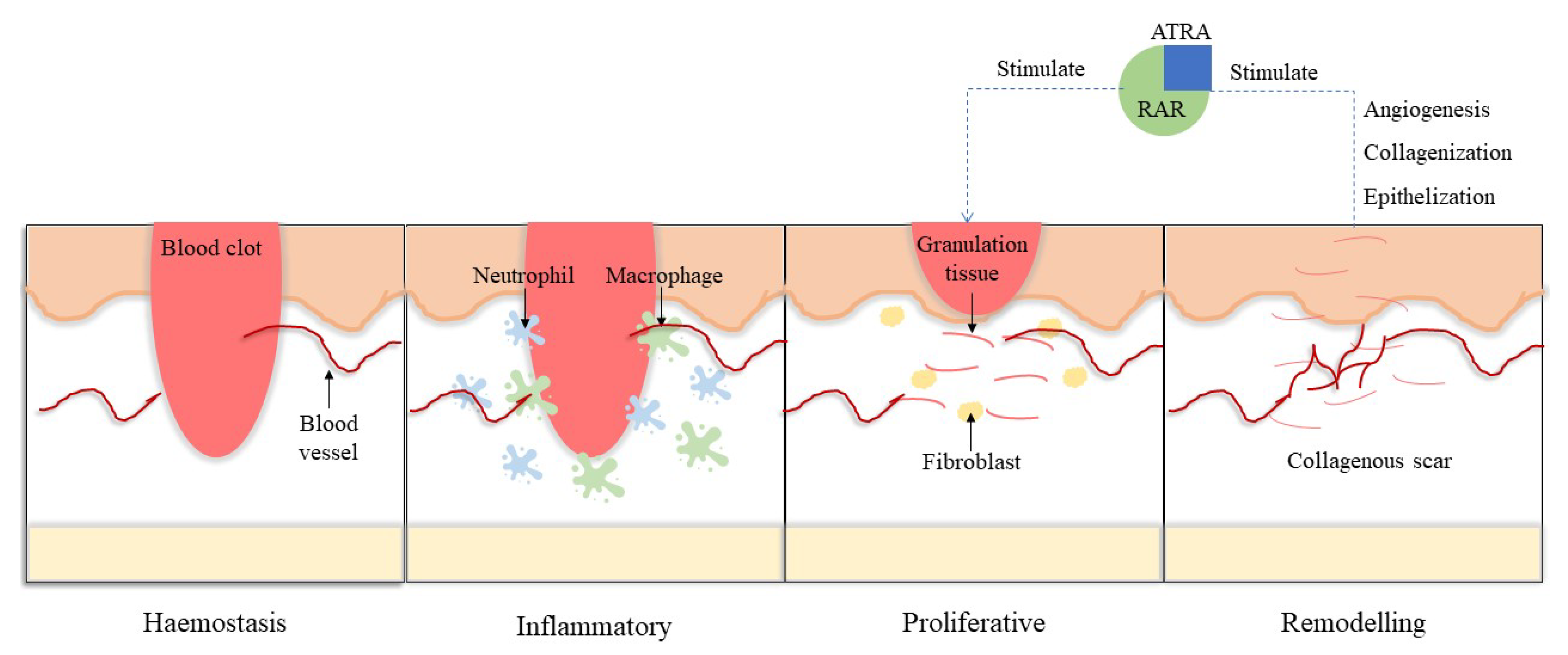
2.4. Wound Healing

Wounds result from an injury that causes a break or a cut in the skin from a harsh impact. Wounds can be classified as acute, such as cuts and burns, and chronic that involve an underlying condition, such as a diabetic wound [34]. Wound healing occurs in three main stages: the inflammatory, proliferative, and remodelling stages [35]. When a wound occurs, the blood vessels constrict and blood coagulates, forming a fibrin network to stop the bleeding, establish haemostasis, and build a barrier. The inflammatory stage is a localized protective response to lesions that begins with the influx of leukocytes, mainly neutrophils. The inflammatory response contributes to wound healing by the release of antimicrobial substances, resolving the fibrin network, the clean-up of cell debris, encouraging angiogenesis, and reepithelialisation [35,36]. However, the most important factors in the transition from inflammation to proliferation are macrophages. Macrophages can be classified into M1 and M2. M1 macrophages are involved in phagocytic activity and act as proinflammatory mediators and M2 macrophages are anti-inflammatory mediators that also produce extracellular matrix, prompt fibroblast proliferation, and phagocytize neutrophils [36]. The proliferative stage involves wound contraction and reepithelization, occurring with the help of fibroblasts, which proliferate into granulation tissue, and keratinocyte migration. Finally, the remodelling stage is when wound closure takes place and a collagenous scar is formed through granulation tissue remodelling [35,36].
Retinoic acid is able to enhance wound healing through its effect on the epidermis and dermis of the wound [37]. The inhibiting effects of cortisone on the wound healing process can be reversed by retinoic acid. Retinoic acid binds to and activates intranuclear RAR, stimulating protein synthesis, which will stimulate granulation tissue, angiogenesis, collagen production, epithelialization, and fibroplasia, as shown in Figure 6 [34,37]. A study of retinoic acid application on acute wounds in normal and aged rat models showed that retinoic acid increased collagen synthesis in aged rats and returned the healing process to a normal state [37]. A study on chronic wounds using an animal model of diabetes found an improvement in wounds treated with retinoic acid compared with the control. The wounds showed an increase in the levels of collagen, reduced leukocyte infiltrate, and less scar tissue [38]. Single and repeated application of tretinoin on a full-thickness acute wound on rats showed significantly higher fibroblast cells during the proliferation phase compared to control [39]. The ability of retinoic acid to promote wound healing extends to corneal epithelial cells, where retinoic acid is necessary for the proliferation and differentiation of corneal epithelial cells. Early barrier function recovery, wound size reduction, increased glycoprotein synthesis, and mucin release are achieved with retinoic acid use [40]. Thus, retinoic acid plays an integral part in all stages of wound healing [34].

3. Retinoic Acid Side Effects

Retinoic acid is mostly used topically to treat acne, psoriasis, photodamage, and wound healing. Despite its ability to treat and alleviate these conditions, retinoic acid also has some unacceptable side effects that cause low tolerability and adherence [3]. One of the most common and well-known side effects that occurs with most topical retinoids is the retinoid reaction that consists of pruritus, burning sensation, scaling, xerosis, peeling, erythema, and oedema. These dose-dependent symptoms are more common with retinoic acid and tazarotene than other retinoids and usually appear in the early treatment stages and gradually resolve [5,41]. This reaction occurs in up to 95% of retinoic acid users and 15% of users stop using retinoic acid topical therapy [42]. The retinoid reaction often occurs in patients with ‘sensitive skin’ and is characterized by feeling of tightness, stinging, burning, tingling, pain, or pruritus [5,43]. Research findings show that retinoids are still under prescribed as acne treatments. Retinoids are not prescribed by 41.2% of dermatologists and 67.6% of non-dermatologists, which may be because of concerns regarding side effects and patient compliance [44].
A possible mechanism for these retinoid reactions is via cytokines in the inflammatory pathway, mainly human monocyte chemoattractant protein-1 and human interleukin-8, which are detected through increased levels of mRNA expression and protein secretion. The mRNA levels were detected through protein levels in the culture supernatants of human epidermal keratinocytes, fibroblasts, and melanocytes treated with retinol/retinoic acid [45]. Another pathway that may contribute to the retinoid reaction is the activation of the irritant receptor of capsaicin, which is a compound in chilli peppers that evokes pain and inflammation. The irritant receptor of capsaicin is also known as the transient receptor potential channel vanilloid subtype-1, which has been identified as an ionotropic receptor for retinoids [46].

4. Type of Delivery Systems for Retinoic Acid

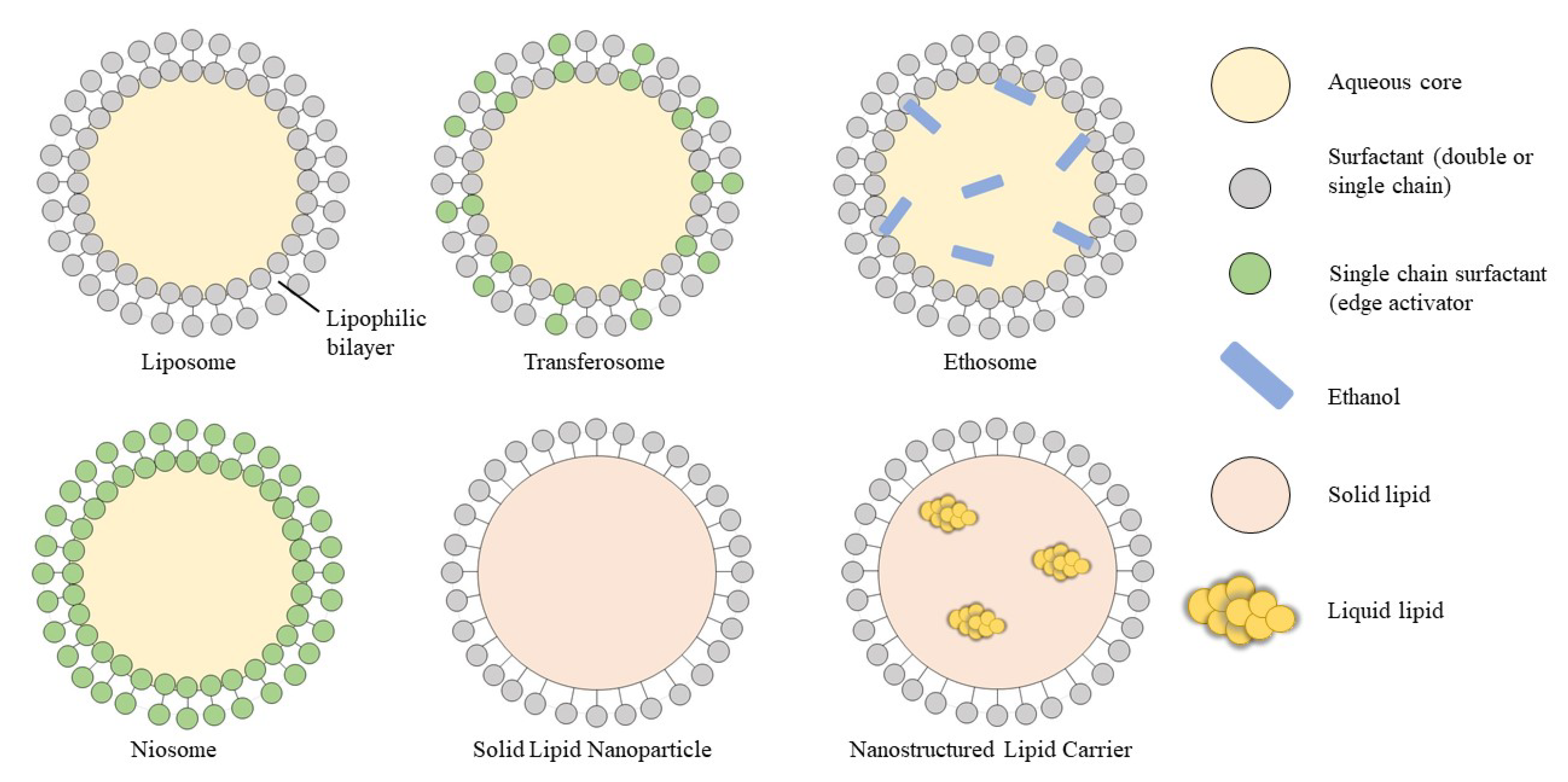
ATRA has been found to be effective in the management of multiple dermatologic diseases and has been developed into various topical formulations. However, several limitations of ATRA require further research to alleviate the adverse effects and improve its stability while maintaining or enhancing the activity towards the target. ATRA has proven to be effective and potent, but its lack of solubility in water imparts a challenge for its delivery. Furthermore, ATRA is unstable in the presence of air, light, and heat [47]. When considering the topical dosage form, retinoic acid is also limited by the skin reactions that it causes, which include erythema, irritation, and peeling [48,49]. Hence, encapsulation of ATRA in a colloidal carrier system is the best strategy to overcome these challenges. Some of the drug delivery systems used for ATRA include solid lipid nanoparticles (SLNs), nanostructured lipid carriers (NLCs), liposomes, ethosomes, and niosomes, as illustrated in Figure 7. The different types of formulations and its method of preparation are also summarised in Table 2.

4.1. Veisuclar Drug Delivery Systems

Many different drug delivery systems have been extensively explored in the past few decades to determine the best delivery systems for different drugs to target different diseases. Considerable attention has been given to vesicular drug delivery systems. A vesicular drug delivery system is a highly ordered assembly of concentric bilayers formed in the presence of water by self-assembling amphiphilic building blocks [59]. Liposomes are a type of vesicular carrier that have been well studied because of their various forms. Liposomes are spherical carriers with an internal aqueous core surrounded by drug molecules [47]. Liposomes are able to increase the efficacy and stability of drugs while reducing the toxicity and, most importantly, enabling targeted delivery [60]. A study on ATRA encapsulated in liposomes showed that the presence of cholesterol or a negative charge inducer had opposite effects on ATRA release. Cholesterol significantly lowered the percentage of ATRA released from the liposomes inducing a sustained release effect, whereas the negative charge inducer significantly increased ATRA release. Clinically, ATRA in liposomes has shown superior efficacy against non-inflammatory lesions and less irritation potential for erythema compared with free ATRA and a marketed product [61]. However, issues of low solubility, leaking, and fusing of the encapsulated drug as well as the high production cost have stimulated further research [59,62].
As a result, niosomes, ethosomes, transferosomes, and many other delivery systems have been developed and applied to the delivery of ATRA. Niosomes are vesicular carriers with non-ionic surfactants; ethosomes contain a high percentage of ethanol and transfersomes, or deformable liposomes, and use a surfactant as an edge activator to destabilize the lipid bilayers [60,63,64]. Liposomes, hexosomes, glycerosomes, and ethosomes could all produce vesicles with similar zeta potentials and polydispersity indices; however, the entrapment efficiency and particle sizes were significantly better in hexosomes than in the other formulations. In in vitro and in vivo studies, the highest skin retention in the stratum corneum, epidermis, and dermis was observed with hexosomes and the least retention with liposomes. Liposomes also showed some permeation through the skin, unlike the other formulations, indicating that the other vesicular formulations were able to provide a targeted effect in the skin layers while reducing systemic side effects because there was less skin permeation. Hexosomes also showed superior anti-rosacea activity, whereas liposomes were unable to suppress inflammatory cell recruitment [51].
When liposomes were compared to niosomes in penetration enhancer containing vesicles (PEVs), niosomes were found to have similar properties and performance to liposomes in terms of drug incorporation and ATRA skin delivery when the penetration enhancer Labrasol® was added. Liposome, liposome-PEV, noisome, and niosome-PEV formulations were prepared using Labrasol® as the penetration enhancer in the PEV formulation. Both liposome formulations had a smaller particle size, a more negative zeta potential, and higher entrapment efficiency compared with the niosome formulations. The niosome characteristics were significantly improved with the addition of Labrasol®. None of the formulations permeated through the skin, instead ATRA accumulated in the skin with the liposomes showing the highest skin drug deposition. Niosomes had the lowest drug deposition, but this was significantly improved by the addition of Labrasol® [52].
ATRA encapsulated in deformable liposomes was also studied through a full factorial design with nine formulations using different ratios of soy phosphatidylcholine (SPC) and transcutol. Higher levels of SPC showed better encapsulation efficiency and slower drug release to a certain extent, whereas a high amount of transcutol had the opposite effect. A drug release study showed an initial burst and then reduced release of ATRA, except for formulation seven (SPC/transcutol = 25:5). Optimized formulations were prepared with ratios of SPC/transcutol of 15.5:14.5, 24:7, and 25:5. The levels of penetration of the optimized formulations were all higher than that of tretinoin cream, with the 15.5:14.5 formulation having the highest penetration as the optimized amount of transcutol acted as a solubilizer. The PEVs had low irritation compared with tretinoin cream in an in vivo skin irritation study [47]. Formulation in liposomes also improved the characteristics of ATRA, and good permeation through the skin was observed because of the nature of the liposomes. In an advanced version of the formulation, the properties were improved with better permeation as observed with hexosomes. Liposomes have the flexibility to be adjusted and modified to suit different goals in disease management.

4.2. Lipid Nanoparticles

The use of lipid nanoparticles is an advanced alternative to traditional colloidal carrier systems suitable for lipophilic drug delivery, such as emulsions. With the benefits of colloidal carriers but with fewer shortcomings, lipid nanoparticles can encapsulate both hydrophilic and lipophilic drugs equally well. Lipid nanoparticles can be categorized into four types: SLNs, NLCs, lipid drug conjugates, and polymer lipid hybrid nanoparticles [65,66]. The lipid matrix of SLNs consists of solid lipids with a melting point above 40 °C, ensuring that the matrix remains solid at normal room and body temperatures [54,67]. SLNs have the advantages of reduced toxicity, increased loading capacity, controlled release, enhanced stability, biodegradability, and ease of upscaling to an industrial scale because of the low cost and organic-solvent-free process [23,38,49]. SLNs also have a large surface area to enable better penetration through the skin and to release the active molecules in a controlled manner [55].
A formulation of ATRA loaded into SLNs showed improvements in the ATRA properties and activity. The efficacy of SLNs loaded with ATRA was assessed based on the desired use. For a topical acne formulation, a rhino mouse model was used to compare a commercial formulation with SLNs loaded with ATRA. In this model, the encapsulated ATRA produced a comedolytic effect and caused epidermal proliferation similar to marketed products and the epidermal granular layer showed greater thickening compared with the commercial formulations. Furthermore, the study showed a significant reduction in skin irritation because of the gradual distribution of ATRA in the skin through the sustained delivery [23,38]. The effects of ATRA on chronic wound healing were assessed using a diabetic mouse model. Wound healing was improved via a reduction in macrophages and neutrophil infiltration. Collagen deposition was also increased, specifically type III collagen, which is associated with healing with reduced scar tissue. ATRA-SLN improved the wound healing without any detrimental effects although applied directly to the wound bed [38]. A study found that ATRA had a low loading capacity in SLNs because of a low solubilization rate in the lipid but had high encapsulation efficiency, which was attributed to the lipophilicity of ATRA. The addition of chitosan to the formulation resulted in less cytotoxicity towards keratinocytes and better antibacterial properties against C. acnes and Staphylococcus aureus, making chitosan a good candidate for ATRA delivery [53].
The encapsulation efficiency of SLNs for ATRA is, however, quite low. The encapsulation efficiency could be improved by forming an ion pair between ATRA and a lipophilic amine, such as maprotiline or benethamine [38,49]. The use of the ion pair improved the encapsulation efficiency and enhanced the particle size and zeta potential of the particles [38]. To investigate further improvements in the permeation, limonene, a terpene, was added as a skin permeation enhancer, which improved the solubility of ATRA, thus also increasing the drug loading capacity. As a skin penetration enhancer, limonene improved the ATRA skin permeation by increasing the ATRA solubility within the skin and its partitioning into the skin [55]. SLNs have been shown to be beneficial for the encapsulation of ATRA, not only by improving its efficacy in managing different skin issues, but also in alleviating unwanted side effects. Some problems that occurred because of the chemical properties of ATRA were resolved by adding an ingredient that could act synergistically with ATRA.
NLCs are composed of a mixture of both a solid and a liquid lipid matrix, with a lower melting point compared with SLNs, but still maintain a solid state at room and body temperature. The development of NLCs was aimed to overcome the limitations observed with the lipid matrix of SLNs. With the addition of a liquid lipid that has varied density in the fatty acid chain, a less ordered structure in the lipid matrix is produced, enabling a much greater drug loading capacity. This less ordered structure in the solid lipid matrix also means the carrier system is more thermodynamically stable [48,56,67]. A formulation of ATRA loaded in NLCs for topical application had high entrapment efficiency, especially with the addition of cholesterol. The use of a sonication method produced NLCs with a smaller particle size and polydispersity index. ATRA loaded in NLCs showed a reduction in the initial drug release and a sustained release effect in studies using Franz cells. Skin irritancy tests for ATRA in NLCs indicated there was no irritation or erythema after 7 days of application, which was in contrast to a marketed formulation. The stability was also improved as there are no changes in the particle size and no significant drug loss, after 4 weeks [56].
The effects of different factors, such as the drug amount, type of lipid, and solid to liquid lipid ratio of the NLCs have been investigated and the best NLC formulation was compared with SLN and suspension formulations. All of the formulations showed an encapsulation efficiency above 95%, but oils with less solubility towards ATRA, such as soybean oil, resulted in slightly less encapsulation. All NLC formulations showed a faster and greater cumulative drug release than SLN formulations. A high amount of medium chain triglycerides (MCTs) resulted in the slowest release rate, but MCTs in general showed better release rates than oleic acid, linoleic acid, or soybean oil. Among the NLCs, the permeation was greatest with a high drug amount, a solid/liquid lipid ratio of 2:1, and with oleic acid because of its superior permeation enhancing properties. Formulations with SLNs had the best permeation, followed by NLC and suspension formulations [54].
In a study of SLN, NLC, and nano-emulsion (NE) formulations, all of the formulations had acceptable characteristics, with the NE having the least desirable characteristics in terms of size, yield, and ATRA content. The permeation of ATRA was improved by the addition of a terpene, especially with limonene in comparison with 1,8-cineole, used at a high percentage. The highest permeation was obtained with SLN, followed by NLC, NE, and suspension formulations because of the better occlusion effect provided by the solid lipid [55]. For the encapsulation of retinoic acid with NLCs, the same ion pairing method used for SLNs has been used to improve the encapsulation efficiency of ATRA. A study used benethamine as the lipophilic amine to pair with ATRA, which resulted in higher encapsulation as the lipophilic properties of ATRA were enhanced by benethamine enabling better incorporation of ATRA into the lipid matrix [48]. When tested for anti-psoriatic activity, ATRA in NLCs and liposomes showed enhanced orthokeratosis, which indicated better anti-psoriatic activity compared with marketed gel products and SLN and ethosome formulations [50]. Improvements in the properties of ATRA were also obtained using NLCs; however, when compared with SLNs, NLCs were inferior in terms of the permeation and controlled drug release but could be superior in terms of efficacy.

5. Conclusions

All trans-retinoic acid has a profound role in the treatment and cure of multiple dermatological such as acne, psoriasis, photoaging, and wound healing. Being the most activated retinoid that mediates many cellular functions, ATRA acts through retinoid nuclear receptors that lead to gene transcription and expression. Given its ample benefits, different delivery systems are continuously being researched in order to improve the efficacy of ATRA to manage the targeted disease. Different novel delivery systems have shown many superior traits in comparison with traditional tretinoin formulation in the market. The efficacy and properties of ATRA improves with the use of these carriers. Unwanted side effects have also been tremendously reduced through encapsulation and the controlled release contributed by these carriers. The importance and advantages of each delivery system may differ from one another, yet it can all be beneficial to a certain condition.

Author Contributions

Conceptualization, H.H. and S.S.S.O.; writing—original draft preparation, S.S.S.O.; writing—review and editing, S.S.S.O.; visualization, H.H.; supervision, H.H. All authors have read and agreed to the published version of the manuscript.

Funding

This research received no external funding.

Acknowledgments

The authors acknowledge International Islamic University Malaysia (IIUM) for administrative and technical support. We thank Victoria Muir, from Edanz Group (https://en-author-services.edanz.com/ac, access date 14 January 2021) for editing a draft of this manuscript in January 2021.

Conflicts of Interest

The authors declare no conflict of interest.

References

  1. Barua, A.B.; Furr, H.C. Properties of Retinoids. Mol. Biotechnol. 1998, 10, 167–182. [Google Scholar] [CrossRef] [PubMed]
  2. Kam, R.K.T.; Deng, Y.; Chen, Y.; Zhao, H. Retinoic Acid Synthesis and Functions in Early Embryonic Development. Cell Biosci. 2012, 2, 11. [Google Scholar] [CrossRef] [PubMed] [Green Version]
  3. Baldwin, H.; Webster, G.; Stein Gold, L.; Callender, V.; Cook-Bolden, F.E.; Guenin, E. 50 Years of Topical Retinoids for Acne: Evolution of Treatment. Am. J. Clin. Dermatol. 2021, 22, 315–327. [Google Scholar] [CrossRef] [PubMed]
  4. Zasada, M.; Budzisz, E. Retinoids: Active Molecules Influencing Skin Structure Formation in Cosmetic and Dermatological Treatments. Adv. Dermatol. Allergol. 2019, 36, 392–397. [Google Scholar] [CrossRef] [Green Version]
  5. Hubbard, B.A.; Unger, J.G.; Rohrich, R.J. Reversal of Skin Aging with Topical Retinoids. Plast. Reconstr. Surg. 2014, 133, 481–490. [Google Scholar] [CrossRef] [PubMed]
  6. Kim, M.S.; Lee, S.R.; Rho, H.S.; Kim, D.H.; Chang, I.S.; Chung, J.H. The Effects of a Novel Synthetic Retinoid, Seletinoid G, on the Expression of Extracellular Matrix Proteins in Aged Human Skin In Vivo. Clin. Chim. Acta 2005, 362, 161–169. [Google Scholar] [CrossRef]
  7. Balak, D.M.W. Topical Trifarotene: A New Retinoid. Br. J. Dermatol. 2018, 179, 231–232. [Google Scholar] [CrossRef]
  8. Riahi, R.R.; Bush, A.E.; Cohen, P.R. Topical Retinoids: Therapeutic Mechanisms in the Treatment of Photodamaged Skin. Am. J. Clin. Dermatol. 2016, 17, 265–276. [Google Scholar] [CrossRef]
  9. Faghihi, G.; Fatemi-Tabaei, S.; Abtahi-Naeini, B.; Siadat, A.H.; Sadeghian, G.; Nilforoushzadeh, M.A.; Mohamadian-Shoeili, H. The Effectiveness of a 5% Retinoic Acid Peel Combined with Microdermabrasion for Facial Photoaging: A Randomized, Double-Blind, Placebo-Controlled Clinical Trial. Dermatol. Res. Pract. 2017, 2017, 8516527. [Google Scholar] [CrossRef]
  10. Wu, D.C.; Fletcher, L.; Guiha, I.; Goldman, M.P. Evaluation of The Safety and Efficacy of The Picosecond Alexandrite Laser with Specialized Lens Array for Treatment of The Photoaging Décolletage. Lasers Surg. Med. 2016, 48, 188–192. [Google Scholar] [CrossRef]
  11. Li, Z.; Niu, X.; Xiao, S.; Ma, H. Retinoic Acid Ameliorates Photoaged Skin Through RAR-Mediated Pathway in Mice. Mol. Med. Rep. 2017, 16, 6240–6247. [Google Scholar] [CrossRef] [PubMed] [Green Version]
  12. Bauer, E.A.; Seltzer, J.L.; Eisen, A.Z. Inhibition of Collagen Degradative Enzymes by Retinoic Acid In Vitro. J. Am. Acad. Dermatol. 1982, 6, 603–607. [Google Scholar] [CrossRef] [PubMed]
  13. Arpino, V.; Brock, M.; Gill, S.E. The Role of TIMPs in Regulation of Extracellular Matrix Proteolysis. Matrix Biol. 2015, 44, 247–254. [Google Scholar] [CrossRef] [PubMed]
  14. Man Kim, J.; Wook Kang, S.; Shin, S.-M.; Su Kim, D.; Choi, K.-K.; Kim, E.-C.; Kim, S.-Y. Inhibition of Matrix Metalloproteinases Expression in Human Dental Pulp Cells by All-Trans Retinoic Acid. Int. J. Oral Sci. 2014, 6, 150–153. [Google Scholar] [CrossRef] [PubMed] [Green Version]
  15. Fisher, G.J.; Datta, S.; Wang, Z.Q.; Li, X.Y.; Quan, T.; Chung, J.H.; Kang, S.; Voorhees, J.J. C-Jun-Dependent Inhibition of Cutaneous Procollagen Transcription Following Ultraviolet Irradiation Is Reversed by All-Trans Retinoic Acid. J. Clin. Investig. 2000, 106, 663–670. [Google Scholar] [CrossRef] [PubMed] [Green Version]
  16. Gieler, U.; Gieler, T.; Kupfer, J. Acne and Quality of Life—Impact and Management. J. Eur. Acad. Dermatol. Venereol. 2015, 29, 12–14. [Google Scholar] [CrossRef]
  17. Dréno, B. What Is New in The Pathophysiology of Acne, An Overview. J. Eur. Acad. Dermatol. Venereol. 2017, 31, 8–12. [Google Scholar] [CrossRef] [Green Version]
  18. Barnard, E.; Shi, B.; Kang, D.; Craft, N.; Li, H. The Balance of Metagenomic Elements Shapes The Skin Microbiome in Acne and Health. Sci. Rep. 2016, 6, 39491. [Google Scholar] [CrossRef] [Green Version]
  19. Kunin, A. Topical Skin Care Composition. U.S Patent No. 8,784,852, 22 July 2014. [Google Scholar]
  20. Das, S.; Reynolds, R.V. Recent Advances in Acne Pathogenesis: Implications for Therapy. Am. J. Clin. Dermatol. 2014, 15, 479–488. [Google Scholar] [CrossRef]
  21. Taylor, M.; Gonzalez, M.; Porter, R. Pathways to Inflammation: Acne Pathophysiology. Eur. J. Dermatol. 2011, 21, 323–333. [Google Scholar] [CrossRef]
  22. Azulay, D.R.; Vendramini, D.L. Retinoids. In Daily Routine in Cosmetic Dermatology; Springer International Publishing: Cham, Switzerland, 2016; pp. 1–16. [Google Scholar]
  23. Castro, G.A.; Oliveira, C.A.; Mahecha, G.A.B.; Ferreira, L.A.M. Comedolytic Effect and Reduced Skin Irritation of a New Formulation of All-Trans Retinoic Acid-Loaded Solid Lipid Nanoparticles for Topical Treatment of Acne. Arch. Dermatol. Res. 2011, 303, 513–520. [Google Scholar] [CrossRef] [PubMed]
  24. Kassir, M.; Karagaiah, P.; Sonthalia, S.; Katsambas, A.; Galadari, H.; Gupta, M.; Lotti, T.; Wollina, U.; Abdelmaksoud, A.; Grabbe, S.; et al. Selective RAR Agonists for Acne Vulgaris: A Narrative Review. J. Cosmet. Dermatol. 2020, 19, 1278–1283. [Google Scholar] [CrossRef] [PubMed]
  25. Kosmadaki, M.; Katsambas, A. Topical Treatments for Acne. Clin. Dermatol. 2017, 35, 173–178. [Google Scholar] [CrossRef] [PubMed]
  26. Gollnick, H.P.M. From New Findings in Acne Pathogenesis to New Approaches in Treatment. J. Eur. Acad. Dermatol. Venereol. 2015, 29, 1–7. [Google Scholar] [CrossRef] [PubMed]
  27. Leyden, J.; Stein-Gold, L.; Weiss, J. Why Topical Retinoids Are Mainstay of Therapy for Acne. Dermatol. Ther. 2017, 7, 293–304. [Google Scholar] [CrossRef] [Green Version]
  28. Khalil, S.; Bardawil, T.; Stephan, C.; Darwiche, N.; Abbas, O.; Kibbi, A.G.; Nemer, G.; Kurban, M. Retinoids: A Journey From The Molecular Structures and Mechanisms of Action to Clinical Uses in Dermatology and Adverse Effects. J. Dermatolog. Treat. 2017, 28, 684–696. [Google Scholar] [CrossRef]
  29. Shah, K.N. Diagnosis and Treatment of Pediatric Psoriasis: Current and Future. Am. J. Clin. Dermatol. 2013, 14, 195–213. [Google Scholar] [CrossRef]
  30. Vincent, N.; Ramya Devi, D.; Vedha, H.B. Progress in Psoriasis Therapy via Novel Drug Delivery Systems. Dermatol. Rep. 2014, 6, 15–19. [Google Scholar] [CrossRef]
  31. Szymański, Ł.; Skopek, R.; Palusińska, M.; Schenk, T.; Stengel, S.; Lewicki, S.; Kraj, L.; Kamiński, P.; Zelent, A. Retinoic Acid and Its Derivatives in Skin. Cells 2020, 9, 2660. [Google Scholar] [CrossRef]
  32. Chen, Z.W.; Zhang, Y.B.; Chen, X.J.; Liu, X.; Wang, Z.; Zhou, X.K.; Qiu, J.; Zhang, N.N.; Teng, X.; Mao, Y.Q.; et al. Retinoic Acid Promotes Interleukin-4 Plasmid-Dimethylsulfoxide Topical Transdermal Delivery for Treatment of Psoriasis. Ann. Dermatol. 2015, 27, 121–127. [Google Scholar] [CrossRef]
  33. Balato, A.; Schiattarella, M.; Lembo, S.; Mattii, M.; Prevete, N.; Balato, N.; Ayala, F. Interleukin-1 Family Members Are Enhanced in Psoriasis and Suppressed by Vitamin D and Retinoic Acid. Arch. Dermatol. Res. 2013, 305, 255–262. [Google Scholar] [CrossRef] [PubMed] [Green Version]
  34. Zinder, R.; Cooley, R.; Vlad, L.G.; Molnar, J.A. Vitamin A and Wound Healing. Nutr. Clin. Pract. 2019, 34, 839–849. [Google Scholar] [CrossRef] [PubMed]
  35. Gonzalez, A.C.D.O.; Andrade, Z.D.A.; Costa, T.F.; Medrado, A.R.A.P. Wound Healing—A Literature Review. An. Bras. Dermatol. 2016, 91, 614–620. [Google Scholar] [CrossRef] [PubMed] [Green Version]
  36. Sorg, H.; Tilkorn, D.J.; Hager, S.; Hauser, J.; Mirastschijski, U. Skin Wound Healing: An Update on the Current Knowledge and Concepts. Eur. Surg. Res. 2017, 58, 81–94. [Google Scholar] [CrossRef]
  37. de Campos Peseto, D.; Carmona, E.V.; da Silva, K.C.; Guedes, F.R.V.; Hummel Filho, F.; Martinez, N.P.; Pereira, J.A.; Rocha, T.; Priolli, D.G. Effects of Tretinoin on Wound Healing in Aged Skin. Wound Repair Regen. 2016, 24, 411–417. [Google Scholar] [CrossRef]
  38. Arantes, V.T.; Faraco, A.A.G.; Ferreira, F.B.; Oliveira, C.A.; Martins-Santos, E.; Cassini-Vieira, P.; Barcelos, L.S.; Ferreira, L.A.M.; Goulart, G.A.C. Retinoic Acid-Loaded Solid Lipid Nanoparticles Surrounded by Chitosan Film Support Diabetic Wound Healing in In Vivo Study. Colloids Surf. B Biointerfaces 2020, 188, 110749. [Google Scholar] [CrossRef]
  39. Surjantoro, A.; Zarasade, L.; Hariani, L. Comparison of the Effectiveness Between Single and Repeated Administration of Topical Tretinoin 0.05% on Full-Thickness Acute Wound Healing. Bali Med. J. 2022, 11, 779–783. [Google Scholar] [CrossRef]
  40. Hattori, M.; Shimizu, K.; Katsumura, K.; Oku, H.; Sano, Y.; Matsumoto, K.; Yamaguchi, Y.; Ikeda, T. Effects of All-Trans Retinoic Acid Nanoparticles on Corneal Epithelial Wound Healing. Graefe’s Arch. Clin. Exp. Ophthalmol. 2012, 250, 557–563. [Google Scholar] [CrossRef]
  41. Inokuchi, M.; Ishikawa, S.; Furukawa, H.; Takamura, H.; Ninomiya, I.; Kitagawa, H.; Fushida, S.; Fujimura, T.; Ohta, T. Treatment of Capecitabine-Induced Hand-Foot Syndrome Using a Topical Retinoid: A Case Report. Oncol. Lett. 2014, 7, 444–448. [Google Scholar] [CrossRef] [Green Version]
  42. Veraldi, S.; Barbareschi, M.; Benardon, S.; Schianchi, R. Short Contact Therapy of Acne with Tretinoin. J. Dermatolog. Treat. 2013, 24, 374–376. [Google Scholar] [CrossRef]
  43. Misery, L.; Loser, K.; Ständer, S. Sensitive Skin. J. Eur. Acad. Dermatol. Venereol. 2016, 30, 2–8. [Google Scholar] [CrossRef] [PubMed]
  44. Pena, S.; Hill, D.; Feldman, S.R. Use of Topical Retinoids by Dermatologists and Non-Dermatologists in the Management of Acne Vulgaris. J. Am. Acad. Dermatol. 2016, 74, 1252–1254. [Google Scholar] [CrossRef] [PubMed]
  45. Kim, B.H.; Lee, Y.S.; Kang, K.S. The Mechanism of Retinol-Induced Irritation and Its Application to Anti-Irritant Development. Toxicol. Lett. 2003, 146, 65–73. [Google Scholar] [CrossRef] [PubMed]
  46. Yin, S.; Carlton, S.M.; Hu, H.; Luo, J.; Qian, A.; Du, J.; Yang, Q.; Zhou, S.; Yu, W.; Du, G.; et al. Retinoids Activate the Irritant Receptor TRPV1 and Produce Sensory Hypersensitivity. J. Clin. Investig. 2013, 123, 3941. [Google Scholar] [CrossRef] [PubMed] [Green Version]
  47. Bavarsad, N.; Akhgari, A.; Seifmanesh, S.; Salimi, A.; Rezaie, A. Statistical Optimization of Tretinoin-Loaded Penetration-Enhancer Vesicles (PEV) for Topical Delivery. DARU J. Pharm. Sci. 2016, 24, 7. [Google Scholar] [CrossRef] [PubMed] [Green Version]
  48. Silva, E.L.; Carneiro, G.; Caetano, P.A.; Goulart, G.; Ferreira Costa, D.; De Souza-Fagundes, E.M.; Gomes, D.A.; Ferreira, L.A.M. Nanostructured Lipid Carriers Loaded with Tributyrin as an Alternative to Improve Anticancer Activity of All-Trans Retinoic Acid. Expert Rev. Anticancer Ther. 2015, 15, 247–256. [Google Scholar] [CrossRef] [PubMed] [Green Version]
  49. Silva, E.L.; Lima, F.A.; Carneiro, G.; Ramos, J.P.; Gomes, D.A.; De Souza-Fagundes, E.M.; Ferreira, L.A.M. Improved In Vitro Antileukemic Activity of All-Trans Retinoic Acid Loaded in Cholesteryl Butyrate Solid Lipid Nanoparticles. J. Nanosci. Nanotechnol. 2016, 16, 1291–1300. [Google Scholar] [CrossRef] [PubMed]
  50. Raza, K.; Singh, B.; Lohan, S.; Sharma, G.; Negi, P.; Yachha, Y.; Katare, O.P. Nano-Lipoidal Carriers of Tretinoin with Enhanced Percutaneous Absorption, Photostability, Biocompatibility and Anti-Psoriatic Activity. Int. J. Pharm. 2013, 456, 65–72. [Google Scholar] [CrossRef]
  51. Wang, J.; Guo, F.; Ma, M.; Lei, M.; Tan, F.; Li, N. Nanovesicular System Containing Tretinoin for Dermal Targeting Delivery and Rosacea Treatment: A Comparison of Hexosomes, Glycerosomes and Ethosomes. RSC Adv. 2014, 4, 45458–45466. [Google Scholar] [CrossRef]
  52. Manca, M.L.; Manconi, M.; Nacher, A.; Carbone, C.; Valenti, D.; MacCioni, A.M.; Sinico, C.; Fadda, A.M. Development of Novel Diolein-Niosomes for Cutaneous Delivery of Tretinoin: Influence of Formulation and In Vitro Assessment. Int. J. Pharm. 2014, 477, 176–186. [Google Scholar] [CrossRef]
  53. Ridolfi, D.M.; Marcato, P.D.; Justo, G.Z.; Cordi, L.; Machado, D.; Durán, N. Chitosan-Solid Lipid Nanoparticles as Carriers for Topical Delivery of Tretinoin. Colloids Surf. B Biointerfaces 2012, 93, 36–40. [Google Scholar] [CrossRef] [PubMed]
  54. Charoenputtakhun, P.; Opanasopit, P.; Rojanarata, T.; Ngawhirunpat, T. All-Trans Retinoic Acid-Loaded Lipid Nanoparticles as a Transdermal Drug Delivery Carrier. Pharm. Dev. Technol. 2014, 19, 164–172. [Google Scholar] [CrossRef]
  55. Charoenputtakun, P.; Pamornpathomkul, B.; Opanasopit, P.; Rojanarata, T.; Ngawhirunpat, T. Terpene Composited Lipid Nanoparticles for Enhanced Dermal Delivery of All-Trans-Retinoic Acids. Biol. Pharm. Bull. 2014, 37, 1139–1148. [Google Scholar] [CrossRef] [PubMed] [Green Version]
  56. Ghate, V.M.; Lewis, S.A.; Prabhu, P.; Dubey, A.; Patel, N. Nanostructured Lipid Carriers for the Topical Delivery of Tretinoin. Eur. J. Pharm. Biopharm. 2016, 108, 253–261. [Google Scholar] [CrossRef] [PubMed]
  57. Ghate, V.M.; Kodoth, A.K.; Raja, S.; Vishalakshi, B.; Lewis, S.A. Development of MART for the Rapid Production of Nanostructured Lipid Carriers Loaded with All-Trans Retinoic Acid for Dermal Delivery. AAPS PharmSciTech 2019, 20, 162. [Google Scholar] [CrossRef] [PubMed]
  58. Asfour, M.H.; Kassem, A.A.; Salama, A. Topical Nanostructured Lipid Carriers/Inorganic Sunscreen Combination for Alleviation of All-Trans Retinoic Acid-Induced Photosensitivity: Box-Behnken Design Optimization, In Vitro and In Vivo Evaluation. Eur. J. Pharm. Sci. 2019, 134, 219–232. [Google Scholar] [CrossRef]
  59. Jain, S.; Jain, V.; Mahajan, S.C. Lipid Based Vesicular Drug Delivery Systems. Adv. Pharm. 2014, 2014, 12. [Google Scholar] [CrossRef] [Green Version]
  60. Alavi, M.; Karimi, N.; Safaei, M. Application of Various Types of Liposomes in Drug Delivery Systems. Adv. Pharm. Bull. 2017, 7, 3–9. [Google Scholar] [CrossRef]
  61. Abdel Rahman, S.; Abdelmalak, N.S.; Badawi, A.; Elbayoumy, T.; Sabry, N.; El Ramly, A. Tretinoin-Loaded Liposomal Formulations: From Lab to Comparative Clinical Study in Acne Patients. Drug Deliv. 2016, 23, 1184–1193. [Google Scholar] [CrossRef]
  62. Akbarzadeh, A.; Rezaei-Sadabady, R.; Davaran, S.; Joo, S.W.; Zarghami, N.; Hanifehpour, Y.; Samiei, M.; Kouhi, M.; Nejati-Koshki, K. Liposome: Classification, Preparation, and Applications. Nanoscale Res. Lett. 2013, 8, 102. [Google Scholar] [CrossRef]
  63. Abdulbaqi, I.M.; Darwis, Y.; Khan, N.A.K.; Abou Assi, R.; Khan, A.A. Ethosomal Nanocarriers: The Impact of Constituents and Formulation Techniques on Ethosomal Properties, In Vivo Studies, and Clinical Trials. Int. J. Nanomed. 2016, 11, 2304. [Google Scholar] [CrossRef] [PubMed] [Green Version]
  64. Abd El-Alim, S.H.; Kassem, A.A.; Basha, M.; Salama, A. Comparative Study of Liposomes, Ethosomes and Transfersomes as Carriers for Enhancing the Transdermal Delivery of Diflunisal: In Vitro and In Vivo Evaluation. Int. J. Pharm. 2019, 563, 293–303. [Google Scholar] [CrossRef] [PubMed]
  65. Shah, R.; Eldridge, D.; Palombo, E.; Harding, I. Composition and Structure. In Lipid Nanoparticles: Production, Characterization and Stability; Springer International Publisher: New York, NY, USA, 2015; pp. 11–22. [Google Scholar]
  66. Mishra, V.; Bansal, K.K.; Verma, A.; Yadav, N.; Thakur, S.; Sudhakar, K.; Rosenholm, J.M. Solid Lipid Nanoparticles: Emerging Colloidal Nano Drug Delivery Systems. Pharmaceutics 2018, 10, 191. [Google Scholar] [CrossRef] [Green Version]
  67. Souto, E.B.; Baldim, I.; Oliveira, W.P.; Rao, R.; Yadav, N.; Gama, F.M.; Mahant, S. SLN and NLC for Topical, Dermal, and Transdermal Drug Delivery. Expert Opin. Drug Deliv. 2020, 17, 357–377. [Google Scholar] [CrossRef] [PubMed]
Figure 1. Retinoid structure (vitamin A).
Cosmetics 09 00140 g001
Figure 2. Illustration of the process of activation of retinoid nuclear receptors (RNRs) by all-trans retinoic acid.
Cosmetics 09 00140 g002
Figure 3. Depicting symptoms of photoaged skin in comparison with normal skin.
Cosmetics 09 00140 g003
Figure 4. Action of ATRA inhibiting MMP through TIMP and AP-1 in the collagen degradation pathway of photoaged skin (MMP: matrix metalloproteinase; TIMP: tissue inhibitor of matrix metalloproteinase; AP-1: activator protein 1).
Cosmetics 09 00140 g004
Figure 5. Acne pathogenesis in hair follicle I and comparison with normal hair follicle.
Cosmetics 09 00140 g005
Figure 6. Action of ATRA in the stages of wound healing.
Cosmetics 09 00140 g006
Figure 7. A depiction of nanocarriers used to carry ATRA for the management of different diseases.
Cosmetics 09 00140 g007
Table 1. Retinoid generations.
GenerationRetinoidsStructure
First generation.
Natural non-aromatic retinoids.
Cosmetics 09 00140 i001
Cyclohexane ring
Retinol (Vitamin A)Cosmetics 09 00140 i002
Retinol
Cosmetics 09 00140 i008
Retinaldehyde
Cosmetics 09 00140 i009
Tretinoin
Retinyl palmitate
Retinyl retinoate
Retinyl N-formyl aspartamate
Retinyl acetate
Retinaldehyde (Retinal)
Tretinoin (All-trans retinoic acid)
Isotretinoin (13-cis retinoic acid)
Alitretinoin
Second generation.
Synthetic monoaromatic retinoids.
Cosmetics 09 00140 i003
Benzene ring
EtretinateCosmetics 09 00140 i004
Etretinate
Cosmetics 09 00140 i010
Acitretin
Acitretin
Third Generation
Synthetic polyaromatic retinoids
Cosmetics 09 00140 i005
Multiple benzene rings
AdapaleneCosmetics 09 00140 i006
Adapalene
Cosmetics 09 00140 i011
Tazarotene
Tazarotene
Bexarotene
Table 2. Delivery systems used to encapsulate ATRA.
FormMethodResultsReference
Liposome (L), ethosome (E), SLN, NLC in hydrogelL: thin film hydration technique; E: cold method; SLN/NLC: microemulsion techniqueSLN and NLC showed smaller particle size, lower zeta potential, higher encapsulation efficiency, and better photoprotection compared to vesicular carrier (L, E). Each formulation showed better skin permeation, retention, and less irritation compared with the marketed product. SLN and NLC had higher permeation flux compared to L and E, while L has better skin retention and less irritation followed by NLC then SLN.[50]
Liposome, hexosome, glycerosome, and ethosomeThin film dispersion-ultrasonic methodHexosome showed superior entrapment efficiency, smaller particle size, higher skin retention in stratum corneum, epidermis and dermis, and better anti-rosacea activity than the other formulations. Systemic side effect were reduced owing to the targeted skin delivery.[51]
Deformable liposome/penetration enhancer vesicle (PEV)Fusion methodFormulations were developed using different ratios of soy phosphatidylcholine and transcutol (SPC/T). Three optimized formulation showed higher penetration and low irritation potential compared with tretinoin cream.[47]
Liposome and niosomeThin film hydration techniqueA liposome, liposome-PEV, noisome, and niosome-PEV formulation was prepared. Both liposome formulations have smaller particle size, a negative zeta potential, higher entrapment efficiency, and high skin deposition. While niosomes’ characteristics, although less than liposomes, were significantly improved with the addition of PEV, Labrasol®.[52]
SLNHot melt homogenization method using emulsification ultrasoundFormulation showed comedolytic effect and epidermal thickening similar to the marketed ATRA formulations with the epidermal granular layer showing higher thickening. Significant reduction in skin irritation was achieved after encapsulation, which can be attributed to SLN control release potential.[23]
SLN and SLN with chitosan.Hot high-pressure homogenization methodLow loading capacity and high encapsulation efficiency of ATRA in SLN owing to low solubilization rate in the lipid and the lipophilicity of ATRA, respectively. SLN with chitosan is suitable for ATRA delivery as it improved antibacterial property against P.acnes and S.aureus while reducing cytotoxicity to keratinocytes.[53]
SLN in chitosan filmHot melt homogenization methodAddition of an amine, maprotiline hydrochloride, resulted in the formation of an ion pair that enhanced the entrapment efficiency of ATRA in SLN. ATRA-SLN provided controlled release, showed improvement in wound healing, and reduced skin irritation.[38]
SLN and NLC with different drug amount, oil amount, and oil typeUltrasonication methodAll formulations showed high encapsulation efficiency. NLCs showed best permeation with a high drug amount, solid/liquid lipid ratio 2:1, and oleic acid use. Formulation wise, SLN has the best permeation followed by NLC and suspension.[54]
SLN, NLC, and Nano emulsion (NE) De-novo emulsification methodAll formulations had acceptable characteristics. Highest permeation was obtained with SLN followed by NLC, NE, and finally suspension. Permeation of ATRA was improved with the addition of terpene as permeation enhancer.[55]
NLCHot melt microemulsion method and hot melt probe sonication methodATRA in NLC showed high entrapment efficiency with addition of cholesterol, has great potential for sustained release, and reduced irritation potential on the skin.[56]
NLCThin lipid-film based microwave-assisted rapid technique (MART)The method developed produced NLC at a shorter duration, with a cleaner and greener process. ATRA in NLC produced has desirable physical features, retains in the skin at a high percentage, and does not enter the blood stream. It has sustained and controlled release with a reduction in skin irritation.[57]
NLCHot high pressure homogenization methodATRA was encapsulated in NLC and sunscreen was incorporated into the formulation. Photostability of ATRA was improved with encapsulation with NLC and further improved with addition of sunscreen, leading to reduce photosensitivity and skin irritation.[58]
Publisher’s Note: MDPI stays neutral with regard to jurisdictional claims in published maps and institutional affiliations.