Article

Multi-Ingredient Supplement Supports Mitochondrial Health through Interleukin-15 Signaling in Older Adult Human Dermal Fibroblasts

by
Irena Alexandra Rebalka
1,
Linda May
2,
Joshua Peter Nederveen
2 and
Mark Andrew Tarnopolsky
2,*
1
Department of Pathology and Molecular Medicine, McMaster University, Hamilton, ON L8S 4L8, Canada
2
Department of Pediatrics, McMaster University, Hamilton, ON L8S 4L8, Canada
*
Author to whom correspondence should be addressed.
Cosmetics 2022, 9(3), 47; https://doi.org/10.3390/cosmetics9030047
Submission received: 31 March 2022 / Revised: 21 April 2022 / Accepted: 25 April 2022 / Published: 29 April 2022

Abstract

:
The macroscopic and microscopic deterioration of human skin with age is, in part, attributed to a functional decline in mitochondrial health. We previously demonstrated that exercise attenuated age-associated changes within the skin through enhanced mitochondrial health via IL-15 signaling, an exercise-induced cytokine whose presence increases in circulation following physical activity. The purpose of this investigation was to determine if these mitochondrial-enhancing effects could be mimicked with the provision of a novel multi-ingredient supplement (MIS). Cultured human fibroblasts isolated from older, sedentary women were treated with control media (CON) or CON supplemented with the following active ingredients to create the MIS: coenzyme Q10, alpha lipoic acid, resveratrol, curcumin, zinc, lutein, astaxanthin, copper, biotin, and vitamins C, D, and E. Outcomes were determined following 24 or 72 h of treatment. MIS provision to dermal fibroblasts significantly increased the mRNA abundance of mitochondrial biogenesis activators and downstream IL-15 signaling pathways, and proteins for oxidative phosphorylation subunits and antioxidant defenses. These findings were co-temporal with lower cellular senescence and cytotoxicity following MIS treatment. In summary, MIS supplementation led to exercise-mimetic effects on human dermal fibroblasts and their mitochondria by reproducing the molecular and biochemical effects downstream of IL-15 activation.

1. Introduction

It is widely appreciated by scientists and non-scientists alike that aging is associated with both visible, molecular, and microscopic alterations to skin health and function. This deterioration includes changes to both the outermost epidermis and the underlying dermis, and is attributed to attenuated cell proliferation (i.e., fibroblasts), dysregulation in extracellular matrix activities, and blunted connective tissue synthesis, among other cellular factors [1]. The molecular etiology of the aforementioned age-associated attributes is at least partially linked to mitochondrial capacity, whose functional decline is widely implicated in the aging process, including the skin [2,3,4,5,6]. This occurs via several independent mechanisms including oxidative damage due to electron transport chain generation of reactive oxygen species (ROS), age-associated damage to mitochondrial DNA (mtDNA), and a decline in energy metabolism [4,7,8,9,10].
Corroborating these findings, our group has previously demonstrated that exercise increased mitochondrial content, improved mitochondrial metabolism, attenuated negative age-associated changes, and increased dermal collagen content within the skin in older adults [5]. A major factor mediating these changes was IL-15, an exercise-stimulated cytokine (i.e., ‘exerkine’) whose secretion transiently increases following acute exercise [5,11]. In line with this finding, daily injections of recombinant IL-15 that mimicked an acute bout of exercise led to an increase in mitochondrial function and dermal collagen content that was comparable to the effect observed with exercise training for the same duration in older mice [5]. Despite these favorable outcomes, the use of recombinant IL-15 injections as a therapy for skin health would require extensive pharmacokinetic studies, and extensive safety and efficacy trials prior to approval. In contrast, nutritional compounds could potentially activate endogenous IL-15 expression or activate the downstream IL-15 signaling pathways independent of IL-15, and thus could be an alternative nutraceutical approach to benefit mitochondrial and dermal health.
Aging is associated with reduced oxidative respiration, mitochondrial gene expression, and biogenesis, concomitant with the buildup of ROS-induced cellular damage. Thus, a prominent anti-aging strategy should encompass the use of a combination of antioxidants and mitochondrial-enhancing ingredients to target the common pathways of cellular dysregulation, including mitochondrial dysfunction and oxidative stress. As evidence that a natural combinatorial therapy results in effective mitigation of a pathological phenotype, our group has shown that a multi-nutrient supplement including creatine monohydrate, alpha-lipoic acid (α-LA), coenzyme Q10 (CoQ10), and vitamin E led to improvements in mitochondrial function and reductions in oxidative stress in patients with genetic mitochondrial disease [12]. Supporting the concept of a multi-ingredient nutraceutical approach, CoQ10 in isolation did not produce antioxidant effects in a similar cohort of patients with mitochondrial disease [13]. Similarly, we have shown that three of these ingredients given as a multi-nutrient combination (α-LA, CoQ10, vitamin E) improved mitochondrial function and exercise performance in female mice [14], whereas others using these supplements in isolation found no ergogenic benefit [15,16,17].
Given that IL-15 signaling pathways are linked to the benefits of exercise on skin health [5], we sought to evaluate the potential for a novel multi-ingredient supplement (MIS) to increase endogenous IL-15 expression and/or activate the IL-15 signaling pathways to improve mitochondrial function and antioxidant defenses in dermal fibroblasts. To create this MIS, we combined previously utilized ingredients known to facilitate improvements in mitochondrial function, namely α-LA, CoQ10, and vitamin E [12,14], with other compounds independently shown to exhibit skin health benefits within the scientific literature. These additional ingredients, with several key pieces of literature highlighting their beneficial attributes to skin health referenced for interest, include vitamin C [18,19,20,21,22], resveratrol [22,23,24,25,26], curcumin [25,27,28,29,30], biotin [31], zinc [32,33,34,35], lutein [22,36,37,38], astaxanthin [39,40,41,42,43], vitamin D [25,44,45,46,47], and copper [48,49].
Herein, we demonstrate that our novel MIS reproduces the biochemical effects of IL-15 activation, leading to the activation of gene expression patterns downstream of IL-15, and resultant protein adaptations in key mitochondrial and antioxidant pathways without increasing endogenous IL-15 expression. Furthermore, these adaptations are associated with less cellular senescence and improved survival in dermal fibroblasts, indicating that the benefits of the MIS extend beyond mitochondrial enhancement alone.

2. Materials and Methods

2.1. Cell Culture and Treatment

Cultured human fibroblasts (derived from skin samples collected from human inner forearm skin biopsies) were collected at McMaster University (Hamilton Integrated Research Ethics Board no. 11–114). Fibroblasts were isolated from sedentary females with an average age of 61.6 years. Fibroblasts (passage 3–6; same passage used for all treatment groups for each method of analysis) were cultured in Fibroblast culture media (MEM) supplemented with 10% fetal bovine serum, L-glutamine, and HEPES (all Thermo Fisher Scientific, Waltham, MA, USA). Cells were maintained at 37 °C with 5% CO2, and culture medium was changed every two days.
The following active ingredients were dissolved via gentle stirring into cell culture media (as above) to create the MIS: CoQ10, α-LA, vitamin E, vitamin C (10 μM; Sigma Aldrich, St. Louis, MO, USA), resveratrol and curcumin (5 μM; Sigma Aldrich), zinc (4 μM; Thermo Fisher Scientific), lutein and astaxanthin (3 μM; lutein, Cayman Chemical Company, Ann Arbor, MI, USA; astaxanthin, Sigma Aldrich), vitamin D and copper (0.5 μM; Sigma Aldrich), and biotin (0.25 μM; Sigma Aldrich). Control treatment (CON) was composed solely of cell culture media, as above.
Each treatment group consisted of a minimum of three replicates of dermal fibroblast cultures per donor. Cell harvests occurred following either 24 or 72 h of either CON or MIS treatment. As such, four experimental groups were created: CON-24, MIS-24, CON-72, MIS-72.

2.2. RNA Isolation and cDNA Synthesis

RNA was isolated from 1–2 million cells via needle aspiration for membrane lysis followed by a standardized Trizol extraction protocol. RNA was resuspended in DNase/RNase-free water and purity was validated using a Nanodrop 1000 Spectrophotometer (Thermo Fisher Scientific). Reverse transcription was performed using SuperScript IV VILO Master Mix (Thermo Fisher Scientific; 11,756,500) as per the manufacturer’s protocol. Reaction mixtures were incubated in a T100 Thermal Cycler (Bio-Rad, Hercules, CA, USA) with thermocycling conditions of 25 °C for ten minutes (annealing), 50 °C for ten minutes (elongation), and 85 °C for five minutes (enzyme inactivation).

2.3. Quantitative PCR

The mRNA content was determined on a CFX384 Real-Time System (Bio-Rad) using the TaqMan Fast Advanced Master Mix (Thermo Fisher Scientific; 4,444,557), using the following TaqMan cycling conditions: 20 s at 95 °C for initial denaturation, 40 cycles with three seconds denaturation at 95 °C, and 30 s annealing and elongation at 60 °C. All reactions were conducted in triplicate and a no-template control sample was included. The following TaqMan Assays (Thermo Fisher Scientific; gene and assay ID as follows) were used for used for quantitative real-time qPCR analysis of mRNA content: B2M, Mm00437762_m1; IL15, Hs01003716_m1; STAT5A, Hs00559637_g1; PPARG, Hs01115513_m1; TFAM, Hs01082775_m1; MTCO1, Hs02596864_g1; COX4I1, Hs00971639_m1; ACO2, Hs00426616_g1; GPX1, Hs00829989_gH; CAT, HS00156308_m1; SOD1, Hs00533490_m1; SOD2, Hs00167309_m1.
Fold-increase in mRNA was calculated via the 2−ΔCT method [50]. Content levels were normalized to the reference gene beta-2-microglobulin (B2M) and subsequently to the respective timepoint CON group dataset. B2M content was unchanged between individually collected samples and was not affected by MIS versus CON treatment.

2.4. Western Blotting

Western blot analysis was performed as described previously [51], and probed using total OXPHOS, glutathione peroxidase-1, catalase, SOD1, and SOD2 (1:1000 in 5% BSA; ab110413, ab108427, ab1877, ab16831, ab13534, respectively; Abcam, Cambridge, MA, USA) antibodies. Bands were detected by enhanced chemiluminescence, quantified via densitometry, and normalized to individual sample Ponceau content.

2.5. Cytotoxicity and Cellular Senescence

Cytotoxicity was quantified using cell culture media within the LDH Assay Kit (Abcam; ab65393) using standard manufacturer instructions. Briefly, following treatment as above, ten microliters of media were extracted from each well of cells on a 96-well plate that was plated at 3000 cells per cm2. All assays were conducted in triplicate.
Cellular senescence was quantified using the Beta-galactosidase Senescence Detection Kit (ab65351; Abcam). Briefly, cells were plated at 3000/cm2 within a 12-well dish and treated as above. The detection kit was then carried out as per manufacturer instructions.

2.6. Statistics

All statistical analyses were performed using Prism 9 (GraphPad Software, La Jolla, CA, USA). All data herein are presented as mean ± standard error of the mean (SEM). A * within a figure indicates statistical significance, set at p < 0.05, via unpaired Student’s t-test with the respective timepoint control (i.e., CON-24 or CON-72).

3. Results

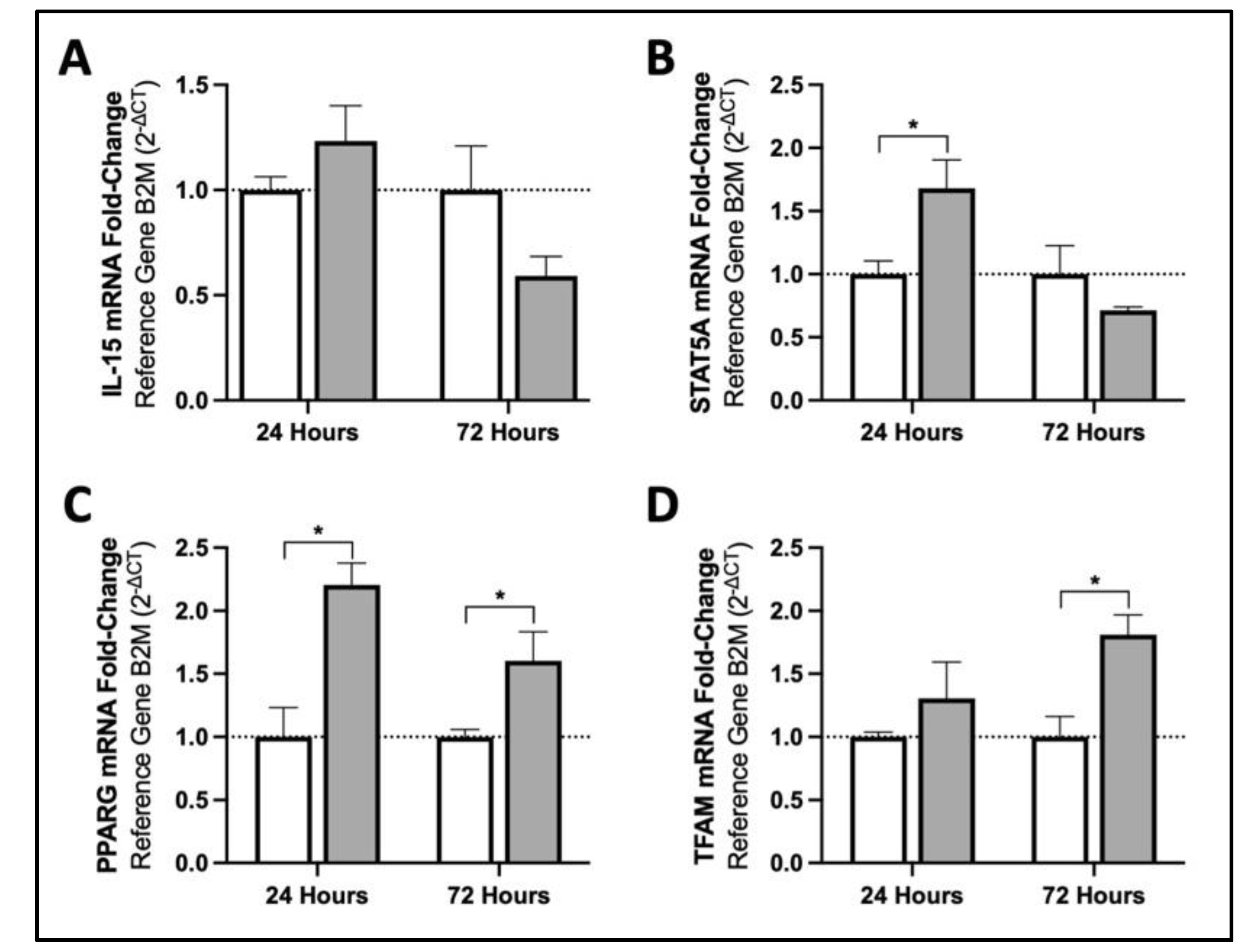
3.1. MIS Treatment Increases mRNA Content of Mitochondrial Biogenesis Activators in Human Fibroblasts via Downstream Targets of IL15

Compared to the respective timepoint control groups, mRNA content of IL15 was not significantly altered by 24 or 72 h of MIS treatment (p = 0.26 and 0.15, respectively; Figure 1A). Downstream effectors of IL15 gene activation, namely STAT5A and PPARG, did show significantly higher mRNA content at 24 h of MIS supplementation (STAT5A 1.68-fold, PPARG 2.21-fold increases when comparing CON-24 and MIS-24; Figure 1B,C). These early elevations in pathway intermediates were associated with a 1.81-fold increase in TFAM mRNA content following 72 h of MIS treatment (Figure 1D).

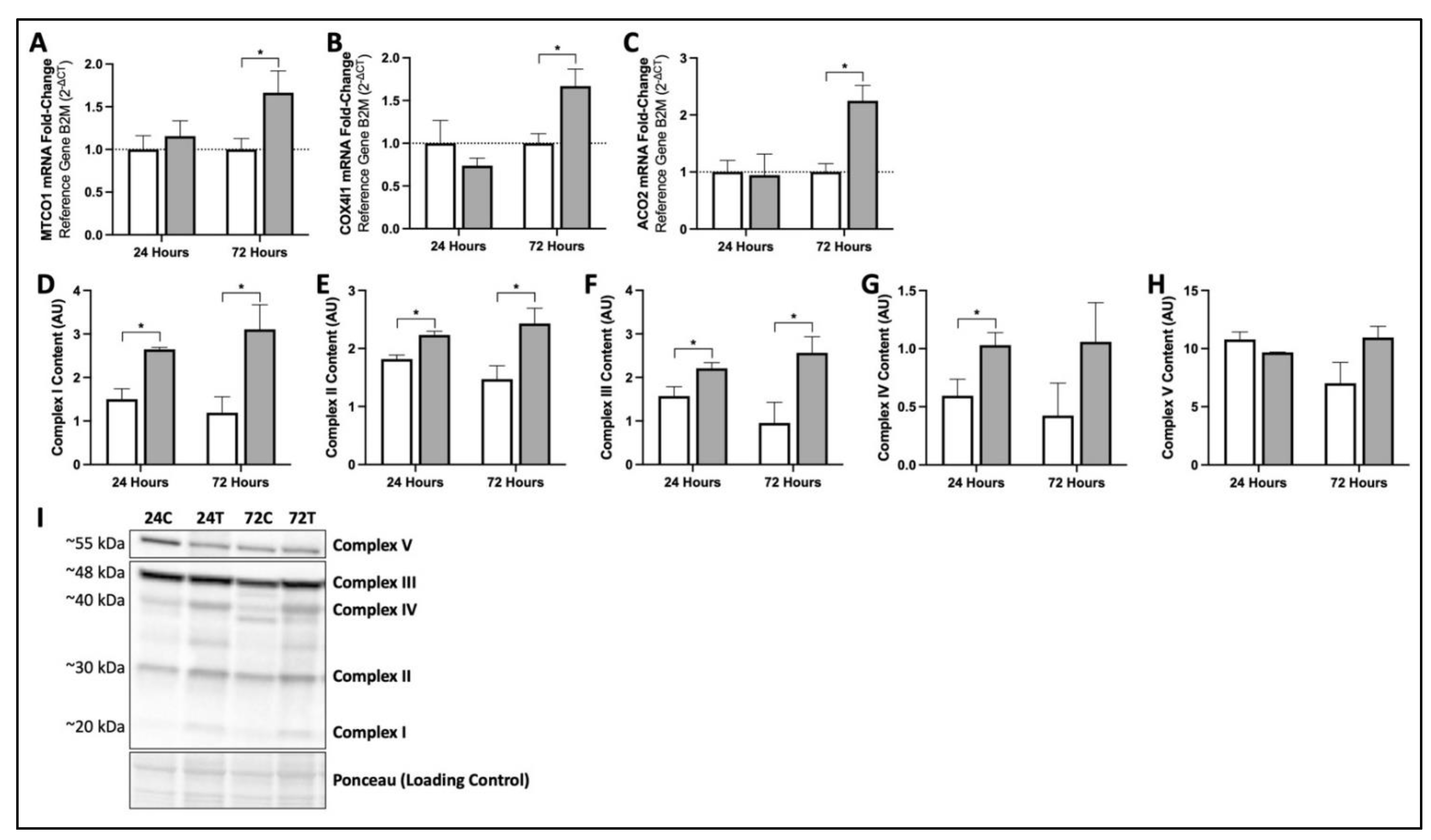
3.2. MIS Treatment Increases Production of Key mRNA and Proteins Responsible for Mitochondrial Function and Oxidative Phosphorylation in Human Fibroblasts

Following 72 h of MIS supplementation, fibroblasts showed significant increases in MTCO1 (1.67-fold), COX4I1 (1.67-fold), and ACO2 (2.25-fold) mRNA content when compared to CON-72 fibroblasts (Figure 2A–C). These elevations were also reflected by positive changes in oxidative phosphorylation protein content. When compared to their respective timepoint controls, both MIS-24 and MIS-72 increased Complex I (76% and 161%), Complex II (23% and 65%), and Complex III (41% and 169%) protein content (Figure 2D–F). Increases in Complex IV content with MIS treatment were only significant following 24 h of supplementation (73% increase at 24-h, p = 0.11 at 72-h; Figure 2G). Although trending towards significance, MIS-24 and MIS-72 did not cause significant changes in Complex V protein content (p = 0.08 and 0.06, respectively; Figure 2H).

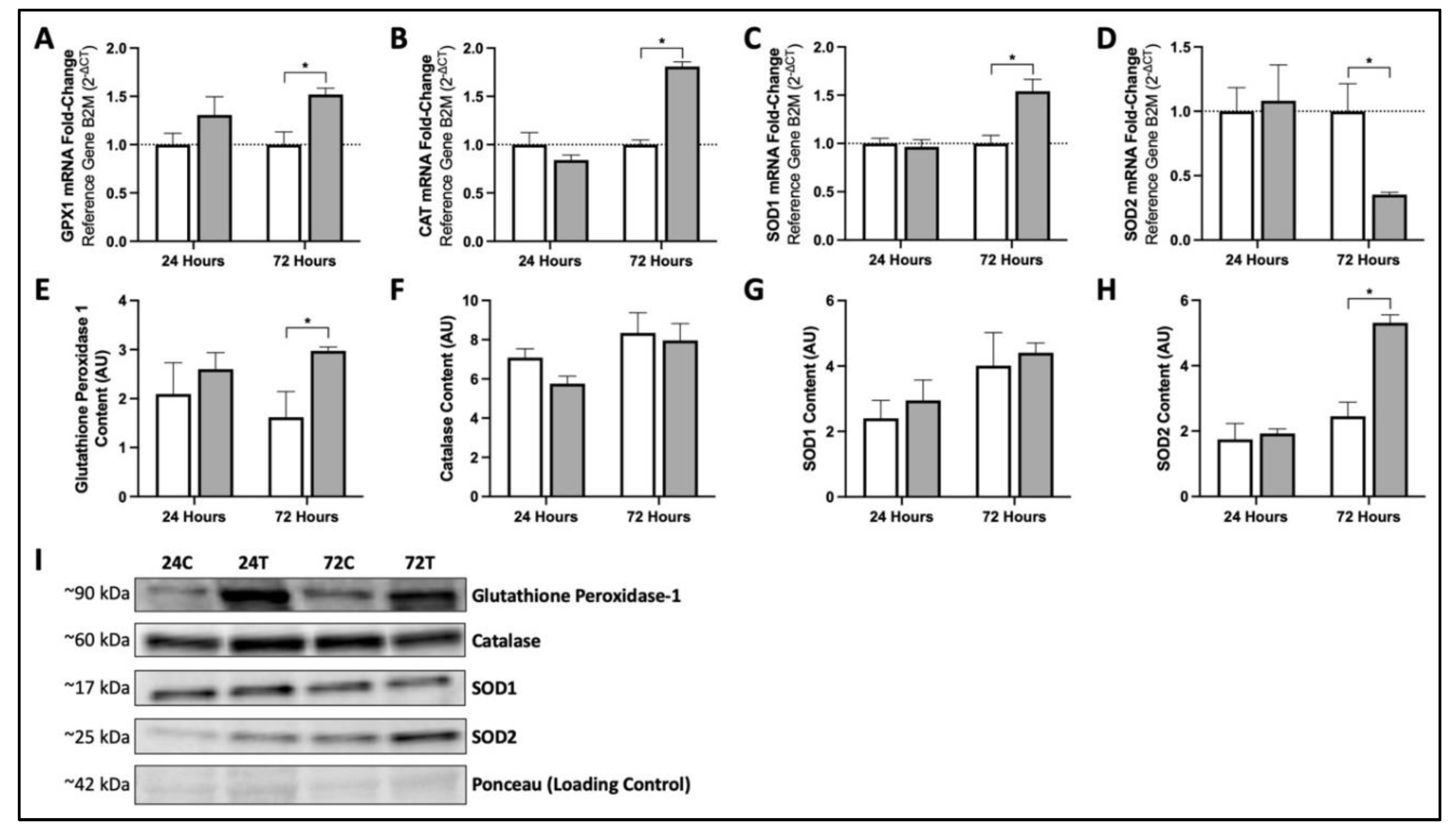
3.3. Seventy-Two Hours of MIS Supplementation Modifies mRNA and Protein Content of Key Cellular Antioxidants in Human Fibroblasts

MIS-72 increased mRNA abundance relative to CON-72 for GPX1 (1.52-fold; Figure 3A), CAT (1.81-fold; Figure 3B), and SOD1 (1.54-fold; Figure 3C). When compared to CON-72, SOD2 mRNA content was lower following 72 h of MIS supplementation (Figure 3D). The aforementioned elevation in GPX1 mRNA abundance was reflected by an 84% increase in GPX1 protein content with MIS-72 treatment (Figure 3E). Although significant increases in CAT and SOD1 mRNA content were seen as a result of MIS-72, these increases did not translate to an increase in catalase or SOD1 protein content following 72 h of MIS supplementation (p = 0.79 and 0.73, respectively; Figure 3F,G). Similar to GPX1, MIS-72 was associated with a significant 117% elevation in SOD2 protein content compared to CON-72 (Figure 3H).

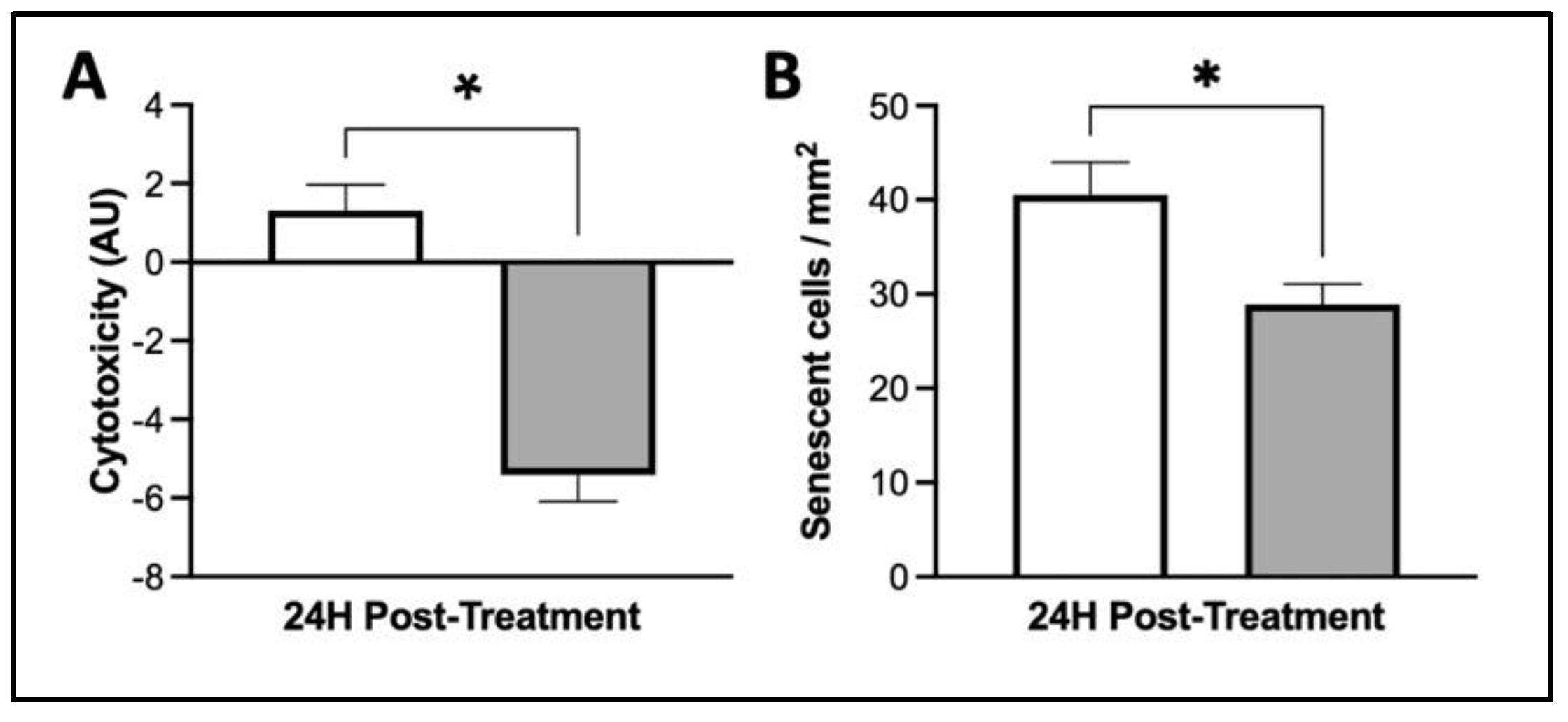
3.4. MIS Treatment Supports Whole-Cell Health in Human Dermal Fibroblasts

After 24 h of MIS treatment, there was a 514% lower relative cellular cytotoxicity in cultured dermal fibroblasts than observed in CON-24 (p < 0.0001, Figure 4A). Furthermore, when compared to CON-24, cellular senescence was 29% lower in fibroblasts after 24 h of MIS supplementation (p = 0.01, Figure 4B).

4. Discussion

The interconnection between poor mitochondrial health, the physiological decline in skin health, and the restoration of these impairments via exercise is an intriguing target for anti-aging strategies [2,3,4,5,6]. Previously, we found IL-15 to be an important regulator of the benefits of exercise on mitochondrial and overall skin health via AMPK- and PGC1α-dependent pathways of mitochondrial biogenesis and an increase in antioxidant defenses [5].
There lies a challenge in translating the science of IL-15 and its benefits on skin health into a meaningful commercial product, because IL-15 cannot be ingested, as it will be degraded in the stomach during digestion. Furthermore, creating a topical IL-15 cosmetic or cream would not likely produce favorable results, as IL-15 would be unstable in a cream/gel matrix, and would not likely penetrate to the dermis to produce visible anti-aging effects. Another challenge to the intravenous or subcutaneous pharmacological administration of IL-15 would lie in selecting an appropriate dose to mimic the exercise “pulse” in vivo, as both positive and negative clinical effects have been observed with different dosages of IL-15 [52]. As such, the most logical approach for recreating the benefits of IL-15 on the skin in humans is via an ingestible MIS that increases endogenous IL-15 expression and/or mimics the downstream effects of IL-15-receptor-mediated signaling. Furthermore, having an MIS in an ingestible format allows for endogenous bathing of the dermis and epidermis upon ingestion in a manner similar to the pulsatile, transient increases in IL-15 release observed following exercise.
Within the current investigation, we indeed mimic the desired exercise-induced effect on the dermal fibroblasts of older adults via provision of a novel MIS containing ingredients known to facilitate mitochondrial or skin health without leading to an increase in endogenous IL-15 expression. Given that IL-15 is not known to be produced in fibroblasts, the cell type used within this body of work, it is not surprising that we did not see an increase in IL-15 mRNA in response to our MIS. Instead, we observed robust activation of the downstream canonical pathways induced by IL-15 receptor binding with MIS supplementation, namely STAT5A, PPARG, and TFAM genes. These downstream pathways are major targets for the pharmaceutical treatment of a variety of mitochondrial-associated disorders and have also been shown to be upregulated by some of our individual MIS ingredients in a variety of cell types [53,54,55,56,57,58,59,60,61].
A potential concern for the usage of a multi-target combinatorial antioxidant supplement is the ability for exogenous antioxidants to have complex and unpredictable interactions [62]. For example, previous studies have reported that combined vitamin C and E supplementation attenuates mRNA responses in mitochondrial proteins and antioxidant enzymes in skeletal muscle, blunting the adaptive response to endurance training [63,64]. Others, however, have not been able to reproduce these findings [65,66], and our group has not found any deleterious metabolic effects of combined vitamin E, CoQ10, and α-LA therapy in humans or rodents [12,14]. Similarly, our recent development of a combination multi-ingredient antioxidant supplement targeting mitochondrial dysfunction in obese mice found no deleterious health effects, and in fact an enhanced benefit of exercise [67]. Similarly, positive effects of MIS supplementation on fibroblast health were observed in the current investigation.
During aging, mitochondrial aconitase, an integral component of the TCA cycle, is deleteriously affected by oxidative damage, resulting in the loss of catalytic function [68]. Within the current investigation, 72 h of MIS supplementation caused an increase in aconitase gene content, potentially indicating the onset of reversal of this aging effect. As accumulation of mtDNA mutations have been associated with phenotypic aging of the skin [69,70,71], the significance of this result is of extended importance due to the additional role of aconitase in associating with and stabilizing mtDNA [72,73]. Furthermore, as mtDNA mutations have also been identified to propel metabolic reprogramming and subsequent cellular senescence [74,75], this may be a key component causing the decrease in cellular senescence observed with MIS supplementation.
Mitochondria accumulate damage with time, resulting in cellular senescence, a process in which cells cease division, develop apoptotic resistance, exhibit altered gene expression patterns, and develop morphological changes [76]. There is clear evidence to suggest that fibroblast senescence is a major driver of age-associated pathology [77,78], making our finding of attenuated senescence with MIS one of clinical relevance to the aging population. In addition to the aforementioned association of mtDNA mutations with cellular senescence, overt mitochondrial dysfunction and oxidative stress also contribute to this pathological state [76,79,80]. As such, additional factors contributing to our decrease in fibroblast senescence likely include the metabolic restoration of mitochondrial health by way of the observed MIS-associated upregulation of mitochondrial respiratory chain complex proteins and antioxidant defenses.
Another highlight of the current investigation is the supra-mitochondrial effect of our MIS in mitigating cytotoxicity. Similar to our findings, resveratrol, a polyphenol within our MIS, has recently been identified to concomitantly decrease both cellular senescence and cytotoxicity on its own in human fibroblasts [81]. Evidently, the benefits of the MIS and its ingredients on mitochondrial health extend beyond this organelle, facilitating whole-cell health and rejuvenation.
In conclusion, the provision of our MIS to aged dermal fibroblasts mimicked exercise through activation of signaling pathways known to be downstream of IL-15 receptor binding. This activation led to elevations in key factors of mitochondrial health, including those involved in mitochondrial biogenesis, oxidative phosphorylation, and antioxidant defense systems. Furthermore, these mitochondrial benefits were associated with improvements in overall fibroblast health. Given these results, future studies will evaluate the benefits of the current MIS in other preclinical disease models associated with aging and mitochondrial dysfunction.

Author Contributions

I.A.R., L.M. and J.P.N. performed experiments and analyzed data. I.A.R. and M.A.T. interpreted experimental results. I.A.R. prepared figures and drafted the manuscript. All authors have read and agreed to the published version of the manuscript.

Funding

This project was funded by Exerkine Corporation. J.P.N was supported by the Canadian Institutes of Health Research (CIHR) Postdoctoral Fellowship.

Institutional Review Board Statement

The study was conducted in accordance with the Declaration of Helsinki. Cultured human fibroblasts used within this investigation were derived from skin samples collected from human inner forearm skin biopsies under the Hamilton Integrated Research Ethics Board, project number 11-114.

Informed Consent Statement

Written informed consent has been obtained from the patients to publish this paper.

Data Availability Statement

Not applicable.

Conflicts of Interest

Exerkine Corporation is a biotechnology company that develops and commercializes therapies based on nutritional supplements, exercise-derived factors (‘exerkines’), and extracellular vesicles to treat and diagnose genetic disorders, chronic diseases, and aging. M.A.T. is the founder, C.E.O., and C.S.O. of Exerkine Corporation, which provided support in the form of a salary for I.A.R. Exerkine Corporation owns shares in HERB + FLORA, a clean beauty corporation, who has marketed a commercial product that contains the same ingredients as the MIS embodied within this research. Exerkine has patents for the use of IL-15 in skin and muscle health (Patent Number CA 2917944).

References

  1. Tobin, D.J. Introduction to skin aging. J. Tissue Viability 2017, 26, 37–46. [Google Scholar] [CrossRef] [PubMed] [Green Version]
  2. López-Otín, C.; Blasco, M.A.; Partridge, L.; Serrano, M.; Kroemer, G. The Hallmarks of Aging. Cell 2013, 153, 1194. [Google Scholar] [CrossRef] [PubMed] [Green Version]
  3. Menon, G.K.; Farra, C.D.; Botto, J.-M.; Domloge, N. Mitochondria: A new focus as an anti-aging target in skin care. J. Cosmet. Dermatol. 2010, 9, 122–131. [Google Scholar] [CrossRef] [PubMed]
  4. Sreedhar, A.; Aguilera-Aguirre, L.; Singh, K.K. Mitochondria in skin health, aging, and disease. Cell Death Dis. 2020, 11, 444. [Google Scholar] [CrossRef] [PubMed]
  5. Crane, J.D.; Macneil, L.G.; Lally, J.S.; Ford, R.J.; Bujak, A.L.; Brar, I.K.; Kemp, B.E.; Raha, S.; Steinberg, G.R.; Tarnopolsky, M.A. Exercise-stimulated interleukin-15 is controlled by AMPK and regulates skin metabolism and aging. Aging Cell 2015, 14, 625–634. [Google Scholar] [CrossRef]
  6. Stout, R.; Birch-Machin, M. Mitochondria’s Role in Skin Ageing. Biology 2019, 8, 29. [Google Scholar] [CrossRef] [Green Version]
  7. Ma, Y.S.; Wu, S.B.; Lee, W.Y.; Cheng, J.S.; Wei, Y.H. Response to the increase of oxidative stress and mutation of mitochondrial DNA in aging. Biochim. Biophys. Acta-Gen. Subj. 2009, 1790, 1021–1029. [Google Scholar] [CrossRef]
  8. Poljšak, B.; Dahmane, R.G.; Godic, A. Intrinsic skin aging: The role of oxidative stress. Acta Dermatovenerol Alp Pannonica Adriat. 2012, 21, 33–36. [Google Scholar] [CrossRef]
  9. Feichtinger, R.G.; Sperl, W.; Bauer, J.W.; Kofler, B. Mitochondrial dysfunction: A neglected component of skin diseases. Exp. Dermatol. 2014, 23, 607–614. [Google Scholar] [CrossRef]
  10. Rinnerthaler, M.; Bischof, J.; Streubel, M.K.; Trost, A.; Richter, K. Oxidative Stress in Aging Human Skin. Biomolecules 2015, 5, 545. [Google Scholar] [CrossRef] [Green Version]
  11. Tamura, Y.; Watanabe, K.; Kantani, T.; Hayashi, J.; Ishida, N.; Kaneki, M. Upregulation of circulating IL-15 by treadmill running in healthy individuals: Is IL-15 an endocrine mediator of the beneficial effects of endurance exercise? Endocr. J. 2011, 58, 211–215. [Google Scholar] [CrossRef] [PubMed] [Green Version]
  12. Rodriguez, M.C.; MacDonald, J.R.; Mahoney, D.J.; Parise, G.; Beal, M.F.; Tarnopolsky, M.A. Beneficial effects of creatine, CoQ10, and lipoic acid in mitochondrial disorders. Muscle Nerve 2007, 35, 235–242. [Google Scholar] [CrossRef] [PubMed]
  13. Glover, E.I.; Martin, J.; Maher, A.; Thornhill, R.E.; Moran, G.R.; Tarnopolsky, M.A. A randomized trial of coenzyme Q10 in mitochondrial disorders. Muscle Nerve 2010, 42, 739–748. [Google Scholar] [CrossRef] [PubMed]
  14. Abadi, A.; Crane, J.D.; Ogborn, D.; Hettinga, B.; Akhtar, M.; Stokl, A.; MacNeil, L.; Safdar, A.; Tarnopolsky, M. Supplementation with α-Lipoic Acid, CoQ10, and Vitamin E Augments Running Performance and Mitochondrial Function in Female Mice. PLoS ONE 2013, 8, e60722. [Google Scholar] [CrossRef] [PubMed]
  15. Laaksonen, R.; Fogelholm, M.; Himberg, J.J.; Laakso, J.; Salorinne, Y. Ubiquinone supplementation and exercise capacity in trained young and older men. Eur. J. Appl. Physiol. Occup. Physiol. 1995, 72, 95–100. [Google Scholar] [CrossRef] [PubMed]
  16. Porter, D.A.; Costill, D.L.; Zachwieja, J.J.; Fink, W.J.; Wagner, E.; Folkers, K. The effect of oral coenzyme Q10 on the exercise tolerance of middle-aged, untrained men. Int. J. Sports Med. 1995, 16, 421–427. [Google Scholar] [CrossRef] [PubMed]
  17. Gaeini, A.A.; Rahnama, N.; Hamedinia, M.R. Effects of vitamin E supplementation on oxidative stress at rest and after exercise to exhaustion in athletic students-ProQuest. J. Sports Med. Phys. Fit. 2006, 46, 458–461. [Google Scholar]
  18. Boyera, N.; Galey, I.; Bernard, B.A. Effect of vitamin C and its derivatives on collagen synthesis and cross-linking by normal human fibroblasts. Int. J. Cosmet. Sci. 1998, 20, 151–158. [Google Scholar] [CrossRef]
  19. Pullar, J.M.; Carr, A.C.; Vissers, M.C.M. The Roles of Vitamin C in Skin Health. Nutrients 2017, 9, 866. [Google Scholar] [CrossRef] [Green Version]
  20. Offord, E.A.; Gautier, J.C.; Avanti, O.; Scaletta, C.; Runge, F.; Krämer, K.; Applegate, L.A. Photoprotective potential of lycopene, β-carotene, vitamin E, vitamin C and carnosic acid in UVA-irradiated human skin fibroblasts. Free Radic. Biol. Med. 2002, 32, 1293–1303. [Google Scholar] [CrossRef]
  21. Jurkiewicz, B.A.; Bissett, D.L.; Buettner, G.R. Effect of Topically Applied Tocopherol on Ultraviolet Radiation-Mediated Free Radical Damage in Skin. J. Investig. Dermatol. 1995, 104, 484–488. [Google Scholar] [CrossRef] [PubMed] [Green Version]
  22. Petruk, G.; Del Giudice, R.; Rigano, M.M.; Monti, D.M. Antioxidants from plants protect against skin photoaging. Oxid. Med. Cell. Longev. 2018, 2018, 1454936. [Google Scholar] [CrossRef] [PubMed] [Green Version]
  23. Davinelli, S.; Bertoglio, J.C.; Polimeni, A.; Scapagnini, G. Cytoprotective Polyphenols Against Chronological Skin Aging and Cutaneous Photodamage. Curr. Pharm. Des. 2018, 24, 99–105. [Google Scholar] [CrossRef] [PubMed]
  24. Afaq, F.; Adhami, V.M.; Ahmad, N. Prevention of short-term ultraviolet B radiation-mediated damages by resveratrol in SKH-1 hairless mice. Toxicol. Appl. Pharmacol. 2003, 186, 28–37. [Google Scholar] [CrossRef]
  25. Schagen, S.K.; Zampeli, V.A.; Makrantonaki, E.; Zouboulis, C.C. Discovering the link between nutrition and skin aging. Dermato-Endocrinol. 2012, 4, 298. [Google Scholar] [CrossRef] [Green Version]
  26. Giardina, D.; Michelotti, A.; Zavattini, G.; Finzi, S.; Ghisalberti, C.; Marzatico, F. Efficacy study in vitro: Assessment of the properties of resveratrol and resveratrol + N-acetyl-cysteine on proliferation and inhibition of collagen activity. Minerva Ginecol. 2010, 62, 195–201. [Google Scholar] [PubMed]
  27. Heng, M.C.Y. Curcumin targeted signaling pathways: Basis for anti-photoaging and anti-carcinogenic therapy. Int. J. Dermatol. 2010, 49, 608–622. [Google Scholar] [CrossRef]
  28. Lima, C.F.; Pereira-Wilson, C.; Rattan, S.I.S. Curcumin induces heme oxygenase-1 in normal human skin fibroblasts through redox signaling: Relevance for anti-aging intervention. Mol. Nutr. Food Res. 2011, 55, 430–442. [Google Scholar] [CrossRef] [Green Version]
  29. Vaughn, A.R.; Branum, A.; Sivamani, R.K. Effects of Turmeric (Curcuma longa) on Skin Health: A Systematic Review of the Clinical Evidence. Phytother. Res. 2016, 30, 1243–1264. [Google Scholar] [CrossRef]
  30. Laura, V.; Mattia, F.; Roberta, G.; Federico, I.; Emi, D.; Chiara, T.; Luca, B.; Elena, C. Potential of Curcumin in Skin Disorders. Nutrients 2019, 11, 2169. [Google Scholar] [CrossRef] [Green Version]
  31. McMahon, R.J. Biotin in metabolism and molecular biology. Annu. Rev. Nutr. 2003, 22, 221–239. [Google Scholar] [CrossRef] [PubMed]
  32. Vollmer, D.L.; West, V.A.; Lephart, E.D. Enhancing Skin Health: By Oral Administration of Natural Compounds and Minerals with Implications to the Dermal Microbiome. Int. J. Mol. Sci. 2018, 19, 3059. [Google Scholar] [CrossRef] [PubMed] [Green Version]
  33. Nitzan, Y.B.; Cohen, A.D.; Bibi Nitzan, Y.; Cohen, A.D. Zinc in skin pathology and care. J. Dermatol. Treat. 2009, 17, 205–210. [Google Scholar] [CrossRef] [PubMed]
  34. Schwartz, J.R.; Marsh, R.G.; Draelos, Z.D. Zinc and Skin Health: Overview of Physiology and Pharmacology. Dermatol. Surg. 2005, 31, 837–847. [Google Scholar] [CrossRef] [PubMed]
  35. Rostan, E.F.; DeBuys, H.V.; Madey, D.L.; Pinnell, S.R. Evidence supporting zinc as an important antioxidant for skin. Int. J. Dermatol. 2002, 41, 606–611. [Google Scholar] [CrossRef] [PubMed] [Green Version]
  36. Grether-Beck, S.; Marini, A.; Jaenicke, T.; Stahl, W.; Krutmann, J. Molecular evidence that oral supplementation with lycopene or lutein protects human skin against ultraviolet radiation: Results from a double-blinded, placebo-controlled, crossover study. Br. J. Dermatol. 2017, 176, 1231–1240. [Google Scholar] [CrossRef] [PubMed]
  37. Lee, E.H.; Faulhaber, D.; Hanson, K.M.; Ding, W.; Peters, S.; Kodali, S.; Granstein, R.D. Dietary Lutein Reduces Ultraviolet Radiation-Induced Inflammation and Immunosuppression. J. Investig. Dermatol. 2004, 122, 510–517. [Google Scholar] [CrossRef]
  38. Palombo, P.; Fabrizi, G.; Ruocco, V.; Ruocco, E.; Fluhr, J.; Roberts, R.; Morganti, P. Beneficial Long-Term Effects of Combined Oral/Topical Antioxidant Treatment with the Carotenoids Lutein and Zeaxanthin on Human Skin: A Double-Blind, Placebo-Controlled Study. Ski. Pharmacol. Physiol. 2007, 20, 199–210. [Google Scholar] [CrossRef]
  39. Komatsu, T.; Sasaki, S.; Manabe, Y.; Hirata, T.; Sugawara, T. Preventive effect of dietary astaxanthin on UVA-induced skin photoaging in hairless mice. PLoS ONE 2017, 12, e0171178. [Google Scholar] [CrossRef] [Green Version]
  40. Yuan, J.P.; Peng, J.; Yin, K.; Wang, J.H. Potential health-promoting effects of astaxanthin: A high-value carotenoid mostly from microalgae. Mol. Nutr. Food Res. 2011, 55, 150–165. [Google Scholar] [CrossRef]
  41. Lyons, N.M.; O’Brien, N.M. Modulatory effects of an algal extract containing astaxanthin on UVA-irradiated cells in culture. J. Dermatol. Sci. 2002, 30, 73–84. [Google Scholar] [CrossRef]
  42. Tominaga, K.; Hongo, N.; Karato, M.; Yamashita, E. Cosmetic benefits of astaxanthin on humans subjects. Acta Biochim. Pol. 2012, 59, 43–47. [Google Scholar] [CrossRef] [PubMed]
  43. Davinelli, S.; Nielsen, M.E.; Scapagnini, G. Astaxanthin in Skin Health, Repair, and Disease: A Comprehensive Review. Nutrients 2018, 10, 522. [Google Scholar] [CrossRef] [PubMed] [Green Version]
  44. Weber, G.; Heilborn, J.D.; Jimenez, C.I.C.; Hammarsjö, A.; Törmä, H.; Ståhle, M. Vitamin D induces the antimicrobial protein hCAP18 in human skin. J. Investig. Dermatol. 2005, 124, 1080–1082. [Google Scholar] [CrossRef] [Green Version]
  45. Chang, A.L.S.; Fu, T.; Amir, O.; Tang, J.Y. Association of facial skin aging and vitamin D levels in middle-aged white women. Cancer Causes Control 2010, 21, 2315–2316. [Google Scholar] [CrossRef] [PubMed] [Green Version]
  46. Bastianetto, S.; Dumont, Y.; Duranton, A.; Vercauteren, F.; Breton, L.; Quirion, R. Protective action of resveratrol in human skin: Possible involvement of specific receptor binding sites. PLoS ONE 2010, 5, e12935. [Google Scholar] [CrossRef] [Green Version]
  47. Schauber, J.; Gallo, R.L. The vitamin D pathway: A new target for control of the skin’s immune response? Exp. Dermatol. 2008, 17, 633–639. [Google Scholar] [CrossRef] [Green Version]
  48. Borkow, G.; Gabbay, J.; Lyakhovitsky, A.; Huszar, M. Improvement of facial skin characteristics using copper oxide containing pillowcases: A double-blind, placebo-controlled, parallel, randomized study. Int. J. Cosmet. Sci. 2009, 31, 437–443. [Google Scholar] [CrossRef]
  49. Borkow, G. Using Copper to Improve the Well-Being of the Skin. Curr. Chem. Biol. 2014, 8, 89. [Google Scholar] [CrossRef] [Green Version]
  50. Schmittgen, T.D.; Livak, K.J. Analyzing real-time PCR data by the comparative CT method. Nat. Protoc. 2008, 3, 1101–1108. [Google Scholar] [CrossRef]
  51. Rebalka, I.A.; Monaco, C.M.F.; Varah, N.E.; Berger, T.; D’souza, D.M.; Zhou, S.; Mak, T.W.; Hawke, T.J. Loss of the adipokine lipocalin-2 impairs satellite cell activation and skeletal muscle regeneration. Am. J. Physiol.-Cell Physiol. 2018, 315, C714–C721. [Google Scholar] [CrossRef] [PubMed]
  52. Marçais, A.; Cherfils-Vicini, J.; Viant, C.; Degouve, S.; Viel, S.; Fenis, A.; Rabilloud, J.; Mayol, K.; Tavares, A.; Bienvenu, J.; et al. The metabolic checkpoint kinase mTOR is essential for IL-15 signaling during the development and activation of NK cells. Nat. Immunol. 2014, 15, 749–757. [Google Scholar] [CrossRef] [PubMed]
  53. Legchenko, E.; Chouvarine, P.; Borchert, P.; Fernandez-Gonzalez, A.; Snay, E.; Meier, M.; Maegel, L.; Mitsialis, S.A.; Rog-Zielinska, E.A.; Kourembanas, S.; et al. PPARγ agonist pioglitazone reverses pulmonary hypertension and prevents right heart failure via fatty acid oxidation. Sci. Transl. Med. 2018, 10, eaao0303. [Google Scholar] [CrossRef] [Green Version]
  54. Yin, Y.; Wu, Y.; Zhang, X.; Zhu, Y.; Sun, Y.; Yu, J.; Gong, Y.; Sun, P.; Lin, H.; Han, X. PPA1 Regulates Systemic Insulin Sensitivity by Maintaining Adipocyte Mitochondria Function as a Novel PPARγ Target Gene. Diabetes 2021, 70, 1278–1291. [Google Scholar] [CrossRef] [PubMed]
  55. Hoseini, R.; Damirchi, A.; Babaei, P. Vitamin D increases PPARγ expression and promotes beneficial effects of physical activity in metabolic syndrome. Nutrition 2017, 36, 54–59. [Google Scholar] [CrossRef] [PubMed]
  56. Aloysius Dhivya, M.; Aberami, S.; Nikhalashree, S.; Biswas, J.; Liu, W.; Irudayaraj, J.; Sulochana, K.N.; Coral, K.; Bharathi Devi, S.R. Copper mediates mitochondrial biogenesis in retinal pigment epithelial cells. Biochim. Biophys. Acta. Mol. Basis Dis. 2020, 1866, 165843. [Google Scholar] [CrossRef]
  57. Bernardo, A.; Plumitallo, C.; De Nuccio, C.; Visentin, S.; Minghetti, L. Curcumin promotes oligodendrocyte differentiation and their protection against TNF-α through the activation of the nuclear receptor PPAR-γ. Sci. Rep. 2021, 11, 4952. [Google Scholar] [CrossRef]
  58. Zhang, X.; Zhao, W.E.; Hu, L.; Zhao, L.; Huang, J. Carotenoids inhibit proliferation and regulate expression of peroxisome proliferators-activated receptor gamma (PPARγ) in K562 cancer cells. Arch. Biochem. Biophys. 2011, 512, 96–106. [Google Scholar] [CrossRef]
  59. Nanjaiah, H.; Vallikannan, B. Lutein upregulates the PGC-1α, NRF1, and TFAM expression by AMPK activation and downregulates ROS to maintain mtDNA integrity and mitochondrial biogenesis in hyperglycemic ARPE-19 cells and rat retina. Biotechnol. Appl. Biochem. 2019, 66, 999–1009. [Google Scholar] [CrossRef]
  60. Lee, D.; Kim, K.Y.; Shim, M.S.; Kim, S.Y.; Ellisman, M.H.; Weinreb, R.N.; Ju, W.K. Coenzyme Q10 ameliorates oxidative stress and prevents mitochondrial alteration in ischemic retinal injury. Apoptosis 2014, 19, 603–614. [Google Scholar] [CrossRef] [Green Version]
  61. Zhou, J.; Yang, Z.; Shen, R.; Zhong, W.; Zheng, H.; Chen, Z.; Tang, J.; Zhu, J. Resveratrol Improves Mitochondrial Biogenesis Function and Activates PGC-1α Pathway in a Preclinical Model of Early Brain Injury Following Subarachnoid Hemorrhage. Front. Mol. Biosci. 2021, 8, 223. [Google Scholar] [CrossRef] [PubMed]
  62. Chen, X.; Li, H.; Zhang, B.; Deng, Z. The synergistic and antagonistic antioxidant interactions of dietary phytochemical combinations. Crit. Rev. Food Sci. Nutr. 2021. online ahead of print. [Google Scholar] [CrossRef] [PubMed]
  63. Morrison, D.; Hughes, J.; Della Gatta, P.A.; Mason, S.; Lamon, S.; Russell, A.P.; Wadley, G.D. Vitamin C and E supplementation prevents some of the cellular adaptations to endurance-training in humans. Free Radic. Biol. Med. 2015, 89, 852–862. [Google Scholar] [CrossRef]
  64. Paulsen, G.; Cumming, K.T.; Holden, G.; Hallén, J.; Rønnestad, B.R.; Sveen, O.; Skaug, A.; Paur, I.; Bastani, N.E.; Østgaard, H.N.; et al. Vitamin C and E supplementation hampers cellular adaptation to endurance training in humans: A double-blind, randomised, controlled trial. J. Physiol. 2014, 592, 1887–1901. [Google Scholar] [CrossRef] [PubMed]
  65. Yfanti, C.; Åkerström, T.; Nielsen, S.; Nielsen, A.R.; Mounier, R.; Mortensen, O.H.; Lykkesfeldt, J.; Rose, A.J.; Fischer, C.P.; Pedersen, B.K. Antioxidant supplementation does not alter endurance training adaptation. Med. Sci. Sports Exerc. 2010, 42, 1388–1395. [Google Scholar] [CrossRef]
  66. Higashida, K.; Kim, S.H.; Higuchi, M.; Holloszy, J.O.; Han, D.H. Normal adaptations to exercise despite protection against oxidative stress. Am. J. Physiol. Endocrinol. Metab. 2011, 301, E779–E784. [Google Scholar] [CrossRef] [Green Version]
  67. Nederveen, J.P.; Manta, K.; Bujak, A.L.; Simone, A.C.; Fuda, M.R.; Nilsson, M.I.; Hettinga, B.P.; Hughes, M.C.; Perry, C.G.R.; Tarnopolsky, M.A. A Novel Multi-Ingredient Supplement Activates a Browning Program in White Adipose Tissue and Mitigates Weight Gain in High-Fat Diet-Fed Mice. Nutrients 2021, 13, 3726. [Google Scholar] [CrossRef]
  68. Yan, L.J.; Levine, R.L.; Sohal, R.S. Oxidative damage during aging targets mitochondrial aconitase. Proc. Natl. Acad. Sci. USA 1997, 94, 11168. [Google Scholar] [CrossRef] [Green Version]
  69. Krutmann, J.; Schroeder, P. Role of Mitochondria in Photoaging of Human Skin: The Defective Powerhouse Model. J. Investig. Dermatol. Symp. Proc. 2009, 14, 44–49. [Google Scholar] [CrossRef] [Green Version]
  70. Eshaghian, A.; Vleugels, R.A.; Canter, J.A.; McDonald, M.A.; Stasko, T.; Sligh, J.E. Mitochondrial DNA deletions serve as biomarkers of aging in the skin, but are typically absent in nonmelanoma skin cancers. J. Investig. Dermatol. 2006, 126, 336–344. [Google Scholar] [CrossRef] [Green Version]
  71. Lee, H.C.; Wei, Y.H.; Yang, J.H. Photoageing-associated mitochondrial DNA length mutations in human skin. Arch. Dermatol. Res. 1995, 287, 641–648. [Google Scholar] [CrossRef] [PubMed]
  72. Shadel, G.S. Mitochondrial DNA, aconitase “wraps” it up. Trends Biochem. Sci. 2005, 30, 294–296. [Google Scholar] [CrossRef] [PubMed]
  73. Chen, X.Z.; Hu, C.W.; Gao, Z.M. Aconitase couples metabolic regulation to mitochondrial DNA maintenance. Science 2005, 307, 714–717. [Google Scholar] [CrossRef] [PubMed]
  74. Wiley, C.D.; Velarde, M.C.; Lecot, P.; Liu, S.; Sarnoski, E.A.; Freund, A.; Shirakawa, K.; Lim, H.W.; Davis, S.S.; Ramanathan, A.; et al. Mitochondrial Dysfunction Induces Senescence with a Distinct Secretory Phenotype. Cell Metab. 2016, 23, 303–314. [Google Scholar] [CrossRef] [Green Version]
  75. Lauri, A.; Pompilio, G.; Capogrossi, M.C. The mitochondrial genome in aging and senescence. Ageing Res. Rev. 2014, 18, 1–15. [Google Scholar] [CrossRef]
  76. Kumari, R.; Jat, P. Mechanisms of Cellular Senescence: Cell Cycle Arrest and Senescence Associated Secretory Phenotype. Front. Cell Dev. Biol. 2021, 9, 485. [Google Scholar] [CrossRef]
  77. Low, E.; Alimohammadiha, G.; Smith, L.A.; Costello, L.F.; Przyborski, S.A.; von Zglinicki, T.; Miwa, S. How good is the evidence that cellular senescence causes skin ageing? Ageing Res. Rev. 2021, 71, 101456. [Google Scholar] [CrossRef]
  78. Takenaka, Y.; Inoue, I.; Nakano, T.; Ikeda, M.; Kakinuma, Y. Prolonged disturbance of proteostasis induces cellular senescence via temporal mitochondrial dysfunction and subsequent mitochondrial accumulation in human fibroblasts. FEBS J. 2021, 289, 1650–1667. [Google Scholar] [CrossRef]
  79. Van Deursen, J.M. The role of senescent cells in ageing. Nature 2014, 509, 439. [Google Scholar] [CrossRef] [Green Version]
  80. Campisi, J.; D’Adda Di Fagagna, F. Cellular senescence: When bad things happen to good cells. Nat. Rev. Mol. Cell Biol. 2007, 8, 729–740. [Google Scholar] [CrossRef]
  81. Birar, V.C.; Sheerin, A.N.; Ostler, E.L.; Faragher, R.G.A. Novel resveratrol derivatives have diverse effects on the survival, proliferation and senescence of primary human fibroblasts. Biogerontology 2020, 21, 817–826. [Google Scholar] [CrossRef] [PubMed]
Figure 1. MIS elevates mRNA abundance for mitochondrial biogenesis activators in human fibroblasts via downstream targets of IL-15. (A) IL15 mRNA content remains unchanged by MIS supplementation. (B) Early elevations in STAT5A and (C) PPARG mRNA content are associated with (D) later elevations in TFAM mRNA content. White bars indicate CON treatment, grey bars indicate MIS treatment. n = 3 independent donor biopsies per group. Data are mean ± SEM. * indicates p < 0.05 via unpaired t-test.
Cosmetics 09 00047 g001
Figure 2. MIS elevates production of key mRNA species and proteins responsible for mitochondrial function and oxidative phosphorylation in human fibroblasts. MIS supplementation results in significantly greater amounts of mRNA compared to CON for: (A) MTCO1, (B) COX4I1, and (C) ACO2 in human fibroblasts. (DF) Both 24 and 72 h of MIS supplementation results in greater Complex I, II, and III protein content than CON, while (G) a greater Complex IV content is significant following 24 h of supplementation. (H) Complex V protein content remains unchanged as a result of treatment. (I) Representative Western blot images depict protein content for Complexes I to V. White bars indicate CON treatment, grey bars indicate MIS treatment. n = 3 independent donor biopsies per group. Data are mean ± SEM. * indicates p < 0.05 via unpaired t-test.
Cosmetics 09 00047 g002
Figure 3. Seventy-two hours of MIS supplementation modifies mRNA and protein content of key cellular antioxidants in human fibroblasts. (A) GPX1, (B) CAT, and (C) SOD1 mRNA content are significantly greater following 72 h of MIS supplementation. (D) SOD2 mRNA content is lower following 72 h of MIS supplementation. (EH) Protein content of the formerly interrogated genes parallels the absence of change following 24 h of MIS supplementation. Following 72 h of MIS supplementation, however, glutathione peroxidase and SOD2 protein content are significantly higher. (I) Representative Western blot images depict protein content for proteins interrogated in E to H. White bars indicate CON treatment, grey bars indicate MIS treatment. n = 3 independent donor biopsies per group. Data are mean ± SEM. * indicates p < 0.05 via unpaired t-test.
Cosmetics 09 00047 g003
Figure 4. MIS supports whole-cell health by decreasing (A) cytotoxicity and (B) cellular senescence in fibroblasts after 24 h of treatment. White bars indicate CON treatment, grey bars indicate MIS treatment. n = 3 independent donor biopsies per group. Data are mean ± SEM. * indicates p < 0.05 via unpaired t-test.
Cosmetics 09 00047 g004
Publisher’s Note: MDPI stays neutral with regard to jurisdictional claims in published maps and institutional affiliations.

