Article

Microalgae as a Sustainable, Natural-Oriented and Vegan Dermocosmetic Bioactive Ingredient: The Case of Neochloris oleoabundans

by
Ana Lucía Morocho-Jácome
1,*,
Bruna Bertoloni dos Santos
1,
João Carlos Monteiro de Carvalho
2,
Tânia Santos de Almeida
3,
Patrícia Rijo
3,
Maria Valéria Robles Velasco
1,
Catarina Rosado
3,* and
André Rolim Baby
1,*
1
Pharmacy Department, Faculty of Pharmaceutical Sciences, University of São Paulo, São Paulo 05508-000, Brazil
2
Biochemical and Pharmaceutical Technology Department, Faculty of Pharmaceutical Sciences, University of São Paulo, São Paulo 05508-000, Brazil
3
CBIOS—Universidade Lusófona’s Research Center for Biosciences and Health Technologies, 1749-024 Lisboa, Portugal
*
Authors to whom correspondence should be addressed.
Cosmetics 2022, 9(1), 9; https://doi.org/10.3390/cosmetics9010009
Submission received: 29 November 2021 / Revised: 26 December 2021 / Accepted: 6 January 2022 / Published: 9 January 2022

Abstract

:
“Vegan” and “sustainable” characteristics are strong claim trends behind the development of innovative skincare, fragrances, and makeup products. This created a need in the market for compliant ingredients. To date, there have been no records evidencing the use of the microalgae Neochloris oleoabundans (NA) in dermocosmetics. Therefore, we studied the applicability of such a natural compound in this context. NA was cultivated, and the scavenging activity (SA) of the NA extracts was evaluated. The highest SA was from the aqueous extract (54.8% ± 2.1%), being higher than that of the positive control. Two hydrogels were prepared with 1.0% ammonium acryloyldimethyltaurate/VP copolymer: (1) control gel; and (2) gel with a 1.0% NA aqueous extract. In vivo experiments were performed in healthy male and female volunteers with skin phototypes of II–IV. The stratum corneum (SC) hydration and the transepidermal water loss (TEWL) were measured in the forearm of participants to determine their biocompatibility. This parameter was determined by skin bioengineering measurements, confirming that SC hydration and TEWL were not affected by the samples. The laser Doppler measurements results showed a delayed erythema onset in the sites, where the NA hydrogel was applied. The results confirmed the biocompatibility and the anti-inflammatory activity of an innovative ingredient derived from microalgae suitable for a natural and vegan lifestyle.

1. Introduction

One of the most promising future developments of dermocosmetics is the sustainable exploration of marine resources. Currently, microalgal-derived compounds have particularly gained more attention as cosmetic ingredients due to their pigments or polysaccharides, among other interesting compounds [1,2]. In addition, psychological research about skin care, fragrances, and makeups revealed an emotion control scheme in consumers’ daily life to induce the choice of determined trademarks or novel actives [3] and, even more recently, the increase of the appeal for vegan and sustainable ingredients in the cosmetic market. The lifestyle called veganism, based on the absence of the use of animal-derived products in daily life, is becoming more prevalent and has created a need in the market for cosmetic products that are vegan-compliant. The latest report predicts the global vegan cosmetics market will go above $21 billion by 2027 [4]. This angle seems to provide a further rationale for the exploration of the cosmetic applicability of microalgae. Similarly, growing concerns about not only the global warming by the abuse of animal-derived materials, but also the cruelty-free concept, may induce consumers of vegan cosmetics to select novel proven compounds from sustainable resources, such as microalgae.
In fact, there is a massive variety of microalgal species in nature [5]; however, only a few of them are already cultivated for commercial purposes [6]. Microalgae industrial cultivation could produce different biomass compositions, when the environmental conditions are changed. Thus, microalgae biomass can be cultivated in custom-made circumstances to obtain biocompounds or extracts that can be employed as raw materials for chemicals, biofuels [7,8], and even cosmetic formulations [9,10]. As an interesting advantage, these photosynthetic microorganisms have higher photosynthetic efficiencies, higher rates of carbon dioxide filtration, and faster growth rates with consequently higher biomass productivity when compared with plants [11].
For instance, Neochloris oleoabundans (N. oleoabundans; Chlorophyceae) has been reported in the literature as a renewable lipid source (35–55% dry cell weight total lipids) for biofuel production [11,12,13]. However, it presents a high applicability potential as a cosmetic ingredient, not only for its lipidic content, but also for containing significant amounts of carotenoids, mainly lutein, cantaxanthin, zeaxanthin, as well as astaxanthin monoesters and diesters [14], with different skin benefits recently reported [2]. Furthermore, the antiproliferative activity against human colon cancer cells of bioactive carotenoids from N. oleoabundans extracts was evaluated [7]. Nevertheless, to date, there has been no information in the literature about other putative active compounds from its biomass. There are few records in the literature evidencing the use of such microalgae in cosmetic preparations, nor is there any information on their safety and efficacy.
Furthermore, the development of cosmetic formulations has been optimized, establishing the effect–concentration relationships of their active ingredients. Different non-invasive in vivo methodologies based in bioengineering devices have been employed in efficacy assays, such as those measuring stratum corneum (SC) hydration or transepidermal water loss (TEWL). However, limitations to this approach have been identified [15]. Dynamic approaches based on the cutaneous response to stress have been developed, such as the measurement of erythema induced by nicotinic acid derivatives [16,17,18,19]. Recently, this approach has been applied to the in vivo studies of the topical antioxidant activity [20,21]. Compounds with this bioactivity are able to interfere with the inflammatory cascade, which leads to the vasodilation caused by nicotinates. Therefore, the in vivo assessment of the antioxidant effect can be based on the ability to decrease the extent of a nicotinate-induced erythema response by laser Doppler flowmetry [16,20], which is a technique that determines changes in the cutaneous microcirculation. In addition, it has been used for the quantitative measurements of the erythema induced by vasodilatory compounds [22], as well as to evaluate the follicular influence on the percutaneous absorption of topical drugs [19]. Complementarily, the cutaneous biocompatibility of cosmetics formulations can be evaluated using the conventional non-invasive skin bioengineering methods, measuring SC hydration, skin barrier function [15,23], and erythema [21].
In this research, we cultivated N. oleoabundans and prepared extracts with acetone, methanol, and water. Those were tested for the in vitro antioxidant capacity by the DPPH assay, and the one with the best performance was studied in vivo. We then proceeded to investigate in participants both the biocompatibility of a custom-made gel formulation containing the N. oleoabundans aqueous extract and its putative antioxidant activity, aiming to probe its applicability as an active ingredient for dermocosmetics while focusing on the nature-orientated and veggie markets.

2. Materials and Methods

2.1. Inoculum Maintenance and Culture

N. oleoabundans, NA (UTEX-1185), University of Texas Culture Collection, was maintained at 25 °C in agar up to three months. A small amount of such microalgae was used to inoculate a 10 mL glass tube with 5 mL Bold medium (2.94 mM NaNO3) [24] in a rotatory shaker (Infors HT® Multitron) at 25 ± 1 °C under a continuous white light intensity of 60 µmol photons m−2 s−1, until the biomass concentration reached almost 100 mg L−1. After that, 500 mL Erlenmeyer flasks containing approximately 200 mL Bold media [24] were inoculated with N. oleoabundans at the same environmental conditions in a batch mode with NaNO3 as a nitrogen source. The biomass concentration was measured by the optical density and harvested from the culture media by centrifugation (Sorvall® RC-5C Plus) at 1400× g for 20 min and then lyophilized. Biomass productivity (Px) was calculated as the ratio between the total amount of biomass produced per unit volume and the cultivation time (Tc) using:
Px   =   ( Xm Xo ) Tc ,
where Xm is the maximum biomass concentrations of each culture and Xo is the starting biomass concentration (50 mg L−1).

2.2. Extracts Preparation

Wet biomass was washed twice with purified water to eliminate all adhered residual salts before freezing. After lyophilization, dry biomass was weighted to prepare extracts at 10% (w v−1) in acetone, methanol, or water. Ultrasound-assisted extraction was performed in an ultrasonic bath (Sonorex® Super RK 510 H; Bandelin, Berlin, Germany) for 1 h [25] and repeated at least five times. The methanolic and acetonic extracts were evaporated, whereas the aqueous extract was lyophilized.

2.3. In Vitro Antioxidant Capacity

The in vitro antioxidant activities of the extracts were evaluated by the 2,2-diphenyl-1-picrylhydrazyl (DPPH•) radical scavenging activity (SA) method described by Falé et al. [26], with a slight modification. Concisely, 10 μL of each extract sample were added to 990 μL of the DPPH• solution (0.002% in methanol). These solutions were incubated for 30 min at room temperature. The absorbance was measured at 517 nm (the maximum absorption wavelength for DPPH) against a corresponding blank (methanol) using a Thermo Scientific® Evolution 300 UV-Vis spectrophotometer along with a Stuart Scientific® Autovortex SA6, and the SA was calculated as SA% defined as the scavenging activity of the free radical according to:
SA   % =   ( A DPPH A sample ) A DPPH   ×   100 ,
where ADPPH is the absorption of DPPH against the blank and Asample is the absorption of the extract or control against the blank. The reference standard was butylated hydroxytoluene (BHT). The procedure was carried out in triplicate.

2.4. Dermocosmetic Prototype Formulation

Aristoflex® AVC (ammonium acryloyldimethyltaurate/VP copolymer) was used as a gelling agent. Two 1.0% Aristoflex® AVC hydrogels were prepared in this study: (1) a control gel; and (2) a gel with 1.0% N. oleoabundans aqueous extract, of which the compositions are described in Table 1.

2.5. Cutaneous Compatibility of N. oleoabundans Dermocosmetic

Cutaneous compatibility was carried out with 14 healthy male (5) and female (9) volunteers, aged 31.5 ± 12.6 years, after receiving their written consent. The approval to the project was obtained from the local committee on human experimentation, and ethical standards were used in the procedures, in accordance with the Helsinki Declaration. The whole study was conducted on the volar forearm. A control (untreated) and two treated sites were attributed at random to each volunteer. The left or right forearm and the upper or lower region of the forearm were the randomized parameters. A 1-day pre-trial washout period was recommended to the volunteers, in which they avoided applying any cosmetic product containing moisturizing ingredients in that site. The measurements were carried out in an air-conditioned room (21 ± 2 °C and relative humidity of 40–60%). The basal measurements of SC hydration and TEWL were assessed with a Corneometer® CM825 (CK Electronics GmbH, Köln, Germany) and Tewameter® TM 300 (CK Electronics GmbH, Köln, Germany), respectively. The formulations described in item 2.4 were then applied on the skin under occlusion, using epicutaneous patches (Finn Chambers®, Epitest Ltd., Oy, Finland) for 24 h. Two hours after the patch removal, SC hydration and TEWL were measured. The results were analyzed as the ratio between the values measured after the patch application and the basal ones, to reduce the inter-individual variability effect [20].

2.6. In Vivo Anti-Inflammatory Activity

Thirteen healthy male (5) and female (8) volunteers were included in this study, aged 31.5 ± 12.6 years after giving their oral and written consent. The volunteers had skin phototypes of II–IV. The whole study was conducted on the ventral forearm skin. Volunteers with dermatological pathologies and with superficial irregularities in the skin were excluded from the study. Furthermore, the volunteers were advised not to consume coffee or other stimulant beverages in the day of the experiment and not to apply any cosmetic or medical product in the forearm. Two randomized areas of 4 cm2 (one for the N. oleoabundans gel and the other for the untreated control) were outlined on the volar forearm of each volunteer. The measurements were carried out in an air-conditioned room (21 ± 2 °C and relative humidity of 40–60%). The basal values of cutaneous microcirculation perfusion were determined using a laser Doppler flowmeter (PeriFlux System® 500, Perimed, Stockholm, Sweden). A single application of 30 μL of N. oleoabundans gel was used to pre-treat the site for two hours. After this time, an erythema was induced by application during 1 min of a filter paper (1 cm2) saturated with an aqueous solution of methyl nicotinate (MN) (0.5%, m V−1) in both the control and pre-treated sites. Immediately after this time, each application site was wiped dry, and the blood flow was measured continuously for 20 min with laser Doppler flowmetry. The results were analyzed as the ratio between the values obtained at each sample site and the control values for all volunteers in the parameters analyzed, to minimize the effect of the inter-individual variability. The parameters evaluated in this part of the study were as following: erythema onset time (t onset), the slope of the line in the hyperemia stage (S), and the area under the curve (AUC) [20,22].

2.7. Statistical Analysis

Experimental results were carried out in triplicate and expressed as mean values ± SD. One-way ANOVA was conducted followed by the Tukey test, with a 95% confidence interval (p < 0.05). Minitab® release 16 (State College, PA, USA) was used for this purpose.

3. Results

3.1. N. oleoabundans Cultivation and Biomass Composition

The maximum biomass concentration (Xm, mg L−1) was reached at the 16th day (263 ± 15 mg L−1) corresponding to its biomass productivity (Px = 13.4 ± 1.0 mg d−1 L−1). The dry biomass contained about 16%, 39%, and 44% of lipids, proteins, and carbohydrates, respectively.

3.2. Extracts Preparation and Cosmetic Formulation

After many preliminary tests, our study used ultrasound-assisted extraction with ethanol, acetone, and water, as isolated solvents, with “green” purposes and the lowest toxicity. We obtained extraction yields using different solvents as follows: water > methanol > acetone. Indeed, a stable formulation (data not shown) according to Table 1 was developed with the N. oleoabundans aqueous extract.

3.3. In Vitro Antioxidant Activity

The SA with the DPPH method was used to evaluate the in vitro antioxidant activity of the N. oleoabundans extracts. We evaluated the ability of the compounds to scavenge-free radicals. The results from the DPPH assay showed differences between the scavenging activities obtained for the three extracts using methanol, acetone, and water. The methanol and acetone extracts exhibited a lower antioxidant activity than the positive control (BHT). However, the aqueous extract had a higher antioxidant activity (54.8% ± 2.1%) than BHT (44.1% ± 1.0%) (Figure 1). The direct relationship between the aqueous extract concentration (AEC, mg mL−1) and the SA (%) was confirmed by the proper fit of the equation obtained after linear regression as following: SA = 0.5813EAC + 49.083 (R2 = 0.9946).
In addition to this, no antimicrobial activity followed by the negative results for Artemia salina toxicity assay were obtained in the N. oleoabundans extracts (data not shown). Therefore, based on not only these results, but also the literature information and even after the statistical analyses (Figure 1), we incorporated the aqueous extract in the dermicosmetic formulation for the following experiments with the volunteers.

3.4. Cutaneous Compatibility of the N. oleoabundans Gel

Figure 2a shows that none of the gels increased the TEWL, and thus, they did not negatively affect the skin barrier function. Our results also showed that SC hydration was maintained in all the participants after the application of both the control sample and the gel containing N. oleoabundans (Figure 2b).

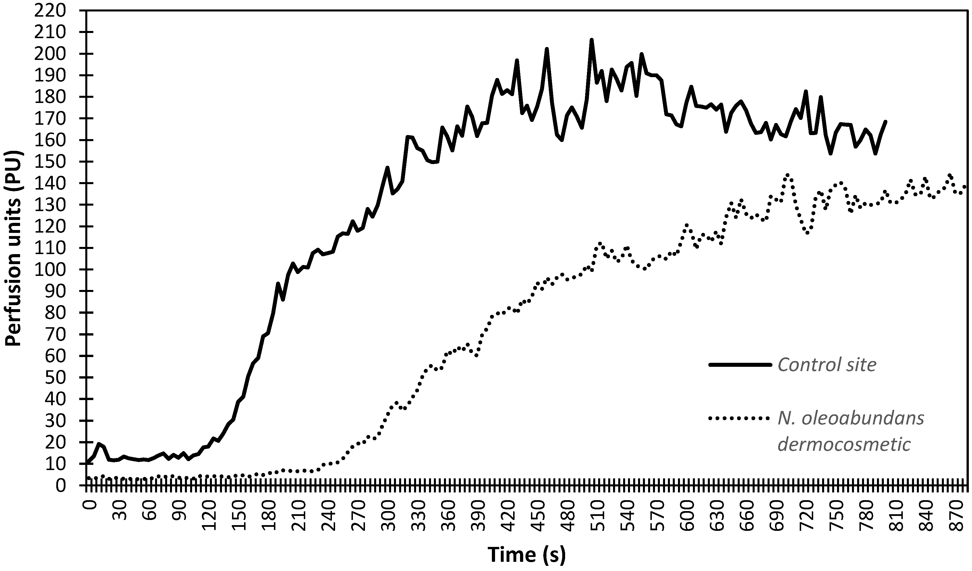
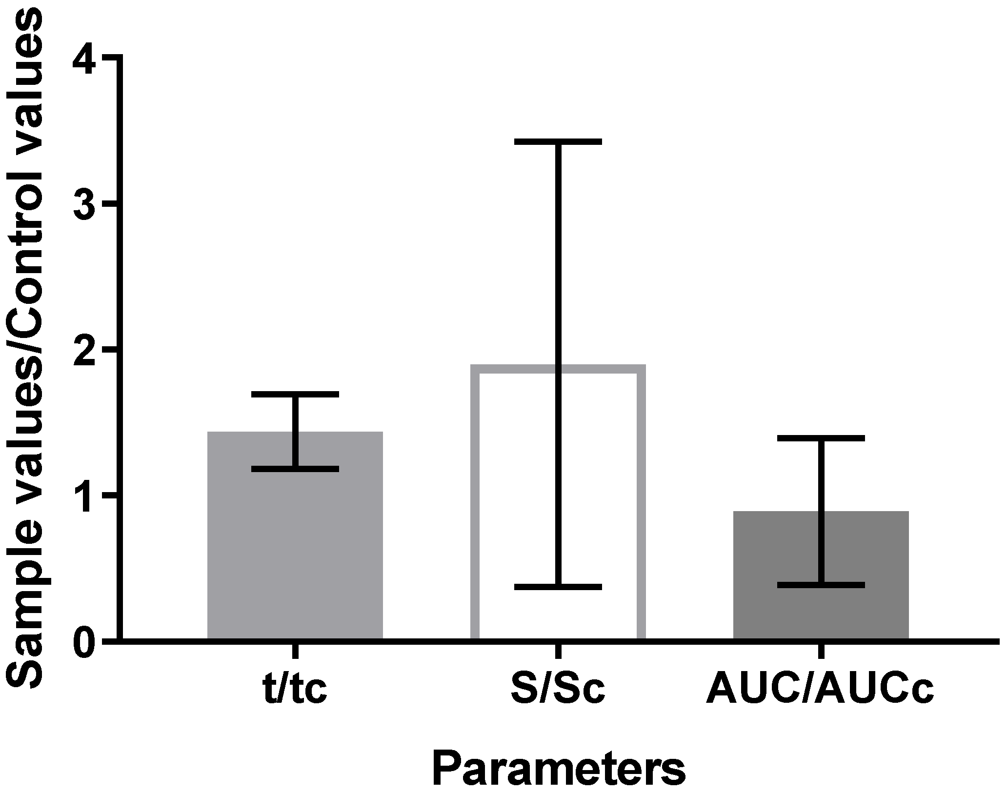
3.5. In Vivo Anti-Inflammatory Activity

To assure the real in vivo response for the microalgae aqueous extract, we tested previously the control gel (without the microalgae extract) following the same experimental protocol. The application of the control gel produced no effects over the volunteers’ forearms (data not showed), and thus, we continued with the experiments using the gel containing the microalgae aqueous extract. Figure 3 shows a typical nicotinate-induced perfusion response during this investigation, where the blood flow increase was less marked in the site pre-treated with microalgae cosmetic than in the untreated site (control site).
The majority of the subjects were of phototype III. It can also be observed that, accordingly, longer t onset times were observed in the pre-treated site when compared with in the control (Figure 4). The parameters slope of the tangent line in the hyperemia (S) and the AUC were also used to assess the anti-inflammatory capacity of the microalgae aqueous extract incorporated into the dermocosmetic sample (Figure 4). The ratio value obtained for S indicated a higher slope in the treated site, but no differences were established, which could be attributed to a high inter-individual variability. Moreover, lower AUCs were observed in the treated site, but again, differences were not obtained.

4. Discussion

Compounds or extracts from microalgae have been associated with diverse industrial uses, but recently an interest in their applications as a cosmetic ingredient has arisen, mainly as part of environmentally sustainable products [2,9]. The batch mode was used to culture N. oleoabundans under phototrophic conditions with no nitrogen deprivation to avoid lipid accumulation. Several studies have shown that cultivation with low nitrogen amounts is intended to produce a great amount of lipids on biomass for biodiesel applications [27,28]. Closed photobioreactors offer more productivity during the process [29]. Thus, we chose the batch mode cultivation to guarantee the maximum biomass production to probe this material as a future ingredient for dermocosmetics. Our biomass composition was in accordance with previously reported values (12.3–15.4% lipids and 26.7–30.1% proteins) [30].
Although multiple reports can be found regarding microalgal bioactivities, such as increase in skin moisturization, promotion of microcirculation, and anti-inflammatory action [31], microalgae can still be considered an underexplored natural resource of natural bioactive compounds for cosmetics, mainly antioxidants. Thus, the first part of this work aimed to establish the SA of the N. oleoabundans extracts using the DPPH assay to evaluate in vitro its antioxidant activity. As reported in the specialized literature, phenolic compounds are linked to the in vitro antioxidant activity in cyanobacteria extracts [32]. However, not only phenolic, but also other bioactive compounds, such as pigments, polysaccharides, and proteins, could be responsible for the antioxidant activities of microalgal biomass, such those present in Chlorella marina methanol extracts [33]. Potent antioxidant activity has been established for other microalgal species, particularly for aqueous extracts with variable colors including violet, green, light blue, blue, and pink [34]. Such research suggested that highly polar compounds, such as phycobilins, phenolic compounds, and polysaccharides, could be responsible for this activity [34]. These results are consistent with the higher SA values obtained herein for the water extract, showing that water allows the further extraction of polar compounds that may be responsible for the higher antioxidant activity observed. Moreover, a recent study evaluating the chemical compositions of some marine microalgal biomass and the total phenolic content, expressed as mg gallic acid equivalent (GAE) per gram of dry weight (DW), showed that N. oleoabundans UTEX-1185 biomass contained 9.8 mg GAE g−1 DW [30]. Considering not only this information, but also the SAs observed for the three N. oleoabundans extracts (Figure 1), we decided to incorporate the aqueous extract into a prototype dermocosmetic formulation to be further tested in vivo for skin compatibility and anti-inflammatory activity. Thus, an inexpensive, sustainable and biocompatible solvent was used to extract compounds from the tested microalgae even to avoid toxic and pollutant residues, which can be produced by another potentially pollutant solvents such as benzene and chloroform.
When probing skin compatibility, the redness, hydration, and TEWL are the most commonly assessed parameters [23,35]. In vivo skin tolerance assays were conducted using different non-invasive bioengineering approaches. According to our results, we confirmed that the SC hydration and the TEWL measured in the sites treated with the control gel and the microalgae gel were not different from those obtained in the untreated control. Thus, the dermocosmetic containing the N. oleobundans extract did not disturb the skin barrier function and could be further investigated, providing a good insight into the biocompatibility of this material.
The in vivo assessment of the anti-inflammatory effect of formulations with the N. oleoabundans aqueous extract was based on the ability to decrease the onset and extent of an erythema induced by the MN application using laser Doppler flowmetry. Briefly, laser Doppler flowmetry measures the blood flow in the range of 0–5 kHz that reflects the flow in the micro-capillaries in the superficial dermal plexus. The arbitrary units generated by the equipment (perfusion units-PU) maintain a direct relationship with the blood flow [19,36]. Moreover, the cutaneous microcirculation is affected by the application of nicotinates by a mechanism involving the activation of epidermal Langerhans cells and the release of prostaglandins D2 and E2, which induces a strong capillary vasodilation in the dermis–epidermis region [18]. Thus, the presence of molecules with anti-inflammatory activity can impact the inflammatory cascade and, thus, diminish the vasodilation [37]. The time required to the onset of erythema after an MN application on skin, named t onset, can also be used to probe the skin barrier function, as well as percutaneous penetration [19].
Since the free radical SA of the N. oleoabundans aqueous extract was established in the first part of this study, the longer t onset and the lower AUC after the application of 0.1% N. oleoabundans dermocosmetic could be explained by the presence of bioactive compounds in the microalgal extract, such as phenolic compounds or even small polysaccharides with proven antioxidant properties [30,34]. This study also suggests that the topical bioavailability of these molecules was highly affected by the inter-individual variability. Therefore, further studies with an increased number of volunteers and probing different concentrations of the extract should be conducted. Nonetheless, the results showed that despite this variability, the treated area presented a longer t onset time and a lower AUC, which indicated an improved skin barrier function.
Finally, N. oleoabundans cultivated under controlled conditions can produce biomass with biocompatible compounds to be used as novel naturally oriented vegan ingredient to aid in the development products for the skin care. Thus, it appears to be a promising species to obtain novel bioactive ingredients with both vegan and sustainable claims in the flourishing scenario of vegan and clean beauty.

5. Conclusions

We cultivated N. oleoabundans and extracted its active compounds with ultrasound-assisted extraction using different solvents. Our work not only evaluated the in vitro antioxidant activities of these extracts and showed an evidence of the good biocompatibility in vivo of the aqueous extract (the most antioxidant sample), but also indicated a possible anti-inflammatory activity of the developed topical hydrogel containing 0.1% N. oleoabundans aqueous extract. These promising results pointed out the relevance of continuing to establish the cutaneous attributes/properties of the N. oleoabundans extract as a multifunctional active ingredient in future studies. In fact, our preliminary results from safety and efficacy assays carried out in vivo seemed to confirm the potential of using microalgae derivatives in dermocosmetics with antiaging claims or even soothing properties as a novel attribute in the increasingly relevant vegan-driven cosmetic market.

Author Contributions

Conceptualization, A.L.M.-J. and C.R.; methodology, P.R.; validation, P.R. and J.C.M.d.C.; formal analysis, A.R.B. and M.V.R.V.; investigation, A.L.M.-J.; resources, A.L.M.-J.; data curation, B.B.d.S., P.R. and C.R.; writing—original draft preparation, A.L.M.-J. and A.R.B.; writing—review and editing, A.R.B., T.S.d.A. and C.R.; supervision, A.R.B.; project administration, A.L.M.-J., A.R.B. and C.R; funding acquisition, A.L.M.-J., A.R.B., J.C.M.d.C. and C.R. All authors have read and agreed to the published version of the manuscript.

Funding

This research was supported by São Paulo Research Foundation (FAPESP, Processes 2015/11194-6, 2016/22000-0, and 2019/16169-0); Portuguese funds through the FCT-Foundation for Science and Technology to CBIOS, under the projects UIDB/04567/2020 and UIDP/04567/2020; and Conselho Nacional de Desenvolvimento Científico e Tecnológico (CNPq), process 305250/2019-1.

Institutional Review Board Statement

The study was conducted according to the guidelines of the Declaration of Helsinki, and approved by the Universidade Lusófona’s Ethics Committee of School of Health Sciences and Technologies (protocol code 1/2016, 7 December 2016).

Informed Consent Statement

Written informed consent has been obtained from the patient(s) to publish this paper.

Conflicts of Interest

The authors declare no conflict of interest.

References

  1. Hayase, M. Introduction to Cosmetic Materials. In Cosmetic Science and Technology; Sakamoto, K., Lochhead, R.Y., Maibach, H.I., Yamashita, Y., Eds.; Elsevier: Amsterdam, The Netherlands, 2017; pp. 149–154. [Google Scholar]
  2. Morocho-Jácome, A.L.; Ruscinc, N.; Martinez, R.M.; Carvalho, J.C.M.; Santos de Almeida, T.; Rosado, C.; Costa, J.G.; Velasco, M.V.R.; Baby, A.R. (Bio)Technological Aspects of Microalgae Pigments for Cosmetics. Appl. Microbiol. Biotechnol. 2020, 104, 9513–9522. [Google Scholar] [CrossRef]
  3. Abe, T. Psychology of Cosmetic Behavior. In Cosmetic Science and Technology; Sakamoto, K., Lochhead, R.Y., Maibach, H.I., Yamashita, Y., Eds.; Elsevier: Amsterdam, The Netherlands, 2016; pp. 101–113. [Google Scholar]
  4. Giliver, L. Global Vegan Cosmetics Market to Exceed $21 Billion by 2027, Predicts New Report. Available online: https://plantbasednews.org/lifestyle/beauty/global-vegan-cosmetics-market-exceed-21-billion/ (accessed on 3 September 2021).
  5. Queiroz, M.I.; Vieira, J.G.; Maroneze, M.M. Morphophysiological, Structural, and Metabolic Aspects of Microalgae. In Handbook of Microalgae-Based Processes and Products; Jacob-Lopes, E., Maroneze, M.M., Queiroz, M.I., Zepka, L.Q., Eds.; Elsevier: Amsterdam, The Netherlands, 2020; pp. 25–48. [Google Scholar]
  6. Cezare-Gomes, E.A.; Mejia-da-Silva, L.C.; Pérez-Mora, L.S.; Matsudo, M.C.; Ferreira-Camargo, L.S.; Singh, A.K.; Carvalho, J.C.M. Potential of Microalgae Carotenoids for Industrial Application. Appl. Biochem. Biotechnol. 2019, 188, 602–634. [Google Scholar] [CrossRef] [PubMed]
  7. Castro-Puyana, M.; Pérez-Sánchez, A.; Valdés, A.; Ibrahim, O.H.M.; Suarez-Álvarez, S.; Ferragut, J.A.; Micol, V.; Cifuentes, A.; Ibáñez, E.; García-Cañas, V. Pressurized Liquid Extraction of Neochloris Oleoabundans for the Recovery of Bioactive Carotenoids with Anti-Proliferative Activity against Human Colon Cancer Cells. Food Res. Int. 2017, 99, 1048–1055. [Google Scholar] [CrossRef] [PubMed] [Green Version]
  8. Günerken, E.; D’Hondt, E.; Eppink, M.H.M.; Garcia-Gonzalez, L.; Elst, K.; Wijffels, R.H. Cell Disruption for Microalgae Biorefineries. Biotechnol. Adv. 2015, 33, 243–260. [Google Scholar] [CrossRef] [PubMed]
  9. Ariede, M.B.; Candido, T.M.; Morocho-Jácome, A.L.; Velasco, M.V.R.; Carvalho, J.C.M.; Baby, A.R. Cosmetic Attributes of Algae—A Review. Algal Res. 2017, 25, 483–487. [Google Scholar] [CrossRef]
  10. Baby, A.R.; Morocho-Jácome, A.L. Dermocosmetic Applications of Microalgal Pigments. Adv. Appl. Microbiol. 2021; in press. [Google Scholar]
  11. Gouveia, L.; Marques, A.E.; Da Silva, T.L.; Reis, A. Neochloris Oleabundans UTEX #1185: A Suitable Renewable Lipid Source for Biofuel Production. J. Ind. Microbiol. Biotechnol. 2009, 36, 821–826. [Google Scholar] [CrossRef] [PubMed]
  12. Pruvost, J.; Van Vooren, G.; Cogne, G.; Legrand, J. Investigation of Biomass and Lipids Production with Neochloris Oleoabundans in Photobioreactor. Bioresour. Technol. 2009, 100, 5988–5995. [Google Scholar] [CrossRef] [Green Version]
  13. Li, H.-B.; Cheng, K.-W.; Wong, C.-C.; Fan, K.-W.; Chen, F.; Jiang, Y. Evaluation of Antioxidant Capacity and Total Phenolic Content of Different Fractions of Selected Microalgae. Food Chem. 2007, 102, 771–776. [Google Scholar] [CrossRef]
  14. Castro-Puyana, M.; Herrero, M.; Urreta, I.; Mendiola, J.A.; Cifuentes, A.; Ibáñez, E.; Suárez-Alvarez, S. Optimization of Clean Extraction Methods to Isolate Carotenoids from the Microalga Neochloris Oleoabundans and Subsequent Chemical Characterization Using Liquid Chromatography Tandem Mass Spectrometry. Anal. Bioanal. Chem. 2013, 405, 4607–4616. [Google Scholar] [CrossRef] [Green Version]
  15. Rosado, C.; Pinto, P.; Rodrigues, L.M. Assessment of Moisturizers and Barrier Function Restoration Using Dynamic Methods. Ski. Res. Technol. 2009, 15, 77–83. [Google Scholar] [CrossRef] [PubMed]
  16. Müller, B.; Kasper, M.; Surber, C.; Imanidis, G. Permeation, Metabolism and Site of Action Concentration of Nicotinic Acid Derivatives in Human Skin: Correlation with Topical Pharmacological Effect. Eur. J. Pharm. Sci. 2003, 20, 181–195. [Google Scholar] [CrossRef]
  17. Wagemaker, T.A.L.; Rijo, P.; Rodrigues, L.M.; Maia Campos, P.M.B.G.; Fernandes, A.S.; Rosado, C. Integrated Approach in the Assessment of Skin Compatibility of Cosmetic Formulations with Green Coffee Oil. Int. J. Cosmet. Sci. 2015, 37, 506–510. [Google Scholar] [CrossRef] [PubMed]
  18. Benyó, Z.N.; Gille, A.; Bennett, C.L.; Rn, B.; Clausen, E.; Offermanns, S. Nicotinic Acid-Induced Flushing Is Mediated by Activation of Epidermal Langerhans Cells. Mol. Pharmacol. 2006, 70, 1844–1849. [Google Scholar] [CrossRef] [PubMed]
  19. Vandersee, S.; Erdmenger, U.; Patzelt, A.; Beyer, M.; Meinke, M.C.; Darvin, M.E.; Koscielny, J.; Lademann, J. Significance of the Follicular Pathway for Dermal Substance Penetration Quantified by Laser Doppler Flowmetry. J. Biophotonics 2016, 9, 276–281. [Google Scholar] [CrossRef] [PubMed]
  20. Oliveira, C.A.; Dario, M.F.; Sarruf, F.D.; Mariz, I.F.A.; Velasco, M.V.R.; Rosado, C.; Baby, A.R. Safety and Efficacy Evaluation of Gelatin-Based Nanoparticles Associated with UV Filters. Colloids Surf. B Biointerfaces 2016, 140, 531–537. [Google Scholar] [CrossRef]
  21. Vertuani, S.; Ziosi, P.; Solaroli, N.; Buzzoni, V.; Carli, M.; Lucchi, E.; Valgimigli, L.; Baratto, G.; Manfredini, S. Determination of Antioxidant Efficacy of Cosmetic Formulations by Non-Invasive Measurements. Ski. Res. Technol. 2003, 9, 245–253. [Google Scholar] [CrossRef]
  22. Wahlberg, J.E. Erythema-Inducing Effects of Solvents Following Epicutaneous Administration to Man—Studied by Laser Doppler Flowmetry. Scand. J. Work. Environ. Health 1984, 10, 159–162. [Google Scholar] [CrossRef]
  23. Peres, D.A.; Oliveira, C.A.; Costa, M.S.; Tokunaga, V.K.; Mota, J.P.; Rosado, C.; Consiglieri, V.O.; Kaneko, T.M.; Velasco, M.V.R.; Baby, A.R. Rutin Increases Critical Wavelength of Systems Containing a Single UV Filter and with Good Skin Compatibility. Ski. Res. Technol. 2016, 22, 325–333. [Google Scholar] [CrossRef] [PubMed]
  24. UTEX Culture Collection of Algae UTEX. Available online: https://utex.org/products/modified-bold-3n-medium (accessed on 16 April 2018).
  25. Rijo, P.; Falé, P.L.; Serralheiro, M.L.; Simões, M.F.; Gomes, A.; Reis, C. Optimization of Medicinal Plant Extraction Methods and Their Encapsulation through Extrusion Technology. Measurement 2014, 58, 249–255. [Google Scholar] [CrossRef]
  26. Falé, P.L.; Borges, C.; Madeira, P.J.A.; Ascensão, L.; Araújo, M.E.M.; Florêncio, M.H.; Serralheiro, M.L.M. Rosmarinic Acid, Scutellarein 4′-Methyl Ether 7-O-Glucuronide and (16S)-Coleon E Are the Main Compounds Responsible for the Antiacetylcholinesterase and Antioxidant Activity in Herbal Tea of Plectranthus Barbatus (Falso Boldo). Food Chem. 2009, 114, 798–805. [Google Scholar] [CrossRef]
  27. Abu Hajar, H.A.; Riefler, R.G.; Stuart, B.J. Cultivation of the Microalga Neochloris Oleoabundans for Biofuels Production and Other Industrial Applications (A Review). Appl. Biochem. Microbiol. 2017, 53, 640–653. [Google Scholar] [CrossRef]
  28. Avila-León, I.A.; Matsudo, M.C.; Ferreira-Camargo, L.S.; Rodrigues-Ract, J.N.; Carvalho, J.C.M. Evaluation of Neochloris Oleoabundans as Sustainable Source of Oil-Rich Biomass. Braz. J. Chem. Eng. 2020, 37, 41–48. [Google Scholar] [CrossRef]
  29. Borowitzka, M.A. High-Value Products from Microalgae—Their Development and Commercialisation. J. Appl. Phycol. 2013, 25, 743–756. [Google Scholar] [CrossRef]
  30. Tibbetts, S.M.; Milley, J.E.; Lall, S.P. Chemical Composition and Nutritional Properties of Freshwater and Marine Microalgal Biomass Cultured in Photobioreactors. J. Appl. Phycol. 2015, 27, 1109–1119. [Google Scholar] [CrossRef] [Green Version]
  31. Oliveira, A.C.; Morocho-Jácome, A.L.; Castro Lima, C.R.; Marques, G.A.; Bispo, M.O.; Barros, A.B.; Costa, J.G.; Santos de Almeida, T.; Rosado, C.; Carvalho, J.C.M.; et al. Cosmetics Applications. In Microalgae; Galanakis, C.M., Ed.; Academic Press: London, UK, 2020; pp. 313–338. [Google Scholar]
  32. Trabelsi, L.; Mnari, A.; Abdel-Daim, M.M.; Abid-Essafi, S.; Aleya, L. Therapeutic Properties in Tunisian Hot Springs: First Evidence of Phenolic Compounds in the Cyanobacterium Leptolyngbya Sp. Biomass, Capsular Polysaccharides and Releasing Polysaccharides. BMC Complement. Altern. Med. 2016, 16, 515. [Google Scholar] [CrossRef] [Green Version]
  33. Manivannan, K.; Anantharaman, P.; Balasubramanian, T. Evaluation of Antioxidant Properties of Marine Microalga Chlorella Marina (Butcher, 1952). Asian Pac. J. Trop. Biomed. 2012, 2, S342–S436. [Google Scholar] [CrossRef]
  34. Shanab, S.M.M.; Mostafa, S.S.M.; Shalaby, E.A.; Mahmoud, G.I. Aqueous Extracts of Microalgae Exhibit Antioxidant and Anticancer Activities. Asian Pac. J. Trop. Biomed. 2012, 2, 608–615. [Google Scholar] [CrossRef] [Green Version]
  35. Berardesca, E.; Loden, M.; Serup, J.; Masson, P.; Rodrigues, L.M. The Revised EEMCO Guidance for the in Vivo Measurement of Water in the Skin. Ski. Res. Technol. 2018, 24, 351–358. [Google Scholar] [CrossRef]
  36. Remane, Y.; Leopold, C.S. Time of Erythema Onset after Application of Methyl Nicotinate Ointments as Response Parameter: Influence of Penetration Kinetics and Enhancing Agents. Ski. Pharmacol. Physiol. 2006, 19, 303–310. [Google Scholar] [CrossRef]
  37. Sauce, R.; Pinto, C.A.S.d.O.; Velasco, M.V.R.; Rosado, C.; Baby, A.R. Ex Vivo Penetration Analysis and Anti-Inflammatory Efficacy of the Association of Ferulic Acid and UV Filters. Eur. J. Pharm. Sci. 2021, 156, 105578. [Google Scholar] [CrossRef] [PubMed]
Figure 1. Variation in the scavenging activities (SAs; %) of three N. oleoabundans extracts. The error bars correspond to the SD (n = 3). One-way ANOVA followed by the Tukey test was used for comparisons between groups. p-values (descriptive level) obtained by ANOVA were considered significant for p < 0.05. Different capital letters were used for means significantly different according to ANOVA.
Cosmetics 09 00009 g001
Figure 2. Transepidermal water loss (TEWL) (a) and stratum corneum (SC) hydration (b) after the application of N. oleoabundans dermocosmetic. The error bars correspond to the SD (n = 14). One-way ANOVA followed by the Tukey test was used for comparisons between groups. p-values (descriptive level) obtained by ANOVA were considered significant for p < 0.05. Different capital letters were used for means significantly different according to ANOVA.
Cosmetics 09 00009 g002
Figure 3. An example of the perfusion units (PUs) variation measured by laser Doppler flowmetry in a volunteer after the applications of methyl nicotinate, i.e., control site (represented by the solid line) and N. oleoabundans gel (represented by the dotted line).
Cosmetics 09 00009 g003
Figure 4. Variation in the onset time (t/tc), the slope of the line of hypermemia (S/Sc), and the area under the curve (AUC/AUCc) after the application of the N. oleoabundans gel. The error bars correspond to the SD (n = 13).
Cosmetics 09 00009 g004
Table 1. Quali/quantitative compositions of dermocosmetics formulation.
Composition *Concentration (% w/w)
Control GelNeochloris Oleoabundans (N. oleoabundans) Gel
Ammonium acryloyldimethyltaurate/VP copolymer (Aristoflex® AVC)1.01.0
N. oleoabundans aqueous extract -1.0
Derivatives of parabens 0.80.8
Aquaqsp 100%qsp 100%
* Qualitative description according to the International Nomenclature of Cosmetic Ingredient (INCI).
Publisher’s Note: MDPI stays neutral with regard to jurisdictional claims in published maps and institutional affiliations.

