
Addressing Human Skin Ethnicity: Contribution of Tissue Engineering to the Development of Cosmetic Ingredients

Abstract
:1. Introduction
2. Materials and Methods
2.1. Ethical Compliance
2.2. Isolation of Ethnic Cells (Keratinocytes and Melanocytes)
2.3. Reconstruction of Pigmented Epidermis
2.4. Silencing
2.5. Histology and Immunohistochemistry
2.6. Fontana-Masson Staining
2.7. Statistics
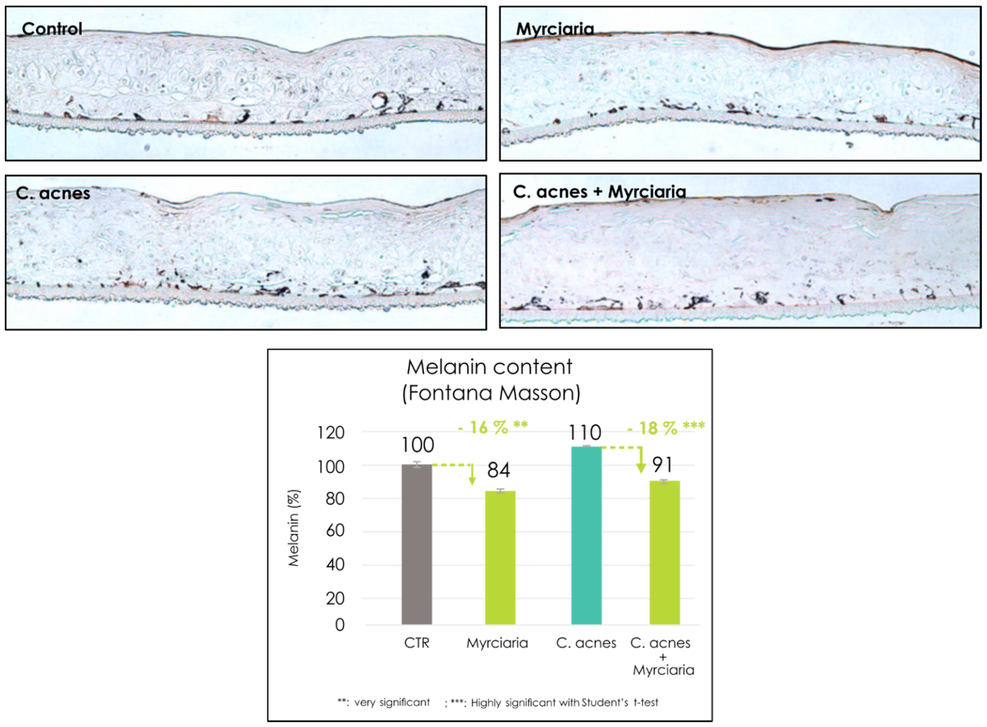
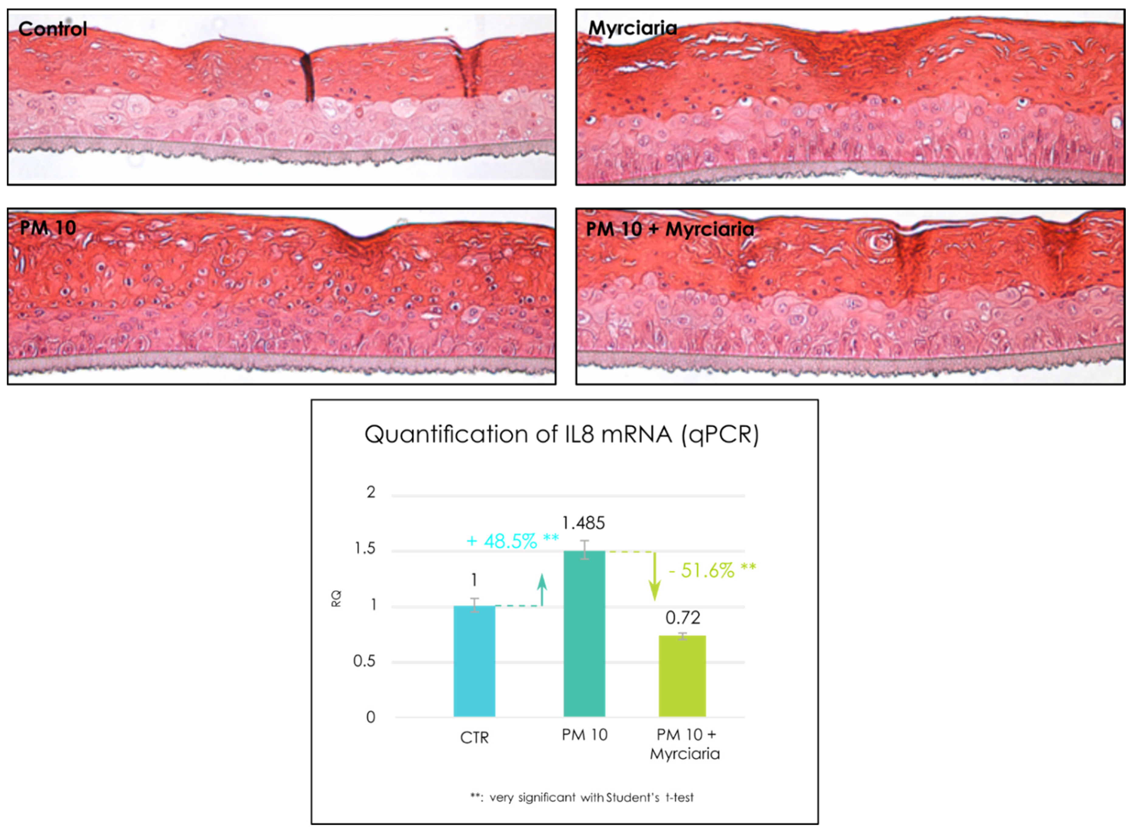
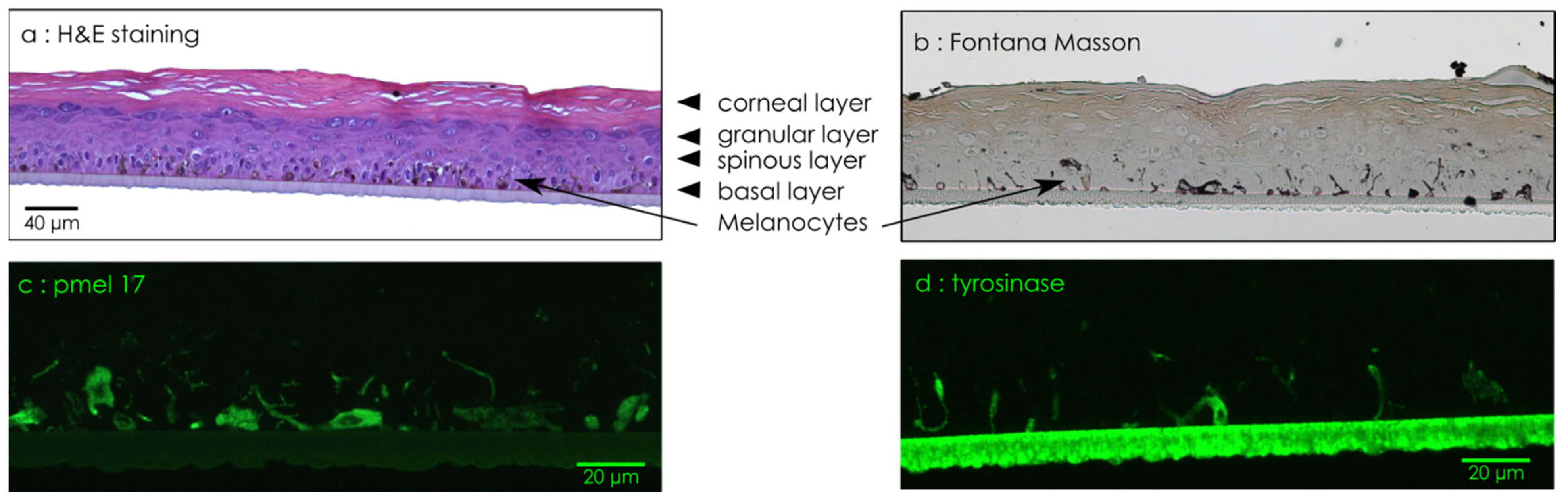
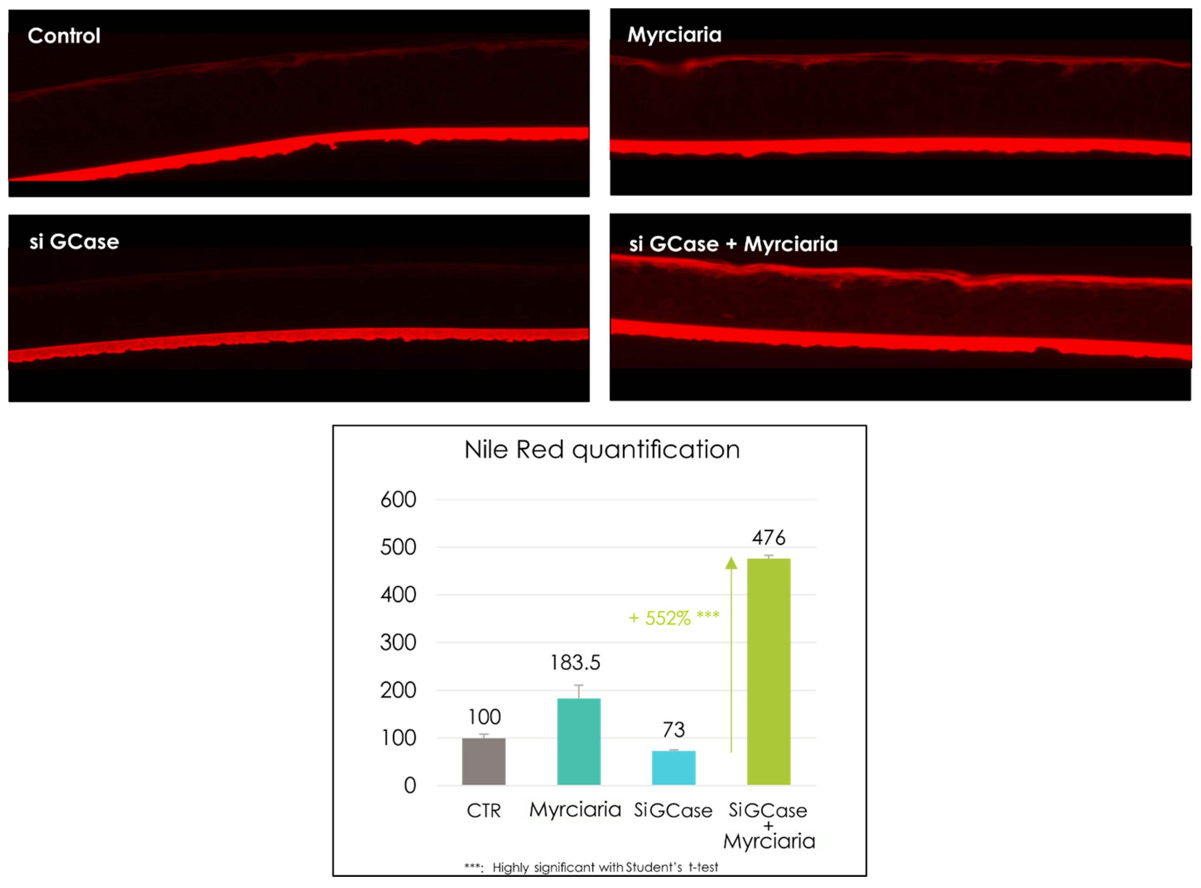
3. Results
4. Conclusions
Author Contributions
Funding
Institutional Review Board Statement
Informed Consent Statement
Data Availability Statement
Acknowledgments
Conflicts of Interest
References
- Fitzpatrick, T.B. The Validity and Practicality of Sun-Reactive Skin Types I Through VI. Arch. Dermatol. 1988, 124, 869–871. [Google Scholar] [CrossRef]
- Lancer, H.A. Lancer ethnicity scale (LES). Lasers Surg. Med. 1998, 22, 9. [Google Scholar] [CrossRef]
- Goldman, M. Universal Classification of Skin Type. In Simplified Facial Rejuvenation; Shiffman, M., Mirrafati, S., Lam Cueteaux, C., Eds.; Springer: Berlin/Heidelberg, Germany, 2008. [Google Scholar]
- Wang, M.; Xiao, K.; Wuerger, S.; Cheung, V.; Ronnier Luo, M. Measuring Human Skin Colour; Society for Imaging Science and Technology: Leeds, UK, 2015. [Google Scholar]
- Gold, E.; Sternfeld, B.; Kelsey, J.; Brown, C.; Mouton, C.; Reame, N.; Salamone, L.; Stellato, R. Relation of Demographic and Lifestyle Factors to Symptoms in a Multi-Racial/Ethnic Population of Women 40–55 Years of Age. Am. J. Epidemiol. 2000, 152, 463–473. [Google Scholar] [CrossRef] [Green Version]
- Yanpei, G.; Jianxin, H.; Chunpeng, J.; Ying, Z. Biomarkers, Oxidative Stress and Autophagy in Skin Aging. Ageing Res. Rev. 2020, 59, 101036. [Google Scholar]
- Cole, M.A.; Quan, T.; Voorhees, J.J.; Fisher, G.J. Extracellular matrix regulation of fibroblast function: Redefining our perspective on skin aging. J. Cell Commun. Signal 2018, 12, 35–43. [Google Scholar] [CrossRef] [PubMed] [Green Version]
- Vashi, N.A.; de Castro Maymone, M.B.; Kundu, R.V. Aging Differences in Ethnic Skin. J. Clin. Aesthet. Dermatol. 2016, 9, 31–38. [Google Scholar] [PubMed]
- Callaghan, D.J.; Singh, B.; Reddy, K.K. Skin Aging in Individuals with Skin of Color. In Dermato Anthropology of Ethnic Skin and Hair; Vashi, N., Maibach, H., Eds.; Springer: Cham, Switzerland, 2017. [Google Scholar]
- Lim, H.; Kang, S.; Chien, A.L. Photodamage in Skin of Color; Cutaneous Photoaging: Manchester UK, 2019. [Google Scholar]
- Perkins, A.; Cheng, C.; Hillebrand, G.; Miyamoto, K.; Kimball, A. Comparison of the epidemiology of acne vulgaris among Caucasian, Asian, Continental Indian and African American women. J. Eur. Acad. Dermatol. Venereol. 2011, 25, 1054–1060. [Google Scholar] [CrossRef] [PubMed]
- Cunliffe, W.J.; Holland, D.B.; Clark, S.M.; Stables, G.I. Comedogenesis: Some new aetiological, clinical and therapeutic strategies. Br. J. Dermatol. 2000, 142, 1084–1091. [Google Scholar] [CrossRef]
- Nisticò, S.P.; Tolone, M.; Zingoni, T.; Tamburi, F.; Scali, E.; Bennardo, L.; Cannarozzo, G. A New 675 nm Laser Device in the Treatment of Melasma: Results of a Prospective Observational Study. Photobiomodul. Photomed. Laser Surg. 2020, 38, 560–564. [Google Scholar] [CrossRef]
- Davis, E.C.; Callender, V.D. A review of acne in ethnic skin: Pathogenesis, clinical manifestations, and management strategies. J. Clin. Aesthet. Dermatol. 2010, 3, 24–38. [Google Scholar]
- Al Qarqaz, F.; Al-Yousef, A. Skin microneedling for acne scars associated with pigmentation in patients with dark skin. J. Cosmet. Dermatol. 2018, 17, 390–395. [Google Scholar] [CrossRef]
- Rullan, P.P.; Olson, R.; Lee, K.C. A Combination Approach to Treating Acne Scars in All Skin Types: Carbolic Chemical Reconstruction of Skin Scars, Blunt Bi-level Cannula Subcision, and Microneedling—A Case Series. J. Clin. Aesthetic. Dermatol. 2020, 3, 19–23. [Google Scholar]
- Narumol, S.A.; Indermeet, K.; Suteeraporn, C.; Henry, W.L. Postinflammatory hyperpigmentation: A comprehensive overview: Epidemiology, pathogenesis, clinical presentation, and noninvasive assessment technique. J. Am. Acad. Dermatol. 2017, 77, 591–605. [Google Scholar]
- Girardeau-Hubert, S.; Deneuville, C.; Pageon, H.; Abed, K.; Tacheau, C.; Cavusoglu, N.; Donovan, M.; Bernard, D.; Asselineau, D. Reconstructed Skin Models Revealed Unexpected Differences in Epidermal African and Caucasian Skin. Sci Rep. 2019, 9, 7456. [Google Scholar] [CrossRef] [PubMed]
- Krutmann, J.; Moyal, D.; Liu, W.; Kandahari, S.; Lee, G.S.; Nopadon, N.; Xiang, L.F.; Seité, S. Pollution and acne: Is there a link? Clin. Cosmet. Investig. Dermatol. 2017, 10, 199–204. [Google Scholar] [CrossRef] [PubMed] [Green Version]
- Park, S.-Y.; Byun, E.J.; Lee, J.D.; Kim, S.; Kim, H.S. Air Pollution, Autophagy, and Skin Aging: Impact of Particulate Matter (PM10) on Human Dermal Fibroblasts. Int. J. Mol. Sci. 2018, 19, 2727. [Google Scholar] [CrossRef] [Green Version]
- Araviiskaia, E.; Berardesca, E.; Bieber, T.; Gontijo, G.; Sanchez Viera, M.; Marrot, L.; Chuberre, B.; Dreno, B. The impact of airborne pollution on skin. J. Eur. Acad. Dermatol. Venereol. 2019, 33, 1496–1505. [Google Scholar] [CrossRef]
- Cole, P.D.; Hatef, D.A.; Taylor, S.; Bullocks, J.M. Skin care in ethnic populations. Semin. Plast. Surg. 2009, 23, 168–172. [Google Scholar] [CrossRef] [Green Version]
- Uchida, Y.; Park, K. Ceramides in Skin Health and Disease: An Update. Am. J. Clin. Dermatol. 2021, 1–14. [Google Scholar]
- Boer, D.E.C.; van Smeden, J.; Bouwstra, J.A. Glucocerebrosidase: Functions in and Beyond the Lysosome. J. Clin. Med. 2020, 9, 736. [Google Scholar] [CrossRef] [Green Version]
- Rheinwald, J.G.; Green, H. Serial cultivation of strains of human epidermal keratinocytes: The formation of keratinizing colonies from single cells. Cell 1975, 6, 331–343. [Google Scholar] [CrossRef]
- Peehl, D.; Ham, R. Growth and differentiation of human keratinocytes without a feeder layer or conditioned medium. In Vitro 1980, 16, 516–525. [Google Scholar] [CrossRef]
- Bell, E.; Ehrlich, H.P.; Buttle, D.J.; Nakatsuji, T. Living tissue formed in vitro and accepted as skin-equivalent tissue of full thickness. Science 1981, 211, 1052–1054. [Google Scholar] [CrossRef]
- Prunerias, M.; Régnier, M.; Woodley, D. Methods for cultivation of keratinocytes with an air-liquid interface. J. Investig. Dermatol. 1983, 81, 28s–33s. [Google Scholar] [CrossRef] [PubMed] [Green Version]
- Boyce, S.; Hansbrough, J. Biologic attachment, growth, and differentiation of cultured human epidermal keratinocytes on a graftable collagen and chondroitin-6-sulfate substrate. Surgery 1988, 103, 421–431. [Google Scholar] [PubMed]
- Tinois, E.; Tiollier, J.; Gaucherand, M.; Dumas, H.; Tardy, M.; Thivolet, J. In vitro and post-transplantation differentiation of human keratinocytes grown on the human type IV collagen film of a bilayered dermal substitute. Exp. Cell Res. 1991, 193, 310–319. [Google Scholar] [CrossRef]
- Ponec, M.; Weerheim, A.; Kempenaar, J.; Mulder, A.; Gooris, G.; Bouwstra, J.; Mommaas, A. The formation of competent barrier lipids in reconstructed human epidermis requires the presence of vitamin C. J. Investig. Dermatol. 1997, 109, 348–355. [Google Scholar] [CrossRef] [Green Version]
- El Ghalbzouri, A.; Siamari, R.; Willemze, R.; Ponec, M. Leiden reconstructed human epidermal model as a tool for the evaluation of the skin corrosion and irritation potential according to the ECVAM guidelines. Toxicol. In Vitro 2008, 22, 1311–1320. [Google Scholar] [CrossRef]
- Rosdy, M.; Pisani, A.; Ortonne, J.P. Production of basement membrane components by a reconstructed epidermis cultured in the absence of serum and dermal factors. Br. J. Dermatol. 1993, 129, 227–234. [Google Scholar] [CrossRef]
- Plaza, C.; Meyrignac, C.; Botto, J.M.; Capallere, C. Characterization of a new full thickness in vitro skin model. Tissue Eng. Part C Methods 2021, 27, 411–420. [Google Scholar] [CrossRef]
- Bessou, S.; Surleve-Bazeille, J.-E.; Sorbier, E.; TAÏEB, A. Ex Vivo Reconstruction of the Epidermis with Melanocytes and the Influence of UVB. Pigment. Cell Res. 1955, 8, 241–249. [Google Scholar] [CrossRef] [PubMed]
- Zhang, Z.; Michniak-Kohn, B.B. Tissue engineered human skin equivalents. Pharmaceutics 2012, 4, 26–41. [Google Scholar] [CrossRef] [PubMed] [Green Version]
- Capallere, C.; Plaza, C.; Meyrignac, C.; Marianne, A.; Marie, B.; Valère, B.; Imane, G.; Éric, B.; Jean-Marie, B. Property characterization of reconstructed human epidermis equivalents, and performance as a skin irritation model. Toxicol. In Vitro 2018, 53, 45–56. [Google Scholar] [CrossRef]
- Kaufman, B.P.; Aman, T.; Alexis, A.F. Post inflammatory hyperpigmentation: Epidemiology, clinical presentation, pathogenesis and treatment. Am. J. Clin. Dermatol. 2018, 19, 489–503. [Google Scholar] [CrossRef] [PubMed]
- Garcia, I.; Capallere, C.; Arcioni, M.; Brulas, M.; Plaza, C.; Meyrignac, C.; Bauza, É.; Botto, J.M. Establishment and performance assessment of an in-house 3D Reconstructed Human Cornea-Like Epithelium (RhCE) as a screening tool for the identification of liquid chemicals with potential eye hazard. Toxicol. In Vitro 2019, 61, 104604. [Google Scholar] [CrossRef]
- OECD. Test No. 431: In Vitro Skin Corrosion: Reconstructed Human Epidermis (RHE) Test Method, OECD Guidelines for the Testing of Chemicals, Section 4; OECD Publishing: Paris, France, 2019. [Google Scholar]
- OECD. Test No. 439: In Vitro Skin Irritation: Reconstructed Human Epidermis Test Method, OECD Guidelines for the Testing of Chemicals, Section 4; OECD Publishing: Paris, France, 2021. [Google Scholar]
- OECD. Test No. 492: Reconstructed Human Cornea-Like Epithelium (RhCE) Test Method for Identifying Chemicals not Requiring Classification and Labelling for Eye Irritation or Serious Eye Damage; OECD Guidelines for the Testing of Chemicals, Section 4; OECD Publishing: Paris, France, 2019. [Google Scholar]
- Zhang, B.; Choi, Y.M.; Lee, J.; An, I.S.; Li, L.; He, C.; Dong, Y.; Bae, S.; Meng, H. Toll-like receptor 2 plays a critical role in pathogenesis of acne vulgaris. Biomed. Dermatol. 2019, 3, 4. [Google Scholar] [CrossRef]
- Rembiesa, J.; Ruzgas, T.; Engblom, J.; Holefors, A. The Impact of Pollution on Skin and Proper Efficacy Testing for Anti-Pollution Claims. Cosmetics 2018, 5, 4. [Google Scholar] [CrossRef] [Green Version]
- Pan, T.L.; Wang, P.W.; Aljuffali, I.A.; Huang, C.T.; Lee, C.W.; Fang, J.Y. The impact of urban particulate pollution on skin barrier function and the subsequent drug absorption. J. Dermatol. Sci. 2015, 78, 51–60. [Google Scholar] [CrossRef]
- Fu, C.; Chen, J.; Lu, J.; Yi, L.; Tong, X.; Kang, L.; Zeng, Q. Roles of inflammation factors in melanogenesis (Review). Mol. Med. Rep. 2020, 21, 1421–1430. [Google Scholar] [CrossRef] [PubMed] [Green Version]
- Gimenez-Arnau, A.M. Xerosis Means “Dry Skin”: Mechanisms. Skin Conditions, and Its Management. In Filaggrin; Thyssen, J., Maibach, H., Eds.; Springer: Berlin/Heidelberg, Germany, 2014. [Google Scholar]
- Sochorová, M.; Staňková, K.; Pullmannová, P.; Kováčik, A.; Zbytovská, J.; Vávrová, K. Permeability Barrier and Microstructure of Skin Lipid Membrane Models of Impaired Glucosylceramide Processing. Sci. Rep. 2017, 7, 6470. [Google Scholar] [CrossRef] [PubMed] [Green Version]
- Boer, D.; van Smeden, J.; Al-Khakany, H.; Melnik, E.; van Dijk, R. Skin of atopic dermatitis patients shows disturbed β-glucocerebrosidase and acid sphingomyelinase activity that relates to changes in stratum corneum lipid composition. Biochim. Et Biophys. Acta (BBA)–Mol. Cell Biol. Lipids 2020, 1865, 158673. [Google Scholar] [CrossRef]


Publisher’s Note: MDPI stays neutral with regard to jurisdictional claims in published maps and institutional affiliations. |
© 2021 by the authors. Licensee MDPI, Basel, Switzerland. This article is an open access article distributed under the terms and conditions of the Creative Commons Attribution (CC BY) license (https://creativecommons.org/licenses/by/4.0/).
Share and Cite
Capallere, C.; Arcioni, M.; Restellini, L.; Imbert, I. Addressing Human Skin Ethnicity: Contribution of Tissue Engineering to the Development of Cosmetic Ingredients. Cosmetics 2021, 8, 98. https://doi.org/10.3390/cosmetics8040098
Capallere C, Arcioni M, Restellini L, Imbert I. Addressing Human Skin Ethnicity: Contribution of Tissue Engineering to the Development of Cosmetic Ingredients. Cosmetics. 2021; 8(4):98. https://doi.org/10.3390/cosmetics8040098
Chicago/Turabian StyleCapallere, Christophe, Marianne Arcioni, Laura Restellini, and Isabelle Imbert. 2021. "Addressing Human Skin Ethnicity: Contribution of Tissue Engineering to the Development of Cosmetic Ingredients" Cosmetics 8, no. 4: 98. https://doi.org/10.3390/cosmetics8040098
APA StyleCapallere, C., Arcioni, M., Restellini, L., & Imbert, I. (2021). Addressing Human Skin Ethnicity: Contribution of Tissue Engineering to the Development of Cosmetic Ingredients. Cosmetics, 8(4), 98. https://doi.org/10.3390/cosmetics8040098