Communication

The Protective Effect of Hamamelis virginiana Stem and Leaf Extract on Fine Dust-Induced Damage on Human Keratinocytes

1
Research Center, The Garden of Naturalsolution, Osan-si 18103, Korea
2
Department of Physiology, College of Medicine, Gachon University, Incheon 21999, Korea
*
Author to whom correspondence should be addressed.
Cosmetics 2021, 8(4), 119; https://doi.org/10.3390/cosmetics8040119
Submission received: 30 November 2021 / Revised: 13 December 2021 / Accepted: 14 December 2021 / Published: 16 December 2021
(This article belongs to the Special Issue Green Ingredients in Cosmetics and Food (Volume II))

Abstract

:
Witch hazel extracts have been used for decades as cosmetic ingredients in skin care products. Our present study aims to evaluate its potential in anti-pollution products using a previously reported in vitro model. Calcium is a universal second messenger, and we used human respiratory and skin cells to detect changes in intracellular Ca2+ concentrations upon particulate matter contact. Both an increase in pro-inflammatory markers and a decrease in tight junction proteins were confirmed, as previously reported. Witch hazel stem and leaf extract showed significant attenuation of Ca2+ response upon the challenge; it displayed systematic regulations of the signal generator, PAR-2; a pro-inflammatory marker, NF-κB; and a tight junction protein, Occludin. We identified hexagalloylglucose from the extract and concluded that it is a major component regulating protection from particulate matter. Based on these results, witch hazel extract containing hexagalloylglucose is an active ingredient in anti-pollution skin care products.

1. Introduction

Hamamelis virginiana or witch hazel, is a useful shrub or small tree and is native to the eastern parts of North America. Native Americans used it for dowsing, observed by early European immigrants, and various preparations of its stem and leaves, such as extracts, tinctures, distillates, and creams, have been applied for skin troubles [1,2]. It is now widely accepted as an anti-inflammatory agent to treat sunburn, atopic eczema, and irritated skin, and it has astringent and antiphlogistic properties [3]. Moreover, beneficial compounds from the leaves or bark of witch hazel have been identified, mostly polyphenols such as hydroxycinnamic acids, flavonoids, and tannins [4,5,6,7,8]. It has been shown to contain many hydrolysable tannins, including the major component hammamelitannin, gallic acid, and carbohydrates with five (pentagalloylglucose) to ten (tannic acid) galloyl moieties. Among these, hammamelitannin displayed inhibition of tumor necrosis factor (TNF) α-induced cell death and showed other immunomodulatory effects.
One of the pioneering studies showed that the topical use of a formulation containing witch hazel distillate was highly effective to soothe UV-irritated skin [1]. This is a fine example showing that it can be adopted for the products that consumers want to use to protect their skin from the everyday stresses caused by “exposome” [9]. During the COVID-19 pandemic, a significant number of people around the world have complained of irritated skin, called “maskne”, due to the heavy use of facemasks [10]. Therefore, the cosmetics industry has been facing ever-growing demands for soothing products, specifically targeting environmental factors. As more consumers are looking for natural ingredients in their personal care products, we attempted to identify effective herbal extracts for protecting skin cells from air pollution. Previously, we introduced an innovative test platform where particulate matter 2.5 (PM2.5) mimics could directly challenge human bronchial/tracheal smooth muscle (HBT-SM) cells or keratinocytes (HaCaT cells), and produced a consistent output through transient induction of intracellular calcium concentration ([Ca2+]i) [11]. We revealed that the Ca2+ signal was generated by proteinase-activated receptor-2 (PAR-2) and was accompanied by both an increase in pro-inflammatory cytokines and a decrease in tight junction proteins. These results demonstrated systematic damage by PM2.5 on skin cells at the molecular level, supported by later studies demonstrating that skin barrier function is affected by PMs; one such study reported the clinical significance of these findings [12,13]. Over the last year, similar studies reached similar conclusions, confirming the crucial impact of PMs on keratinocytes [14,15]. Moreover, ER stress elicited by PM could also raise [Ca2+]i, eventually increasing melanin production [16,17].
In the present work, we could reveal that H. virginiana stem and leaf extract (HVE) could lower the Ca2+ influx in HaCaT cells by fine dust. The response involved PAR-2, NF-κB, and Occludin, like in previous work, and HVE could effectively reverse the damaging outputs. We further identified hexagalloylglucose (HGG) as a novel active ingredient of HVE to modulate [Ca2+]i upon PM stress.

2. Materials and Methods

2.1. Treating Cells with PM, HVE, and HGG

DPM NIST® SRM® 1650b, Fura-2/AM and Pluronic™ F-127 were purchased from Merck (Darmstadt, Germany) and Invitrogen (Waltham, MA, USA). DPM solution was prepared as a stock (25 mg/mL) in dimethyl sulfoxide (DMSO) and sonication in a water bath was applied prior to use, as previously mentioned [11]. We tried to keep the same conditions as the DPM solution, because it cannot be homogeneous all the time. Witch hazel stem and leaves were purchased from Cailleau Herboristerie (Chemillé, France). The doses of HVE and HGG were determined based on cytotoxicity (Supplementary Figure S1).

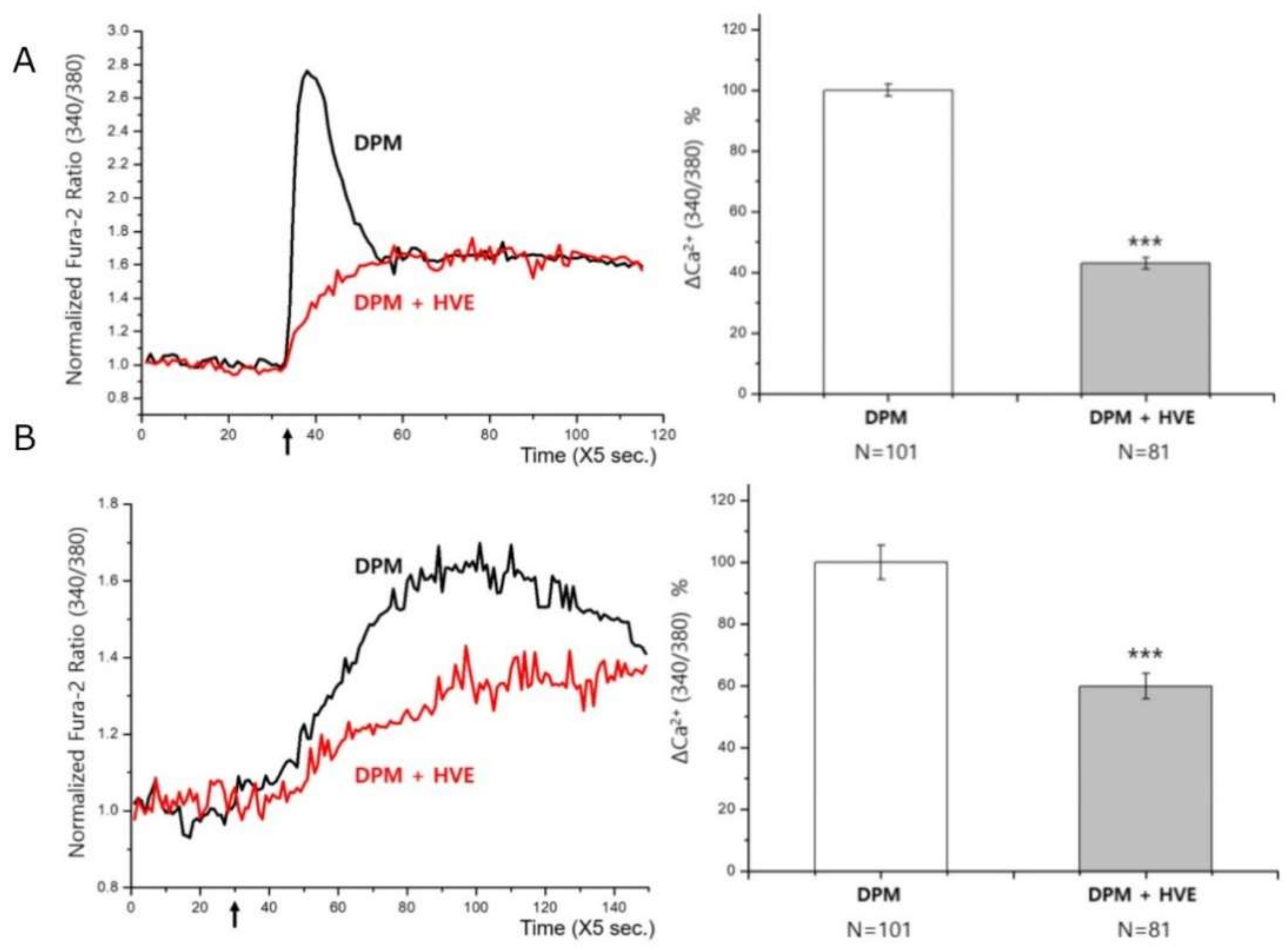
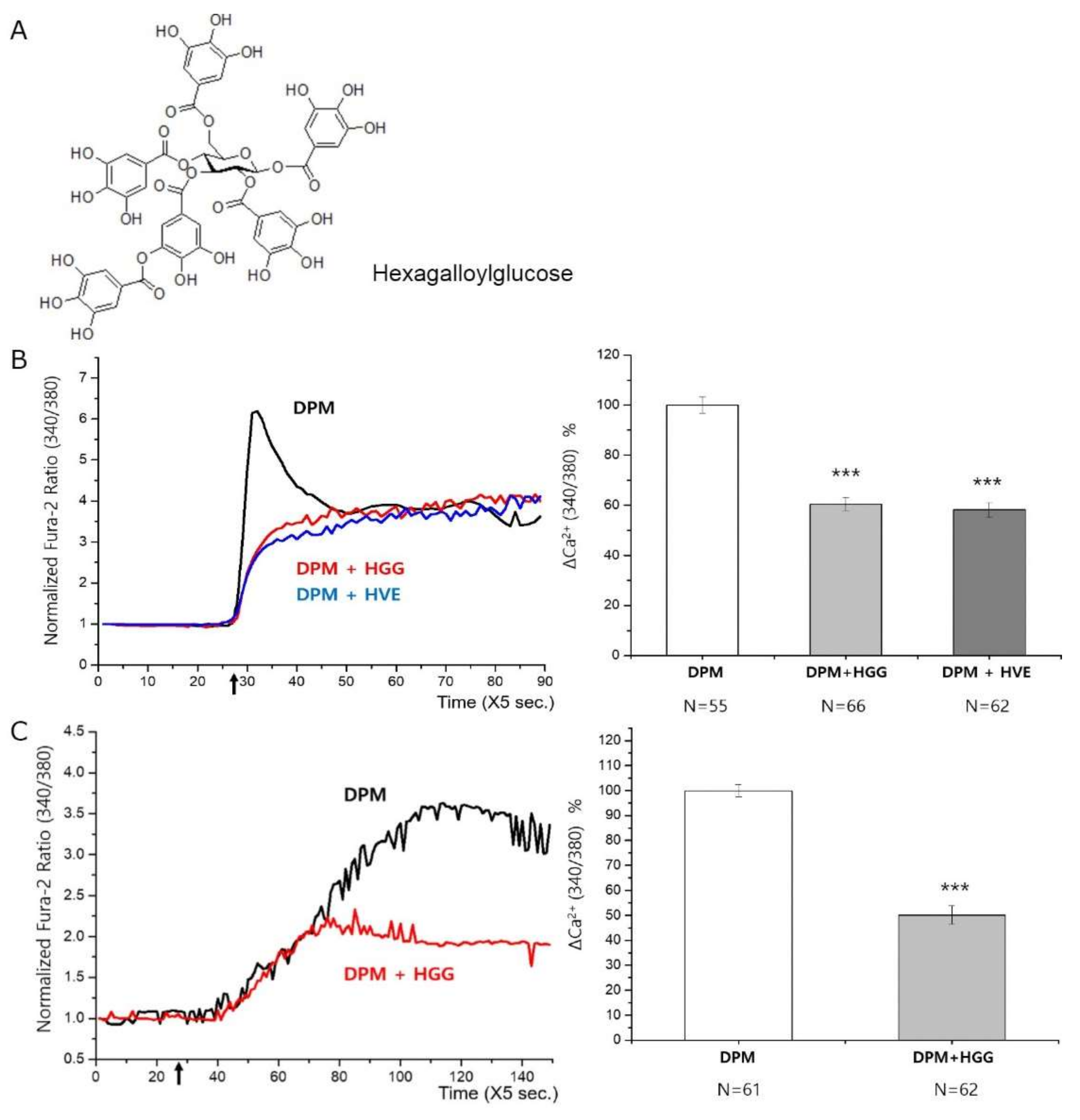
2.2. Measuring Intracellular Ca2+ Concentration

HBT-SM cells were purchased from LIFELINE Cell Technology (Frederick, MD, USA) and HaCaT cells were from AddexBio (San Diego, CA, USA). As previously described [11], the cells preloaded with 5 μM Fura-2/AM (Invitrogen, Carlsbad, CA, USA) and 0.1% Pluronic™ F-127 (Invitrogen), were attached to the coverslip, where [Ca2+]i was measured in the perfusion system with 340 and 380 nm excitation and an emission of 510 nm. The same amount of extraction vehicle (butylene glycol for HVE, or DMSO for HGG) was added to the control.

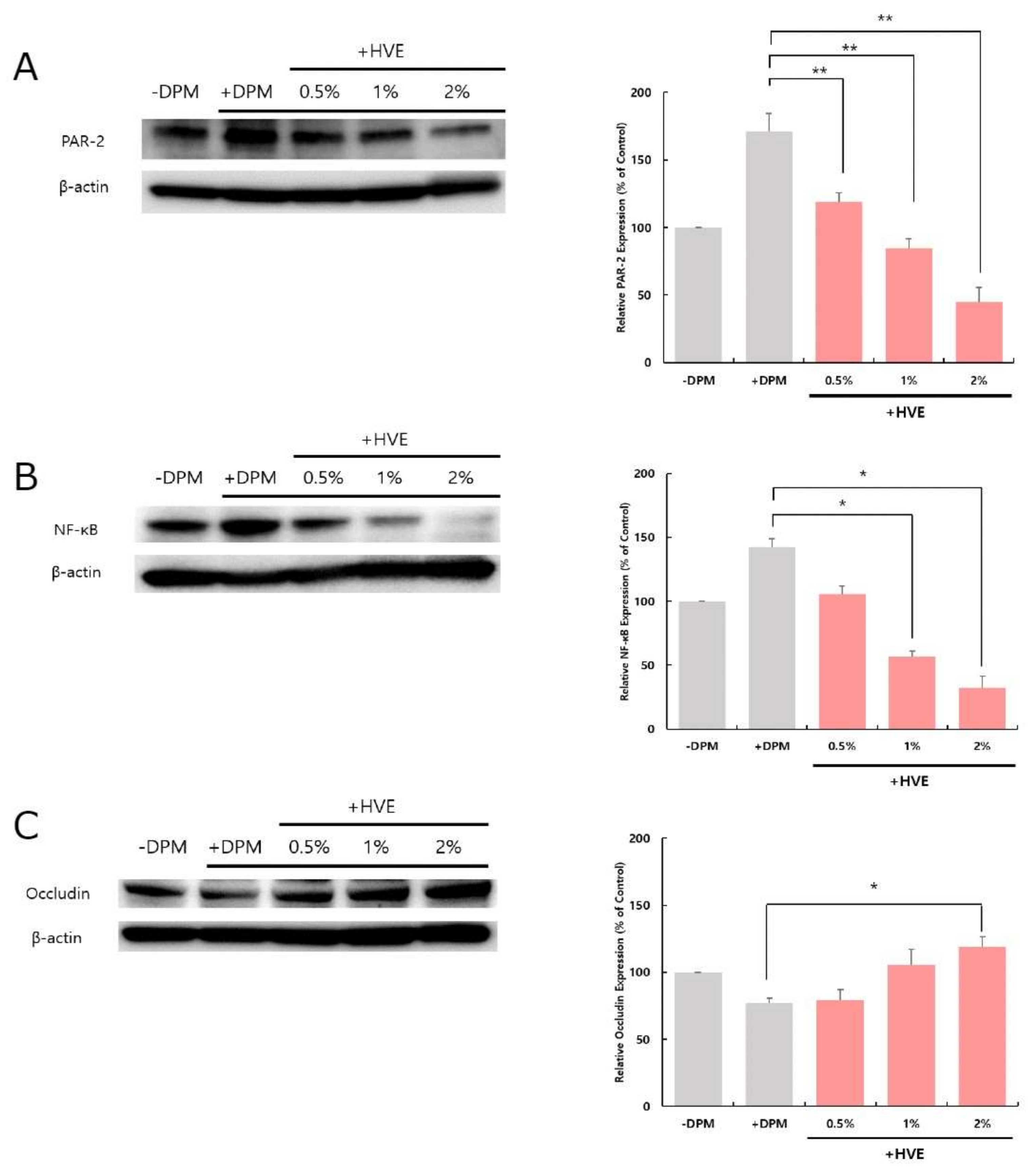
2.3. Western Blot Analysis

HaCaT cells were cultured and treated with DPM (25 μg/mL) and HVE or HGG as previously described [11]. The cells were lysed in RIPA buffer with protease inhibitor cocktail, and the same amount of protein was resolved in 4–12% SDS polyacrylamide gels for Western blotting. Antibodies were diluted to 1:1000 (PAR-2, NF-κB, and Occludin) or 1:10,000 (β-actin). PAR-2 (#6976), NF-κB (#8242), and Occludin (#91131) antibodies were purchased from Cell Signaling Technology (Beverly, MA, USA). The antibody against β-actin (A1978) was from Sigma Aldrich (St. Louis, MO, USA). Image Quant LAS 4000 (GE Healthcare, IL, USA) hardware and software were used to detect and analyze Western blot results. We also used Image J (NIH, Bethesda, MD, USA) for analyses.

2.4. Identifying Active Compounds in HVE

The stem and leaf of H. virginiana were extracted in water once, and residual raw materials were extracted again in ethanol. The ethanol extract was further fractionated by Sephadex LH-20 (Merck, Germany) to isolate HGG, the structure of which was determined with NMR spectroscopy (Supplementary Figure S2).
Hexagalloylglucose: 13C-NMR (100 MHz, CD3OD) δ 169.1, 167.9, 167.3, 167.0, 166.9, 166.2 (all galloyl carbonyl C), 146.5, 146.4 (×3), 143.2 (all galloyl C-3′, 5′), 140.7, 140.3 (×2), 140.1, 140.0, 139.7 (all galloyl C-4′), 121.4, 121.0, 120.3, 120.2 (×2), 119.7 (all galloyl C-1′), 110.7 (×2), 110.5 (×5), 110.4 (×3), 110.1 (×2) (all galloyl C-2′, 6′), 93.8 (glucose C-1), 74.3 (glucose C-5), 74.1 (glucose C-3), 72.2 (glucose C-2), 69.8 (glucose C-4), 63.1 (glucose C-6) [18].

3. Results

We screened natural products (extracted in 40% 1,3-butylene glycol with 1:50 ratio) frequently used in cosmetics, to find one that could lower the Ca2+ influx upon DPM-treatment. When we challenged HBT-SM cells with 25 μg/mL of DPM, a steep increase in [Ca2+]i was observed like in previous results (Figure 1A). Under the same conditions, 2% HVE in the media could strongly reduce initial increase in [Ca2+]i, instead slowly increasing to a steady-state. Consistent with the results from HBT-SM cells, 2% HVE could also reduce the increase in [Ca2+]i in HaCaT cells upon DPM-treatment (Figure 1B). In both types of cells, [Ca2+]i slowly increases to reach a steady-state, indicating that HVE may not completely block the Ca2+ influx. However, the rapid increase in [Ca2+]i was the most prominent outcome of DPM-treatment and directly indicated the engagement of PAR-2 with physiological responses in the previous experiments [11]. Thus, we chose HVE to further analyze the molecular markers in the downstream responses to DPM.
To estimate the DPM-induced damage state of HaCaT cells, we first checked the level of PAR-2 proteins with different concentrations of HVE in the media. As we expected, HVE could revert the increase in PAR-2 elicited by DPM-treatment, to even lower levels than normal (Figure 2A). DPM increased the level of PAR-2 as in the previous finding, and we could observe PAR-2 reduction by HVE in a concentration-dependent manner. Ca2+ influx via PAR-2 is linked with several downstream pathways, such as an increase in pro-inflammatory cytokines and a decrease in tight junction proteins. Next, we measured the level of NF-κB to monitor the inflammatory pathway elicited by DPM. Significant induction of NF-κB protein by DPM was effectively reversed by HVE (Figure 2B). Like PAR-2, NF-κB was decreased to much lower levels by increasing the amount of HVE, indicating that HVE could be an effective anti-inflammatory agent, especially under the attack by air pollution. Finally, the level of Occludin protein of HaCaT cells was investigated as an indicator of tight junction integrity. In Figure 2C, DPM decreased the level of Occludin as in the previous result, but HVE enabled cells to recover it in a concentration-dependent manner. The series of Western blot analyses led us to conclude that HVE could not only modulate the Ca2+ influx by DPM, but also protect cells from a damaged state.
Plenty of natural products have been developed as active ingredients in cosmetics, but most share common issues of consistency and quality control. Therefore, it is very important to identify the compound, which can be a major component for the shown activities, although the actual mechanism of whole extracts could be much more complex. We identified hexagalloylglucose (HGG, 3-O-digalloyl-1,2,4,6-tetra-O-galloyl-β-D-glucose, Figure 3A) from HVE. By checking [Ca2+]i curves, we verified that HGG might be the most active component of HVE in the model. First, HGG alone could inhibit the fast Ca2+ influx by DPM-treatment of HBT-SM cells as efficiently as HVE (Figure 3B). The reduction in [Ca2+]i was almost identical, but the amount of HGG (100 μg/mL in the media) was excessive, considering its low concentration in HVE. We could conclude that HGG is an active compound of HVE to protect cells from the DPM damage. However, we have to admit that the effect of HVE should be a combined effect of diverse compounds, most of which we could not identify. We then confirmed that HGG acts on HaCaT cells as well (Figure 3C).

4. Discussion

In this study, we report that H. virginiana can be an active ingredient in cosmetics, protecting skin cells from damage by air pollutants, such as PM. Our platform, which monitors [Ca2+]i during the treatment of cells with DPM, showed highly effective blocking of signal generation by HVE. Like a previous report [11], this platform could successfully identify novel active compounds or natural products that can protect epidermal cell damage induced by PM. Another recent study demonstrated an increase in [Ca2+]i by PM2.5, in which the endoplasmic reticulum stress-related proteins are involved [17]. Although the detailed mechanisms and related molecules are different, both studies implicated [Ca2+]i as a useful indicator to monitor cellular damage by PMs. Calcium (Ca2+) is called a universal second messenger, and can regulate diverse biological functions, but its role in skin health has been relatively overlooked. Dissection of the pathways from PMs to downstream events, such as inflammation, skin barrier function, and pigmentation, should be investigated, and further studies using 3D models or the tests of the chronic effect of HVE and HGG in vivo would help to understand the physiological significance of these findings.
Next, we confirmed the reduction in damage signal by HVE with known molecular markers. PAR-2 expression was downregulated by HVE, back to the normal level from that increased by DPM. NF-κB was also downregulated, suggesting that the inflammatory state by DPM was controlled by HVE. Finally, the tight junction protein Occludin was upregulated in the presence of HVE and all of these results were in good accordance with previous reports on the protecting effects under DPM attack. Various tannins were identified from H. virginiana and were studied for their biological activities [7,8,19]. However, the role of HGG in HVE was not yet reported. Here, we addressed HGG as another active compound of HVE that can specifically modulate [Ca2+]i via the PAR-2 pathway to protect skin cells. Together with these results, we can introduce an innovative skin care ingredient for those who are searching for anti-pollution products. The chronic toxicity, however, should be further analyzed before being applied in consumer products.

5. Conclusions

Here, we demonstrated that witch hazel could be a novel skincare ingredient, especially for anti-pollution products. HVE could protect keratinocytes from the inflammatory condition by PMs and simultaneously help to recover their barrier function via tight junction protein expression.

Supplementary Materials

The following are available online at https://www.mdpi.com/article/10.3390/cosmetics8040119/s1, Figure S1: Cytotoxicity of HVE and HGG, Figure S2: Identification of HGG by NMR spectra.

Author Contributions

Conceptualization, J.C. (Joonseok Cha); methodology, G.Y.H., D.Y., J.C. (Joonseok Cha); validation, J.C. (Joonseok Cha); investigation, J.C. (Jiyoung Choi), M.Y.M., G.Y.H. and D.Y.; resources, D.Y. and M.S.C.; writing—original draft preparation, J.C. (Jiyoung Choi) and J.C. (Joonseok Cha); writing—review and editing, J.C. (Joonseok Cha); supervision, M.S.C. and J.C. (Joonseok Cha); funding acquisition, M.S.C. All authors have read and agreed to the published version of the manuscript.

Funding

This research received no external funding.

Institutional Review Board Statement

Not applicable.

Informed Consent Statement

Not applicable.

Data Availability Statement

Data are contained within the article or Supplementary Materials.

Conflicts of Interest

J.C. (Jiyoung Choi); M.Y.M.; G.Y.H.; M.S.C.; J.C. (Joonseok Cha) are members of a company, The Garden of Naturalsolution. The authors have no conflict of interest to declare.

References

  1. Korting, H.C.; Schäfer-Korting, M.; Hart, H.; Schmid, M. Anti-inflammatory activity of hamamelis distillate applied topically to the skin: Influence of vehicle and dose. Eur. J. Clinic. Pharmacol. 1993, 44, 315–318. [Google Scholar] [CrossRef] [PubMed]
  2. Korting, H.C.; Schäfer-Korting, M.; Klövekorn, W.; Klövekorn, G.; Martin, C.; Laux, P. Comparative efficacy of hamamelis distillate and hydrocortisone cream in atopic eczema. Eur. J. Clinic. Pharmacol. 1995, 48, 461–465. [Google Scholar] [CrossRef] [PubMed]
  3. Masaki, H.; Atsumi, T.; Sakurai, H. Protective activity of hamamelitannin on cell damage induced by superoxide anion radicals in murine dermal fibroblasts. Biol. Pharm. Bull. 1995, 18, 59–63. [Google Scholar] [CrossRef] [PubMed] [Green Version]
  4. Vennat, B.; Pourrat, H.; Pouget, M.P.; Gross, D.; Pourrat, A. Tannins from Hamamelis virginiana: Identification of proanthocyanidins and hamamelitannin quantification in leaf, bark, and stem extracts. Planta Med. 1988, 54, 454–457. [Google Scholar] [CrossRef]
  5. Deters, A.; Dauer, A.; Schnetz, E.; Fartasch, M.; Hensel, A. High molecular compounds (polysaccharides and proanthocyanidins) from Hamamelis virginiana bark: Influence on human skin keratinocyte proliferation and differentiation and influence on irritated skin. Phytochemistry 2001, 58, 949–958. [Google Scholar] [CrossRef]
  6. Habtemariam, S. Hamamelitannin from Hamamelis virginiana inhibits the tumour necrosis factor-alpha (TNF)-induced endothelial cell death in vitro. Toxicon 2002, 40, 83–88. [Google Scholar] [CrossRef]
  7. Wang, H.; Provan, G.J.; Helliwell, K. Determination of hamamelitannin, catechins and gallic acid in witch hazel bark, twig and leaf by HPLC. J. Pharm. Biomed. Anal. 2003, 33, 539–544. [Google Scholar] [CrossRef]
  8. Duckstein, S.M.; Stintzing, F.C. Investigation on the phenolic constituents in Hamamelis virginiana leaves by HPLC-DAD and LC-MS/MS. Anal. Bioanal. Chem. 2011, 401, 677–688. [Google Scholar] [CrossRef] [PubMed]
  9. Krutmann, J.; Bouloc, A.; Sore, G.; Bernard, B.A.; Passeron, T. The skin aging exposome. J. Dermatol. Sci. 2016, 85, 152–161. [Google Scholar] [CrossRef] [PubMed] [Green Version]
  10. Teo, W. Diagnostic and management considerations for "maskne" in the era of COVID-19. J. Am. Acad. Dermatol. 2021, 84, 520–521. [Google Scholar] [CrossRef] [PubMed]
  11. Choi, J.; Moon, M.Y.; Han, G.Y.; Chang, M.S.; Yang, D.; Cha, J. Phellodendron amurense Extract Protects Human Keratinocytes from PM2.5-Induced Inflammation via PAR-2 Signaling. Biomolecules 2020, 11, 23. [Google Scholar] [CrossRef] [PubMed]
  12. Park, T.H.; Park, S.; Cho, M.K.; Kim, S. Associations of particulate matter with atopic dermatitis and chronic inflammatory skin diseases in South Korea. Clin. Exp. Dermatol. 2021. accepted. [Google Scholar] [CrossRef] [PubMed]
  13. Kim, B.E.; Kim, J.; Goleva, E.; Berdyshev, E.; Lee, J.; Vang, K.A.; Lee, U.H.; Han, S.; Leung, S.; Hall, C.F.; et al. Particulate matter causes skin barrier dysfunction. JCI Insight 2021, 6, e145185. [Google Scholar] [CrossRef] [PubMed]
  14. Teng, W.; Huang, P.; Wang, H.; Tseng, C.; Yen, F. Pterostilbene Attenuates Particulate Matter-Induced Oxidative Stress, Inflammation and Aging in Keratinocytes. Antioxidants 2021, 10, 1552. [Google Scholar] [CrossRef] [PubMed]
  15. Dias, M.K.H.M.; Madusanka, D.M.D.; Han, E.J.; Kim, H.; Jeon, Y.; Jee, Y.; Kim, K.; Lee, K.; Fernando, I.P.S.; Ahn, G. Sargassum horneri (Turner) C. Agardh ethanol extract attenuates fine dust-induced inflammatory responses and impaired skin barrier functions in HaCaT keratinocytes. J. Ethnopharmacol. 2021, 273, 114003. [Google Scholar] [CrossRef] [PubMed]
  16. Ahn, Y.; Lee, E.J.; Luo, E.; Choi, J.; Kim, J.Y.; Kim, S.; Kim, S.; Bae, Y.J.; Park, S.; Lee, J.; et al. Particulate Matter Promotes Melanin Production through Endoplasmic Reticulum Stress-Mediated IRE1α Signaling. J. Investig. Dermatol. 2021. Available online: https://www.sciencedirect.com/science/article/abs/pii/S0022202X21023484 (accessed on 30 October 2021). [CrossRef] [PubMed]
  17. Molagoda, I.M.N.; Kavinda, M.H.D.; Choi, Y.H.; Lee, H.; Kang, C.; Lee, M.; Lee, C.; Kim, G. Fisetin Protects HaCaT Human Keratinocytes from Fine Particulate Matter (PM2.5)-Induced Oxidative Stress and Apoptosis by Inhibiting the Endoplasmic Reticulum Stress Response. Antioxidants 2021, 10, 1492. [Google Scholar] [CrossRef] [PubMed]
  18. Hwang, J.K.; Kong, T.W.; Baek, N.I.; Pyun, Y.R. Alpha-Glycosidase inhibitory activity of hexagalloylglucose from the galls of Quercus infectoria. Planta Med. 2000, 66, 273–274. [Google Scholar] [CrossRef] [PubMed]
  19. González, M.J.; Torres, J.L.; Medina, I. Impact of thermal processing on the activity of gallotannins and condensed tannins from Hamamelis virginiana used as functional ingredients in seafood. J. Agric. Food Chem. 2010, 58, 4274–4283. [Google Scholar] [CrossRef] [PubMed]
Figure 1. [Ca2+]i of HBT-SM (A) and HaCaT (B) cells were monitored. Both cells showed previously reported Ca2+ influx by DPM addition at the indicated time (arrows, 25 μg/mL in the media), which HVE (2% in the media) successfully inhibited (red curves). Ns indicate the number of cells that we analyzed for the quantification. After normalization, the difference between DPM only and DPM+HVE was analyzed using a Student’s t-test. *** p < 0.001.
Cosmetics 08 00119 g001
Figure 2. HVE supported HaCaT cells to recover from the DPM-induced damage state. The levels of PAR-2 (A) and NF-κB (B) were increased by DPM treatment (25 μg/mL in the media), but HVE could reduce them to or below the normal level. On the contrary, the level of Occludin (C) was decreased by DPM, but returned to the normal level by HVE. Densitometry data were normalized by β-actin and the significance was analyzed using a paired Student’s t-test. * p < 0.05; ** p < 0.01.
Cosmetics 08 00119 g002
Figure 3. HGG is an active compound that can manifest the protective effect of HVE on the cells. (A) The structure of HGG resolved by NMR. (B) HGG (100 μg/mL in the media) showed almost identical effect with HVE (2% in the media) on the [Ca2+]i of HBT-SM cells upon DPM treatment at the indicated time (arrows). (C) HGG could also decrease the [Ca2+]i of HaCaT cells, suggesting the protection from the DPM damage. After normalization, the significance of difference to the control (DPM only) was analyzed using a Student’s t-test. *** p < 0.001.
Cosmetics 08 00119 g003
Publisher’s Note: MDPI stays neutral with regard to jurisdictional claims in published maps and institutional affiliations.

