Article

Codonopsis pilosula Extract Protects Melanocytes against H2O2-Induced Oxidative Stress by Activating Autophagy

Department of Pharmaceutics & Biotechnology, Konyang University, Daejeon 35365, Korea
Cosmetics 2021, 8(3), 67; https://doi.org/10.3390/cosmetics8030067
Submission received: 18 June 2021 / Revised: 20 July 2021 / Accepted: 27 July 2021 / Published: 28 July 2021

Abstract

:
Recently, as the anti-aging role of melanin in the skin and the inhibition of melanin production has been identified, the development of materials capable of maintaining skin homeostasis has been attracting attention. In this study, we further investigated the anti-melanogenic effect of Codonopsis pilosula extract (CPE) and, under oxidative stress, the cytoprotective effect in Melan-a melanocytes exposed to H2O2. First, CPE treatment significantly reduced melanin production by inhibiting melanogenesis-associated proteins, including microphthalmia-associated transcription factor (MITF), tyrosinase, and tyrosinase-related protein 2 (TRP 2), as a result of the phosphorylation of MAPK/JNK in Melan-a cells. Next, to investigate the protective effects of the CPE on oxidative-stress-induced skin injury and its molecular mechanism, we determined the effect of CPE after inducing oxidative stress by exposing melanocytes to H2O2. CPE protected cells from H2O2-induced cytotoxicity by reducing the expression of the gene encoding the Bax pro-apoptotic protein, whereas it induced the genes encoding the B-cell lymphoma 3 (Bcl2) family and MITF, which is a transcriptional regulator that promotes melanocyte differentiation. Furthermore, our results show that CPE enhanced the production of autophagy-related proteins such as Beclin-1 and light chain 3 (LC3) II; this was substantially reversed by 3-methyladenin (MA, an autophagy inhibitor) pretreatment. Collectively, our findings demonstrate that CPE treatment exhibits not only an anti-melanogenic effect in normal melanocytes, but also a cytoprotective effect in melanocytes subjected to oxidative stress by inducing autophagy and MITF expression. Therefore, we believe that CPE is a potent candidate for cell maintenance in melanocytes.

1. Introduction

Human skin is the largest organ of the human body and protects the body from environmental toxins, allergens, and oxidative stress. Melanin, which is mainly produced by melanocytes, is known to play an important role in preventing skin diseases and is present in various tissues of the human body [1,2]. However, the excessive production and accumulation of reactive oxygen species (ROS) due to stress, ultraviolet (UV) radiation, and aging result in many skin disorders, such as hyperpigmentation, melasma, and eventually the degradation of melanocytes. Several studies on the treatment of melanogenesis have focused on regulating the expression of microphthalmia-associated transcription factor (MITF) and tyrosinase activity [3,4,5]. Additionally, recent studies have shown that skin melanogenesis is mediated via several melanogenic signaling pathways, including mitogen-activated protein kinase (MAPK) signaling, protein kinase A (PKA), and the cyclic adenosine monophosphate (cAMP)-mediated pathway [6].
The cellular autophagy system is well known as a cellular self-digestion process that degrades damaged proteins or isolates dysfunctional organelles in a cell and then decomposes them in lysosomes to maintain cell homeostasis [7,8]. Recent studies have identified autophagy as being involved in the normal function of melanocytes and in regulating the expression of the melanin-forming transcription factor MITF. Therefore, autophagy-inducing agents have the potential to limit the damage caused by UV rays and lipid oxidation and to maintain homeostasis in melanocytes and keratinocytes [9,10,11]. However, there is little information on the application of natural autophagy-inducing preparations that protect melanocytes against oxidative stress.
Codonopsis pilosula is the root of Codonopsis pilosula (Fr.) Nannf., belonging to the Campanula family. It is grown or cultivated in the mountains of Gangwon-do, Korea, and is also distributed in the northern and western regions of China, such as the Ganxi and Shanxi regions [12]. In folk medicine, it has been used as a substitute for ginseng for hundreds of years, since it has the same pharmacological activities, such as replenishing energy, strengthening the immune system, lowering gastrointestinal diseases, and regulating blood pressure, but at a lower price. Phytochemical research shows that C. pilosula contains large amounts of sucrose, polysaccharides, triterpenes, saponins, phytosterols, phenolic glycosides, alkaloids, and polyacetylenes [13]. Among them, lobetyolin, a major acetylene of C. pilosula, is known to activate NF-κB and has an immune-stimulating effect [14]. In addition, according to recent studies on the effect of C. pilosula extract, an ethanol extract of C. pilosula significantly inhibits allergic reactions caused by ovalbumin, while a water extract decreases the plasma glucose level, and a butanol extract shows free-radical-scavenging activity and an inhibitory effect on lipid peroxidation in rat brain homogenate [15,16,17].
Previously, the antioxidant activity of C. pilosula was found to vary due to differences in polyphenol content depending on the extraction solvent used [18]. Thus, we investigated the melanogenic inhibitory effect of C. pilosula extract (CPE) under normal conditions through mechanistic signaling pathways, as well as the cytoprotective effect against H2O2-induced oxidative stress in Melan-a melanocytes.

2. Materials and Methods

2.1. Reagents

RPMI 1640 and fetal bovine serum (FBS) were purchased from Welgene (Daegu, Korea). Penicillin–streptomycin was purchased from GibcoBRL (Eggenstein, Germany). Phorbol 12-myristate 13-acetate (TPA) was purchased from TOCRIS (Bristol, UK). Hydrogen peroxide (H2O2), 3-(4,5-dimethylthiazol-2-yl)-2,5-diphenyltetrazolium bromide (MTT), 4′,6-diamidino-2-phenylindole (DAPI), and 3-MA were purchased from Sigma-Aldrich (St Louis, MO, USA). All the other chemicals and reagents were of analytical grade.

2.2. Preparation of the Codonopsis Pilosula Extract

The C. pilosula root (CP) was simply chopped, and 100 g of CP was immersed in 1 L of 50% ethanol (w/v); then, it was extracted three times using ultrasonic waves at 30 °C. After each extract was centrifuged to collect the supernatant, the supernatant was concentrated and freeze-dried to prepare the C. pilosula extract (CPE).

2.3. Cell Culture and Stock Preparation of CPE

The melanocyte Melan-a cells were grown and maintained in RPMI 1640 supplemented with 10% FBS, penicillin (100 U/mL), streptomycin (100 U/mL), and 200 nM phorbol 12-myristate 13-acetate (TPA) and maintained at 37 °C in a 5% CO2 incubator. After culturing 3 × 10⁵ cells per well in a six-well plate, the Melan-a cells were placed in an incubator for 48 h until the color of the medium changed to black. The stock solutions (100 mg/mL) of CPE were dissolved in sterile water and stored at −20 °C.

2.4. Measurement of Cell Viability

The cell viability of CPE in Melan-a cells was determined using an MTT colorimetric assay and flow cytometry. The cells were maintained until they reached 80% confluence in 96-well plates and then treated with CPE and/or 0.5 mM H2O2 for 24 h. Cell medium with 10 μL of MTT solution (5 mg/mL) was added, and the cells were incubated for an additional 4 h. After the incubation of the cells, the insoluble formazan crystals were dissolved in 100 μL of dimethyl sulfoxide. The absorbance at 540 nm was measured by spectrophotometry using a microplate reader. Dead cells were determined with a PI/Annexin V cell death detection kit (EMD Milipore Corporation, Billercia, MA, USA) according to the manufacturer’s protocol. Briefly, the cells were washed and incubated for 15 min at RT in the dark containing FITC-Annexin V and PI. Afterwards, dead cells were analyzed by the MuseTM cell analyser.

2.5. In Vitro Melanin Estimation

Approximately 2 × 10⁵ cells/well were grown in a six-well plate. After treatment with CPE and/or 0.5 mM H2O2 as previously described [19], PBS-washed cells were harvested, lysed in 1× PBS containing 1% Triton X-100, and centrifuged at 4 °C at 13,000 rpm for 15 min. The cellular melanin content was solubilized using 1 N NaOH. An ELISA plate reader was used to quantify the melanin by measuring the absorbance at 405 nm. Data were obtained from three experiments, and the melanin content was calculated and is denoted as the fold change compared with the control cells.

2.6. Protein Isolation and Western Blot Assay

Protein samples were extracted as explained in our previous work [20]. Then, the protein concentration of the cell lysate was measured using a BCA protein assay kit (Thermo Scientific, Waltham, MA, USA). Equivalent amounts of denatured cell lysate (30 μg of cell lysate per sample) were separated by 10% sodium dodecyl sulfate–polyacrylamide gel electrophoresis (SDS-PAGE) and transferred to a nitrocellulose membrane. The membrane was incubated with the primary antibody diluted in 5% skim milk powder in PBS containing 0.05% Tween 20 (PBST) overnight at 4 °C.
Following primary antibody incubation, the membranes were washed and incubated in the HRP-conjugated secondary antibody diluted in 5% skim milk in PBST for 1 h at room temperature. Protein expression was analyzed using an enhanced chemiluminescence (ECL) detection system (AI680, GE Healthcare, Uppsala, Sweden).

2.7. Estimation of Intracellular ROS by DCFH-DA Staining

The cellular ROS levels in the H2O2-treated Melan-a cells were estimated by the DCFH-DA fluorescence microscopy method [21]. The levels of intracellular DCF fluorescence are proportional to the intracellular ROS produced. First, 2 × 10⁵ cells/well were treated with individual concentrations of CPE and/or H2O2 for 24 h, then DCFH2-DA was added 2 h before the end of the reaction. Data were collected from three or more independent experiments.

2.8. Statistical Processing

All the experimental results are presented as the means ± standard deviations (SDs) of three biological replicates. The statistical significance among the multiple mean values was assessed by one-way analysis of variance (ANOVA), followed by Duncan’s multiple-comparison test, using SPSS 18.0 (SPSS Inc., Chicago, IL, USA), as indicated in the legends. A p-value < 0.05 was considered to indicate a statistically significant difference.

3. Results

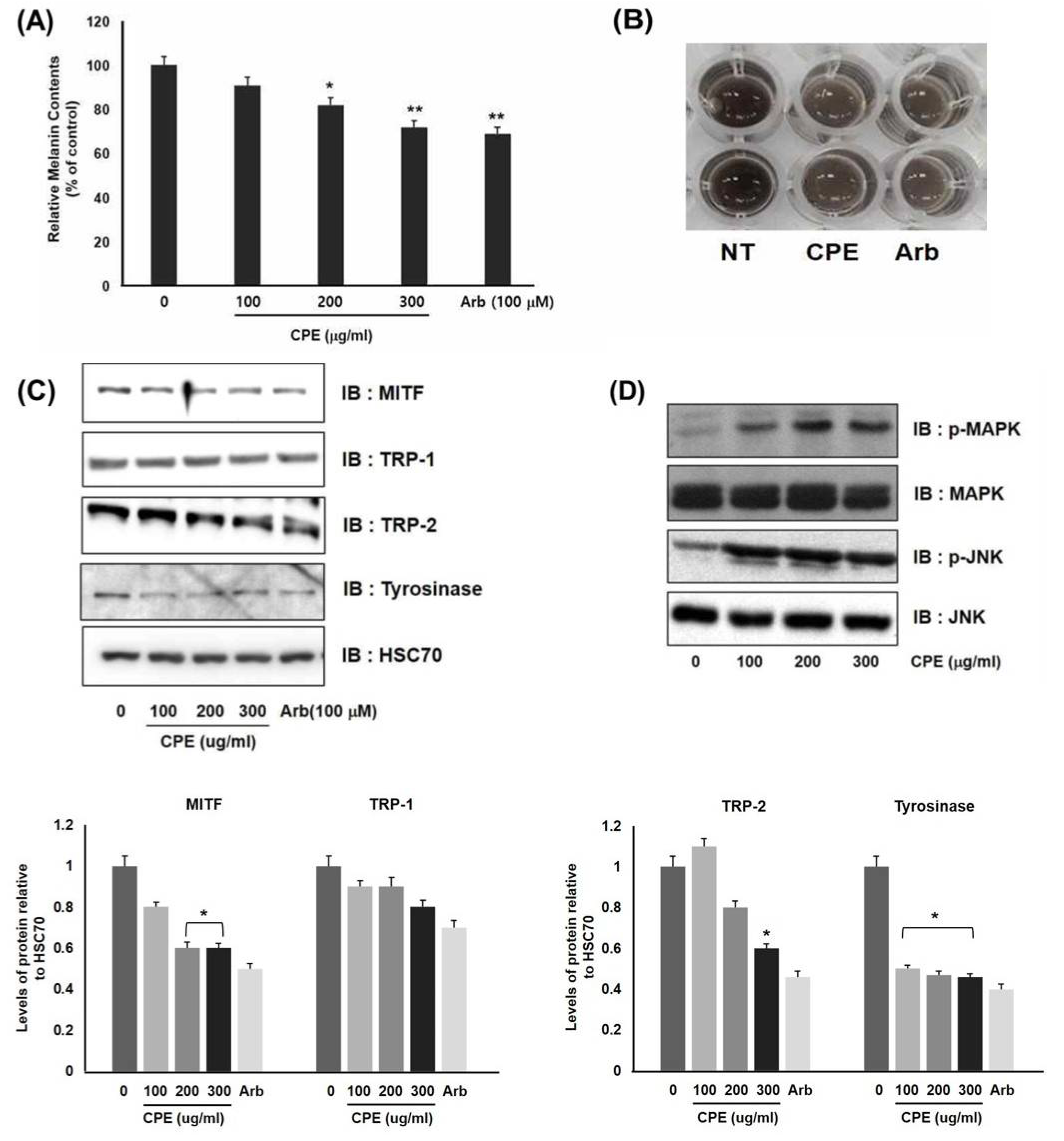
3.1. Downregulation of Melanogenesis in Melanocytes by CPE

Melanocytes synthesize melanin in response to external stimuli, such as UV irradiation. Major factors such as α-melanocyte-stimulating hormone (α-MSH), stem cell factor (SCF), and endothelin-1 (ET-1) are secreted from melanocytes and increase the expression of MITF, thereby promoting melanogenesis [22,23]. First, we examined whether CPE treatment inhibited melanin production and melanogenesis-related proteins.
CPE treatment at 100, 200, and 300 μg/mL significantly reduced melanin production compared with the negative control. In particular, treatment with 300 μg/mL of CPE resulted in a reduction similar to that induced by arbutin treatment (Figure 1A,B). In addition, we investigated the melanogenesis proteins tyrosinase, TRP-1, TRP-2, and MITF by Western blotting. Treatment with CPE at concentrations of 300 μg/mL significantly decreased the levels of the MITF, TRP-2, and tyrosinase proteins (Figure 1C). As it was confirmed that CPE inhibits the expression of MITF-related proteins, inhibiting melanin formation, we further examined whether CPE affected the MAPK signaling pathway. CPE increased MAPK phosphorylation in a dose-dependent manner, specifically inducing JNK phosphorylation (Figure 1D). These results suggest that the melanogenesis inhibition by CPE results from the downregulation of MITF and tyrosinase expression by the induction of JNK/MAPK phosphorylation.

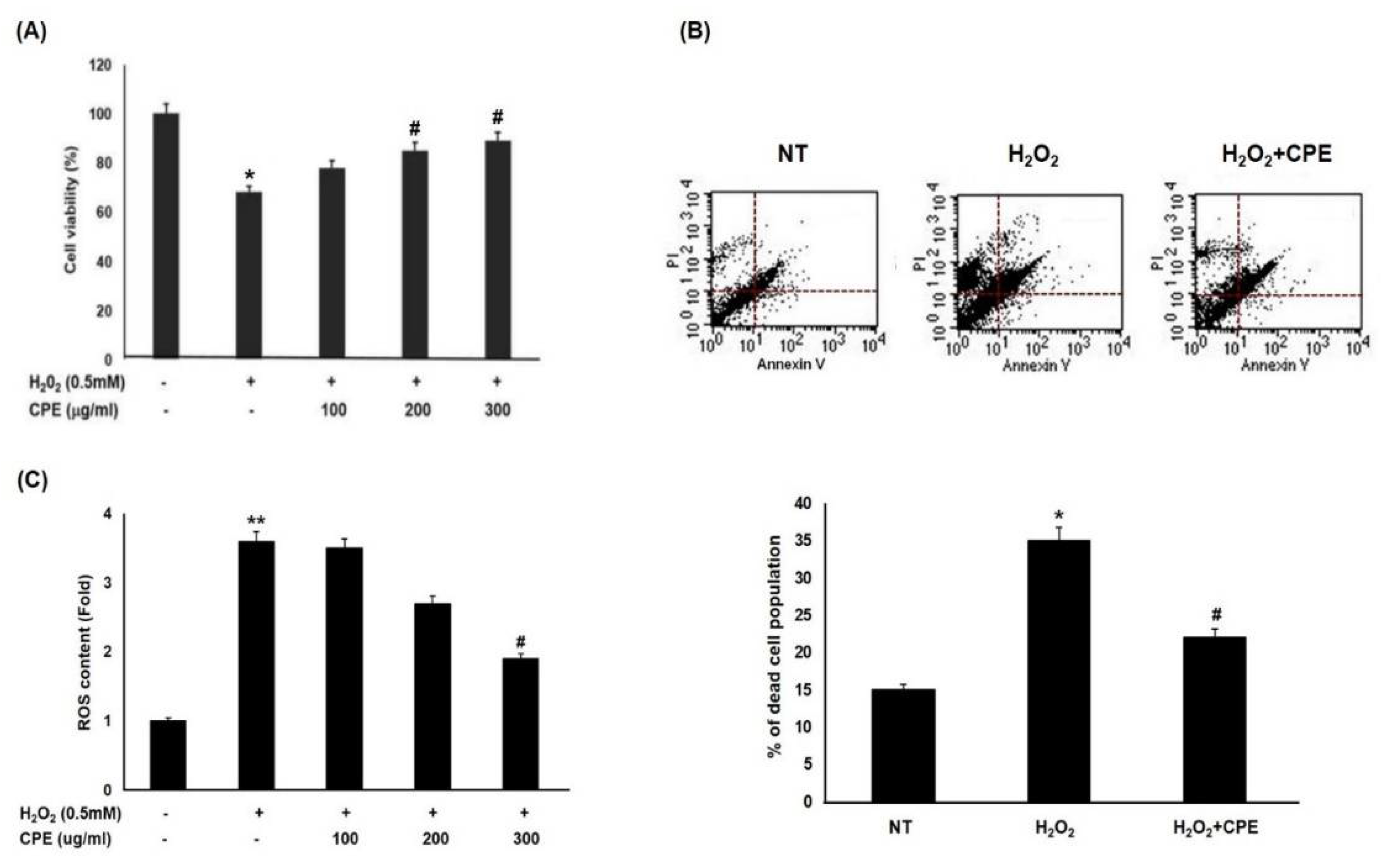
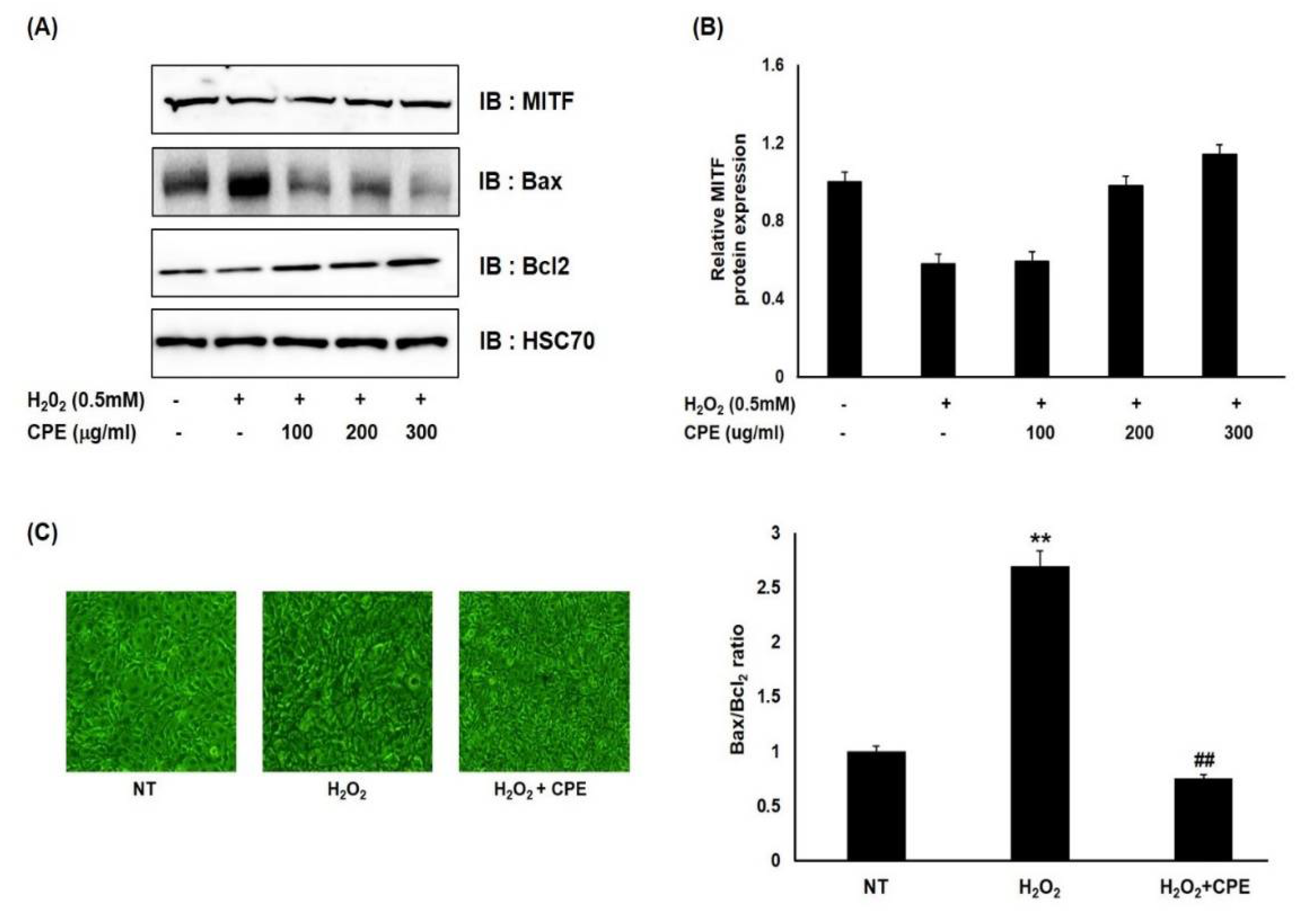
3.2. Effect of CPE on H2O2-Induced Cell Death in Melanocytes

Skin cells, such as keratinocytes and melanocytes, react sensitively to ROS, such as hydrogen peroxide (H2O2) and superoxide anion, to maintain a normal status. However, an imbalance in the oxidant–antioxidant status due to excessive cellular ROS can lead to the accumulation of damaged proteins or organelles, eventually leading to apoptotic cell death [24,25]. Recently, it has been reported that melanin promotes skin aging by stimulating melanin formation depending on the external environment of the skin, as well as skin health being maintained by intracellular melanin content being retained [26]. Thus, we investigated the effect of CPE extract on cells subjected to oxidative stress via H2O2 treatment. The H2O2-treated cells showed a decrease in cell viability of 32.8% compared with the untreated cells. However, CPE treatment reduced the inhibition of cell viability in a concentration-dependent manner under H2O2 treatment. In particular, CPE at 300 μg/mL restored cell viability to 91.9% (Figure 2A). In addition, the apoptotic death of the CPE- and/or H2O2-treated cells was detected by double staining with Annexin V-FITC/PI, followed by flow cytometric analysis. Following H2O2 treatment, the percentage of apoptotic cells increased to 34.5%, compared with 14.8% in the untreated cells. However, CPE treatment markedly inhibited H2O2-induced apoptotic death, with the percentage of Annexin V-stained cells being 22.2% (Figure 2B). As this shows the same pattern as the ROS content, these results show that CPE maintains cell viability by inhibiting cell death via protecting against H2O2-induced ROS (Figure 2C).

3.3. Effect of CPE on H2O2-Induced Cell Death in Melanocytes

As previously confirmed, CPE maintains cell viability by inhibiting the increase in apoptosis following H2O2 treatment. Thus, we next investigated the effect of CPE on the expression of the proteins involved in cell death and of the MITF protein, which regulates melanin production in the presence of H2O2. As shown in Figure 3, in the cells treated with H2O2, MITF expression was slightly reduced, on the other hand, in the cells treated with CPE, MITF expression was almost the same as in the untreated cells. In addition, H2O2 increased the expression of the apoptosis-inducing Bax protein [27,28], but decreased Bax expression in a concentration-dependent manner in CPE-treated cells. By contrast, H2O2 decreased the expression of the Bcl2 protein, which promotes cell survival [28,29], but the expression of Bcl2 was increased by CPE treatment in a concentration-dependent manner. When we calculated the expression of these two proteins to determine the Bax/Bcl2 ratio, the same tendency was observed. Therefore, under H2O2-induced oxidative stress, the expression of MITF was reduced by the induction of cell death, whereas CPE exhibited a potent protective effect, preventing H2O2-induced cell death and maintaining the expression of MITF.

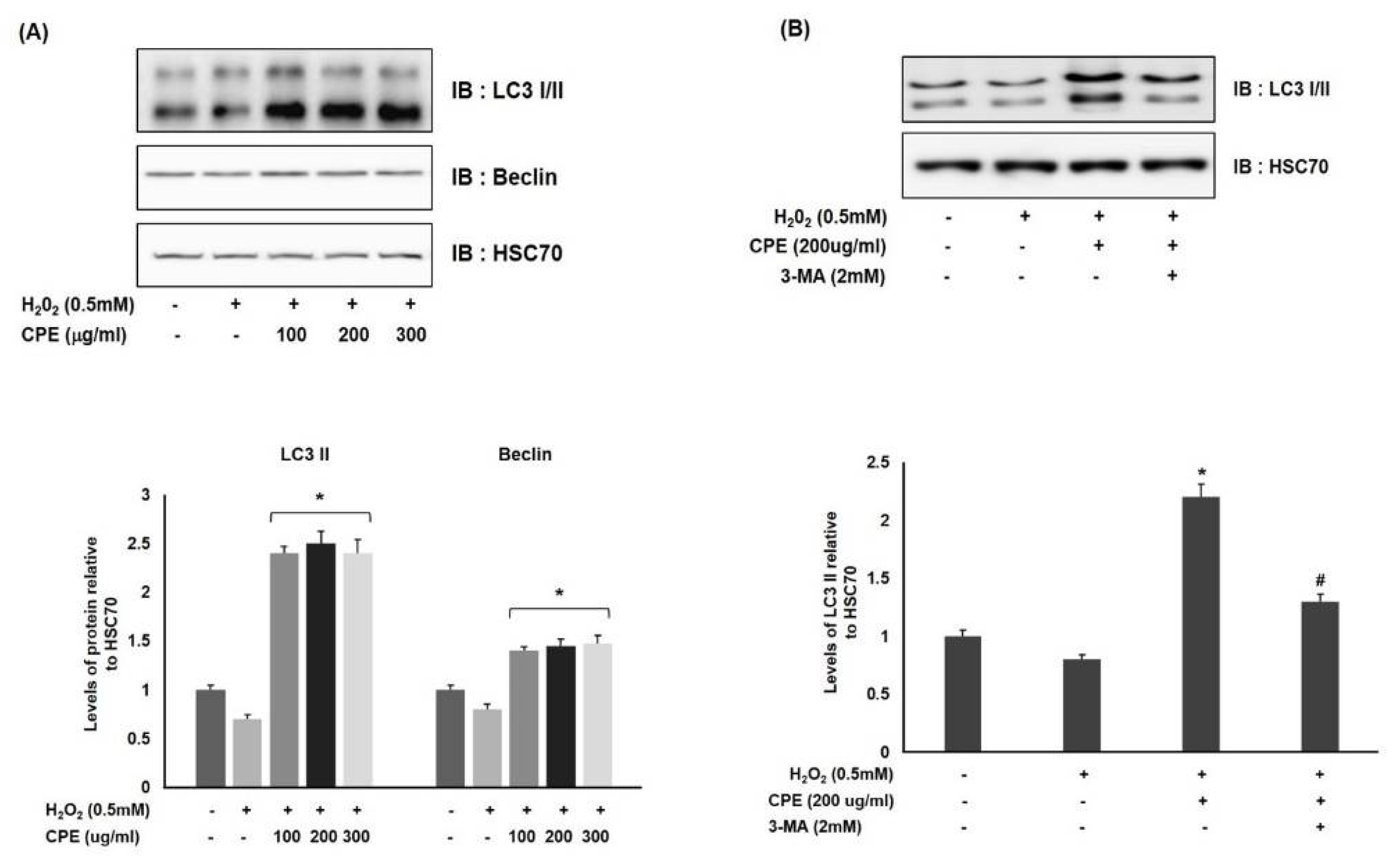
3.4. Effect of CPE on Autophagy Activation in Oxidative-Stress-Induced Melanocytes

Recent studies have revealed that autophagy and its regulator play a crucial role in the antioxidative response against ROS-induced oxidative stress in human cells [26,30]. Feng et al. [31] showed that Apocynum venetum leaf extract protects injured neurons against H2O2-induced apoptosis by reducing the ROS-induced activation of autophagy. Since the association between the modulation of autophagy and anti-melanogenesis has been established, the role of autophagy in CPE’s protective effect against H2O2-induced oxidative stress was investigated in Melan-a cells. The levels of LC3, an autophagosome protein, were used as an indicator of autophagy. The Western blot data indicated that CPE significantly upregulated the expression of the pro-autophagic LC3-II and Beclin proteins, whose expression was decreased by H2O2 treatment. This suggests that CPE has a cytoprotective effect through autophagy activation under intracellular oxidative stress (Figure 4A). Next, the intra-relationship between the cytoprotective effect and autophagy of CPE in H2O2-treated Melan-a cells was further analyzed using a potent autophagic inhibitor, 3-MA. Compared with previous data, cells pretreated with 3-MA and CPE and stimulated with H2O2 showed decreased LC3-II expression levels (Figure 4B). Overall, these data imply that inhibiting autophagy negatively affects the cytoprotective efficacy of CPE in H2O2-treated cells, indicating that autophagy is essential for CPE’s cytoprotective effects.

4. Discussion

We previously established the optimal extraction method resulting in the highest antioxidant activity as well as polyphenol content [18]. In this study, we applied the high-yield ultrasonic extraction method and found that the extract exhibits a cytoprotective effect by activating autophagy under oxidative stress conditions, as well as by inhibiting melanogenesis in Melan-a cells.
First, we further investigated the anti-melanogenic effect of CPE. CPE downregulated melanogenesis by decreasing the expression of genes related to melanogenesis, such as MITF, tyrosinase, and TRP-2. Several studies have demonstrated that the biosynthetic mechanisms of melanogenesis related to enzyme expression are mediated by various signaling pathways, including mitogen-activated protein kinase (MAPK), protein kinase A (PKA), and PI3K/Akt [32]. Among them, activation of the MAPK signaling pathway repressed MITF at the protein stability level, as well as the transcriptional level, in human primary melanocytes [33,34]. The phosphorylation of MAPK and the signaling cascades of the extracellular responsive kinase (ERK), c-Jun N-terminal kinase (JNK), and p38 also regulate melanin production. Among them, the JNK activator suppresses melanogenesis through the phospho-inhibition of CREB-regulated transcription coactivator 3 (CRTC3)-dependent MITF expression [35,36]. Our results show that the phosphorylation of MAPK, especially JNK, effectively increases following CPE treatment compared with control cells. These findings suggest that CPE-induced depigmentation in Melan-a melanocytes may occur by MITF/JNK-regulated signaling pathways.
Melanocytes produce the melanin pigment and transfer it to keratinocytes, where the pigments help to protect the skin from oxidative environmental stressors such as air pollutants, chemical products, and UV damage [37,38,39]. MITF is one of the major transcriptional regulators responsible for key genes that promote melanocyte development and differentiation, as well as regulating melanogenesis. Moreover, MITF regulates the expression of certain genes that maintain cell homeostasis, including those encoding the proteins involved in proliferation (e.g., CDK2) and apoptosis (e.g., Bcl2) [40,41,42]. Steingrimsson et al. [43] identified MITF mutant mice as affected by hearing loss and pigmentary disturbances as a result of the loss of melanocytes, as well as faulty osteoclasts and mast cells. This suggests that MITF plays important roles in the development of these cell types.
CPE is a derivative of herbal medicines used to treat gastrointestinal and allergic diseases due to its anti-aging and anti-allergic effects [15,44]. However, whether CPE can protect melanocytes from oxidative stress remains unknown. To estimate the effect of CPE on melanocyte survival in response to oxidative stress, we first observed cell death in the melanocytes exposed to H2O2. Following treatment with 0.5 mM H2O2 for 24 h, cell viability was decreased compared with that of the untreated cells. However, the viability of the CPE-treated cells was restored by reducing the ROS content; then the Bax/Bcl2 ratio and MITF expression were increased, unlike in the H2O2-treated cells.
Autophagy is the major intracellular degradation system by which the damage of lysosomes results in their degradation. Mizushima et al. [45] demonstrated that, under conditions of starvation, inflammation, or oxidative stress, the autophagic process can regulate the degradation of damaged proteins and organelles in cells, so that cellular renovation and homeostasis can be achieved. Microtubule-associated protein 1 light chain 3 (LC3) and Beclin-1 play major roles in mammalian autophagy. LC3 exists in two forms, namely, a cytosolic form (LC3 I) and a lipid phosphatidylethanolamine-conjugated form (LC3 II) that is inserted into both the inner and outer membranes of the growing autophagosome [46,47]. Zhang et al. [48] used the conversion of LC3 I to LC3 II as a biochemical marker to analyze the status of autophagy in melanocytes. Further research indicated that LC3 is a marker of final autophagosome formation, whereas Beclin-1 is involved in the initial step of autophagosome formation, activating autophagy [49]. The constitutive autophagic activity plays a key role in preventing oxidative damage in melanocytes, but there have been few studies on the molecular mechanisms regulating autophagy in melanocytes under oxidative stress.
Consequently, we investigated the implication of CPE-mediated autophagy for cell survival in melanocytes subjected to oxidative stress. Our results show that, after CPE treatment, autophagy was activated through the increased expression of Beclin-1 and induced conversion of LC3 I to LC3 II. This suggests that CPE exerts a cytoprotective effect through autophagy activation in melanocytes subjected to oxidative stress.
In conclusion, the present study demonstrated that CPE treatment not only has an anti-melanogenic effect through the MITF/JNK pathway in normal melanocytes, but also, under H2O2-induced oxidative stress, maintained cell survival and exerted cytoprotection through MITF, resulting in the sustained activation of autophagy in Melan-a cells.

Funding

This research was supported by the Konyang University Research Fund in 2019.

Institutional Review Board Statement

Not applicable.

Informed Consent Statement

Not applicable.

Data Availability Statement

The data presented in this study are available on request from the corresponding author.

Acknowledgments

The author acknowledges Hyun-Ji Park and Mi-Jin Kwon for their support during the experimental work. The author have consented to the acknowledgement.

Conflicts of Interest

No potential conflict of interest are reported by the author.

References

  1. Ichihashi, M.; Ueda, M.; Budiyanto, A.; Bito, T.; Oka, M.; Fukunage, M.; Tsuru, K.; Horikawa, T. UV-induced skin damage. Toxicology 2003, 189, 21–39. [Google Scholar] [CrossRef]
  2. Lukiewicz, S. The biological role of melanin. I. New concepts and methodical approaches. Folia Histochem. Cytochem. (Krakow) 1972, 10, 93–108. [Google Scholar]
  3. Hearing, V.J. Biogenesis of pigment granules: A sensitive way to regulate melanocyte function. J. Dermatol. Sci. 2005, 37, 3–14. [Google Scholar] [CrossRef] [PubMed]
  4. Ganesan, A.K.; Ho, H.; Bodemann, B.; Petersen, S.; Aruri, J.; Koshy, S.; White, M.A. Genome-wide siRNA-based functional genomics of pigmentation identifies novel genes and pathways that impact melanogenesis in human cells. PLoS Genet. 2008, 4, e1000298. [Google Scholar] [CrossRef] [PubMed] [Green Version]
  5. Ko, M.S.; Lee, H.J.; Kang, M.J. Antioxidant activities and whitening effects of extracts from Hippophae rhamnoides L. J. East Asian Soc. Diet Life 2012, 22, 812–817. [Google Scholar]
  6. Ahn, J.H.; Jin, S.H.; Kang, H.Y. LPS induces melanogenesis through p38 MAPK activation in human melanocytes. Arch. Dermatol. Res. 2008, 300, 325–329. [Google Scholar] [CrossRef]
  7. Navone, F.; Genevini, P.; Borgese, N. Autophagy and neurodegeneration: Insights from a cultured cell model of ALS. Cells 2015, 4, 354–386. [Google Scholar] [CrossRef]
  8. Zhou, Y.Y.; Li, Y.; Jiang, W.Q.; Zhou, L.F. MAPK/JNK signalling: A potential autophagy regulation pathway. Biosci. Rep. 2015, 35, e00199. [Google Scholar] [CrossRef]
  9. Raposo, G.; Marks, M.S. Melanosomes—Dark organelles enlighten endosomal membrane transport. Nat. Rev. Mol. Cell Biol. 2007, 8, 786–797. [Google Scholar] [CrossRef] [Green Version]
  10. Ho, H.; Ganesan, A.K. The pleiotropic roles of autophagy regulators in melanogenesis. Pigment Cell Melanoma Res. 2011, 24, 595–604. [Google Scholar] [CrossRef]
  11. Rubinsztein, D.C.; Gestwicki, J.E.; Murphy, L.O.; Klionsky, D.J. Potential therapeutic applications of autophagy. Nat. Rev. Drug Discov. 2007, 6, 304–312. [Google Scholar] [CrossRef]
  12. Hu, X.Y.; Qin, F.Y.; Lu, X.F.; Zhang, L.S.; Cheng, Y.X. Three new polyynes from Codonopsis pilosula and their activities on lipid metabolism. Molecules 2018, 23, 887. [Google Scholar] [CrossRef] [Green Version]
  13. He, J.Y.; Ma, N.; Zhu, S.; Komatsu, K.; Li, Z.Y.; Fu, W.M. The genus Codonopsis (Campanulaceae): A review of phytochemistry, bioactivity and quality control. J. Nat. Med. 2015, 69, 1–21. [Google Scholar] [CrossRef] [PubMed] [Green Version]
  14. Wang, J.; Tong, X.; Li, P.; Liu, M.; Peng, W.; Cao, H.; Su, W. Bioactive components on immuno-enhancement effects in the traditional Chinese medicine Shenqi Fuzheng injection based on relevance analysis between chemical HPLC fingerprints and in vivo biological effects. J. Ethnopharmacol. 2014, 155, 405–415. [Google Scholar] [CrossRef] [PubMed]
  15. Kang, Y.S.; Jung, K.J.; Lee, K.S.; Lee, H.S.; Park, Y.K. Effects of the ethanol extract of Codonopsis Pilosula Radix on ovalbumin-induced allergic responses in mice. Korea J. Herbol. 2013, 28, 9–15. [Google Scholar] [CrossRef] [Green Version]
  16. He, K.; Li, X.; Chen, X.; Ye, X.; Huang, J.; Li, Y.; Deng, Y.; Jin, Q.; Shi, Q.; Shu, H. Evaluation of antidiabetic potential of selected traditional Chinese medicines in STZ-induced diabetic mice. J. Ethnophamacol. 2011, 137, 1135–1142. [Google Scholar] [CrossRef]
  17. Ng, T.B.; Liu, F.; Wang, H.X. The antioxidant effects of aqueous and organic extracts of panax quinquefolium, panax notoginseng, Codonopsis pilosula, Pseudostellaria heterophylla and Glehnia littoralis. J. Enthnopharmacol. 2004, 93, 285–288. [Google Scholar] [CrossRef] [PubMed]
  18. Park, H.J.; Ahn, S.H.; Choi, W.Y.; Lee, G.W.; Kwon, M.J.; Cho, Y.H. Changes in whitening activity of Codonopsis pilosula extracts according to extraction solvents. KSBB J. 2019, 34, 1–9. [Google Scholar] [CrossRef]
  19. Park, H.J. Anti-inflammatory mechanisms of gamma-irradiated fibroin protein. Master’s Thesis, Konyang University, Daejeon, Korea, 2020. [Google Scholar]
  20. Xin, Q.; Kwon, M.J.; Lee, J.W.; Kim, K.S.; Chen, H.; Campos, M.G.; Tundis, R.; Cui, C.B.; Cho, Y.H.; Cao, H. Gamma irradiated Rhodiola sachalinensis extract ameliorates testosterone-induced benign prostatic hyperplasia by downregulating 5-alpha reductase and restoring testosterone in rats. Molecules 2019, 24, 3981. [Google Scholar] [CrossRef] [Green Version]
  21. Kim, H.; Xue, X. Detection of total reactive oxygen species in adherent cells by 2′,7′-dichlorodihydrofluorescein diacetate staining. J. Vis. Exp. 2020, 160. [Google Scholar] [CrossRef] [PubMed]
  22. Murase, D.; Hachiya, A.; Amano, Y.; Ohuchi, A.; Kitahara, T.; Takema, Y. The essential role of p53 in hyperpigmentation of the skin via regulation of paracrine melanogenic cytokine receptor signaling. J. Biol. Chem. 2009, 284, 4343–4353. [Google Scholar] [CrossRef] [Green Version]
  23. Niwano, T.; Terazawa, S.; Nakajima, H.; Wakabayashi, Y.; Imokawa, G. Astaxanthin and withaferin a block paracrine cytokine interactions between uvb-exposed human keratinocytes and human melanocytes via the attenuation of endothelin-1 secretion and its downstream intracellular signaling. Cytokine 2015, 73, 184–197. [Google Scholar] [CrossRef] [PubMed]
  24. Trachootham, D.; Lu, W.; Ogasawara, M.A.; Valle, N.R.-D.; Huang, P. Redox regulation of cell survival. Antioxid. Redox Signal 2008, 10, 1343–1374. [Google Scholar] [CrossRef] [Green Version]
  25. Halliwell, B. Free radicals and antioxidants—Quo vadis? Trends Pharmacol. Sci. 2011, 32, 125–130. [Google Scholar] [CrossRef] [PubMed]
  26. Qiao, Z.; Xu, Z.; Xiao, Q.; Yang, Y.; Ying, J.; Xiang, L.; Zhang, C. Dysfunction of ATG7-dependent autophagy dysregulates the antioxidant response and contributes to oxidative stress-induced biological impairments in human epidermal melanocytes. Cell Death Discov. 2020, 6, 31. [Google Scholar] [CrossRef] [PubMed]
  27. Korsmeyer, S.J.; Shuttler, J.R.; Veis, D.J. Bcl2/Bax: A rheostat that regulates an anti-oxidant pathway and cell death. Semin. Cancer Biol. 1993, 4, 327–332. [Google Scholar]
  28. Khodapasand, E.; Jafarzadeh, N.; Farrokhi, F.; Kamalidehghan, B.; Houshmand, M. Is Bax/Bcl-2 ratio considered as a prognostic marker with age and tumor location in colorectal cancer? Iran. Biomed. J. 2015, 19, 69–75. [Google Scholar] [PubMed]
  29. Larsen, C.J. The Bcl2 gene is the prototype of a gene family that controls programmed cell death (apoptosis). Ann. Genet. 1994, 37, 121–134. [Google Scholar]
  30. Emanuele, S.; Notaro, A.; Piccionello, A.P.; Maggio, A.; Lauricella, M.; D’Anneo, A.; Cernigliaro, C.; Calvaruso, G.; Giuliano, M. Sicilian litchi fruit extracts induce autophagy versus apoptosis switch in human colon cancer cells. Nutrients 2018, 10, 1490. [Google Scholar] [CrossRef] [Green Version]
  31. Feng, Y.; Jiang, C.; Yang, F.; Chen, Z.; Li, Z. Apocynum venetum leaf extract protects against H2O2-induced oxidative stress by increasing autophagy in PC12 cells. Biomed. Rep. 2020, 6. [Google Scholar] [CrossRef]
  32. Han, J.S.; Sung, J.H.; Lee, S.K. Anti-melanogenesis activity of hydrolyzed ginseng extract (GINST) via inhibition of JNK mitogen-activated protein kinase in B16F10 cells. J. Food Sci. 2016, 81, H2085–H2092. [Google Scholar] [CrossRef]
  33. Haq, R.; Shoag, J.; Andreu-Perez, P.; Yokoyama, S.; Edelman, H.; Rowe, G.C.; Frederick, D.T.; Hurley, A.D.; Nellore, A.; Kung, A.L.; et al. Oncogenic BRAF regulates oxidative metabolism via PGC1α and MITF. Cancer Cell 2013, 23, 302–315. [Google Scholar] [CrossRef] [Green Version]
  34. Garraway, L.A.; Widlund, H.R.; Rubin, M.A.; Getz, G.; Berger, A.J.; Ramaswamy, S.; Beroukhim, R.; Milner, D.A.; Granter, S.R.; Du, J.; et al. Integrative genomic analyses identify MITF as a lineage survival oncogene amplified in malignant melanoma. Nature 2005, 436, 117–122. [Google Scholar] [CrossRef]
  35. Wu, M.; Hemesath, T.J.; Takemoto, C.M.; Horstmann, M.A.; Wells, A.G.; Price, E.R.; Fisher, D.Z.; Fisher, D.E. c-Kit triggers dual phosphorylations, which couple activation and degradation of the essential melanocyte factor Mi. Genes Dev. 2000, 4, 301–312. [Google Scholar]
  36. Kim, J.H.; Hong, A.R.; Kim, Y.H.; Yoo, H.J.; Kang, S.W.; Chang, S.E.; Song, Y.S. JNK suppresses melanogenesis by interfering with CREB-regulated transcription coactivator 3-dependent MITF expression. Theranostics 2020, 10, 4017–4029. [Google Scholar] [CrossRef]
  37. Slominski, A.; Tobin, D.J.; Shibahara, S.; Wortsman, J. Melanin pigmentation in mammalian skin and its hormonal regulation. Physiol. Rev. 2004, 84, 1155–1228. [Google Scholar] [CrossRef] [PubMed]
  38. D’Orazio, J.; Jarrett, S.; Amar-Ortiz, A.; Scott, T. UV radiation and the skin. Int. J. Mol. Sci. 2013, 14, 12222–12248. [Google Scholar] [CrossRef] [PubMed] [Green Version]
  39. Meyskens, F.L., Jr.; Farmer, P.; Fruehauf, J.P. Redox regulation in human melanocytes and melanoma. Pigment Cell Res. 2001, 14, 148–154. [Google Scholar] [CrossRef] [Green Version]
  40. Hoek, K.S.; Schlegel, N.C.; Eichhoff, O.M.; Widmer, D.S.; Praetorius, C.; Einarsson, S.O.; Valgeirsdottir, S.; Bergsteinsdottir, K.; Schepsky, A.; Dummer, R.; et al. Novel MITF targets identified using a two-step DNA microarray strategy. Pigment Cell Melanoma Res. 2008, 21, 665–676. [Google Scholar] [CrossRef] [PubMed]
  41. Strub, T.; Giuliano, S.; Ye, T.; Bonet, C.; Keime, C.; Kobi, D.; Gras, S.L.; Cormont, M.; Ballotti, R.; Bertolotto, C.; et al. Essential role of microphthalmia transcription factor for DNA replication, mitosis and genomic stability in melanoma. Oncogene 2011, 30, 2319–2332. [Google Scholar] [CrossRef] [Green Version]
  42. Kawakami, A.; Fisher, D. The master role of microphthalmia-associated transcription factor in melanocyte and melanoma biology. Lab. Investig. 2017, 97, 649–656. [Google Scholar] [CrossRef] [PubMed] [Green Version]
  43. Steingrímsson, E.; Moore, K.J.; Lamoreux, M.L.; Ferré-D’Amaré, A.R.; Burley, S.K.; Sanders Zimring, D.C.; Skow, L.C.; Hodgkinson, C.A.; Arnheiter, H.; Copeland, N.G.; et al. Molecular basis of mouse microphthalmia (mi) mutations helps explain their developmental and phenotypic consequences. Nat. Genet. 1994, 8, 256–263. [Google Scholar] [CrossRef] [PubMed]
  44. Meng, J.; Liu, J.; Chen, D.; Kang, J.; Huang, Y.; Li, D.; Duan, Y.; Wang, J. Integration of lncRNA and mRNA profiles to reveal the protective effects of Codonopsis pilosula extract on the gastrointestinal tract of mice subjected to D-galactose-induced aging. Int. J. Mol. Med. 2021, 47, 1. [Google Scholar] [CrossRef] [PubMed]
  45. Mizushima, N.; Komatsu, M. Autophagy: Renovation of cells and tissues. Cell 2011, 147, 728–741. [Google Scholar] [CrossRef] [PubMed] [Green Version]
  46. Kabeya, Y.; Mizushima, N.; Ueno, T.; Yamamoto, A.; Kirisako, T.; Noda, T.; Kominami, E.; Ohsumi, Y.; Yoshimori, T. LC3, a mammalian homologue of yeast Apg8p, is localized in autophagosome membranes after processing. EMBO J. 2000, 19, 5720–5728. [Google Scholar] [CrossRef]
  47. Chifenti, B.; Locci, M.T.; Lazzeri, G.; Guagnozzi, M.; Dinucci, D.; Chiellini, F.; Filice, M.E.; Salerno, M.G.; Battini, L. Autophagy-related protein LC3 and Beclin-1 in the first trimester of pregnancy. Clin. Exp. Reprod. Med. 2013, 40, 33–37. [Google Scholar] [CrossRef]
  48. Zhang, C.F.; Gruber, F.; Ni, C.; Mildner, M.; Koenig, U.; Karner, S.; Barresi, C.; Rossiter, H.; Narzt, M.S.; Nagelreiter, I.M.; et al. Suppression of autophagy dysregulates the antioxidant response and causes premature senescence of melanocytes. J. Investig. Dermatol. 2015, 135, 1348–1357. [Google Scholar] [CrossRef] [PubMed] [Green Version]
  49. Eskelinen, E.L.; Saftig, P. Autophagy: A lysosomal degradation pathway with a central role in health and disease. Biochim. Biophys. Acta 2009, 1793, 664–673. [Google Scholar] [CrossRef] [Green Version]
Figure 1. Codonopsis pilosula extract (CPE) decreased melanogenesis by inhibiting the MITF/MAPK pathway in melanocytes. (A) Melan-a cells were treated with CPE (100, 200, and 300 μg/mL) and arbutin (100 μM) for 72 h. Then, the melanin content was measured using an ELISA reader. (B) The melanin level in Melan-a cells was visualized after dissolving cell pellets in 1 N NaOH. (C) The expression of MITF, TRP-1, TRP-2, and tyrosinase, as well as that of (D) MAPK and JNK and their phosphorylation, were determined by Western blotting. HSC70 was used as the internal standard. All data are shown as the means ± SDs of at least three independent experiments (* p < 0.05, ** p < 0.01 vs. NT).
Cosmetics 08 00067 g001
Figure 2. CPE protects melanocytes against H2O2-induced oxidative stress. (A) Melan-a cells were first treated with CPE and then with 0.5 mM H2O2 for 24 h. The cell viability was determined with an MTT assay. (B) The apoptotic ratio was measured by flow cytometry though Annexin V/PI double staining. (C) The fluorescence intensity of the DCF-stained cells for each condition was quantified. All data are shown as the means ± SDs of at least three independent experiments (* p < 0.05, ** p < 0.01 vs. NT, # p < 0.05 vs. H2O2 treated cells).
Cosmetics 08 00067 g002
Figure 3. CPE protects melanocytes against oxidative-stress-induced cell damage. (A) Melan-a cells were first treated with CPE and then with H2O2 for 24 h. The protein expression of MITF and Bcl2/Bax was measured by Western blot analysis, and HSC70 was used as an internal standard. (B) MITF and the Bax/Bcl2 ratio were calculated according to the areas of the expression bands. (C) The cell image was taken under a fluorescence microscope. All data are shown as the means ± SDs of at least three independent experiments (** p < 0.01 vs. NT, ## p < 0.01 vs. H2O2 treated cells).
Cosmetics 08 00067 g003
Figure 4. CPE protects melanocytes by activation of autophagy, protecting against H2O2-induced oxidative stress. (A) The Melan-a cells were first treated with CPE for 1 h and then with H2O2 for 24 h. Cells were harvested and subjected to Western blot analysis with LC3 and Beclin antibodies. (B) The Melan-a cells were treated with H2O2 or CPE in the presence and absence of 3-MA. All data are shown as the means ± SDs of at least three independent experiments (* p < 0.05 vs. NT, # p < 0.05 vs. H2O2 treated cells).
Cosmetics 08 00067 g004
Publisher’s Note: MDPI stays neutral with regard to jurisdictional claims in published maps and institutional affiliations.

