Article

Facial Treatment with 3-O-Cetyl Ascorbic Acid for Improvement of Skin Texture: Uptake, Effectiveness, and In Vitro Carcinogenicity Assessment

1
Department of Life Sciences, Faculty of Life and Environmental Sciences, Prefectural University of Hiroshima, Shoubara, Hiroshima 727-0023, Japan
2
Research & Development Division, PIAS Corporation, Kobe, Hyogo 651-2241, Japan
*
Author to whom correspondence should be addressed.
Cosmetics 2021, 8(2), 38; https://doi.org/10.3390/cosmetics8020038
Submission received: 13 April 2021 / Revised: 12 May 2021 / Accepted: 13 May 2021 / Published: 18 May 2021
(This article belongs to the Special Issue Feature Papers in Cosmetics in 2021)

Abstract

:
Ascorbic acid (AA) is a water-soluble vitamin that is found at high concentrations in normal skin. The important and well-known benefits of using AA in skin health include the stimulation of collagen synthesis and the assistance of protection against photo-oxidative damages. To maintain stability and improve drug delivery to the active site, a variety of AA derivatives have been chemically synthesized. Among these compounds, we focus here on a lipophilic derivative, 3-O-cetyl ascorbic acid (3-CetylAA), which remains poorly characterized for cosmetic applications. Uptake analysis in three healthy human volunteers’ skin was conducted using a serial tape-stripping technique detecting 3-CetylAA (on average, 128 ± 27 pmol per µg) in the stratum corneum after a 5-h topical treatment when treated with 25 mM 3-CetylAA-containing cream for 13 days twice daily and continuously. Time-of-flight secondary ion mass spectrometry (ToF-SIMS) imaging of vertical cryosections of pig skin revealed the presence of 3-CetylAA in the epidermal layer after topical treatment with 3-CetylAA-containing cream. In sun-exposed human skin, 3-CetylAA improved the texture after treatment with 25 mM 3-CetylAA-containing cream for 4 weeks or more when used twice daily or continuously. An in vitro transformation assay using BALB/c 3T3 A31-1-1 cells demonstrated that 10 µM 3-CetylAA, which is the same concentration exhibited in vitro biological activities in another lipophilic AA derivative, 2-O-octadecyl ascorbic acid, was non-carcinogenic and did not potentiate the UVC-induced transformation frequency when applied for 3 days after UVC irradiation. These results demonstrate that 3-CetylAA is a promising candidate as a lipophilic derivative of AA for cosmetic purposes.

1. Introduction

Ascorbic acid (AA) is an essential nutrient that has multiple biological functions, including those important to skin health [1,2,3]. In normal human skin, high levels of AA are known to be present in both the epidermis and dermis [4,5,6]. The levels are lower in aged and photodamaged skin and are decreased in ultraviolet (UV)-exposed skin [7,8].
For cosmetic purposes, the expected benefits and merits of using AA include upregulated collagen generation and antioxidative actions [9,10]. In addition, AA has been or is being evaluated as a cosmetic ingredient for other activities, such as the repression of melanogenesis or the modulation of cell signaling and transcriptional pathways [11,12,13,14,15].
AA is unstable and readily oxidized in the formulation of cosmetics, and it is difficult to deliver into the skin in an adequate quantity [16,17]. Consequently, considerable efforts to improve its topical application have been directed toward the development of new synthetic derivatives and novel delivery methods [18,19,20,21,22,23,24,25].
The lipophilic modification of AA is an example of these efforts to improve its incorporation into the plasma membrane phospholipid bilayers [26,27,28]. Here, we focus on 3-O-cetyl ascorbic acid (3-CetylAA), which is one such lipophilic AA derivative.
To date, 3-CetylAA has been suggested for use in cosmetic products for skin whitening and for managing wrinkles [29]. Additionally, 3-CetylAA has been found to promote collagen synthesis [29,30]. However, no study has yet analyzed its uptake and distribution in the skin or confirmed its effectiveness and adverse effects.
In the present study, we demonstrate that topically applied 3-CetylAA is detected in the epidermal layer and is effective in improving skin texture. Furthermore, through an in vitro carcinogenicity assessment, we demonstrate that 3-CetylAA is non-carcinogenic at a concentration that has biological activities associated with skin improvement in vitro. Thus, 3-CetylAA is an attractive candidate for cosmetic purposes.

2. Materials and Methods

2.1. 3-CetylAA

AA and 3-CetylAA were purchased from Wako Pure Chemical Industries (Osaka, Japan) and Nikko Chemicals Co. (Tokyo, Japan), respectively. Both chemicals were dissolved in dimethyl sulfoxide (DMSO).

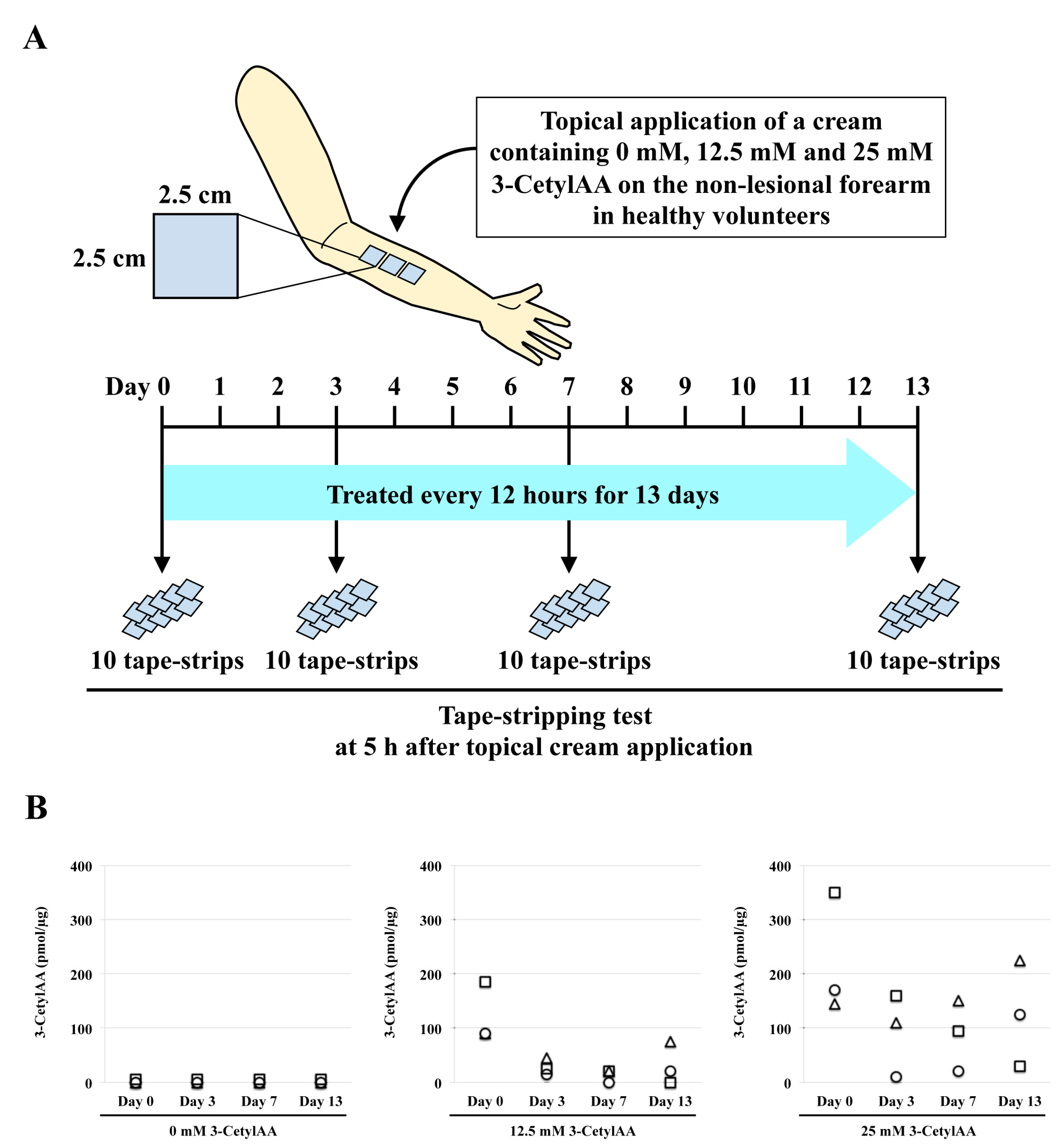
2.2. Tape-Stripping Analysis

One hour before topical application, the forearms of three healthy male volunteers (27−32 years) were cleaned with a mild soap solution (PIAS Co., Kobe, Japan), rinsed with water, and dried. Then, 25 mg of a cream (ingredients: purified water, mineral oil, petrolatum, cetostearyl alcohol, butylene glycol, sodium lauryl sulfate, isopropyl palmitate, sodium acrylate/sodium acryloyldimethyltaurate copolymer, and methylparaben) containing 3-CetylAA (0, 12.5, and 25 mM) was topically applied to a 6.25 cm2 area of the non-lesional forearm (4 mg/cm2), as shown in Figure 1A, following a previously published protocol [31]. The cream was washed away as above, after a 5-h topical application, and the stratum corneum was obtained with a standard tape-stripping procedure [32] using 6.25 cm2 (2.5 cm × 2.5 cm) of Scotch Book tape 845 (3M, St. Paul, MN, USA). Ten serial tape-strips were obtained and used to perform the following analysis for 3-CetylAA quantification.
The recovery period after stratum corneum removal is dependent on the degree of disruption, but the recovery response leads to restoration of the barrier function within hours to days [33,34,35,36,37]. Furthermore, complete stratum corneum removal requires over 70 tape strips [38,39]. Therefore, here, the same test area was repeatedly treated with 3-CetylAA cream for 13 days twice daily after the first ten tape-strippings, and the abovementioned serial tape-stripping procedure was performed on days 0, 3, 5, 7, and 13 on the same volunteers.
The content of 3-CetylAA on the tape-strips was quantified by high-performance liquid chromatography (HPLC) (Shimadzu, Tokyo, Japan) with coulometric electrochemical detection, followed by solvent extraction, as shown previously [16].
To estimate clearance, an additional area of the forearm in the same volunteers was used. Five hours after topical application of a cream containing 25 mM 3-CetylAA, the forearms were cleaned as above, without the tape-stripping procedure. The stratum corneum was obtained after 7 h, and the amounts of 3-CetylAA on the tape-strips were quantified and compared with the amounts on the tape-strips obtained after a 5-h topical application.

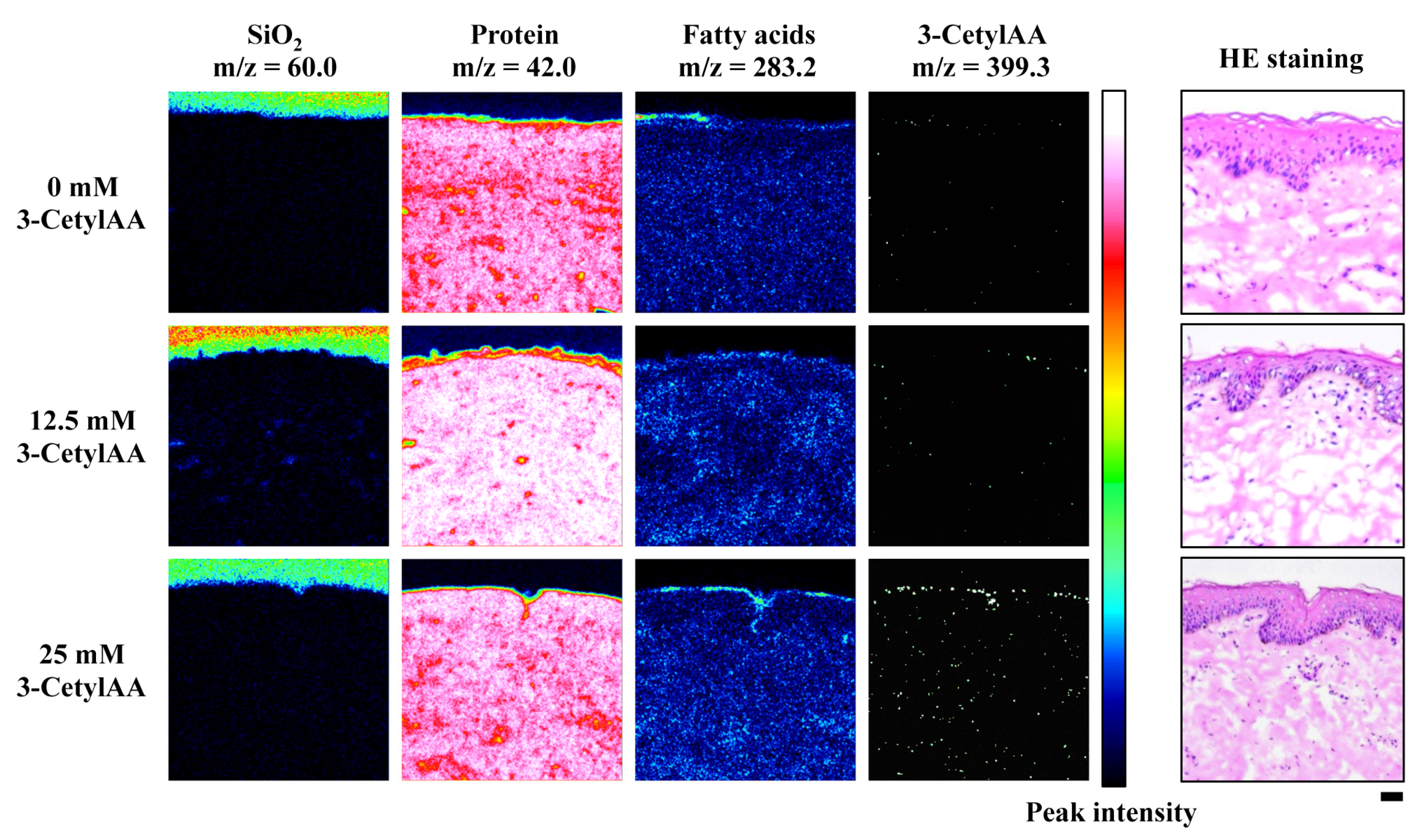
2.3. Time-of-Flight Secondary Ion Mass Spectrometry (ToF-SIMS) Imaging Analysis

An in vitro skin model (Micro-Yucatan Pig Skin-set) was obtained from Charles River Laboratories Japan (Tokyo, Japan). Frozen (−80 °C) skin was thawed at 20 °C for 30 min and cut into appropriate sizes following removal of the adhering fat layer using scissors and a grater. The skin was mounted on a modified Franz diffusion cell apparatus with a 1.1 cm2 effective area [40]. A cream containing 0, 12.5, or 25 mM 3-CetylAA (4 mg/cm2: an equivalent amount for in vivo uptake analysis) was applied to the mounted skin, and the apparatus was maintained in a humidified CO2 incubator at 32 °C for 24 h with 16 mL receptor solution (PBS: phosphate buffered saline). The skin sample was harvested and immediately frozen to obtain vertical cryosections following the removal of the residual skin surface components by washing with PBS. The sectioning was performed using a cryostat (Leica CM1520) at −20 °C with a thickness of 10 μm per slice.
ToF-SIMS imaging was performed with a TOF-SIMS V (ION-TOF, Münster, Germany), with a focused 30 keV Bi3++ primary ion beam at a typical current of 0.2 pA. The profiles were obtained by scanning the primary ion beam over an analysis area of 500 × 500 µm2 using high mass resolution mode (m/Δm = 7000; lateral resolution, 2 µm). The charge compensation of the sample was performed by irradiating the sample with a pulsed beam of electrons using an electron flood gun. Data analysis was performed at Tokyo Headquarters at the Material Science and Technology of Japan (Tokyo, Japan).
Hematoxylin and eosin (HE) staining of the same sections was performed after the ToF-SIMS analysis.

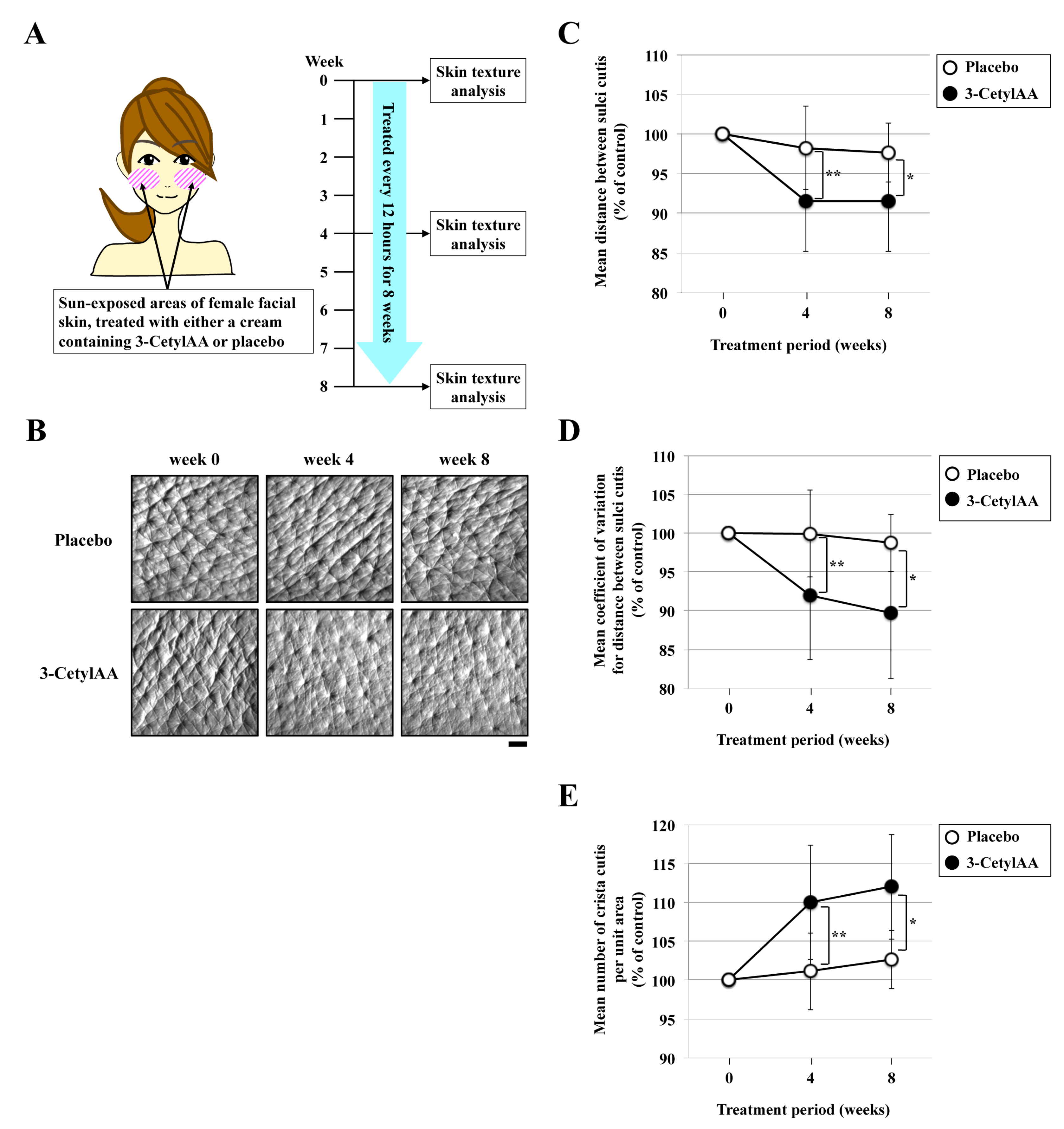
2.4. Skin Texture Analysis

To investigate the effects of 3-CetylAA treatment on the texture of sun-exposed human skin, 64 mg of a cream containing 25 mM 3-CetylAA and a placebo cream were topically applied to a 16 cm2 area of the facial skin of 14 and 13 healthy female volunteers (30−58 years), respectively. This amount (4 mg/cm2) is equivalent to that for in vivo uptake analysis. All volunteers underwent twice-daily treatments for 8 weeks from September to November at 10:30 AM and 10:30 PM, and the test areas were exposed to the sun for 3 h per day (7:00 AM−8:30 AM and 12:30 PM−14:00 PM). During the test period, none of the volunteers used any skin formulation containing steroids or any other compounds, other than the test agent on their skin. No adverse events, including itching, erythema, or others, on the test area were observed in any of the volunteers, and none of them had any skin problems that hindered the evaluation.
The effects of 3-CetylAA treatment on human skin texture were assessed by using the 3D Skin Roughness Analysis Measurement System (ASA-03RXD, Asahi Biomed Co., Kanagawa, Japan). This system is a reflective replica analysis system for skin surface, allowing the quantitative evaluation of skin surface texture in a non-invasive manner. According to the manufacturer’s instructions, skin texture was evaluated using three parameters: (i) distance between the sulci cutis, (ii) the coefficient of variation between the sulci cutis, and (iii) number of crista cutis per unit area. The decreased distance, decreased coefficient of variation for distance and increased number of crista cutis indicated improved skin. The data from three different areas (20 mm × 20 mm) in each volunteer were obtained on day 0 before topical application, and on days 28 and 56 at 3:30 PM, namely after a 5-h topical application.

2.5. In Vitro Transformation Assay

A BALB/c 3T3 A31-1-1 cell transformation assay system was utilized in this study [41]. The cells were cultured in Eagle’s minimal essential medium supplemented with 10% fetal bovine serum (Lot No. 015021, Biological Industries, Beit HaEmek, Israel) in 5% CO2 at 37 °C. Exponentially growing cells were seeded at 102 and 103/60 mm in a culture dish for the colony formation assay and at 104 cells/60 mm in a culture dish for the transformation assay. At day 1 after inoculation, these cells were exposed to 0 or 10 J/m2 of UVC. The irradiated cells were cultured for 24 h in fresh medium, and then the cells were treated for 72 h with DMSO, 100 µM AA or 10 µM 3-CetylAA. For the colony formation assay, the treated cells were further cultured for 2 days and the dishes were fixed with formalin (Nacalai Tesque, Kyoto, Japan) and stained with Giemsa Stain (Sigma-Aldrich Japan, Tokyo, Japan). Formed colonies of greater than 50 cells were considered to indicate survival. For the transformation assay, the treated cells were further cultured for 27 days by refeeding them with fresh medium twice a week, and the dishes were fixed and stained. Formed transformed foci, as determined using standard criteria, were counted [41].

2.6. Statistics

The paired Student’s t-test was used to detect significant differences between 3-CetylAA-treated test groups and untreated control groups. Significant differences in the transformation frequency were analyzed using the Mann–Whitney U test.

2.7. Ethical Considerations

The study involved 32 healthy Japanese females, aged between 30 and 55 years, and without acute or chronic diseases, including skin diseases. All of them voluntarily signed an informed consent form and were available for follow-up during the testing period. This research, performed on humans, complied with the principles of the Declaration of Helsinki and Japan, and it was reviewed and approved by the Institutional Review Board in the legally incorporated medical institution ‘Kenshokai’, Osaka, Japan (Permission number: 20170830-1), based on Japanese Guidance for the Safety Evaluation of Cosmetics 2015.
Other studies using pig skin and cultured cells were approved by the local ethics committees (the PIAS Corporation Institutional Ethics Committee, project number 2017-54, and the Ethics Committee of Prefectural University of Hiroshima, project number H28-R3-004). All methods were carried out in accordance with the relevant guidelines and regulations.

3. Results

3.1. Tape-Stripping of 3-CetylAA in Human Skin

A lipophilic ascorbic acid derivative, 3-CetylAA, has emerged as a possible material for cosmetic use, yet there are very few data available regarding the delivery of 3-CetylAA into the skin after topical application. First, we determined the degree of uptake in three healthy human volunteers’ skin using a serial tape-stripping technique (Figure 1A). Subsequent analysis by HPLC revealed that 10 consecutive pooled tapes contained, on average, 48 ± 15 and 128 ± 27 pmol/µg 3-CetylAA after a 5-h topical treatment when treated with 12.5 and 25 mM 3-CetylAA-containing cream for 13 days twice daily and continuously (Figure 1B). Moreover, 3-CetylAA in the stratum corneum after a 5-h uptake displayed a >90% clearance within 7 h (Table 1).

3.2. ToF-SIMS Imaging of 3-CetylAA in Pig Skin

The serial tape-stripping analysis revealed the presence of 3-CetylAA in the stratum corneum. We then studied the skin permeability of topically applied 3-CetylAA using pig skin. Our approach using ToF-SIMS imaging successfully visualized the presence of topically applied 3-CetylAA in the stratum corneum (Figure 2).

3.3. Effects of 3-CetylAA on Skin Texture in Sun-Exposed Areas

Next, we tested whether 3-CetylAA had an impact on the texture of sun-exposed facial skin. Its effects were quantified using a reflective replica analysis system, designed to detect the three-dimensional skin roughness. When treated with 25 mM 3-CetylAA-containing cream twice daily and continuously (Figure 3A), the skin’s surface texture significantly improved compared to the placebo group (Figure 3B), resulting in densely packed furrows of sulci cutis (Figure 3C). We also found that the variation in the distance between the sulci cutis decreased, smoothening the rough and uneven texture of the skin (Figure 3D). In addition, the number of crista cutis increased in the treated skin (Figure 3E). Thus, 3-CetylAA can significantly improve the textural characteristics of sun-exposed skin.

3.4. Toxic and Carcinogenic Effects of 3-CetylAA

Finally, we examined the toxic and carcinogenic effects of 3-CetylAA using a BALB/c 3T3 A31-1-1 cell transformation assay system, which is widely used to detect both genotoxic and non-genotoxic carcinogens. When treated with 10 µM 3-CetylAA for 72 h after exposure to 0 or 10 J/m2 of UVC, the clonogenic ability of A31-1-1 cells was slightly but not significantly increased compared with the DMSO-treated control (Figure 4B). The transformation frequency was also slightly but not significantly increased compared with the DMSO-treated control (Figure 4C). From these data, we conclude that 3-CetylAA is non-carcinogenic during at least the initiation step of two-stage cell transformation processes in A31-1-1 cells.

4. Discussion

In healthy adults, levels of AA are 340−3640 pmol/mg in the epidermis and 170−734 pmol/mg in the dermis [2]. In the present study, we demonstrate that 3-CetylAA is detected in the stratum corneum after a 5-h topical treatment with 25 mM 3-CetylAA-containing cream (Figure 1 and Figure 2). The amounts of 3-CetylAA in the stratum corneum were not increased even by repeated topical application in the same site (Figure 1B). This observation possibly reflects the saturation uptake capacity of the stratum corneum for the formulation we used here. Alternatively, it may be possible that side effects of repeated tape stripping of the same site in 3-6 days interval cannot be excluded, despite a rapid recovery after stratum corneum removal within a few days [33,34,35,36,37]. Nevertheless, we provided additional information on clearance (Table 1). Depending on the volunteers after a 7-h clearance, the amount of 3-CetylAA decreased by 63% to 96%. This clearance could explain the absence of accumulation in stratum corneum instead of saturation in stratum corneum or repeated tape-stripping on the same area. Anyway, this concentration is approximately 1000-fold less than that of AA, and 3-CetylAA can improve skin texture in this concentration range (Figure 3).
Previous studies have shown that 3-CetylAA is effective in reducing skin-wrinkling by increasing fibroblast-derived type I procollagen synthesis [29]. Nitric oxide production and HSP47 expression have been considered as possible mechanisms responsible for this [30]. Here, topically applied 3-CetylAA was mainly found in the stratum corneum. A recent study revealed that analysis by the serial tape-stripping technique could be used to estimate the function of topically applied chemicals input into viable tissue [31]. Clearance of 3-CetylAA from the stratum corneum, as shown in Table 1, suggests its involvement in the link between skin texture and anti-wrinkle efficacy through epidermal differentiation to reach the viable part of the epidermis, and possibly penetrate more deeply through the epidermal layer from the stratum granulosum to the basal area via metabolic conversion and diffusion. Further studies investigating full skin distribution with quantification of 3-CetylAA and its metabolites are necessary, and may lead to this goal.
We recently discovered that 2-O-octadecylascorbic acid (2-OctadecylAA) prevents misoriented cell divisions with repression of RhoGDIβ expression [42]. Moreover, 2-OctadecylAA is lipophilic, as is 3-CetylAA. Therefore, a possible mechanism behind the effects of 3-CetylAA on skin texture may comprise the prevention of misoriented cell divisions, which is mediated through RhoGDIβ repression [42,43]. Another possible mechanism may involve the elimination of unfit keratinocytes, which is mediated through type XVII collagen synthesis [44]. Both mechanisms are likely to contribute to the improvement of skin texture. Further study is required to test the effects of 3-CetylAA on skin homeostasis.
It is important to determine whether 3-CetylAA is carcinogenic and has synergistic effects with UV irradiation. Here, we tested 10 µM 3-CetylAA for these effects because another lipophilic AA derivative, 2-OctadecylAA, was observed to have in vitro biological activities in the same concentration range [42]. Using a BALB/c 3T3 A31-1-1 cell transformation assay system, we demonstrated that 10 µM 3-CetylAA has no transformation-initiation activity, and also that 10 µM 3-CetylAA does not potentiate the UVC-induced killing effects and the UVC-induced transformation frequency. Thus, 3-CetylAA may be suitable for use for cosmetic purposes with no serious adverse effects; however, it remains challenging to assess its transformation-promotion effects, which are caused by non-genotoxic carcinogens. In relation to this, we observed that both UVC-induced effects are slightly but not significantly increased (Figure 4). Further investigations are needed to assess the character and degree of safety concerns.
In conclusion, our investigation demonstrates that 3-CetylAA is a promising candidate for application in cosmetics. Furthermore, the development of drug delivery approaches for 3-CetylAA is being carried out to improve its percutaneous absorption [45].

Author Contributions

Conceptualization, K.N., K.H. and M.T. (Masaaki Tatsuka); methodology, Y.Y., K.N. and M.T. (Masaaki Tatsuka); software, Y.Y., M.T. (Misaki Toyoshima) and K.N.; validation, N.D., K.N. and M.T. (Masaaki Tatsuka); formal analysis, N.D., Y.Y., M.T. (Misaki Toyoshima) and Y.K.; investigation, N.D., Y.Y., M.T. (Misaki Toyoshima) and Y.K.; resources, K.N., K.H. and M.T. (Masaaki Tatsuka); data curation, K.N. and M.T. (Masaaki Tatsuka); writing—original draft preparation, M.T. (Masaaki Tatsuka); writing—review and editing, N.D., K.N. and M.T. (Masaaki Tatsuka); visualization, N.D. and K.N.; supervision, K.N., K.H. and M.T. (Masaaki Tatsuka); project administration, K.N., K.H. and M.T. (Masaaki Tatsuka); funding acquisition, N.D. and M.T. (Masaaki Tatsuka). All authors have read and agreed to the published version of the manuscript.

Funding

This research was funded by the Prefectural University of Hiroshima, grant number S-H30 and S-R1. N.D. was funded by the Prefectural University of Hiroshima Graduate School of Comprehensive Scientific Research Projects.

Institutional Review Board Statement

The study was conducted according to the guidelines of the Declaration of Helsinki and approved by the Institutional Review Board in the legally incorporated medical institution ‘Kenshokai’, Osaka, Japan (Permission number: 20170830-1), and the local ethics committees (the PIAS Corporation Institutional Ethics Committee, project number 2017-54, and the Ethics Committee of Prefectural University of Hiroshima, project number H28-R3-004).

Informed Consent Statement

Informed consent was obtained from all subjects involved in the study.

Acknowledgments

We thank Takahide Ota for critical comments on the work and also thank Hiro Togari, Kenji Minagi, Takatoshi Sugimoto, Kaede Takeshima, Masahiro Mori, and Megumi Morisaki for their technical assistance.

Conflicts of Interest

The authors declare no conflict of interest.

References

  1. Telang, P.S. Vitamin C in dermatology. Indian Dermatol. Online J. 2013, 4, 143–146. [Google Scholar] [CrossRef] [PubMed]
  2. Pullar, J.M.; Carr, A.C.; Vissers, M.C.M. The Roles of Vitamin C in Skin Health. Nutrition 2017, 9, 866. [Google Scholar] [CrossRef] [Green Version]
  3. Ravetti, S.; Clemente, C.; Brignone, S.; Hergert, L.; Allemandi, D.; Palma, S. Ascorbic Acid in Skin Health. Cosmetics 2019, 6, 58. [Google Scholar] [CrossRef] [Green Version]
  4. Shindo, Y.; Witt, E.; Han, D.; Epstein, W.; Packer, L. Enzymic and Non-Enzymic Antioxidants in Epidermis and Dermis of Human Skin. J. Investig. Dermatol. 1994, 102, 122–124. [Google Scholar] [CrossRef] [PubMed] [Green Version]
  5. Rhie, G.-E.; Shin, M.H.; Seo, J.Y.; Choi, W.W.; Cho, K.H.; Kim, K.H.; Park, K.C.; Eun, H.C.; Chung, J.H. Aging- and Photoaging-Dependent Changes of Enzymic and Nonenzymic Antioxidants in the Epidermis and Dermis of Human Skin In Vivo. J. Investig. Dermatol. 2001, 117, 1212–1217. [Google Scholar] [CrossRef] [PubMed] [Green Version]
  6. McArdle, F.; Rhodes, L.; Parslew, R.; Jack, C.; Friedmann, P.; Jackson, M. UVR-induced oxidative stress in human skin in vivo: Effects of oral vitamin C supplementation. Free. Radic. Biol. Med. 2002, 33, 1355–1362. [Google Scholar] [CrossRef]
  7. Shindo, Y.; Witt, E.; Packer, L. Antioxidant Defense Mechanisms in Murine Epidermis and Dermis and Their Responses to Ultraviolet Light. J. Investig. Dermatol. 1993, 100, 260–265. [Google Scholar] [CrossRef] [Green Version]
  8. Shindo, Y.; Witt, E.; Han, D.; Packer, L. Dose-Response Effects of Acute Ultraviolet Irradiation on Antioxidants and Molecular Markers of Oxidation in Murine Epidermis and Dermis. J. Investig. Dermatol. 1994, 102, 470–475. [Google Scholar] [CrossRef] [Green Version]
  9. Gref, R.; Deloménie, C.; Maksimenko, A.; Gouadon, E.; Percoco, G.; Lati, E.; Desmaële, D.; Zouhiri, F.; Couvreur, P. Vitamin C–squalene bioconjugate promotes epidermal thickening and collagen production in human skin. Sci. Rep. 2020, 10, 16883. [Google Scholar] [CrossRef]
  10. Abdullah, M.; Jamil, R.T.; Attia, F.N. Vitamin c (ascorbic acid). In Statpearls; StatPearls Publishing: Treasure Island, FL, USA, 2021. [Google Scholar]
  11. Duarte, T.L.; Cooke, M.S.; Jones, G.D. Gene expression profiling reveals new protective roles for vitamin C in human skin cells. Free. Radic. Biol. Med. 2009, 46, 78–87. [Google Scholar] [CrossRef]
  12. Lee, S.-A.; Son, Y.-O.; Kook, S.-H.; Choi, K.-C.; Lee, J.-C. Ascorbic acid increases the activity and synthesis of tyrosinase in B16F10 cells through activation of p38 mitogen-activated protein kinase. Arch. Dermatol. Res. 2011, 303, 669–678. [Google Scholar] [CrossRef] [PubMed]
  13. Panich, U.; Tangsupa-a-nan, V.; Onkoksoong, T.; Kongtaphan, K.; Kasetsinsombat, K.; Akarasereenont, P.; Wongkajornsilp, A. Inhibition of UVA-mediated melanogenesis by ascorbic acid through modulation of antioxidant defense and nitric oxide system. Arch. Pharmacal Res. 2011, 34, 811–820. [Google Scholar] [CrossRef] [PubMed]
  14. De Tullio, M.C. Beyond the Antioxidant: The Double Life of Vitamin C. Prokaryotic Cytoskelet. 2011, 56, 49–65. [Google Scholar] [CrossRef]
  15. Sanadi, R.M.; Deshmukh, R.S. The effect of Vitamin C on melanin pigmentation A systematic review. J. Oral Maxillofac. Pathol. 2020, 24, 374–382. [Google Scholar] [CrossRef]
  16. Pinnell, S.R.; Yang, H.; Omar, M.; Riviere, N.M.; DeBuys, H.V.; Walker, L.C.; Wang, Y.; Levine, M. Topical L-Ascorbic Acid: Percutaneous Absorption Studies. Dermatol. Surg. 2001, 27, 137–142. [Google Scholar] [CrossRef]
  17. Al-Niaimi, F.; Chiang, N.Y.Z. Topical Vitamin C and the Skin: Mechanisms of Action and Clinical Applications. J. Clin. Aesthetic Dermatol. 2017, 10, 14–17. [Google Scholar]
  18. Stamford, N.P.J. Stability, transdermal penetration, and cutaneous effects of ascorbic acid and its derivatives. J. Cosmet. Dermatol. 2012, 11, 310–317. [Google Scholar] [CrossRef]
  19. Duarah, S.; Durai, R.D.; Narayanan, V.B. Nanoparticle-in-gel system for delivery of vitamin C for topical application. Drug Deliv. Transl. Res. 2017, 7, 750–760. [Google Scholar] [CrossRef]
  20. Wang, Q.; Zhang, H.; Huang, J.; Xia, N.; Li, T.; Xia, Q. Self-double-emulsifying drug delivery system incorporated in natural hydrogels: A new way for topical application of vitamin C. J. Microencapsul. 2018, 35, 90–101. [Google Scholar] [CrossRef]
  21. Macan, A.M.; Kraljević, T.G.; Raić-Malić, S. Therapeutic Perspective of Vitamin C and Its Derivatives. Antioxidants 2019, 8, 247. [Google Scholar] [CrossRef] [Green Version]
  22. Machado, B.H.B.; Frame, J.; Zhang, J.; Najlah, M. Comparative Study on the Outcome of Periorbital Wrinkles Treated with Laser-Assisted Delivery of Vitamin C or Vitamin C Plus Growth Factors: A Randomized, Double-blind, Clinical Trial. Aesthetic Plast. Surg. 2020, 1–13. [Google Scholar] [CrossRef]
  23. Caritá, A.C.; de Azevedo, J.R.; Buri, M.V.; Bolzinger, M.-A.; Chevalier, Y.; Riske, K.A.; Leonardi, G.R. Stabilization of vitamin C in emulsions of liquid crystalline structures. Int. J. Pharm. 2021, 592, 120092. [Google Scholar] [CrossRef] [PubMed]
  24. Elhabak, M.; Ibrahim, S.; Abouelatta, S.M. Topical delivery of l-ascorbic acid spanlastics for stability enhancement and treatment of UVB induced damaged skin. Drug Deliv. 2021, 28, 445–453. [Google Scholar] [CrossRef] [PubMed]
  25. Wang, Y.-S.; Yang, W.-H.; Gao, W.; Zhang, L.; Wei, F.; Liu, H.; Wang, S.-Y.; Li, Y.-Y.; Zhao, W.-M.; Ma, T.; et al. Combination and efficiency: Preparation of dissolving microneedles array loaded with two active ingredients and its anti-pigmentation effects on guinea pigs. Eur. J. Pharm. Sci. 2021, 160, 105749. [Google Scholar] [CrossRef] [PubMed]
  26. Tai, A.; Goto, S.; Ishiguro, Y.; Suzuki, K.; Nitoda, T.; Yamamoto, I. Permeation and metabolism of a series of novel lipophilic ascorbic acid derivatives, 6-O-acyl-2-O-α-d-glucopyranosyl-l-ascorbic acids with a branched-acyl chain, in a human living skin equivalent model. Bioorganic Med. Chem. Lett. 2004, 14, 623–627. [Google Scholar] [CrossRef]
  27. Tripathi, R.; Singh, B.; Bisht, S.; Pandey, J. L-Ascorbic Acid in Organic Synthesis: An Overview. Curr. Org. Chem. 2009, 13, 99–122. [Google Scholar] [CrossRef]
  28. Díaz, Y.D.L.M.Z.; Menghi, K.; Guerrero, M.L.; Nocelli, N.; Fanani, M.L. l-Ascorbic acid alkyl esters action on stratum corneum model membranes: An insight into the mechanism for enhanced skin permeation. Colloids Surfaces B Biointerfaces 2020, 185, 110621. [Google Scholar] [CrossRef]
  29. Park, C.M.; Lee, S.Y.; Joung, M.S.; Choi, J.W. Anti-wrinkle effect of 3-o-cetyl-l-ascorbic acid. J. Soc. Cosmet. Sci. Korea 2008, 34, 303–309. [Google Scholar]
  30. Kaburagi, S. The active ingredient of stimulating the biosynthesis of dermal component. Fragr. J. 2008, 34, 42–45. [Google Scholar]
  31. Hoppel, M.; Tabosa, M.A.M.; Bunge, A.L.; Delgado-Charro, M.B.; Guy, R.H. Assessment of Drug Delivery Kinetics to Epidermal Targets In Vivo. AAPS J. 2021, 23, 1–11. [Google Scholar] [CrossRef]
  32. N’Dri-Stempfer, B.; Navidi, W.C.; Guy, R.H.; Bunge, A.L. Improved Bioequivalence Assessment of Topical Dermatological Drug Products Using Dermatopharmacokinetics. Pharm. Res. 2008, 26, 316–328. [Google Scholar] [CrossRef] [PubMed]
  33. Sahle, F.F.; Gebre-Mariam, T.; Dobner, B.; Wohlrab, J.; Neubert, R.H. Skin Diseases Associated with the Depletion of Stratum Corneum Lipids and Stratum Corneum Lipid Substitution Therapy. Ski. Pharmacol. Physiol. 2015, 28, 42–55. [Google Scholar] [CrossRef] [PubMed]
  34. Kim, H.S.; Lim, S.H.; Song, J.Y.; Kim, M.-Y.; Lee, J.H.; Park, J.G.; Kim, H.O.; Park, Y.M. Skin barrier function recovery after diamond microdermabrasion. J. Dermatol. 2009, 36, 529–533. [Google Scholar] [CrossRef]
  35. Milewski, M.; Brogden, N.K.; Stinchcomb, A.L. Current aspects of formulation efforts and pore lifetime related to microneedle treatment of skin. Expert Opin. Drug Deliv. 2010, 7, 617–629. [Google Scholar] [CrossRef] [PubMed] [Green Version]
  36. Song, J.Y.; Kang, H.A.; Kim, M.-Y.; Park, Y.M.; Kim, H.O. Damage and Recovery of Skin Barrier Function After Glycolic Acid Chemical Peeling and Crystal Microdermabrasion. Dermatol. Surg. 2004, 30, 390–394. [Google Scholar] [CrossRef]
  37. Andrews, S.; Lee, J.W.; Prausnitz, M. Recovery of Skin Barrier After Stratum Corneum Removal by Microdermabrasion. AAPS PharmSciTech 2011, 12, 1393–1400. [Google Scholar] [CrossRef] [Green Version]
  38. Weigmann, H.-J.; Lademann, J.; Meffert, H.; Schaefer, H.; Sterry, W. Determination of the Horny Layer Profile by Tape Stripping in Combination with Optical Spectroscopy in the Visible Range as a Prerequisite to Quantify Percutaneous Absorption. Ski. Pharmacol. Physiol. 1999, 12, 34–45. [Google Scholar] [CrossRef]
  39. Lademann, J.; Ilgevicius, A.; Zurbau, O.; Liess, H.-D.; Schanzer, S.; Weigmann, H.-J.; Antoniou, C.; Von Pelchrzim, R.; Sterry, W. Penetration studies of topically applied substances: Optical determination of the amount of stratum corneum removed by tape stripping. J. Biomed. Opt. 2006, 11, 054026. [Google Scholar] [CrossRef]
  40. Ishikawa, A.; Fujii, M.; Morimoto, K.; Yamada, T.; Koizumi, N.; Kondoh, M.; Watanabe, Y. Oil-in-water emulsion lotion providing controlled release using 2-methacryloyloxyethyl phosphorylcholine n-butyl methacrylate copolymer as emulsifier. Results Pharma Sci. 2012, 2, 16–22. [Google Scholar] [CrossRef] [Green Version]
  41. Benedict, W.F.; Chouroulinkov, I.; Fisher, P.B.; Kakunaga, T.; Marquardt, H.; Pienta, R.J.; Yamasaki, H. Transformation assay of established cell lines: Mechanisms and application. In Proceedings of the A Workshop Organized by Iarc in Collaboration with the Us National Cancer Institute and the Us Environmental Protection Agency, Lyon, France, 15–17 February 1984; International Agency for Research on Cancer: Lyon, France, 1985; Volume 67, pp. 1–255. [Google Scholar]
  42. Doi, N.; Togari, H.; Minagi, K.; Iwaoka, Y.; Tai, A.; Nakaoji, K.; Hamada, K.; Tatsuka, M. 2-O-Octadecylascorbic acid represses RhoGDIβ expression and ameliorates DNA damage-induced abnormal spindle orientations. J. Cell. Biochem. 2021. [Google Scholar] [CrossRef] [PubMed]
  43. Doi, N.; Togari, H.; Minagi, K.; Nakaoji, K.; Hamada, K.; Tatsuka, M. Protective Effects of Salicornia europaea on UVB-Induced Misoriented Cell Divisions in Skin Epithelium. Cosmetics 2020, 7, 44. [Google Scholar] [CrossRef]
  44. Liu, N.; Matsumura, H.; Kato, T.; Ichinose, S.; Takada, A.; Namiki, T.; Asakawa, K.; Morinaga, H.; Mohri, Y.; De Arcangelis, A.; et al. Stem cell competition orchestrates skin homeostasis and ageing. Nat. Cell Biol. 2019, 568, 344–350. [Google Scholar] [CrossRef] [PubMed]
  45. Fushimi, T.; Uchino, T.; Miyazaki, Y.; Hatta, I.; Asano, M.; Fujino, H.; Suzuki, R.; Fujimori, S.; Kamiya, D.; Kagawa, Y. Development of phospholipid nanoparticles encapsulating 3-O-cetyl ascorbic acid and tocopherol acetate (TA-Cassome) for improving their skin accumulation. Int. J. Pharm. 2018, 548, 192–205. [Google Scholar] [CrossRef]
Figure 1. Amounts of 3-CetylAA in the stratum corneum after a 5-h uptake: (A) Study design and treatment schedule. A cream containing 0 mM, 12.5 mM, and 25 mM 3-CetylAA was topically applied to the ventral side of the non-lesional forearm in three healthy volunteers. All volunteers underwent twice-daily treatments for 13 days. At the indicated time points, ten serial tape-strips (2.5 cm × 2.5 cm) were obtained. (B) Quantification of 3-CetylAA in the stratum corneum. The amounts of 3-Cetyl AA were quantified in the first ten strips from the three volunteers by high-performance liquid chromatography analysis, and they are indicated as the total amount of ten tapes in each volunteer.
Cosmetics 08 00038 g001
Figure 2. ToF-SIMS images (left to right: SiO2, protein, fatty acids, and 3-CetylAA) and optical micrograph (HE staining), with the same analysis area (500 µm × 500 µm). Scale bar, 50 µm.
Cosmetics 08 00038 g002
Figure 3. Improvements in the texture of sun-exposed skin after treatment with 3-CetylAA: (A) Study design and treatment schedule. A cream containing 25 mM 3-CetylAA and a placebo cream were topically applied to the sun-exposed facial skin of 14 and 13 healthy volunteers, respectively. All volunteers underwent twice-daily treatments for 4 and 8 weeks. The skin texture was analyzed before and 4 and 8 weeks after the treatment. (B) Typical reflective replica images from skin treated with either 3-CetylAA or placebo. The images were obtained using the 3D Skin Roughness Analysis Measurement System. Scale bar, 1000 µm. (C) Effects of 3-CetylAA on the distance between sulci cutis. The data were obtained from three different areas (20 mm × 20 mm) in each volunteer. Values indicate means ± S.D. * Significant difference (p < 0.03). ** Significant difference (p < 0.01). (D) Effects of 3-CetylAA on coefficient of variation between sulci cutis in the above data. Values indicate means ± S.D. * Significant difference (p < 0.02). ** Significant difference (p < 0.007). (E) Effects of 3-CetylAA on number of crista cutis per unit area. The data were obtained from three different areas (20 mm × 20 mm) in each volunteer. Values indicate means ± S.D. * Significant difference (p < 0.002). ** Significant difference (p < 0.0008).
Cosmetics 08 00038 g003
Figure 4. Effects of 3-CetylAA-induced cell killing and cell transformation and their UVC-induced potentiation effects: (A) Study design and treatment schedule. Exponentially growing BALB/c 3T3 A31-1-1 cells were used for clonogenic assay and transformation assay. (B) Depiction of colony formation on Giemsa-stained dishes from clonogenic assay. (C) Quantification of the number of colonies. The data were obtained from three independent experiments using triplicated dishes. Values indicate means ± S.E. N.S., not significant. (D) Depiction of transformed foci on Giemsa-stained dishes from transformation assay. (E) Quantification of the number of transformed foci as determined using standard criteria. The data were obtained from three independent experiments using 10−12 dishes. Values indicate means ± S.E. * Significant difference ( p < 0.03). N.S., not significant.
Cosmetics 08 00038 g004
Table 1. Amounts of 3-CetylAA in the stratum corneum after uptake and clearance.
Volunteer #1Volunteer #2Volunteer #3
5 h uptake14.5 1 (0%) 212 (0%)35 (0%)
7 h clearance5.5 (63%)0.5 (96%)8 (77%)
1 3-CetylAA in the stratum corneum (pmol/mg). 2 % Clearance.
Publisher’s Note: MDPI stays neutral with regard to jurisdictional claims in published maps and institutional affiliations.

