Article

Rhodomyrtus tomentosa Fruit Extract and Skin Microbiota: A Focus on C. acnes Phylotypes in Acne Subjects

1
R& D Department, Greentech. Biopôle Clermont-Limagne, 63360 Saint Beauzire, France
2
R&D Department, Phylogene S.A., 62 RN 113, 30620 Bernis, France
3
UMR 1019 INRA-UcA, UNH (Human Nutrition Unity), ECREIN Team, University Clermont Auvergne, 63000 Clermont-Ferrand, France
*
Author to whom correspondence should be addressed.
Cosmetics 2020, 7(3), 53; https://doi.org/10.3390/cosmetics7030053
Submission received: 29 May 2020 / Revised: 28 June 2020 / Accepted: 1 July 2020 / Published: 7 July 2020
(This article belongs to the Special Issue Feature Papers in Cosmetics in 2020)

Abstract

:
Knowing that Rhodomyrtus tomentosa is known to have antibacterial effects, this study investigated the skin microbiota with a focus on Cutibacterium acnes (C. acnes) phylotypes in subjects with acne, and determined microbiota changes after 28 days of treatment with berries Rhodomyrtus tomentosa as an active ingredient (RT). Skin swabs from seventeen acne subjects were collected and the skin microbiome was analyzed using 16S rRNA gene sequencing. A culture-independent next-generation sequencing (NGS)-based SLST (single-locus sequence typing) approach was aimed at evaluating RT extract effects on C. acnes phylotype repartition. Clinical evaluations (lesion counts) were performed at baseline (D0) and after 28 days (D28) of twice-daily application of the RT active ingredient. We determined: (1) the skin microbiota at D0 was dominated by Actinobacteria followed by Firmicutes and Proteobacteria; (2) at the genus level, Cutibacterium was the most abundant genus followed by Staphylococcus and Corynebacterium; (3) C. acnes was the major species in terms of mean abundance, followed by Staphylococcus epidermidis (S. epidermidis) and Staphylococcus hominis (S. hominis); and (4) phylotype IA1 was most represented, with a predominance of SLST type A1, followed by phylotypes II, IB, IA2, IC, and III. After 28 days of RT extract treatment, phylotype repartition were modified with a decrease in abundance (approximately 4%) of phylotype IA1 and an increase in phylotype II and III. Cutibacterium granulosum (C. granulosum) abundance also decreased. Reduction of retentional and inflammatory lesions was also noted only after RT treatment; thus, RT extract acts as a microbiota-regulating agent.

1. Introduction

The skin is the largest organ of the human body. Four phyla dominate the skin including Actinobacteria, Proteobacteria, Bacteroidetes, and Firmicutes, along with most of the bacteria belonging to the Staphylococcus, Corynebacterium, and Cutibacterium genera [1]. The relationship between members of this cutaneous microbiota is essential for the maintenance of healthy skin, of which microbial variations and loss of diversity may result in the induction of cutaneous diseases such as acne [2]. Besides increased sebum production, inflammatory mediators of the skin, and follicular keratinization of the pilosebaceous ducts, which are well-known to be involved in acne development, it seems that Cutibacterium acnes (C. acnes), which prevents colonization from harmful pathogens, is also recognized as an opportunistic pathogen in acne vulgaris [3]. Nevertheless, Fitz-Gibbon et al. [4] showed that the relative abundance of C. acnes (in metagenomics studies) is similar among patients with acne and healthy individuals.
More precisely, C. acnes’ protective role as a commensal bacterium of healthy skin microbiota is well documented, helping to maintain a low skin pH and blocking pathogens such as Staphylococcus aureus (S. aureus) and Streptococcus [5]. For Szabo et al. [6], specific metabolic features allow C. acnes to preserve the stability of resident skin microbiota. However, several mechanisms have been proposed by which C. acnes aggravates acne, including augmentation of lipogenesis, comedone formation, and host inflammation [7]. Additionally, recent studies have reported that C. acnes phylotypes might play a critical role in acne onset [6,8].
Early typing methods noted two distinct C. acnes phenotypes named type I and II. An additional phylogenetic group called type III was also included [9]. Recent genomic studies have highlighted the presence of subgroups among phylotypes according to genome analyses called multi-locus sequence typing (MLST) and single-locus sequence typing (SLST). Presently, the C. acnes type I clade can be subdivided into closely related subtypes: IA1, IA2, IB, and IC. These subtypes contain many different clonal complexes (CC) with three major phylogenetic clades I, II and III having been identified. However, it should be noted that nomenclature differs between studies (Aarhus scheme I-1a, I-1b, I-2, Belfast scheme IA1, IA2, IB, IC) [10].
Despite controversial data in line with the population samplings, anatomic sites, and typing methods, most studies report that strains from type IA1 preferentially colonize skin with acne while others do not or poorly present in acne lesions (IB, II and III) [8,11].
Dagnelie et al. [3] also reported that inflammatory severe acne of both the face and back is associated with diversity loss of C. acnes phylotypes, and a high predominance of phylotype IA1, both on the face (72.7%) and the back (95.6%). They also suggested that acne could be associated with the proliferation of one specific phylotype.
Thus, it seems that the severity of acne may be more related to the selection of its subtypes than its proliferation. It is also important to note that other factors such as androgens and hormonal fluctuation or imbalance, poor nutrition, stress, pollution, and habits are important in the development and persistence of the disease [12].
C. acnes has been shown to coexist on the skin surface and in the pilosebaceous follicle with other Cutibacterium spp., including C. granulosum and C. avidum.
C. acnes and C. granulosum are most abundant in the sebaceous gland-rich sites of the skin, which includes the face and upper trunk. Although C. acnes is best known for its connection with acne, it is speculated that other bacteria might also indirectly contribute to the inflammatory process. In a metagenomic analysis of acne patients, Barnard et al. [13] noted a higher relative abundance of C. granulosum in healthy individuals, compared to those with acne. In contrast, early culture-based studies reported that C. granulosum is more prevalent in comedones and pustules compared to uninvolved follicles of patients with acne [14]. Moreover, C. granulosum was reported to demonstrate greater lipase activity compared to C. acnes [15]. Finally, Rajiv et al. [16] observed that C. acnes and Staphylococcus epidermidis were more prevalent in acne patients than in the control population. The applicability of the finding was tested using explant models and S. epidermidis was found to prevent acne and exert antimicrobial activity. S. epidermidis has been reported to produce antimicrobial peptides such as epidermin, phenol-soluble modulins, Pep5, and epilancin. Wang et al. [17] claimed that S. epidermidis strains release succinic acid, which has an anti-C. acnes effect. Christensen et al. [18] reported that S. epidermidis secretes polymorphic toxins that inhibit C. acnes growth. In addition, S. epidermidis was shown to generate staphylococcal lipoteichoic acid, which dampens C. acnes-related inflammation by increasing the expression of miR-143 and blocking TLR-2 expression in keratinocytes [19].
Topical retinoids, antibiotics, benzoyl peroxide (BPO), which suppresses bacterial proliferation, hyperkeratinization and inflammation, and systemic antibiotics are currently the first-line treatments for mild-to-moderate acne [20]. Although highly effective, topical treatments affect skin barrier integrity and are often associated with side effects such as dryness, irritation, itching, and redness [21]. For Dursun et al. [22], current treatment approaches for acne are becoming inefficient. Moreover, using antibiotics daily can induce multidrug resistance [23].
Novel therapies are in high demand and an ethnopharmacological approach to discover new plant sources of anti-acne therapeutics could fill this void of effective therapies. Exploitation of natural resources, especially medicinal plants and their derived products, are considered promising alternative agents for the treatment of diseases.
Among them, Rhodomyrtus tomentosa (RT) is a flowering plant belonging to the family Myrtaceae, native to southern and southeastern Asia. All parts of this plant (leaves, roots, buds, and fruits) have been used in traditional Vietnamese, Chinese, and Malaysian medicine. In traditional medicine, RT fruits have been used to treat diarrhea and dysentery, and to boost the immune system [24].
RT has been reported to contain various phytochemical compounds in many parts of the plant. Ellagitannins, stilbenes, anthocyanins, flavonols, and phenolic acids are the phenolic compounds found in the fruit [25]. Among them, piceatannol, a stilbene having potent biological activities, including antioxidant, anticancer, anti-inflammatory, and antimicrobial properties, is a major phenolic compound identified in the fruit [26]. Organic acids are also largely represented in the fruit by malic acid, quinic acid, and citric acid, having astringent properties and antibacterial properties [27,28]. Recently, the acylphloroglucinol rhodomyrtone from RT fruit was evidenced as a potential inhibitor of inflammation. Moreover, rhodomyrtone showed strong antibacterial activity against a wide range of gram-positive pathogenic bacteria, as well as anti-biofilm property against Staphylococci causing severe infections. More specifically, rhodomyrtone inhibits C. acnes proliferation [29].
This study investigated the skin microbiota with a focus on Cutibacterium acnes (C. acnes) phylotypes in subjects with acne and determined microbiota changes after 28 days of treatment with berries RT as the active ingredient.

2. Material and Methods

2.1. Participants

Seventeen volunteers were included by a dermatologist. Written informed consent and photography were obtained from each subject prior to enrollment.
Subjects were aged 27.3 ± 1.0 years with mild to moderate acne (Global Acne Severity Scale [GEA] II to III), presenting at least five papules and pustules on the face (not located on the nasal pyramid), and at least six closed and open comedones on the face, including at least three on the forehead [30]. Exclusion criteria were presence of any cutaneous lesions affecting the face apart from ongoing acne (i.e., vitiligo, psoriasis, and seborrheic dermatitis), or any chronic or acute progressive disease, which may interfere with the study. Patients who applied facial topical treatments during the preceding month including anti-acneic agents (retinoids, zinc, benzoyle peroxide), antimicrobial agents (antibiotics, antiseptic), corticosteroids or nonsteroid anti-inflammatory drugs (NSAIDs), or patients who took systemic anti-acneic treatment during the preceding month with antibiotics (tetracycline, macrolides, macrolide derivatives) or zinc, or patients treated with NSAIDs, corticosteroid, or antibiotics other than anti-acneic taken during the preceding month, were not included. Patients were also excluded if they had been treated with oral retinoids or any hormonal treatment for contraceptive or anti-acneic purpose initiated or modified during the 12 preceding weeks. During treatment, none of the aforementioned treatments were allowed.
Permission to conduct the study was granted by the local university’s human research ethics committee, and all experiments were conducted in accordance with the Declaration of Helsinki.

2.2. Procedure

Two visits were performed for subjects: at inclusion (D0) and at the end of the study (D28). At baseline, microbiological sampling was performed on 17 acne patients for microbiota studies. In addition, the dermatologist performed a clinical examination of the face at D0 and D28. Concomitant treatments, adverse events, and treatment compliance were recorded.

2.3. Study Product

The study products were a gel containing 2% Rhodomyrtus tomentosa fruit extract and a placebo based on the same gelling agent acrylates/C10-30 alkyl acrylate cross polymer, wherein RT extract was replaced by demineralized water. Each formulation provided the same penetration capacity due to the presence of the surfactant PEG-8 Caprylic/Capric Glycerides (1%). The study products were applied twice daily, on each half-face. Subjects were instructed not to change their hygiene habits or to apply other skin care products or topical drugs during the study on their face. All toxicological tests (eye irritation, skin irritation, sensitization, phototoxicity, and mutagenicity) were realized.

2.4. Preparation of Extract

Extractions of compounds from Rhodomyrtus tomentosa (RT) fruits were conducted with hydroalcoholic solution (ethanol/water 50/50) over 12 h at room temperature. The extract solution was filtrated through a 15 µm plate. The resulting solution was concentrated at 5% of dry matter in a matrix water/propane 1,3-diol (50/50). Solvent was removed by rotary evaporation at 60 °C. At the end of the extraction process, the extract was decontaminated by filtration at 0.45 µm.Table 1 summarizes the characteristics of RT extract. The RT active ingredient was not an eye irritant at 2% in a formula (HET-CAM-Hen’s Egg-Chorioallantoic Membrane test), had a very good skin compatibility (patch test), had no sensitizing effect (HRIPT- Human Repeat Insult Patch Test), was not phototoxic, and was not mutagenic at 5%.

2.5. Microbiota Sampling

On the first day of the study (Day 0), skin samplings (swabs) were performed on each volunteer. Samplings were collected by the same investigator on the cheeks, forehead, temple, or chin of each patient’s face (depending on the location of acne lesions). Then, volunteers were asked to apply active formula on one side of the face, and placebo formula on the other side, twice a day for 4 weeks. New samplings were performed at the end of treatment on day 28 (Day 28) for each volunteer and face side.
After sampling, swabs were immediately placed in PowerBead tubes containing cell lysing solution and beads, and were stored at −80 °C until DNA extraction was performed. DNA extraction and purification were performed using DNeasy PowerSoil Kit (Qiagen, Hilden, Germany). Briefly, cells on swabs were lysed by the combination of mechanical (bead beating) and chemical treatments (detergent). Extracted DNA was then purified on silica membrane and eluted in 100 μL.
Then, 16S metagenomics 16SrDNA variables regions V1 to V3 were amplified using primer 27F (AGAGTTTGATCCTGGCTCAG) and 534R (ATTACCGCGGCTGCTGG); 16s DNA sequencing was performed on Illumina Miseq using paired-end technology.
All sequence processing was performed using QIIME2 suite (https://qiime2.org/) using the default settings. After quality filtering, read pairs were merged by overlapping and clustered and chimeric sequences were removed. Operational taxonomic units (OTUs) were assigned to sequences against Silva 132 rDNA database.
To evaluate whether the active ingredient affects populations of five bacterial species (S. epidermidis, S. aureus, S. hominis, C. acnes, and C. granulosum, and one genus Corynebacterium), genus/species quantification using quantitative (real-time) PCR (qPCR) was carried out. All qPCRs were performed with LightCycler 480 II (Roche, Basel, Switzerland) thermocycler using LightCycler® Multiplex DNA Master (Roche, Basel, Switzerland) core qPCR kit and fluorescent TaqMan probe chemistry for species-specific qPCR, and LightCycler® 480 SYBR Green I Master (Roche, Basel, Switzerland) for genus-specific qPCR.
For each qPCR, standards made of known amounts of corresponding genomic DNA extracted from pure strains (105 to 10 genome equivalent) were integrated to qPCR assay.
Variable C. acnes regions described by Scholtz et al. [31] were amplified and sequenced on Illumina Miseq using paired-end technology with modified primers. Original C. acnes SLST method amplifies a region of 612 base pairs, which is too large to be sequenced using Illumina technology. New primers amplifying a region or 497 base pairs were designed, forward primer being placed 74 nucleotides downstream original and reverse 26 nucleotides upstream. This modification will not allow distinction between SLST types A1 and A6 or SLST types E3 and E7. In both cases, these variants belong to the same phylotype. All sequence processing was performed using QIIME2 suite (https://qiime2.org/) with default settings. After quality filtering, read pairs were merged by overlapping and clustered and chimeric sequences were removed. SLST (Single Locus Sequence Type) was assigned to sequences against an SLST reference database (http://medbac.dk/slst/pacnes) [31].

2.6. Clinical Evaluations

Clinical evaluations were conducted by the same investigator on Day 0 and Day 28 and included the scoring of lesions (blackheads, microcysts, papules, and pustules) on each hemi-face (except nasal pyramid, the vermillion border, the crease of the chin, and the rim of the scalp) and the reporting of local tolerance issues, acne signs, and symptoms. The variations (D28–D0) in the number of lesions were calculated for each kind of lesion. Sebum was evaluated by sebumeter® on the right and left wings of nose. Redness was evaluated by VISIA-CR and photos were analyzed by IPP soft at the forehead and the chin. Comparisons between placebo and RT extract were realized.
A subjective evaluation questionnaire was filled in by the subjects at the end of the study on Day 28 to subjectively evaluate the properties and the efficacy of the studied products.

2.7. Statistical Analysis

Raw data were used to determine taxa abundances and α-diversity indices after removing OTU with less than 2 counts. Filtered and normalized data were used to compare populations between treatments and days (β-diversity) and to compare taxa abundances between the same sample groups. Abundances for five taxonomic levels (Phylum to Genus) were calculated by summing sequence numbers assigned to same taxa for the considered level. Shannon diversity index was calculated using PAST 3.25 software (University of Oslo, Oslo, Norway), wherein index increases with diversity.
Concerning the genomic quantitative study of major genus/species of skin microbiome, statistics were performed on normalized data. Normalization was performed for each sample by dividing observed taxon abundance by the sum of all taxa abundances. Statistical comparison of taxa abundances was performed uniquely for taxa identified in at least half of tested samples. Missing values (i.e., samples where no species or genus was detected) were replaced by a random value sampled around the minimum value +/− 50%. Taxa abundances were compared using either the parametric paired t-test or the non-parametric Wilcoxon and Sign rank tests. Normality of data distribution was verified using the Shapiro–Wilk test.
SLST types and phylotype abundances and diversities were calculated for each sample and compared to evaluate treatment effect between groups made up of volunteers treated with active formula and volunteers treated with placebo formula and treatment effect overtime using the non-parametric Wilcoxon Test.
For in vivo results, comparisons between D0 and D28 were performed using the paired Student’s t-test or the Wilcoxon signed-rank test.
For all hypothesis testing, p-value threshold was set at 0.05 to determine whether observed differences were statistically significant. All statistical analyses were performed using PAST version 3.25 software (University of Oslo, Oslo, Norway).

3. Results

3.1. Microbiota

3.1.1. Before the Treatment

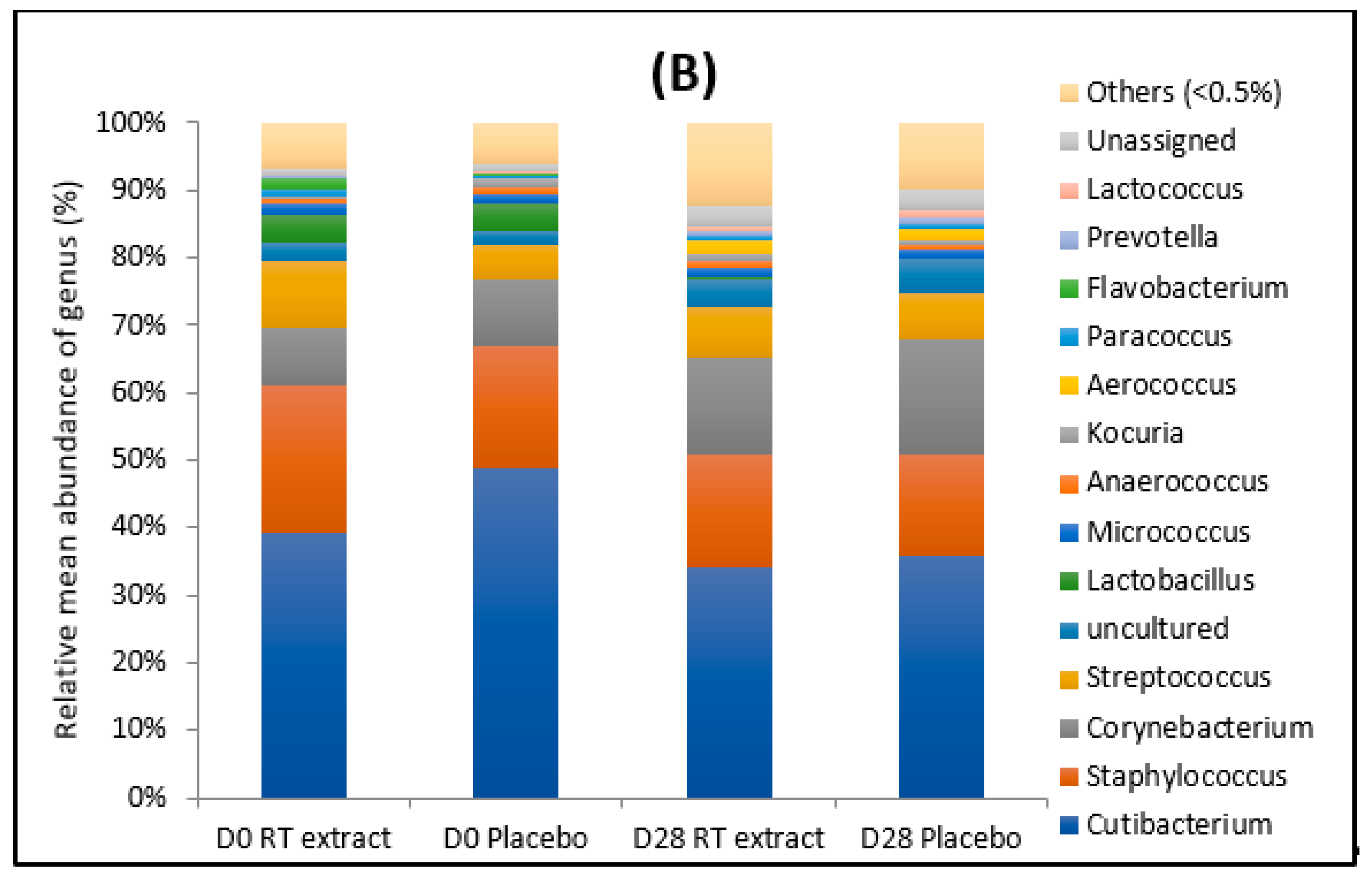
As shown in Figure 1, major phyla were Actinobacteria (average 56%) followed by Firmicutes (average 32%). Cutibacterium was the most abundant genus (average 40%), followed by Staphylococcus (average 18%), Corynebacterium (average 12%), and Streptococcus (average 7%) both at D0 and D28, with no significant differences between placebo and RT extract treatment.
At D0, C. acnes was the major species in term of mean abundance, followed by S. epidermidis. S. hominis was also present with a mean abundance close to that of S. epidermidis.
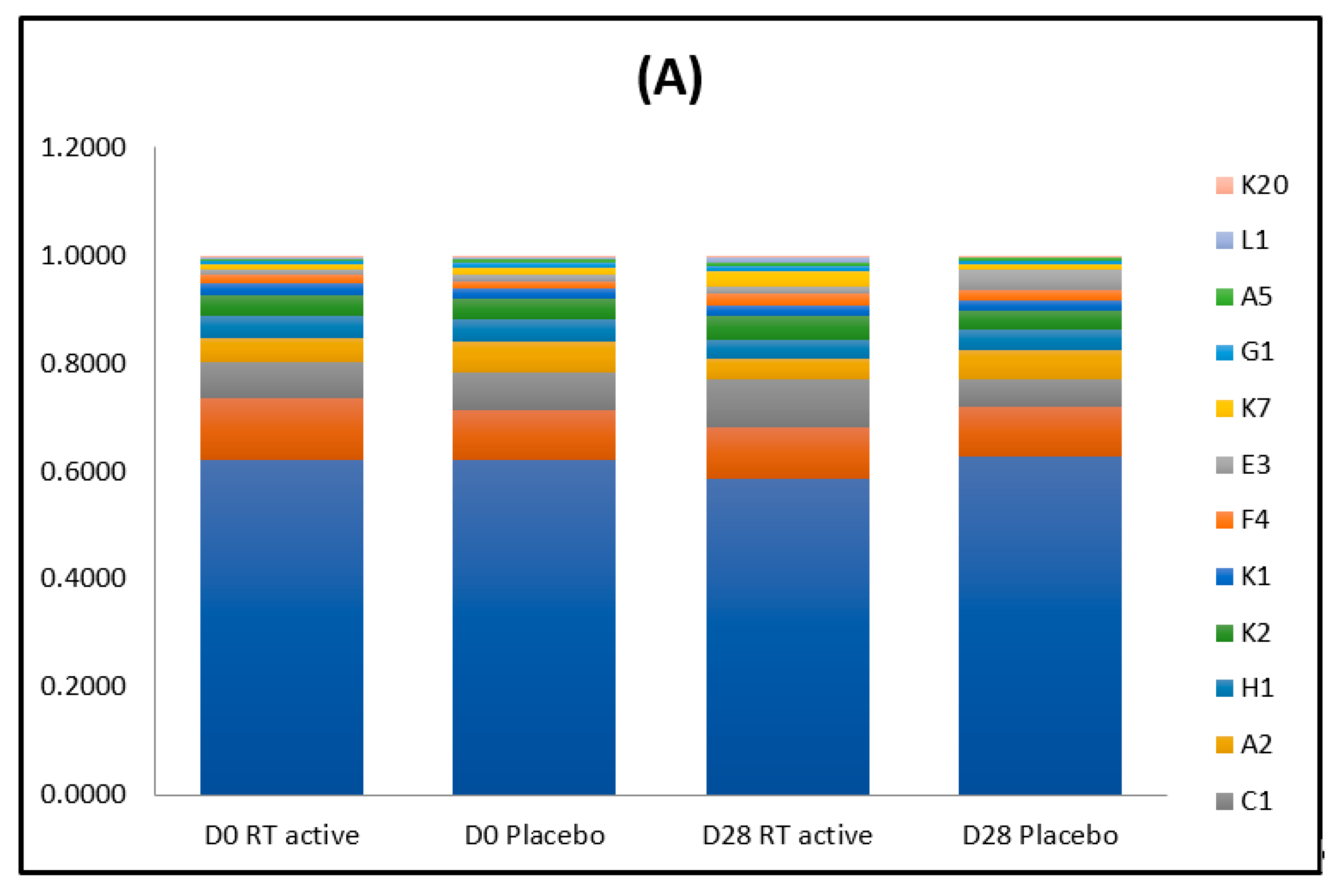
As shown in Figure 2, in most samples and all groups, phylotype IA1 was most represented, followed by phylotypes II, IB, IA2, IC, and III.

3.1.2. After the Treatment

When the diversity index was compared between treatments using the non-parametric Wilcoxon test, diversity expressed by the Shannon index appeared to be statistically higher (p < 0.05) after 28 days of treatment with RT active ingredient when compared with D0.
A significant increase in Corynebacterium genus (p < 0.008) was noted only for the placebo treatment. Mean raw abundance values of C. granulosum were decreased after the RT active ingredient treatment when compared with D0 and placebo. This was not noted in the placebo group.
A decrease in abundance (approximately 4%) of phylotype IA1 was noted when samples treated with RT active ingredient were compared to samples treated with placebo or to samples at baseline (p < 0.05). Contrasting this, a significant increase of phylotype II was observed (p < 0.02). Relative abundance of phylotype III also increased with RT treatment. When diversity indices were compared between sample groups using the non-parametric Wilcoxon test, diversity expressed by the Simpson index appeared to be statistically higher (p < 0.05) after 28 days of treatment with RT when compared with D0.

3.2. Clinical Evaluation

The RT active ingredient induced a significant decrease in blackheads, papules, general non-inflammatory lesions, and global inflammatory lesions (Table 2). No changes were observed with the placebo treatment. Significant differences were noted between treatments concerning the number of papules, blackheads, and inflammatory and non-inflammatory lesions.
The RT active ingredient significantly reduced, by 41%, the skin sebum at D28 compared to D0. This improvement was significantly different (p < 0.05) from the placebo treatment.
Results also showed that the RT active ingredient significantly reduced skin redness at D28, with this decrease being significantly different from placebo at D28 (21% versus 4% for the placebo; p < 0.05).
A total of 83% of subjects noted that RT extract treatment reduced imperfections (36% after the placebo treatment). A total of 77% of subjects observed that it eliminated sebum excess (53% for the placebo treatment) and 71% of subjects observed that it tightened the pores (30% for the placebo).

4. Discussion

The aim of this exploratory study was to determine the microbiota on the surface of skin with acne lesions and to evaluate changes in the microbiota profile and C. acnes phylotype biodiversity after 28 days of twice daily application of RT extract or placebo. This study reveals that prior to the application of RT extract, the skin surface microbiota in acne subjects was dominated by Actinobacteria followed by Firmicutes and Proteobacteria. At the genus level, Cutibacterium (Actinobacteria) was the most abundant genus followed by Staphylococcus (Firmicutes) and Corynebacterium (Actinobacteria), with no changes after RT active ingredient treatment. Comparable repartition has also been reported for patients suffering from acne [2]. At D28, Corynebacterium significantly increased only after placebo treatment, as this genus was identified as dominant mediators of skin immunity and inflammation, and recognized as pathogens, particularly among immunocompromised hosts [32]. The skin of patients with atopic dermatitis was also characterized by high relative abundance of Corynebacterium [33]. This result suggests that RT extract has a beneficial effect on this genus. Of the five bacterial species evaluated, C. acnes was the major species in terms of mean abundance, followed by Staphylococcus epidermidis. Both these taxa were detected in all tested samples at D0. Staphylococcus hominis, a skin commensal bacteria and opportunistic pathogen, was detected in almost all samples (84%). Its abundance was not affected by the treatment.
It is worth mentioning that excess C. acnes colonization might not be an important factor in acne pathogenesis, with some studies reporting little difference in the comparative amount of C. acnes in individuals with and without acne [4]. C. acnes acts as a pathogen or a commensal bacterium according to the strain and balance among the metagenomic elements [3]. In the era of genomic research, different DNA-based methods used for bacterial typing identify various C. acnes phylogenic groups. However, different nomenclatures have been described. Scholz et al. [31] proposed a SLST scheme comparable to that of existing MLST schemes, but unlike these schemes, it can be used for mapping of multiple strains in a complex microbial environment. Three main phylotypes (I, II, and III) led to the recent proposal of their reclassification in subspecies: phylotype I as C. acnes subsp. acnes, type II as C. acnes subsp. defendens, and type III as C. acnes subsp. elongatum [34]. In this study, we used a denomination of phylotypes bases on the initial phylotyping (IA1, IA2, IB, II, and III) to facilitate the comprehension. These different C. acnes phylotypes have different inflammatory expressions of various putative virulence factors, explaining their distinct involvement in acne disease [35]. These factors include lipase and heat shock proteins. It also appears that phylotype IA1 strains isolated from patients with acne produce significantly higher levels of porphyrins than healthy skin associated with phylotype II strains [36]. A Japanese study using the SLST method, as in our study, showed that phylotype IA1 was predominant in each acne severity category (with 60%, 57.1%, and 63.3% of strains in the severe, moderate, and mild acne groups, respectively) [37]. Phylotype II is described as less abundant on acne skin than healthy skin [3]. Additionally, C. acnes isolates belonging to phylotype III were not found in acne lesions, but comprised approximately 20% of isolates from healthy skin [38]. However, the data are controversial. In fact, Romano-Bertrand et al. [39] suggested that phylotype III may be involved in C. acnes deep tissue infections along with phylotype IB.
At D0, phylotype IA1 was the most represented, followed by phylotypes II, IB, IA2, IC, and III. High relative abundance of phylotype IA1 is also observed when skin from patients with severe and mild acne was compared [40]. A higher incidence of phylotype IA1 in acne occurrence or severity may be due to its higher capacity to adhere to skin and form biofilms. Another hypothesis is that phylotype IA1 and II strains show different hyaluronate lyase activity, wherein phylotype IA1 is less active and partial degradation of hyaluronic acid may produce various oligosaccharides that could be related to the inflammatory process induced by acne. After 28 days of RT extract treatment, phylotypes repartition was modified with a decrease in abundance (approximately 4%) of phylotype IA1 and an increase in phylotype II and III. The changes were not observed after placebo treatment (Figure 2). Phylotype III, which is characterized by high pro-inflammatory potential in skin explants, does not seem to be related to acne [41]. Phylotype II participates in the skin microbiome and is not relevant in the pathology of acne [34]. We hypothesize a different antibacterial sensitivity of the diverse phylotypes after RT extract treatment.
Besides C. acnes, mean raw abundance values of C. granulosum were also decreased only after the RT active ingredient treatment. C. granulosum, reported as present on skin, is also possibly more aggressive than C. acnes [2]. However, the data are controversial. In a metagenomic analysis of acne patients, Barnard et al. [13] found higher relative abundance of C. granulosum in healthy individuals compared to individuals with acne, suggesting a commensal role for this species. In contrast, early culture-based studies reported that C. granulosum is more prevalent in comedones and pustules compared to uninvolved follicles of acne patients. Moreover, C. granulosum was reported to possess greater lipase activity compared to C. acnes [15]. However, the limited genome data currently available for this understudied bacterium indicates a limited repertoire of virulence-associated genes, with notable absences of sialidases and hyaluronate lyases, thought to contribute to C. acnes-host interactions during disease. Thus, further investigations are required to determine the potential health contribution of this Cutibacterium species [2].
Concerning the clinical study, more than 75% of subjects noted that RT extract treatment reduced imperfections and eliminated sebum excess and 71% of subjects reported pore tightening effects. These perceptions corroborate the results expressed using VISIA-CR and the sebumeter. In fact, after 28 days of treatment, the skin is significantly less red and the skin sebum reduces significantly.
Having shown that the RT active ingredient induces modifications in the skin microbiota and has positive repercussions on skin health, it is necessary to understand the link between the chemical characterization of RT extract and the skin microbiota. Generally speaking, it has been noted that R. tomentosa has a strong antimicrobial and antifungal activity [24]. In fact, the fruit and leaf extract of R. tomentosa exhibited such activities against Bacillus cereus and Candida albicans. The leaves, stem, twig, and fruit of the plant also showed activity against Salmonella typhi and C. acnes.
Phytochemical screening of the RT extract used in this study as presented in Table 1 revealed that the major constituents in the extract were flavonoids, stilbene represented by piceatannol, phenolic acids such as gallic acid and ellagic acid, acylphloroglucinol such as rhodomyrtone, and quinic acid. These kinds of molecules have been described previously in Rhodomyrtus tomentosa [42,43]. Previous articles have mentioned that these molecules have antimicrobial properties and more particularly against Cutibacterium acnes and Staphylococci species, which are two predominant bacterial groups in the case of acne, as shown in our study. More precisely, Abu-Qatouseh et al. [44] noted that polyphenolic extracts of medicinal herbs have anti-Cutibacterium acnes and anti-inflammatory effects. Wang et al. [45] reported that flavonoids extracted from many plants have antibacterial activity against Cutibacterium strains, and showed that quercetin and isoquercetin, well-known flavonoids, have antibacterial efficacy towards C. acnes and Staphyloccoci species (S. aureus and S. epidermidis). Phenolic acids such as gallic acid also have antibacterial properties against pathogenic bacteria like S. aureus, and ellagic acid, another phenolic acid, inhibits C. acnes biofilm formation [46]. According to Docherty [47], piceatannol, a molecule belonging to stilbene family, has antimicrobial properties against C. acnes, where the IC50 is 123 mg·L−1 and IC100 234 mg·L−1after 24 h. The IC50 and IC100 are the concentrations that reduce bacterial growth by 50% and 100%, respectively. More recently, Zu et al. [48] have reported that piceatannol inhibits C. acnes-induced human keratinocyte proliferation and migration by activating the antioxidant Nrf2 pathway and inhibiting the inflammatory NF-κB pathway, suggesting the potential of piceatannol to treat acne vulgaris. Thus, the presence of the piceatannol in our extract may help to fight against the pathogenic bacteria C. acnes. Saising and Voravuthikunchai [49] reported that rhodomyrtone contained in Rhodomyrtus tomentosa had an antibacterial activity against C. acnes with an MIC90 value of 0.5 mg·L−1. The formulation tested in this study contains 2% of Rhodomyrtus tomentosa extract and more particularly 0.1 mg·L−1 of rhodomyrtone, which may contribute to the efficacy against the pathogenic bacteria C. acnes. Our RT extract also contains acid compounds, such as malic acid, citric acid, shikimic acid and quinic acid, that are interesting for acne treatment, because of their chemical exfoliating properties, useful for fighting against excessive keratinization in acne pathology [50]. Moreover, Amrutha et al. [28] mentioned that organic acids such as citric acid have antibacterial properties and can inhibit biofilm formation. Finally, RT extract contains sugars, mainly monosaccharides (glucose and fructose) as described by Lai et al. [25], and also polysaccharides such as xylan (polymer of xylose), arabinogalactan or arabinogalactan-protein, and pectin. A synergistic effect of all these present molecules could explain the efficacy of RT extract against pathogenic bacteria such as C. acnes which is involved in the acne pathology. Finally, Kusuma et al. [51] showed that the ethanolic extract of the fruit of Rhodomyrtus tomentosa, containing flavonoids, triterpenoids and carbohydrates, has antimicrobial properties against C. acnes.
Further phytochemical analysis of the active compounds from R. tomentosa should be conducted given their diverse and extensive traditional uses and potential therapeutic applications. It could be interesting to check the presence of ellagitannins, as described by Lai et al. [25], because of the astringent activity of these kind of molecules and so their potential efficacy in the treatment of acne.

5. Conclusions

This study opens up new areas of research into innovative alternative treatments for mild acne using botanical therapy. Using 16S rRNA profiling and a single-locus sequence typing (SLST) scheme for C. acnes, our data confirm the link between the presence of some phylotypes and acne. We also showed that RT active ingredient modified phylotypes repartition with a decrease in abundance of phylotype IA1 and an increase in phylotype II and III. Thus, we hypothesize a different antibacterial sensitivity of the diverse phylotypes after RT extract treatment. This treatment significantly increased the Shannon index as both the richness and the evenness of the community increased. These modifications were associated with a decrease in acne lesions. It is known that RT has various beneficial health effects including antioxidant, antibacterial, antifungal, anti-inflammatory, and anticancer activities. This study showed that RT extract can also be applied for treatment of skin diseases such as acne. However, this study has a limitation linked to the lack of healthy subjects, in order to compare the characterization of healthy skin microbiota versus microbiota of acne patients. Moreover, our results may sometimes be compared to those of investigations which have not always used the same sequencing methodologies. However, to our knowledge, there is no standardization in the sequencing methodology, which makes comparison between studies difficult. To conclude, various and novel treatments focusing on C. acnes acne-associated phylotypes are worthy of further investigation for acne management. The next steps will be: (1) to conduct further phytochemical studies of the active compounds from R. tomentosa; (2) to evaluate whether RT extract is able to modify C. acnes expression virulence factors; and (3) to evaluate RT extract on Candida and Malassezia sp. In fact, as suggested by studies, yeast sp. can intensify acne, eczema, psoriasis, and atopic dermatitis, and the lipase gene might be one of the virulence factors of Candida and Malassezia yeasts in the causation of acne.

Author Contributions

E.F. and J.-Y.B. conceived the study; E.R. and M.C. worked on plant extract; E.G. and S.G. performed the experiments and analyzed the data; I.M. was involved in coordination of data collection and collected data. S.G. was involved in statistical analysis. G.S. managed data collection. E.F. and E.R. prepared the manuscript. All authors critically revised the manuscript. All authors have read and agreed to the published version of the manuscript.

Funding

This research received no external funding.

Acknowledgments

We thank Kevin Kodo for assisting with the active ingredient.

Conflicts of Interest

The authors have declared no conflicting of interest.

References

  1. Rocha, M.A.; Bagatin, E. Skin barrier and microbiome in acne. Arch. Dermat. Res. 2017, 310, 181–185. [Google Scholar] [CrossRef] [PubMed]
  2. Lee, Y.B.; Byun, E.J.; Kim, H.S. Potential role of the microbiome in acne: A comprehensive review. J. Clin. Med. 2019, 8, 987. [Google Scholar] [CrossRef] [PubMed] [Green Version]
  3. Dagnelie, M.A.; Corvec, S.; Saint-Jean, M.; Bourdès, V.; Nguyen, J.M.; Khammari, A.; Dréno, B. Decrease in Diversity of Propionibacterium acnes Phylotypes in Patients with Severe Acne on the Back. Acta Dermat. Vener. 2018, 7, 262–267. [Google Scholar] [CrossRef] [PubMed] [Green Version]
  4. Fitz-Gibbon, S.; Tomida, B.H.; Chiu, L.; Nguyen, C.; Du, M.; Liu, D.; Elashoff, M.C.; Erfe, A.; Loncaric, J.; Kim, L.; et al. Propionibacterium acnes strain populations in the human skin microbiome associated with acne. J. Investig. Dermatol. 2013, 133, 2152–2160. [Google Scholar] [CrossRef] [PubMed] [Green Version]
  5. Christensen, G.J.; Bruggemann, H. Bacterial skin commensals and their role as host guardians. Benef. Microbes. 2014, 5, 201–215. [Google Scholar] [CrossRef] [PubMed]
  6. Szabo, K.; Erdei, L.; Bolla, B.S.; Tax, G.; Biro, T.; Kemeny, L. Factors shaping the composition of the cutaneous microbiota. Br. J. Dermat. 2017, 176, 344–351. [Google Scholar] [CrossRef] [Green Version]
  7. Xu, H.; Li, H. Acne, the Skin Microbiome, and Antibiotic Treatment. Am. J. Clin. Dermat. 2019, 20, 335–344. [Google Scholar] [CrossRef]
  8. Lomholt, H.B.; Scholz, C.F.P.; Brüggemann, H.; Tettelin, H.; Kilian, M. A comparative study of Cutibacterium (Propionibacterium) acnes clones from acne patients and healthy controls. Anaerobe 2017, 47, 57–63. [Google Scholar] [CrossRef]
  9. McDowell, A.; Perry, A.L.; Lambert, P.A.; Patrick, S. A new phylogenetic group of Propionibacterium acnes. J. Med. Microbiol. 2008, 57, 218–524. [Google Scholar] [CrossRef] [Green Version]
  10. Yu, Y.; Champer, J.; Garbán, H.; Kim, J. Typing of Propionibacterium acnes: A review of methods and comparative analysis. Br. J. Dermatol. 2015, 172, 1204–1209. [Google Scholar] [CrossRef]
  11. McLaughlin, J.; Watterson, S.; Layton, A.M.; Bjourson, A.J.; Barnard, E.; McDowell, A. Propionibacterium acnes and Acne Vulgaris: New Insights from the Integration of Population Genetic, Multi-Omic, Biochemical and Host-Microbe Studies. Microorganisms 2019, 13, 7. [Google Scholar] [CrossRef] [PubMed] [Green Version]
  12. Leyden, J.J. New understandings of the pathogenesis of acne. J. Am. Acad. Dermatol. 1995, 32, 15–25. [Google Scholar] [CrossRef]
  13. Barnard, E.; Shi, B.; Kang, D.; Craft, N.; Li, H. The balance of metagenomic elements shapes the skin microbiome in acne and health. Sci. Report. 2016, 6. [Google Scholar] [CrossRef] [PubMed] [Green Version]
  14. Gehse, M.; Hoffler, U.; Gloor, M.; Pulverer, G. Propionibacteria in patients with acne vulgaris and in healthy persons. Arch. Dermat. Res. 1983, 275, 100–104. [Google Scholar] [CrossRef] [PubMed]
  15. Whiteside, J.A.; Voss, J.G. Incidence and lipolytic activity of Propionibacterium acnes (Corynebacterium acnes group I) and P. granulosum (C. acnes group II) in acne and in normal skin. J. Investig. Dermatol. 1973, 60, 94–97. [Google Scholar] [CrossRef] [Green Version]
  16. Rajiv, P.; Nitesh, K.; Raj, K.; Hemant, G. Staphylococcus epidermidis in Human Skin Microbiome associated with Acne: A Cause of Disease or Defence? Res. J. Biotech. 2013, 8, 78–82. [Google Scholar]
  17. Wang, Y.; Kuo, S.; Shu, M.; Yu, J.; Huang, S.; Dai, A.; Two, A.; Gallo, R.L.; Huang, C.M. Staphylococcus epidermidis in the human skin microbiome mediates fermentation to inhibit the growth of Propionibacterium acnes: Implications of probiotics in acne vulgaris. Appl. Microbiol. Biotechnol. 2014, 98, 411–424. [Google Scholar] [CrossRef] [Green Version]
  18. Christensen, G.J.; Scholz, C.F.; Enghild, J.; Rohde, H.; Kilian, M.; Thurmer, A.; Brzuszkiewicz, E.; Lomholt, H.B. Antagonism between Staphylococcus epidermidis and Propionibacterium acnes and its genomic basis. BMC Med. Genom. 2016, 17, 152. [Google Scholar] [CrossRef] [Green Version]
  19. Xia, X.; Li, Z.; Liu, K.; Wu, Y.; Jiang, D.; Lai, Y. Staphylococcal LTA-Induced miR-143 Inhibits Propionibacterium acnes-Mediated Inflammatory Response in Skin. J. Investig. Dermatol. 2016, 136, 621–630. [Google Scholar] [CrossRef] [Green Version]
  20. Fisk, W.A.; Lev-Tov, H.A.; Sivamani, R.K. Botanical and phytochemical therapy of acne: A systematic review. Phyto. Res. 2014, 28, 1137–1152. [Google Scholar] [CrossRef]
  21. Pineau, R.M.; Hanson, S.E.; Lyles, J.T.; Quave, C.L. Growth inhibitory activity of callicarpa americana leaf extracts against cutibacterium acnes. Front. Pharm. 2019, 15, 1206. [Google Scholar] [CrossRef] [PubMed] [Green Version]
  22. Dursun, R.; Daye, M.; Durmaz, K. Acne and rosacea: What’s new for treatment? Dermatol. Ther. 2019, 32, e13020. [Google Scholar] [CrossRef] [PubMed]
  23. Humphrey, S. Antibiotic resistance in acne treatment. Skin Ther. Lett. 2012, 9, 1–3. [Google Scholar]
  24. Vo, T.S.; Ngo, D.H. The Health Beneficial Properties of Rhodomyrtus tomentosa as Potential Functional Food. Biomelecules 2019, 9, 76. [Google Scholar] [CrossRef] [Green Version]
  25. Lai, T.N.H.; André, C.; Rogez, H.; Mignolet, E.; Nguyen, T.B.T.; Larondelle, Y. Nutritional composition and antioxidant properties of the sim fruit (Rhodomyrtus tomentosa). Food Chem. 2015, 168, 410–416. [Google Scholar] [CrossRef] [PubMed]
  26. Zhao, G.; Zhang, R.; Liu, L.; Deng, Y.; Wei, Z.; Zhang, Y.; Ma, Y.; Zhang, M. Different thermal drying methods affect the phenolic profiles, their bioaccessibility and antioxidant activity in Rhodomyrtus tomentosa. Food Sci. Technol. 2017, 79, 260–266. [Google Scholar] [CrossRef]
  27. Kukreja, A.; Wadhwa, N.; Tiwari, A. Therapeutic Role of Resveratrol and Piceatannol in Disease Prevention. J. Blood Dis. Transfus. 2014, 5, 240. [Google Scholar] [CrossRef] [Green Version]
  28. Amrutha, B.; Sundar, K.; Shetty, P.H. Effect of organic acids on biofilm formation and quorum signaling of pathogens from fresh fruits and vegetables. Microb. Pathog. 2017, 111, 156–162. [Google Scholar] [CrossRef]
  29. Wunnoo, S.; Saising, J.; Voravuthikunchai, S.P. Rhodomyrtone inhibits lipase production, biofilm formation, and disorganizes established biofilm in Propionibacterium acnes. Anaerobe 2017, 43, 61–68. [Google Scholar] [CrossRef]
  30. Dreno, B.; Poli, F.; Pawin, H.; Beylot, C.; Faure, M.; Chivot, M.; Auffret, N.; Moyse, D.; Ballanger, F.; Revuz, J. Development and evaluation of a Global Acne Severity Scale (GEA Scale) suitable for France and Europe. J. Eur. Acad. Dermatol. Venereol. 2011, 25, 43–48. [Google Scholar] [CrossRef]
  31. Scholtz, C.; Jensen, A.; Lomholt, H.B.; Brüggemann, H.; Kilian, M. A Novel High-Resolution Single Locus Sequence Typing Scheme for Mixed Populations of Propionibacterium acnes in vivo. PLoS ONE 2014, 9, e104199. [Google Scholar]
  32. Ridaura, V.K.; Bouladoux, N.; Claesen, J.; Chen, Y.E.; Byrd, A.L.; Constantinides, M.G.; Merrill, E.D.; Tamoutounour, S.; Fischbach, M.A. Contextual control of skin immunity and inflammation by Corynebacterium. J. Exp. Med. 2018, 215, 785. [Google Scholar] [CrossRef] [Green Version]
  33. Moosbrugger-Martinz, V.; Hackl, H.; Gruber, R.; Pilecki, M.; Knabl, L.; Orth-Höller, D.; Dubrac, S. First evidences of distinguishable bacterial and fungal dysbiosis in the skin of patients with atopic dermatitis of netherton Syndrome. J. Invest. Dermatol. 2020, S0022-202X, 31668. [Google Scholar] [CrossRef] [PubMed]
  34. McDowell, A.; Barnard, E.; Liu, J.; Li, H.; Patrick, S. Corrigendum: Proposal to reclassify Propionibacterium acnes type I as Propionibacterium acnes subsp. acnes subsp. nov. and Propionibacterium acnes type II as Propionibacterium acnes subsp. defendens subsp. nov. Int. J. Syst. Evol. Microbiol. 2017, 67, 4880. [Google Scholar] [CrossRef] [PubMed] [Green Version]
  35. Omer, H.; McDowell, A.; Alexeyev, O.A. Understanding the role of Propionibacterium acnes in acne vulgaris: The critical importance of skin sampling methodologies. Clin. Dermatol. 2017, 35, 118–129. [Google Scholar] [CrossRef] [PubMed]
  36. Johnson, T.; Kang, D.; Barnard, E.; Li, H. Strain-level differences in porphyrin production and regulation in Propionibacterium acnes elucidate disease associations. mSphere 2016, 1, 1–12. [Google Scholar] [CrossRef] [Green Version]
  37. Nakase, K.; Hayashi, N.; Akiyama, Y.; Aoki, S.; Noguchi, N. Antimicrobial susceptibility and phylogenetic analysis of Propionibacterium acnes isolated from acne patients in Japan between 2013 and 2015. J. Dermat. 2017, 44, 1248–1254. [Google Scholar] [CrossRef]
  38. McDowell, A.; Nagy, I.; Magyari, M.; Barnard, E.; Patrick, S. The opportunistic pathogen Propionibacterium acnes: Insights into typing, human disease, clonal diversification and CAMP factor evolution. PLoS ONE 2013, 8, e70897. [Google Scholar] [CrossRef] [Green Version]
  39. Romano-Bertrand, S.; Beretta, M.; Jean-Pierre, H. Propionibacterium acnes populations involved in deep pathological samples and their dynamics along the cardiac surgical pathway. Eur. J. Clin. Microbiol. Infect. Dis. 2015, 34, 287–301. [Google Scholar] [CrossRef]
  40. Paugam, C.; Corvec, S.; Saint Jean, M.; Le Moigne, M.; Khammari, A.; Boisrobert, A.; Nguyen, J.M.; Gaultier, A.; Dreno, B. Propionibacterium acnes phylotypes and acne severity: An observational prospective study. J. Eur. Acad. Dermatol. Venereol. 2017, 31, e398–e399. [Google Scholar] [CrossRef]
  41. Pécastaings, S.; Roques, C.; Nocera, T.; Peraud, C.; Mengeaud, V.; Khammari, A.; Dreno, B. Characterisation of Cutibacterium acnes phylotypes in acne and in vivo exploratory evaluation of Myrtacine®. J. Eur. Dermatol. Venereol. 2018, 32, 15–23. [Google Scholar] [CrossRef] [PubMed] [Green Version]
  42. Lai, T.; Herent, M.F.; Quetin-Leclercq, J.; Nguyen, T.B.T.; Rogez, H.; Larondelle, Y.; Andre, C.M. Piceatannol, a potent bioactive stilbene, as major phenolic component in Rhodomyrtus tomentosa. Food Chem. 2013, 138, 1421–1430. [Google Scholar] [CrossRef] [PubMed]
  43. Hamid, H.A.; Mutazah, S.S.; Yusoff, M.M. Rhodomyrtus tomentosa: A phytochemical and pharmacological review. Am. J. Pharm. Clin. Res. 2017, 10, 10–16. [Google Scholar] [CrossRef] [Green Version]
  44. Abu-Qatouseh, L.; Mallah, E.; Mansour, K. Evaluation of anti-Propionibacterium acnes and anti-inflammatory effects of polyphenolic extracts of medicinal herbs in Jordan. Biomed. Pharm. J. 2019, 12, 211–217. [Google Scholar] [CrossRef]
  45. Wang, L.; Yang, X.; Qin, P.; Shan, F.; Ren, G. Flavonoid composition, antibacterial and antioxidant properties of tartary buckwheat bran extract. Ind. Crops Prod. 2013, 49, 312–317. [Google Scholar] [CrossRef]
  46. Sivasankar, C.; Maruthupadiyan, S.; Balamurugan, K.; James, P.B.; Krishnan, V.; Pandian, S.K. A combinaison of ellagic acid and tetracycline inhibits biofilm formation and the associated virulence of Propionibacterium acnes in vitro and in vivo. Biofouling 2016, 32, 397–410. [Google Scholar] [CrossRef]
  47. Docherty, J.J.; McEwen, H.A.; Sweet, T.J.; Bailey, E.; Booth, T.D.J. Resveratrol inhibition of Propionibacterium acnes. J. Antimicrob. Chemother. 2007, 59, 1182–1184. [Google Scholar] [CrossRef] [Green Version]
  48. Zhu, T.; Fang, F.; Sun, S.; Yang, X.; Zhang, X.; Yu, L.; Yang, L. Piceatannol Inhibits P. acnes-Induced Keratinocyte Proliferation and Migration by Downregulating Oxidative Stress and the Inflammatory Response. Inflammation 2020, 43, 347–357. [Google Scholar] [CrossRef]
  49. Saising, J.; Voravuthikunchai, S.P. Anti Propionibacterium acnes activity of rhodomyrtone, an effective compound from Rhodomyrtus tomentosa (Aiton) Hassk. leaves. Anaerobe 2012, 18, 400–404. [Google Scholar] [CrossRef]
  50. Kapuscinska, A.; Nowak, I. Use of organic acids in acne and skin discolorations therapy. Postepy Higieny I Medycyny Doswiadczalnej 2015, 69, 374–383. [Google Scholar] [CrossRef]
  51. Kusuma, I.W.; Ainiyati, N.; Suwinarti, W. Search for biological activities from an invasive shrub species rose myrtle (Rhodomyrtus tomentosa). Nusantara Biosci. 2016, 8, 55–59. [Google Scholar] [CrossRef]
Figure 1. Mean relative phyla (A) and genera (B) abundances for each group of samples. For the sake of clarity, only phyla with mean relative abundance superior than 0.1% and genera with mean relative abundance superior than 0.5% are represented.
Cosmetics 07 00053 g001aCosmetics 07 00053 g001b
Figure 2. (A) C. acnes single-locus sequence typing (SLST) type distribution on placebo at D0 (before treatment) and after 28 days of placebo or RT treatment. (B) C. acnes phylotype distribution on placebo at D0 (before treatment) and after 28 days of placebo or RT treatment.
Cosmetics 07 00053 g002aCosmetics 07 00053 g002b
Table 1. Characterization of Rhodomyrtus tomentosa (RT) extract.
CompoundsConcentration (mg·L−1) in RT Extract
Phenolic Compounds
Polyphenols (catechin)5000
Stilbenes
Piceatannol500
Flavonoids (rutin)250
Phenolic acids
Gallic acid50
Ellagic acid50
Acylphloroglucinol
Rhodomyrtone5
Acid Compounds
Organic acids1000
Malic Acid300
Citric Acid700
Shikimic acid150
Quinic acid1250
Sugars
Monosaccharides
Glucose15,000
Fructose17,500
Polysaccharides3750
Table 2. Inflammatory and non-inflammatory lesion variations between D0 and D28 for the placebo and RT extract groups.
ParametersVariation
Placebo Group
Variation
RT Extract Group
p Value
Placebo Versus RT Extract Group
Blackheads−0.3 ± 1.4−3.5 ± 1.1 **0.01
Papules−0.4 ± 0.9−1.6 ± 0.4 ***0.05
Global NIL−1.9 ± 0.2−4.1 ± 1.4 **0.01
GIL−0.6 ± 0.5−2.2 ± 0.6 ***0.05
**: p < 0.01; ***: p < 0.001: versus D0; GNIL: Global non-inflammatory lesions; GIL: Global inflammatory lesions.