Article

A Preliminary Study Evaluating the Potential of a Topical Hydrating Formulation Enriched with Platelet-Rich Plasma for Improving Skin Texture

by
Cristina-Ionela Stănciulescu
1,
Andreea-Roxana Ungureanu
2,*,
Virgil Pătrașcu
1,*,
Cornelia Bejenaru
3,
Marilena-Viorica Hovaneţ
4 and
Gabi Topor
5
1
Department of Dermatology, Faculty of Medicine, University of Medicine and Pharmacy of Craiova, 2 Petru Rareş Street, 200349 Craiova, Romania
2
Department of Pharmacy, Sanador Clinical Hospital, 9 Sevastopol Street, 010991 Bucharest, Romania
3
Department of Pharmaceutical Botany, Faculty of Pharmacy, University of Medicine and Pharmacy of Craiova, 2 Petru Rareş Street, 200349 Craiova, Romania
4
Department of Pharmaceutical Botany and Cell Biology, Faculty of Pharmacy, Carol Davila University of Medicine and Pharmacy, 6 Traian Vuia Street, 020945 Bucharest, Romania
5
Department of Pharmaceutical Sciences, Faculty of Medicine and Pharmacy, “Dunărea de Jos“ University of Galati, 35 Al. I Cuza Street, 800010 Galati, Romania
*
Authors to whom correspondence should be addressed.
Cosmetics 2026, 13(4), 175; https://doi.org/10.3390/cosmetics13040175
Submission received: 2 May 2026 / Revised: 27 June 2026 / Accepted: 6 July 2026 / Published: 8 July 2026
(This article belongs to the Section Cosmetic Dermatology)

Abstract

Intrinsic and extrinsic skin aging alter skin structure and function, while hydrating products and regenerative therapies such as platelet-rich plasma (PRP) support the restoration of skin homeostasis and clinical appearance. This in vivo study aimed to evaluate whether a combination of a hydrating cream and PRP improves skin texture. Three groups of participants were treated over 90 days with hydrating cream, intradermal PRP, and hydrating cream enriched with PRP (HC-PRP). The evaluation was conducted by dermoanalysis at each 30 days with two main indices, moisture and texture, and six complementary indices, oil, complexion, 3D, pigment, phlogosis, and pores. The best texture outcomes were observed in the HC-PRP group, which showed a significant improvement over time and better results than the cream alone (significant after Bonferroni correction). For the moisture index, the hydrating cream alone gave the largest increase, consistent with its primary action, although HC-PRP was also associated with a moderate increase. These exploratory results point to a potential benefit of the HC-PRP combination for skin texture and offer a starting point for future research. Further randomized studies on larger samples are required to validate these observations and to establish the contribution of PRP.

1. Introduction

The skin is a highly specialized organ whose structure and function are tightly linked to its ability to maintain hydration. At the structural level, hydration is essential to the integrity and function of the skin barrier and at the molecular level it influences biochemical processes [1]. The stratum corneum, as the outermost layer of the epidermis acts as the primary barrier against trans-epidermal water loss (TEWL) and extrinsic factors [2]. Its structure (corneocytes embedded in a lipid matrix) enables water retention, preserves elasticity and protects overall skin integrity [3]. The water within the stratum corneum is largely retained by the natural moisturizing factor (NMF) consisting of hygroscopic molecules derived by the breakdown of filaggrin (as pyrrolidone carboxylic acid, urocanic acid, lactic acid, urea, glycerol) [4]. The amount of filaggrin in the skin is a determinant of proper moistness. Mutations in the gene encoding its precursor (pro-filaggrin) are associated with skin disorders characterized by dry skin [5,6]. Additionally, aquaporins, particularly aquaporin-3 (AQP3), are membrane proteins that facilitate the transport of water and glycerol between keratinocytes, contributing to intracellular hydration and maintaining osmotic balance, while intercellular lipids form a barrier that limits water loss [7,8,9]. In the dermis, glycosaminoglycans such as hyaluronic acid play a key role providing turgor and supporting diffusion of nutrients and signaling molecules [10,11].
Over the course of life, the whole body undergoes an aging process. For skin, due to endogenous factors (genetics, metabolism) and exogenous factors (lifestyle, environment exposure), several molecular and structural changes impair the hydration system. There is a decline in lipid synthesis, especially ceramides, leading to disrupted lamellar organization and increased TEWL. Filaggrin expression decreases, reducing NMF content and thus the water-binding capacity of the stratum corneum. Moreover, hyaluronic acid levels diminish and aquaporin expression becomes less efficient, further compromising water transport and retention [12,13,14].
Disruption of these molecular mechanisms, allow easier penetration of pathogens, irritants, and allergens, increasing the risk of infections, inflammation, hypersensitivity and impairing wound healing. Adequate hydration is essential not only for aesthetic appearance, but also for preserving the skin’s defensive and regenerative functions. The World Health Organization (WHO) emphasizes the importance of awareness, early intervention, and access to appropriate dermatological care, considering skin diseases as a public health priority [15].
Skin texture is an important indicator of skin appearance and reflects the microtopography of the skin surface, including roughness, pore visibility, fine lines, and surface uniformity. Changes in skin texture are commonly associated with intrinsic aging, photoaging, and impaired skin barrier function. Advances in non-invasive imaging technologies have enabled objective assessment of skin texture [16,17]. Skin hydration is closely linked to skin texture. Adequate water content within the stratum corneum contributes to a smoother and more uniform skin surface, whereas dehydration and barrier dysfunction increase TEWL, leading to textural irregularities. Indirect assessments of changes in skin texture can be made by measuring hydration [18].
The application of moisturizing products directly targets skin’s hydration deficits through three main mechanisms: occlusion (hydrophobic film that reduces TEWL), humectancy (attract and bind water from the atmosphere and deeper skin layers) and emollience (restore the intercellular matrix and improve the barrier function) [19,20,21].
In recent years, platelet-rich plasma (PRP) has emerged as a promising regenerative approach in dermatology and aesthetic medicine [22]. PRP is an autologous concentration of platelets containing numerous growth factors, including platelet-derived growth factor (PDGF), transforming growth factor-β (TGF-β), vascular endothelial growth factor (VEGF), and epidermal growth factor (EGF). These bioactive molecules stimulate fibroblast proliferation and extracellular matrix remodeling, promoting the synthesis of collagen, elastin, and hyaluronic acid [23,24]. Clinical studies have shown measurable improvements in texture parameters following PRP treatment, suggesting its role in dermal remodeling and surface refinement. These effects are primarily attributed to increased collagen synthesis and fibroblast activation, which enhance skin structural integrity and surface smoothness [25,26].
The topical use of platelet concentrates in cosmetic products remains largely under-investigated, whereas intradermal PRP has been widely studied. This knowledge gap provided the rationale for the current study. Although the high-molecular-weight components of PRP are unlikely to penetrate into the dermis when applied topically, they may exert superficial effects at the level of the stratum corneum, and a contribution to the skin microenvironment has been suggested [27]. Rather than addressing the mechanism of action, the present study focused on whether a topical formulation enriched with PRP produces an improvement in skin texture. Accordingly, the aim of this preliminary study was to determine whether a hydrating cream enriched with PRP (HC-PRP) is more effective than the hydrating cream (HC) or intradermal PRP alone in improving skin texture, assessed as the primary outcome, with additional indices, including the moisture index, assessed as secondary outcomes.

2. Materials and Methods

2.1. Study Design

The present study is a prospective, interventional, three-arm in vivo analysis designed to investigate the comparative efficacy in terms of skin texture for topical hydrating cream, PRP, and their combination. All participants were evaluated by a dermatologist before being included in the study to certificate eligibility.
Eligible participants were self-assigned to one of three treatment groups (non-randomized study): 1. topical hydrating cream therapy, 2. intradermal PRP therapy, and 3. topical combined therapy (HC-PRP). The primary outcome was evaluation of skin texture and the secondary outcomes were evaluation of other parameters characterizing the skin (moisture, sebum level, 3D, complexion, phlogosis).
Participants’ eligibility for the study was determined based on the following inclusion criteria: 1. over 18 years; 2. present signs of skin aging (including fine lines and wrinkles, decreased elasticity, uneven skin texture and mild-to-moderate photoaging, assessed by dermatologist); 3. willingness to provide informed consent; and 4. ability to follow-up assessments; and exclusion criteria: 1. active skin disorders (eczema, acne exacerbations or infections); 2. dermatological treatments in the last six months (home skincare exfoliating products, daily moisturizing routine, use of sunscreen); 3. had previously undergone any facial aesthetic procedures (e.g., laser treatments, microneedling, injectable treatments such as fillers or botulinum toxin); 4. systemic medications affecting skin physiology; and 5. inability to comply with study procedures.
The study was conducted in accordance with the principles of the Declaration of Helsinki and Good Clinical Practice guidelines. This study was approved by the Ethics Committee of the University of Medicine and Pharmacy of Craiova, number 72/2021. Written informed consent, which also covered the publication of the study data, was obtained from all participants prior to inclusion in the study. In this study, adherence to Good Clinical Practice refers to the ethical conduct of the study, including ethics committee approval, written informed consent, and standardized data collection and handling, and not to a randomized controlled design.
The study included 40 participants who self-identify as women, residing in Romania, aged over 18 years (average age for each group was around 45 years, with seven participants over the age of 50). The participants self-selected their treatment group based on preference, particularly considering the invasive nature of intradermal PRP injection. The groups were formed as follows: first group included 20 participants who a received hydrating cream and applied topically on facial skin once daily, in the evening, after washing and cleansing; second group (as active comparator group) included five participants who received PRP as intradermal facial administration; and the third group included 15 participants who received HC-PRP, applied topically to the facial skin once daily, in the evening, after washing and cleansing. Skin assessments were performed at baseline (before treatment) and monthly thereafter for three consecutive months (at 30, 60 and 90 days).

2.2. Study Materials

The hydrating cream was a standardized, hypoallergenic and non-comedogenic cream commercially available on the market in Romania. The main properties are intense hydration and skin relief, effects imprinted by active ingredients such as humectants (thermal water, glycerin, sodium hyaluronate, pentylene glycol, 1,2-hexanediol, betaine, methyl gluceth-20), emollients and occlusive agents (dimethicone, caprylic triglyceride, squalene, triethylhexanoin, isocetyl stearoyl stearate, Cucurbita pepo seed oil). Other components with technical roles are emulsifiers (polyglyceryl-10 myristate, C12-20 alkyl glucoside, C14-22 alcohols), texture enhancers (carbomer, acrylates, dimethyl silane), pH correctors (citric acid, potassium hydroxide, sodium citrate), and others (BHT, fragrance).
Participants performed the skin cleansing procedure using water and mild soap prior to the application of the study products. No additional cleansing was performed immediately before skin measurements. Participants were instructed to avoid using other cosmetic products during the study.
Platelet-rich plasma (PRP) was prepared according to the following protocol: 7 mL of venous blood was collected from each participant into a vacutainer tube and centrifuged using an XC 3000 Spinplus PRF/PRP centrifuge (Jiangdong Instruments, Shanghai, China) at 3000 rpm for 10 min at room temperature. Following centrifugation, the blood separated into two layers: a lower layer consisting of red blood cells (sediment) and an upper layer consisting of platelet-rich plasma (supernatant). The resulting plasma was collected and used for the study.
For the participants in the second group PRP was intradermally administered using a 30G needle. The area was pretreated with anesthetic cream (5 g of lidocaine/prilocaine cream 25 mg/g), applied topically at least 30 min before procedure. Intradermal plasma administration was performed for each participant three times at an interval of 30 days.
For the participants in the third group, the PRP was obtained in the same conditions, each had a monthly collection of 7 mL of venous blood on the day of the evaluation. The plasma was divided equally, immediately after centrifugation into 30 syringes (0.1 mL/syringe) and frozen to −20 °C. The syringes were kept in the freezer. An extemporaneous topical formulation of HC-PRP was prepared by gently mixing 1 g of cream with 0.1 mL of thawed autologous PRP until a visually homogeneous semi-solid dispersion was achieved. The product was prepared in single-use aliquots immediately prior to application to ensure consistency and minimize potential degradation or phase separation. The formulation was applied topically to the face by each participant every evening over a period of three months.
In the topical treatment groups, one fingertip of cream was applied to each of four predefined facial regions (the forehead, the right cheek, the left cheek and the chin) and then distributed evenly across the entire face with a gentle massage until fully absorbed.

2.3. Data Acquisition

The data were collected by scanning the face skin of each participant at the beginning of the study, after 30 days, 60 days and 90 days of therapy using the IntuiSkin dermoanalyzer (Rowe, Bucharest, Romania). The imaging system is based on a 1/3.25 inch Complementary Metal-Oxide Semiconductor (CMOS) sensor, this type of sensor being effectively used in modern imaging systems [28,29,30]. The device was operated under room temperature (15–25 °C) and relative humidity 40–60%, having a maximum resolution of 1600 × 1200 pixels and a magnification factor of up to 50×. Considering potential variations in ambient temperature, participants underwent a 15 min acclimatization period prior to measurements. The sensor was placed on the skin of the participants, at facial level, being able to provide, by its internal integrated software (Multi Skin Analysis System), digital images of the skin and eight indices: moisture, oil, texture, complexion, 3D, pigment, phlogosis and pores. The results were stored for each participant and allowed for the monitoring of changes before and after treatment.
For standardization purposes, measurements were performed exclusively on the right cheek at all study visits. The cheek region was selected as a representative facial site due to its accessibility and relatively homogeneous skin characteristics, thereby minimizing inter-site variability [31].

2.4. Data Analysis

Results are presented as mean values per group and standard deviation.
The texture index was chosen for statistical analysis because it reflects the combined influence of multiple skin characteristics, including hydration, sebum level, and surface irregularities, thus providing a comprehensive indicator of overall skin condition [16]. The other indices were considered exploratory outcomes. These parameters were assessed descriptively without formal statistical analysis.
The analyzed numerical data were not normally distributed; therefore, non-parametric tests were used for comparisons; Mann–Whitney–Wilcoxon test for two groups (comparisons between treatments at the same time of assessment) and Friedman test (for three or more paired groups; comparisons for the same type of treatment between reevaluation times) [32].
Because of the baseline imbalance, between-group comparisons at the follow-up time points were performed using analysis of covariance (ANCOVA) with the baseline value as a covariate; effect sizes were reported as partial eta-squared and as baseline-adjusted mean differences with 95% confidence intervals.
Effect sizes (Kendall’s W for Friedman test, r for Mann–Whitney/Wilcoxon tests, and baseline-adjusted mean differences with 95% confidence intervals for ANCOVA) were calculated and reported to complement p-values. Multiple comparisons were adjusted using the Bonferroni correction.
The statistics were performed using IBM SPSS Statistics software version 26.0. (IBM Corp., Armonk, NY, USA). Microsoft Excel version 2019 (Microsoft Corporation, Redmond, WA, USA) was additionally used for the calculation of the Mann–Whitney–Wilcoxon effect size. The chosen level of significance was set at 0.05 (p < 0.05) for all analyses.

3. Results

3.1. Texture Analysis

Overall skin texture was estimated by texture index of IntuiSkin apparatus, with the results presented in Figure 1. Texture index reflects skin surface roughness; lower values correspond to a smoother and more even skin surface. Regarding the group treated with hydrating cream, the average texture index increased constantly from 2.33 to 3.64 (56.62%). A decrease in the texture index for participants treated with PRP was noticed, falling from a value of 3.39 after the first administration to a value of 2.84 after the last administration (17.62%). This represented a downward trend with fluctuations. The best results were observed for the third group; HC-PRP showed a constant decrease in the texture index. After 30 days, a decrease of 13.64% was registered, and at the end of the study the index decreased to 32.71%.
In the HC group, a highly significant change in the texture index was observed across the four time points (Friedman test, p < 0.0001; Kendall’s W = 0.40, a medium effect), reflecting a progressive increase in the mean texture index and suggesting that the hydrating cream alone did not improve skin texture. In the PRP group, no statistically significant change was observed over time (p = 0.2407; Kendall’s W = 0.28); although the texture index improved slightly, this non-significant result most likely reflects a type II error, given the small sample size (N = 5) and the consequently limited statistical power. In the HC-PRP group, a statistically significant change was observed (p = 0.0002; Kendall’s W = 0.43, a medium effect), corresponding to a progressive decrease in the mean texture index. The two significant changes remained significant after Bonferroni correction (Table 1).
Between-group comparisons (Mann–Whitney–Wilcoxon test, Table 2) showed that, at baseline, the HC and HC-PRP groups differed in their texture index (p = 0.0108), although this difference did not remain significant after Bonferroni correction. At the follow-up time points, the HC-PRP group showed a progressively lower texture index than the HC group, the difference becoming statistically significant after Bonferroni correction at 60 days (p = 0.0324, r = 0.51, a large effect) and at 90 days (p < 0.0001, r = 0.76, a large effect). No other between-group comparison remained significant after correction; in particular, none of the comparisons involving the PRP group reached significance, consistent with its small sample size. The effect sizes for the HC vs. HC-PRP comparison increased over time (r = 0.36, 0.51 and 0.76 at 30, 60 and 90 days, respectively), indicating a widening difference between the two groups.
After adjusting for the baseline value (ANCOVA), the HC-PRP group had a significantly lower texture index than the HC group at all three follow-up time points, with the adjusted mean difference increasing over time (+0.76 at 30 days, +1.29 at 60 days and +1.73 at 90 days; all significant after Bonferroni correction). At 90 days, the PRP group also showed a significantly lower adjusted index than the HC group (+1.01, p = 0.0438 after Bonferroni correction); however, given the small size of this group (n = 5), this result should be regarded as exploratory. Because these differences persisted (and increased) after adjusting for the baseline value, they cannot be attributed to the baseline imbalance or to regression to the mean (Table 3). As the two creams differed only by the addition of PRP, this difference is consistent with a contribution of PRP, although unmeasured differences between the non-randomized groups cannot be excluded.
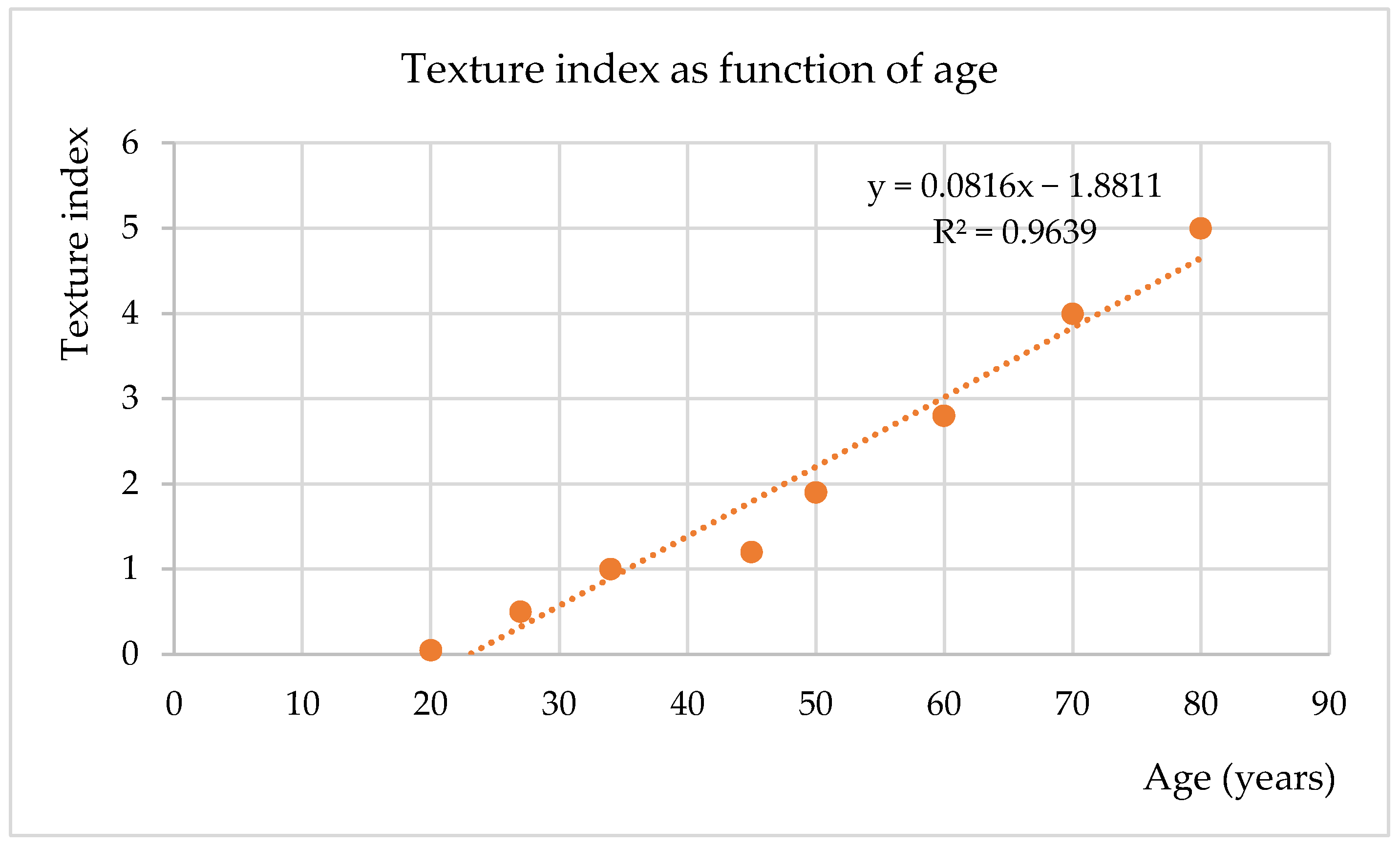
The collected data were used to obtain an exploratory fitted curve describing facial skin texture index as a function of age (Figure 2). The observed texture index values ranged from 0.05 to 5. The fitted line indicated that a texture index of about 4 corresponds to an age of approximately 70 years. At baseline, texture analysis identified values above 4 in many participants under the age of 60.
At baseline, participants treated with PRP and HC-PRP showed a mean texture index that, when projected onto the exploratory age–texture curve (used as an illustrative reference) corresponded to a level typically observed around the age of 60 years, even though their mean chronological age was about 45 years. After 90 days of therapy, the mean texture index of both groups improved, shifting toward levels corresponding to approximately 57 years in the PRP group and 50 years in the HC-PRP group.
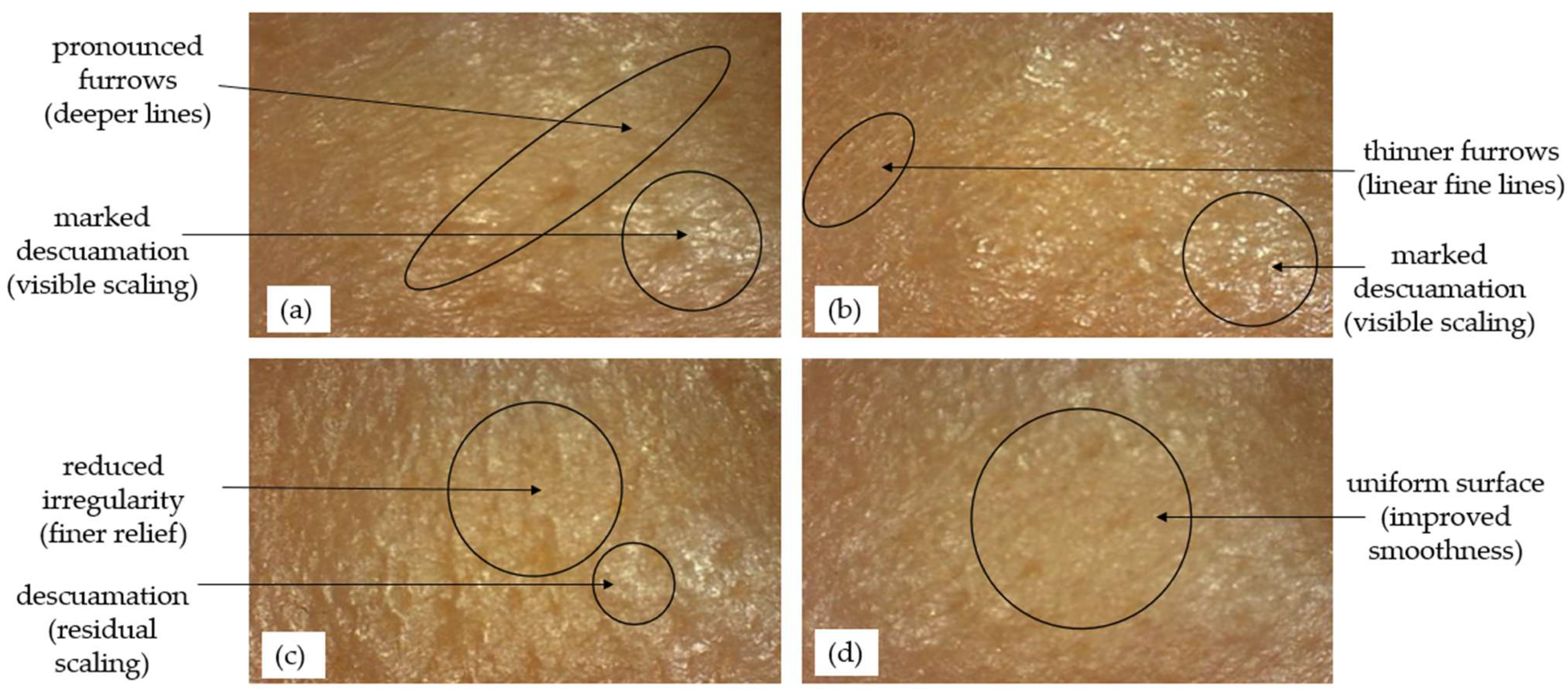
Dynamic dermoanalyzer captures during the study for a participant (46 years) from the HC-PRP group are shown in Figure 3.

3.2. Epidermal Hydration

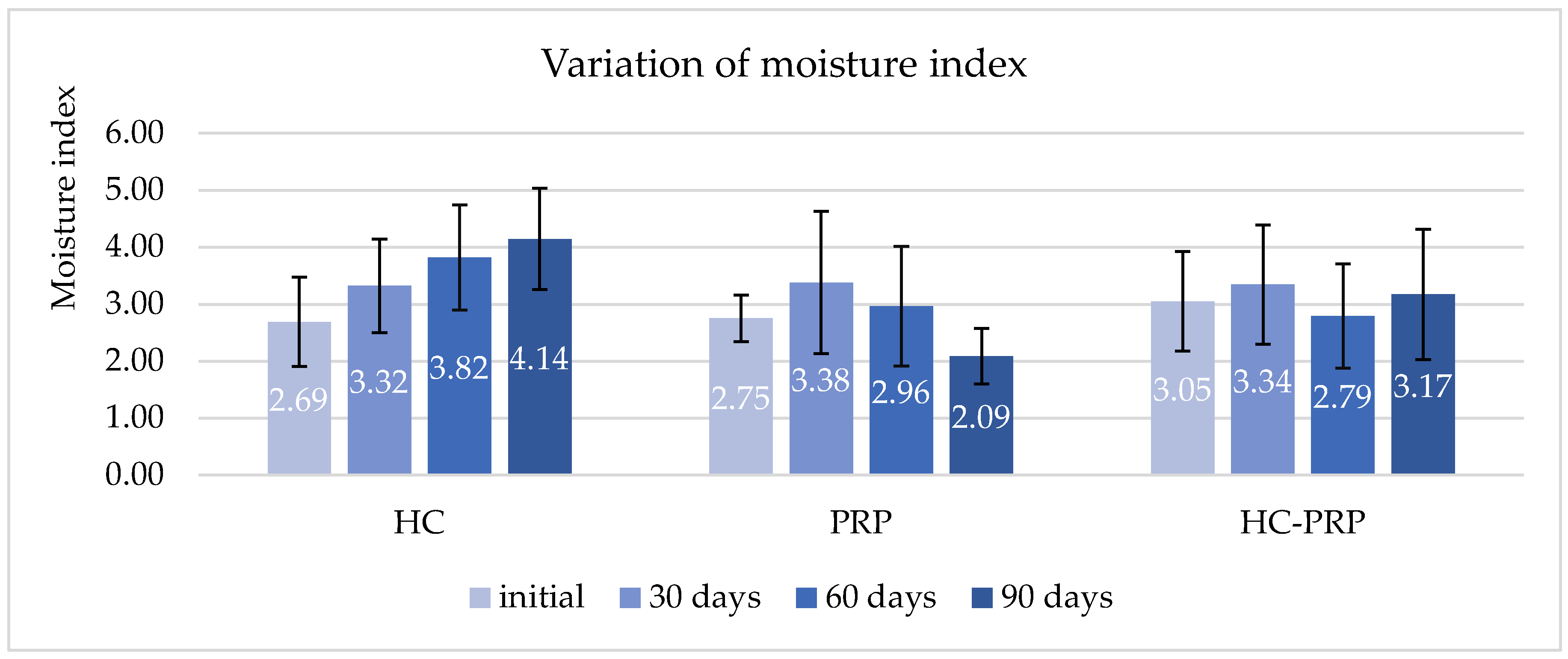
The results of moisture index, which evaluate the epidermal hydration level, are shown in Figure 4. This index indicates the relative water content of the stratum corneum; higher values reflect improved skin hydration. The group of participants treated with the hydrating cream showed the best results: starting with an initial moisture index of 2.69, it continuously increased at each 30 day interval and reached a value of 4.14 after 90 days, representing an overall increase of 53.96%. Although the PRP-treated group showed an initial increase in the moisture index after 30 days, a decrease from baseline was observed by the end of the study. For the third group, treated with HC-PRP, an increase in the moisture index of 4.02% was observed compared to its initial value.
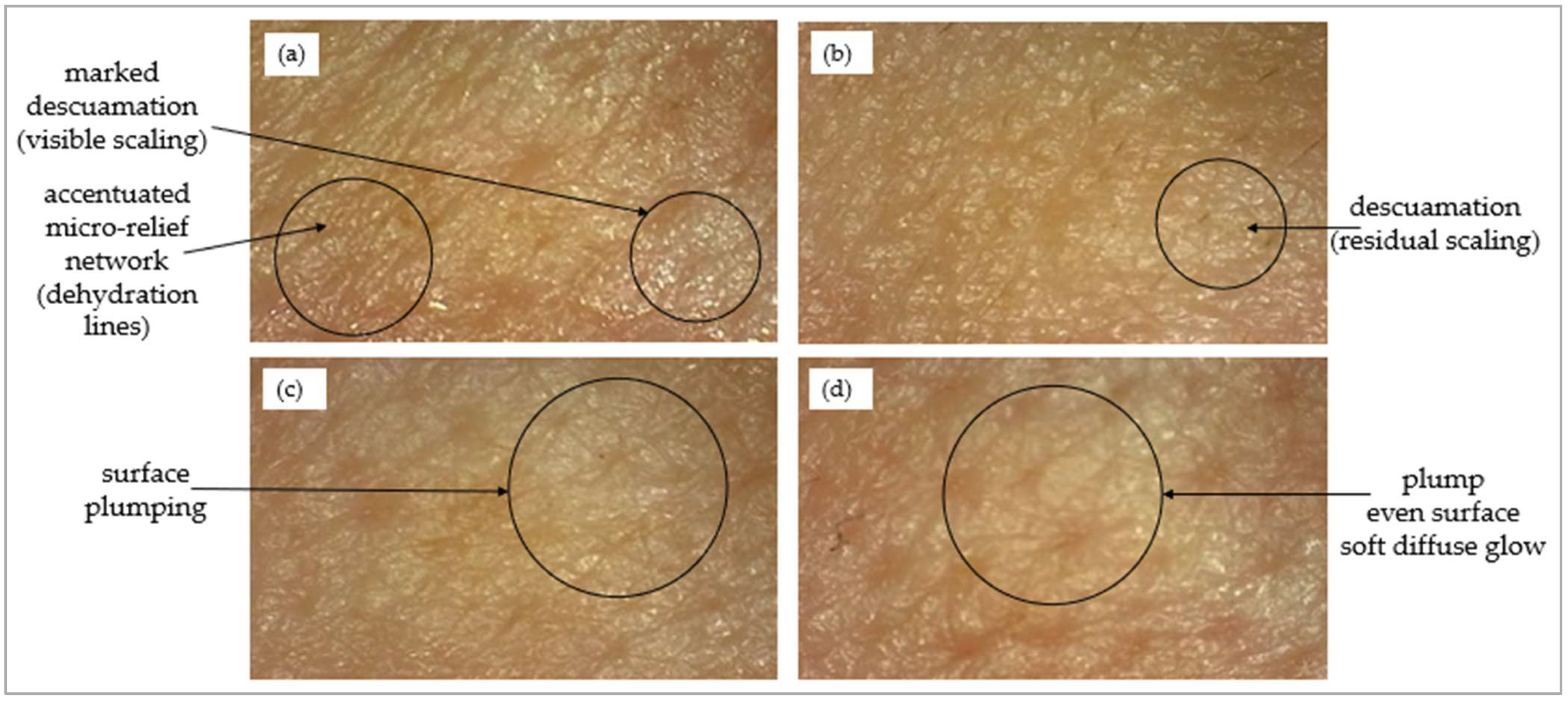
Notable results for moisture were observed for a 47-year-old participant from the first group, for whom the index reached a value above the group mean from the first 60 days. Dermoanalyzer captures are presented in Figure 5.
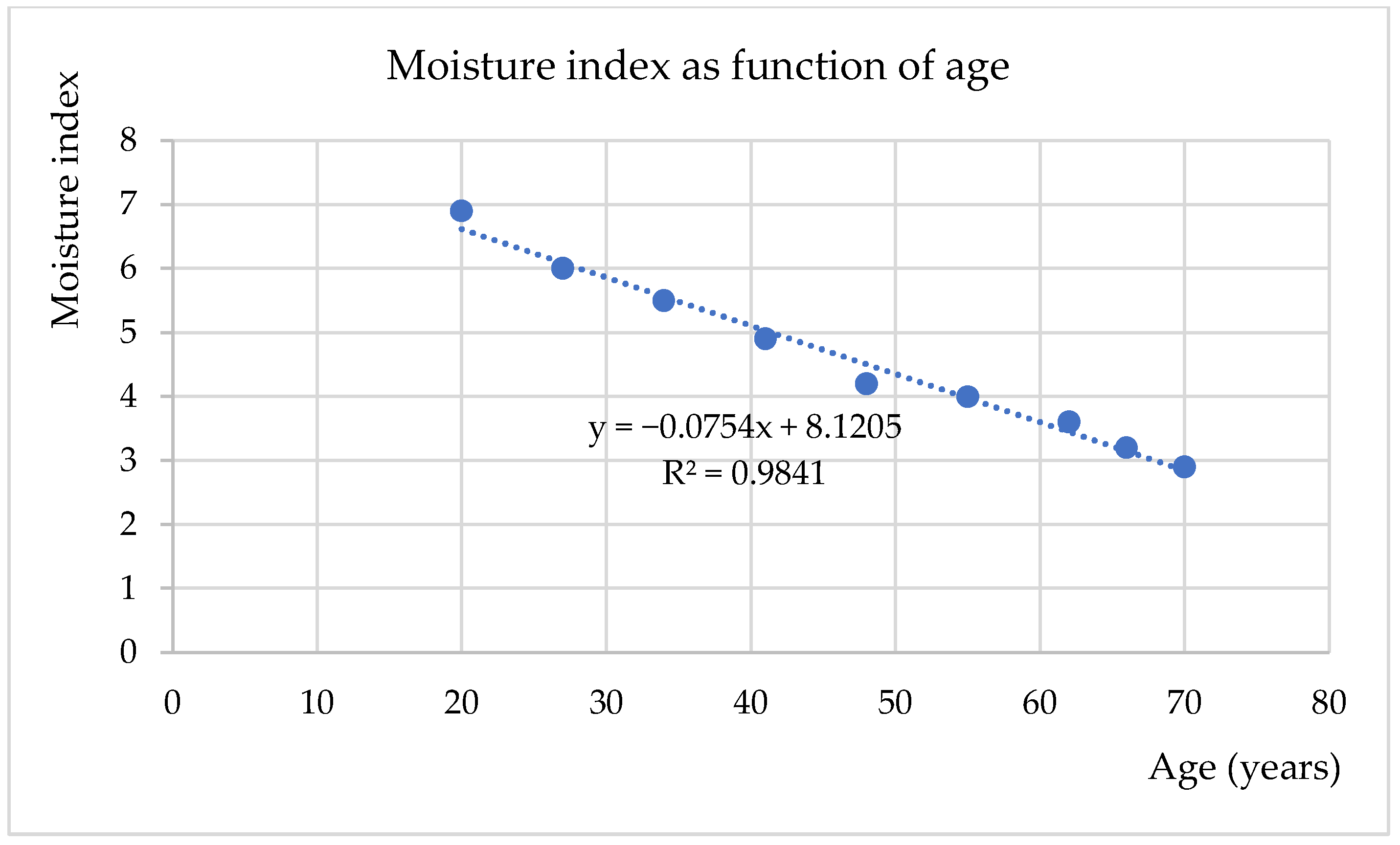
The experimental data were used to generate an exploratory fitted curve describing facial skin hydration (moisture index) as a function of age. This is represented by a downward curve, starting from the value of 6.9 for the age of 20 years, decreasing to the value of 2.9 at the age of 70 years (Figure 6).
When the measured values were projected onto the fitted age–moisture curve (Figure 6)—used here only as an illustrative reference and not as a validated predictor of biological age—the first group (hydrating cream) showed a marked improvement in hydration, shifting from a baseline moisture level typically observed around the age of 70 years toward a level corresponding to approximately 55 years. The third group (HC-PRP) showed a more moderate shift (from a level corresponding to approximately 67 years toward approximately 65 years).

3.3. Complementary Indices

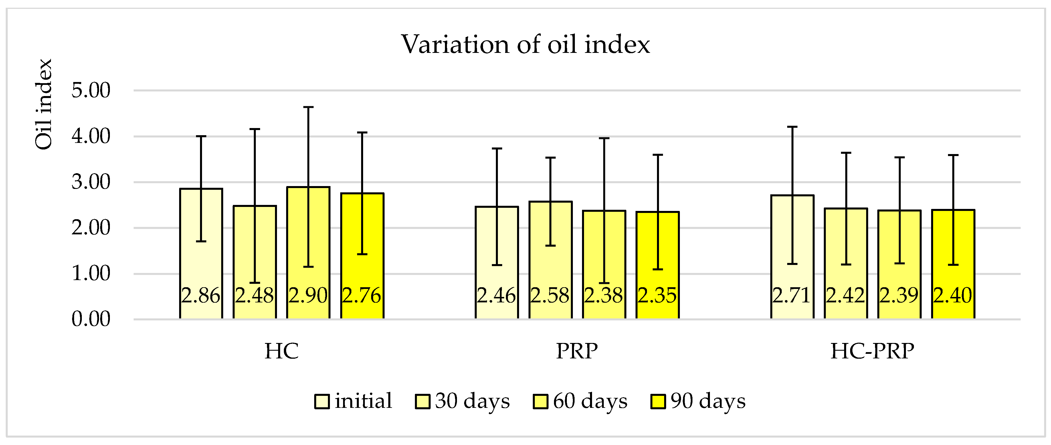
In addition to the main parameters, a set of complementary indices was evaluated to achieve a more comprehensive characterization of the skin. Oil index measures sebum levels on the skin surface; higher values indicate increased skin oiliness; these results are shown in Figure 7. The index values remained relatively constant throughout the study period for all groups.
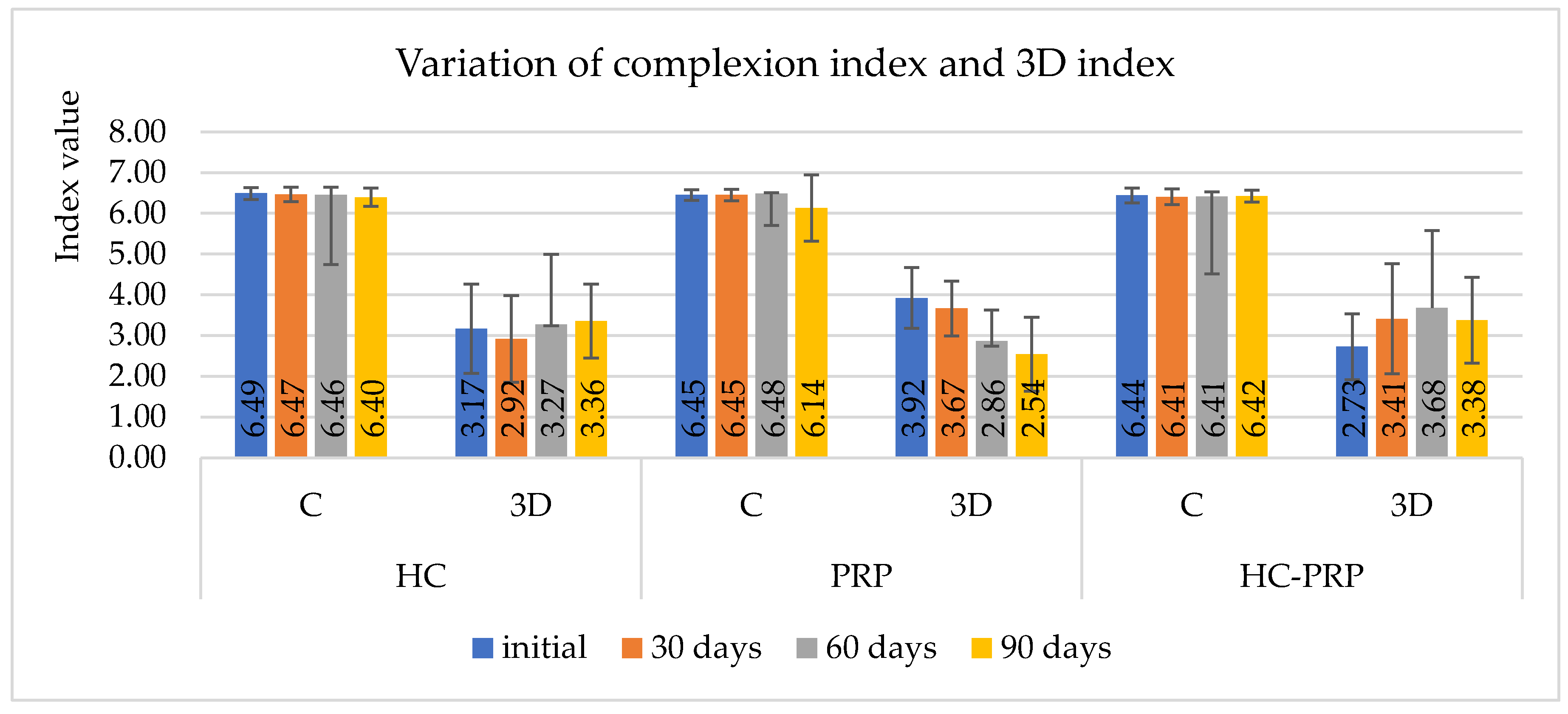
The complexion index and the 3D index were analyzed to provide a comprehensive evaluation of skin appearance and structure, with the results illustrated in Figure 8. The complexion index assesses overall skin tone and brightness; higher values correspond to a more even and radiant appearance. The 3D index evaluates surface topography, including fine lines and micro-relief; lower values reflect reduced roughness and smoother contours. While the complexity index has no remarkable changes over the study period for the three groups, in contrast, the 3D index showed the most important variability for the groups with PRP. For the group with intradermal PRP the values decreased at the end of the study with 35.24% compared to initial value, and for the group with topic PRP, at 90 days, decreased with 8.19% compared to the value at 60 days.
Pigment index quantifies localized skin pigmentation or spots; lower values indicate reduction in hyperpigmentation. Phlogosis index reflects local erythema or inflammation; lower values correspond to reduced redness or irritation. The pores index estimates pore visibility and size (a device-estimated score, not a direct value of pore size); lower values indicate smaller and less visible pores. All these indices showed negligible variation between groups, and between participants, remaining close to a baseline value of 1 (ex. 1–1.10), suggesting no meaningful differences under the studied conditions.

4. Discussion

In this preliminary study, we evaluated whether enriching a hydrating cream with PRP (HC-PRP) improves skin texture, comparing it with a hydrating cream alone (as a principal comparator) and with intradermal PRP (as a secondary, exploratory comparator) over a 90-day period.
The dermoanalysis yielded eight dermatological indices (moisture, texture, oil, complexion, 3D, pigment, phlogosis, and pores) which were used for the subsequent comparisons. Among these, texture (which describes the skin’s surface relief and is particularly relevant in anti-aging studies [33]) and hydration [34] are the most extensively studied skin characteristics. Their use in clinical and cosmetic trials is well established, as they are highly sensitive to topical interventions and can be reliably quantified using non-invasive techniques [35,36,37]. In our study, the indices assessing texture (texture index) and hydration (moisture index) showed the most marked variations.
The hydrating cream acts mainly by optimizing the level of hydration as shown by the moisture index, which increased by more than 50% after 90 days of daily use. The increase in texture index may be secondary to this intensive hydration. Humectants attract water into the corneocytes, causing them to swell unevenly, which can accentuate the surface relief at the microscopic level. A study investigating the effect of hydration on corneocytes (obtained by tape stripping from the forearm) revealed that they swell approximately 50% relative to their dry volume and become more elastic, although prolonged water exposure disrupts the lipid lamellae [38,39]. In parallel, occlusive agents and emollients form a superficial film that modifies the skin surface [40]. These effects are generally related to a physical reorganization of the superficial layer rather than to structural changes in the skin. The literature indicates that the moisturizing effect of topical hydrating formulations typically extends to approximately 15 μm into the stratum corneum [41]. This initial stage of well-hydrated surface may create favorable conditions for the deeper skin layers to stabilize their structural function. The progressive increase in hydration over the 90 days of moisturizer use may reflect an extension of hydration to deeper layers; some studies report increases in hydration in the viable epidermis after repeated applications [42,43]. The dissociation observed between improved hydration and a higher texture index in the HC group may reflect the complex biophysical response of the stratum corneum to topical moisturization. This finding contrasts with most published reports, in which improved hydration is generally associated with reduced skin roughness [44,45]. Increased water content can induce transient corneocyte swelling and altered light scattering or surface roughness, which may negatively influence device-derived texture metrics despite improved hydration status. These differences should be interpreted cautiously, as the underlying biological processes cannot be determined from the present study design and may involve multiple interacting factors. Further histological and ultrastructural studies are needed to better elucidate the underlying mechanisms of the observed surface changes.
Skin lipids, comprising surface lipids such as sebum and barrier lipids such as ceramides, cholesterol, and fatty acids, protect the skin and help maintain its hydration. The oil index did not show notable variations in our study, suggesting that the interventions did not alter surface lipids. Among ceramides, the 6-hydroxysphingosine/phytosphingosine ratio is significantly correlated with surface texture and imbalances in this ratio have been associated with a rough and flaky skin texture [46]. The cream used in our study was only a basic moisturizer, containing occlusives, emollients, and humectants that act mainly on the skin surface, whereas ceramide-containing products may act more deeply by supporting skin repair. Such products not only enhance hydration but may also contribute to a more complete and functional skin recovery over time [47,48], which may be important for improving overall texture. Thus, in our study, the hydrating cream has proven to be more effective at improving hydration than at repairing the skin or improving texture.
Several studies have investigated the activity of PRP combinations for skin rejuvenation. One study on a combination of hyaluronic acid and PRP revealed that facial appearance and skin elasticity were significantly improved compared with either agent used alone [49,50]. Another study evaluated the PRP-hyaluronic acid association on neck skin, (PRP used as three monthly intradermal injection): PRP stimulates fibroblasts, collagen production and extracellular matrix remodeling, while hyaluronic acid enhances this process by hydrating the tissue, leading to improvements in firmness, hydration, pigmentation and sebum regulation [51]. In our study, by contrast, PRP was incorporated into a topical hydrating cream containing sodium hyaluronate as one of its active ingredients; these deep dermal mechanisms cannot be assumed for our formulation. The intradermal administration route used for PRP in the second group limits our ability to isolate the contribution of topical PRP within the combined formulation, a limitation further compounded by the absence of a topical PRP control group, which would have allowed a more precise attribution of its individual effect.
At the molecular level, when delivered into the tissue, PRP has been shown to increase the synthesis of glycosaminoglycans, stimulate angiogenesis through VEGF-mediated pathways [52,53], improve microcirculation (leading to improved skin radiance [54]), and enhance keratinocyte proliferation and filaggrin expression [55,56], thereby supporting the water-binding capacity of the stratum corneum. These findings suggest that PRP may act as a regenerative approach targeting biological processes involved in skin hydration rather than merely masking the signs of dehydration, as occurs with conventional hydrating creams. However, these mechanisms were not directly investigated in the present study and should therefore be considered hypothetical explanations for the observed improvements. Moreover, the exact mechanism by which topically applied PRP may influence skin parameters remains unclear. Given the molecular size of platelet-derived growth factors, direct penetration through the intact stratum corneum is unlikely. The observed effects may instead result from indirect mechanisms, including modulation of the superficial epidermal environment, enhancement of hydration and barrier function by the formulation vehicle (HC), or the generation of smaller peptides, through degradation of platelet-derived components, as bioactive peptides derived from PRP have been shown to retain regenerative activity in experimental models (in vitro) [57], although whether this occurs after topical application remains to be demonstrated.
The intradermal PRP group did not show a marked improvement in texture index and showed a decrease in hydration at the final time point. Given the small size of this group (5 participants), these results are most likely affected by limited statistical power and high inter-individual variability, and may also reflect the complex relationships between dermal interventions and superficial hydration metrics. The response to PRP for skin rejuvenation is heterogeneous [50], and even a randomized controlled trial reported no significant benefit of intradermal PRP over saline for photoaged facial skin [25]. Texture and hydration indices may not necessarily evolve in parallel following intradermal treatment. These results should therefore be interpreted cautiously rather than as evidence against the regenerative potential of PRP.
Regarding the relationship between age and PRP treatment, the literature reports heterogeneous findings. Some studies suggest that younger fibroblasts exhibit a stronger response to PRP, while reduced cellular responsiveness in older individuals may limit regenerative outcomes [58]. In contrast, other reports have found that the effectiveness of PRP in improving skin is not age dependent [59]. In the present study, the mean age of participants was 46 years; over 90 days a significant improvement in skin texture was observed in the HC-PRP group, with a similar but non-significant trend in the intradermal PRP group. The age–index (moisture/texture) fitting analysis was performed for exploratory purposes to visualize potential trends; it was not intended as a validated predictive model.
Several studies have focused on the topical administration of PRP, whose main advantage is that it is non-invasive and well-tolerated, avoiding the discomfort and risks associated with intradermal injections. In a study on perioral wrinkles, topical PRP produced significant improvements in moisture, collagen fiber content, elasticity and both satisfaction scales (patient assessment and surgeon lifting assessment) [60]. In another study, topical PRP applied after electroporation increased collagen type I expression and upregulated collagen-related mRNA, indicating enhanced dermal regeneration [61]. Notably, this effect was obtained with electroporation, a penetration-enhancing technique, rather than with simple topical application. In our study, topical PRP associated with hydrating cream significantly improved skin texture, although, in the absence of a penetration-enhancing step, this effect was most likely superficial, consistent with the limited penetration of high-molecular-weight components.
No treatment-related adverse events were reported or observed during the study period. In the topical treatment groups (HC and HC-PRP), no local irritation, contact dermatitis or allergic reactions were noted. In the intradermal PRP group, no injection-site pain, ecchymosis, infection or granuloma formation was observed. As PRP is an autologous blood-derived product, the risk of immunogenic or allergic reactions is considered low [62]. Moreover, current evidence on PRP-related adverse events derives largely from isolated case reports, and higher-quality studies are needed to establish a causal relationship between PRP and specific adverse events [63]. This is consistent with the safety profile generally reported for PRP, whose autologous origin entails a minimal risk of immunogenic or allergic reactions [62]. Nevertheless, given the relatively short follow-up (90 days) and the small sample size, the long-term safety aspects discussed above could not be fully assessed in the present study.
An important limitation regarding effectiveness is that quantitative assessment of platelet-derived growth factors and their stability over the study period was not performed; therefore, variability in the bioactive content of PRP and potential degradation during storage cannot be excluded. PRP can be stored for 1–3 months at −20 °C to −80 °C; however, key growth factors (including PDGF, TGF-β, and VEGF) are sensitive to handling and to repeated freeze–thaw cycles [64,65,66]. Accordingly, in our study PRP was stored at −20 °C (as single-use aliquots) and was subjected to a single freeze–thaw cycle. In addition, the HC-PRP was prepared extemporaneously to minimize the possible degradation. Evidence from a previous study indicates that a PRP-based topical product remained stable at 4 °C over a period of 90 days [61]. Nevertheless, the stability of PRP after incorporation into the cream was not assessed and remains an important topic for future investigation.
The current study has several additional limitations. The sample size was small, particularly in the intradermal PRP group (n = 5), reflecting patient preference for non-invasive treatments, which limited the statistical power of the between-group comparisons. The lack of randomization introduces an important potential for selection bias. Generalizability is further limited by the single-center design and the inclusion of only self-defined female participants, since population-specific demographic and environmental factors can influence skin characteristics. The use of a single device for evaluating skin parameters hinders the comparisons with other technologies. Regarding measurement, the use of a single device prevented comparison with results obtained using other technologies; studies comparing devices based on different measurement principles (e.g., electrical capacitance and high-frequency conductance) have highlighted the value of combining multiple assessment methods [67]. Although measurements were performed under standardized indoor conditions, minor variations in environmental temperature across study visits cannot be excluded and may have influenced the skin biophysical parameters. Despite these limitations, the study provides a valuable preliminary basis for evaluating the potential of HC-PRP.
Regarding the HC and HC-PRP groups, both received the same hydrating base; the two formulations differing only by the addition of PRP. In the HC group, the moisture index improved, whereas a corresponding improvement in skin texture was not observed, with the texture index increasing over time. It should be noted, however, that the HC group started from the most favorable (lowest) baseline texture index of the three groups; consequently, this increase may reflect, at least in part, regression to the mean and natural variability within a small, non-randomized group, rather than a genuine deleterious effect of the hydrating cream itself. In contrast, regression to the mean cannot account for the improvement observed in the HC-PRP group. This group started from a higher (less favorable) baseline value and its texture index nevertheless decreased below the overall mean (in the opposite direction to that expected from regression to the mean). Moreover, because the groups differed at baseline, the between-group comparison was based on ANCOVA adjusted for the baseline value, and the difference in favor of HC-PRP persisted and even increased after this adjustment, whereas regression to the mean would be expected to attenuate it. These two observations are therefore complementary rather than contradictory: regression to the mean may contribute to the within-group trajectory of the HC group, while the adjusted between-group comparison supports a real difference between the HC-PRP and HC formulations. This may indicate an additional contribution of PRP-derived bioactive factors. A comparable role of topical PRP has been suggested by a vehicle-controlled study in which PRP incorporated into a cosmetic base was associated with histologic and collagen-related changes in facial skin [61], although the study used electroporation to enhance penetration unlike the plain topical application as in our study. However, because in our study the groups were not randomized, unmeasured differences between the self-selected groups cannot be excluded, and the specific contribution of PRP cannot be confirmed within the present design. These findings should therefore be regarded as preliminary and hypothesis-generating, requiring confirmation in larger, balanced and randomized studies.
Dermatological analysis with the dermoanalyzer is limited to superficial skin parameters and does not allow assessment of the deeper dermal processes needed for a complete understanding of the histological changes. However, this highlights the value of complementary investigations that could provide insight into tissue architecture and biological mechanisms involved. Future studies could also investigate how long the observed benefits on texture persist after treatment and explore the use of HC-PRP in various skin conditions.

5. Conclusions

In conclusion, the hydrating cream alone was associated with an improvement in the moisture index, whereas a corresponding improvement in skin texture was not observed in this group. In contrast, the PRP-enriched cream (HC-PRP) was associated with a progressive improvement in skin texture and yielded better texture outcomes than the cream alone. Although HC and HC-PRP differ only by the addition of PRP, the findings should be interpreted as exploratory, as the analysis was conducted on non-randomized groups. They indicate a favorable effect of the HC-PRP topical combination on skin texture and provide a basis for further investigation. Additional studies on larger samples are needed to confirm these results and to better define the role of PRP in topical formulation.

Author Contributions

Conceptualization, C.-I.S. and V.P.; methodology, C.-I.S. and V.P.; software, C.-I.S., A.-R.U., V.P. and G.T.; validation, C.-I.S., A.-R.U., V.P., C.B., M.-V.H. and G.T.; formal analysis, C.-I.S., A.-R.U., V.P., C.B., M.-V.H. and G.T.; investigation, C.-I.S. and V.P.; resources, C.-I.S., A.-R.U., V.P., C.B., M.-V.H. and G.T.; data curation, C.-I.S., A.-R.U., V.P., C.B., M.-V.H. and G.T.; writing—original draft preparation, C.-I.S. and A.-R.U.; writing—review and editing, C.-I.S., A.-R.U., V.P., C.B., M.-V.H. and G.T.; visualization, C.-I.S., A.-R.U., V.P., C.B., M.-V.H. and G.T.; supervision, C.-I.S., A.-R.U., V.P., C.B., M.-V.H. and G.T.; project administration, C.-I.S., V.P. and G.T.; All authors have read and agreed to the published version of the manuscript.

Funding

This research received no external funding.

Institutional Review Board Statement

The study was conducted in accordance with the Declaration of Helsinki, and approved by the Ethics Committee of the University of Medicine and Pharmacy of Craiova (72/11.05.2021).

Informed Consent Statement

Informed consent was obtained from all participants involved in the study.

Data Availability Statement

The original contributions presented in this study are included in the article. Further inquiries can be directed to the corresponding authors.

Conflicts of Interest

The authors declare no conflicts of interest.

Abbreviations

The following abbreviations are used in this manuscript:
AQP3Aquaporin-3
CMOSComplementary Metal-Oxide Semiconductor
EGFEpidermal Growth Factor
HCHydrating Cream
HC-PRPHydrating Cream Enriched with Platelet-Rich Plasma
NMFNatural Moisturizing Factor
PDGFPlatelet-Derived Growth Factor
PRPPlatelet-Rich Plasma
TEWLTrans-epidermal Water Loss
TGF-bTransforming Growth Factor beta
VEGFVascular Endothelial Growth Factor
WHOWorld Health Organization

References

  1. Sagrafena, I.; Morin, M.; Paraskevopoulos, G.; Nilsson, E.J.; Hrdinová, I.; Kováčik, A.; Björklund, S.; Vávrová, K. Structure and function of skin barrier lipids: Effects of hydration and natural moisturizers in vitro. Biophys. J. 2024, 123, 3951–3963. [Google Scholar] [CrossRef] [PubMed]
  2. Bouwstra, J.A.; Nădăban, A.; Bras, W.; McCabe, C.; Bunge, A.; Gooris, G.S. The skin barrier: An extraordinary interface with an exceptional lipid organization. Prog. Lipid Res. 2023, 92, 101252. [Google Scholar] [CrossRef] [PubMed]
  3. Samadi, A.; Yazdanparast, T.; Shamsipour, M.; Hassanzadeh, H.; Hashemi Orimi, M.; Firooz, R.; Firooz, A. Stratum corneum hydration in healthy adult humans according to the skin area, age and sex: A systematic review and meta-analysis. J. Eur. Acad. Dermatol. Venereol. 2022, 36, 1713–1721. [Google Scholar] [CrossRef] [PubMed]
  4. Gunnarsson, M.; Mojumdar, E.H.; Topgaard, D.; Sparr, E. Extraction of natural moisturizing factor from the stratum corneum and its implication on skin molecular mobility. J. Colloid Interface Sci. 2021, 604, 480–491. [Google Scholar] [CrossRef] [PubMed]
  5. Hoober, J.K.; Eggink, L.L. The Discovery and Function of Filaggrin. Int. J. Mol. Sci. 2022, 23, 1455. [Google Scholar] [CrossRef] [PubMed]
  6. Moosbrugger-Martinz, V.; Leprince, C.; Méchin, M.C.; Simon, M.; Blunder, S.; Gruber, R.; Dubrac, S. Revisiting the Roles of Filaggrin in Atopic Dermatitis. Int. J. Mol. Sci. 2022, 23, 5318. [Google Scholar] [CrossRef] [PubMed]
  7. Bi, Y.; Pang, S.; Liu, Y.; Cheng, J.; Ma, Q.; Song, A.; Yin, X.; Wang, J. Aquaporins in lipid metabolism: Functions and regulation in health and disease. Lipids Health Dis. 2025, 24, 336. [Google Scholar] [CrossRef] [PubMed]
  8. Camilion, J.V.; Khanna, S.; Anasseri, S.; Laney, C.; Mayrovitz, H.N. Physiological, Pathological, and Circadian Factors Impacting Skin Hydration. Cureus 2022, 14, e27666. [Google Scholar] [CrossRef] [PubMed]
  9. Qin, H.; Zheng, X.; Zhong, X.; Shetty, A.K.; Elias, P.M.; Bollag, W.B. Aquaporin-3 in keratinocytes and skin: Its role and interaction with phospholipase D2. Arch. Biochem. Biophys. 2011, 508, 138–143. [Google Scholar] [CrossRef] [PubMed]
  10. Iaconisi, G.N.; Lunetti, P.; Gallo, N.; Cappello, A.R.; Fiermonte, G.; Dolce, V.; Capobianco, L. Hyaluronic Acid: A Powerful Biomolecule with Wide-Ranging Applications—A Comprehensive Review. Int. J. Mol. Sci. 2023, 24, 10296. [Google Scholar] [CrossRef] [PubMed]
  11. Chylińska, N.; Maciejczyk, M. Hyaluronic Acid and Skin: Its Role in Aging and Wound-Healing Processes. Gels 2025, 11, 281. [Google Scholar] [CrossRef] [PubMed]
  12. Hussein, R.S.; Bin Dayel, S.; Abahussein, O.; El-Sherbiny, A.A. Influences on Skin and Intrinsic Aging: Biological, Environmental, and Therapeutic Insights. J. Cosmet. Dermatol. 2025, 24, e16688. [Google Scholar] [CrossRef] [PubMed]
  13. Danby, S.G.; Andrew, P.V.; Kay, L.J.; Pinnock, A.; Chittock, J.; Brown, K.; Williams, S.F.; Cork, M.J. Enhancement of stratum corneum lipid structure improves skin barrier function and protects against irritation in adults with dry, eczema-prone skin. Br. J. Dermatol. 2022, 186, 875–886. [Google Scholar] [CrossRef] [PubMed]
  14. Geusens, B.; Haykal, D. Genetic profiling and precision skin care: A review. Front. Genet. 2025, 16, 1559510. [Google Scholar] [CrossRef] [PubMed]
  15. World Health Organization. Skin Diseases as a Global Public Health Priority: Resolution WHA78.15 Adopted by the Seventy-Eighth World Health Assembly; World Health Organization: Geneva, Switzerland, 2025; Available online: https://apps.who.int/gb/ebwha/pdf_files/WHA78/A78_R15-en.pdf (accessed on 21 March 2026).
  16. Pawlus, Z.; Kruczek, W.; Reizer, R.; Dzierwa, A.; Frątczak, A.; Bergler-Czop, B. Measurements of surface textures in dermatology—A review. Measurement 2026, 257, 118943. [Google Scholar] [CrossRef]
  17. Seck, A.; Dee, H.; Smith, W.; Tiddeman, B. 3D surface texture analysis of high-resolution normal fields for facial skin condition assessment. Skin Res. Technol. 2020, 26, 169–186. [Google Scholar] [CrossRef] [PubMed]
  18. Pequeno, A.L.V.; Bagatin, E. Dermatological ultrasound in assessing skin aging. Front. Med. 2024, 11, 1353605. [Google Scholar] [CrossRef] [PubMed]
  19. Rajkumar, J.; Chandan, N.; Lio, P.; Shi, V. The Skin Barrier and Moisturization: Function, Disruption, and Mechanisms of Repair. Skin Pharmacol. Physiol. 2023, 36, 174–185. [Google Scholar] [CrossRef] [PubMed]
  20. Hong, S.; Maitra, P.; Nguyen, A.; Kadoya, K.; Mehta, R.C. Development of an in vitro Functional Assay to Evaluate the Occlusive Properties of Moisturizers on Dry Skin. Skin Pharmacol. Physiol. 2023, 36, 140–148. [Google Scholar] [CrossRef] [PubMed]
  21. Purnamawati, S.; Indrastuti, N.; Danarti, R.; Saefudin, T. The Role of Moisturizers in Addressing Various Kinds of Dermatitis: A Review. Clin. Med. Res. 2017, 15, 75–87. [Google Scholar] [CrossRef] [PubMed]
  22. White, C.; Brahs, A.; Dorton, D.; Witfill, K. Platelet-Rich Plasma: A Comprehensive Review of Emerging Applications in Medical and Aesthetic Dermatology. J. Clin. Aesthet. Dermatol. 2021, 14, 44–57. [Google Scholar] [PubMed]
  23. Vladulescu, D.; Scurtu, L.G.; Simionescu, A.A.; Scurtu, F.; Popescu, M.I.; Simionescu, O. Platelet-Rich Plasma (PRP) in Dermatology: Cellular and Molecular Mechanisms of Action. Biomedicines 2023, 12, 7. [Google Scholar] [CrossRef] [PubMed]
  24. Wu, W.S.; Chen, L.R.; Chen, K.H. Platelet-Rich Plasma (PRP): Molecular Mechanisms, Actions and Clinical Applications in Human Body. Int. J. Mol. Sci. 2025, 26, 10804. [Google Scholar] [CrossRef] [PubMed]
  25. Alam, M.; Hughart, R.; Champlain, A.; Geisler, A.; Paghdal, K.; Whiting, D.; Hammel, J.A.; Maisel, A.; Rapcan, M.J.; West, D.P.; et al. Effect of Platelet-Rich Plasma Injection for Rejuvenation of Photoaged Facial Skin: A Randomized Clinical Trial. JAMA Dermatol. 2018, 154, 1447. [Google Scholar] [CrossRef] [PubMed]
  26. Du, R.; Lei, T. Effects of autologous platelet-rich plasma injections on facial skin rejuvenation. Exp. Ther. Med. 2020, 19, 3024–3030. [Google Scholar] [CrossRef] [PubMed]
  27. Wyganowska, M.L.; Tyliszczak, F.; Marzec, M.; Klewin-Steinböck, S.; Nowak, I. Instrumental In Vivo Assessment of Cosmetic Emulsions Containing Platelet-Rich Fibrin (PRF) or Recombinant Epidermal Growth Factor (EGF): A Pilot Compatibility Study. Pharmaceuticals 2026, 19, 394. [Google Scholar] [CrossRef] [PubMed]
  28. Kang, H.G.; Song, J.J.; Lee, K.; Nam, K.C.; Hong, S.J.; Kim, H.C. An investigation of medical radiation detection using CMOS image sensors in smartphones. Nucl. Instrum. Methods Phys. Res. Sect. A Accel. Spectrometers Detect. Assoc. Equip. 2016, 823, 126–134. [Google Scholar] [CrossRef]
  29. Li, G.; Zhao, F.; Wang, Z.; Chen, Z. The development and application of CMOS image sensor. Appl. Comput. Eng. 2023, 7, 767–777. [Google Scholar] [CrossRef]
  30. Sarkar, M.; Assaad, M. Noninvasive Non-Contact SpO2 Monitoring Using an Integrated Polarization-Sensing CMOS Imaging Sensor. Sensors 2022, 22, 7796. [Google Scholar] [CrossRef] [PubMed]
  31. Han, H.S.; Kim, B.J. Development and Validation of the Cheek Smoothness Scale: A Photonumeric Assessment Tool for Cheek Skin Texture. J. Cosmet. Dermatol. 2026, 25, e70806. [Google Scholar] [CrossRef] [PubMed]
  32. Kim, H.Y. Statistical notes for clinical researchers: Nonparametric statistical methods: 2. Nonparametric methods for comparing three or more groups and repeated measures. Restor. Dent. Endod. 2014, 39, 329. [Google Scholar] [CrossRef] [PubMed]
  33. Wang, Y.C.; Ma, S.H.; Chang, Y.T.; Chen, C.C. Clinical evaluation of skin aging: A systematic review. Arch. Gerontol. Geriatr. 2025, 139, 105995. [Google Scholar] [CrossRef] [PubMed]
  34. Neha; Joshi, R.; Bhave, A.; Bhagat, S.V.; Veligandla, K.; Rathod, R.; Kotak, B. Efficacy and In-Use Tolerance of Venusia Baby Moisturizer for Skin Hydration in Babies With Dry and/or Normal Skin. Cureus 2023, 15, e45032. [Google Scholar] [CrossRef] [PubMed]
  35. Zhao, S.; Wang, M.; Lai, X.; Yan, Y. Noninvasive Assessment Methods for the Therapeutic Effect Following Facial Rejuvenation. J. Cosmet. Dermatol. 2025, 24, e70482. [Google Scholar] [CrossRef] [PubMed]
  36. Carlos Da Silva, G.; Barbosa, M.B.; Júnior, F.B.C.; Moreira, P.L.; Werka, R.; Martin, A.A. Detection of skin wrinkles and quantification of roughness using a novel image processing technique from a dermatoscope device. Skin Res. Technol. 2023, 29, e13335. [Google Scholar] [CrossRef] [PubMed]
  37. Gougeon, S.; Hernandez, E.; Chevrot, N.; Vergne, T.; Cherel, M.; Prestat-Marquis, E.; Jomier, M.; Burty-Valin, E. Evaluation of a new connected portable camera for the analysis of skin microrelief and the assessment of the effect of skin moisturisers. Skin Res. Technol. 2023, 29, e13190. [Google Scholar] [CrossRef] [PubMed]
  38. Évora, A.S.; Zhang, Z.; Johnson, S.A.; Adams, M.J. The effects of hydration on the topographical and mechanical properties of corneocytes. J. Mech. Behav. Biomed. Mater. 2024, 150, 106296. [Google Scholar] [CrossRef] [PubMed]
  39. Mojumdar, E.H.; Pham, Q.D.; Topgaard, D.; Sparr, E. Skin hydration: Interplay between molecular dynamics, structure and water uptake in the stratum corneum. Sci. Rep. 2017, 7, 15712. [Google Scholar] [CrossRef] [PubMed]
  40. Tamura, E.; Yasumori, H.; Yamamoto, T. The efficacy of a highly occlusive formulation for dry lips. Int. J. Cosmet. Sci. 2020, 42, 46–52. [Google Scholar] [CrossRef] [PubMed]
  41. Gallinger, J.; Kuhn, A.; Wessel, S.; Behm, P.; Heinecke, S.; Filbry, A.; Hillemann, L.; Rippke, F. Depth-dependent hydration dynamics in human skin: Vehicle-controlled efficacy assessment of a functional 10% urea plus NMF moisturizer by near-infrared confocal spectroscopic imaging (KOSIM IR) and capacitance method complemented by volunteer perception. Skin Res. Technol. 2022, 28, 342–349. [Google Scholar] [CrossRef] [PubMed]
  42. Lee, H.J.; Park, S.R.; Kwon, D.I.; Park, M.S.; Lim, D.H. Depth profiling of epidermal hydration inducing improvement of skin roughness and elasticity: In vivo study by confocal Raman spectroscopy. J. Cosmet. Dermatol. 2022, 21, 4810–4817. [Google Scholar] [CrossRef] [PubMed]
  43. Bianchini, J.M.; Zhang, Q.; Hanna, G.; Flach, C.R.; Wang, H.; Southall, M.D.; Mendelsohn, R.; Randhawa, M. A unique gel matrix moisturizer delivers deep hydration resulting in significant clinical improvement in radiance and texture. Clin. Cosmet. Investig. Dermatol. 2019, 12, 229–239. [Google Scholar] [CrossRef] [PubMed]
  44. Ruini, C.; Kendziora, B.; Ergun, E.Z.; Sattler, E.; Gust, C.; French, L.E.; Bağcı, I.S.; Hartmann, D. In vivo examination of healthy human skin after short-time treatment with moisturizers using confocal Raman spectroscopy and optical coherence tomography: Preliminary observations. Skin Res. Technol. 2022, 28, 119–132. [Google Scholar] [CrossRef] [PubMed]
  45. Crowther, J.M.; Matts, P.J. What defines dry skin? Correlating a range of skin hydration parameters with In Vivo Confocal Raman Spectroscopy. Int. J. Cosmet. Sci. 2024, 46, 610–622. [Google Scholar] [CrossRef] [PubMed]
  46. Yokose, U.; Ishikawa, J.; Morokuma, Y.; Naoe, A.; Inoue, Y.; Yasuda, Y.; Tsujimura, H.; Fujimura, T.; Murase, T.; Hatamochi, A. The ceramide [NP]/[NS] ratio in the stratum corneum is a potential marker for skin properties and epidermal differentiation. BMC Dermatol. 2020, 20, 6. [Google Scholar] [CrossRef] [PubMed]
  47. Wang, Y.; Li, S.; Ai, Y.; Lynch, S.; Baalbaki, N.; Zhang, X.; He, X.; Huang, X.; Steel, A.; Hsu, K.; et al. Evaluating the effect of moisturizers containing endogenous lipids on skin barrier properties. J. Dermatol. Sci. Cosmet. Technol. 2024, 1, 100037. [Google Scholar] [CrossRef]
  48. Andrew, P.V.; Williams, S.F.; Brown, K.; Chittock, J.; Pinnock, A.; Poyner, A.; Cork, M.J.; Danby, S.G. Topical supplementation with physiological lipids rebalances the stratum corneum ceramide profile and strengthens skin barrier function in adults predisposed to atopic dermatitis. Br. J. Dermatol. 2025, 193, 729–740. [Google Scholar] [CrossRef] [PubMed]
  49. Hersant, B.; SidAhmed-Mezi, M.; Aboud, C.; Niddam, J.; Levy, S.; Mernier, T.; La Padula, S.; Meningaud, J.-P. Synergistic Effects of Autologous Platelet-Rich Plasma and Hyaluronic Acid Injections on Facial Skin Rejuvenation. Aesthetic Surg. J. 2021, 41, NP854–NP865. [Google Scholar] [CrossRef] [PubMed]
  50. Phoebe, L.K.W.; Lee, K.W.A.; Chan, L.K.W.; Hung, L.C.; Wu, R.; Wong, S.; Wan, J.; Yi, K. Use of platelet rich plasma for skin rejuvenation. Skin Res. Technol. 2024, 30, e13714. [Google Scholar] [CrossRef] [PubMed]
  51. De Leon, S.T.; Zabala, D.A.; Viriyaskultorn, N.; Buranaporn, P.; Bhorntarakcharoen, W.; Hutachoke, T.; Leesanguankul, T.; Wannawittayapa, T.; Tantrapongsathorn, W.; Sittiwanaruk, S.; et al. Efficacy and Safety of Platelet-Rich Plasma and Hyaluronic Acid Combination Treatment for Neck Rejuvenation in Thailand: A Prospective Study. Dermatol. Ther. 2026, 16, 1209–1223. [Google Scholar] [CrossRef] [PubMed]
  52. Alves, R.; Grimalt, R. A Review of Platelet-Rich Plasma: History, Biology, Mechanism of Action, and Classification. Skin Appendage Disord. 2018, 4, 18–24. [Google Scholar] [CrossRef] [PubMed]
  53. Buzalaf, M.A.R.; Levy, F.M. Autologous platelet concentrates for facial rejuvenation. J. Appl. Oral. Sci. 2022, 30, e20220020. [Google Scholar] [CrossRef] [PubMed]
  54. Manole, C.G.; Soare, C.; Ceafalan, L.C.; Voiculescu, V.M. Platelet-Rich Plasma in Dermatology: New Insights on the Cellular Mechanism of Skin Repair and Regeneration. Life 2024, 14, 40. [Google Scholar] [CrossRef] [PubMed]
  55. Misiura, M.; Guszczyn, T.; Oscilowska, I.; Baszanowska, W.; Palka, J.; Miltyk, W. Platelet-Rich Plasma Promotes the Proliferation of Human Keratinocytes via a Progression of the Cell Cycle. A Role of Prolidase. Int. J. Mol. Sci. 2021, 22, 936. [Google Scholar] [CrossRef] [PubMed]
  56. Xian, L.J.; Roy Chowdhury, S.; Bin Saim, A.; Bt Hj Idrus, R. Concentration-dependent effect of platelet-rich plasma on keratinocyte and fibroblast wound healing. Cytotherapy 2015, 17, 293–300. [Google Scholar] [CrossRef] [PubMed]
  57. Demidova-Rice, T.N.; Wolf, L.; Deckenback, J.; Hamblin, M.R.; Herman, I.M. Human Platelet-Rich Plasma- and Extracellular Matrix-Derived Peptides Promote Impaired Cutaneous Wound Healing In Vivo. PLoS ONE 2012, 7, e32146. [Google Scholar] [CrossRef] [PubMed]
  58. Nanda, S.; Chauhan, K.; Shetty, V.; Dashore, S.; Bhatia, S. Platelet-rich plasma in aesthetics. Indian Dermatol. Online J. 2021, 12, 41. [Google Scholar] [CrossRef] [PubMed]
  59. Majewska, L.; Kijowski, J.; Dorosz, K. Effect of Patient Age on Platelet-Rich Plasma (PRP) and Fibrin Treatments for Skin Density and Thickness: A Single-Center Ultrasound Study. Life 2025, 15, 308. [Google Scholar] [CrossRef] [PubMed]
  60. Araco, A. A prospective study comparing topic platelet-rich plasma vs. placebo on reducing superficial perioral wrinkles and restore dermal matrix. J. Cosmet. Laser Ther. 2019, 21, 309–315. [Google Scholar] [CrossRef] [PubMed]
  61. Draelos, Z.D.; Rheins, L.A.; Wootten, S.; Kellar, R.S.; Diller, R.B. Pilot study: Autologous platelet-rich plasma used in a topical cream for facial rejuvenation. J. Cosmet. Dermatol. 2019, 18, 1348–1352. [Google Scholar] [CrossRef] [PubMed]
  62. Asubiaro, J.; Avajah, F. Platelet-Rich Plasma in Aesthetic Dermatology: Current Evidence and Future Directions. Cureus 2024, 16, e66734. [Google Scholar] [CrossRef] [PubMed]
  63. Arita, A.; Tobita, M. Adverse events related to platelet-rich plasma therapy and future issues to be resolved. Regen. Ther. 2024, 26, 496–501. [Google Scholar] [CrossRef] [PubMed]
  64. Wang, Y.; Fu, X.; Cao, D. Effect of storage temperature and time on growth factor concentration in single-harvest platelet-rich plasma. Transfus. Apher. Sci. 2025, 64, 104197. [Google Scholar] [CrossRef] [PubMed]
  65. Kim, J.I.; Bae, H.C.; Park, H.J.; Lee, M.C.; Han, H.S. Effect of Storage Conditions and Activation on Growth Factor Concentration in Platelet-Rich Plasma. J. Orthop. Res. 2020, 38, 777–784. [Google Scholar] [CrossRef] [PubMed]
  66. Beitia, M.; Delgado, D.; Mercader, J.; Gimeno, I.; Espregueira-Mendes, J.; Aizpurua, B.; Sánchez, M. The effect of short-term cryopreservation on the properties and functionality of platelet-rich plasma. Platelets 2023, 34, 2210243. [Google Scholar] [CrossRef] [PubMed]
  67. Koseki, K.; Kawasaki, H.; Atsugi, T.; Nakanishi, M.; Mizuno, M.; Naru, E.; Ebihara, T.; Amagai, M.; Kawakami, E. Assessment of skin barrier function using skin images with topological data analysis. npj Syst. Biol. Appl. 2020, 6, 40. [Google Scholar] [CrossRef] [PubMed]
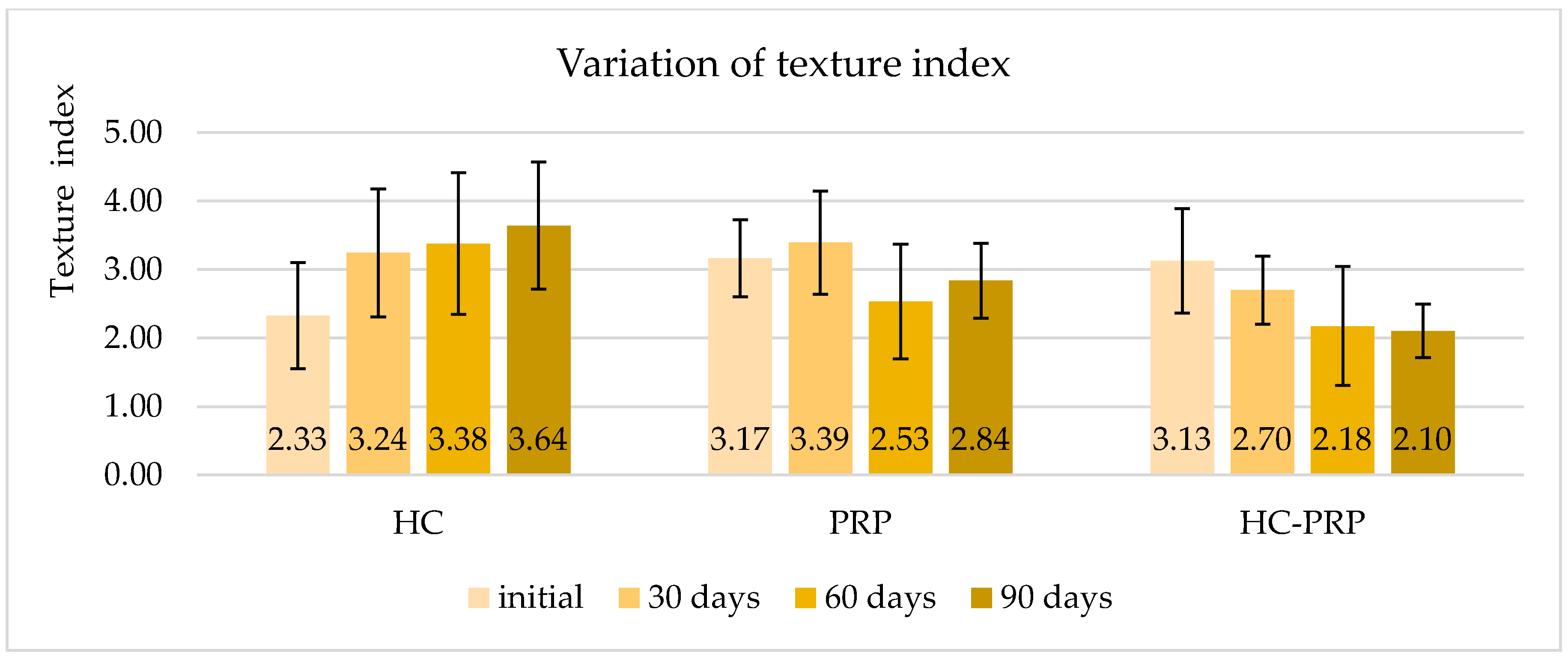
Figure 1. Texture index values (HC = hydrating cream group; PRP = platelet-rich plasma group; HC-PRP = hydrating cream enriched with platelet-rich plasma group).
Cosmetics 13 00175 g001
Figure 2. Exploratory fitted curve of facial skin texture index as a function of age. The regression line is used as an illustrative reference only and not as a validated predictor of biological age.
Cosmetics 13 00175 g002
Figure 3. Dermoanalyzer captures for a participant from the third group (HC-PRP), showing skin at the start of the study with a texture index of 4.35, corresponding to >70 years age (a); skin capture at 30 days, texture index of 2.87, corresponding to approximately 60 years age (b); skin at 60 days, texture index of 2.70, corresponding to 55 years age, reduction in irregularities and wrinkles (c); skin at the end of the study, texture index of 2.28, corresponding to approximately 50 years, and gained uniformity (d).
Cosmetics 13 00175 g003
Figure 4. Moisture index variation over study period (HC = hydrating cream group; PRP = platelet-rich plasma group; HC-PRP = hydrating cream enriched with platelet-rich plasma group).
Cosmetics 13 00175 g004
Figure 5. Dermoanalyzer captions for a participant from the first group. Face at the beginning of the study with a moisture index value of 2.74; (a) skin after 30 days with improvement in surface irregularities and a moisture index of 3.83; (b) face after 60 days with blurred wrinkles, moisture value of 4.62; (c) and the skin at the end of the study, rendering the brightness of the complexion, with a moisture index of 4.65 (d).
Cosmetics 13 00175 g005
Figure 6. Exploratory fitted curve of facial skin moisture index as a function of age. The regression line is used as an illustrative reference only and not as a validated predictor of biological age.
Cosmetics 13 00175 g006
Figure 7. Oil index values (HC = hydrating cream group; PRP = platelet-rich plasma group; HC-PRP = hydrating cream enriched with platelet-rich plasma group).
Cosmetics 13 00175 g007
Figure 8. Complexion and 3D indices values (HC = hydrating cream group; PRP = platelet-rich plasma group; HC-PRP = hydrating cream enriched with platelet-rich plasma group, C = complexion index, 3D = 3D index).
Cosmetics 13 00175 g008
Table 1. Within-group change in skin texture index over time (Friedman test).
GroupNpp BonferroniKendall’s W
HC20<0.0001<0.00010.40
PRP50.24070.72210.28
HC-PRP150.00020.00060.43
HC = hydrating cream group; HC-PRP = hydrating cream enriched with platelet-rich plasma group; N = number of participants; Kendall’s W = effect size for the Friedman test: 0.1 ≤ W ≤ 0.3 (small), 0.3 ≤ W ≤ 0.5 (moderate) and W ≥ 0.5 (large).
Table 2. Skin texture index by group and between-group comparisons at each time point (Mann–Whitney–Wilcoxon test).
PeriodHC
(Mean ± SD)
PRP
(Mean ± SD)
HC-PRP
(Mean ± SD)
HC vs. PRPHC vs. HC-PRPPRP vs. HC-PRP
initial2.33 ± 0.803.17 ± 0.633.13 ± 0.79p = 0.0173
Bonferroni = 0.2076
r = 0.48
p = 0.0108
Bonferroni = 0.1296
r = 0.43
p = 0.8660
Bonferroni = 1.0000
r = 0.05
30 days3.24 ± 0.963.39 ± 0.842.70 ± 0.51p = 0.9215
Bonferroni = 1.0000
r = 0.03
p = 0.0329
Bonferroni = 0.3948
r = 0.36
p = 0.0806
Bonferroni = 0.9672
r = 0.40
60 days3.38 ± 1.062.53 ± 0.942.18 ± 0.90p = 0.1294
Bonferroni = 1.0000
r = 0.31
p = 0.0027
Bonferroni = 0.0324
r = 0.51
p = 0.8614
Bonferroni = 1.0000
r = 0.05
90 days3.64 ± 0.932.84 ± 0.612.10 ± 0.39p = 0.1636
Bonferroni = 1.0000
r = 0.28
p ≤ 0.0001
Bonferroni ≤ 0.0001
r = 0.76
p = 0.0328
Bonferroni = 0.3936
r = 0.48
HC = hydrating cream group; HC-PRP = hydrating cream enriched with platelet-rich plasma group; SD = standard deviation; p = uncorrected Mann–Whitney–Wilcoxon p-value; p Bonferroni = p value corrected for the 12 between-group comparisons, r = effect size: 0.1 ≤ r ≤ 0.3 (small), 0.3 ≤ r ≤ 0.5 (moderate) and r ≥ 0.5 (large). Comparisons in bold are significant after Bonferroni correction (Bonferroni p < 0.05).
Table 3. Between-group comparisons adjusted for the baseline value (ANCOVA).
PeriodComparisonAdjusted Mean Difference *
(95% CI)
pp (Bonferroni)
30 daysHC vs. PRP0.08 (−0.77, 0.93)0.85271.0000
30 daysHC vs. HC-PRP0.76 (0.15, 1.37)0.01570.0471
30 daysPRP vs. HC-PRP0.68 (−0.14, 1.51)0.10150.3045
60 daysHC vs. PRP0.94 (−0.14, 2.02)0.08570.2571
60 daysHC vs. HC-PRP1.29 (0.51, 2.06)0.00180.0054
60 daysPRP vs. HC-PRP0.35 (−0.70, 1.40)0.50271.0000
90 daysHC vs. PRP1.01 (0.21, 1.81)0.01460.0438
90 daysHC vs. HC-PRP1.73 (1.16, 2.30)<0.0001<0.0001
90 daysPRP vs. HC-PRP0.72 (−0.05, 1.50)0.06700.2010
HC = hydrating cream group; HC-PRP = hydrating cream enriched with platelet-rich plasma group; CI = confidence interval; p (Bonferroni) = p value corrected for the three pairwise comparisons at each time point; comparisons in bold are significant after Bonferroni correction (Bonferroni p < 0.05). * ANCOVA was adjusted for the baseline texture value. The adjusted mean difference is the first group minus the second group at the mean baseline value; a positive value indicates a higher (less smooth) texture index in the first group.
Disclaimer/Publisher’s Note: The statements, opinions and data contained in all publications are solely those of the individual author(s) and contributor(s) and not of MDPI and/or the editor(s). MDPI and/or the editor(s) disclaim responsibility for any injury to people or property resulting from any ideas, methods, instructions or products referred to in the content.