Review

Use of a 532 nm Green Laser for Solar Lentigines: Case Series and Review

1
Department of Clinical Sciences, Sapienza University of Rome, 00185 Rome, Italy
2
Dermatology Unit, Department of Experimental and Clinical Medicine, “Magna Graecia” University of Catanzaro, 88100 Catanzaro, Italy
*
Author to whom correspondence should be addressed.
Cosmetics 2026, 13(3), 128; https://doi.org/10.3390/cosmetics13030128
Submission received: 6 April 2026 / Revised: 18 May 2026 / Accepted: 20 May 2026 / Published: 22 May 2026
(This article belongs to the Section Cosmetic Dermatology)

Abstract

Background: Solar lentigines are common epidermal hyperpigmented macules associated with chronic ultraviolet exposure and photoaging. Objective: To describe a standardized 532 nm green laser protocol for solar lentigines and to place these observations within a narrative review with a structured PubMed/Medline literature search. Methods: Five patients (two women and three men; age range 42–65 years, mean 53.6 years; Fitzpatrick skin phototypes II–III) with solar lentigines underwent treatment with a 532 nm green laser (QuadroStarPRO GREEN, Asclepion) using a standardized, single-session protocol. Outcomes were assessed at the final available follow-up (day 21) by 2 independent dermatologists using a retrospective categorical response classification (complete response/partial response/no response) based on paired baseline and day 21 image documentation only; patient satisfaction was recorded at day 21 on a 0–10 visual analog scale (VAS). A narrative review with a structured PubMed/Medline literature search was conducted to identify clinical studies evaluating 532 nm KTP/green laser devices for lentigines, freckles, and ephelides. Results: All five target lesions were classified as complete response at day 21 (5/5 complete response), with a mean VAS satisfaction score of 8.6/10 (range, 7–10) and no discordance between dermatologists. Mild transient erythema was observed immediately after treatment and improved within the first day; no persistent adverse events, dyschromia, or scarring were observed during the available 21-day follow-up. Conclusions: In this small case series, a single-session millisecond 532 nm green laser protocol was associated with complete-response classification at day 21 in five target lesions. Published clinical studies indicate that outcomes with 532 nm devices vary with device type, pulse structure, and treatment settings; larger comparative studies with objective pigment measures and longer follow-ups are needed.

1. Introduction

Solar lentigines are benign, well-demarcated brown macules on sun-exposed skin and among the earliest visible signs of photoaging [1]. Their clinical impact is largely aesthetic, but treatment still needs to balance pigment clearance against the risk of adverse events such as post-inflammatory hyperpigmentation (PIH), particularly in darker skin types [1,2]. Laser treatment is grounded in selective photothermolysis, with melanin as the target chromophore and parameter selection aimed at limiting collateral injury [3,4]. At approximately 532 nm, melanin absorption is high, which makes green laser devices a plausible option for superficial epidermal pigmented lesions [5,6]. Solar lentigines are a common target, yet published outcomes remain heterogeneous [1,7,8]. That variability likely reflects more than wavelength alone. Device family, pulse structure, fluence, spot size, endpoint selection, and post-treatment care all differ across Q-switched, picosecond, and longer-pulsed 532 nm platforms [2,5,7,8,9]. Against this backdrop, the present work describes a reproducible, clinically oriented 532 nm millisecond protocol and places the case series within a narrative review with a structured PubMed/Medline literature search, with particular attention to reporting features that shape interpretability, including device type, pulse structure, parameter selection, endpoint definition, and follow-up [7,8]. Longer-pulsed 532 nm platforms have been explored as an alternative technical approach in selected pigment-related indications, especially when conservative endpoints are used, whereas Q-switched and picosecond systems operate through different pulse structures and tissue interactions [3,5,10,11]. Available clinical studies, however, do not establish a definitive or superior benefit of longer-pulsed 532 nm lasers over shorter-pulsed (Q-switched or picosecond) systems for solar lentigines; current evidence supports longer-pulsed platforms as a technical alternative rather than as a preferred modality [5,10,11]. Non-laser options such as cryotherapy, intense pulsed light (IPL), and chemical peels can also improve lentigines, but clearance, downtime, and PIH risk vary across modalities and skin types [12,13,14]. In addition, in vivo imaging studies suggest that solar lentigines may involve structural alteration of the dermal papillae and other biologic features that could influence treatment response or recurrence over time [15]. Because pigmented facial macules may mimic lesions with malignant potential, careful clinical and dermoscopic assessment remains essential before cosmetic laser treatment, with biopsy considered whenever diagnostic uncertainty persists [16,17,18,19]. Despite this expanding literature, clinical reports remain heterogeneous in device family, pulse structure, parameter selection, endpoint definition, and follow-up, which limits cross-study comparability and the practical translation of published evidence into routine practice. To address this reporting gap, the aim of the present work is to describe a standardized, single-session millisecond 532 nm green laser protocol for solar lentigines in a small descriptive case series and to contextualize these short-term observations within a structured narrative review of clinical studies on 532 nm KTP/green laser devices for solar lentigines and related superficial epidermal pigmented lesions.

2. Materials and Methods

2.1. Clinical Study Design

This manuscript reports a small retrospective descriptive case series of solar lentigines treated at the Dermatology Unit, Magna Graecia University of Catanzaro, Catanzaro, Italy, with a standardized 532 nm green laser protocol [1]. Patients were treated during 2025, and clinical and dermoscopic image documentation, together with follow-up data, was retrospectively reviewed from January 2026 onward.
Participants and lesions. A total of 5 patients (2 women and 3 men; Fitzpatrick phototypes II to III; age range 42–65 years) were included. One target lesion per patient was evaluated. Target lesions were located on the forehead, temple, cheek, or hand (Table 1).
Dermoscopic inclusion criteria. All target lesions were clinically and dermoscopically classified as solar lentigines by two independent dermatologists before treatment, using contact dermoscopy. To qualify for inclusion, lesions had to show a sharply demarcated, light- to medium-brown homogeneous or finely reticular pigmentation, often with a fingerprint-like or moth-eaten edge, and had to lack dermoscopic features suggestive of evolution toward another diagnosis—in particular, seborrheic keratosis (milia-like cysts, comedo-like openings, cerebriform or fissures-and-ridges pattern), lichen planus-like keratosis (granular or peppered grey-blue dots compatible with regression), pigmented actinic keratosis on facial skin (rhomboidal structures, asymmetric pigmented follicular openings), and early lentigo maligna (slate-grey dots and globules, annular-granular pattern, asymmetric pigmented follicular openings, dark rhomboids) [16,17,18,19]. Lesions with any equivocal feature were excluded from the protocol and considered for biopsy.
Ethics and consent. This retrospective descriptive case series was conducted in routine dermatologic practice in accordance with the Declaration of Helsinki. All participants provided written informed consent for treatment and for publication of de-identified clinical and dermoscopic images.
Photography and dermoscopic documentation. Standardized contact dermoscopic images were obtained at baseline and at day 21 follow-up for all 5 target lesions. In addition, 2 illustrative target lesions (Case 1 and Case 2) were documented with serial contact dermoscopic imaging at intermediate time points to qualitatively show short-term lesion evolution. Outcome assessment was based on paired baseline and day 21 image documentation. Pre- and post-treatment care: Continuous cold-air cooling (e.g., Zimmer Cryo 6 or equivalent device) was applied to the treatment field during the procedure to improve tolerability. All participants were instructed to use broad-spectrum photoprotection (SPF 50+) and a moisturizing cream for at least 1 month after treatment and to avoid intentional ultraviolet exposure [1,7]. Descriptive reporting: Given the very small sample size, reporting was descriptive only. Response categories, follow-up, adverse events, and VAS satisfaction scores are presented at the patient level without formal hypothesis testing. Laser device and parameters: Treatments were performed with a 532 nm laser device (QuadroStarPRO GREEN, Asclepion Laser Technologies, Jena, Germany) using a 1 mm spot size, fluence 16–18 J/cm2, pulse duration 16–18 ms, and repetition rate 2–4 Hz, with a single pass per lesion and minimal overlap between adjacent pulses. The treatment field was gently cleansed with sterile saline (0.9% NaCl) immediately before irradiation; residual make-up, if present, was removed beforehand with a mild non-occlusive cleanser. Because longer-pulsed 532 nm modalities have been explored as an alternative technical approach to some shorter-pulse 532 nm configurations, endpoint selection prioritized conservative, lesion-limited whitening or greying and avoidance of excessive overlap [2,5].
Outcome assessment: Baseline and day 21 standardized dermoscopic images and clinical follow-up documentation were available for all 5 target lesions. Intermediate serial dermoscopic time points were available only for the 2 illustrative cases (Case 1 and Case 2). Two independent dermatologists retrospectively classified response at day 21 using paired baseline and day 21 clinical and dermoscopic image documentation only. “Complete response” was defined as complete resolution of the target lesion at day 21; “partial response” as visible improvement without complete resolution; and “no response” as no visible improvement. Patient satisfaction was recorded at day 21 using a 0–10 VAS. Adverse events and their duration were documented.

2.2. Narrative Review with a Structured PubMed/Medline Literature Search

A narrative review with a structured PubMed/Medline literature search was conducted to identify human clinical studies evaluating 532 nm KTP/green laser technologies for solar lentigines or closely related superficial epidermal pigmented lesions, including freckles and ephelides [7]. Search terms included combinations of (lentigo OR lentigines OR freckles OR ephelides OR ‘solar lentigo’) AND (532 OR ‘green laser’ OR KTP OR ‘potassium titanyl phosphate’ OR ‘frequency-doubled Nd:YAG’) AND (laser OR Q-switched OR picosecond OR long-pulsed). Titles and abstracts were screened in PubMed/Medline, and the review was limited to human clinical studies published in English. Eligible reports included randomized controlled trials, prospective comparative studies, retrospective clinical series, and case series reporting efficacy and safety outcomes for 532 nm KTP/green devices, including frequency-doubled Nd:YAG platforms. Studies focused exclusively on non-lentiginous dermal pigmentary disorders were excluded. Table 2 provides a selected contextual synthesis of the principal eligible studies most directly relevant to solar lentigines and related superficial epidermal pigmented lesions. Among the eligible records retrieved by the structured search, the works summarized in Table 2 were selected on the following criteria: (i) explicit reporting of efficacy and/or safety outcomes for solar lentigines or closely related superficial epidermal pigmented lesions (freckles, ephelides) treated with a 532 nm KTP/green laser; (ii) preference for randomized controlled, split-face/split-hand, prospective comparative, or otherwise well-characterized clinical designs; and (iii) inclusion of studies addressing different 532 nm device families (Q-switched, picosecond, and longer-pulsed systems) and a range of Fitzpatrick phototypes, in order to capture the heterogeneity of the published evidence. Records were not included in Table 2 if they (a) focused exclusively on non-lentiginous indications (e.g., melasma, vascular lesions, dermal pigmentary disorders such as nevus of Ota, tattoo removal), (b) used 532 nm sources only as ancillary or combination treatment without separately reporting 532 nm outcomes for lentigines, or (c) were conference abstracts, non-English reports, or non-clinical (in vitro or animal) studies. Two reviewers independently screened titles and abstracts and extracted key study features, including design, population or phenotype, anatomic site, device type and pulse structure, number of sessions, endpoints, follow-up, efficacy measures, and adverse events. Disagreements were resolved by consensus. Given the heterogeneity of designs, devices, outcome definitions, and follow-up schedules, formal risk-of-bias scoring was not performed and the findings were synthesized narratively. Accordingly, this work is reported as a narrative review with a structured literature search, rather than as a systematic or scoping review, because no protocol was pre-registered, only one database was searched, and no formal risk-of-bias appraisal was conducted. Existing review articles were used only to cross-check coverage of key clinical studies and to contextualize variability across devices and patient populations [7,8,20]. Detailed protocol parameters and quantitative efficacy/safety outcomes for the 532 nm arms of the studies summarized in Table 2 are reported in Supplementary Table S1.

3. Results

Case Series Outcomes

At day 21, all five target lesions were classified as complete responses by both independent dermatologists (5/5 complete responses; no discordance). The mean VAS satisfaction score at day 21 was 8.6/10 (range, 7–10). Mild transient erythema was observed immediately after treatment and improved within the first day. No persistent adverse events, dyschromia, or scarring was observed during the available 21-day follow-up. Illustrative dermoscopic cases.
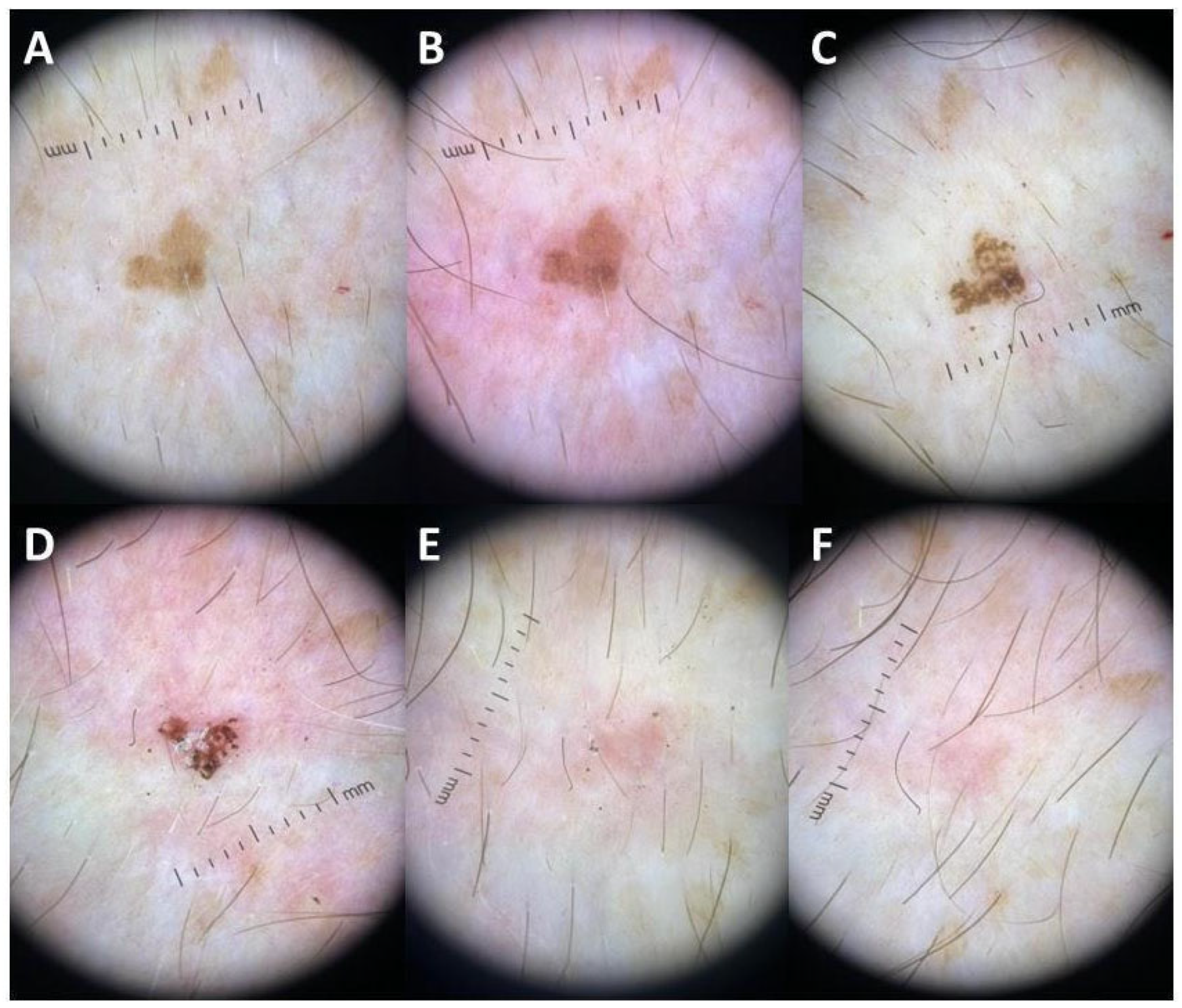
A representative solar lentigo from Case 1 (65-year-old man, Fitzpatrick skin type II, dorsum of the hand) is shown as a six-panel serial contact dermoscopic sequence after a single treatment session using the protocol described above (Figure 1A–F).
The sequence qualitatively captures early post-treatment change, including mild erythema and transient pigment darkening or crusting-like change at intermediate time points, followed by progressive attenuation at the final available follow-up.
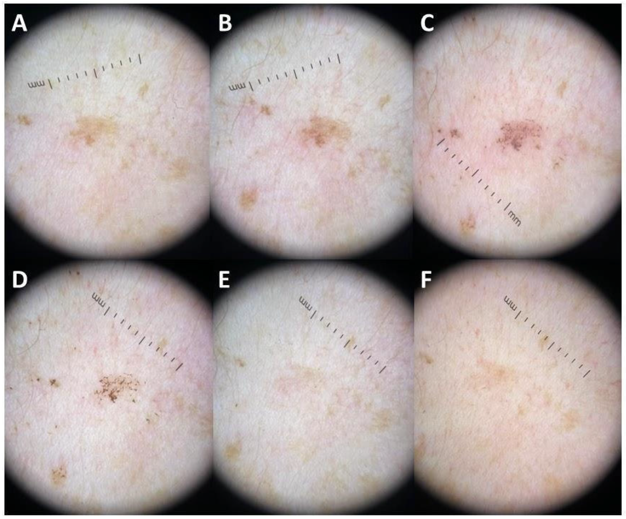
A second representative solar lentigo from Case 2 (42-year-old woman, Fitzpatrick skin type II, forehead) is shown as a separate six-panel serial contact dermoscopic sequence after a single treatment session (Figure 2A–F). This sequence likewise captures a staged short-term response pattern, with early post-treatment erythema and focal pigment alteration followed by progressive lightening at the final available follow-up.

4. Discussion

In this case series, a single-session, 532 nm millisecond green laser protocol was associated with complete-response classification at day 21 for all five target lesions, high patient-reported satisfaction, and only mild transient erythema resolving within the first day. These are short-term descriptive observations from a very small routine-practice series. The published literature suggests that 532 nm devices can be effective for solar lentigines and related superficial epidermal pigmented lesions, but outcomes are shaped by device type, pulse structure, endpoint selection, and treatment settings rather than by wavelength alone [7,8,32]. That distinction matters clinically because Q-switched, picosecond, and longer-pulsed 532 nm platforms do not behave identically. Comparative and randomized studies indicate that meaningful improvement can be achieved with several 532 nm approaches, while adverse-event profiles vary across devices, patient populations, and endpoint intensity [2,5,9,10,11]. In Q-switched and nanosecond-oriented protocols, studies in lentigines suggest that more aggressive immediate whitening strategies may increase PIH risk without a clear efficacy advantage, which supports conservative endpoint selection, particularly in darker phototypes [2,14,21,22,24]. Picosecond 532 nm studies also report favorable clinical improvement in selected settings, with some comparative studies suggesting lower PIH rates or higher satisfaction than certain Q-switched comparators, although outcome definitions and follow-up remain heterogeneous [9,25,26,28,29,30,33]. For longer-pulsed 532 nm approaches, the literature supports a narrower and more technical reading. These studies do not establish superiority over shorter-pulse systems, but they do support the plausibility of a more gradual, controlled treatment strategy when conservative endpoints, careful overlap control, and phototype-aware management are used [5,10,11]. Diagnostic triage remains essential before laser treatment of facial pigmented lesions. Reports of melanoma diagnosed after prior laser treatment underscore the need for careful clinical and dermoscopic evaluation and for a low threshold for biopsy when any lesion is equivocal [18,19,21,22,34]. The two serial illustrative cases qualitatively document the short-term treatment trajectory seen in the overall series. In both, early erythema and transient pigment alteration were followed by later attenuation in the final images. These image sequences remain descriptive rather than quantitative, but they help clarify endpoint selection and short-term evolution in a small case series. IPL and cryotherapy remain relevant comparators in routine practice. Available studies suggest that these modalities can improve lentigines, but PIH risk, downtime, and durability vary across devices, phototypes, and treatment protocols [12,13,27]. Biologic studies further suggest that solar lentigines are not merely static melanin deposits. Structural and cellular changes within chronically photoexposed skin may contribute to variable treatment response and to recurrence risk over time, which reinforces the need for longer follow-up in future studies [15,35,36,37,38,39,40,41,42]. Limitations include the very small sample size, the absence of a comparator group, the routine-practice nature of the series, possible selection bias, and the use of retrospective categorical response assessment rather than objective pigment quantification. Follow-up was limited to 21 days, so recurrence, durability, delayed PIH, and later adverse events could not be assessed. Pain was not formally quantified, and only two cases had intermediate serial dermoscopic documentation. The review component was intentionally targeted rather than fully systematic, was limited to PubMed/Medline and English-language studies, and did not include formal risk-of-bias scoring. Within these limits, the report is best read as a small technical and descriptive contribution. Its main value lies in presenting a clearly reported single-session 532 nm millisecond protocol and in placing those short-term observations within a cautious reading of the heterogeneous published literature.

5. Conclusions

For five target lesions treated with a single-session millisecond 532 nm green laser protocol, complete-response classification was observed at the final available day 21 follow-up, with high VAS satisfaction and no persistent adverse events documented within that short interval. Published clinical studies suggest that outcomes with 532 nm devices depend on device type, pulse structure, and treatment settings, and that larger comparative studies with objective pigment measures and longer follow-up are still needed.

Supplementary Materials

The following supporting information can be downloaded at https://www.mdpi.com/article/10.3390/cosmetics13030128/s1, Table S1: Detailed protocol parameters and quantitative efficacy/safety outcomes for the 532 nm treatment arms of the clinical studies summarized in Table 2.

Author Contributions

Conceptualization, G.C. and M.S.; methodology, G.C., S.P.N. and A.D.; validation, G.P., S.P.N. and A.D.; formal analysis, L.G. (Luca Guarino) and E.D.D.; investigation, A.C., L.G. (Luca Guarino), L.G. (Luca Gargano) and G.R.; resources, E.Z., G.R. and E.D.D.; data curation, E.Z.; writing—original draft preparation, E.Z. and L.G. (Luca Guarino); writing—review and editing, L.G. (Luca Gargano) and E.Z.; visualization, E.Z.; supervision, G.P., G.C. and S.P.N. All authors have read and agreed to the published version of the manuscript.

Funding

This research received no external funding.

Institutional Review Board Statement

All procedures performed in studies involving human participants were in accordance with the ethical standards of the institutional and/or national research committee (Committee “Calabria Centro” with reference number 373/2019, 17 December 2019) and with the 1964 Helsinki Declaration and its later amendments or comparable ethical standards.

Informed Consent Statement

Informed consent was obtained from all subjects involved in the study.

Data Availability Statement

The data presented in this study are available on request from the corresponding author. The data are not publicly available due to privacy restrictions concerning patient images and identifiable clinical information.

Conflicts of Interest

The authors declare no conflicts of interest.

References

  1. Ortonne, J.P.; Pandya, A.G.; Lui, H.; Hexsel, D. Treatment of solar lentigines. J. Am. Acad. Dermatol. 2006, 54, S262–S271. [Google Scholar] [CrossRef]
  2. Negishi, K.; Akita, H.; Tanaka, S.; Yokoyama, Y.; Wakamatsu, S.; Matsunaga, K. Comparative study of treatment efficacy and the incidence of post-inflammatory hyperpigmentation with different degrees of irradiation using two different quality-switched lasers for removing solar lentigines on Asian skin. J. Eur. Acad. Dermatol. Venereol. 2013, 27, 307–312. [Google Scholar] [CrossRef] [PubMed]
  3. Anderson, R.R.; Parrish, J.A. Selective photothermolysis: Precise microsurgery by selective absorption of pulsed radiation. Science 1983, 220, 524–527. [Google Scholar] [CrossRef] [PubMed]
  4. Goldman, L.; Blaney, D.J.; Kindel, D.J., Jr.; Richfield, D.; Franke, E.K. Pathology of the effect of the laser beam on the skin. Nature 1963, 197, 912–914. [Google Scholar] [CrossRef]
  5. Chan, H.H.; Fung, W.K.; Ying, S.Y.; Kono, T. An in vivo trial comparing the use of different types of 532 nm Nd:YAG lasers in the treatment of facial lentigines in Oriental patients. Dermatol. Surg. 2000, 26, 743–749. [Google Scholar] [CrossRef]
  6. Thawabteh, A.M.; Jibreen, A.; Karaman, D.; Thawabteh, A.; Karaman, R. Skin Pigmentation Types, Causes and Treatment-A Review. Molecules 2023, 28, 4839. [Google Scholar] [CrossRef]
  7. Mardani, G.; Nasiri, M.J.; Namazi, N.; Farshchian, M.; Abdollahimajd, F. Treatment of solar lentigines: A systematic review of clinical trials. J. Cosmet. Dermatol. 2025, 24, e70133. [Google Scholar] [CrossRef] [PubMed]
  8. Mukovozov, I.; Roesler, J.; Kashetsky, N.; Gregory, A. Treatment of lentigines: A systematic review. Dermatol. Surg. 2023, 49, 17–24. [Google Scholar] [CrossRef]
  9. Vachiramon, V.; Iamsumang, W.; Triyangkulsri, K. Q-switched double frequency Nd:YAG 532-nm nanosecond laser vs. double frequency Nd:YAG 532-nm picosecond laser for the treatment of solar lentigines in Asians. Lasers Med. Sci. 2018, 33, 1941–1947. [Google Scholar] [CrossRef]
  10. Negishi, K.; Tanaka, S.; Tobita, S. Prospective, randomized, evaluator-blinded study of the long pulse 532-nm KTP laser alone or in combination with the long pulse 1064-nm Nd:YAG laser on facial rejuvenation in Asian skin. Lasers Surg. Med. 2016, 48, 844–851. [Google Scholar] [CrossRef]
  11. Carniol, P.J.; Farley, S.; Friedman, A. Long-pulse 532-nm diode laser for nonablative facial skin rejuvenation. Arch. Facial Plast. Surg. 2003, 5, 511–513. [Google Scholar] [CrossRef][Green Version]
  12. Seirafi, H.; Fateh, S.; Farnaghi, F.; Ehsani, A.H.; Noormohammadpour, P. Efficacy and safety of long-pulse pulsed dye laser delivered with compression versus cryotherapy for treatment of solar lentigines. Indian J. Dermatol. 2011, 56, 48–51. [Google Scholar] [CrossRef]
  13. Kawada, A.; Shiraishi, H.; Asai, M.; Kameyama, H.; Sangen, Y.; Aragane, Y.; Tezuka, T. Clinical improvement of solar lentigines and ephelides with an intense pulsed light source. Dermatol. Surg. 2002, 28, 504–508. [Google Scholar] [CrossRef]
  14. Iraji, F.; Mousavi, A.; Poostiyan, N.; Saber, M. Q-switched frequency-doubled Nd:YAG (532 nm) laser versus trichloroacetic acid 35% peeling in the treatment of dorsal hand solar lentigo: An assessor-blind split-hand randomized controlled trial. J. Cosmet. Dermatol. 2022, 21, 6776–6782. [Google Scholar] [CrossRef]
  15. Pollefliet, C.; Corstjens, H.; González, S.; Hellemans, L.; Declercq, L.; Yarosh, D. Morphological characterization of solar lentigines by in vivo reflectance confocal microscopy: A longitudinal approach. Int. J. Cosmet. Sci. 2013, 35, 149–155. [Google Scholar] [CrossRef] [PubMed]
  16. Zipser, M.C.; Mangana, J.; Oberholzer, P.A.; French, L.E.; Dummer, R. Melanoma after laser therapy of pigmented lesions--circumstances and outcome. Eur. J. Dermatol. 2010, 20, 334–338. [Google Scholar] [CrossRef] [PubMed]
  17. Delker, S.; Livingstone, E.; Schimming, T.; Schadendorf, D.; Griewank, K.G. Melanoma diagnosed in lesions previously treated by laser therapy. J. Dermatol. 2017, 44, 23–28. [Google Scholar] [CrossRef] [PubMed]
  18. Pohl, L.; Kaiser, K.; Raulin, C. Pitfalls and recommendations in cases of laser removal of decorative tattoos with pigmented lesions: Case report and review of the literature. JAMA Dermatol. 2013, 149, 1087–1089. [Google Scholar] [CrossRef]
  19. Hibler, B.P.; Connolly, K.L.; Lee, E.H.; Rossi, A.M.; Nehal, K.S. Lentigo maligna melanoma with a history of cosmetic treatment: Prevalence, surgical outcomes and considerations. Lasers Surg. Med. 2017, 49, 819–826. [Google Scholar] [CrossRef]
  20. Nisticò, S.P.; Cannarozzo, G.; Provenzano, E.; Tamburi, F.; Fazia, G.; Sannino, M.; Negosanti, F.; Del Duca, E.; Patruno, C.; Bennardo, L. Nanosecond Q-switched 1064/532 nm laser to treat hyperpigmentations: A double center retrospective study. Clin. Pract. 2021, 11, 708–714. [Google Scholar] [CrossRef]
  21. Todd, M.M.; Rallis, T.M.; Gerwels, J.W.; Hata, T.R. A comparison of 3 lasers and liquid nitrogen in the treatment of solar lentigines: A randomized, controlled, comparative trial. Arch. Dermatol. 2000, 136, 841–846. [Google Scholar] [CrossRef]
  22. Ho, S.G.Y.; Chan, N.P.Y.; Yeung, C.K.; Shek, S.Y.; Chan, H.H.L. A retrospective analysis of the management of freckles and lentigines using four different pigment lasers on Asian skin. J. Cosmet. Laser Ther. 2012, 14, 74–80. [Google Scholar] [CrossRef]
  23. Kim, J.S.; Nam, C.H.; Kim, J.Y.; Gye, J.W.; Hong, S.P.; Kim, M.H.; Park, B.C. Objective evaluation of the effect of Q-switched Nd:YAG (532 nm) laser on solar lentigo by using a colorimeter. Ann. Dermatol. 2015, 27, 326–328. [Google Scholar] [CrossRef] [PubMed]
  24. Vachiramon, V.; Panmanee, W.; Techapichetvanich, T.; Chanprapaph, K. Comparison of Q-switched Nd:YAG laser and fractional carbon dioxide laser for the treatment of solar lentigines in Asians. Lasers Surg. Med. 2016, 48, 354–359. [Google Scholar] [CrossRef]
  25. Guss, L.; Goldman, M.P.; Wu, D.C. Picosecond 532 nm neodymium-doped yttrium aluminium garnet laser for the treatment of solar lentigines in darker skin types: Safety and efficacy. Dermatol. Surg. 2017, 43, 456–459. [Google Scholar] [CrossRef]
  26. Negishi, K.; Akita, H.; Matsunaga, Y. Prospective study of removing solar lentigines in Asians using a novel dual-wavelength and dual-pulse width picosecond laser. Lasers Surg. Med. 2018, 50, 851–858. [Google Scholar] [CrossRef] [PubMed]
  27. Friedmann, D.P.; Peterson, J.D. Efficacy and safety of intense pulsed light with a KTP filter for the treatment of solar lentigines. Lasers Surg. Med. 2019, 51, 500–508. [Google Scholar] [CrossRef]
  28. Kim, J.Y.; Yang, J.; Huh, G.; Choi, Y.J.; Kim, W.S. A split-face, single-blinded, randomized controlled comparison of 532 nm picosecond neodymium-doped yttrium aluminum garnet laser versus 532 nm Q-switched neodymium-doped yttrium aluminum garnet laser in the treatment of solar lentigines. Ann. Dermatol. 2020, 32, 8–13. [Google Scholar] [CrossRef] [PubMed]
  29. Vachiramon, V.; Namasondhi, A.; Anuntrangsee, T.; Jurairattanaporn, N. Randomized, evaluator-blinded comparative study of a potassium titanyl phosphate (KTP) 532-nm picosecond laser and an alexandrite 755-nm picosecond laser for the treatment of solar lentigines in Asians. J. Cosmet. Dermatol. 2022, 21, 4370–4377. [Google Scholar] [CrossRef]
  30. Liu, L.; Che, Q.; Zhou, Z.; Tang, Q.; Gao, Y.; He, Q.; Xie, Y.; Wa, Q. 730-nm, 532-nm and 694-nm laser in the treatment of freckles and solar lentigines (a randomized clinical trial). Lasers Med. Sci. 2025, 40, 309. [Google Scholar] [CrossRef]
  31. Lee, S.Y.; Kim, K.R.; Kim, S.; Han, H.S.; Hong, J.Y.; Seok, J.; Park, K.Y. Refractory solar lentigines successfully treated with 532-nm nanosecond Nd:YAG vasculature salvage laser surgery (VSLS) system: Case series. Skin. Res. Technol. 2024, 30, e70055. [Google Scholar] [CrossRef]
  32. Zawodny, P.; Zawodny, P.; Kulaszyńska, M.; Stój, E.; Knap-Czechowska, A.; Skonieczna-Żydecka, K.; Sieńko, J. The efficacy of 532/755 nm laser therapy for facial pigmented and vascular lesions: A systematic review and meta-analysis. J. Clin. Med. 2025, 14, 2546. [Google Scholar] [CrossRef]
  33. Bohnert, K.; Dorizas, A.; Sadick, N. A prospective, randomized, double-blinded, split-face pilot study comparing Q-switched 1064-nm Nd:YAG versus 532-nm Nd:YAG laser for the treatment of solar lentigines. J. Cosmet. Laser Ther. 2018, 20, 395–397. [Google Scholar] [CrossRef]
  34. Bukvić Mokos, Z.; Lipozenčić, J.; Ceović, R.; Stulhofer Buzina, D.; Kostović, K. Laser therapy of pigmented lesions: Pro and contra. Acta Dermatovenerol. Croat. 2010, 18, 185–189. [Google Scholar] [PubMed]
  35. Andersen, W.K.; Labadie, R.R.; Bhawan, J. Histopathology of solar lentigines of the face: A quantitative study. J. Am. Acad. Dermatol. 1997, 36, 444–447. [Google Scholar] [CrossRef] [PubMed]
  36. Nakamura, M.; Morita, A.; Seité, S.; Haarmann-Stemmann, T.; Krutmann, J. Environment-induced lentigines: Formation of solar lentigines beyond ultraviolet radiation. Exp. Dermatol. 2015, 24, 407–411. [Google Scholar] [CrossRef]
  37. Goorochurn, R.; Viennet, C.; Granger, C.; Fanian, F.; Varin-Blank, N.; Roy, C.L.; Humbert, P. Biological processes in solar lentigo: Insights brought by experimental models. Exp. Dermatol. 2016, 25, 174–177. [Google Scholar] [CrossRef]
  38. Chen, N.; Hu, Y.; Li, W.H.; Eisinger, M.; Seiberg, M.; Lin, C.B. The role of keratinocyte growth factor in melanogenesis: A possible mechanism for the initiation of solar lentigines. Exp. Dermatol. 2010, 19, 865–872. [Google Scholar] [CrossRef]
  39. Imokawa, G. Melanocyte activation mechanisms and rational therapeutic treatments of solar lentigos. Int. J. Mol. Sci. 2019, 20, 3666. [Google Scholar] [CrossRef] [PubMed]
  40. Rashid, T.; Hussain, I.; Haider, M.; Haroon, T.S. Laser therapy of freckles and lentigines with quasi-continuous, frequency-doubled, Nd:YAG (532 nm) laser in Fitzpatrick skin type IV: A 24-month follow-up. J. Cosmet. Laser Ther. 2002, 4, 81–85. [Google Scholar] [CrossRef]
  41. Vejjabhinanta, V.; Elsaie, M.L.; Patel, S.S.; Patel, A.; Caperton, C.; Nouri, K. Comparison of short-pulsed and long-pulsed 532 nm lasers in the removal of freckles. Lasers Med. Sci. 2010, 25, 901–906. [Google Scholar] [CrossRef]
  42. Wu, P.J.; Chen, S.T.; Liao, Y.H.; Sun, C.K. In vivo harmonic generation microscopy for monitoring the height of basal keratinocytes in solar lentigines after laser depigmentation treatment. BioMed Opt. Express 2021, 12, 6129–6142. [Google Scholar] [CrossRef] [PubMed]
Figure 1. Representative serial contact dermoscopic images of a dorsal hand solar lentigo from Case 1 documenting short-term lesion evolution after a single 532 nm green laser treatment through day 21. Panel timepoints were (A) = before treatment, (B) = immediately after treatment, (C) = day 1, (D) = day 8, (E) = day 12, and (F) = day 21.
Cosmetics 13 00128 g001
Figure 2. Representative serial contact dermoscopic images of Case 2 (panels (A)–(F)) documenting short-term lesion evolution after a single 532 nm green laser treatment through day 21. Panel timepoints were (A) = before treatment, (B) = immediately after treatment, (C) = day 1, (D) = day 8, (E) = day 12, and (F) = day 21.
Cosmetics 13 00128 g002
Table 1. Patient-level characteristics and outcomes. All five patients received a single laser session under the protocol described in Section 2.1 and were followed up on at day 21, when both independent dermatologists classified the target lesion as a complete response.
CaseSexAge (Years)Skin TypeSiteVAS (0–10)
Case 1M65IIDorsum of the hand9
Case 2F42IIForehead8
Case 3M58IIForehead10
Case 4M56IIICheek7
Case 5F47IILeft temple9
Abbreviations: F = female; M = male; VAS = visual analog scale. “Skin type” refers to Fitzpatrick skin phototype. The immediate reaction was mild transient erythema, which resolved within day 1 in all five patients. No persistent AEs were documented during the available 21-day follow-up.
Table 2. Selected clinical studies and contextual comparative evidence on 532 nm KTP/green laser devices for solar lentigines and related superficial epidermal pigmented lesions.
StudyDesign/NPopulation/SiteDevice/Pulse (Comparators)Key Outcomes and Safety
Todd et al., 2000 [21]RCT, n = 27Hand solar lentiginesQS 532 Nd:YAG vs. krypton vs. 532 vanadate vs. cryotherapyQS 532 Nd:YAG is most likely to provide significant lightening and has the fewest adverse effects; most patients preferred laser therapy.
Chan et al., 2000 [5]Randomized split-face, n = 34Facial lentigines (Chinese patients)Versapulse QS 532 vs. long-pulsed 532 vs. conventional QS 532Long-pulsed 532 was associated with comparable improvement to conventional QS 532 and lower complication risk than one QS platform.
Negishi et al., 2013 [2]Comparative randomized groups, 355 lesions/193 casesAsian skin (III-V)QS ruby vs. QS 532 Nd:YAG; aggressive vs. mild IW endpointsAggressive immediate whitening increased PIH without a clear efficacy advantage; mild endpoints reduced PIH risk.
Ho et al., 2012 [22]Retrospective analysisAsian skin; freckles/lentiginesMultiple pigment lasers (including 532 nm devices)Real-world outcomes across platforms underscore the heterogeneity of devices, endpoints, and PIH reporting.
Kim et al., 2015 [23]Prospective, n = 20Facial lentiginesQS 532 Nd:YAG; objective colorimetryImprovement is documented with physician/patient assessment and colorimetry, illustrating the value of objective pigment measures.
Vachiramon et al., 2016 [24]RCT, n = 25Upper extremity lentigines (Thai, III-IV)QS 532 Nd:YAG vs. fractional CO2QS 532 improved pigmentation more; fractional CO2 had faster healing and less pain; PIH did not differ significantly.
Vachiramon et al., 2018 [9]RCT, 30 paired lesions (28 completed)Upper extremities (Asian)KTP 532 ns vs. KTP 532 psBoth improved luminance; no clear difference in clearance; higher satisfaction with picosecond; similar adverse events.
Guss et al., 2017 [25]Prospective case seriesDarker skin typesPicosecond 532 nm Nd:YAGHigh clearance reported with acceptable safety when conservative endpoints were used.
Negishi et al., 2018 [26]Clinical trialAsian phototypes III-IVPicosecond 532 nm Nd:YAG (dual-wavelength/pulse width)High proportion of lesions achieved >75% clearance; low PIH; histology supported reduced collateral damage versus QS.
Friedmann & Peterson, 2019 [27]Prospective trial, n = 16Facial and hand solar lentigines (types II-III)IPL with KTP-like filter (525–585 nm)Significant pigment improvement with minimal downtime; the effect gradually declined over 6 months.
Kim et al., 2020 [28]Split-face RCT, n = 20Facial lentigines532 ps Nd:YAG vs. 532 QS Nd:YAGBoth effective; the picosecond arm was associated with greater improvement and lower PIH at 4 weeks.
Iraji et al., 2022 [14]Split-hand RCTDorsal hand solar lentigoQS 532 Nd:YAG vs. TCA 35% peelQS 532 was associated with greater lesion lightening and satisfaction; adverse events were generally mild.
Vachiramon et al., 2022 [29]Evaluator-blinded RCTSolar lentigines (Asian patients)KTP 532 ps vs. alexandrite 755 psBoth improved; the trial reported greater L* improvement with 755 ps; adverse events were similar.
Liu et al., 2025 [30]Randomized clinical trial, n = 42Freckles and solar lentigines (face)730 ps vs. 532 ps vs. 694 QSAll were effective; pain and histologic changes differed; 532 ps was associated with moderate vacuolar degeneration in basal cells.
Negishi et al., 2016 [10]Prospective split-face RCT, n = 22Asian skin; facial photodamage with bilateral solar lentiginesLP 532 KTP alone vs. LP 532 KTP + LP 1064 Nd:YAGImprovement in pigment indices occurred on both sides; there was no significant clinical difference, but subtle surface changes were detected by 3D analysis; adverse events were minor.
Lee et al., 2024 [31]Case series, n = 6Refractory solar lentigines (Korean patients)532 nm nanosecond Nd:YAG (VSLS system), single sessionComplete lesion removal reported without PIH; mild transient erythema.
Abbreviations: CO2 = carbon dioxide; IPL = intense pulsed light; IW = immediate whitening; KTP = potassium titanyl phosphate; L* = lightness coordinate in the CIELAB color space; LP = long-pulse; Nd:YAG = neodymium-doped yttrium aluminum garnet; ns = nanosecond; PIH = post-inflammatory hyperpigmentation; ps = picosecond; QS = Q-switched; RCT = randomized controlled trial; TCA = trichloroacetic acid.
Disclaimer/Publisher’s Note: The statements, opinions and data contained in all publications are solely those of the individual author(s) and contributor(s) and not of MDPI and/or the editor(s). MDPI and/or the editor(s) disclaim responsibility for any injury to people or property resulting from any ideas, methods, instructions or products referred to in the content.