Abstract
Hair loss and scalp dysfunction are prevalent concerns with limited non-medicinal long-term solutions. Growth factors and plant-derived extracellular vesicle (EV) represent promising regenerative approaches. In this exploratory randomized controlled trial, 60 healthy adults (18–60 years) were randomly assigned into five groups: (A) placebo; (B) base formula with 0.1% caffeine and panthenol; (C) base + recombinant Fc-fusion long-acting insulin-like growth factor-1 (rIGF-1) and fibroblast growth factor-7 (rFGF-7); (D) base + Centella asiatica (C. asiatica) EV; and (E) base + rIGF-1, rFGF-7, and C. asiatica EV. Participants applied their assigned product once daily for 56 days. Scalp and hair parameters, including sebum content, hair length, thickness, density, and hair loss, were assessed at baseline and Days 14, 28, 42, and 56. The combination of C. asiatica EV with rIGF-1 and rFGF-7 (Group E) showed the greatest improvements across all endpoints, including significant increases in hair thickness, density, and length, and a reduction in sebum content and hair loss by Day 56 compared with placebo. The results support further study of topical use of C. asiatica-derived EV with recombinant long-acting growth factors as a novel, naturally derived, cosmetic intervention for scalp and hair care.
1. Introduction
Hair loss and scalp dysfunction are prevalent conditions, with androgenetic alopecia (AGA) being the most common, affecting up to 50% of men and 30–40% of women during their lifetime []. Beyond aesthetic concerns, hair loss can profoundly affect self-esteem, psychosocial well-being, and overall quality of life [,]. The pathogenesis of AGA involves a complex interplay of genetic predisposition, androgen regulation such as the action of dihydrotestosterone (DHT) and its receptor, and follicular microenvironment signaling pathways such as Wnt/β-catenin, TGF-β, and IGF-1 [,].
Although FDA-approved treatments such as minoxidil and finasteride are widely used, their effectiveness requires continuous long-term use. Minoxidil prolongs the hair anagen (growth) phase but may cause rebound in hair loss after discontinuation []. Finasteride has been associated with sexual dysfunction, hormone-related side effects, and teratogenic risks upon exposure during pregnancy [,]. As a result, safer and more sustainable alternatives for promoting scalp and hair health are being explored, including growth factor treatment, stem cell-derived exosomes, and low-level laser therapy [,,,].
Plant extracts have gained interest as natural alternatives for topical hair formulations for their regenerative and antioxidant properties. Traditional botanicals such as saw palmetto (Serenoa repens), green tea (Camellia sinensis), and grey mangrove (Avicennia marina) inhibit 5α-reductase activity, while rosemary (Salvia rosmarinus) and grape seed (Vitis vinifera) improve scalp blood circulation and stimulate follicular proliferation [,]. Centella asiatica L. Urb., a medicinal herb native to Asia, possesses well-documented wound-healing, anti-inflammatory, and antioxidant properties attributed to triterpenes such as asiaticoside and madecassoside []. In vitro studies have shown that C. asiatica extract promotes collagen synthesis, dermal papilla cell health, and enhances the inductive capacity []. Hair lotions containing C. asiatica extract can strengthen hair follicles and roots by upregulating vascular endothelial growth factor (VEGF) and increasing blood supply around the hair follicle [,].
Extracellular vesicles (EVs) are nanosized lipid bilayer particles secreted by cells that transport bioactive proteins, nucleic acids, lipids, and secondary metabolites, exerting diverse physiological regulatory effects []. Compared to conventional extracts, EVs offers enhanced stability, cellular uptake, and targeted delivery, making them attractive natural delivery systems for skin applications [,]. Plant-derived EVs are produced during intercellular communication and contain many of the plant’s original bioactive ingredients which can be readily absorbed by mammalian cells []. EVs from C. asiatica and other plants have been shown to promote collagen synthesis, stimulate keratinocyte and fibroblast proliferation, and exhibit anti-inflammatory effects more effectively than crude extracts [,,]. Building on our previous work demonstrating the skin benefits of C. asiatica EV–based formulations in preclinical and clinical application, this study applies C. asiatica EV technology to scalp and hair care [,]
Growth factors such as insulin-like growth factor-1 (IGF-1) and fibroblast growth factor-7 (FGF-7, also known as keratinocyte growth factor, KGF), are essential regulators of hair follicle morphogenesis and regeneration [,,]. IGF-1 is primarily synthesized by dermal papilla cells, hair matrix cells, and dermal fibroblasts and exerts proliferative and anti-apoptotic effects on follicular keratinocytes via the PI3K/AKT signaling pathway [,,]. It prolongs the anagen phase, inhibits the transition of hair follicles into the catagen (shrinking) phase, and promotes follicular cell differentiation while exerting antioxidant and anti-inflammatory effects [,,,]. FGF-7 supports epithelial–mesenchymal interactions, stimulates hair matrix proliferation, and regulates outer root sheath cell differentiation through the MAPK/CREB and Wnt/β-catenin pathways [,,,]. By prolonging the anagen phase and delaying transition to catagen, FGF-7 contributes to hair shaft elongation and follicular renewal [,]. Recent advances in protein engineering have enabled the development of recombinant growth factors fused to the human IgG antibody Fc region (Fc-fusion) with extended half-lives and enhanced pharmacokinetics stability, enhancing their topical efficacy [,].
Other supportive ingredients, including caffeine and vitamin B5, also contribute to scalp and hair health. Caffeine, a natural alkaloid found in coffee and tea, stimulates hair matrix keratinocyte proliferation and counteract the inhibitory effects of DHT [,]. It enhances IGF-1 activity, prolongs the anagen phase, and supports stronger hair through activation of the cAMP pathway and antioxidant protection [,]. Vitamin B5 (pantothenic acid) and its derivative D-panthenol promote dermal papilla cell and outer root sheath cell proliferation, upregulating Ki-67, β-catenin, and VEGF, while suppressing TGF-β1 and caspase-associated apoptotic signals [,].
In this study, we developed a novel topical hair care essence that combines recombinant Fc-fusion long-acting IGF-1 (rIGF-1) and FGF-7 (rFGF-7) with C. asiatica EV in a base formulation containing caffeine and panthenol. We aimed to evaluate their synergistic effects on scalp and hair parameters in a prospective, randomized, controlled clinical trial. This approach integrates plant-derived biomaterials with engineered growth factors, offering a potential new direction in non-drug-based scalp and hair care.
2. Materials and Methods
2.1. Participants and Study Design
This study was a prospective, randomized, placebo-controlled, double-blinded clinical trial conducted to evaluate the effects of a topical scalp revitalizing essence on hair growth and hair loss. Participants were healthy adults of any gender aged 18 to 60 (inclusive) years.
Inclusion criteria include healthy adults defined as individuals without chronic diseases, significant illnesses (including cancer, post-stroke disorders, paralysis, acute myocardial infarction, coronary artery bypass surgery, end-stage renal disease, and major organ transplant or hematopoietic stem cell transplant), or allergic constitutions, and who are not currently taking any medication or using any scalp care products.
Exclusion criteria include individuals who are pregnant or breastfeeding, as well as males or females with chronic diseases, significant illnesses, or allergic constitutions, currently using scalp care products, students or employees affiliated with the principal investigator, individuals with scalp wounds, those who have participated in cosmetic product testing, and those who have undergone scalp aesthetic treatments within the past three months. Participants were randomly assigned to five groups (n = 12 per group) as detailed in the Description of Intervention below.
Participants were asked to apply approximately 1 mL of their assigned product once daily in the evening after shampooing. The product was applied over the entire scalp using a cotton pad and gently tapped into the scalp with fingertips to promote absorption. Participants were asked not to change their daily hair care routine other than the application of the test product. Usage was self-recorded in a provided diary. The duration of intervention was 8 weeks (56 days), with measurements taken at baseline (Day 0) and on Days 14, 28, 42, and 56.
Description of Intervention
The following formulations were evaluated:
- Group A: Placebo control (Base formula without caffeine and panthenol, see below)
- Group B: Base formula (active ingredients: 0.1% caffeine and panthenol)
- Group C: Base formula + recombinant long-acting insulin growth factor-1 (rIGF-1) and recombinant long-acting fibroblast growth factor-7 (rFGF-7)
- Group D: Base formula + Centella asiatica extracellular vesicles (C. asiatica EV).
- Group E: Base formula + rIGF-1, rFGF-7, and C. asiatica EV.
The composition of the formulations is as in Table 1:
Table 1.
Composition of the formulations. Plus (+) or minus (−) sign indicates the presence or absence of the component in the formulation.
C. asiatica EVs were isolated as previously described with INCi names Centella Asiatica Leaf/Petiole Vesicles (INCI ID: 39425) and Centella Asiatica Callus Vesicles (INCI ID: 40060) at a final concentration of 1011 particles/mL [].
rIGF-1 and rFGF-7 are recombinant proteins modified for long-acting by fusing human IGF-1 or human FGF-7 to a human IgG1 Fc region fragment with a flexible linker, with the following INCI names:
- rIGF-1: sr-(Methionyl sh-Oligopeptide-2 Dipeptide-46 Oligopeptide-163 sh-Polypeptide-181), INCI ID:40196
- rFGF-7: sr-(Methionyl sh-Polypeptide-3 Dipeptide-46 Oligopeptide-163 sh-Polypeptide-181), INCI ID:40201
2.2. Randomization and Blinding
Participants were randomly assigned in a 1:1:1:1:1 ratio into five parallel groups (n = 12 per group) using block randomization with a fixed block size of five, without stratification. The random allocation sequence was generated by the principal investigator and implemented using sequentially numbered, opaque, sealed envelopes (SNOSE) prepared by an independent staff member who was not involved in participant recruitment, assessment, or intervention delivery. The envelopes, identical in appearance and sealed with tamper-proof tape, were stored in a locked cabinet accessible only to the designated unblinded coordinator. Allocation was revealed only after eligibility confirmation and receipt of written informed consent.
A double-blind design was employed for this study. Participants, care providers administering the interventions, outcome assessors, and data analysts remained blinded to group allocation until the database lock, except in cases requiring emergency unblinding for safety reasons. Blinding was maintained by preparing all intervention and control products in identical containers with matched appearance, labeling, texture, viscosity, and scent. Each product was labeled only with a participant identification number, without an indication of group assignment. The randomization code was securely held by the independent, unblinded coordinator and was not released until study completion.
2.3. Assessment of Outcomes
Measurements were performed using two non-invasive diagnostic systems at standardized scalp sites (left, right, and vertex):
- Scalp sebum levels were quantified using C+K Multi Probe Adaptor MPA580 system with Sebumeter® SM815 probe (Courage-Khazaka Electronic GmbH, Koln, Germany) via grease-spot photometry.
- Hair length, thickness, and density were assessed with the ScalpX Intelligent Scalp Diagnostic System (VAST Technologies Inc., Taipei City, Taiwan) with a 5-megapixel handheld USB digital microscope (DMC1213_USB 200X, Coresync Technology Corp., New Taipei City, Taiwan) to capture high-resolution scalp images and analyze using machine learning and artificial intelligence with an internal database.
Hair loss was assessed by standardized 60-stroke combing test []. Participants were instructed to wash their hair on the day prior to the assessment. On the following day, under dry hair conditions, the hair was combed 60 times using a standardized comb. The shed hairs were collected on a clean surface and subsequently counted manually.
All assessments were conducted onsite at Hungkuang University (Taichung City, Taiwan) under controlled environmental conditions (20 ± 1 °C, 50 ± 5% RH) in a sealed room free of direct sunlight and airflow from vents.
For parameters such as sebum content, hair density, and hair loss, values were normalized as percentage improvement over baseline:
For hair length and hair thickness, absolute changes (in mm or μm) from baseline were used due to the linear nature of hair growth and narrow biological range, respectively.
Results for each participant were reported and calculated as the average of measurements from the left, right, and vertex regions of each participant to avoid pseudo-replication.
2.4. Statistical Analysis
Statistical analyses were performed using the built-in statistical package of GraphPad Prism 6.01 (Boston, MA, USA). Results were presented in mean values with 95% confidence intervals. For comparing demographic baseline values, the χ2 test was used for gender and the one-way ANOVA was used for continuous variables. To compare within-group changes over time, a repeated-measures ANOVA with the Greenhouse–Geisser correction for sphericity, followed by Dunnett’s post hoc test for multiple comparisons was used. For between-group comparisons at each timepoint, one-way ANOVA followed by Tukey’s HSD test for multiple comparisons. Both post hoc procedures include family-wise error rate correction to control for multiple comparisons. A p-value < 0.05 was considered statistically significant. All tests were two-tailed. The sample size in this clinical study was not based on any statistical assumptions or calculations, as this was an exploratory study aimed at observing the effects of the formulations.
3. Results
3.1. Participants
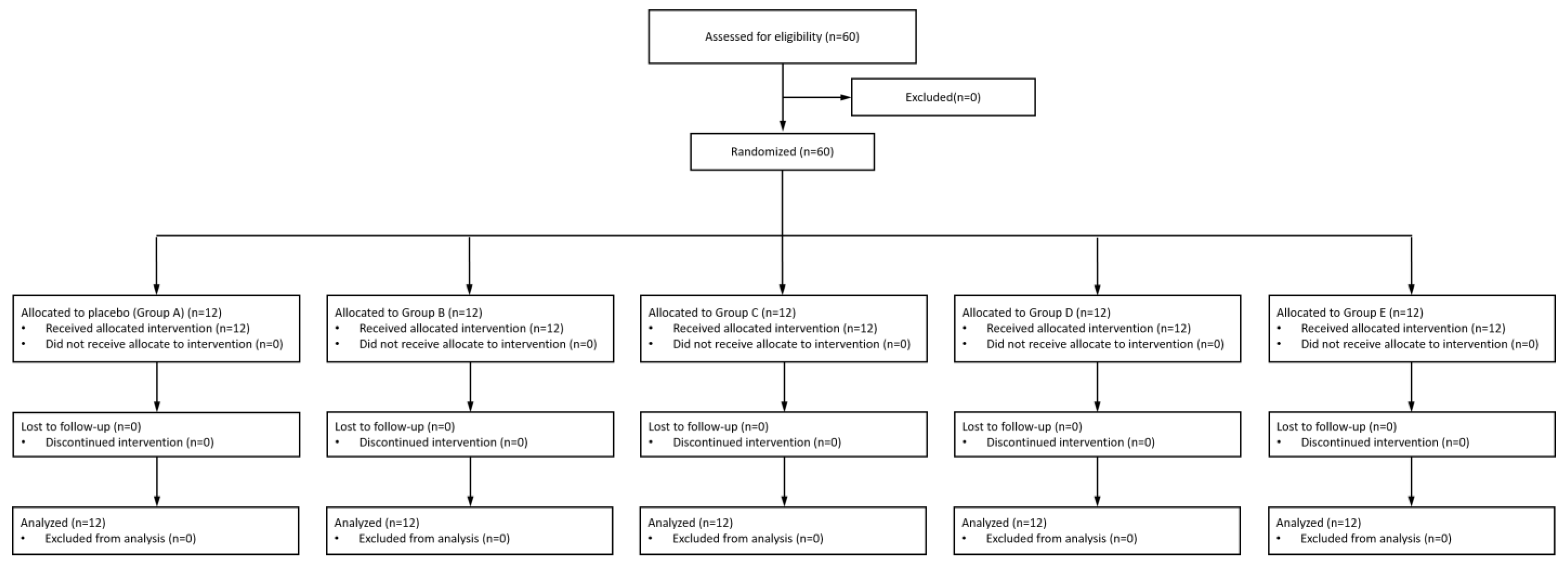
Between 8 April 2025 and 31 July 2025, a total of 60 healthy adult participants were enrolled and randomly assigned to five groups (n = 12 per group). All participants completed the study without major protocol violations or deviations. The CONSORT flow diagram summarizing participant recruitment, allocation, and follow-up is presented in Figure 1, and baseline demographic characteristics are shown in Table 2.
Figure 1.
CONSORT flowchart of the study.
Table 2.
Demographic characteristics and baseline values of the participants.
3.2. Assessments of Outcomes
3.2.1. Sebum Content
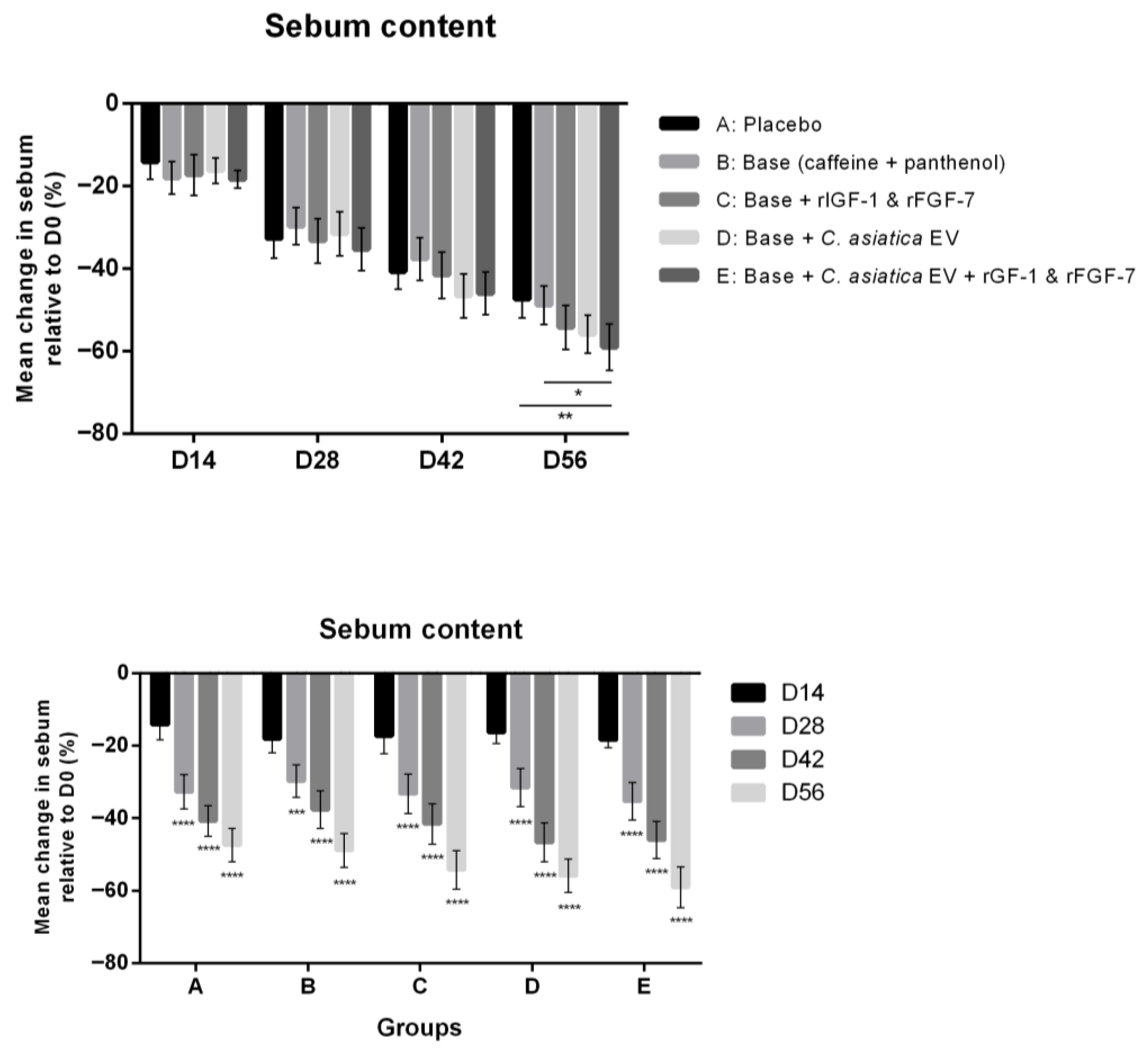
Sebum levels decreased over time across all groups, with formulations containing C. asiatica EV with rIFG-1 and rFGF-7 (Group E) resulting in significantly lower sebum content compared to both the base formulation (Group B) and placebo control by Day 56 (p < 0.05 and p < 0.01, respectively) (Figure 2). By Day 56, Group E achieved an average of 59% reduction from baseline, compared to 47.7% in the placebo group. In contrast, minimal changes were observed in Groups B to D relative to placebo (not statistically significant). Thus, the combined use of C. asiatica EVs, IGF-1, and FGF-7s could reduce sebum production in the scalp.
Figure 2.
Change (%) in sebum content compared to baseline (Day 0) at 14, 28, 42, and 56 days after start of the study. (Top): Comparison of between-group at each timepoint with statistical significance calculated using one-way ANOVA followed by Tukey’s HSD test; (Bottom): Comparison of within-group changes over time with statistical significance calculated using repeated measures ANOVA followed by Dunnett’s post hoc test. Results are presented in mean values with error bars representing 95% confidence intervals. * p < 0.05, ** p < 0.01, *** p < 0.001, **** p < 0.0001.
3.2.2. Hair Length
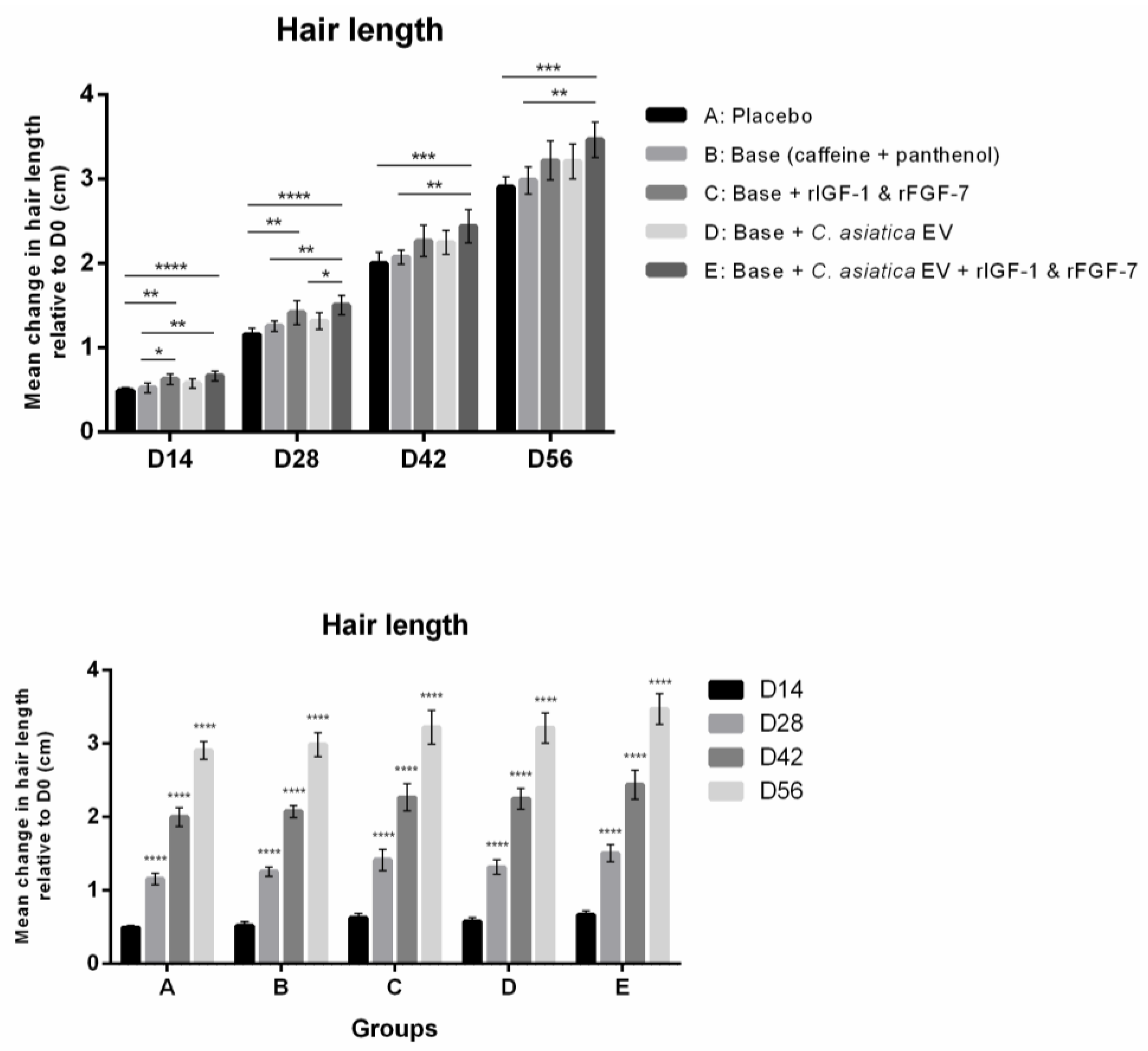
As expected, hair length increased in all groups due to natural hair growth over time (Figure 3, bottom). Group E achieved the greatest mean cumulative growth by Day 56 (3.5 cm), significantly exceeding both placebo (p < 0.001) and Group B (p < 0.01) (Figure 3 top). Interestingly, the difference between Group E and placebo was already significant on Day 14 (p < 0.0001) and remained so throughout the study. These results suggest that formulations containing C. asiatica EV and recombinant growth factors accelerate hair growth.
Figure 3.
Change (cm) in hair length compared to baseline (Day 0) at 14, 28, 42, and 56 days after start of the study. (Top): Comparison of between-group at each timepoint with statistical significance calculated using one-way ANOVA followed by Tukey’s HSD test; (Bottom): Comparison of within-group changes over time with statistical significance calculated using repeated measures ANOVA followed by Dunnett’s post hoc test. Results are presented in mean values with error bars representing 95% confidence intervals. * p < 0.05, ** p < 0.01, *** p < 0.001, **** p < 0.0001.
3.2.3. Hair Thickness
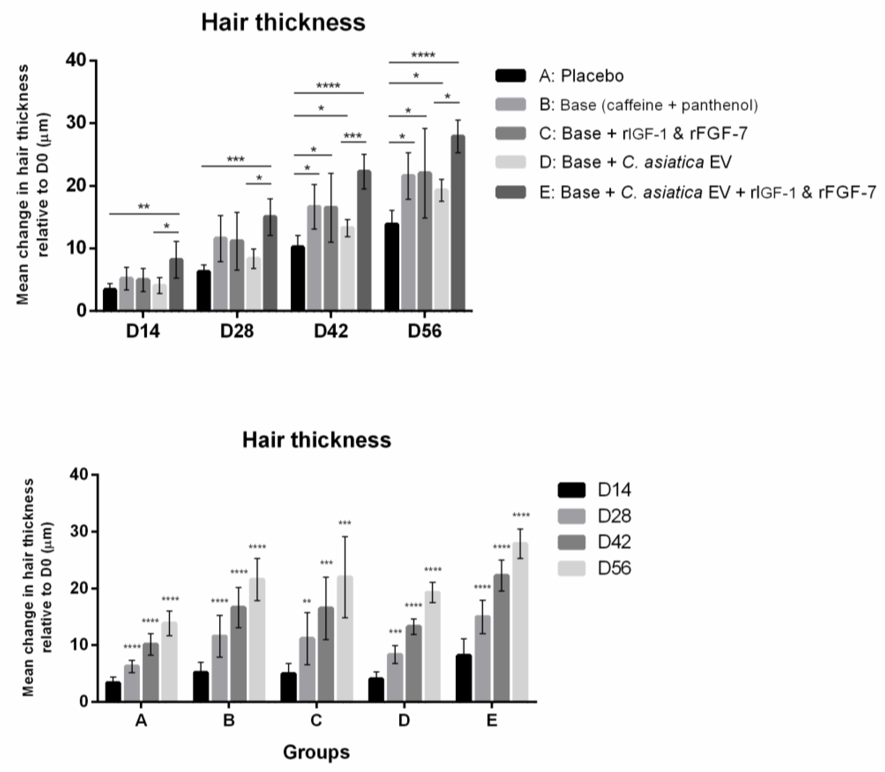
All treatment groups showed a progressive increase in hair shaft diameter (Figure 4 bottom). Group E again exhibited the greatest effect, with a mean increase of 27.9 μm at Day 56 compared to 13.9 μm of placebo (p < 0.0001) (Figure 4 top). Other formulations also showed significant improvements compared to placebo (p < 0.05). Notably, the inclusion of rIGF-1 and rFGF-7 with C. asiatica EV in Group E led to a significantly greater increase in hair thickness (p < 0.05) compared to C. asiatica EV alone in Group D (27.9 μm vs. 19.3 μm), suggesting a synergistic effect between EV and growth factors.
Figure 4.
Change (µm) in hair thickness compared to baseline (Day 0) at 14, 28, 42, and 56 days after start of the study. (Top): Comparison of between-group at each timepoint with statistical significance calculated using one-way ANOVA followed by Tukey’s HSD test; (Bottom): Comparison of within-group changes over time with statistical significance calculated using repeated measures ANOVA followed by Dunnett’s post hoc test. Results are presented in mean values with error bars representing 95% confidence intervals. * p < 0.05, ** p < 0.01, *** p < 0.001, **** p < 0.0001.
3.2.4. Hair Density
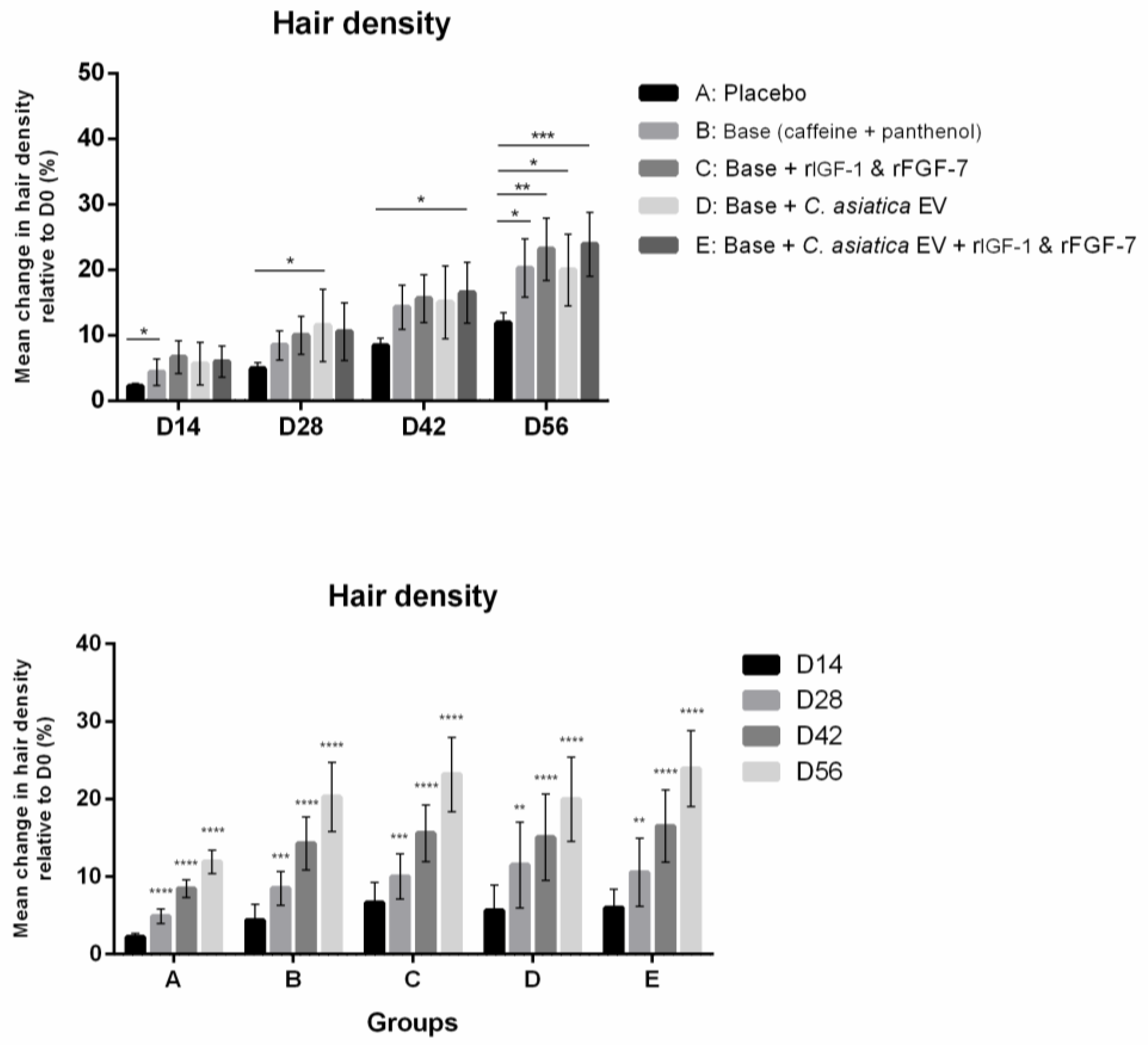
Hair density increased in all treatment groups, including the placebo group (Figure 5, bottom). Group E showed the most pronounced improvement, with a mean increase of approximately double (23.9% vs. 11.9% increase relative to baseline, p < 0.001) that of placebo by Day 56 (Figure 5 top). Group C also showed significant increases compared to placebo (23.2% vs. 11.9%, p < 0.01), while Groups B and D showed moderate but significant improvement (20.3% vs. 11.9%; 20.0% vs. 11.9%, p < 0.05). As seen in the hair thickness results, the synergistic effects of EV and growth factor may contribute to their greater effect in increasing follicular density.
Figure 5.
Change (%) in hair density compared to baseline (Day 0) at 14, 28, 42, and 56 days after start of the study. (Top): Comparison of between-group at each timepoint with statistical significance calculated using one-way ANOVA followed by Tukey’s HSD test; (Bottom): Comparison of within-group changes over time with statistical significance calculated using repeated measures ANOVA followed by Dunnett’s post hoc test. Results are presented in mean values with error bars representing 95% confidence intervals. * p < 0.05, ** p < 0.01, *** p < 0.001, **** p < 0.0001.
3.2.5. Hair Loss
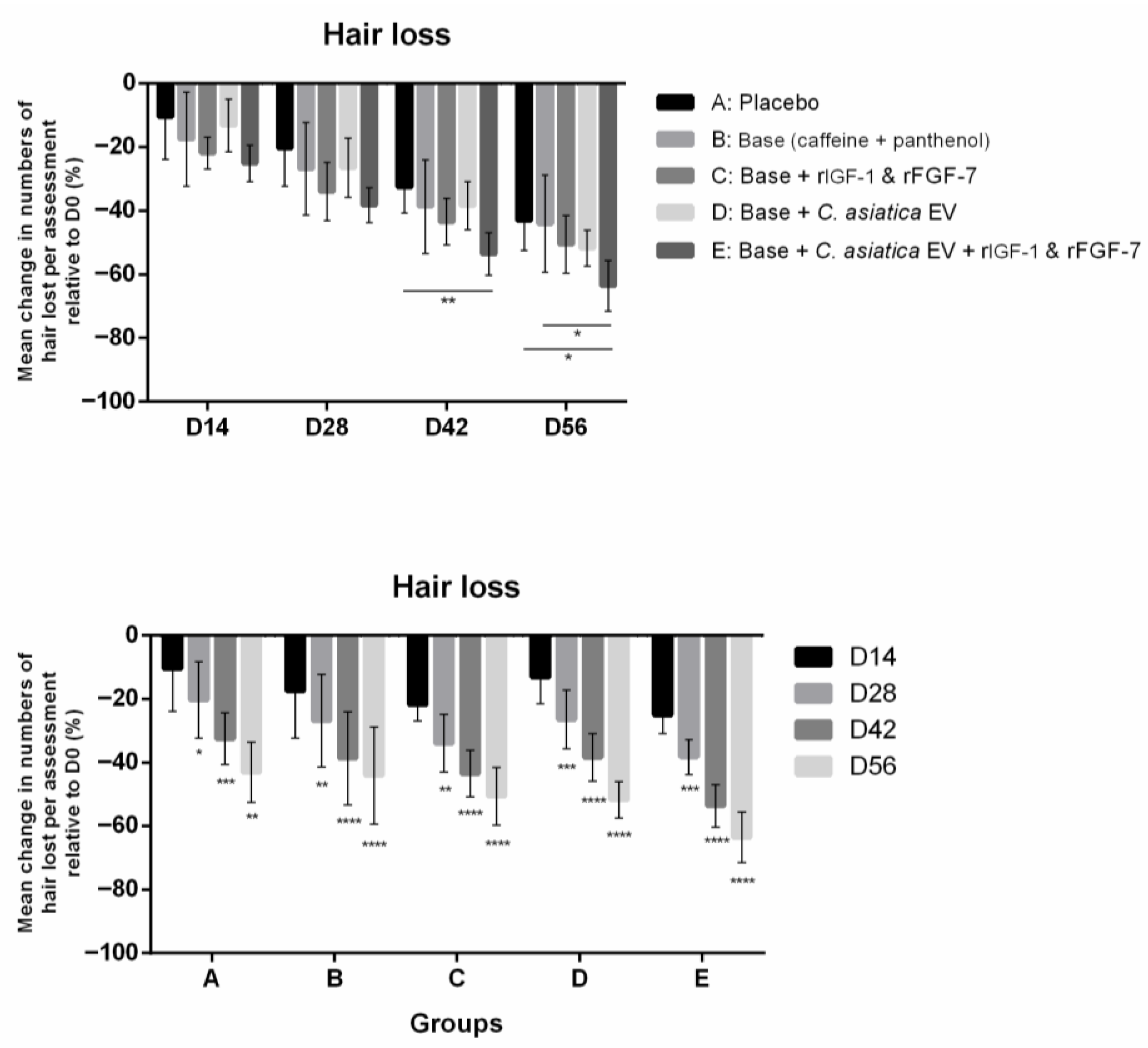
Hair shedding decreased progressively across all groups during the course of the study, including the placebo group (Figure 6 bottom). Group E had the greatest reduction in hair loss, with decreases of 53.6% and 63.6% at Days 42 and 56, respectively, compared to 32.5% and 43.1% in the placebo group (p < 0.01 and p < 0.05) (Figure 6 top). Other formulations did not result in a significant reduction in hair loss compared to placebo.
Figure 6.
Change (%) in hair lost during the assessment event compared to baseline (Day 0) at 14, 28, 42, and 56 days after start of the study. (Top): Comparison of between-group at each timepoint with statistical significance calculated using one-way ANOVA followed by Tukey’s HSD test; (Bottom): Comparison of within-group changes over time with statistical significance calculated using repeated measures ANOVA followed by Dunnett’s post hoc test. Results are presented in mean values with error bars representing 95% confidence intervals. * p < 0.05, ** p < 0.01, *** p < 0.001, **** p < 0.0001.
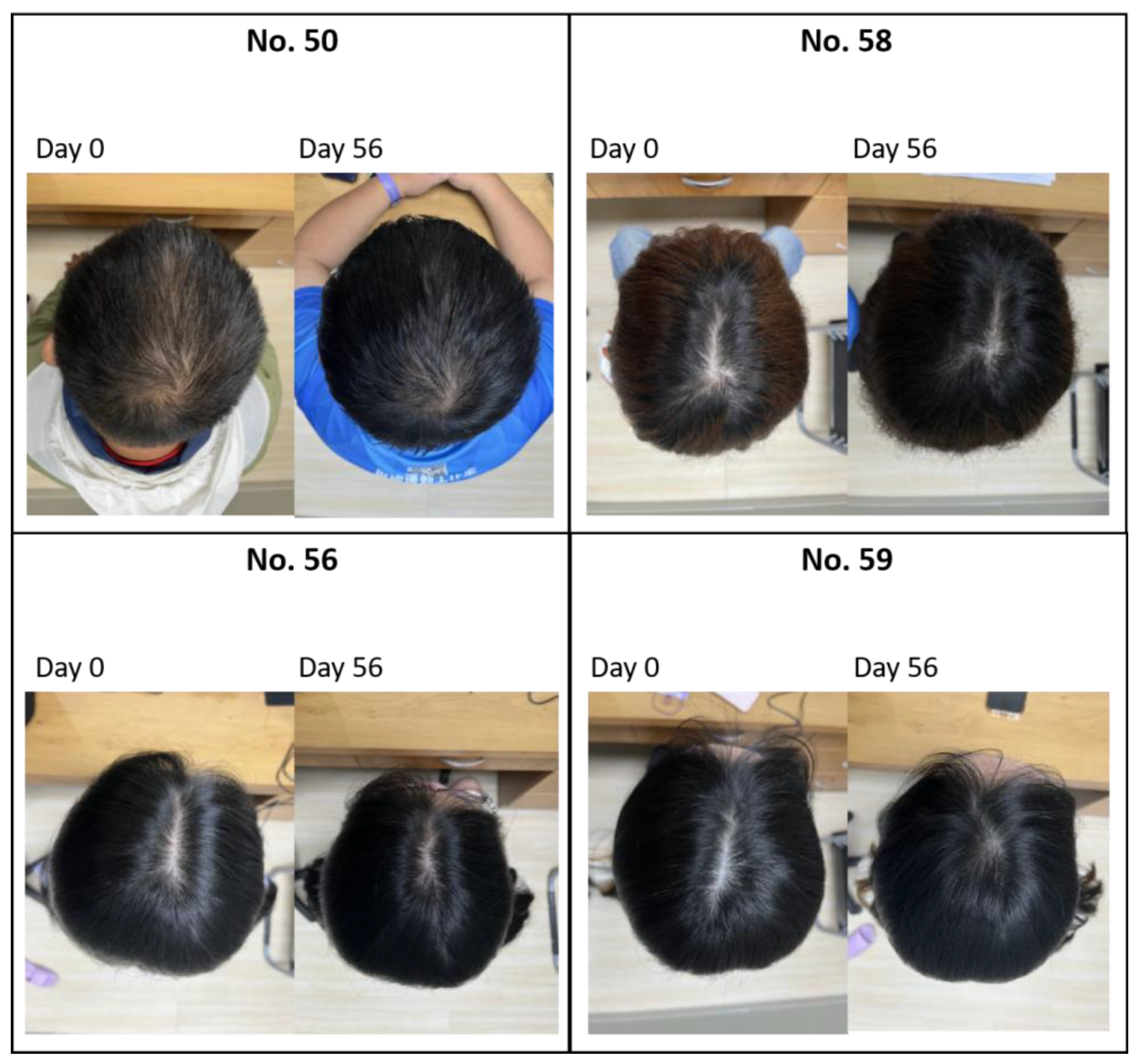
Representative photographs from Group E participants before treatment (Day 0) and after 56 days are shown in Figure 7, demonstrating visibly increased hair density, particularly at the vertex region.
Figure 7.
Representative photographs of the participants in Group E (Base formula with rIGF-1, rFGF-7, and C. asiatica EV) on Day 0 and Day 56 after treatment.
4. Discussion
This was a randomized placebo-controlled trial to evaluate the efficacy of topical formulations containing rIGF-1, rFGF-7, and C. asiatica EV in a base formulation with caffeine and panthenol on scalp and hair parameters over a 56-day period. Our findings demonstrate that the combined formulation Group E had the most consistent and pronounced effects in sebum and hair loss reduction, and hair growth (length, thickness, and density). By Day 56, hair shaft thickness increased by 27.9 μm, approximately twice that of placebo (13.9 μm, p < 0.0001). Similarly, hair density increased by 23.9%, also nearly double that of the placebo group (p < 0.001). These findings support a synergistic action between C. asiatica EV, rIGF-1, and rFGF-7, which are known to modulate signaling pathways involved in follicle development and dermal papilla function [,,,,,].
Hair length results further supported this, with Group E showing 3.5 cm of cumulative growth by Day 56, exceeding placebo (p < 0.001) and showing significant differences from as early as Day 14 (p < 0.0001) (Figure 3). While hair naturally grows at a relatively fixed rate, the enhancement observed here may reflect activation of IGF-1– and FGF-7–mediated signaling pathways that promote keratinocyte proliferation and prolong the anagen phase, although mechanistic pathways of our results have yet to be been elucidated [,,]. Similarly, hair loss decreased most markedly in Group E (63.6% reduction vs. 43.1% in the placebo group, p < 0.05). This is consistent with IGF-1’s known role in inhibiting follicular apoptosis and caffeine’s ability to counteract DHT-mediated follicular inhibition [,,,].
Sebum production was reduced in all groups, but significantly only in Group E with 59% reduction relative to baseline, significantly outperforming both placebo (p < 0.01) and the base formulation Group B (p < 0.05). C. asiatica EV can contain triterpenoids, lipids, proteins, and anti-inflammatory compounds as well as mRNAs that may regulate sebaceous glands and microvascular perfusion [,,]. While the effect of C. asiatica EV on hair follicles has yet to be reported, C. asiatica extract is known to promote dermal papilla activity and vascular growth via VEGF signaling [,].
Our results obtained here are in agreement with previous studies demonstrating the roles of IGF-1 and FGF-7 in promoting dermal papilla proliferation, follicle regeneration, and keratinocyte survival [,,,,,,]. While topical minoxidil and finasteride remain the most widely used growth stimulants, they are known to cause rebound shedding upon discontinuation and side effects that impact quality of life [,,]. Therefore, combining growth factor with plant-derived EVs may offer a multi-targeted approach to scalp care. To our knowledge, this is the first human trial to combine plant-derived EV with engineered growth factors as a promising cosmetic application for hair and scalp care [,,,]. The observed improvements in scalp and hair parameters reflect cosmetic enhancement consistent with improvement in follicular and scalp microenvironment. However, regenerative mechanisms remain hypothetical in the absence of molecular and mechanistic data.
Nevertheless, several limitations of the study must be acknowledged. First, modest improvements were observed even in the placebo group, likely attributable to natural telogen-to-anagen cycling, standardized hair care routines, and psychological effects of study participation (Hawthorne effect) [,,]. Some placebo components, such as glycerin and menthol, may have contributed physiologically even though they are not active ingredients per se. Glycerin is commonly used as a humectant in cosmetics that can retain water in the stratum corneum and help skin barrier repair, leading to improved scalp hydration []. Menthol and menthyl lactate activate TRPM8 channels to provide a cooling sensation while also stimulating cutaneous blood flow, possibly leading to increased perfusion of follicular metabolism and reduced skin irritation []. Second, as an exploratory study, the short duration (56 days) may be insufficient to capture the complete picture of hair growth cycles or delayed responses, which typically span several months. Third, the relatively young (mean age 36 years) and predominantly female sample may limit the generalizability of the findings to older and male populations with more advanced androgenetic alopecia or age-related scalp conditions [,]. The small sample size also precludes subgroup analysis by age, gender, or baseline scalp condition. Since follicular renewal capacity declines with age, further studies in middle-aged and older individuals using a balanced gender ratio are warranted to confirm whether the synergistic effects of C. asiatica EV, rIGF-1 and rFGF-7 are equally effective across different age groups. Therefore, the findings should be interpreted as preliminary evidence to support future studies, including long-term safety and outcomes, mechanistic biomarkers, and scalp biopsies to investigate the long-term and increased frequency of use of the product.
In conclusion, the topical application of an essence containing C. asiatica EVs, IGF-1, FGF-7, and caffeine led to significant improvements in hair thickness, density, length, and sebum control, along with a reduction in hair loss after 56 days of use. These effects suggest a synergistic interaction between plant-derived EVs, rIGF-1, and rFGF-7, offering a cosmetic approach to scalp and hair health distinct from conventional drug-based alopecia treatment.
Author Contributions
Conceptualization, supervision and project administration: T.-M.C., C.C. and T.-Y.K. Visualization and formal analysis: T.-M.C., W.-J.H. and L.T.-C.L. Investigation, data acquisition and curation: T.-M.C., H.-C.H. and J.-Y.L. Methodology: T.-M.C., C.-C.W., H.-C.H., J.-Y.L., C.-H.C., P.-L.K., W.-H.T., W.-Y.Q., I.P. and T.-Y.K. Resources: C.-C.W., C.-H.C., P.-L.K., W.-H.T. and T.-Y.K. Writing—original draft preparation: T.-M.C., W.-J.H. and L.T.-C.L. Writing—review and editing: T.-M.C., W.-J.H., L.T.-C.L., W.-Y.Q., I.P., C.C. and T.-Y.K. All authors have read and agreed to the published version of the manuscript.
Funding
Schweitzer Biotech Company provided the necessary funding for the study as well as provided the test products, but had no role in enrollment, participation, data collection, data analysis, or completion of final report of the clinical study. All participants provided written informed consent prior to the start of the study.
Institutional Review Board Statement
The finalized protocol, case report form, advertisement, and informed consent form were reviewed and approved by the institutional review board of the Antai Tian-Sheng Memorial Hospital (IRB No. 25-016-A, approved 6 February 2025). All participants provided written informed consent prior to the start of the study. This study was conducted in compliance with the Declaration of Helsinki and in compliance with all International Conference on Harmonization Good Clinical Practice Guidelines. This clinical study was registered on ClinicalTrials.gov under NCT06985121 on 22 May 2025.
Data Availability Statement
All relevant data is provided within the manuscript.
Acknowledgments
We thank colleagues at Schweitzer Biotech Company for reviewing and providing feedback for the manuscript preparation.
Conflicts of Interest
Authors C.-C.W., C.-H.C., P.-L.K., W.-H.T., W.-J.H., L.T.-C.L., C.C., and T.-Y.K. are employed by Schweitzer Biotech Company. T.-M.C., W.-Y.Q., and I.P. are consultants employed by Schweitzer Biotech Company. The remaining authors declare that the research was conducted in the absence of any commercial or financial relationships that could be construed as a potential conflict of interest.
References
- Norwood, O.T. Male pattern baldness: Classification and incidence. South Med. J. 1975, 68, 1359–1365. [Google Scholar] [CrossRef]
- Cash, T.F. The psychology of hair loss and its implications for patient care. Clin. Dermatol. 2001, 19, 161–166. [Google Scholar] [CrossRef]
- Alfonso, M.; Richter-Appelt, H.; Tosti, A.; Viera, M.S.; Garcia, M. The psychosocial impact of hair loss among men: A multinational European study. Curr. Med. Res. Opin. 2005, 21, 1829–1836. [Google Scholar] [CrossRef] [PubMed]
- Trueb, R.M. Molecular mechanisms of androgenetic alopecia. Exp. Gerontol. 2002, 37, 981–990. [Google Scholar] [CrossRef] [PubMed]
- Randall, V.A. Androgens and hair growth. Dermatol. Ther. 2008, 21, 314–328. [Google Scholar] [CrossRef] [PubMed]
- Messenger, A.G.; Rundegren, J. Minoxidil: Mechanisms of action on hair growth. Br. J. Dermatol. 2004, 150, 186–194. [Google Scholar] [CrossRef]
- Kaufman, K.D.; Olsen, E.A.; Whiting, D.; Savin, R.; DeVillez, R.; Bergfeld, W.; Price, V.H.; Van Neste, D.; Roberts, J.L.; Hordinsky, M.; et al. Finasteride in the treatment of men with androgenetic alopecia. J. Am. Acad. Dermatol. 1998, 39, 578–589. [Google Scholar] [CrossRef]
- Irwig, M.S. Persistent sexual side effects of finasteride: Could they be permanent? J. Sex. Med. 2012, 9, 2927–2932. [Google Scholar] [CrossRef]
- Yuan, A.R.; Bian, Q.; Gao, J.Q. Current advances in stem cell-based therapies for hair regeneration. Eur. J. Pharmacol. 2020, 881, 173197. [Google Scholar] [CrossRef]
- Avci, P.; Gupta, G.K.; Clark, J.; Wikonkal, N.; Hamblin, M.R. Low-level laser (light) therapy (LLLT) for treatment of hair loss. Lasers Surg. Med. 2014, 46, 144–151. [Google Scholar] [CrossRef]
- Trueb, R.M. Further clinical evidence for the effect of IGF-1 on hair growth and alopecia. Skin Appendage Disord. 2018, 4, 90–95. [Google Scholar] [CrossRef]
- Danilenko, D.M.; Ring, B.D.; Yanagihara, D.; Benson, W.; Wiemann, B.; Starnes, C.O.; Pierce, G.F. Keratinocyte growth factor is an important endogenous mediator of hair follicle growth, development, and differentiation. Am. J. Pathol. 1995, 147, 145–154. [Google Scholar]
- Dhariwala, M.Y.; Ravikumar, P. An overview of herbal alternatives in androgenetic alopecia. J. Cosmet. Dermatol. 2019, 18, 966–975. [Google Scholar] [CrossRef] [PubMed]
- Jain, R.; Monthakantirat, O.; Tengamnuay, P.; De-Eknamkul, W. Identification of a new plant extract for androgenic alopecia treatment using a non-radioactive human hair dermal papilla cell-based assay. BMC Complement. Altern. Med. 2015, 16, 18. [Google Scholar] [CrossRef] [PubMed]
- Bandopadhyay, S.; Mandal, S.; Ghorai, M.; Jha, N.K.; Kumar, M.; Radha, N.; Ghosh, A.; Proćków, J.; Perez de la Lastra, J.M.; Dey, A. Therapeutic Properties and Pharmacological Activities of Asiaticoside and Madecassoside: A Review. J. Cell. Mol. Med. 2023, 27, 593–608. [Google Scholar] [CrossRef] [PubMed]
- Choi, Y.M.; An, S.; Lee, J.; Lee, J.H.; Lee, J.N.; Kim, Y.S.; Ahn, K.J.; An, I.S.; Bae, S. Titrated extract of Centella asiatica increases hair inductive property through inhibition of STAT signaling pathway in three-dimensional spheroid cultured human dermal papilla cells. Biosci. Biotechnol. Biochem. 2017, 81, 2323–2329. [Google Scholar] [CrossRef]
- Togni, S.; Meneghin, M.; Eggenhoffner, R.; Giacomelli, L. Strengthening hair with Centella asiatica: A report of clinical and subjective efficacy of a local treatment with a 0.5% hair lotion. Esperienze Dermatol. 2018, 20 (Suppl. S2), 27–30. [Google Scholar] [CrossRef]
- Saansoomchai, P.; Surangkul, D.; Srikummool, T.C.M. Enhanced VEGF expression in hair follicle dermal papilla cells by Centella asiatica Linn. CMU J. Nat. Sci. 2018, 17, 25–37. [Google Scholar] [CrossRef]
- Jeppesen, D.K.; Zhang, Q.; Franklin, J.L.; Coffey, R.J. Extracellular vesicles and nanoparticles: Emerging complexities. Trends Cell Biol. 2023, 33, 667. [Google Scholar] [CrossRef]
- Moghassemi, S.; Dadashzadeh, A.; Sousa, M.J.; Vlieghe, H.; Yang, J.; León-Félix, C.M.; Amorim, C.A. Extracellular vesicles in nanomedicine and regenerative medicine: A re-view over the last decade. Bioact. Mater. 2024, 36, 126. [Google Scholar] [CrossRef]
- Yang, G.H.; Lee, Y.B.; Kang, D.; Choi, E.; Nam, Y.; Lee, K.H.; You, H.J.; Kang, H.J.; An, S.H.; Jeon, H. Overcome the barriers of the skin: Exosome therapy. Biomater. Res. 2023, 25, 22. [Google Scholar] [CrossRef]
- Liu, R.; Zhang, F.; He, X.; Huang, K. Plant derived exosome-like nanoparticles and their therapeutic applications in glucolipid metabolism diseases. J. Agric. Food Chem. 2025, 73, 6385–6399. [Google Scholar] [CrossRef] [PubMed]
- Chang, T.M.; Wu, C.C.; Huang, H.C.; Wang, S.S.; Chuang, C.H.; Kao, P.L.; Tang, W.H.; Liu, L.T.C.; Qiu, W.Y.; Percec, I.; et al. In Vitro Characterization of Centella asiatica Extracellular Vesicles and Their Skin Repair Effects in a UVB-Irradiated Mouse Model. Int. J. Mol. Sci. 2025, 26, 8982. [Google Scholar] [CrossRef] [PubMed]
- Li, A.; Li, D.; Gu, Y.; Liu, R.; Tang, X.; Zhao, Y.; Qi, F.; Wei, J.; Liu, J. Plant-Derived Nanovesicles: Further Exploration of Biomedical Function and Application Potential. Acta Pharm. Sin. 2023, 13, 3300–3320. [Google Scholar] [CrossRef] [PubMed]
- Kim, M.H.; Yoon, E.J.; Kim, J.S.; Park, S.J.; Lee, H. Comparative analysis of the transcriptome and efficacy of bioactive Centella asiatica exosomes on skin cells. Res. Sq. 2023. [Google Scholar] [CrossRef]
- Chang, T.-M.; Wu, C.-C.; Huang, H.-C.; Wang, S.-S.; Chuang, C.-H.; Kao, P.-L.; Tang, W.-H.; Liu, L.T.-C.; Qiu, W.-Y.; Percec, I.; et al. A 28-Day Pilot Study of the Effects on Facial Skin Hydration, Elasticity, and Texture of a Centella asiatica Extracellular Vesi-cle-Based Skin Care Formulation. Cosmetics 2025, 12, 186. [Google Scholar] [CrossRef]
- Su, H.Y.; Hickford, J.G.; Bickerstaffe, R.; Palmer, B.R. Insulin-like growth factor 1 and hair growth. Dermatol. Online J. 1999, 5, 1. [Google Scholar] [CrossRef]
- Li, J.; Yang, Z.; Li, Z.; Gu, L.; Wang, Y.; Sung, C. Exogenous IGF-1 promotes hair growth by stimulating cell proliferation and down regulating TGF-beta1 in C57BL/6 mice in vivo. Growth Horm. IGF Res. 2014, 24, 89–94. [Google Scholar] [CrossRef]
- Zhao, J.; Harada, N.; Okajima, K. Dihydrotestosterone inhibits hair growth in mice by inhibiting insulin-like growth factor-I production in dermal papillae. Growth Horm. IGF Res. 2011, 21, 260–267. [Google Scholar] [CrossRef]
- Philpott, M.P.; Sanders, D.A.; Kealey, T. Effects of insulin and insulin-like growth factors on cultured human hair follicles: IGF-I at physiologic concentrations is an important regulator of hair follicle growth in vitro. J. Investig. Dermatol. 1994, 102, 857–861. [Google Scholar] [CrossRef]
- Hayashi, Y.; Yamamoto, N.; Nakagawa, T.; Ito, J. Insulin-like growth factor 1 inhibits hair cell apoptosis and promotes the cell cycle of supporting cells by activating different downstream cascades after pharmacological hair cell injury in neonatal mice. Mol. Cell. Neurosci. 2013, 56, 29–38. [Google Scholar] [CrossRef] [PubMed]
- Li, Y.; Shelat, H.; Geng, Y.J. IGF-1 prevents oxidative stress induced-apoptosis in induced pluripotent stem cells which is mediated by microRNA-1. Biochem. Biophys. Res. Commun. 2012, 426, 615–619. [Google Scholar] [CrossRef] [PubMed]
- Xia, Y.; Chen, J.; Ding, J.; Zhang, J.; Chen, H. IGF1- and BM-MSC-incorporating collagen-chitosan scaffolds promote wound healing and hair follicle regeneration. Am. J. Transl. Res. 2020, 12, 6264–6276. [Google Scholar] [PubMed]
- Schlake, T. FGF signals specifically regulate the structure of hair shaft medulla via IGF-binding protein 5. Development 2005, 132, 2981–2990. [Google Scholar] [CrossRef]
- Perez-Mora, S.; Ocampo-Lopez, J.; Gomez-Garcia, M.D.C.; Perez-Ishiwara, D.G. BFNB enhances hair growth in C57BL/6 mice through the induction of EGF and FGF7 factors and the PI3K-AKT-beta-catenin pathway. Int. J. Mol. Sci. 2023, 24, 12110. [Google Scholar] [CrossRef]
- Kinoshita-Ise, M.; Tsukashima, A.; Kinoshita, T.; Yamazaki, Y.; Ohyama, M. Altered FGF expression profile in human scalp-derived fibroblasts upon WNT activation: Implication of their role to provide folliculogenetic microenvironment. Inflamm. Regen. 2020, 40, 35. [Google Scholar] [CrossRef]
- Iino, M.; Ehama, R.; Nakazawa, Y.; Iwabuchi, T.; Ogo, M.; Tajima, M.; Arase, S. Adenosine stimulates fibroblast growth factor-7 gene expression via adenosine A2b receptor signaling in dermal papilla cells. J. Investig. Dermatol. 2007, 127, 1318–1325. [Google Scholar] [CrossRef]
- Guo, L.; Degenstein, L.; Fuchs, E. Keratinocyte growth factor is required for hair development but not for wound healing. Genes Dev. 1996, 10, 165–175. [Google Scholar] [CrossRef]
- Lee, T.; Lee, D.; Jung, E.; Son, M.; Koo, K.; Choi, J. Engineering of long-acting human growth hormone-Fc fusion proteins: Effects of valency, fusion position, and linker design on pharmacokinetics and efficacy. PLoS ONE 2025, 20, e0323791. [Google Scholar] [CrossRef]
- Kim, S.J.; Kwak, H.H.; Cho, S.Y.; Sohn, Y.B.; Park, S.W.; Huh, R.; Kim, J.; Ko, A.R.; Jin, D.K. Pharmacokinetics, pharmacodynamics, and efficacy of a novel long-acting human growth hormone: Fc fusion protein. Mol. Pharm. 2015, 12, 3759–3765. [Google Scholar] [CrossRef]
- Fischer, T.W.; Hipler, U.C.; Elsner, P. Effect of caffeine and testosterone on the proliferation of human hair follicles in vitro. Int. J. Dermatol. 2007, 46, 27–35. [Google Scholar] [CrossRef]
- Fischer, T.W.; Herczeg-Lisztes, E.; Funk, W.; Zillikens, D.; Biro, T.; Paus, R. Differential effects of caffeine on hair shaft elongation, matrix and outer root sheath keratinocyte proliferation, and transforming growth factor-beta2/insulin-like growth factor-1-mediated regulation of the hair cycle in male and female human hair follicles in vitro. Br. J. Dermatol. 2014, 171, 1031–1043. [Google Scholar] [PubMed]
- Fischer, T.W.; Bergmann, A.; Kruse, N.; Kleszczynski, K.; Skobowiat, C.; Slominski, A.T.; Paus, R. New effects of caffeine on corticotropin-releasing hormone (CRH)-induced stress along the intrafollicular classical hypothalamic-pituitary-adrenal (HPA) axis (CRH-R1/2, IP3-R, ACTH, MC-R2) and the neurogenic non-HPA axis (substance P, p75NTR and TrkA) in ex vivo human male androgenetic scalp hair follicles. Br. J. Dermatol. 2021, 184, 96–110. [Google Scholar] [PubMed]
- Szendzielorz, E.; Spiewak, R. Caffeine as an active molecule in cosmetic products for hair loss: Its mechanisms of action in the context of hair physiology and pathology. Molecules 2025, 30, 167. [Google Scholar] [CrossRef] [PubMed]
- Sin, J.Y.; Kim, J.; Choi, Y.-H.; Kang, N.-G.; Lee, S. Dexpanthenol Promotes Cell Growth by Preventing Cell Senescence and Apoptosis in Cultured Human Hair Follicle Cells. Curr. Issues Mol. Biol. 2021, 43, 1361–1373. [Google Scholar] [CrossRef]
- Wang, Z.; Nan, W.; Si, H.; Wang, S.; Zhang, H.; Li, G. Pantothenic acid promotes dermal papilla cell proliferation in hair follicles of American minks via inhibitor of DNA Binding 3/Notch signaling pathway. Life Sci. 2020, 252, 117667. [Google Scholar] [CrossRef]
- Shieh, J.-S.; Chin, Y.-T.; Yeh, T.-T.; Guo, J.J.; Chang, F.-W.; Cheng, H.-R.; Hsu, H.-H.; Huang, W.-L.; Huang, H.-H.; Hsieh, Y.-Y.; et al. Topical Application of Bio-Pulsed Avian MSC-Derived Extracellular Vesicles Enhances Hair Regrowth and Skin Rejuvenation: Evidence from Clinical Evaluation and miRNA Profiling. Curr. Issues Mol. Biol. 2025, 47, 539. [Google Scholar] [CrossRef]
- Braunholtz, D.A.; Edwards, S.J.; Lilford, R.J. Are randomized clinical trials good for us (in the short term)? Evidence for a “trial effect”. J. Clin. Epidemiol. 2001, 54, 217–224. [Google Scholar] [CrossRef]
- Fluhr, J.W.; Darlenski, R.; Surber, C. Glycerol and the skin: Holistic approach to its origin and functions. Br. J. Dermatol. 2008, 159, 23–34. [Google Scholar] [CrossRef]
- Craighead, D.H.; Alexander, L.M. Topical menthol increases cutaneous blood flow. Microvasc. Res. 2016, 107, 39–45. [Google Scholar] [CrossRef]
Disclaimer/Publisher’s Note: The statements, opinions and data contained in all publications are solely those of the individual author(s) and contributor(s) and not of MDPI and/or the editor(s). MDPI and/or the editor(s) disclaim responsibility for any injury to people or property resulting from any ideas, methods, instructions or products referred to in the content. |
© 2025 by the authors. Licensee MDPI, Basel, Switzerland. This article is an open access article distributed under the terms and conditions of the Creative Commons Attribution (CC BY) license (https://creativecommons.org/licenses/by/4.0/).