Article

A Polyherbal Formulation That Mitigates Cellular Damage in Narrowband UVB-Irradiated HaCaT Cells

by
Sineenad Teerapatpaisan
1,
Alisa Naladta
2,
Suthasinee Thapphasaraphong
1 and
Natsajee Nualkaew
1,*
1
Faculty of Pharmaceutical Sciences, Khon Kaen University, Khon Kaen 40002, Thailand
2
Faculty of Sciences, Khon Kaen University, Khon Kaen 40002, Thailand
*
Author to whom correspondence should be addressed.
Cosmetics 2025, 12(6), 241; https://doi.org/10.3390/cosmetics12060241
Submission received: 20 September 2025 / Revised: 23 October 2025 / Accepted: 27 October 2025 / Published: 31 October 2025
(This article belongs to the Section Cosmetic Formulations)

Abstract

Narrowband ultraviolet B (NB-UVB) phototherapy, used for treating skin diseases, can induce skin aging, cause inflammation, and reduce cell viability due to reactive oxygen species (ROS) generation. To mitigate these adverse effects, a multi-target polyherbal mixture for topical application was developed. This study investigated the effects of a polyherbal combination comprising Zingiber officinale (ZH), Garcinia mangostana (GE), and Centella asiatica (CAEw) extracts against NB-UVB-induced damage in HaCaT cells. Extracts were prepared to obtain high levels of specific biomarkers (compound D, α-mangostin, and asiaticoside). They were characterized for total phenolic and total flavonoid content, antioxidant properties, and anti-collagenase activity. The ability to enhance HaCaT cell viability after NB-UVB exposure was evaluated to determine the optimal polyherbal mixture ratios. Both the individual extracts and polyherbal formulations significantly improved irradiated HaCaT cell viability. Subsequent treatment with 100 µg/mL of the polyherbal mixture ZH:GE:CAEw (1:1:1) increased cell viability from 62.3% to 80.1% and decreased intracellular ROS (63.6%) without reducing cell apoptosis. It also downregulated the gene expression of cyclooxygenase-2, inducible nitric oxide synthase, matrix metalloproteinase-1 (MMP-1), and MMP-9, allowing their expression to reach the normal level of the non-irradiated cells. In conclusion, the polyherbal mixture effectively attenuated NB-UVB-induced damage and premature aging in HaCaT keratinocytes.

Graphical Abstract

1. Introduction

Exposure to ultraviolet (UV) radiation, including ultraviolet A (UVA) at wavelengths of 320–400 nm and ultraviolet B (UVB) at wavelengths of 290–320 nm, results in the formation of reactive oxygen species (ROS), including singlet oxygen (O2), hydroxyl radicals (•OH), superoxide anion (O2), and peroxyl radicals (ROO•). ROS lead to skin damage through DNA breakage, inflammation, and oxidative stress, resulting in cell death via apoptosis [1]. An imbalance between skin antioxidants and ROS enhances the activity of matrix metalloproteinases (MMPs), such as collagenases, which degrade collagen and contribute to photoaging and premature aging. Moreover, chronic UV exposure is a risk factor for skin cancer.
Broadband (BB)-UVB (270–350) is used for psoriasis treatment. It was found that wavelengths of 312–313 nm are more effective [2,3] and carry a lower risk of carcinogenesis [4]. Narrowband UVB (NB-UVB), at 313 nm, is now a common phototherapy for several skin diseases, including psoriasis, vitiligo, eczema, and photodermatoses [5]. However, prolonged use of NB-UVB can induce skin aging and promote skin cancer by affecting DNA damage, oxidative stress, and inflammation [6].
UVB irradiation reduces cell viability via several pathways, including DNA damage, intracellular ROS accumulation, and inflammation. Increased MMP synthesis and decreased collagen synthesis also occur, leading to wrinkles, fine lines, and reduced skin elasticity. Many studies on herbal extracts have focused on their UVB-preventive effects and their UV-filter properties to protect against UVB-induced skin damage when used as pre-treatment before exposure to UVB. Therefore, this study focused on reducing the adverse effects after NB-UVB exposure in human keratinocytes.
Polyherbal formulations tend to enhance therapeutic efficacy while reducing potential adverse effects of single-herb preparations [7]. Since different bioactive molecules act through distinct mechanisms, a polyherbal mixture can provide multiple therapeutic effects via multi-target pathways. This approach may create synergistic effects or lead to reduced amounts of individual herbal components in each formula [8]. Successful polyherbal formulations depend on several factors, including the rational selection of ingredients and their appropriate proportions [9].
To date, few studies have reported the polyherbal formulation for photoprotection, chosen based on its photoprotective effects, or elucidated the attenuated effects of that polyherbal mixture to NB-UVB-exposed keratinocytes. There are two main strategies to achieve a polyherbal formulation with a potential photoprotection effect. Firstly, based on the traditional recipe that contains herbs with anti-aging properties, and secondly, from a new combination of the target herbs. The Harak formula, (HRF), a Thai traditional herbal antifever remedy composed of five herbs (Ficus racemosa L., Capparis micracantha DC., Clerodendrum petasites (Lour.) S. Moore, Harrisonia perforata (Blanco) Merr., and Tiliacora triandra (Colebr.) Diels), has proven potential for preventing UVA-induced photoaging as a new indication, based on its bioactive compounds’ antioxidant, anti-inflammatory, and anti-collagenase properties [10]. The traditional Chinese medicinal plants combination of Camellia sinensis, Vitis vinifera, and Silybum marianum, which prevents antioxidative damage of skin cells through synergistic effects, has been developed due to the antioxidant properties of each herb to be an innovative polyherbal sunscreen for enhanced antioxidant activity, safer, and more effective photoprotection [11].
In this study, a polyherbal formulation was developed to attenuate NB-UVB-induced skin damage. This new formulation combines extracts from selected herbal medicines, namely, Zingiber cassumunar Roxb., Garcinia mangostana L., and Centella asiatica (L.) Urb., as shown in Figure 1. Zingiber. cassumunar possesses anti-inflammatory effects through the inhibition of the p38, ERK, and Akt signaling pathways. It consists mainly of phenylbutenoids, of which (E)-4-(3′,4′-dimethoxyphenyl)but-3-en-1-ol (compound D) is a potent anti-inflammatory substance [12,13,14]. Centella asiatica promotes extracellular matrix accumulation [15,16] and exhibits anti-photoaging activity [17]. It contains triterpenoids, of which asiaticoside is the main active ingredient and is a potential compound against photoaging by upregulating protein expression in the TGF-β1/Smad pathway in UV-induced HaCat cells [18]. Garcinia mangostana (mangosteen) contains α-mangostin as the major bioactive compound. Both mangosteen peel extract and α-mangostin possess photoprotective properties, such as antioxidant and anticancer properties [19,20]. To formulate the polyherbal mixture, we determined the ratio of these extracts that achieved the greatest increase in the cell viability of NB-UVB-exposed human keratinocytes, the outermost layer of human skin directly exposed to UVB radiation. The effects of the extract mixture on these cells and gene expression related to cell damage and skin aging were elucidated. Each herbal bioactive substance, namely, compound D, α-mangostin, and asiaticoside, were targeted to evaluate the suitable extraction method and used as biomarkers of the polyherbal mixture. The HPLC method was developed to quantify the biomarker content of each extract. The results will assist in developing a polyherbal topical product to reduce the side effects of phototherapy treatments.

2. Materials and Methods

2.1. Chemicals

All general solvents were analytical grade. HPLC-grade solvents were used for the HPLC analysis. Standard (E)-4-(3′,4′-dimethoxyphenyl)but-3-en-1-ol (compound D) was purified in this study using the preparative TLC method and was identified by comparing TLC chromatogram with a standard compound D obtained from Associate Professor Dr. Somsak Nualkaew, Mahasarakham University, Thailand; α-mangostin was purchased from Biopurify (Sichuan, China), and asiaticoside was obtained from Cayman (Ann Arbor, MI, USA). 2,7-Dichlorofluorescein diacetate (DCFH-DA), 2,2-diphenyl-1-picrylhydrazyl (DPPH), and 3-(4,5-dimethylthiazol-2-yl)-2,5-diphenyltetrazolium bromide (MTT) were purchased from Sigma-Aldrich (St. Louis, MO, USA). A dead cell apoptosis kit with annexin V Alexa Fluor™ 488 and propidium iodide was obtained from Invitrogen (Bend, OR, USA). Trizol reagent, a RevertAid First Strand cDNA Synthesis Kit, and Maxima SYBR Green qPCR master mix were purchased from Thermo Fisher Scientific (Waltham, MA, USA).

2.2. Plant Materials

Fresh Zingiber cassumunar rhizomes were purchased from a local market in Khon Kaen province. Dried Garcinia mangostana (mangosteen) pericarps were obtained from Chumphon province. Fresh Centella asiatica was collected from Amphawa, Ratchaburi province, Thailand. All the plant materials were identified by Associate Prof. Dr. Somsak Nualkaew, Faculty of Pharmacy, Mahasarakham University, Thailand. The voucher specimen was kept in the Faculty of Pharmaceutical Sciences, Khon Kaen University, Thailand. All the herbs were washed, chopped, dried in a hot-air oven at 50 °C, ground, and sieved through a 60-mesh sieve to obtain dry powder with a particle size of 250 µm.

2.3. Extract Preparation

The dried plant material powder was macerated in solvents three times, each time for 3 days, in a ratio of herb (g) to solvent (mL) of 1:10. Then, the mixture was filtered through filter paper (Whatman no. 1) and dried using a rotary evaporator, followed by a freeze dryer, except for the hexane layer and hexane extract, which were not freeze-dried.
The dried Z. cassumunar (300 g) powder was macerated with 3000 mL of 95% EtOH to yield 95% EtOH extract 60.66 g. The EtOH extract (10 g) was partitioned between H2O (50 mL) and hexane (50 mL) three times. The hexane layers were pooled and dried to obtain the hexane layer extract, weighing 0.96 g. The aqueous layer was further partitioned three times with 50 mL of EtOAc to obtain the dried EtOAc layer extract (1.58 g). The Z. cassumunar hexane extract was prepared from 100 g of Z. cassumunar, macerated with hexane to yield 14.76 g of the hexane extract (ZH).
The dried G. mangostana fruit pericarp powder (200 g) was separately macerated with 3000 mL of 95% EtOH, CH2Cl2, and EtOAc solvents to obtain the 95% EtOH extract (GE), weighing 34.16 g, the CH2Cl2 extract, weighing 30.4 g, and the EtOAc extract, weighing 33.0 g. The dried G. mangostana powder (100 g) was also macerated with n-BuOH to obtain an n-BuOH extract (8.65 g).
The dried C. asiatica powder (100 g) was macerated with 1000 mL of 75% EtOH to yield 21.26 g of extract. The 75% EtOH extract (10 g) was sequentially partitioned with EtOAc and n-BuOH. The EtOAc layer was 821 mg, while the BuOH layer was 986 mg. The other solvent extraction was performed using dried C. asiatica powder, 100 g, macerated with 1000 mL of acetone–EtOH (2.6:7.4) to obtain an AE extract of 24.12 g. The AE extract was then defatted by partitioning between H2O and hexane. The hexane layer was discarded, and the aqueous layer was collected and dried to obtain CAEw, weighing 3.17 g.

2.4. High-Performance Thin-Layer Chromatography (HPTLC)

Standard 1 mg/mL solutions in MeOH of compound D, α-mangostin, and asiaticoside and 15 mg/mL extracts in MeOH were used. A sample volume of 5 µL was applied to a TLC silica gel GF254 precoated plate (Merck, Darmstadt, Germany) using an autosampler (CAMAG Linomat IV 4 Automatic TLC Plate Sampler, CAMAG, Muttenz, Switzerland). The samples were developed at an 18 cm distance in a 20 mL mobile phase, as shown in Table 1. The peak area indicating the amount of compound D, α-mangostin, or asiaticoside as a biomarker for each extract was determined using TLC–densitometry (CAMAG Scanner 3), equipped with CAMAG winCATS version.
The HPTLC peak area of the biomarker in the extract was compared between the extraction methods. The extracts with the highest peak area for their bioactive compound were chosen, which were ZH for the Z. cassumunar extract, GE for the G. mangostana extract, and CAEw for the C. asiatica extract. All the extracts were kept at −20 °C until use.

2.5. High-Performance Liquid Chromatography (HPLC) Analysis

HPLC was performed using an HPLC instrument with an autosampler (Agilent 1260, Agilent Technologies, Santa Clara, CA, USA). The peaks were monitored using a UV diode array detector (DAD). The column RP-18 (Synergi™ 4 µm Fusion-RP 80 Å, size LC column 250 × 4.6 mm, 5 µm, Phenomenex, Torrance, CA, USA) was used. A 5 mg/mL sample concentration was loaded into the column as a volume of 10 µL. A flow rate of 1 mL/min was set. To detect the compound D and α-mangostin biomarkers in the Z. cassumunar and G. mangostana extracts, mobile phase I was used, which consisted of solvent A (acetonitrile) and solvent B (0.2% CH3COOH in H2O). The column was equilibrated in 20% A. Gradient elution was performed as follows: at 5 min, 35% A; at 20 min, 50% A; at 25 min, 65% A; and at 30 min, 100% A. Peaks were monitored at 254 nm. The standard graph of compound D and α-mangostin were y = 31,038x + 123.58 (R2 = 0.992) and y = 48,510x + 221.08 (R2 = 0.9903).
Mobile phase II, used to detect asiaticoside in the C. asiatica extract, was modified from [21]. It was composed of solvent A (acetonitrile) and solvent B (0.01% orthophosphoric acid in H2O) with the following gradient: 0 min, 20% A; 5 min, 40% A; 5 min, 50% A; and 28 min, 50% A. The UV detector was set at 210 nm. The standard graph of asiaticoside was y = 1789.8 x + 83.856 (R2 = 0.996).
The compound D, α-mangostin, and asiaticoside contents were calculated using the calibration graph (Figure S1). The HPLC method was validated for the quantitative analysis of compound D, α-mangostin, and asiaticoside, as shown in the Table S1.

2.6. Chemical Characterization of the Herbal Extracts

2.6.1. Determination of Total Phenolic Contents

The total phenolic contents (TPCs) of the extracts were determined by the Folin–Ciocalteu method [22]. The reaction involved a 20 µL sample, 10% (v/v) Folin–Ciocalteu’s reagent (100 µL), and 7% (w/v) Na2CO3 (80 µL), which was incubated at room temperature in the dark. The absorbance was measured at a wavelength of 760 nm using a microplate reader (Ensight™, Multimode, Perkin Elmer, 120 V, Waltham, MA, USA). A calibration curve was established using gallic acid. TPCs were presented as mg gallic acid equivalent (GAE)/g extract.

2.6.2. Determination of Total Flavonoid Contents

The total flavonoid contents (TFCs) of the extracts were quantified using the aluminum chloride method [23]. The reaction in a 96-well plate consisted of 100 μL of the extract and 20 μL of 5% (w/v) NaNO2, which was left for 5 min at room temperature. Then, 35 μL of 10% (w/v) AlCl3 was added, mixed, and incubated for 6 min at room temperature. The absorbance was measured using a microplate reader at 430 nm. A calibration curve of quercetin was established. TFC values were reported as mg of quercetin equivalent (QE) per g of extract.

2.7. Antioxidant Assays

2.7.1. 2,2-Diphenyl-1-Picrylhydrazyl (DPPH) Free Radical Scavenging Assay

The DPPH free radical scavenging assay was performed in a 96-well plate. The extract (100 μL) and 0.2 mM DPPH (100 μL) were mixed and incubated at room temperature for 30 min in the absence of light; then, the absorbance was measured at 517 nm using a microplate reader. The percent free radical scavenging was calculated as shown in the following Equation [24]:
% DPPH free radical scavenging = [(ADPPH − A sample)/ADPPH] × 100
where ADPPH is the absorbance of DPPH in MeOH and Asample is the absorbance of the reaction between the extract and DPPH. The results were expressed as IC50 (µg/mL), the concentration required to scavenge 50% of the free radical DPPH. Gallic acid was used as the positive control.

2.7.2. Superoxide Anion Scavenging Activity

A total volume of 200 µL was assayed in a 96-well plate. The reaction consisted of 43 µM nitro blue tetrazolium chloride (NBT) (60 µL), 166 µM β-nicotinamide adenine dinucleotide (NADH) 60 µL, and an herbal extract in 19 mM phosphate buffer (pH 7.4). Then, 2.7 µM phenazine methosulfate (PMS), 60 µL, was added. The reaction was incubated at room temperature for 15 min, and the absorbance was measured at 560 nm using a microplate reader (EnSight, Perkin Elmer, Waltham, MA, USA). The percent superoxide anion scavenging was calculated as shown in the following equation:
% superoxide anion scavenging = [(Acontrol − A sample)/Acontrol] × 100
where Acontrol is the absorbance of the reaction without a sample and Asample is the absorbance of the reaction of the extract. IC50 values (µg/mL) were calculated. Rutin was used as a positive control.

2.8. Anti-Collagenase Activity Assay

The assay was performed in a 96-well black plate using an EnzChek collagenase/gelatinase assay kit (Invitrogen, Carlsbad, CA, USA). The 100 µL reaction mixture consisted of 48.75 µL of a sample, 1 mg/mL DQ gelatin (1.25 µL), and 50 µL of 4 U/mL collagenase (ChC) from Clostridium histolyticum. It was then incubated at room temperature in the dark for 2 h. Fluorescence intensity was measured using a microplate reader (EnSight, Perkin Elmer, Waltham, MA, USA) at an excitation wavelength of 495 nm and an emission wavelength of 515 nm. 1,10-Phenanthroline monohydrate, a collagenase inhibitor, was used as the positive control. The percentage collagenase inhibition was calculated using the following formula:
% collagenase inhibition = ((Fcontrol − Fsample)/Fsample) × 100
where Fcontrol is the fluorescence intensity of the reaction without a sample and Fsample is the fluorescence intensity of the reaction of the extract. IC50 (µg/mL) values were determined.

2.9. NB-UVB-Exposed HACAT Assays

2.9.1. Cell Culture

Non-tumorigenic human keratinocytes (HaCaT) were purchased from the Cell Line Service (CLS, Eppelheim, Germany) Cat. no: 300493. The cells were cultured in complete medium, which was DMEM (Dulbecco’s modified Eagle’s medium) supplemented with 10% fetal bovine serum (FBS) and 1% penicillin–streptomycin, at 37 °C and 5% CO2.

2.9.2. Cell Viability Assay

A cell viability assay was performed to determine the non-cytotoxic concentration range of the extracts using the MTT assay [25]. The assays were performed in 96-well plates. HaCaT cells (7500 cells/well) were seeded for 24 h, treated with various concentrations of the extracts in a serum-free medium, and incubated at 37 °C with 5% CO2 for 24 h. The cell culture medium containing the extract was discarded, and the cells were washed with PBS and then incubated with 50 μL of 0.5 mg/mL MTT solution in PBS for 2 h. The formazan product was dissolved in 50 μL DMSO, and the absorbance was measured using a microplate reader (EnSight, Perkin Elmer, Waltham, MA, USA) at 570 nm. The cell viability was calculated. The extract concentration that resulted in a cell viability of at least 80% was selected for further study.

2.9.3. NB-UVB Exposure

HaCaT cells (7500 cells/well) in a complete medium were seeded into 96-well plates and incubated for 24 h. The medium was discarded, and the cells were washed with PBS. An NB-UVB lamp (PL-S 9W/01/2P, 311 nm, Philips, Warsaw, Poland) was positioned 10 cm from the plate, which irradiated the HaCaT cells for 15 min. The NB-UVB dose was measured by a Digital UV-AB Light Meter (General, New York, NY, USA). The prescribed dose used in this study was 120 mJ/cm2.

2.9.4. Cell Proliferation Assay on NB-UVB-Exposed HaCaT Cells

After NB-UVB exposure, the HaCaT cells were treated with the herbal extracts in serum-free cell culture medium for 24 h. Cell viability was assessed using the MTT assay.

2.9.5. Apoptosis Assay

Cell apoptosis was detected using a Dead Cell Apoptosis Kit with annexin V (Alexa Fluor™ 488) and propidium iodide (PI). HaCaT cells were seeded in 6-well plates (1 × 10⁶ cells/plate) for 24 h, irradiated with NB-UVB, and treated with 25 and 100 µg/mL polyherbal extract in serum-free culture medium for 24 h. The cells were then collected by trypsinization, washed with PBS, and centrifuged at 100× g for 5 min. The cell pellets were resuspended in annexin V-binding buffer, counted using a cell counter (Countess®II Automated Cell Counter, Thermo Fisher Scientific, Waltham, MA, USA), and adjusted to a volume of 400 µL containing 1 × 10⁵ cells. The cell solution 100 µL was stained with annexin V 2.5 µL and 100 µg/mL PI 0.5 µL and then examined using a fluorescence flow cytometer (Attune NxT flow cytometer, Thermo Fisher Scientific, Waltham, MA, USA). Data were presented as percentages of the cell number in the cell sample, including viable cells (Q1), early apoptotic cells (Q2), late apoptotic cells (Q3), and necrotic cells (Q4).

2.9.6. Intracellular ROS Assay

HaCaT cells were seeded at a density of 2 × 10⁴ cells/well in a 96-well black plate with a clear bottom and incubated at 37 °C and 5% CO2 for 24 h. Then, the cells were irradiated with NB-UVB (120 mJ/cm2) and subsequently post-treated with a ZH:GE:CAEw (1:1:1) mixture in a serum-free medium at concentrations of 25 and 100 µg/mL for 24 h. Quercetin 20 µM was used as the positive control. Cell viability was evaluated using Presto Blue cell viability reagent (Invitrogen, Carlsbad, CA, USA) and measured as fluorescence intensity at 560/590 nm. Then, the cells were washed with PBS and incubated with 100 µL of 20 µM DCFH-DA solution at 37 °C for 45 min in the dark. The fluorescence intensity was measured at excitation/emission wavelengths of 485/535 nm, normalized to cell viability, and presented as the relative fluorescence intensity compared to the non-NB-UVB control.

2.9.7. Quantitative Real-Time PCR (qPCR) of NB-UVB-Exposed HaCaT Cells

HaCaT cells (1 × 10⁶ cells/well) were seeded in a 6-well plate and incubated at 37 °C with 5% CO2 for 24 h. The cells were exposed to NB-UVB at a dose of 120 mJ/cm2 and treated with the ZH:GE:CAEw (1:1:1) extract at concentrations of 25 and 100 µg/mL in a serum-free medium for 24 h. Total RNA was extracted using Trizol reagent according to the manufacturer’s protocol. RNA concentration was determined using a spectrophotometer (BioDrop, Cambridge, UK). First-strand cDNA was synthesized from 250 ng of total RNA using a RevertAid First Strand cDNA Synthesis Kit following the manufacturer’s instructions. The synthesized cDNA was stored at −20 °C until use.
Real-time PCR was performed using the CFX96 Real-Time PCR Detection System (Bio-Rad, Hercules, CA, USA). Specific primers (Table 2) were designed for COX-2, iNOS, MMP-1, and MMP-9, and GAPDH was used as a housekeeping gene. The PCR mixture of 10 µL contained 5 µL of 2× SYBR Green master mix, 0.2 µL each of 10 µM forward and reverse primers, 3 µL of diluted cDNA, and 1.6 µL of nuclease-free water. The PCR cycling conditions consisted of initial denaturation at 95 °C for 3 min, followed by 40 cycles of denaturation at 95 °C for 20 s, annealing at 57 °C for 20 s, and extension at 72 °C for 30 s. Melting curve analysis was performed to verify the amplification specificity. Relative mRNA expression was calculated using the 2−ΔΔCT method [26].

2.10. Statistical Analysis

Statistical significance was determined using SPSS version 23.0 software (IBM Corp., New York, NY, USA). The post hoc test for multiple comparisons was performed using Tukey’s Honestly Significant Difference (HSD) test. Significance was set at p < 0.05.

3. Results

3.1. Extraction Yield and HPTLC Peak Area of Biomarkers

The extraction solvent affected both the yield and biomarker content, as shown in Table 3. The HPTLC peak area of compound D ((E)-4-(3′,4′-dimethoxyphenyl)but-3-en-1-ol), α-mangostin, and asiaticoside was compared among the different solvent extractions. The Z. cassumunar ethanolic extract yielded more compound D than the hexane extract. As a high amount of compound D was required, the hexane extract (ZH) was chosen for further studies. The G. mangostana fruit pericarp extraction using 95% ethanol yielded the extract (GE) with the highest amount of α-mangostin. The 75% C. asiatica ethanolic extract resulted in a high yield with a low amount of asiaticoside. Partitioning of this ethanolic extract between aqueous and n-BuOH could elevate the asiaticoside level in the n-BuOH layer, but the yield was low. Extraction with acetone–EtOH (2.6:7.4) resulted in an extract with both a higher yield and asiaticoside; it was defatted with hexane to remove the non-polar part. The obtained defatted extract, CAEw, was used in the further analyses.

3.2. Chemical Characterization of the Extracts

3.2.1. HPLC Analysis of the Extracts

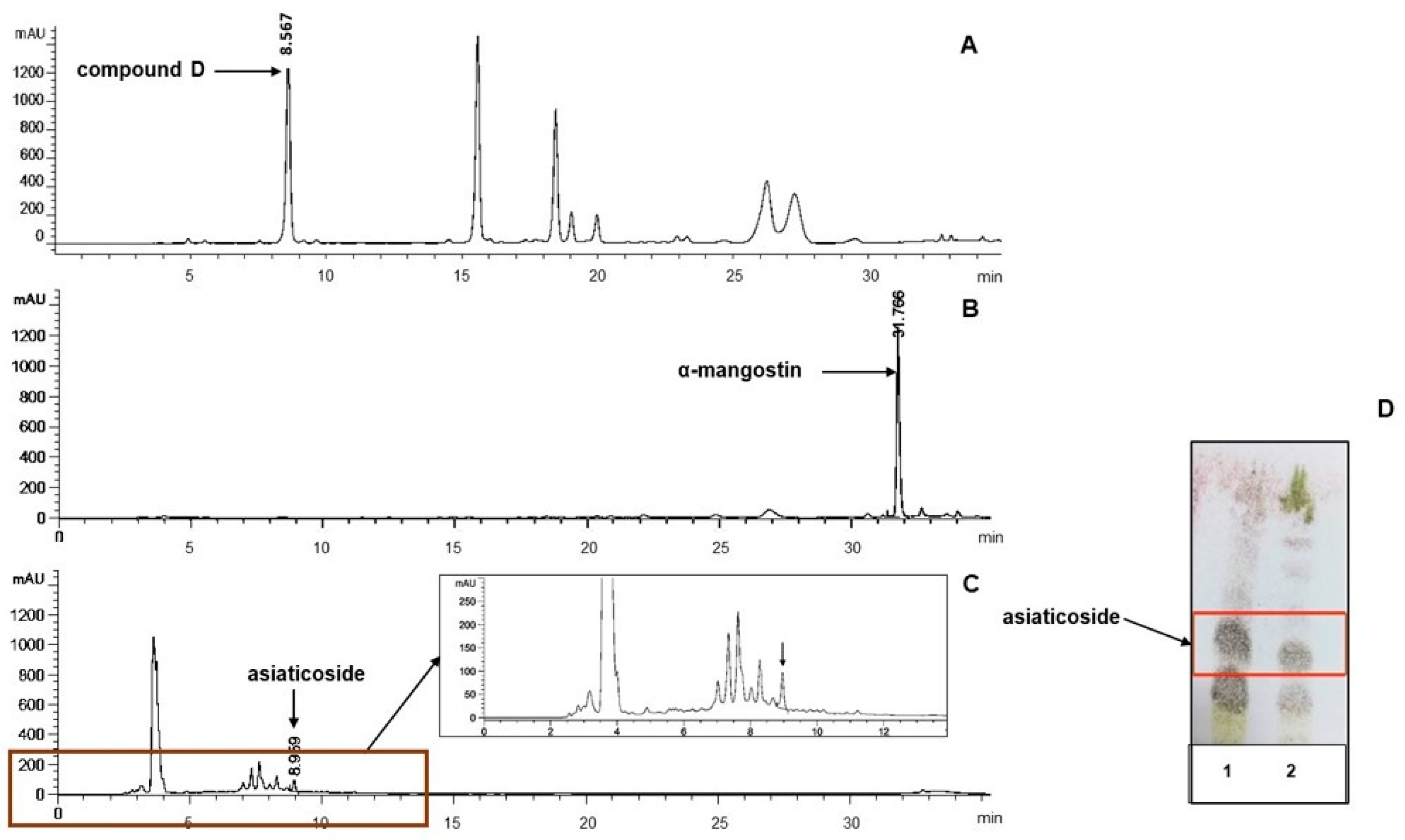
The HPLC chromatograms of ZH, GE, and CAEw demonstrated the presence of biomarkers (Figure 2). Peaks of compound D, α-mangostin, and asiaticoside were observed at retention times of 8.567, 31.766, and 8.959 min, respectively. Compound D and α-mangostin were the major peaks in ZH and GE, respectively. Asiaticoside was detected as only a trace peak in CAEw due to the absence of a strong chromophore in the molecule. TLC of the BuOH extract and CAEw from C. asiatica revealed the presence of asiaticoside as the dominant compound, as shown in Figure 2D.
The biomarker contents in the ZH, GE, and CAEw dried extracts obtained with the HPLC method were compound D, at 77.51 mg/g (7.8%), α-mangostin, at 75.12 mg/g (7.5%), and asiaticoside, at 98.73 mg/g (9.8%), respectively, which contained a high content of the markers.

3.2.2. Total Phenolic Contents (TPCs) and Total Flavonoid Contents (TFCs)

The TPCs and TFCs of ZH, GE, and CAEw are listed in Table 4. The G. mangostana extract (GE) had the highest TPC and TFC values, while those of CAEw were the lowest.

3.3. Antioxidant and Anti-Collagenase Activities of the Extracts

ZH exhibited the lowest antioxidant potencies and could not inhibit collagenase activity, whereas compound D had moderate and mild effects, respectively. GE exhibited the most potent antioxidant activities, including superoxide anion and DPPH free radical scavenging, as well as the most potent collagenase inhibitory effect. The potency of GE was comparable to that of α-mangostin in DPPH free radical scavenging and collagenase inhibitory effects. CAEw demonstrated a moderate superoxide anion inhibitory effect and mild collagenase inhibition, whereas DPPH free radical scavenging activity was comparable to that of GE and asiaticoside (Table 5).

3.4. Effect of the Herbal Extracts on the Viability of NB-UVB-Exposed HaCaT

NB-UVB irradiation reduced the viability of NB-UVB-exposed HaCaT cells in a dose-dependent manner. Exposure to NB-UVB at the irradiation dose of 120 mJ/cm2 caused a 30% reduction in cell viability (Figure 3), which was suitable for further experiments.
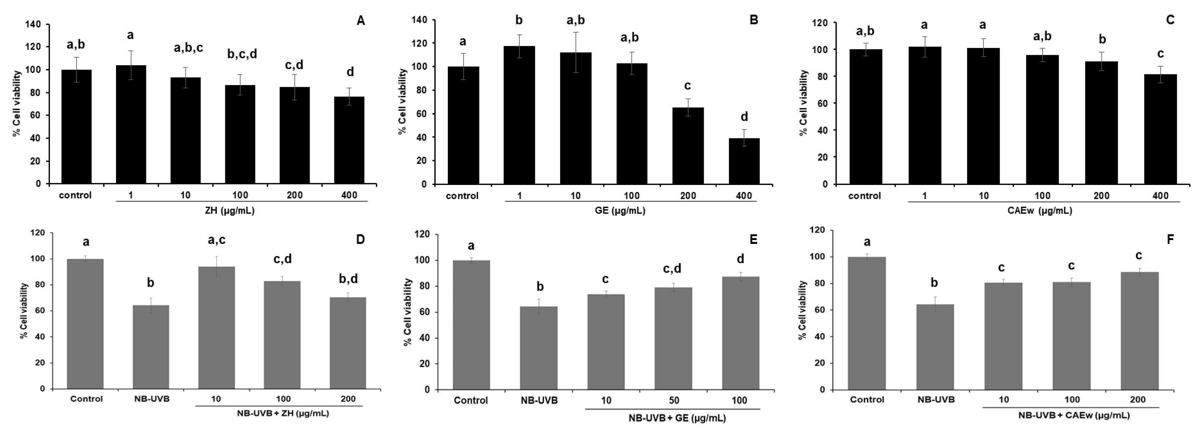
The extract concentration that provided more than 80% cell viability in the MTT assay was considered non-cytotoxic. ZH and CAEw were non-cytotoxic to HaCaT cells at concentrations of 1–400 µg/mL, whereas GE was non-cytotoxic at 1–100 µg/mL. At a low dose (1 µg/mL), GE potentially stimulated cell proliferation (p < 0.05), as shown in Figure 4A–C.
Post-treatment with ZH, GE, and CAEw after NB-UVB irradiation improved cell viability to a greater extent than irradiation alone (Figure 4D–F). At a concentration of 10 µg/mL, ZH exhibited the strongest stimulation of cell proliferation (94.08%) and showed a declining effect at higher concentrations. GE enhanced cell viability in a dose-dependent manner, and CAEw showed steady cell proliferation at 10–200 µg/mL.

3.5. Effect of Polyherbal Mixture Ratios on the Viability of NB-UVB-Exposed HaCaT

The 1:1:1, 1:2:1, and 10:1:10 ratios of the extract mixture consisting of ZH:GE:CAEw were evaluated for enhancing cell viability after NB-UVB exposure. The 1:1:1 extract mixture at a concentration of 100 µg/mL showed the highest ability to induce proliferation in NB-UVB-exposed HaCaT cells (p < 0.05) after 24 h treatment, as shown in Table 6. Hence, the formula ZH: GE: CAEw (1: 1: 1) was selected.
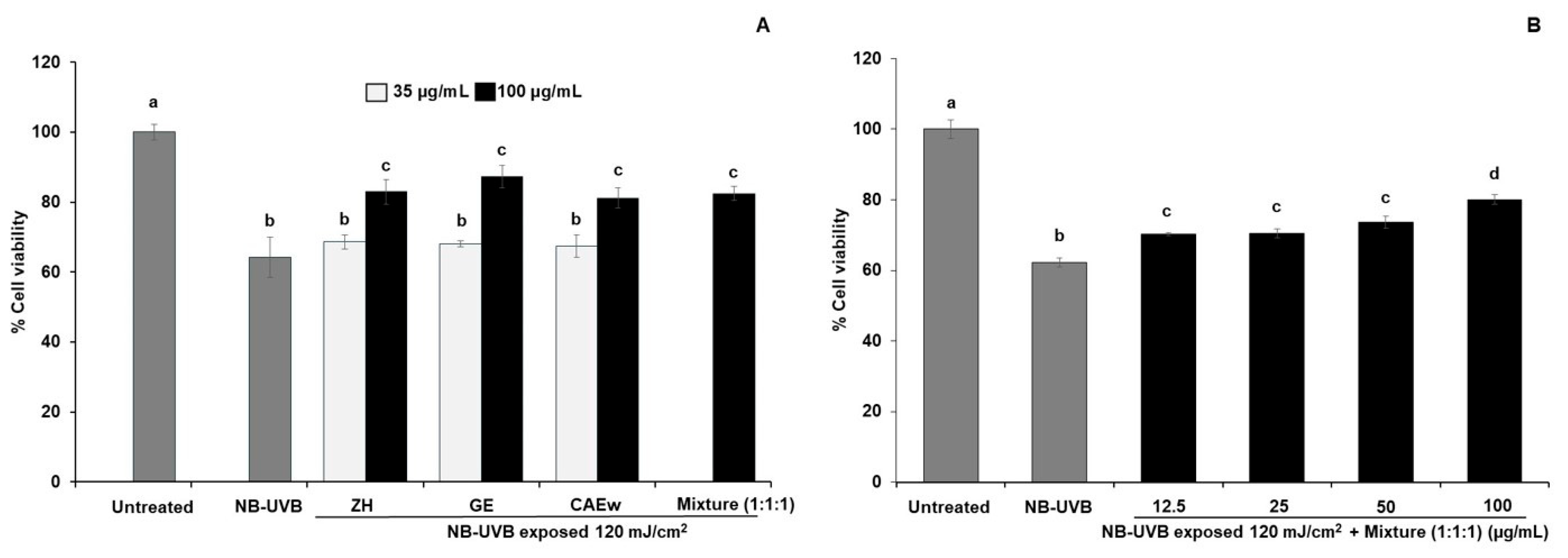
To investigate the additive effect of the extracts in the ZH:GE:CAEw (1:1:1) mixture, cell viability assays were performed with the single herbal extracts, at 35 µg/mL and 100 µg/mL, and with the herbal formulation, at 100 µg/mL. The results showed that each extract at 35 µg/mL did not increase cell viability after NB-UVB exposure, whereas at 100 µg/mL, ZH, GE, and CAEw increased cell survival to 82.96%, 87.34%, and 81.18%, respectively. The polyherbal extract at a total concentration of 100 µg/mL exhibited a significant stimulatory effect (p < 0.05) from 62.3% to 80.09%, or a 28.4% increase in cell viability after NB-UVB exposure (Figure 5A). The effect of a lower concentration of the polyherbal mixture (1:1:1) was then investigated; a slightly increase in cell viability (8%) appeared starting from a total concentration of 12.5 µg/mL (p < 0.05) (Figure 5B), while each single extract at 35 µg/mL did not enhance cell viability (Figure 5A). The results suggested that using the herbal extracts as a polyherbal mixture could achieve the effect at lower concentrations than the single extract.

3.6. Effects of the Polyherbal Mixture on Intracellular Reactive Oxygen Species (ROS) and Cell Apoptosis on NB-UVB-Exposed HaCaT

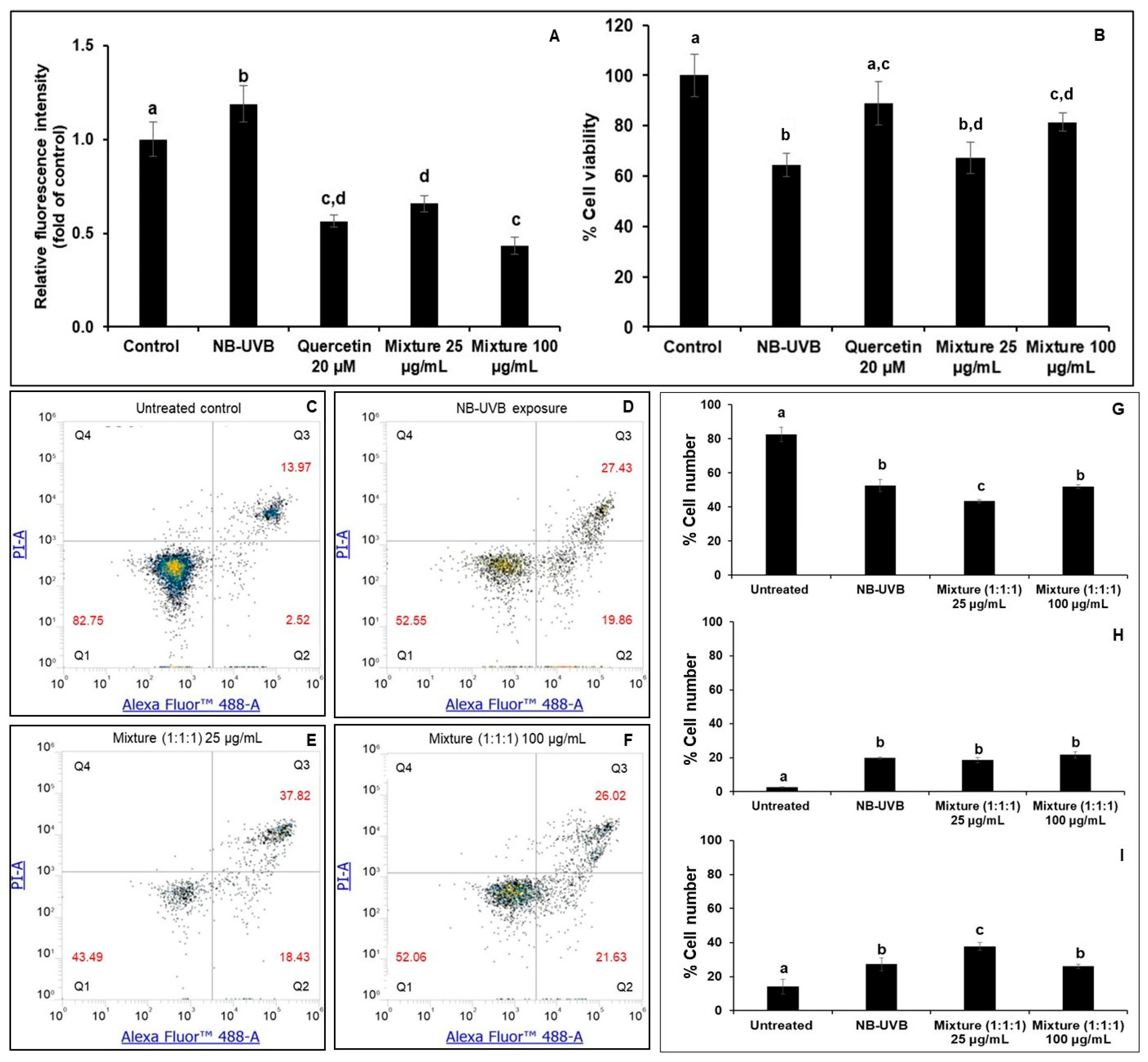
After 24 h of NB-UVB irradiation, increased intracellular ROS levels by approximately 19%, decreased cell viability, and induced apoptosis of HaCaT cells at early and late stages were observed (Figure 6). Treatment with the herbal mixture (1:1:1) reduced the ROS level by 44.6% and 63.6% from the NB-UVB exposed cells at 25 and 100 µg/mL, respectively, without suppressing cell apoptosis. At 25 µg/mL, it did not increase viable cells or stop the progression of cell apoptosis, which appeared as more late apoptotic cells than early-stage ones. Treatment with 100 µg/mL of the polyherbal mixture enhanced cell viability (p < 0.05) and lowered intracellular ROS to the same level as 20 µM quercetin (positive control), but it did not decrease the number of apoptotic cells compared to the NB-UVB group. The results indicated that the polyherbal mixture did not reduce cell apoptosis induced by NB-UVB irradiation. The increase in cell viability might be due to the proliferation of viable cells.

3.7. Gene Expression Analysis

NB-UVB upregulated COX-2, iNOS, MMP-1, and MMP-9 expression after 24 h of exposure. Treatment with the polyherbal mixture at 25 and 100 µg/mL after irradiation significantly reduced the expression of these genes to the same level as the non-irradiated cells at 24 h of incubation time, as shown in Figure 7.

4. Discussion

Zingiber cassumunar, Garcinia mangostana, and Centella asiatica are well-known herbs with long-term use as topical products, suggesting their safety and efficacy. All three herbs exhibit anti-photoaging properties and are used as ingredients in some topical products. To date, a polyherbal mixture combination of these herbs has not been analyzed for a photoprotective effect on post-NB-UVB-exposed keratinocytes. This formulation could be a new ingredient for alleviating the adverse skin effects from phototherapy using NB-UVB irradiation.
Zingiber cassumunar rhizomes have been used to relieve muscle pain and inflammation-related symptoms. The active constituents include volatile oils and phenylbutanoids. Hexane is the most effective solvent for extracting phenylbutanoids from this plant [14]. The principle phenylbutanoids include (E)-1-(3,4)-dimethoxyphenyl)butadiene (DMPBD), (E)-4-(3′,4′-dimethoxyphenyl)but-3-en-1-ol (compound D), and (E)-1-(3,4-dimethoxyphenyl)but- 3-en-1-yl acetate (compound D acetate). In this study, only compound D was selected as a targeting compound and biomarker. It appeared as a peak in the three dominant peaks in ZH, along with the HPLC chromatogram of the non-polar extract of this plant, together with peaks of compound D acetate and DMPBD [13]. Compound D could be quantitatively analyzed with HPTLC [27] and the HPLC method [13]. Due to its hydrophobic properties, compound D was found to be locally accumulated after topical administration in newborn pig skin in an in vitro skin permeation study [28]. A niosome gel from Zingiber cassumunar extract was developed, which provides comparable anti-inflammatory effects to steroids and NSAIDs [29]. Mangosteen pericarp contains many bioactive xanthones, such as α-mangostin and γ-mangostin, as well as anthocyanins. α-Mangostin is the principal compound of the mangosteen pericarp extract, which could be detected and quantitatively analyzed using HPTLC at 320 nm [30]. The quantitative analysis of this compound in a 95% EtOH mangosteen peel extract using the HPTLC method was validated. Centella asiatica is an herbal medicine that is widely used for wound-healing treatment [31]. It contains significant amounts of madecassoside and asiaticoside but low amounts of asiatic and madecassic acid [32]. In this study, asiaticoside was used as a targeted compound to assess the optimal extraction condition of the C. asiatica extract due to its anti-elastase and matrix metalloproteinase (MMP)-inhibitory activities [16], making asiaticosides a potential anti-photoaging compound [18]. As the asiaticoside structure lacks chromophores, detection of this compound with the HPTLC method could be performed by derivatization, such as using a 2-naphthol sulfuric acid reagent to provide a colored band in the visible range [33] and measuring absorbance at 530 nm. The result of Centella asiatica extraction showed that the BuOH layer of the 75% EtOH extract contained a high amount of asiaticoside. The 117.7 °C boiling point of n-BuOH makes it difficult to evaporate to dryness, which would be a problem in large-scale production; therefore, an alternative green solvent, acetone–EtOH (2.6:7.4), was used.
Compound D, α-mangostin, and asiaticoside, the targeted compounds for developing the extraction solvents, are also commonly used as biomarkers for the quality control of each herbal material. Compound D and asiaticoside are available in the Thai Herbal Pharmacopoeia for Zingiber cassumunar and Centella asiatica, respectively [34].
According to the ICH Q3C guideline 2024, hexane is a class II solvent with a Permissible Daily Exposure (PDE) of 2.9 mg/day. It has a concentration limit of 290 ppm in pharmaceutical products, whereas acetone and EtOH are class 3 (solvents with low toxic potential). Hence, the solvents used to prepare the herbal extracts in this study are safe for further topical product development. Moreover, all solvents could be removed by rotary evaporator under controlled high vacuum in 45 °C due to their low boing point properties; therefore, these solvents are safe for extraction.
The superoxide anion scavenging activities, DPPH free radical scavenging properties, and collagen inhibitory effect suggest potential photoaging protection from the oxidative reactions. The observed antioxidant and anti-collagenase properties of GE and α-mangostin are similar to those reported in previous studies [35]. Mangosteen peel extracts possess anti-elastase activities, and α-mangostin is a potent anti-collagenase compound [36]. Mangosteen peel extracts also contain γ-mangostin, a xanthone of potent anti-hyaluronidase related to the photoprotective properties of mangosteen. This suggests that several active compounds in the GE provide photoprotective effects. The topical application of an ethanolic extract on mice skin after NB-UVB exposure supports mangosteen’s enhanced collagen synthesis activity as it increased the thickness and density of collagen in mice skin [37]. C. asiatica enhances collagen synthesis, where asiaticoside induces type I collagen synthesis [37]. Therefore, the mild and moderate anti-collagenase effects of CAEw and asiaticoside (Table 5) suggest that they may not increase collagen synthesis through collagenase inhibitory activity.
UVB exposure increases ROS production, inducing oxidative stress and damaging cells [38]. NB-UVB exposure can cause sunburned skin. Sunburned cells are apoptotic keratinocytes induced by DNA damage, characterized by cell shrinkage, membrane blebbing, chromatin condensation, and genomic DNA fragmentation [39]. NB-UVB also induces apoptosis in HaCaT cells [40]. ROS production triggers inflammation and activates collagenase, which degrades collagen, leading to premature skin aging. ROS are also a major cause of DNA damage and finally result in cell death or skin cancer. Hence, to diminish those harmful effects, herbal extracts should act on multiple targets. Using a polyherbal mixture may result in synergistic effects, broadened mechanisms of action, and increased effectiveness at lower concentrations. The minimal dose is also advantageous for reducing potential adverse effects and saving costs.
Enhancing the cell viability of UVB-irradiated keratinocytes is crucial for photoprotection. It leads to the recovery of the skin barrier and reflects the neutralizing effect of UVB-induced cell damage by the photoprotective mechanism, such as antioxidant properties, reducing intracellular ROS levels, preventing DNA damage, and anti-inflammatory effects [41]. The polyherbal mixture containing ZH, GE, and CAEw, in various ratios, was primarily screened for cell proliferation-enhancing effects on irradiated HaCaT cells. The ratio (1:1:1) was designed based on similar non-cytotoxic concentration ranges of each extract. The 1:2:1 ratio consisted of a 2-fold concentration of GE due to its cell recovery effect. The 10:1:10 ratio was designed based on the potent anti-inflammatory effects of Z. cassumunar extract and the stimulation of the collagen synthesis property of C. asiatica extract [42]. The polyherbal mixture (1:1:1) at 100 µg/mL provided the greatest increase in cell viability in NB-UVB-exposed HaCaT cells. Although the mixture ratio (1:2:1) composed of the highest amount of GE had the highest antioxidant (DPPH and superoxide anion scavenging effects), anti-collagenase activities, TPC, and TFC values (Table 4 and Table 5), it increased cell viability less than the 1:1:1 ratio. One explanation for this is that the increasing of cell numbers of the irradiated cells resulted from various mechanism of actions, not only from the scavenging mechanism of antioxidant activities, and anti-collagenase properties. In addition, not only phenolic and flavonoid substances exhibit a photoprotective effect, but some triterpenoids also have this effect. Furthermore, the weakest induction of cell proliferation was observed in the 10:1:10 ratio may be caused by the high ZH and low GE contents. The higher concentration of ZH reduced cell viability in the irradiated HaCaT cells, as shown in Figure 4D.
No studies have reported a post-treatment cell viability enhancement after NB-UVB exposure to HaCaT cells by Z. cassumunar, mangosteen peel, or C. asiatica extracts alone or by a polyherbal mixture of those herbal extracts. However, an increase in HaCaT cell viability was found after pre-treatment with triterpenoids from C. asiatica [17] and α-mangostin in UVB-induced-HaCaT [43,44]. Hence, the results of this study support the development of the polyherbal mixture as an innovative ingredient to attenuate the adverse effects from post-exposure to UVB or NB-UVB phototherapy. Therefore, the mechanisms underlying the photoprotective effects of the polyherbal mixture were investigated and compared between a low dose of 25 µg/mL and the most effective dose of 100 µg/mL.
Cell damage induced by UVB exposure is expressed immediately after irradiation and increases during incubation [45]. An increase in intracellular ROS, leading to apoptosis-mediated cell death, was found after 24 h NB-UVB irradiation of HaCaT cells in this study. ROS levels decreased following the polyherbal mixture treatments at 25 and 100 µg/mL, but the 25 µg/mL concentration may not be high enough to increase cell viability or to suppress the progression of apoptosis from early to late stages. The 100 µg/mL polyherbal mixture attenuated cell damage by inducing cell proliferation and lowering ROS levels after 24 h irradiation, without reducing the number of apoptotic cells. This may be beneficial for photoprotection against cancer, as damaged cells can progress to carcinogenic cells, as demonstrated by the laver (Porphyra yezoensis) extract’s ability to induce apoptosis effects [46].
NB-UVB irradiation of HaCaT cells upregulates MMP-1 and MMP-9 and facilitates the expression of the inflammatory mediator’s cyclooxygenase-2 (COX-2) and iNOS, as previously described. The accumulation of ROS from UVB irradiation activates inflammatory responses. It also increases the expression of matrix metalloproteinases (MMPs) in human skin, including MMP-1 (collagenase that cleaves type I and type III collagen), MMP-3, and MMP-9. MMPs are enzymes that play a role in degrading extracellular matrix (ECM) molecules, such as collagen, elastin, and gelatin, as well as glycoproteins, which are associated with skin aging. MMP inhibitors and antioxidants can counteract skin aging caused by ultraviolet radiation or oxidative stress, such as resveratrol on HaCat cells and ICR mice [47] and diphlorethohydroxycarmalol, an antioxidant from brown algae [48]. Moreover, controlling inflammatory responses, such as suppressing the expression of cyclooxygenase-2 (COX-2) and nitric oxide synthase (iNOS), is an effective strategy for preventing photodamage-related diseases. The protective effect of antioxidant properties on UVB-induced HaCaT cells was shown in a study that used an Entada phaseoloides extract [49]. Therefore, mRNA downregulation of COX-2, iNOS, MMP-1, and MMP-9 by the polyherbal mixture indicated photoprotective effects related to anti-inflammation and anti-aging in the NB-UVB-exposed HaCaT at 24 h after irradiation at the gene expression level. This effect could be from GE, ZH, and CAEw. The EtOH extract and α-mangostin downregulated the mRNA expression of MMP-1, MMP-2, MMP-9, IL-6, and TNF-α; alleviated the inflammatory response by inhibiting the nuclear factor kB (NF-kB) signaling pathway and the mitogen-activated protein kinase (MAPK) signaling pathway in UVB-HaCaT; and protected them against UVB-mediated apoptosis [43]. Topical administration of a hexane-soluble Z. cassumunar extract downregulated MMP-1 expression after UVB irradiation in Wistar rats [50]. Post-treatment of asiaticoside decreased MMP-9 mRNA expression in UVB-exposed HaCaT cells [18]. It was found that treatment with the ZH:GE:CAEw mixture at 25 µg/mL could affect gene regulation even though a slight or non-effect on cell viability was observed. This effect was also found in post-treatment with Derris scandens extract in irradiated NB-UVB cells [51].
In summary, the polyherbal mixture of ZH, GE, and CAEw had the advantage of multi-target action from the different characteristics of each extract. The 100 µg/mL polyherbal mixture contained less extract (33.3 µg/mL each) in the formula and provided the same cell recovery as the single extract at the same concentration (100 µg/mL); therefore, it can lower the possible unwanted effects of the individual extract and save costs by utilizing a lower concentration. The mixture reduced the NB-UVB effect at the gene expression level and decreased intracellular ROS at a low concentration (25 µg/mL).
This study has some limitations related to the fact that the herbal ingredient for targeting biological activities is still under development. It still lacks in vivo studies and clinical translation. As the activities were only investigated in NB-UVB-exposed HaCaT cells, in vivo assays should be further evaluated. Although all the herbs in this formulation have been well-studied for mechanisms related to photoprotective effects, the combination of extracts might result in unexpected interactions among the chemical components; in addition, synergistic effects may occur, which should be further analyzed. The biphasic phenomenon of phytochemicals should also be considered. This means that opposite effects of phytochemicals can occur between low and high concentrations, which occurs in many herbal substances, such as quercetin, resveratrol, berberine, and curcumin, as reviewed by Jodyns-Lebert in 2020 [52]. A biphasic effect is also reported in the Korean traditional herbal recipe for HRMC5 extract, composed of five herbs. This extract increases cell survival at a low dose but reduces cell survival at a high dose on UVB- and non-UVB exposed HaCaT cells, indicating the narrow range of therapeutic concentrations [53]. Curcumin, a chemical ingredient purified from the CHCl3 layer of Zingiber cassumunar MeOH extract [54], also exhibits a biphasic effect: it acts as an antioxidant at a low dose but has other functions at a high dose, such as the induction of autophagy and cell death [55]. Therefore, the mechanism of action of the ZH, GE, and CAEw polyherbal mixture should be investigated. Since the formulation was newly established by the combination of herbal extracts, it is necessary to conduct toxicity tests for short- and long-term uses.

5. Conclusions

In this study, we developed a novel polyherbal formulation combining compound D-rich Zingiber cassumunar extract, α-mangostin-rich Garcinia mangostana extract, and asiaticoside–rich Centella asiatica extracts in a 1:1:1 ratio, which effectively protected human keratinocytes (HaCaT cells) from NB-UVB–induced damage by enhancing viable cell proliferation, reducing intracellular reactive oxygen species, and facilitating the apoptotic removal of damaged cells. Moreover, this polyherbal formulation significantly downregulated mRNA expression of pro-inflammatory mediators (COX-2 and iNOS) and collagen-degrading enzymes (MMP-1 and MMP-2), demonstrated both anti-inflammatory and anti-photoaging activities. This polyherbal formulation was impressive in reducing individual extract doses while maintaining efficacy comparable to that of a single herbal extract. These findings highlight the potential of this formulation as a topical product to mitigate oxidative stress and premature skin aging from NB-UVB phototherapy. Future work should focus on elucidating the molecular mechanisms underlying its anti-photoaging effects and on conducting in vivo efficacy and toxicity studies.

Supplementary Materials

The following supporting information can be downloaded at https://www.mdpi.com/article/10.3390/cosmetics12060241/s1; Figure S1: HPLC standard graphs of biomarkers; Table S1: Method validation of biomarkers compound D in Zingiber cassumunar extract (ZH), α-mangostin in Garcinia mangostana extract (GE), and asiaticoside in Centella asiatica extract (CAEw).

Author Contributions

Conceptualization, N.N.; formal analyses, S.T. (Sineenad Teerapatpaisan) and N.N.; investigation, S.T. (Sineenad Teerapatpaisan) and A.N.; resources, N.N.; writing—original draft preparation, S.T. (Sineenad Teerapatpaisan), A.N. and N.N.; writing—review and editing, S.T. (Sineenad Teerapatpaisan), N.N. and S.T. (Suthasinee Thapphasaraphong); supervision, N.N. and S.T. (Suthasinee Thapphasaraphong); project administration N.N.; funding acquisition, N.N. All authors have read and agreed to the published version of the manuscript.

Funding

Faculty of Pharmaceutical Sciences, Khon Kaen University, Thailand: 1(3)/2563 and 65-2(1)/2566; and the NSRF via the Program Management Unit for Human Resources & Institutional Development, Research and Innovation (grant number B38G670003).

Institutional Review Board Statement

Not applicable.

Informed Consent Statement

Not applicable.

Data Availability Statement

The authors confirm that the data supporting the findings of this study.

Conflicts of Interest

The authors declare no conflicts of interest.

References

  1. Marrot, L.; Meunier, J.R. Skin DNA Photodamage and Its Biological Consequences. J. Am. Acad. Dermatol. 2008, 58, S139–S148. [Google Scholar] [CrossRef] [PubMed]
  2. Parrish, J.A.; Jaenicke, K.F. Action Spectrum for Phototherapy of Psoriasis. J. Investig. Dermatol. 1981, 76, 359–362. [Google Scholar] [CrossRef]
  3. Yuehua, Y.; Khalaf, A.T.; Xiaoxiang, Z.; Xinggang, W. Narrow-Band Ultraviolet B and Conventional UVB Phototherapy in Psoriasis: A Randomised Controlled Trial. J. Appl. Sci. 2008, 5, 905–908. [Google Scholar] [CrossRef]
  4. Coelho, M.M.V.; Apetato, M. The Dark Side of the Light: Phototherapy Adverse Effects. Clin. Dermatol. 2016, 34, 556–562. [Google Scholar] [CrossRef]
  5. Goulden, V.; Ling, T.C.; Babakinejad, P.; Dawe, R.; Eadie, E.; Fassihi, H.; Fityan, A.; Garibaldinos, T.; Ibbotson, S.H.; Novakovic, L.; et al. British Association of Dermatologists and British Photodermatology Group Guidelines for Narrowband Ultraviolet B Phototherapy 2022. Br. J. Dermatol. 2022, 187, 285–286. [Google Scholar] [CrossRef]
  6. Ade, I.; Wanandi, S.I.; Wuyung, P.E.; Hoemardani, A.S.D. Effect of Narrowband Ultraviolet B (311 nm) Exposure on Skin Carcinogenesis in Wistar Rats. J. Adv. Vet. Anim. Res. 2024, 11, 1105–1113. [Google Scholar] [CrossRef]
  7. Patel, V.R.; Saini, S.; Dwivedi, J.; Gupta, A.K.; Shrivastava, A.K.; Misra, A. Exploring the Concept and Scope of Polyherbal Formulations: A Comprehensive Review. Int. J. Herb. Med. 2025, 13, 9–16. [Google Scholar] [CrossRef]
  8. Karole, S.; Shrivastava, S.; Thomas, S.; Soni, B.; Khan, S.; Dubey, J.; Dubey, S.P.; Khan, N.; Jain, D.K. Polyherbal Formulation Concept for Synergic Action: A Review. J. Drug Deliv. Ther. 2019, 9, 453–466. [Google Scholar] [CrossRef]
  9. Singh, M.; Gautam, A.; Kumar, P. Chemical Stability of Polyherbal Formulations. Int. J. Environ. Health Sci. 2024, 6, 24–37. [Google Scholar] [CrossRef]
  10. Lohakul, J.; Chaiprasongsuk, A.; Jeayeng, S.; Saelim, M.; Muanjumpon, P.; Thanachaiphiwat, S.; Tripatara, P.; Soontrapa, K.; Lumlerdkij, N.; Akarasereenont, P.; et al. The Protective Effect of Polyherbal Formulation, Harak Formula, on UVA-Induced Photoaging of Human Dermal Fibroblasts and Mouse Skin via Promoting Nrf2-Regulated Antioxidant Defense. Front. Pharmacol. 2021, 12, 649820. [Google Scholar] [CrossRef] [PubMed]
  11. Jayaprakash, N.; Arumugam, S.; Paramasivam, B.; Narasimhan, T.; Jayaraman, S.; Venkateasan, P.; Rajendran, S.; Elumalai, K. Formulation and Characterization of a Polyherbal Sunscreen Containing Camellia sinensis, Vitis vinifera, and Silybum marianum Extracts. Pharmacogn. Res. 2025, 17, 836–849. [Google Scholar] [CrossRef]
  12. Panthong, A.; Kanjanapothi, D.; Niwatananant, W.; Tuntiwachwuttikul, P.; Reutrakul, V. Anti-Inflammatory Activity of Compound D {(E)-4-(3′,4′-Dimethoxyphenyl)but-3-en-2-ol} Isolated from Zingiber cassumunar Roxb. Phytomedicine 1997, 4, 207–212. [Google Scholar] [CrossRef]
  13. Gundom, T.; Sukketsiri, W.; Panichayupakaranant, P. Phytochemical Analysis and Biological Effects of Zingiber cassumunar Extract and Three Phenylbutenoids: Targeting NF-κB, Akt/MAPK, and Caspase-3 Pathways. BMC Complement. Med. Ther. 2025, 25, 180. [Google Scholar] [CrossRef]
  14. Kaewchoothong, A.; Tewtrakul, S.; Panichayupakaranant, P. Inhibitory Effect of Phenylbutanoid-Rich Zingiber cassumunar Extracts on Nitric Oxide Production by Murine Macrophage-Like RAW264.7 Cells. Phytother. Res. 2012, 26, 1789–1792. [Google Scholar] [CrossRef]
  15. Maquart, F.X.; Chastang, F.; Simeon, A.; Birembaut, P.; Gillery, P.; Wegrowski, Y. Triterpenes from Centella asiatica Stimulate Extracellular Matrix Accumulation in Rat Experimental Wounds. Eur. J. Dermatol. 1999, 9, 289–296. [Google Scholar]
  16. Nema, N.K.; Maity, N.; Sarkar, B.K.; Mukherjee, P.K. Matrix Metalloproteinase, Hyaluronidase and Elastase Inhibitory Potential of Standardized Extract of Centella asiatica. Pharm. Biol. 2013, 51, 1182–1187. [Google Scholar] [CrossRef] [PubMed]
  17. Dang, Y.; Liu, T.; Liu, Y.; Li, J.; Jing, Y.; Yang, M.; Zhang, H.; Jiang, M.; Wu, H.; Yang, W.; et al. Anti-Photoaging Activity of Triterpenoids Isolated from Centella asiatica. Phytochemistry 2024, 228, 114246. [Google Scholar] [CrossRef]
  18. Jiang, H.; Zhou, X.; Chen, L. Asiaticoside Delays Senescence and Attenuates Generation of ROS in UV Exposure Cells through Regulation of TGFβ1/Smad Pathway. Exp. Ther. Med. 2022, 24, 667. [Google Scholar] [CrossRef]
  19. Kalick, L.S.; Khan, H.A.; Maung, E.; Baez, Y.; Atkinson, A.N.; Wallace, C.E.; Day, F.; Delgadillo, B.E.; Mondal, A.; Watanapokasin, R.; et al. Mangosteen for Malignancy Prevention and Intervention: Current Evidence, Molecular Mechanisms, and Future Perspectives. Pharmacol. Res. 2023, 188, 106630. [Google Scholar] [CrossRef]
  20. Wang, F.; Ma, H.; Liu, Z.; Huang, W.; Xu, X.; Zhang, X. α-Mangostin Inhibits DMBA/TPA-Induced Skin Cancer through Inhibiting Inflammation and Promoting Autophagy and Apoptosis by Regulating PI3K/Akt/mTOR Signaling Pathway in Mice. Biomed. Pharmacother. 2017, 92, 672–680. [Google Scholar] [CrossRef] [PubMed]
  21. Monton, C.; Luprasong, C.; Suksaeree, J.; Songsak, T. Validated High Performance Liquid Chromatography for Simultaneous Determination of Stability of Madecassoside and Asiaticoside in Film Forming Polymeric Dispersions. Rev. Bras. Farmacogn. 2018, 28, 289–293. [Google Scholar] [CrossRef]
  22. Gong, Y.; Liu, X.; He, W.H.; Xu, H.G.; Yuan, F.; Gao, Y.X. Investigation into the Antioxidant Activity and Chemical Composition of Alcoholic Extracts from Defatted Marigold (Tagetes erecta L.) Residue. Fitoterapia 2012, 83, 481–489. [Google Scholar] [CrossRef]
  23. Zhishen, J.; Mengcheng, T.; Jianming, W. The Determination of Flavonoid Contents in Mulberry and Their Scavenging Effects on Superoxide Radicals. Food Chem. 1999, 64, 555–559. [Google Scholar] [CrossRef]
  24. Sithisarn, P.; Rojsanga, P.; Sithisarn, P.; Kongkiatpaiboon, S. Antioxidant Activity and Antibacterial Effects on Clinical Isolated Streptococcus suis and Staphylococcus intermedius of Extracts from Several Parts of Cladogynos orientalis and Their Phytochemical Screenings. Evid.-Based Complement. Altern. Med. 2015, 2015, 908242. [Google Scholar] [CrossRef]
  25. Mosmann, T. Rapid Colorimetric Assay for Cellular Growth and Survival: Application to Proliferation and Cytotoxicity Assays. J. Immunol. Methods 1983, 65, 55–63. [Google Scholar] [CrossRef]
  26. Livak, K.J.; Schmittgen, T.D. Analysis of Relative Gene Expression Data Using Real-Time Quantitative PCR and the 2−ΔΔCT Method. Methods 2001, 25, 402–408. [Google Scholar] [CrossRef] [PubMed]
  27. Tangyuenyongwatana, P. HPTLC Analysis of (E)-4-(3′,4′-Dimethoxyphenyl)but-3-en-1-ol in Zingiber cassumunar Roxb Rhizome Extract. Thai J. Pharm. Sci. 2017, 41, 1–6. [Google Scholar]
  28. Suksaeree, J.; Charoenchai, L.; Madaka, F.; Monton, C.; Sakunpak, A.; Charoonratana, T.; Pichayakorn, W. Zingiber cassumunar Blended Patches for Skin Application: Formulation, Physicochemical Properties, and In Vitro Studies. Asian J. Pharm. Sci. 2015, 10, 341–349. [Google Scholar] [CrossRef]
  29. Priprem, A.; Janpim, K.; Nualkaew, S.; Mahakunakorn, P. Topical Niosome Gel of Zingiber cassumunar Roxb. Extract for Anti-Inflammatory Activity Enhanced Skin Permeation and Stability of Compound D. AAPS PharmSciTech 2015, 17, 631–639. [Google Scholar] [CrossRef] [PubMed]
  30. Pothitirat, W.; Gritsanapan, W. HPLC Quantitative Analysis Method for the Determination of α-Mangostin in Mangosteen Fruit Rind Extract. Thai J. Agric. Sci. 2009, 42, 7–12. [Google Scholar]
  31. Arribas-López, E.; Zand, N.; Ojo, O.; Snowden, M.J.; Kochhar, T. A Systematic Review of the Effect of Centella asiatica on Wound Healing. Int. J. Environ. Res. Public Health 2022, 19, 3266. [Google Scholar] [CrossRef]
  32. Hashim, P.; Sidek, H.; Helan, M.H.M.; Sabery, A.; Palanisamy, U.D.; Ilham, M. Triterpene Composition and Bioactivities of Centella asiatica. Molecules 2011, 16, 1310–1322. [Google Scholar] [CrossRef]
  33. Chaisawadi, A.; De-Eknamkul, W. Development of a New Analytical Method for Determination of Asiaticoside Content in Centella asiatica. Thai J. Pharm. Sci. 2012, 36, 205–208. [Google Scholar] [CrossRef]
  34. Department of Medical Sciences, Ministry of Public Health. Thai Herbal Pharmacopoeia 2016; Department of Medical Sciences: Nonthaburi, Thailand, 2016; ISBN 9786161131913. [Google Scholar]
  35. Widowati, W.; Ginting, C.N.; Lister, I.N.E.; Girsang, E.; Amalia, A.; Wibowo, S.H.B.; Kusuma, H.S.W.; Rizal. Anti-Aging Effects of Mangosteen Peel Extract and Its Phytochemical Compounds: Antioxidant Activity, Enzyme Inhibition and Molecular Docking Simulation. Trop. Life Sci. Res. 2020, 31, 127–144. [Google Scholar] [CrossRef]
  36. Fitri, E.W. The Effectiveness of Topical Mangosteen Pericarp Extract on the Collagen of Mice Skin Exposed to Ultraviolet B. Am. J. Clin. Exp. Med. 2016, 4, 88–93. [Google Scholar] [CrossRef]
  37. Lee, J.; Jung, E.; Kim, Y.; Park, J.; Park, J.; Hong, S.; Kim, J.; Hyun, C.; Kim, Y.S.; Park, D. Asiaticoside Induces Human Collagen I Synthesis through TGFβ Receptor I Kinase (TβRI Kinase)-Independent Smad Signaling. Planta Med. 2006, 72, 324–328. [Google Scholar] [CrossRef] [PubMed]
  38. Shukla, S.; Chopra, D.; Patel, S.K.; Negi, S.; Srivastav, A.K.; Ratnasekhar, C.; Bala, L.; Dwivedi, A.; Ray, R.S. Superoxide Anion Radical Induced Phototoxicity of 2,4,5,6-Tetraminopyrimidine Sulfate via Mitochondrial-Mediated Apoptosis in Human Skin Keratinocytes at Ambient UVR Exposure. Food Chem. Toxicol. 2022, 164, 112990. [Google Scholar] [CrossRef]
  39. Kulms, D.; Schwarz, T. Molecular Mechanisms of UV-Induced Apoptosis. Photodermatol. Photoimmunol. Photomed. 2000, 16, 195–201. [Google Scholar] [CrossRef]
  40. Aufiero, B.M.; Talwar, H.; Young, C.; Krishnan, M.; Hatfield, J.S.; Lee, H.K.; Wong, H.K.; Hamzavi, I.; Murakawa, G.J. Narrow-Band UVB Induces Apoptosis in Human Keratinocytes. J. Photochem. Photobiol. B 2006, 82, 132–139. [Google Scholar] [CrossRef]
  41. Lin, W.T.; Chen, Y.J.; Kuo, H.N.; Kumar, S.; Abomughaid, M.M.; Kumar, S. Ultraviolet B-Induced Oxidative Damage in Human Skin Keratinocytes Is Alleviated by Pinus morrisonicola Leaf Essential Oil through Activation of the Nrf2-Dependent Antioxidant Defense System. Redox Rep. 2025, 30, 2527427. [Google Scholar] [CrossRef] [PubMed]
  42. Bylka, W.; Znajdek-Awiżeń, P.; Studzińska-Sroka, E.; Dańczak-Pazdrowska, A.; Brzezińska, M. Centella asiatica in Dermatology: An Overview. Phytother. Res. 2014, 28, 1117–1124. [Google Scholar] [CrossRef]
  43. Im, A.-R.; Kim, Y.-M.; Chin, Y.-W.; Chae, S. Protective Effects of compounds from Garcinia mangostana L. (Mangosteen) Against UVB Damage in HaCaT Cells and Hairless Mice. Int. J. Mol. Med. 2017, 40, 1941–1949. [Google Scholar] [CrossRef] [PubMed]
  44. Jin, J.; Bao, Y.; Wang, Y.; Zheng, H.; Guo, H.; Zhang, L.; Guo, R.; Yang, L. Protective Activity of Alpha-Mangostin Against UVB-Induced Injury in HaCaT Cells by Modulating the Ceramide and MAPK and NF-ΚB Signaling Pathways. J. Food Biochem. 2023, 2023, 4702866. [Google Scholar] [CrossRef]
  45. Khalil, C.; Shebaby, W. UVB Damage Onset and Progression 24 h Post Exposure in Human-Derived Skin Cells. Toxicol. Rep. 2017, 4, 441–449. [Google Scholar] [CrossRef]
  46. Kim, S.; You, D.H.; Han, T.; Choi, E.M. Modulation of Viability and Apoptosis of UVB-Exposed Human Keratinocyte HaCaT Cells by Aqueous Methanol Extract of Laver (Porphyra yezoensis). J. Photochem. Photobiol. B 2014, 141, 301–307. [Google Scholar] [CrossRef] [PubMed]
  47. Cui, B.; Wang, Y.; Jin, J.; Yang, Z.; Guo, R.; Li, X.; Yang, L.; Li, Z. Resveratrol Treats UVB-Induced Photoaging by Anti-MMP Expression, Through Anti-Inflammatory, Antioxidant, and Antiapoptotic Properties, and Treats Photoaging by Upregulating VEGF-B Expression. Oxid. Med. Cell. Longev. 2022, 2022, 6037303. [Google Scholar] [CrossRef]
  48. Piao, M.J.; Susara Ruwan Kumara, M.H.; Kim, K.C.; Kang, K.A.; Kang, H.K.; Lee, N.H.; Hyun, J.W. Diphlorethohydroxycarmalol Suppresses Ultraviolet B-Induced Matrix Metalloproteinases via Inhibition of JNK and ERK Signaling in Human Keratinocytes. Biomol. Ther. 2015, 23, 557–563. [Google Scholar] [CrossRef]
  49. Mittraphab, Y.; Nagata, M.; Matsumoto, M.; Shimizu, K. Antioxidant and Protective Effect of Acetone Extract of Entada Phaseoloides Leaves on UVB-Irradiated Human Epidermal Keratinocytes (HaCaT cells) by Inhibiting COX-2, iNOS, and Caspase-3 Activation. Nat. Prod. Commun. 2022, 17, 1934578X221078627. [Google Scholar] [CrossRef]
  50. Tyas, D.A.; Wijayanti, N.; Nuringtyas, T.R.; Wahyuono, S. Protective Effects of Zingiber cassumunar Roxb. Extract Against UVB-Induced Oxidative Stress in Wistar Albino Rats (Rattus Novergicus Berkenhout, 1769). Indones. J. Biotechnol. 2024, 29, 25–32. [Google Scholar] [CrossRef]
  51. Sukhonthasilakun, S.; Mahakunakorn, P.; Naladta, A.; Nuankaew, K.; Nualkaew, S.; Yenjai, C.; Nualkaew, N. Anti-Inflammatory Effects of Derris Scandens Extract on Narrowband-Ultraviolet B Exposed HaCaT Human Keratinocytes. J. Ayurveda Integr. Med. 2023, 14, 100693. [Google Scholar] [CrossRef] [PubMed]
  52. Jodynis-Liebert, J.; Kujawska, M. Biphasic Dose-Response Induced by Phytochemicals: Experimental Evidence. J. Clin. Med. 2020, 9, 718. [Google Scholar] [CrossRef] [PubMed]
  53. Ha, R.; Cho, W.K.; Kim, E.; Jang, S.J.; Kim, J.-D.; Yi, C.-G.; Moh, S.H. Exploring the Benefits of Herbal Medicine Composite 5 (HRMC5) for Skin Health Enhancement. Curr. Issues Mol. Biol. 2024, 46, 12133–12151. [Google Scholar] [CrossRef] [PubMed]
  54. Han, A.R.; Lee, E.J.; Min, H.Y.; Kim, H.R.; Lee, S.; Seo, E.K. A Potential Cytotoxic Principle of Zingiber cassumunar. Nat. Prod. Sci. 2003, 9, 109–111. [Google Scholar]
  55. Moghaddam, N.S.A.; Oskouie, M.N.; Butler, A.E.; Petit, P.X.; Barreto, G.E.; Sahebkar, A. Hormetic Effects of Curcumin: What is the Evidence? J. Cell. Physiol. 2018, 234, 10060–10071. [Google Scholar] [CrossRef]
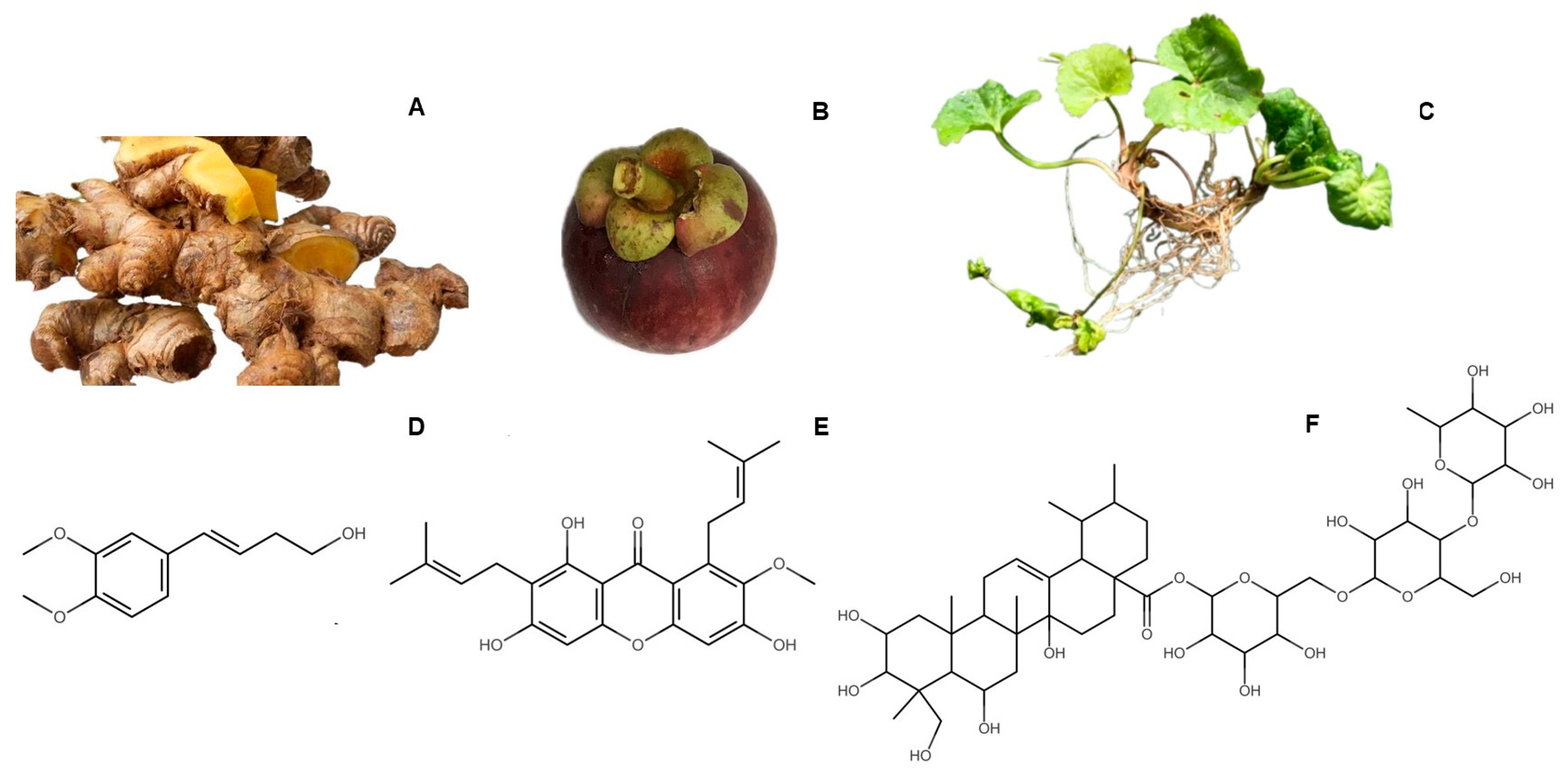
Figure 1. Plant materials used to prepare the polyherbal mixture and the targeted active compounds, which are used as biomarkers. (A): Zingiber cassumunar rhizome; (B): Garcinia mangostana fruit (fruit pericarp was used); (C): Centella asiatica whole plant (leaves were used); (D): (E)-4-(3′,4′-dimethoxyphenyl)but-3-en-1-ol (compound D); (E): α-mangostin; (F): asiaticoside.
Cosmetics 12 00241 g001
Figure 2. Chemical profiles of the selected extracts and their biomarkers. (A): Z. cassumunar extract (ZH); (B): G. mangostana extract (GE); (C): C. asiatica extract (CAEw); (D): TLC of C. asiatica extract (adsorbent: silica gel GF254; mobile phase: CHCl3:MeOH:H2O (30:13:1); detection: anisaldehyde–H2SO4 spraying reagent and heated; lane 1: BuOH layer; lane 2: CAEw).
Cosmetics 12 00241 g002
Figure 3. NB-UVB-exposed HaCaT cells and cell viability after 24 h irradiation. (A): non-irradiated HaCaT cells; (B): NB-UVB-exposed HaCaT cells at a dose of 120 mJ/cm2 after 24 h irradiation. Cells were visualized under an inverted microscope (10×).; (C): cell viability by NB-UVB irradiation in a dose-dependent manner after 24 h irradiation). (Different letters indicate significant differences between groups at p < 0.05 by Tukey’s multiple comparison test, n = 3).
Cosmetics 12 00241 g003
Figure 4. Effects of herbal extracts on HaCaT cell viability. (AC): Cell viability of non-irradiated HaCaT cells. (DF): Cell viability of NB-UVB-exposed HaCaT cells at a dose of 120 mJ/cm2. (Cells were treated following NB-UVB irradiation and incubated for 24 h). (A,D): Cells treated with ZH; (B,E): cells treated with GE; and (C,F): cells treated with CAEw after NB-UVB exposure. (Different letters indicate significant differences between groups at p < 0.05 by Tukey’s multiple comparison test, n = 3).
Cosmetics 12 00241 g004
Figure 5. Effects of ZH, GE, and CAEw and the polyherbal mixture post-treatment on the cell viability of NB-UVB-exposed HaCaT cells. Cells were treated following NB-UVB irradiation and incubated for 24 h. (A): Effect of the single extracts at 35 and 100 µg/mL and the polyherbal mixture at 100 µg/mL. (B): Effect of the polyherbal mixture at various concentrations. (Data are presented as mean ± standard deviation (SD); different letters indicate significant differences between groups at p < 0.05 based on Tukey’s multiple comparison test, n = 3).
Cosmetics 12 00241 g005
Figure 6. Effects of the polyherbal mixture (1:1:1) on cell viability, intracellular reactive oxygen species (ROS), and cell apoptosis in NB-UVB-exposed HaCaT. Cells were treated following NB-UVB irradiation and incubated for 24 h. (A): Intracellular reactive oxygen species (ROS); (B): effects of the 25 and 100 µg/mL mixtures on the cell viability of irradiated HaCaT cells; (CF): scatter plots of PI (y-axis) vs. annexin V (x-axis) of cell apoptosis analysis using flow cytometry: (C): non-treated control (non-irradiation); (D): NB-UVB irradiation; (E): ZH:GE:CEAw (1:1:1) 25 µg/mL; (F): ZH:GE:CEAw (1:1:1) 100 µg/mL (Q1: viable cells; Q2: early apoptotic cells; Q3: late apoptotic cells; Q4: necrotic cells); (GI): percentage of viable, early apoptotic, and late apoptotic cells ((G): viable cells; (H): early apoptotic; and (I): late apoptotic cells). Different letters indicate significant differences between groups at p < 0.05 based on Tukey’s multiple-comparison test.
Cosmetics 12 00241 g006
Figure 7. Effect of the polyherbal mixture on mRNA expression in NB-UVB-irradiated HaCaT cells. Cells were treated following NB-UVB irradiation and incubated for 24 h. (A): COX-2; (B): iNOS; (C): MMP-1; (D): MMP-9. The control was non-NB-UVB-exposed HaCaT cells. Data are presented as mean ± standard deviation (SD); different letters indicate significant differences between groups at p < 0.05 based on Tukey’s multiple-comparison test; n = 3.
Cosmetics 12 00241 g007
Table 1. HPTLC conditions and detection.
SampleBiomarker Mobile Phase Spraying Reagent Wavelength (nm)
Z. cassumunar extractcompound DCHCl3:EtOAc (8.5:1.5) -254
G. mangostana extractα-mangostinHexane–EtOAc (7:3) -317
C. asiatica extractasiaticosideCHCl3:MeOH:H2O (30:13:1)anisaldehyde -H2SO4 530
Table 2. Primer sequences used for RT-qPCR analysis.
GeneForward Primer (5′3′)Reverse Primer (5′3′)Accession No.
GAPDHGAGAAGGCTGGGGCTCATTTAGTGATGGCATGGACTGTGGNM_002046.6
COX-2TTGCATTCTTTGCCCAGCACACCGTAGATGCTCAGGGACTNM_000963.4
iNOSCCTGGAGGTGCTAGAGGAGTATCTCCGGTGTGGTAGGTGANM_000625.4
MMP-1TGTGGTGTCTCACAGCTTCCATCTGGGCTGCTTCATCACCNM_002421.4
MMP-9ACGATGACGAGTTGTGGTCCGGTTTCCCATCAGCATTGCCNM_004994.3
Table 3. The HPTLC peak areas of the biomarkers in the herbal extracts (75 µg).
Plant/
(Biomarker)
Extract% Yield of Dry Weight (w/w)Peak Area of Biomarker
(mAU)
Z. cassumunar/(Compound D)95% EtOH extract20.229856.9
Hexane layer1.9416,765.5
EtOAc layer3.195314.7
Hexane extract (ZH)14.7616,687.4
Compound D (5 µg)-4589.6
G. mangostana/
(α-Mangostin)
95% EtOH extract (GE)17.0848,775.2
CH2Cl2 extract15.2054,572
EtOAc extract16.5016,550.4
n-BuOH extract8.651026.4
α-Mangostin (7.5 µg)-65,191.85
C. asiatica/
(Asiaticoside)
75% Ethanol extract21.263475.7
EtOAc layer1.742317.2
n-BuOH layer2.1017,028.8
Acetone–EtOH (2.6:7.4) extract24.1212,567.2
Hexane layer9.19N.D.
Aqueous layer (CAEw)10.148650.5
Asiaticoside (5 µg)-10,345
Table 4. Total phenolic and flavonoid contents of the chosen extracts.
Extract TPC
(mg GAE/g Extract)
TFC
(mg QE/g Extract)
ZH249.25 ± 5.19148.64 ± 15.17
GE398.29 ± 8.49233.27 ± 9.90
CAEw185.56 ± 4.7854.12 ± 6.32
Table 5. Antioxidant and anti-collagenase activities of the extracts and biomarkers.
SamplesIC50 (µg/mL)
Superoxide Anion
Scavenging
DPPH Free Radical
Scavenging
Collagenase
Inhibition
Z. cassumunar extract (ZH)>250 #218.18 ± 8.46N.A.
G. mangostana extract (GE)52.67 ± 0.2617.68 ± 0.3877.21 ± 0.54
C. asiatica extract (CAEw)188.83 ± 1.9422.16 ± 0.251500.29 ± 23.48
Compound D69.86 ± 0.75115.32 ± 3.881368.35 ± 19.65
α-Mangostin105.04 ± 1.6311.01 ± 0.3163.61 ± 1.51
Asiaticoside289.70 ± 10.9391.64 ± 1.47209.65 ± 2.77
Rutin 27.13 ± 0.30N.D.N.D.
Gallic acid N.D.1.73 ± 0.95N.D.
1,10-Phenanthroline monohydrateN.D.N.D.0.06 ± 0.00 µM
# at a concentration of 250 µg/mL, superoxide anion scavenging activity of ZH was 24.97 ± 1.04%; N.A.: no activity; N.D.: not determined.
Table 6. % Cell viability effect of 100 µg/mL polyherbal mixture on NB-UVB-exposed HaCaT cells.
TreatmentsAmount (µg/mL)% Cell Viability *% Increasing of Cell Viability *
ZHGECAEw
Non-NB-UVB---100.0 ± 2.7 a-
NB-UVB---62.3 ± 1.3 b
ZH: GE: CAEw (1:1:1)33.333.333.380.1 ± 1.3 c28.4
ZH: GE: CAEw (1:2:1)25.050.025.075.2 ± 1.9 d20.6
ZH: GE: CAEw (10:1:10)47.64.847.673.2 ± 0.6 d17.5
* Different letters indicate significant differences between groups at p < 0.05 based on Tukey’s multiple comparison test, n = 3. Cell viability was assessed after 24 h treatment.
Disclaimer/Publisher’s Note: The statements, opinions and data contained in all publications are solely those of the individual author(s) and contributor(s) and not of MDPI and/or the editor(s). MDPI and/or the editor(s) disclaim responsibility for any injury to people or property resulting from any ideas, methods, instructions or products referred to in the content.