Article

Anti-Aging Efficacy of a Multi-Peptides–Silybin Complex: Mechanistic Insights and a 56-Day Clinical Evaluation

Research & Innovation Center, Proya Cosmetics Co., Ltd., Hangzhou 310023, China
*
Authors to whom correspondence should be addressed.
Cosmetics 2025, 12(5), 223; https://doi.org/10.3390/cosmetics12050223
Submission received: 11 September 2025 / Revised: 2 October 2025 / Accepted: 4 October 2025 / Published: 10 October 2025
(This article belongs to the Section Cosmetic Dermatology)

Abstract

Peptides are widely used in cosmetic formulations to stimulate extracellular matrix (ECM) synthesis, while silybin (a flavonolignan from Silybum marianum) offers retinol-like benefits through antioxidant and photoprotective activity. This study evaluated a novel anti-aging cream combining seven bioactive peptides with silybin to assess synergistic effects on ECM regeneration and clinical skin rejuvenation. In vitro assays in human dermal fibroblasts and keratinocytes revealed that the formulation rapidly upregulated gene and protein expression of collagen types I, III, IV, and XVII and lysyl oxidase (LOX) within 4–16 h. Ex-vivo, ultraviolet (UV)-damaged skin explants treated with the peptide–silybin complex showed enhanced recovery of collagen, elastic fibers, and LOX versus untreated controls. A 56-day clinical study (n = 31) demonstrated significant improvements in wrinkle area and volume, elasticity (+12.5%), firmness (+20.7%), and dermal density (+78%, all p < 0.001). No adverse effects were reported, and over 80% of participants noted improved skin texture and firmness. These findings highlight a novel synergy between peptides and silybin, with rapid ECM activation and clinical efficacy. To our knowledge, this is the first evidence of a cosmetic peptide formulation significantly upregulating LOX expression, suggesting a new mechanism for strengthening dermal architecture and improving skin resilience. Future studies should elucidate the mechanisms underlying these effects and assess whether other botanicals confer complementary benefits when combined with peptide blends.

1. Introduction

Skin aging occurs via intrinsic chronologic processes and extrinsic factors (primarily photoaging due to ultraviolet (UV) exposure [1]. While intrinsic aging leads to gradual, inevitable changes, extrinsic aging—especially from cumulative UV radiation—accelerates structural degradation of the skin, manifesting as wrinkles, sagging, laxity, and uneven texture [2,3]. Photoaged skin shows heightened matrix metalloproteinase activity and increased collagen degradation compared to naturally aged skin [1,3]. At the molecular level, these visible aging signs are underpinned by changes in the extracellular matrix (ECM). Collagen fibers and elastin networks become fragmented and depleted with age and UV exposure, and non-enzymatic crosslinks such as advanced glycation end products (AGEs) accumulate, causing the ECM to become stiffer and less compliant [4,5]. The net result is a loss of dermal structural integrity and elasticity.
Among the enzymes crucial for ECM stability is lysyl oxidase (LOX). LOX catalyzes covalent cross-linking of collagen and elastin fibers, thereby enhancing tensile strength and limiting the dermal matrix’s susceptibility to protease-mediated degradation [6,7]. A decline in LOX expression and activity has been observed in adulthood, which contributes to weaker, more fragile collagen bundles and reduced skin elasticity [7,8,9]. Indeed, inadequate collagen/elastin cross-linking resulting from low LOX is thought to accelerate dermal matrix degeneration and promote wrinkle formation in aged skin [5,7,8]. Restoration of collagen, elastin, and LOX levels in the skin is therefore an important strategy for effective anti-aging interventions.
Recent advances in cosmetic science suggest that synergistic combinations of peptides can activate skin repair pathways more effectively than single peptides alone [10,11]. Signal peptides are short bioactive sequences that can mimic fragments of functional proteins, thereby stimulating cells to produce ECM components or modulate cellular signaling relevant to skin structure and functions [12,13]. For instance, topically applied peptide formula has been shown to increase dermal collagen content and reduce wrinkles in clinical studies [12]. In this study, we formulated a unique blend of seven peptides, each chosen for distinct but complementary anti-aging mechanisms: two cyclic peptides and five signal peptides. Cyclic peptides (including Linum usitatissimum seed extract and synthetic cyclopeptide-161) are known for their conformational stability and enhanced skin penetration [14,15,16]. The linseed-derived cyclic peptide has been reported to stimulate elastin expression in fibroblasts, improving elasticity, while cyclopeptide-161 is a neurotransmitter-inhibiting peptide aimed at reducing expression lines (unpublished data) [16,17,18]. The formula also contains tetradecyl aminobutyroylvalylaminobutyric urea trifluoroacetate, which has been shown to promote dermal firming by increasing collagen I production (even under UV-stress conditions) and boosting the production of decorin and lumican, thereby strengthening collagen fibril architecture and reducing skin sagging [19]. Palmitoyl tripeptide-5 is a well-known signal peptide that can activate transforming growth factor beta (TGF-β) pathways similar to thrombospondin-1, leading to increased collagen types I and III synthesis and reduced matrix metalloproteinase activity [20,21,22]. Acetyl tetrapeptide-11 promotes keratinocyte proliferation and has been found to upregulate collagen XVII, a key component of the dermal–epidermal junction (DEJ), thereby improving epidermal–dermal anchoring [21,23]. Acetyl tetrapeptide-9 stimulates the production of collagen I as well as lumican (a proteoglycan involved in collagen fibril organization), supporting dermal matrix density [21]. Finally, hexapeptide-9 is believed to stimulate fibroblasts to increase production of collagens and elastic fibers [24,25]. By combining these seven peptides, our formulation targets multiple biological pathways in parallel, including collagen synthesis, elastin production, and dermal–epidermal junction reinforcement, for a multifaceted anti-aging effect.
Peptides primarily improve structural wrinkles via dermal ECM support; however, they have limited effects on epidermal renewal and lack intrinsic free-radical–scavenging activity, which may constrain their standalone efficacy against photoaging-related oxidative stress [13]. Retinol—particularly when combined with antioxidants such as ferulic acid—has been shown to attenuate ROS levels and oxidative injury in UVB-induced photoaging models [26], thereby complementing the limitations of peptides and providing broader anti-aging benefits. In parallel, retinoid-like botanical antioxidants have gained attention as gentle yet effective anti-aging agents [27]. Silybin, a major flavonolignan isolated from Silybum marianum, has demonstrated notable antioxidant, anti-inflammatory, and photoprotective properties [28,29]. Treatment with silybin induced morphological changes analogous to those triggered by retinoic acid, inhibited the differentiation process of keratinocytes, reduced the expression levels of keratinocyte-specific terminal differentiation markers, and promoted the expression of proteins composing the basement membrane [29]. Silybin has been reported to exhibit superior anti-aging efficacy compared to the well-known retinoid-like compound, bakuchiol [30]. Unlike retinoids, however, silybin does not cause irritation or photosensitivity, making it an appealing “retinol-like” alternative for improving skin aging safely [29]. Recent clinical evidence supports silybin’s efficacy: a formulation with Silybum marianum extract was shown to increase collagen III and hyaluronic acid in human skin, producing anti-wrinkle results comparable to retinol but without the side effects [30]. Despite these benefits, silybin has been relatively underutilized in ECM-focused skincare studies, and its potential synergistic interactions with pro-collagen peptides are largely unexamined.
Building on this background, we developed a comprehensive anti-aging formula that merges the multi-mechanistic peptide approach with silybin’s protective and collagen-boosting properties. To our knowledge, this study is the first to evaluate a cosmetic formulation containing such a broad combination of peptides together with silybin. We conducted a series of investigations to assess its impact from the molecular level up to clinical outcomes: (1) rapid gene activation of collagen subtypes and LOX in cultured human fibroblasts and keratinocytes, (2) restoration of UV-damaged ECM proteins in human skin explants, (3) rapid in vitro activation of anti-aging proteins by the cream containing multi-peptides and silybin, and (4) a 56-day clinical trial to measure improvements in wrinkles, elasticity, and dermal structure in human volunteers. By examining both mechanistic biomarkers and visible skin changes, we aim to determine whether the seven-peptide–silybin complex delivers synergistic anti-aging effects that are superior to peptides alone, and to elucidate a potential new paradigm for skin anti-aging therapy.

2. Materials and Methods

2.1. Research Product

The composition of multi-peptides includes cyclopeptide-161, Linum usitatissimum (linseed) seed extract, tetradecyl aminobutyroylvalylaminobutyric urea trifluoroacetate, palmitoyl tripeptide-5, acetyl tetrapeptide-11, acetyl tetrapeptide-9, and hexapeptide-9. Peptide test articles were used in their as-supplied cosmetic ingredient forms, i.e., peptide solutions or dispersions in inert carriers (e.g., water, glycerin, caprylyl glycol, or triglyceride solvents). Reported concentrations for the ‘multi-peptides’ refer to the as-supplied mixtures unless otherwise noted. Active-equivalent concentrations, calculated from supplier certificates of analysis, are provided in Data S3. Silybin was used as a phospholipid complex (lecithin and 33% silybin (w/w)), and concentrations are expressed as silybin equivalents.
For the model of damage caused by UV irradiation to human skin explants, multi-peptides and silybin were added to a minimal formula without similar efficacy ingredients. The ratio of multi-peptides and silybin was the same as that in the cream. The vehicle (minimal formulation with the least interference to the results, composed only of essential excipients, including emulsifiers, preservatives, thickeners, and penetration enhancers) was prepared. The cream (the ingredients are listed in Data S1) for clinical study containing polypeptides and silybin was provided by Proya Cosmetics Co., Ltd. (Hangzhou, China). Multi-peptides, silybin, and the cream were diluted to different concentration for cell experiments.

2.2. Cell Culture

Human skin fibroblasts (HSFs, Biocell, batch No. Fb220309, Guangzhou, China) were cultured with Dulbecco’s modified Eagle’s medium (DMEM, GIBCO BRL, Grand Island, NY, USA) supplemented with 10% fetal bovine serum (FBS) (Sigma-Aldrich, St. Louis, MO, USA) and 1% penicillin/streptomycin (GIBCO, USA) [31]. Normal human epidermal keratinocytes (NHEKs, Biocell, batch No. Ep23013101, Guangzhou, China) were cultured with KcGrowth (Biocell, Guangzhou, China) culture solution. Human skin explants were immersed in 75% alcohol for 30 s and washed with sterile phosphate buffer saline (PBS) three times. Consequently, the explants were cut into 24 ± 2 mm2 blocks, with the epidermis facing up and the dermis facing down, and put into the culture mold. Then the explants were transferred into the 6-well plates, containing 3.7 mL culture solution (Biocell, Guangzhou, China). The solution was replaced every day. All cells and tissues were maintained at 37 °C in a humidified atmosphere containing 5% CO2.
HSFs and NHEKs were incubated for 24 h and then pretreated with polypeptides, silybin, or the cream. Then the cells were collected after incubation for several hours.

2.3. Antibodies and Reagents

3-(4,5-dimethylthiazol-2-yl)-2,5-diphenyltetrazolium bromide (MTT) was purchased from Sigma-Aldrich, USA. Antibodies of collagen I and collagen III were purchased from Proteintech, Rosemont, IL, USA. Antibodies of collagen IV, collagen XVII, and lysyl oxidase (LOX) were purchased from Abcam, Cambridge, UK. RNAiso Plus, reverse transcription (RT) reagent kit, and fluorochrome were purchased from Accurate Biology, Changsha, China. PBS (Solarbio, Beijing, China) and paraformaldehyde (PFA, Biosharp, Hefei, China) were used for cell culture and fixation. Vitamin C and E (Sigma-Aldrich, St. Louis, MO, USA) and TGF-β1 (PeproTech, Rocky Hill, NJ, USA) were used as a positive control.

2.4. Instruments

A CO2 incubator (Thermo Fisher Scientific, Waltham, MA, USA) and an ultra-clean workbench (Airtech, Suzhou, China) were used for cell culture. Microplate spectrophotometer (BioTek, Winooski, VT, USA), polymerase chain reaction (PCR, BioRad, Hercules, CA, USA), quantitative real-time PCR (qRT-PCR, BioRad, USA), LED optical microscopes (Leica, Wetzlar, Germany), BioImage Lab device (Bio-Rad, USA), and upright light microscopes (Olympus, Tokyo, Japan) were used for analysis. UVA and UVB lamps (Philips, Amsterdam, The Netherlands) were used for irradiation.
In the clinical study, the parameters related to skin wrinkles and pores were measured with Primos CR (Canfield, Parsippany, NJ, USA), Visia 7 (Canfield, USA), and Antera 3D (Miravex, Dublin, Ireland). Dermal density was measured with a DUB® SkinScanner (tpm taberna pro medicum GmbH, Lüneburg, Germany). Skin elasticity and firmness were measured with Cutometer dual MPA580 (Courage + Khazaka electronic GmbH, Köln, Germany).

2.5. UV Irradiation and Drug Treatment

After 2 days of culture, human skin explants were treated with drugs and then irradiated with ultraviolet A (UVA, 30 J/cm2) and ultraviolet B (UVB, 50 mJ/cm2) [32]. Treatment and irradiation were performed at an interval of 8 h every day for four days. Only treatment of drugs performed for the next three days. The model group (negative control, NC) was only irritated by UV without treatment. Then, 100 μg/mL VC + 7 μg/mL VE were added to the medium as the positive control. The human skin explants were treated with multi-peptides alone or in combination with 0.033% silybin. After treatment, the human skin explants were fixed with 4% PFA and then sliced for analysis.
Compared with the multi-peptides group, the synergistic enhancement rate of multi-peptides combined with silybin was calculated by the following formula:
Rate of synergistic enhancement = 100% × (expressionmulti-peptides+0.033% silybin − expressionmulti-peptides)/expressionmulti-peptides

2.6. MTT Assay

Cell viability was evaluated by MTT assay. The cells were divided into six groups: 0~10 mg/g multi-peptides + NHEKs/HSFs, 0~1 mg/g silybin + NHEKs/HSFs, and 0~10 mg/g cream + NHEKs/HSFs, with five repeats in each group. Absorption at 490 nm was assessed using microplate spectrophotometer [33]. Based on the results of cell viability, a non-cytotoxic dose of multi-peptides, silybin, and the cream were used for in vitro experiments.

2.7. RNA Extraction and qRT-PCR

qRT-PCR was used to determine the relative gene expressions of collagen I, III, IV, XVII, and LOX in cells. All primer sequences were obtained from the National Center for Biotechnology Information (NCBI) database (https://www.ncbi.nlm.nih.gov/). After incubation for 4 h, 8 h, and 16 h, cells were washed twice with PBS. Then, 1 mL RNAiso Plus was added to each well, and the lysed cells were collected. RNA was extracted, and then cDNA was synthesized by reverse transcription [34]. qRT-PCR was performed, and the results were calculated using the 2−ΔΔCT method [35]. The experiment was repeated three times, independently. The primer sequences used are listed in Data S4.

2.8. Immunofluorescence (IF) Staining

IF was used to determine the content of collagen I and IV in HSFs, and collagen I, III, IV, XVII, and LOX in human skin explants. After incubation for 4 h and 16 h, HSFs were fixed with 4% PFA for 30 min. Human skin explants were fixed with 4% PFA for 24 h. Subsequently, immunofluorescence staining was performed. The staining results were observed under a fluorescence microscope, and images were taken [34]. The images were collected and analyzed with Image-Pro® Plus version 6.0 (IPP) software (Media Cybernetics, Rockville, MD, USA). The expression levels of collagen were expressed as integrated optical density (IOD) or mean optical density (MOD, representing the ratio of IOD and area/cell quantity).

2.9. Western Blot (WB) Assay

WB was used to determine the content of collagen III and LOX in HSFs and collagen XVII in NHEKs. After incubation for 4 h and 16 h, the protein content was determined by WB. In addition, the post-incubation time was extended. After 4 h and 16 h of drug treatment, the new medium was replaced; incubation was performed for 20 h and 8 h. Then the expression of collagen III and XVII was determined. The freshly harvested cells were treated with RIPA lysis for 1 h by vortexing every 5 min. After 40 min, the liquid supernatant was collected after centrifugation for 10 min at 13,500 rpm at 4 °C. Then, 10%~12% SDS-PAGE gel was used to isolate the same amount of protein and transfer it to the polyvinylidene fluoride membrane. Primary antibodies were added and incubated for 1.5 h at room temperature. Then secondary antibodies were used and incubated for 1 h. The membranes were washed 3 times for 5 min between each step. β-actin served as the loading control [36]. The protein–antibody complex was detected by the chemiluminescent substrate, the emitted light was captured on an X-ray film, and the intensities of bands were semi-quantified by ImageJ version 1.46 (National Institutes of Health, Bethesda, MD, USA) software. The expression levels of proteins were expressed as IOD.

2.10. Victoria Blue (VB) Staining

VB was used to determine the content of elastic fibers in human skin explants. Human skin explants were fixed with 4% PFA and embedded, and the sections were stained with VB [37]. The section results were kept and observed under microscope. Images were collected and analyzed by IPP software. The expression levels of elastic fibers were expressed as the integrated area.

2.11. Clinical Study

Thirty-one female volunteers aged 32–59 years old (48.81 ± 7.10) participated in this clinical trial. All volunteers were treated with the cream on the face for 56 days, once in the morning and once in the evening. Tests were performed every 4 weeks. All tests were performed at constant temperature and humidity (21 ± 1 °C, 50%~60% RH). Noninvasive measurements of skin physiological parameters were performed at each time point. A dermatologist evaluated the safety of the cream. Volunteers were required to fill in a questionnaire every 4 weeks after using the cream. The differences of the parameters and scores/grades before and after use were statistically analyzed.

2.12. Statistical Analysis

Data were analyzed by SPSS version 28.0 software (IBM, Armonk, NY, USA) using the Student’s t test and are shown as the mean ± standard deviation (SD)/standard error (SE). The Shapiro–Wilk test was performed on the data of the clinical study of normality test. If the data were normally distributed (α > 0.05), the Student’s t-test method was used for statistical analysis. Otherwise, the Wilcoxon rank sum test was used for statistical analysis. The Wilcoxon rank sum test was used for statistical analysis of discrete data (score/grades). Statistical significance was set at p < 0.05 [38].
The rate of change in the clinical study was calculated by the following formula:
Rate of change = 100% × (T28d/56d − T0d)/T0d

3. Results

3.1. Cell Viability

Cell viability exceeding 90% was defined as non-cytotoxic. As illustrated in Figure 1, both NHEKs and HSFs maintained >90% viability with multi-peptides treated across all tested concentrations (0~10 mg/g). Silybin remained non-cytotoxic up to 0.0078 mg/g for NHEKs and 0.0156 mg/g for HSFs, while the cream was non-cytotoxic up to 0.625 mg/g and 0.1563 mg/g, respectively. Based on these results, subsequent experiments were conducted using concentrations within these non-cytotoxic ranges.

3.2. Rapidly Transcriptional Activation of Anti-Aging Proteins by Multi-Peptides and Silybin

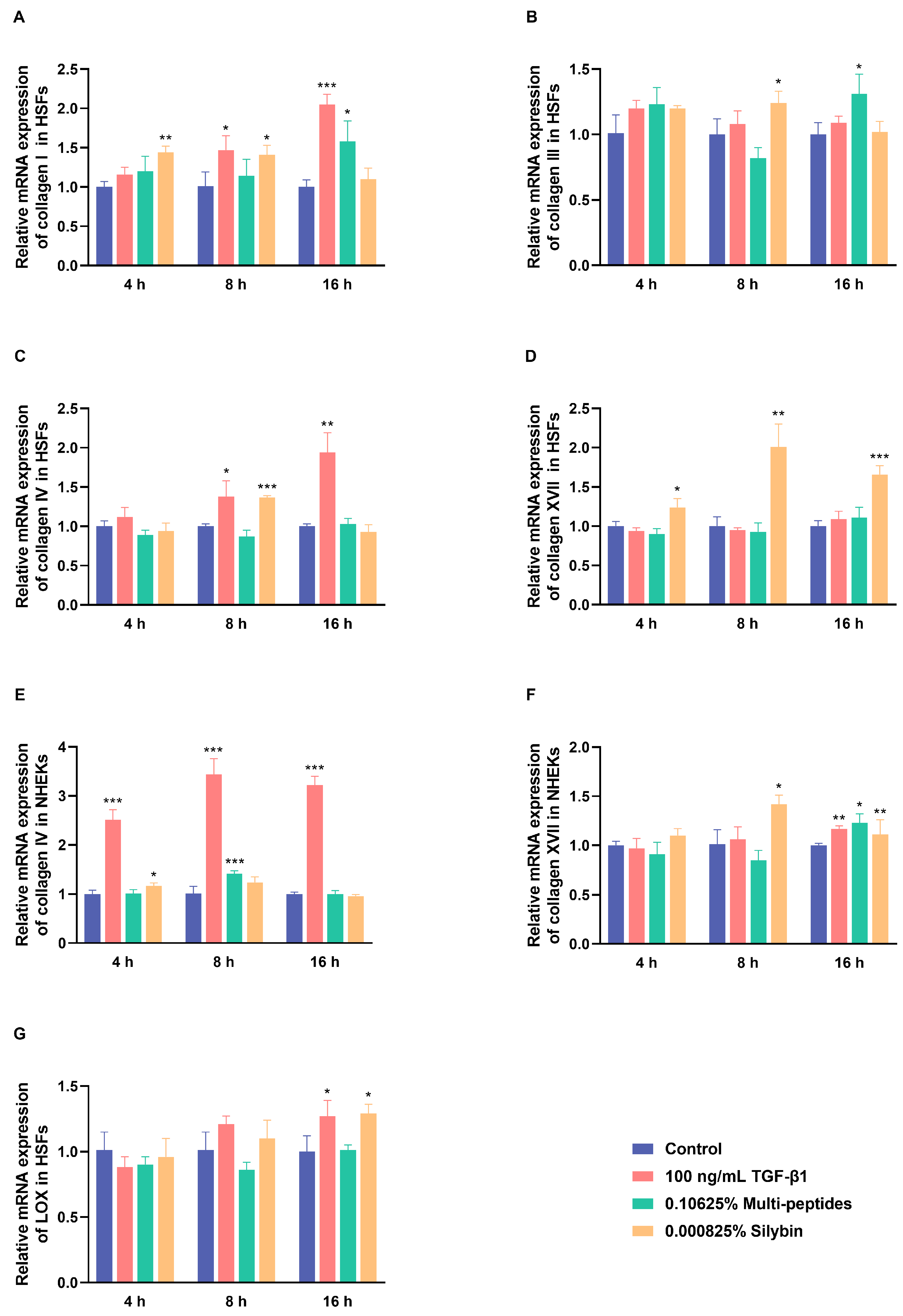
qRT-PCR analysis showed that multi-peptides and silybin induced rapid transcriptional activation of ECM-related genes in both HSFs and NHEKs within 16 h (Figure 2A–G). TGF-β1, used as a positive control, consistently upregulated the relative gene expressions of collagen I, IV, and XVII across multiple time points. At 4 h, silybin significantly increased the relative gene expressions of collagen I and XVII in HSFs and collagen IV in NHEKs, whereas multi-peptides showed minimal effects. At 8 h, multi-peptides upregulated the relative gene expression of collagen IV in NHEKs, while silybin further enhanced the relative gene expressions of collagen I, III, IV, and XVII in HSFs and collagen XVII in NHEKs. At 16 h, multi-peptides elevated the relative gene expressions of collagen I and III in HSFs and collagen XVII in NHEKs, and silybin strongly increased the relative gene expressions of collagen XVII in both cell types. The relative gene expression of LOX was significantly induced only by TGF-β1 and silybin after 16 h. All changes were statistically significant (p < 0.05 vs. control).

3.3. Restoration of UV-Damaged ECM Proteins by Multi-Peptides and Silybin

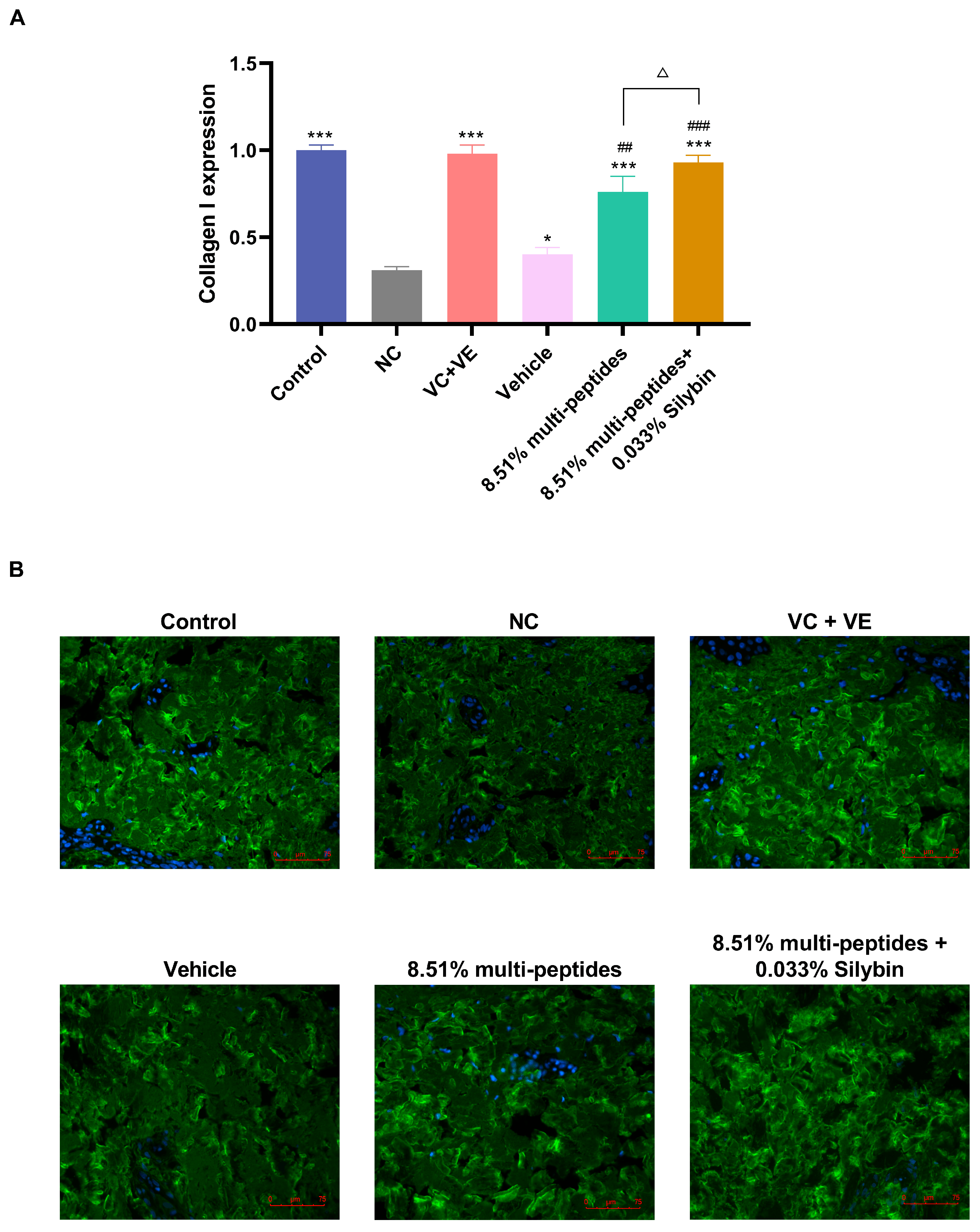
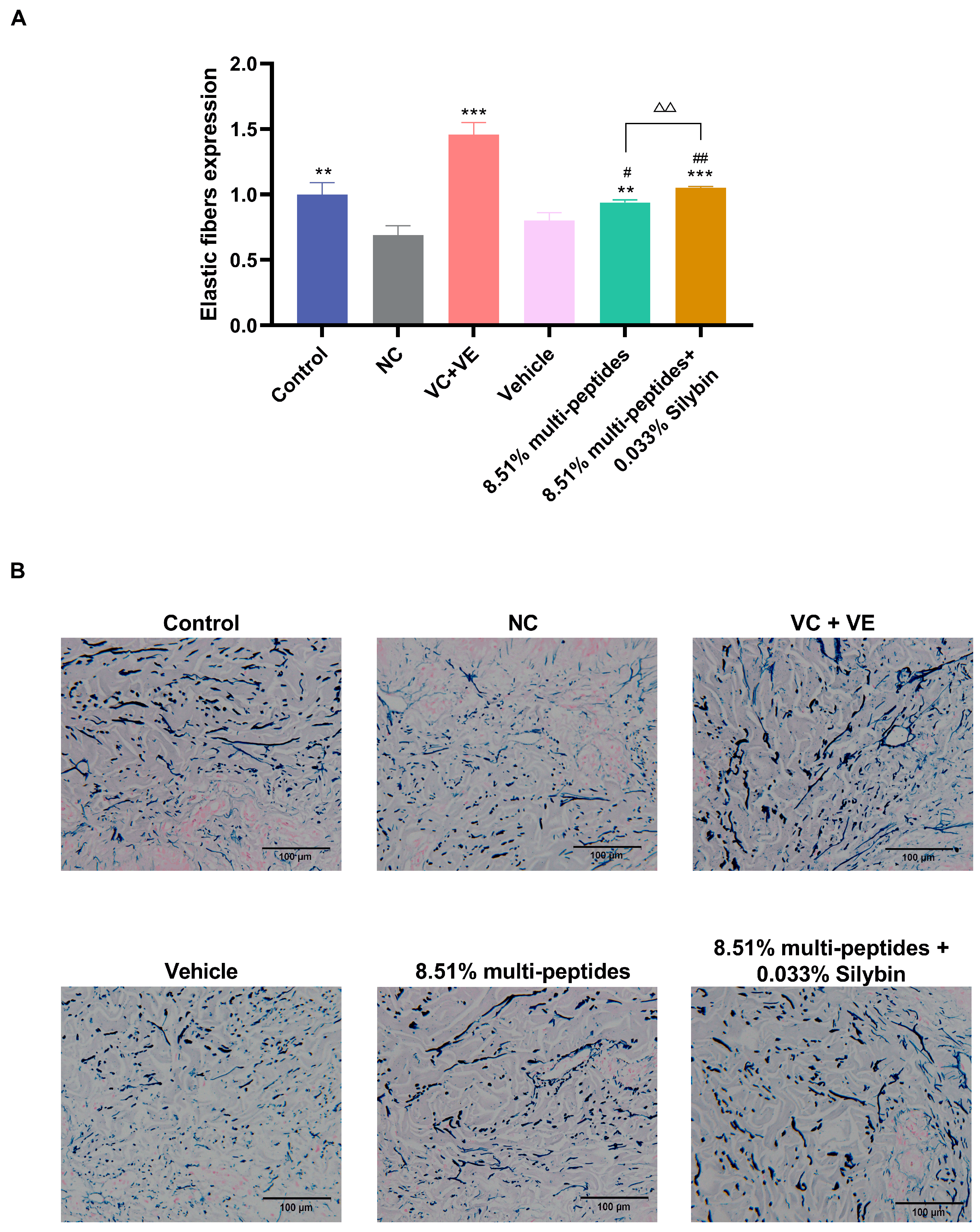
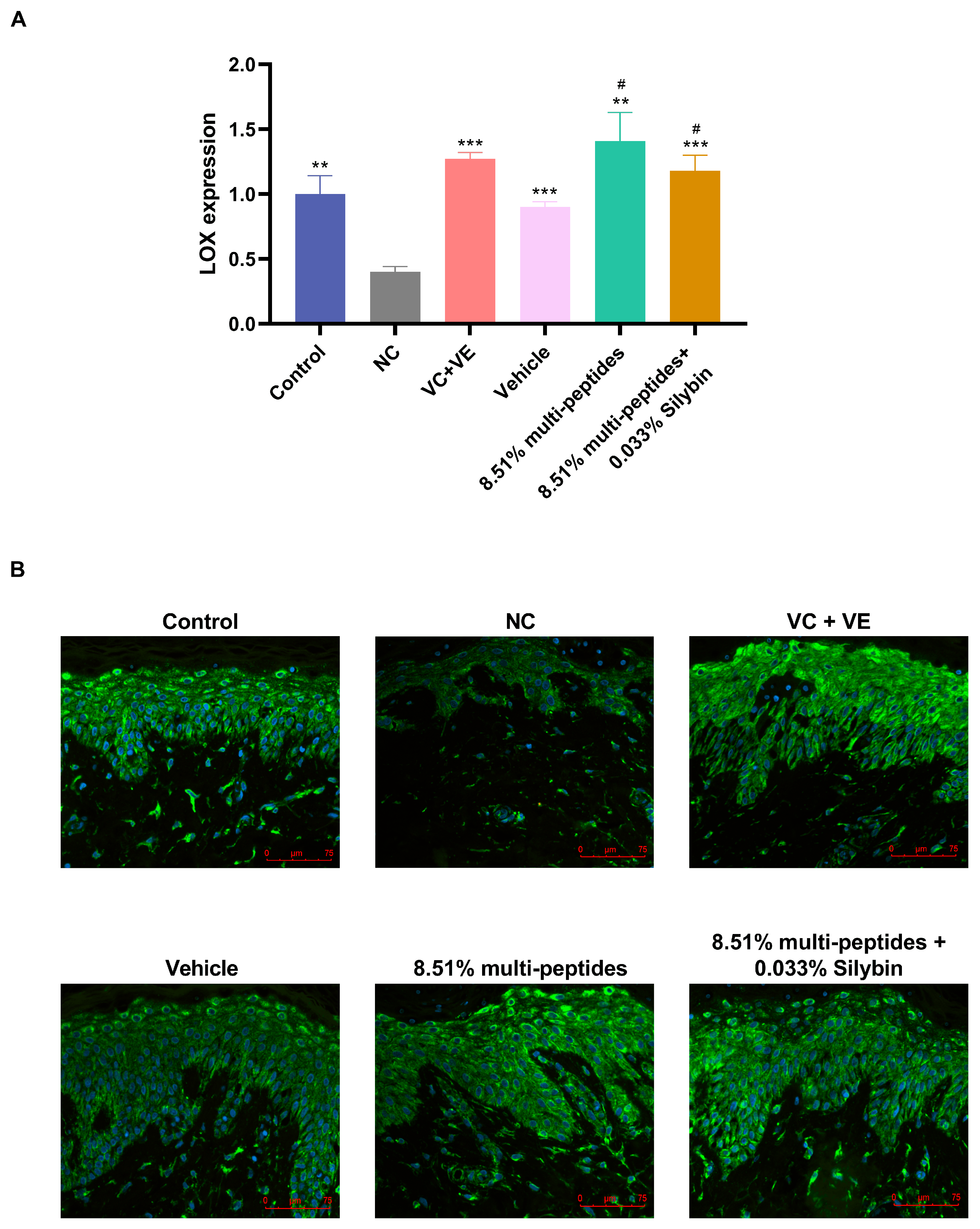
Immunofluorescence (collagen I, III, IV, XVII, LOX) and Victoria blue staining (elastic fibers) demonstrated that UV irradiation markedly decreased ECM protein levels in human skin explants compared with the negative control (Figure 3, Figure 4 and Figure 5 and Figures S1–S3). Treatment with multi-peptides or multi-peptides combined with silybin significantly restored these proteins relative to the negative control (p < 0.05). The positive control (VC + VE) produced a similar recovery.
Vehicle treatment exhibited partial effects, with significant increases limited to collagen I, XVII, and LOX (p < 0.05 vs. negative control). Compared with the vehicle, multi-peptides alone significantly increased collagen I, III, XVII, elastic fibers, and LOX, while the combination of multi-peptides with silybin further enhanced collagen I, III, IV, XVII, and elastic fibers (p < 0.05). The synergistic enhancement rates for collagen I, III, IV, XVII, and elastic fibers were 22.37%, 34.29%, 155.56%, 29.58%, and 11.70%, respectively. No additional effect on LOX was observed with the addition of silybin (p > 0.05 vs. vehicle).
These findings suggest that multi-peptides exert restorative effects on UV-damaged ECM proteins, and co-treatment with silybin provides a synergistic benefit, particularly for collagen subtypes and elastic fibers.

3.4. Rapid In Vitro Activation of Anti-Aging Proteins by the Cream Containing Multi-Peptides and Silybin

Protein-level analyses using immunofluorescence and Western blot demonstrated that the cream induced a time-dependent upregulation of multiple ECM proteins in HSFs and NHEKs (Figure 6A–J).
The positive control (TGF-β1) markedly increased collagen I, III, IV, and XVII protein expression within 4–16 h and further enhanced collagen III and XVII after prolonged post-incubation (4 h + 20 h or 16 h + 8 h).
For the cream treatment (multi-peptides + silybin), early increases in collagen I, collagen IV, and LOX were observed as early as 4 h (p < 0.05 vs. control) and remained elevated at 16 h. Collagen III expression required extended incubation (4 h + 20 h, 16 h or 16 h + 8 h) to show significant upregulation, while collagen XVII enhancement was most marked after prolonged incubation beyond 16 h. IF images of collagen I and collagen IV (Figure 6A–D) revealed increased green fluorescence intensity in cream-treated cells, consistent with the semiquantitative results from Western blot (Figure 6E–J).
In short, these findings confirm that the cream rapidly activates multiple ECM proteins associated with anti-aging effects, with some proteins (collagen I, IV, and LOX) responding early and others (collagen III and XVII) requiring longer exposure or post-incubation to achieve maximal activation.

3.5. Anti-Aging Effects of the Cream on Skin

Clinical evaluation demonstrated progressive improvements in various facial parameters following 28 and 56 days of cream application.
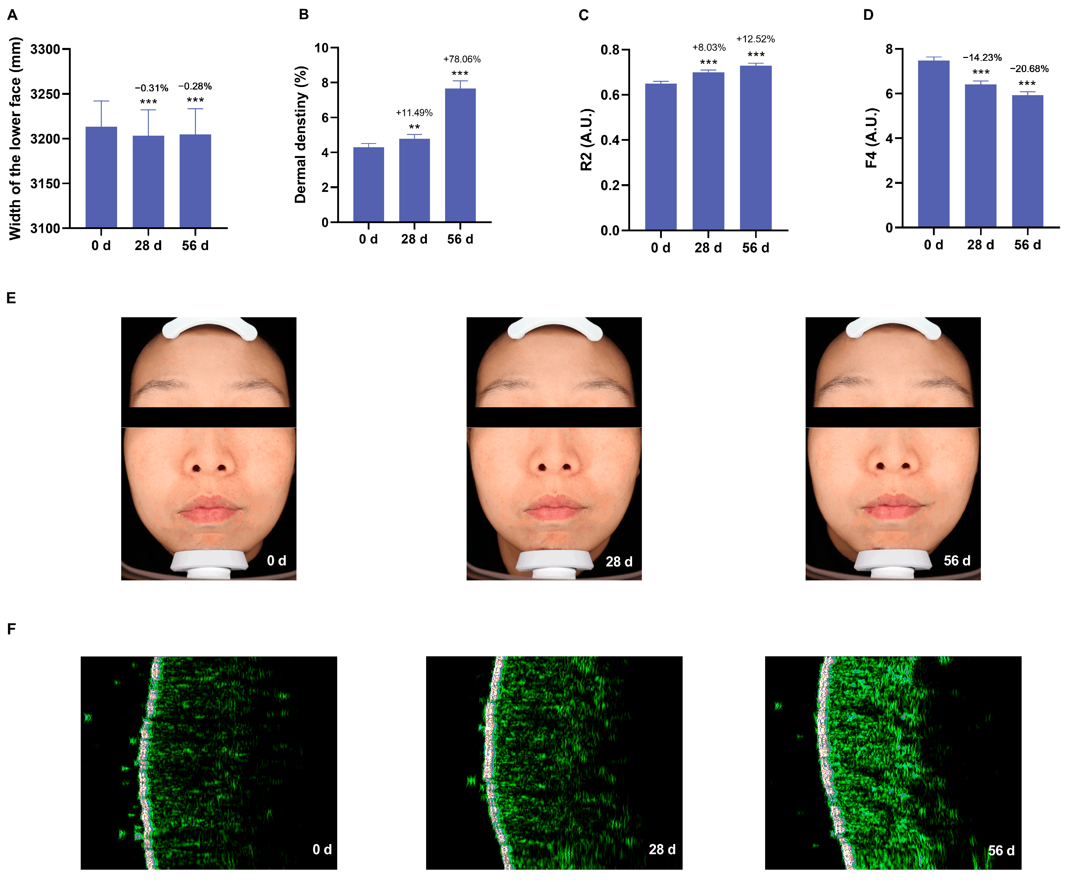
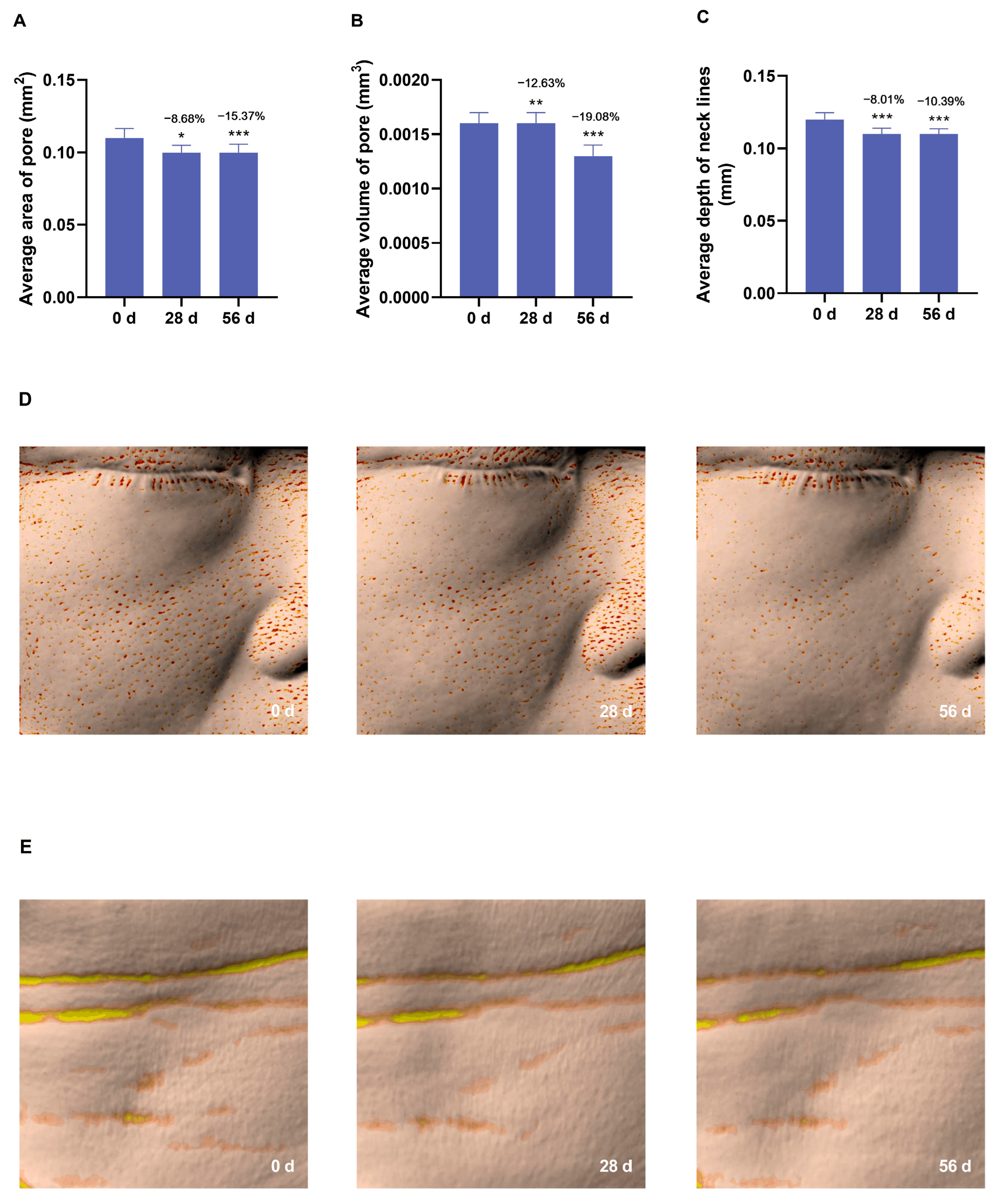
The lower face width decreased by 0.28% and dermal density rose by 78.06% at 56 days (Figure 7A,B). R2 (skin elasticity) increased by 12.52% and F4 (lower F4 presented greater firmness) decreased by 20.68% at 56 days (Figure 7C,D). All p values were less than 0.001. Average pore area and volume were reduced by 15.37% and 19.08% at 56 days (Figure 8A,B), respectively (p < 0.001), while neck stripe depth decreased by 8.01% and 10.39% at 28 and 56 days (p < 0.05) (Figure 8C). Primos CR and Antera 3D analyses revealed significant reductions in wrinkle length, area, and volume across the forehead, nasolabial, glabellar, and cheek regions, with improvements observable as early as 28 days and further enhanced at 56 days (Figure 9). Dermatological assessments confirmed minimal irritation, with no adverse reactions reported (Table 1). Self-reported satisfaction exceeded 80% by day 56, particularly for improvements in firmness, midface lifting, and jawline definition (Table 2).

4. Discussion

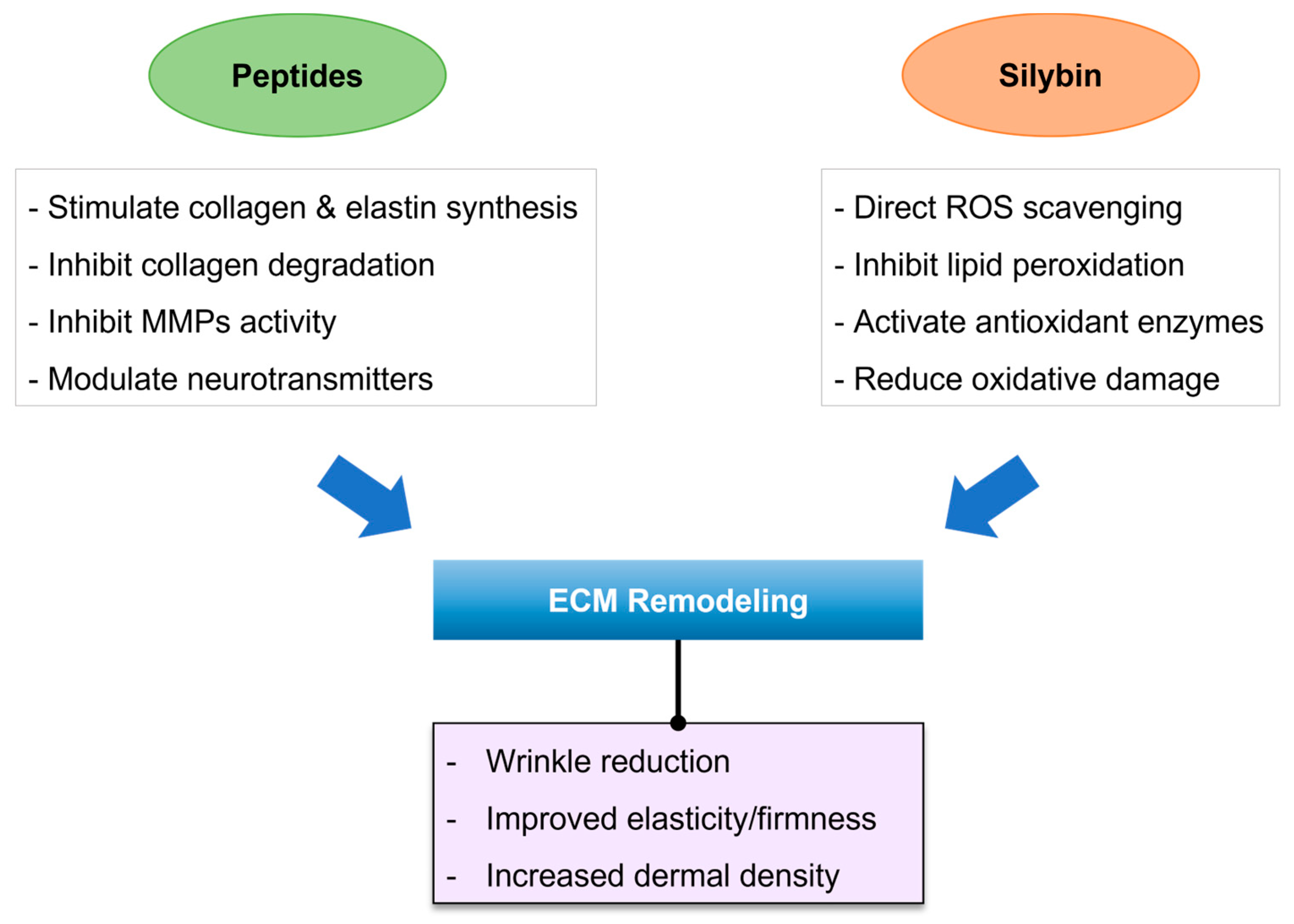
This study demonstrates that a seven-peptide and silybin complex can effectively restore key ECM components and improve clinical signs of skin aging. It is particularly noteworthy as the first report of a topical cosmetic formulation significantly upregulating LOX in both skin cells and tissue explants—a finding relevant to long-term matrix remodeling and tensile strength. While botanical antioxidants like silybin have shown retinol-comparable rejuvenation benefits without irritation [30], and topical peptides are well-established stimulators of dermal matrix renewal [21], their combined effects on skin structure have not been previously documented. Here, we observed a clear synergistic benefit: peptides alone markedly restored collagen I, III, IV, XVII, elastic fibers, and LOX levels in UV-damaged skin explants (vs. untreated controls, p < 0.05), and adding silybin further enhanced the recovery of collagens (I, III, IV, XVII) and elastic fibers beyond the peptide blend’s effects. Such multi-ingredient synergy is consistent with recent in vitro work showing that combinations of peptides can amplify skin-regenerative gene expression compared to single peptides [10]. Our findings thus introduce a compelling new paradigm for next-generation anti-aging cosmetics, whereby a cocktail of functionally diverse peptides plus an antioxidant yields greater ECM benefits than peptides alone. By leveraging diverse peptide functions—collagen stimulation, elastin enhancement, and matrix protection—together with silybin’s antioxidant and photoprotective roles, this formulation achieves a multi-mechanism anti-aging effect. The consistency of results across in vitro, ex vivo, and clinical evaluations further confirms the translational validity of these synergistic effects (Figure 10).
Our use of silybin is particularly timely given recent evidence of its retinol-like efficacy without the side effects. The core constituents of Silybum marianum are flavonolignans collectively known as silymarin, comprising silybin (the most abundant and bioactive component), isosilybin, silychristin, and silydianin [39]. Their reported bioactivities include direct free-radical scavenging, attenuation of ROS-generating enzyme activity, activation of the Nrf2/ARE antioxidant response, inhibition of NF-κB-mediated pro-inflammatory signaling, and the induction of cytoprotective proteins such as heat-shock proteins and thioredoxin [39]. A 2025 clinical study demonstrated that an S. marianum (silymarin) extract produced anti-wrinkle and collagen-boosting outcomes comparable to retinol but with no irritation or photosensitivity [30]. By combining silybin’s protective, collagen-boosting action with peptides’ regenerative stimuli, we introduce a novel dual strategy to combat skin aging. Notably, incorporating silybin into the peptide blend enhanced efficacy without any observable irritation in our trial, underscoring its value as a gentler alternative or complement to retinoids. In our UV-exposed skin explant model, treatment with the seven-peptide blend alone significantly restored ECM proteins—including collagen types I, III, IV, XVII, elastin, and LOX—relative to untreated UV-damaged controls. The addition of silybin provided further increases, particularly for collagens I, III, IV, XVII, and elastin (all p < 0.05 vs. peptide-only), confirming a synergistic interaction. These data suggest that silybin not only adds antioxidant protection but also actively amplifies the matrix-regenerative effects of peptides. Such a multi-target approach addresses several fundamental aspects of skin aging simultaneously, from replenishing structural proteins to fortifying cross-linking enzymes, while avoiding the tolerability issues of harsher actives.
By simultaneously targeting multiple ECM components, our formulation helps rebuild a stronger dermal architecture and DEJ, which are critical for youthful skin function. Collagen I and III are the predominant structural proteins in the dermis, providing tensile strength and resilience; the significant upregulation of both types in our studies suggests robust restoration of the dermal collagen network. Collagen IV is a key constituent of the basement membrane at the DEJ, anchoring the epidermis to the dermis. Chronic UV exposure is known to suppress collagen IV synthesis and degrade the DEJ, contributing to wrinkle formation and loss of skin firmness [40,41]. In our UV-damaged explants, collagen IV levels were nearly restored to normal with peptide–silybin treatment, which likely helps maintain DEJ integrity. Collagen XVII (BP180) is a transmembrane collagen in basal keratinocytes’ hemidesmosomes that helps bind the epidermis to the basement membrane [23]. Loss of collagen XVII with aging or photodamage leads to compromised epidermal–dermal adhesion, impairing skin firmness [42,43]. Few cosmetic ingredients specifically target collagen XVII production; thus, the marked upregulation of collagen XVII observed in our cell and explant models is a unique and valuable finding. Elevating all of these collagens in concert implies that our formulation can reinforce the dermal scaffold and improve epidermal anchoring, thereby enhancing skin firmness and reducing the appearance of wrinkles at the surface.
Another notable innovation of our study is the focus on LOX, an enzyme rarely examined in cosmetic science despite its critical role in stabilizing collagen and elastic fibers. LOX catalyzes the covalent cross-linking of collagen and elastin, a process essential for ECM tensile strength and durability [6,7]. With age, LOX expression declines and contributes to weaker, more lax collagen bundles. To date, few cosmetic interventions have directly aimed to boost LOX or related cross-linking enzymes; one exception is a report that a dill extract can induce LOXL1 (a LOX family member) to increase elastin content in adult skin [8]. We provide the first evidence that both the individual ingredients (multi-peptides and silybin) and the final cream formulation significantly upregulate LOX expression in human fibroblasts and in UV-aged skin tissue. This finding supports the hypothesis that our treatment not only improves superficial skin parameters but also promotes long-term structural integrity of the dermis through true ECM remodeling and fiber cross-link reinforcement. Enhancing LOX-mediated cross-linking could yield a more resilient dermal matrix that resists degradation over time [44]. Elastic fibers are subjected to diverse enzymatic, chemical, and biophysical influences throughout the lifespan of an organism, and because of their low turnover, they gradually accumulate damage. Histologically, aging of elastic fibers is characterized by fragmentation and thinning of elastin [45]. A significant increase in elastin observed in our explant model, especially with the peptide–silybin combination, suggests that upregulating LOX (and LOX-like proteins) may revive elastin assembly by facilitating new cross-links [45]. In essence, our formulation targets not just collagen synthesis but also the quality and durability of the collagen/elastic fibers via LOX—a multifaceted strategy that may confer more long-lasting firmness than approaches that stimulate collagen alone.
Moreover, we observed a rapid induction of procollagen markers. To our knowledge, there is a paucity of studies on such rapid collagen and LOX induction by topical cosmetic actives—within 4 h of exposure. Most published research on peptides or retinoids evaluates molecular changes at least 24 h later [46,47,48]. Here, we showed that silybin (at 0.000825%) and even the finished cream (at just 0.03125%) could significantly upregulate the gene or protein levels of collagen I (by ~40%), collagen III (~45%), collagen IV (~20%), and collagen XVII (~20–30%), as well as LOX (~20–25%) in cultured skin cells within 4 h. These findings introduce the novel concept of a “flash-collagen charging” effect in cosmetic science, wherein our peptide–silybin product triggers immediate ECM fortification well before the timelines traditionally expected for skincare actives. Early activation of collagen production and cross-linking enzymes might jump-start the skin’s regenerative processes, potentially leading to faster and more noticeable improvements in skin quality.

5. Conclusions

In summary, the combination of seven complementary peptides with silybin represents an innovative and clinically effective anti-aging strategy. By concurrently enhancing collagen, elastin, and LOX, this complex addresses the structural causes of skin aging on multiple levels—rebuilding the dermal matrix, strengthening the DEJ, and improving the resilience of collagen/elastic fibers. The synergistic enhancement rates of silybin for collagen and elastic fibers were 11.7% to 155.56%. Early increases in collagen I, collagen IV, and LOX were observed as soon as 4 h after cream treatment and remained evident up to 16 h. Our translational approach, linking molecular markers to clear clinical outcomes, underscores the real-world value of this multi-target formulation. These outcomes highlight the advantages of a synergistic, multi-mechanism approach in skincare, echoing the emerging consensus that combining peptides with other bioactives can yield superior rejuvenation results [10,49,50]. Moreover, the significant improvements observed in wrinkles, elasticity (+12.52%), firmness (−20.68%), and dermal density (+78.06%) after 8 weeks are consistent with other recent clinical interventions using multi-peptide products [46,50], reinforcing the relevance of our findings for cosmetic dermatology. Taken together, our study suggests that intelligently formulated peptide–antioxidant complexes can achieve rapid and sustained skin anti-aging benefits, and it provides a strong scientific basis for the development of next-generation cosmeceuticals targeting both the quantity and quality of the dermal extracellular matrix.

Supplementary Materials

The following supporting information can be downloaded at: https://www.mdpi.com/article/10.3390/cosmetics12050223/s1, Data S1: The ingredients of the cream formula; Data S2: Collagen expression in human skin explants after UV irradiation; Data S3: Composition of as-supplied raw materials and corresponding active-equivalent contents; Data S4: primer sequences used in qRT-PCR.

Author Contributions

Conceptualization, H.Z., H.H., L.W., Y.Y., and J.H.; methodology, H.Z., H.H. and C.X.; software, H.Z. and Y.L.; validation, H.H., C.X., L.W., Y.Y. and Y.L.; formal analysis, H.Z., H.H., and C.X.; investigation, H.Z., H.H., C.X., L.W., Y.Y., J.H., F.L., Y.L. and P.S.; resources, Y.C., F.L. and P.S.; data curation, H.Z., H.H., C.X., L.W., Y.Y. and Y.L.; writing—original draft preparation, H.Z.; writing—review and editing, Y.L., H.Z. and H.H.; visualization, H.H. and C.X.; supervision, J.H., Y.C., F.L., Y.L. and P.S.; project administration, L.W. and Y.Y.; funding acquisition, Y.L. and P.S. All authors have read and agreed to the published version of the manuscript.

Funding

This study was funded in full by Proya Cosmetics Co., Ltd.

Institutional Review Board Statement

The study was conducted in accordance with the Declaration of Helsinki, and the protocol was approved by the Ethics Committee of Shanxi BioCell Biotechnology Co., Ltd. (Approval No. GDLL2023005) on 4 July 2023 and the Ethic Committee for clinical research of SGS-CSTC Standards Technical Services Co., Ltd. (Approval No. 2023051) on 23 June 2023.

Informed Consent Statement

Informed consent was obtained from all subjects involved in the study. Written informed consent was obtained from the patients to publish this paper.

Data Availability Statement

The raw data supporting the conclusions of this article will be made available by the authors on request.

Acknowledgments

All authors sincerely thank the SGS-CSTC Standards Technical Services Co., Ltd., Hangzhou Branch, and BioCell Biotechnology Co., Ltd., for their invaluable technical assistance.

Conflicts of Interest

All the authors are employees of Proya Cosmetics Co., Ltd. The authors declare that the research was conducted in the absence of any commercial or financial relationships that could be construed as a potential conflict of interest.

Abbreviations

The following abbreviations are used in this manuscript:
ECMExtracellular matrix
LOXLysyl oxidase
DEJDermal–epidermal junction
ROSReactive oxygen species
UVUltraviolet
AGEsAdvanced glycation end products
HSFsHuman skin fibroblasts
FBSFetal bovine serum
NHEKsNormal human epidermal keratinocytes
PBSPhosphate buffer saline
MTT3-(4,5-dimethylthiazol-2-yl)-2,5-diphenyltetrazolium bromide
PFAParaformaldehyde
VCVitamin C
VEVitamin E
TGFTransforming growth factor
UVAUltraviolet A
UVBUltraviolet B
NCNegative control
qRT-PCRQuantitative real-time polymerase chain reaction
IFImmunofluorescence
IPPImage-Pro® Plus
IODIntegrated optical density
MODMean optical density
WBWestern blot
VBVictoria blue
RHRelative humidity
SDStandard deviation
SEStandard error

References

  1. Chung, J.H.; Seo, J.Y.; Choi, H.R.; Lee, M.K.; Youn, C.S.; Rhie, G.E.; Cho, K.H.; Kim, K.H.; Park, K.C.; Eun, H.C. Modulation of Skin Collagen Metabolism in Aged and Photoaged Human Skin In Vivo. J. Investig. Dermatol. 2001, 117, 1218–1224. [Google Scholar] [CrossRef]
  2. Warren, R.; Gartstein, V.; Kligman, A.M.; Montagna, W.; Ridder, G.M. Age, sunlight, and facial skin: A histologic and quantitative study. J. Am. Acad. Dermatol. 1991, 25, 751–760. [Google Scholar] [CrossRef]
  3. Fisher, G.J.; Wang, Z.Q.; Datta, S.C.; Varani, J.; Kang, S.; Voorhees, J.J. Pathophysiology of premature skin aging induced by ultraviolet light. N. Engl. J. Med. 1997, 337, 1419–1428. [Google Scholar] [CrossRef]
  4. Rittie, L.; Fisher, G.J. Natural and Sun-Induced Aging of Human Skin. Cold Spring Harb. Perspect. Med. 2015, 5, a015370. [Google Scholar] [CrossRef]
  5. Bailey, A.J. Molecular mechanisms of ageing in connective tissues. Mech. Ageing Dev. 2001, 122, 735–755. [Google Scholar] [CrossRef] [PubMed]
  6. Behmoaras, J.; Slove, S.; Seve, S.; Vranckx, R.; Sommer, P.; Jacob, M.P. Differential expression of lysyl oxidases LOXL1 and LOX during growth and aging suggests specific roles in elastin and collagen fiber remodeling in rat aorta. Rejuvenation Res. 2008, 11, 883–889. [Google Scholar] [CrossRef] [PubMed]
  7. Mäki, J.M. Lysyl oxidases in mammalian development and certain pathological conditions. Histol. Histopathol. 2009, 24, 651–660. [Google Scholar] [CrossRef] [PubMed]
  8. Cenizo, V.; Andre, V.; Reymermier, C.; Sommer, P.; Damour, O.; Perrier, E. LOXL as a target to increase the elastin content in adult skin: A dill extract induces the LOXL gene expression. Exp. Dermatol. 2006, 15, 574–581. [Google Scholar] [CrossRef]
  9. Baccarani-Contri, M.; Vincenzi, D.; Quaglino, D., Jr.; Mori, G.; Pasquali-Ronchetti, I. Localization of human placenta lysyl oxidase on human placenta, skin and aorta by immunoelectronmicroscopy. Matrix 1989, 9, 428–436. [Google Scholar] [CrossRef]
  10. Flagler, M.J.; Tamura, M.; Laughlin, T.; Hartman, S.; Ashe, J.; Adams, R.; Kozak, K.; Cresswell, K.; Mullins, L.; Jarrold, B.B.; et al. Combinations of peptides synergistically activate the regenerative capacity of skin cells in vitro. Int. J. Cosmet. Sci. 2021, 43, 518–529. [Google Scholar] [CrossRef]
  11. Widgerow, A.D.; Ziegler, M.E.; Shafiq, F. TriHex 2.0—Advancing Skin Health Science and the TriHex Technology. J. Cosmet. Dermatol. 2025, 24, e16690. [Google Scholar] [CrossRef]
  12. Gorouhi, F.; Maibach, H.I. Role of topical peptides in preventing or treating aged skin. Int. J. Cosmet. Sci. 2009, 31, 327–345. [Google Scholar] [CrossRef]
  13. Gorouhi, F.; Maibach, H.I. Topical Peptides and Proteins for Aging Skin. In Textbook of Aging Skin; Farage, M.A., Miller, K.W., Maibach, H.I., Eds.; Springer: Berlin/Heidelberg, Germany, 2017; pp. 1865–1896. ISBN 978-3-662-47398-6. [Google Scholar]
  14. Namjoshi, S.; Benson, H.A.E. Cyclic peptides as potential therapeutic agents for skin disorders. Pept. Sci. 2010, 94, 673–680. [Google Scholar] [CrossRef] [PubMed]
  15. Shim, Y.Y.; Gui, B.; Arnison, P.G.; Wang, Y.; Reaney, M.J.T. Flaxseed (Linum usitatissimum L.) bioactive compounds and peptide nomenclature: A review. Trends Food Sci. Technol. 2014, 38, 5–20. [Google Scholar] [CrossRef]
  16. Wang, H.P.; Xing, H.Y.; Liu, Z.G.; Yu, X.Y. An Anti-Wrinkle Cyclic Hexapeptide Compound and Its Preparation Method. CN114634554A, 6 December 2022. [Google Scholar]
  17. Li, Y.; Li, J.L.; Huang, P.; Sun, B.X.; Liu, Z.B.; Wang, Y. A Linseed Extract with Skin Anti-Aging Effect, Its Preparation Method and Application. CN116172921A, 30 May 2023. [Google Scholar]
  18. Abdul Jabbar, O.; Kashmoola, M.A.; Mustafa Al-Ahmad, B.E.; Mokhtar, K.I.; Muhammad, N.; Abdul Rahim, R.; Qouta, L.A. The Effect of Flaxseed Extract on Skin Elasticity of The Healing Wound In Rabbits. IIUM Med. J. Malays. 2019, 18, 5–12. [Google Scholar] [CrossRef]
  19. New ingredients for skin and hair care launched at in-cosmetics. Pharm Cosmet Rev. 2010, 37, 14. [CrossRef]
  20. Murphy-Ullrich, J.E.; Poczatek, M. Activation of latent TGF-β by thrombospondin-1: Mechanisms and physiology. Cytokine Growth Factor. Rev. 2000, 11, 59–69. [Google Scholar] [CrossRef]
  21. Schagen, S.K. Topical Peptide Treatments with Effective Anti-Aging Results. Cosmetics 2017, 4, 16. [Google Scholar] [CrossRef]
  22. Rahnamaeian, M.; Vilcinskas, A. Short antimicrobial peptides as cosmetic ingredients to deter dermatological pathogens. Appl. Microbiol. Biotechnol. 2015, 99, 8847–8855. [Google Scholar] [CrossRef]
  23. Powell, A.M.; Sakuma-Oyama, Y.; Oyama, N.; Black, M.M. Collagen XVII/BP180: A collagenous transmembrane protein and component of the dermoepidermal anchoring complex. Clin. Exp. Dermatol. 2005, 30, 682–687. [Google Scholar] [CrossRef]
  24. Wang, M.; Wang, Z.; Liu, T.; Zhao, Y.; Sun, X.; Lu, B.; Zhang, J.; Liu, Z.; Zhang, J. Pioneering ionic liquids in neuro-soothing: Enhanced transdermal delivery of collagen peptides and their synergistic anti-aging functions. Mater Today Bio 2025, 31, 101527. [Google Scholar] [CrossRef] [PubMed]
  25. Tang, Y.; Nie, T.; Zhang, L.; Liu, X.; Deng, H. Peptides in Cosmetics: From Pharmaceutical Breakthroughs to Skincare Innovations. Cosmetics 2025, 12, 107. [Google Scholar] [CrossRef]
  26. Shu, P.; Mo, J.; Li, Z.; Li, M.; Zhu, W.; Du, Z. Ferulic acid in synergy with retinol alleviates oxidative injury of HaCaT cells during UVB-induced photoaging. Aging 2024, 16, 7153–7173. [Google Scholar] [CrossRef]
  27. Ferreira, M.S.; Magalhães, M.C.; Oliveira, R.; Sousa-Lobo, J.M.; Almeida, I.F. Trends in the Use of Botanicals in Anti-Aging Cosmetics. Molecules 2021, 26, 3584. [Google Scholar] [CrossRef]
  28. Rajnochová Svobodová, A.; Gabrielová, E.; Michaelides, L.; Kosina, P.; Ryšavá, A.; Ulrichová, J.; Zálešák, B.; Vostálová, J. UVA-photoprotective potential of silymarin and silybin. Arch. Dermatol. Res. 2018, 310, 413–424. [Google Scholar] [CrossRef]
  29. Kitajima, S.; Yamaguchi, K. Silybin from Silybum Marianum Seeds Inhibits Confluent-Induced Keratinocytes Differentiation as Effectively as Retinoic Acid without Inducing Inflammatory Cytokine. J. Clin. Biochem. Nutr. 2009, 45, 178–184. [Google Scholar] [CrossRef] [PubMed]
  30. Boira, C.; Chapuis, E.; Lapierre, L.; Tiguemounine, J.; Scandolera, A.; Reynaud, R. Silybum marianum Extract: A Highly Effective Natural Alternative to Retinoids to Prevent Skin Aging Without Side Effects. J. Cosmet. Dermatol. 2025, 24, e16613. [Google Scholar] [CrossRef]
  31. Sharifi, R.; Pasalar, P.; Kamalinejad, M.; Dehpour, A.R.; Tavangar, S.M.; Paknejad, M.; Mehrabani Natanzi, M.; Nourbakhsh, M.; Ahmadi Ashtiani, H.R.; Akbari, M.; et al. The effect of silymarin (Silybum marianum) on human skin fibroblasts in an in vitro wound healing model. Pharm. Biol. 2013, 51, 298–303. [Google Scholar] [CrossRef]
  32. Bäckvall, H.; Wassberg, C.; Berne, B.; Ponten, F. Similar UV responses are seen in a skin organ culture as in human skin in vivo. Exp. Dermatol. 2002, 11, 349–356. [Google Scholar] [CrossRef]
  33. Feng, Z.; Yang, R.; Wu, L.; Tang, S.; Wei, B.; Guo, L.; He, L.; Feng, Y. Atractylodes macrocephala polysaccharides regulate the innate immunity of colorectal cancer cells by modulating the TLR4 signaling pathway. Onco Targets Ther. 2019, 12, 7111–7121. [Google Scholar] [CrossRef]
  34. Iriyama, S.; Ogura, Y.; Nishikawa, S.; Hosoi, J.; Amano, S. Regeneration of collagen fibrils at the papillary dermis by reconstructing basement membrane at the dermal-epidermal junction. Sci. Rep. 2022, 12, 795. [Google Scholar] [CrossRef]
  35. Livak, K.J.; Schmittgen, T.D. Analysis of Relative Gene Expression Data Using Real-Time Quantitative PCR and the 2−ΔΔCT Method. Methods 2001, 25, 402–408. [Google Scholar] [CrossRef] [PubMed]
  36. Cox, T.R.; Bird, D.; Baker, A.M.; Barker, H.E.; Ho, M.W.; Lang, G.; Erler, J.T. LOX-mediated collagen crosslinking is responsible for fibrosis-enhanced metastasis. Cancer Res. 2013, 73, 1721–1732. [Google Scholar] [CrossRef]
  37. Jang, H.-J.; Kim, Y.-m.; Yoo, B.-Y.; Seo, Y.-K. Wound-healing effects of human dermal components with gelatin dressing. J. Biomater. Appl. 2017, 32, 716–772. [Google Scholar] [CrossRef] [PubMed]
  38. Altman, D.G. Practical Statistics for Medical Research; Chapman and Hall/CRC: Boca Raton, FL, USA, 1991; ISBN 0-412-27630-5. [Google Scholar]
  39. Zhang, X.; Liu, M.; Wang, Z.; Wang, P.; Kong, L.; Wu, J.; Wu, W.; Ma, L.; Jiang, S.; Ren, W.; et al. A review of the botany, phytochemistry, pharmacology, synthetic biology and comprehensive utilization of Silybum marianum. Front. Pharmacol. 2024, 15, 1417655. [Google Scholar] [CrossRef] [PubMed]
  40. Haytoglu, N.S.; Gurel, M.S.; Erdemir, A.; Falay, T.; Dolgun, A.; Haytoglu, T.G. Assessment of skin photoaging with reflectance confocal microscopy. Skin. Res. Technol. 2014, 20, 363–372. [Google Scholar] [CrossRef]
  41. Vázquez, F.; Palacios, S.; Alemañ, N.; Guerrero, F. Changes of the basement membrane and type IV collagen in human skin during aging. Maturitas 1996, 25, 209–215. [Google Scholar] [CrossRef]
  42. Langton, A.K.; Halai, P.; Griffiths, C.E.M.; Sherratt, M.J.; Watson, R.E.B. The impact of intrinsic ageing on the protein composition of the dermal-epidermal junction. Mech. Ageing Dev. 2016, 156, 14–16. [Google Scholar] [CrossRef]
  43. Tao, K.; Zhu, H.; Wei, J. Anti-aging effect of low molecular weight recombinant humanized collagen on photo-aging by activating adherence junction signaling pathways. PLoS ONE 2025, 20, e0329460. [Google Scholar] [CrossRef]
  44. Szauter, K.M.; Cao, T.; Boyd, C.D.; Csiszar, K. Lysyl oxidase in development, aging and pathologies of the skin. Pathol. Biol. 2005, 53, 448–456. [Google Scholar] [CrossRef]
  45. Heinz, A. Elastic fibers during aging and disease. Ageing Res. Rev. 2021, 66, 101255. [Google Scholar] [CrossRef] [PubMed]
  46. Jeong, S.; Yoon, S.; Kim, S.; Jung, J.; Kor, M.; Shin, K.; Lim, C.; Han, H.S.; Lee, H.; Park, K.-Y.; et al. Anti-Wrinkle Benefits of Peptides Complex Stimulating Skin Basement Membrane Proteins Expression. Int. J. Mol. Sci. 2020, 21, 73. [Google Scholar] [CrossRef]
  47. Dierckx, S.; Patrizi, M.; Merino, M.; González, S.; Mullor, J.L.; Nergiz-Unal, R. Collagen peptides affect collagen synthesis and the expression of collagen, elastin, and versican genes in cultured human dermal fibroblasts. Front. Med. 2024, 11, 1397517. [Google Scholar] [CrossRef]
  48. Brown, A.; Furmanczyk, M.; Ramos, D.; Ribes, A.; Pons, L.; Bustos, J.; de Henestrosa, A.R.F.; Granger, C.; Jourdan, E. Natural Retinol Analogs Potentiate the Effects of Retinal on Aged and Photodamaged Skin: Results from In Vitro to Clinical Studies. Dermatol. Ther. 2023, 13, 2299–2317. [Google Scholar] [CrossRef]
  49. Jiang, F.; Wu, Y.; Liu, Z.; Hong, M.; Huang, Y. Synergy of GHK-Cu and hyaluronic acid on collagen IV upregulation via fibroblast and ex-vivo skin tests. J. Cosmet. Dermatol. 2023, 22, 2598–2604. [Google Scholar] [CrossRef] [PubMed]
  50. Akulinina, I.; Stefanaki, I.; Pavlíčková, E.; Maiolino, M.; Hajduk, S.; Sápy, M.; Mertin, B.; Rijo, H.; Tekeli, Ö.; Valois, A.; et al. Topical formulation containing peptides and vitamin C in ampoules improves skin aging signs: Results of a large, international, observational study. J. Cosmet. Dermatol. 2022, 21, 3910–3916. [Google Scholar] [CrossRef] [PubMed]
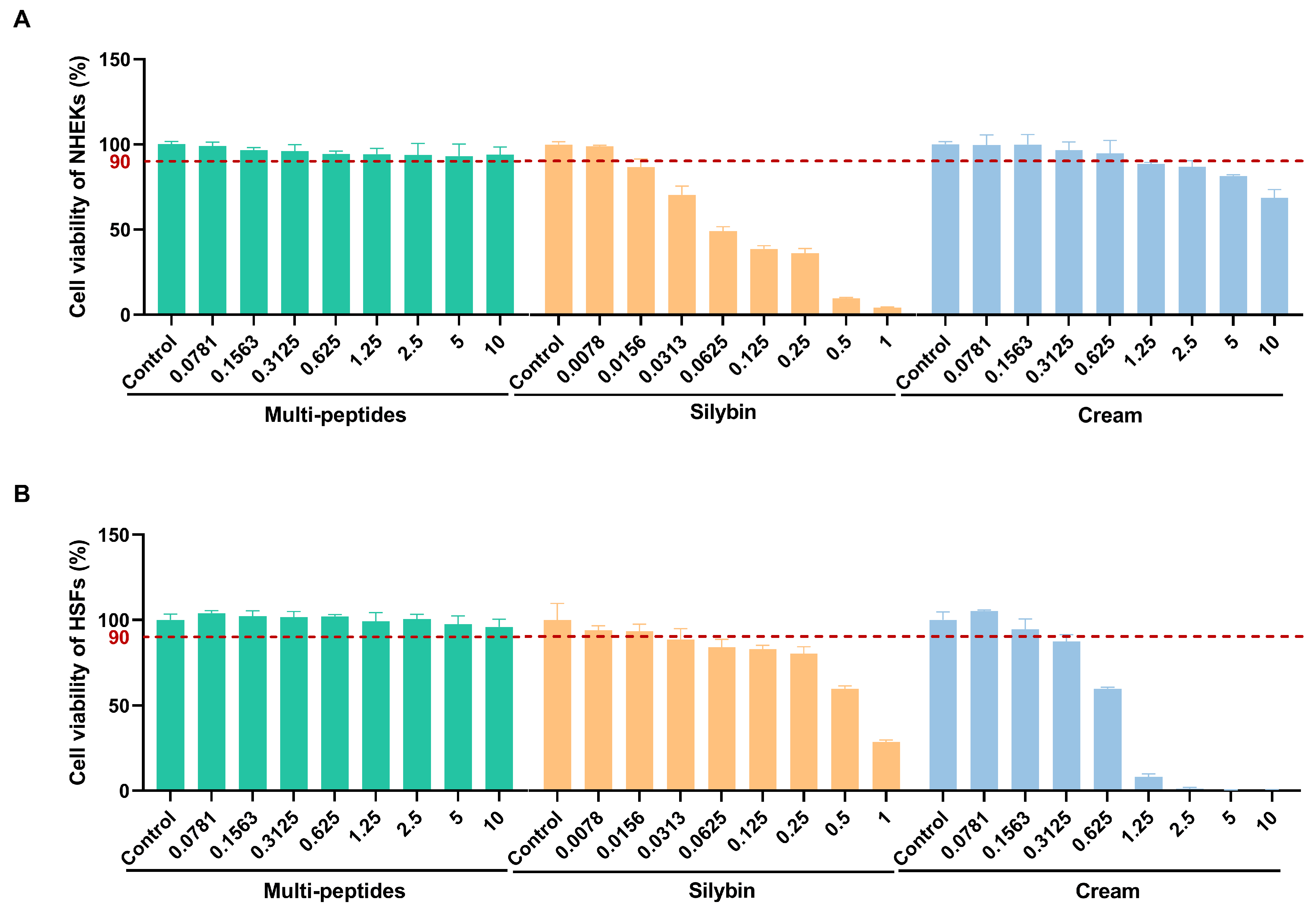
Figure 1. Cell viability of (A) NHEKs and (B) HSFs treated with polypeptides (green), silybin (orange), and cream (blue) at increasing concentrations (0.0078~10 mg/g) for 24 h, determined by MTT assay. The red dashed line at 90% indicates the threshold for non-cytotoxicity. Data are presented as mean ± SD (n = 5). Concentrations selected for subsequent experiments were within the non-cytotoxic range.
Cosmetics 12 00223 g001
Figure 2. Relative mRNA expression of ECM-related genes (collagen I, III, IV, XVII, and LOX) in HSFs or NHEKs after treatment with multi-peptides or silybin for 4, 8, and 16 h, measured by qRT-PCR. (A–D) The relative mRNA expressions of collagen I, III, IV, and XVII in HSFs. (E,F) The relative mRNA expressions of collagen IV and XVII in NHEKs. (G) The relative mRNA expressions of LOX in HSFs. Experimental group: blue = control, red = 100 ng/mL TGF-β1 (positive control), green = 0.10625% multi-peptides, orange = 0.000825% silybin. Data are presented as mean ± SD (n = 3). Statistical analysis was performed using Student’s t-test versus control at each time point. Statistical significance: * p < 0.05, ** p < 0.01, *** p < 0.001.
Cosmetics 12 00223 g002
Figure 3. Effect of 8.51% multi-peptides and 8.51% multi-peptides + 0.033% silybin on UV-induced reduction of collagen I in human skin explants. (A) Semiquantitative analysis of collagen I expression in different groups, with IOD used to represent the level of expression. (B) Representative immunofluorescence (IF) images of collagen I (green) and nucleus (blue, DAPI). Higher green fluorescence indicates higher collagen I expression. Experimental groups: untreated group (control), UV-induced only (negative control, NC), positive control (VC + VE), minimalist formula (vehicle), 8.51% multi-peptides, and 8.51% multi-peptides + 0.033% Silybin. Data are presented as mean ± SD (n = 3). Statistical analysis was performed using Student’s t-test in each group. Statistical significance: * p < 0.05, *** p < 0.001 vs. NC; ## p < 0.01, ### p < 0.001 vs. vehicle; Δ p < 0.05 vs. multi-peptides.
Cosmetics 12 00223 g003
Figure 4. Effect of 8.51% multi-peptides and 8.51% multi-peptides + 0.033% silybin on UV-induced reductionof elastic fibers in human skin explants. (A) Semiquantitative analysis of elastic fibers expression in different groups, with integrated area used to represent the level of expression. (B) Representative Victoria blue (VB) images of elastic fibers (blue). Darker blue indicates higher elastic fiber content. Experimental groups: untreated group (control), UV-induced only (negative control, NC), positive control (VC + VE), minimalist formula (vehicle), 8.51% multi-peptides, and 8.51% multi-peptides + 0.033% Silybin. Data are presented as mean ± SD (n = 3). Statistical analysis was performed using Student’s t-test in each group. Statistical significance: ** p < 0.01, *** p < 0.001 vs. NC; # p < 0.05, ## p < 0.01 vs. vehicle; ΔΔ p < 0.01 vs. multi-peptides.
Cosmetics 12 00223 g004
Figure 5. Effect of 8.51% multi-peptides and 8.51% multi-peptides + 0.033% silybin on UV-induced reduction of LOX in human skin explants. (A) Semiquantitative analysis of LOX expression in different groups, with IOD used to represent the level of expression. (B) Representative IF images of LOX (green) and nucleus (blue, DAPI). Higher green fluorescence indicates higher LOX expression. Experimental groups: untreated group (control), UV-induced only (negative control, NC), positive control (VC + VE), minimalist formula (vehicle), 8.51% multi-peptides, and 8.51% multi-peptides + 0.033% silybin. Data are presented as mean ± SD (n = 3). Statistical analysis was performed using Student’s t-test in each group. Statistical significance: ** p < 0.01, *** p < 0.001 vs. NC; # p < 0.05 vs. vehicle.
Cosmetics 12 00223 g005
Figure 6. Protein expression of ECM-related markers (collagen I, III, IV, XVII, and LOX) in HSFs or NHEKs after treatment with the cream containing multi-peptides and silybin. (A,C,E,G,I) Semiquantitative analysis of protein expression levels determined by immunofluorescence or Western blot. The expression of collagen I and IV was represented by MOD, whereas the expression of collagen III, XVII, and LOX was represented by IOD. (B,D) Representative IF images of collagen I and collagen IV (green) with nucleus counterstaining (blue, DAPI); higher green fluorescence indicates higher protein expression. (F,H,J) Representative Western blot bands of collagen III, collagen XVII, and LOX with β-actin as the loading control. Experimental groups: dark blue = untreated control, pink = 100 ng/mL TGF-β1 (positive control), light blue = 0.03125% cream (HSFs), or 0.125% cream (NHEKs). Data are presented as mean ± SD (n = 3). Statistical analysis was performed using Student’s t-test versus control at each time point. Statistical significance: * p < 0.05, ** p < 0.01, *** p < 0.001 vs. control.
Cosmetics 12 00223 g006
Figure 7. Improvement in facial firmness after 28 and 56 days of clinical cream application. (A,E) Lower face width (mm) assessed by Visia 7 (cross-polarized light) showing facial contour changes over time. (B,F) Dermal density (%) measured by DUB SkinScanner (green areas indicate higher density). (C,D) Skin elasticity (R2) and firmness (F4) measured by Cutometer dual MPA580. Data are presented as mean ± SE (n = 31). Statistical significance: ** p < 0.01, *** p < 0.001 vs. baseline (0 d). (E) Subject 19, 42 years old; (F) Subject 32, 56 years old.
Cosmetics 12 00223 g007
Figure 8. Improvement of pores and neck stripes after 28 and 56 days of clinical cream application. (A–C) Average area (mm2)/volume (mm3) of pore and average depth of neck stripe (mm) were measured by Antera 3D. (D,E) Images of pores and neck stripes acquired from Antera 3D. Data are presented as mean ± SE (n = 31). Statistical significance: * p < 0.05; ** p < 0.01; *** p < 0.001 vs. baseline (0 d). (D) Subject 1, 47 years old; (E) Subject 10, 53 years old.
Cosmetics 12 00223 g008
Figure 9. Reduction of wrinkles in varying degrees after 28 and 56 days of clinical cream application. (A,C,E) represent wrinkle length (mm), area (mm2), and volume (mm3), respectively. (B,D,F,G) Grayscale and color height maps of forehead, nasolabial, frown, and cheek captured by Primos CR. The darker the color, the more sunken it was. Data were presented as mean ± SE (n = 31). Statistical significance: ** p < 0.01, *** p < 0.001 vs. baseline (0 d). (B) Subject 19, 42 years old; (D) Subject 15, 34 years old; (F) Subject 7, 54 years old; (G) Subject 26, 51 years old.
Cosmetics 12 00223 g009
Figure 10. Synergistic mechanism of peptides and silybin.
Cosmetics 12 00223 g010
Table 1. Dermatologists’ ratings of cream safety in clinical study (n = 31).
Evaluation ItemScore 1
0 d28 d56 d
Pruritus0.060.030.03
Burning0.000.000.00
Tightness0.390.260.19
Tingling0.000.000.00
Dryness/scaling0.100.030.03
Edema0.000.000.00
Desquamation0.000.000.00
Erythema0.000.000.00
1 The score is from 0 to 3, with 0 as “no symptoms”, 1 as “mild”, 2 as “moderate”, and 3 as “obvious”. Data were presented as mean.
Table 2. Self-assessment questionnaire for subjects (n = 31).
Evaluation ItemConsumer Satisfaction 1
28 d56 d
Improvement in pore-related concerns81%90%
Improved jawline definition81%90%
Reduction of nasolabial folds81%90%
Reduction of glabellar lines84%87%
Reduction of neck lines84%84%
Reduction in submental fat81%87%
Improved skin firmness and elasticity87%90%
Refined and smoother skin87%87%
Mild and non-irritating87%97%
1 Consumer satisfaction is defined as: 100% × (Number of subjects with the score > 3)/(Total subjects). Scoring criteria: 1 to 5 rating scale is used (5—Satisfied, 4—Fairly satisfied, 3—Neutral, 2—Fairly dissatisfied, 1—Dissatisfied).
Disclaimer/Publisher’s Note: The statements, opinions and data contained in all publications are solely those of the individual author(s) and contributor(s) and not of MDPI and/or the editor(s). MDPI and/or the editor(s) disclaim responsibility for any injury to people or property resulting from any ideas, methods, instructions or products referred to in the content.