Article

Green Synthesis of Iron Oxide Nanoparticles for Use in Pickering Emulsions: In Vitro UV-Absorbing and Antimicrobial Properties

by
Ahmet Doğan Ergin
1,*,†,
Zeynep Betül Erbay
1,†,
Müberra Karaca
1,
Suzan Ökten
2,
Gülcan Kuyucuklu
3,
Camillo Benetti
4 and
Ayça Altay Benetti
4,*
1
Department of Pharmaceutical Technology, Faculty of Pharmacy, Trakya University, Edirne 22030, Turkey
2
Department of Pharmaceutical Microbiology, Faculty of Pharmacy, Trakya University, Edirne 22030, Turkey
3
Department of Medical Microbiology, Faculty of Medicine, Kırklareli University, Kırklareli 39100, Turkey
4
Department of Pharmacy and Pharmaceutical Science, National University of Singapore, Singapore 117559, Singapore
*
Authors to whom correspondence should be addressed.
These authors contributed equally to this work.
Cosmetics 2025, 12(5), 208; https://doi.org/10.3390/cosmetics12050208
Submission received: 12 May 2025 / Revised: 4 August 2025 / Accepted: 9 September 2025 / Published: 17 September 2025
(This article belongs to the Special Issue Feature Papers in Cosmetics in 2025)

Abstract

The integration of nanotechnology with green chemistry presents sustainable strategies for developing multifunctional cosmeceutical formulations. In this study, iron oxide nanoparticles (IONPs) were successfully synthesized using antioxidant-rich green tea extract via an eco-friendly method. The nanoparticles were incorporated into a novel Pickering emulsion comprising coconut oil and green tea extract, targeting UV protection and antimicrobial performance. The green-synthesized IONPs displayed strong UV absorption properties, achieving an SPF of 6.20 at 1.0 M concentration, outperforming standard TiO2 nanoparticles (SPF 3.98). The optimized Pickering emulsion formulation showed stability and skin-friendly pH. Antimicrobial studies revealed significant inhibition of Cutibacterium acnes and Staphylococcus aureus, with over 97% microbial reduction observed within 2 h of exposure. This dual-functional system, combining UV protection and antimicrobial effects, demonstrates the potential of green nanomaterials for developing safe, effective, and sustainable skincare formulations. The study provides new insight into the application of iron-based green nanotechnology in surfactant-free emulsions, supporting further innovation in the field of natural photoprotective cosmeceuticals.

1. Introduction

Ultraviolet (UV) radiation, particularly UVB (290–320 nm) and UVA (320–400 nm) rays, is known to contribute significantly to skin damage, including sunburn, premature aging, and skin cancer. Effective protection against UV radiation remains a key aspect of dermatological health. According to the American Academy of Dermatology (AAD), minimizing exposure to UV radiation and using protective strategies such as physical barriers and topical agents with UV-absorbing properties can help reduce the harmful effects of both UVA and UVB rays [1]. Excessive exposure to ultraviolet (UV) radiation is a major cause of skin damage, leading to sunburn, premature aging, and even skin cancer [2]. Conventional sunscreens rely on organic UV filters (e.g., oxybenzone, avobenzone) and inorganic blockers (e.g., titanium dioxide, zinc oxide) to reduce effects of harmful UV rays. However, growing concerns over the phototoxicity, environmental impact, and potential systemic absorption of these compounds have driven research toward safer and more sustainable alternatives [3].
Nanoparticles, particularly titanium dioxide (TiO2) and zinc oxide (ZnO), are widely utilized for their ability to provide broad-spectrum UV protection (Broad-spectrum refers to the ability to protect against both UVA and UVB radiation [4]. Their nanoscale size allows these particles to be transparent on the skin, addressing consumer preferences for aesthetic appeal while effectively blocking harmful UV radiation [5]. Research indicates that nano-sized metal oxide particles may exhibit enhanced UV-interacting characteristics compared to their bulk counterparts. For instance, Singh and Nanda demonstrated that these nanoparticles show improved UV absorption and scattering properties, which are critical for reducing UV radiation penetration and may be beneficial in formulations designed for topical skin protection [6]. Furthermore, studies have shown that the absorption and heat distribution properties of TiO2 nanoparticles of varying sizes (62 and 122 nm) under UV radiation at wavelengths of 310 nm and 400 nm. The results show that smaller nanoparticles at 310 nm enhance UV protection primarily through absorption, while at 400 nm, scattering properties dominate. The presence of these nanoparticles significantly reduces UV penetration into deeper skin layers and lowers surface temperature, mitigating the risk of heat-induced damage [7]. On the other hand, TiO2 nanoparticles can have cytotoxicity potential [8].
Iron oxide nanoparticles (IONPs) have gained significant attention in the field of photoprotection, particularly as components in tinted sunscreens, due to their ability to block visible light (VL) alongside ultraviolet radiation (UVR). The combination of these two effects is crucial, as visible light exposure has been associated with various skin issues, including pigmentation disorders such as melasma [9,10].
Iron oxides (Fe2O3, Fe3O4), including their nano-sized forms, are permitted cosmetic ingredients and are primarily regulated as colorants (CI 77491, CI 77492, and CI 77499) in both the European Union (EU) and the United States (US). Although they are not explicitly approved as UV filters under current regulations, they are frequently used in tinted sunscreens, foundations, and BB creams to provide both coloration and photoprotection, particularly against visible and blue light [11,12,13,14].
In the EU, iron oxides are listed in Annex IV of the EU Cosmetics Regulation (1223/2009/EC), which covers approved colorants. The regulation does not restrict particle size, meaning that nano-forms are implicitly allowed, provided that they are safe and non-penetrative. However, manufacturers must demonstrate safety through a Cosmetic Product Safety Report (CPSR), particularly for nano-materials. In summary, while iron oxide nanoparticles are not classified as primary UV filters like titanium dioxide or zinc oxide, their use as secondary photoprotective agents and colorants is well-established and permitted in topical cosmetic formulations, provided that safety is demonstrated. Their inclusion in formulations, especially those based on green synthesis methods, aligns with the principles of safer-by-design nanomaterials.
Studies indicate that tinted sunscreens containing iron oxides are particularly beneficial for individuals with skin of color or those prone to hyperpigmentation. For instance, Zhou et al. highlight that these formulations not only prevent melasma relapses but also enhance the effectiveness of other topical treatments [9]. Additionally, Dumbuya et al. demonstrated that the application of iron oxide-containing formulations significantly reduced visible light-induced pigmentation compared to non-tinted formulations, reinforcing the idea that iron oxides play a dual role in both protecting against and masking existing skin discoloration. Moreover, Dewi describes that the effectiveness of tinted sunscreens is partly attributed to concentrated formulations of iron oxide and titanium dioxide that provide substantial photoprotection against VL [15]. This is supported by findings from Tanaka et al., which suggest that dark-colored iron oxides are effective in blocking a broad spectrum of radiation, including UV and visible light, thus providing comprehensive skin protection [14]. Furthermore, Cole et al. emphasize the particular need for photoprotection strategies that incorporate these elements, especially for populations at greater risk of VL-related skin damage [10]. Iron oxides not only serve as effective physical blockers but also show a notable capacity for improving the overall aesthetic quality of sunscreens. They physically filter UV and VL radiation while also enhancing the skin’s appearance by providing a tint that can camouflage existing pigmentation issues [13]. This synergy between protective efficacy and cosmetic appeal is critical, as it encourages consistent use among individuals with skin conditions related to pigmentation.
Green synthesis of iron oxide nanoparticles has gained attention as an eco-friendly method with diverse applications. This method minimizes the use of toxic chemicals and enhances the biocompatibility of the synthesized nanoparticles, making them suitable for various applications, including biomedical, environmental, and agricultural fields.
One prominent method for synthesizing iron oxide nanoparticles involves the use of plant extracts. Recent studies have successfully synthesized iron nanoparticles using various plant extracts, including green tea leaf, sorghum bran, Chlorella-K01, and microalgal extracts [16,17]. The autonomous synthesis of iron (hydr)oxide nanoparticles through the precipitation of iron(II) ions using in situ-generated hydroxide ions demonstrates a pH-controlled method for nanoparticle formation [18]. These examples illustrate the versatility of plant extracts in synthesizing iron oxide nanoparticles with desirable characteristics. These green-synthesized iron oxide nanoparticles have exhibited potential for enhancing plant growth, acting as antifungal agents, and showing antimicrobial properties [17,19]. The use of plant extracts in the synthesis process not only reduces environmental impact but also provides a cost-effective and sustainable alternative to traditional chemical methods [20]. The stability and characteristics of these nanoparticles, influenced by the presence of organic acids in plant extracts, show the importance of green synthesis in nanoparticle production [21]. This method not only reduces environmental impact but also enhances the biocompatibility and functionality of nanoparticles. In recent years, titanium dioxide (TiO2) nanoparticles have also been widely studied for green synthesis using plant-based extracts due to their photocatalytic, antimicrobial, and UV-protective properties. For instance, Kaur et al. synthesized Citrus limon/TiO2 nanoparticles through a one-pot biogenic method, demonstrating both strong photocatalytic degradation efficiency and antibacterial activity [22]. Similarly, another study reported the synthesis of Elettaria cardamomum-wrapped TiO2 nanoparticles with promising biological and environmental applications [23].
Despite their broad applicability, TiO2 nanoparticles have limitations such as potential phototoxicity, low compatibility with darker skin tones due to the white-cast effect, and inadequate protection against visible light. In contrast, iron oxide nanoparticles (IONPs) offer distinct advantages. They provide protection not only in the ultraviolet but also in the visible light spectrum—an important factor in managing hyperpigmentation disorders such as melasma. Additionally, IONPs exhibit superior skin compatibility and aesthetic blending due to their natural pigmentation, and they possess intrinsic antimicrobial activity when synthesized via polyphenol-rich plant extracts such as green tea. These characteristics make IONPs especially suitable for multifunctional cosmeceutical formulations, combining UV shielding, antimicrobial action, and consumer acceptability.
In our study, we produced iron oxide nanoparticles with aqueous green tea extracts. The green tea extract served as both a reducing and stabilizing agent, facilitating the conversion of ferric ions (Fe3+) into iron oxide nanoparticles (Fe2O3 or Fe3O4) through a simple and eco-friendly synthesis method. The polyphenolic compounds present in green tea, such as catechins, played a crucial role in the reduction process, effectively donating electrons to the ferric ions and promoting the nucleation and growth of the nanoparticles [24]. Additionally, green tea extracts have exhibited anti-inflammatory and antimicrobial effects, supporting their potential application in topical skincare formulations [25]. Clinical studies have also suggested that green tea acts as a chemoprotectant and sunscreen agent, offering a range of protective activities against carcinogenesis and skin damage [26].
Pickering emulsions are a type of emulsion stabilized by solid particles rather than traditional surfactants. These emulsions possess the unique properties of solid particles, which adsorb at the oil-water interface, leading to enhanced stability against coalescence and phase separation. The use of iron oxide nanoparticles (IONPs) in Pickering emulsions has been applied due to their magnetic properties, biocompatibility, and ability to provide additional functionalities to the emulsions. Iron oxide nanoparticles can effectively stabilize Pickering emulsions by forming a robust interfacial layer that prevents droplet coalescence. The adsorption of IONPs at the oil-water interface is influenced by their size, shape, and surface chemistry, which can be tailored during the synthesis process. For instance, green synthesis methods utilizing plant extracts have been shown to produce iron oxide nanoparticles with desirable characteristics for emulsion stabilization [27]. Recent studies have demonstrated that IONPs can synergistically enhance stability when incorporated into Pickering emulsions—a surfactant-free system stabilized by solid particles [28]. The interfacial layer of nanoparticles not only improves emulsion stability but also provides a physical UV barrier, reducing skin penetration of harmful rays [29].
In this study, the Pickering emulsion consists of coconut oil as the oil phase, iron oxide nanoparticles (IONPs) as the stabilizing particles, and green tea extract as the water phase. Coconut oil is primarily recognized for its emollient and moisturizing properties. Some studies have reported that it may exhibit minor UV-absorbing activity, particularly in the UVB range; however, its contribution to UV protection is considered minimal and not sufficient for standalone photoprotective applications [30,31]. This property makes it a valuable ingredient in sunscreen formulations, especially when combined with other natural components that enhance its protective effects.
The incorporation of coconut oil in the Pickering emulsion contributes to the overall stability and effectiveness of the formulation. Its fatty acid composition, particularly the presence of medium-chain fatty acids, aids in forming a uniform and long-lasting film on the skin, which is essential for effective sun protection [32]. Moreover, coconut oil has been shown to have antioxidant properties, which can further protect the skin from oxidative stress induced by UV exposure [33]. The combination of coconut oil and green tea extract, rich in polyphenols, enhances the antioxidant capacity of the emulsion, providing a synergistic effect that may improve skin protection against UV-induced damage [34].
Although several studies have explored the green synthesis of iron oxide nanoparticles using tea extract or other plant-based systems, most of these works have focused primarily on synthesis optimization or preliminary characterization. To our knowledge, no previous study has combined green tea-mediated iron oxide nanoparticles with coconut oil in a stable Pickering emulsion system for simultaneous UV protection and antimicrobial application. Furthermore, the dual evaluation of both SPF values and antimicrobial activity in such a formulation remains underexplored. This study fills this gap by developing a surfactant-free, naturally derived emulsion stabilized by green-synthesized IONPs and systematically evaluating its physicochemical, rheological, photoprotective, and antimicrobial properties. This multifunctional approach contributes a novel direction in the field of green nanotechnology-based cosmeceuticals. Based on this rationale, the aim of the present study is to develop and evaluate a green-synthesized IONP-based Pickering emulsion with dual functionality.

2. Materials and Methods

2.1. Materials

Iron(II) chloride tetrahydrate (FeCl2·4H2O, ≥99% purity, Sigma-Aldrich, St. Louis, MO, USA) was used as the iron precursor. Titanium dioxide (TiO2, anatase phase, nanopowder, 30–50 nm, ≥99.5% purity) was obtained from HW NANO (Hangzhou, Zhejiang, China). Green tea leaves (Camellia sinensis) were sourced from a certified herbalist and used without further purification. Virgin coconut oil (cosmetic grade, cold-pressed) was obtained from TheLifeCo (Muğla, Türkiye). All other chemicals used in the study were of analytical grade and used as received without further modification.
Antimicrobial reference agents including ciprofloxacin (≥98%), gentamicin sulfate (≥98%), ceftazidime (≥98%) and clarithromycin (≥98%) were obtained from Sigma-Aldrich (St. Louis, MO, USA). Tween-80 (polyoxyethylene (20) sorbitan monooleate, analytical grade) was also purchased from Sigma-Aldrich. The microbial strains used in this study were Staphylococcus aureus ATCC 29213, Staphylococcus epidermidis NCTC 11047, and Cutibacterium acnes ATCC 6919, which were obtained from the American Type Culture Collection (ATCC, Manassas, VA, USA) and the National Collection of Type Cultures (NCTC, UK). Culture media, including Tryptic Soy Broth (TSB) and Plate Count Agar, were procured from LabM (Heywood, UK). All materials and media were handled in accordance with standard microbiological practices to ensure sterility and experimental accuracy.

2.2. Preparation of Green Tea Extract

Thirty grams of green tea leaves were accurately weighed using a precision balance and subsequently transferred into a 1000 mL round-bottom flask with the aid of a funnel. Following this, 500 mL of deionized water, measured using a graduated cylinder, was added to the round-bottom flask. The mixture was then heated on a heating plate at 80 °C for a duration of 1 h. After the heating period, the flask was removed from the heating plate, and a magnetic stir bar was added to facilitate mixing while the solution was allowed to cool on a magnetic stirrer. The reaction was carried out at room temperature (25 ± 2 °C). Once cooled, the mixture was filtered using a clean and sterilized muslin cloth, followed by filtration through Whatman 42 filter paper to obtain the final green tea extract.

2.3. Green Synthesis of Iron Oxide Nanoparticles

Green tea extract was used as the reducing and stabilizing agent in the synthesis of iron nanoparticles. In a separate beaker, 1 M Iron (II) chloride tetrahydrate (FeCl2.4H2O) solution was prepared by dissolving the appropriate amount of FeCl2.4H2O in deionized water. The 20 mL prepared plant extract was slowly added to the 30 mL FeCl2 solution under continuous stirring at 700 rpm. The reaction mixture was stirred for 1 h at room temperature, allowing the formation of iron nanoparticles. The change in color of the mixture indicated the reduction of Fe+2 ions to iron nanoparticles. After formation, pH was adjusted to 12.0 and stirred for one hour. The resulting solution was centrifuged at 4000 rpm for 15 min to separate the nanoparticles. The supernatant was discarded, and the iron nanoparticles were washed 2 times with deionized water and 1 time with ethanol to remove any unreacted plant extract and other impurities. The purified nanoparticles were then dried in a fume hood at room temperature for 24 h and stored in airtight containers for further characterization.

2.4. Preparation of Pickering Emulsion

Pickering emulsions were prepared using an ultrasonic homogenizer (15,000 rpm, 2 min) at four different concentrations. Iron nanoparticles were accurately weighed on a precision balance in amounts of 10, 7.5, 5, and 2.5% (w/w) and placed into appropriate beakers. Green tea extract, used as the aqueous phase, was weighed in suitable percentages (60, 62.5, 65, and 67.5% w/w) and added to the iron nanoparticles (Table 1). This mixture was dispersed using an ultrasonic homogenizer at 15,000 rpm for 2 min and heated to 70 °C. For the oil phase, solid coconut oil was weighed and heated to 70 °C. The aqueous phase was gradually added to the oil phase using a pipette, and the mixture was homogenized at 15,000 rpm for 2 min. The resulting emulsions were left to stand overnight at room temperature to check for phase separation. The most stable formulation (PE-1) was selected based on its appearance and stability. The formulation exhibits a brown-black color due to the IONP content.

2.5. Characterization of Iron Oxide Nanoparticles

The hydrodynamic size distribution and polydispersity index (PDI) of the synthesized iron oxide nanoparticles (IONPs) were determined using Dynamic Light Scattering (DLS). Measurements were performed using a Malvern Zetasizer Nano ZS (Malvern Instruments Ltd., Malvern, UK). Samples were appropriately diluted with deionized water to ensure optimal scattering intensity and prevent multiple scattering effects. All measurements were conducted at 25 ± 2 °C, and data were collected in triplicate to obtain an average hydrodynamic diameter and PDI.
The zeta potential of the IONPs was also measured using the same Malvern Zetasizer Nano ZS instrument. Samples were prepared by diluting the IONP dispersion in deionized water, and measurements were performed at 25 ± 2 °C. The zeta potential, which indicates the electrostatic potential at the shear plane of the particles, was calculated from the electrophoretic mobility of the particles. All measurements were carried out in triplicate, and the mean zeta potential value was recorded.
The biosynthesis of iron oxide nanoparticles (IONPs) was analyzed using an Ultraviolet-Visible Light Spectrophotometer (Optizen POP; Mecasys Co., Ltd., Daejeon, Republic of Korea). The identification of functional groups involved in the biosynthesis and stabilization of the nanoparticles, utilizing green extract, FeCl2, and IONPs, was conducted through Fourier Transform Infrared Spectroscopy (FT-IR, Spectrum 100, PerkinElmer, Waltham, MA, USA). Furthermore, the structural characteristics of the IONPs were examined via Scanning Electron Microscopy (SEM, Quattro S ESEM, Thermo Fisher Scientific, Brno, Czech Republic) to assess their morphology.

2.6. Characterization of Pickering Emulsion

The characterizations of the Pickering emulsions involved several key evaluations. Firstly, the physical appearance of the formulations was assessed for phase separation and overall visual properties. The pH measurements were conducted using a pH meter, with each formulation measured three times to obtain an arithmetic mean value. Additionally, microscopic analysis was performed to examine droplet distribution and stability at room temperature (25 ± 2 °C). For this purpose, the emulsions were diluted 100-fold and visually inspected on the 0th, 3rd, and 7th days using an optical microscope (Leica DM 750, Leica Microsystems GmbH, Wetzlar, Germany) to monitor visual changes over time.

2.7. Rheological Analyses

Rheological analyses were performed using the Haake Mars III rheometer (Thermo Fisher Scientific, Waltham, MA, USA). The rheometer was equipped with a coaxial cylindrical rotor CC25 DIN Ti and cup CCB25 DIN, with a gap of 5.3 mm between them. The sample volume used was 16.1 mL, and the temperature of the samples was controlled using a Peltier temperature module TM-PE-C, suitable for cylindrical measurement geometries. Three different methods were used. In the first method, the sample was equilibrated in a temperature-controlled environment before measurement. The rheometer was calibrated using appropriate standards, and measurements were conducted at a constant temperature of 25 °C. Shear rate was increased logarithmically from 0.00006 to 250 s−1, obtaining 100 viscosity values. These values were plotted on a logarithmic scale against shear rate to evaluate the shear-thinning behavior of the samples. The second method aimed to detail the mechanical and viscoelastic properties of the emulsion. Samples were standardized and equilibrated at a designated temperature (25 °C). Rheological properties were measured at increasing frequencies, starting from 0.02 Hz to 16 Hz, recording storage modulus (G′, loss modulus (G″), complex viscosity (|Eta*|), shear stress (Tau), and phase angle (Delta). These measurements were conducted to assess the thermal stability and frequency response of the samples’ viscoelastic behavior. The third method focused on understanding the impact of temperature changes on the mechanical properties of the emulsion and optimizing its characteristics for potential applications. Samples were prepared under standardized conditions and brought to experimental temperature. Rheological properties were measured at 10 °C intervals from 27 °C to 100 °C, determining storage modulus (G′), loss modulus (G″), complex viscosity (|Eta*|), and phase angle (Delta) at each temperature. These measurements were used to analyze the changes in viscoelastic behavior with increasing temperature.

2.8. Determination of Sun Protection Factor (SPF)

For the calculation of Sun Protection Factor (SPF) values, the Mansur method was employed with Equation (1), utilizing in vitro techniques that leverage the absorbance values at various wavelengths [35]. In the first part, 10 mg of 0.1 M, 0.5 M, and 1 M FeCl2-based IONP was prepared by diluting with 200 mL of distilled water and measured. After selection of optimum formulation according to SPF values, Pickering emulsions were prepared with these nanoparticles.
To calculate the SPF values of Pickering emulsions, 0.05 mL of the emulsions containing 10% IONP and 0.1 mL from other concentrations were diluted to 100 mL with distilled water. Distilled water was used as a blank. The absorbance of the formulations was measured in triplicate across the UVB range of 290–320 nm at 5 nm intervals.
The SPF was calculated by multiplying the absorbance values (ABS) by the erythemal effect (EE) and the solar intensity (I) spectrum values, which vary with wavelength. The products for each wavelength were then summed over the UVB range:
SPF   =   CF   ×   290 320 E E ( λ ) ×   I ( λ ) × ABS ( λ )
where CF is a correction factor, EE(λ) represents the erythemal effect at each wavelength, I(λ) denotes the solar intensity spectrum, and ABS(λ) is the measured absorbance. The constants of EE(λ) × I(λ) were used pre-defined by Sayre et al. 1979 [36].
According to Mansur (1986), this method provides a systematic approach to determining the protective efficacy of sunscreen formulations against UV radiation [37].

2.9. Antimicrobial Study of Formulations

This study consists of two parts. In the first part, the antimicrobial effect of iron nanoparticles (IONPs) was evaluated using the Microdilution Method. Following this, an alternative method, referred to as the AATCC 100 test method, was employed for Pickering emulsions containing IONPs. Due to turbidity and phase opacity, the emulsions could not be assessed with microdilution method.
An antimicrobial susceptibility study was conducted using the microdilution method, in accordance with EUCAST guidelines [38]. In this study, iron oxide nanoparticles IONP were prepared at three different molarities (S1: 1 M, S2: 0.5 M, and S3: 0.25 M) in cation-adjusted Mueller–Hinton broth (CA-MHB) with each solution standardized to a 1000 µg/mL concentration. Bacterial suspensions were prepared at a density of 5 × 10⁵ CFU/mL. Following the preparation, 10 µL of each microbial inoculum was added to the first rows of the microplates. Plates were incubated at 37 °C for 16–18 h. After this preparation, 10 µL of each microbial inoculum was added to the respective wells of the microdilution trays. The trays were incubated at 37 °C for bacterial strains and 35 °C for fungal strains. The minimum inhibitory concentration (MIC) endpoint was determined as the lowest concentration of IONP at which no visible growth was observed, indicated by a lack of turbidity in the wells.
Another study was conducted to determine the degree of antimicrobial activity of PE formulations and coconut oil, which are main components of the formulation, using the recommended AATCC 100 test method and the Turkish Standards Institute TS EN 1040/1999 test method [39]. In this study, standard bacterial strains including *Staphylococcus aureus* ATCC 6538, *Staphylococcus epidermidis* NCTC 11047, and *Cutibacterium acnes* ATCC 6919 were utilized. For the experimental setup, sterile 24-well microplates were used, with each well containing 1 mL of Tryptic Soy Broth (TSB, LabM). The excipients and formulations were diluted in TSB at concentrations of 1/2 and 1/4. A bacterial suspension was prepared to achieve a concentration of 0.5 McFarland (approximately 0.5 × 10⁸ CFU/mL), and 100 µL of this suspension was added to each well. The incubation periods were designed to reflect practical scenarios, with samples being incubated for 2, 4, and 6 h, which aligns with previous studies that emphasize the importance of contact time in antimicrobial efficacy assessments.
Following the incubation, Tween-80 was added as a neutralizing agent to halt the antimicrobial activity, and serial dilutions (10−1, 10−2, and 10−3) were performed. From each dilution and from the initial inoculum, 100 µL was spread onto Plate Count Agar for colony counting. The incubation conditions were set at 37 °C for 16–18 h, with aerobic conditions for *Staphylococcus* strains and anaerobic conditions for *Cutibacterium* strains. After incubation, colonies were counted to determine the reduction in microbial count compared to the initial inoculum. The reduction factor (%R) was calculated using Equation (2):
R ( % )   =   100   ×   B A B
where R represents the percentage reduction, B is the initial number of organisms in the sample, and A is the number of organisms present in the neutralization solution after contact with the sample. This method allows for a quantitative comparison of the antimicrobial efficacy of the tested substances against the selected bacterial strains.

2.10. Cell Viability Assay

Human keratinocytes (HaCATs) were maintained under standard culture conditions and seeded in 12-well tissue culture plates at a density of 2 × 10⁵ cells per well. Cells were allowed to adhere overnight before the application of treatments.
Cells were treated with iron-containing formulations at varying concentrations:
FeCl2 in PBS: 1, 10, and 100 µg/mL.
IONP PE in PBS: 1, 10, and 100 µg/mL.
Untreated controls included cells that were not exposed to any treatment. All treatment conditions were performed in triplicate.
After 24 h of exposure, cell viability was assessed using the Alamar Blue Cell Viability Reagent (Thermo Fisher Scientific, USA). A 1% (v/v) solution of Alamar Blue was added directly to each well, and cells were incubated under standard conditions.
Fluorescence was measured using a HIDEX microplate reader at an excitation wavelength of 560 nm and an emission wavelength of 590 nm.
To visually assess the morphology and viability of the keratinocytes following treatment, phase-contrast imaging was performed after 24 h of exposure to the various iron-containing formulations. Images were captured using a Leica DMi8 Inverted LED Motorized Microscope (Leica DM 750, Leica Microsystems GmbH, Wetzlar, Germany) under standard conditions. Representative fields were selected from each treatment group and control condition to ensure consistent comparison. Cell morphology, confluence, and general health status were qualitatively evaluated based on the integrity of the monolayer, presence of cellular debris, and changes in shape or adherence.
Microscopy observations were used to visually confirm the results obtained from the Alamar Blue assay and further support conclusions regarding cell viability and cytotoxicity under the tested conditions.

2.11. Statistical Analysis

All tests were conducted in triplicate (n = 3). Grubb’s test was employed as a means of identifying any potential outliers. Furthermore, the t-test, one-way and two-way ANOVA tests were utilized to assess any potential statistical differences in the pharmacokinetic parameters.

3. Results and Discussion

3.1. Iron-Oxide Nanoparticle Preparation

The successful synthesis of iron oxide nanoparticles (IONPs) using aqueous green tea extract was achieved through a green, one-step method. The polyphenolic content of the extract, particularly catechins, facilitated the reduction of Fe3+ ions and acted as natural capping agents, contributing to nanoparticle stabilization. The formation of IONPs was confirmed through physicochemical analyses, supporting the effectiveness of plant-mediated synthesis. Notably, the use of green tea not only enabled an eco-friendly production route but also imparted potential bioactive properties—such as antimicrobial and anti-inflammatory effects—which align with the intended cosmeceutical applications of the resulting formulations. These inherent characteristics of the green-synthesized nanoparticles form the basis for evaluating their biological activity and topical suitability in the following sections.
In UV-Vis spectrophotometry (Figure 1), the distinct absorbance patterns of the precursor materials and the synthesized nanoparticles provide crucial insights into the success of the green synthesis process. The green tea extract, utilized in the synthesis, exhibits a characteristic and prominent absorbance peak around 270–275 nm, with values reaching nearly 3.0, indicative of its rich polyphenolic content, which is essential for its role as a reducing and stabilizing agent. The FeCl2 precursor shows a generally decreasing absorbance with increasing wavelength, displaying a broad shoulder around 280–290 nm. In contrast, the synthesized iron oxide nanoparticles (IONPs) demonstrate a relatively stable absorbance across the 200–400 nm range. While the IONPs do not present sharp, distinct peaks like the green tea extract, they maintain a consistent absorbance profile, with a subtle elevation around 290–300 nm and notably higher absorbance at 400 nm compared to the FeCl2 precursors. UV Vis spectroscopy results revealed that the Iron Oxide NPs peaked between 230 and 290 nm, which is confirmation of the synthesized Iron Oxide nanoparticles. UV-Vis spectroscopy results revealed that the green-synthesized Iron Oxide Nanoparticles (IONPs) exhibited an absorbance profile consistent with nanoparticle formation, with a notable increase in absorbance observed between 230 and 290 nm. This spectral characteristic, particularly in this region, confirms the successful synthesis of the IONPs using the green tea extract and FeCl2 precursors for sun protection applications [40].
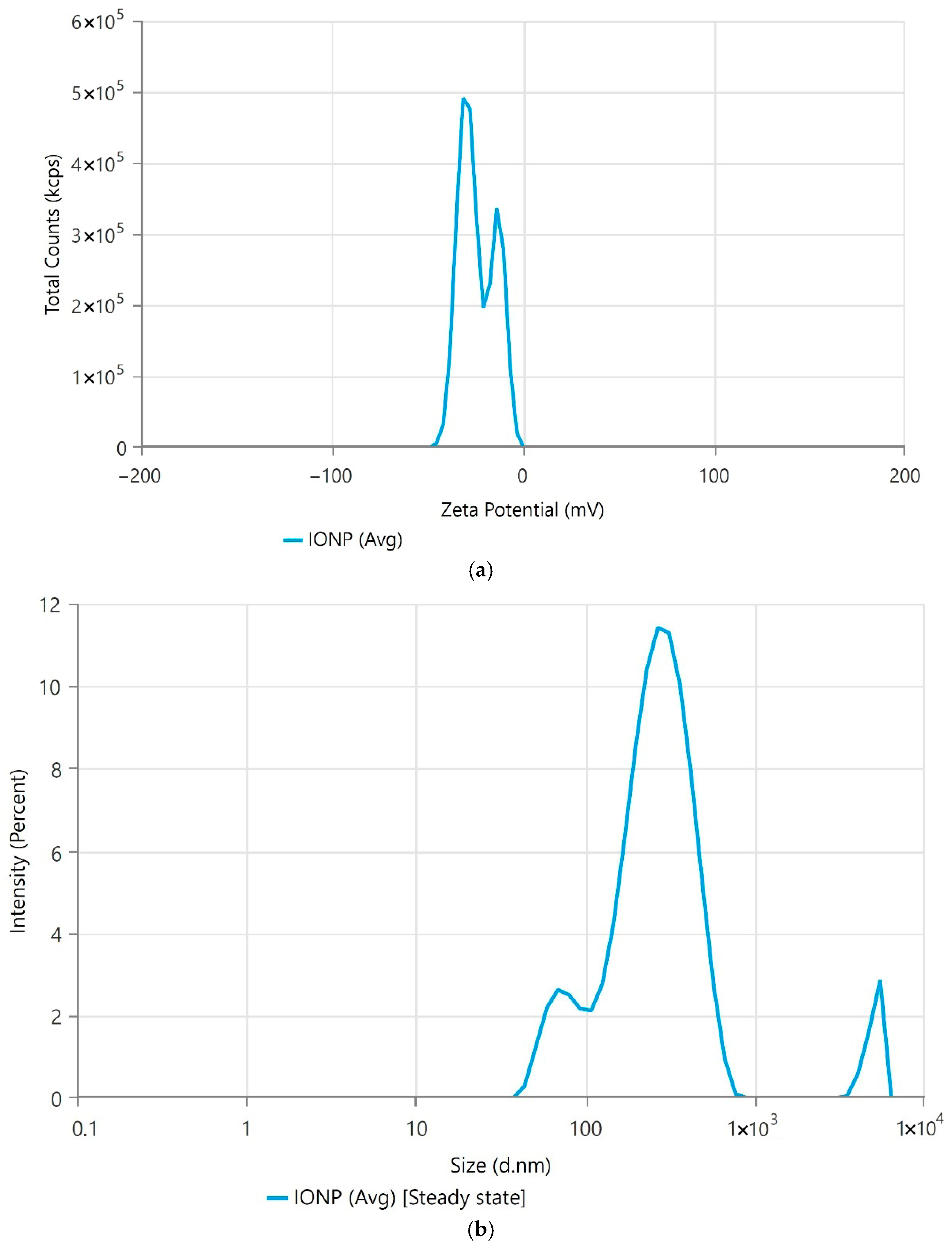
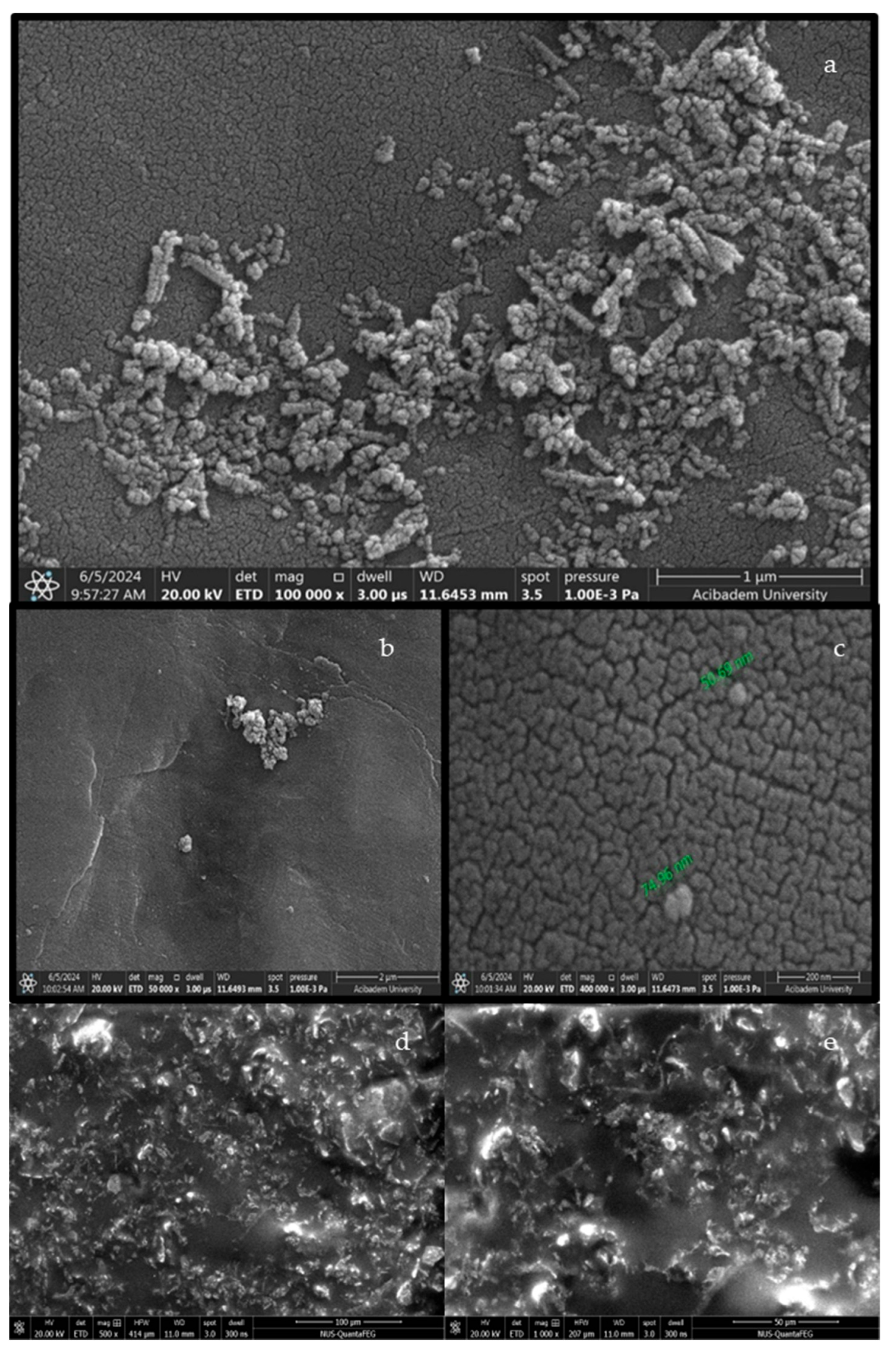
DLS measurements revealed a hydrodynamic size of 238.5 ± 9.67 nm, with a polydispersity index (PDI) of 0.4329 ± 0.0845. This PDI value suggests a relatively broad size distribution in the aqueous dispersion, which is consistent with the observed agglomeration in SEM images where particles formed clusters and networks (Figure 2). The zeta potential of the IONPs was determined to be −23.98 ± 0.33485 mV. This negative zeta potential indicates moderate colloidal stability, implying that the nanoparticles possess sufficient surface charge to prevent rapid aggregation in dispersion, though some level of agglomeration is still evident. This combined characterization from SEM, DLS, and zeta potential provides a more complete picture of the physical state and stability of the synthesized IONPs.
SEM image displays a highly magnified view of a sample with irregularly shaped and sized particles (Figure 3a–c). Figure 3a (100,000×) presents a densely packed surface of interconnected particles, while Figure 3b (1000×) illustrates a lower-magnification overview of nanoparticle deposition. In Figure 3c, green arrows highlight the approximate particle dimensions, confirming the presence of nanoscale entities. The iron particles appear to be agglomerated, forming clusters and networks on the surface. The texture of the background surface indicates a fine granular structure, which contrasts with the larger, more distinct particles. The analysis revealed a wide size distribution, with most particles being relatively small but larger than 10 nm, highlighting the heterogeneity in particle size and distribution within the sample. Although some aggregates exceeded 100 nm, individual primary particles observed in SEM images were below 100 nm, justifying the nanoparticle designation. Figure 3d,e represent SEM images of the Pickering emulsion containing IONPs. The nanoparticles appear embedded within the emulsion matrix, indicating successful integration and potential interfacial stabilization.
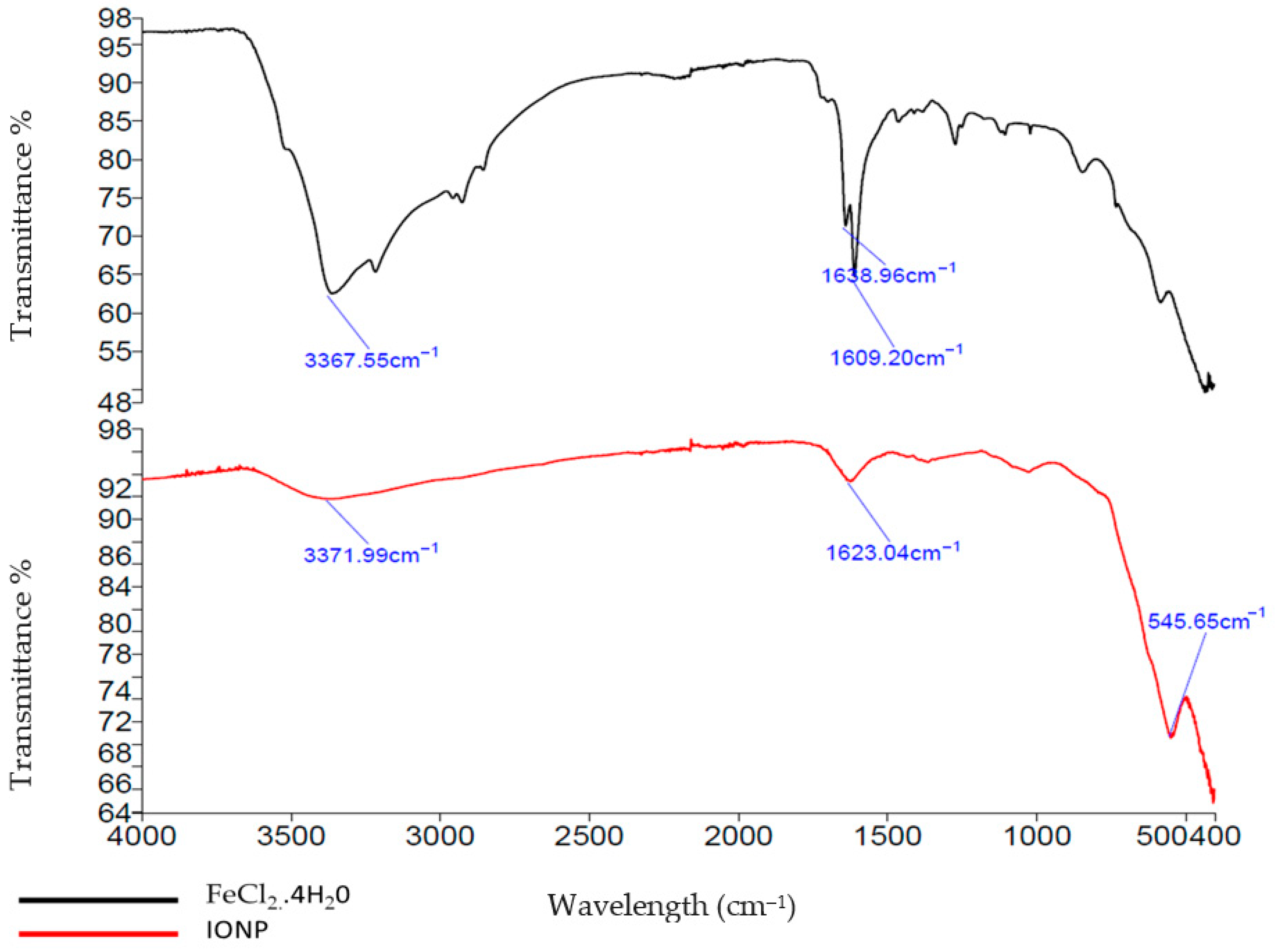
The provided FTIR spectra compare the FeCl2.4H2O (black) with iron nanoparticles synthesized using green synthesis methods (red). The FeCl2.4H2O spectrum exhibits characteristic peaks, such as the broad absorption band at 3367.55 cm−1, indicating O-H stretching vibrations due to water molecules. Additionally, peaks at 1638.96 cm−1 and 1609.20 cm−1 correspond to H-O-H bending vibrations of water (Figure 4).
In contrast, the spectrum of the green synthesized iron nanoparticles shows different features. The broad absorption band at 3371.99 cm−1 still indicates the presence of O-H stretching, likely from hydroxyl groups or residual water. The significant peak shift and intensity change at 1623.04 cm−1 suggest the formation of new bonds, possibly indicating the presence of organic stabilizers or capping agents from the green synthesis method. The peak at 545.65 cm−1, which is absent in the FeCl2.4H2O spectrum, may correspond to Fe-O stretching vibrations, confirming the formation of iron oxide nanoparticles (Figure 3).
The 0.1 M IONP formulation shows a very low SPF of 0.447 ± 0.002, suggesting minimal UV protection. As the concentration of IONP increases, the SPF also increases, with 0.5 M IONP having an SPF of 5.185 ± 0.015 and 1.0 M IONP showing a higher SPF of 6.202 ± 0.013, indicating a concentration-dependent enhancement in UV protection. The TiO2 nanoparticles have an intermediate SPF value of 3.982 ± 0.006, demonstrating moderate UV protection efficacy (Table 2).

3.2. Preparation and Evaluation of Pickering Emulsion

Pickering emulsions consist of at least two phases, and emulsifiers are used to help these phases mix properly. Unlike traditional emulsions, Pickering emulsions use solid particles as stabilizers. In the formulations, iron nanoparticles, which have proven best sun protection properties, were used along with solid coconut oil as the oil phase and green tea extract heated to 70 °C as the aqueous phase. Green tea extract has also demonstrated sun protection efficacy in in vivo studies [41]. Initially, four different formulations were tested and left to sit overnight to observe their physical appearance. Initially, the PE-1 formulation was produced. Subsequently, the amounts of oil phase and nanoparticles were varied and analyzed. In the PE-4, PE-5, and PE-6 formulations, phase separation was observed after 2 h. Therefore, measurements proceeded with the PE-1, PE-2, and PE-3 formulations, which remained stable without phase separation.
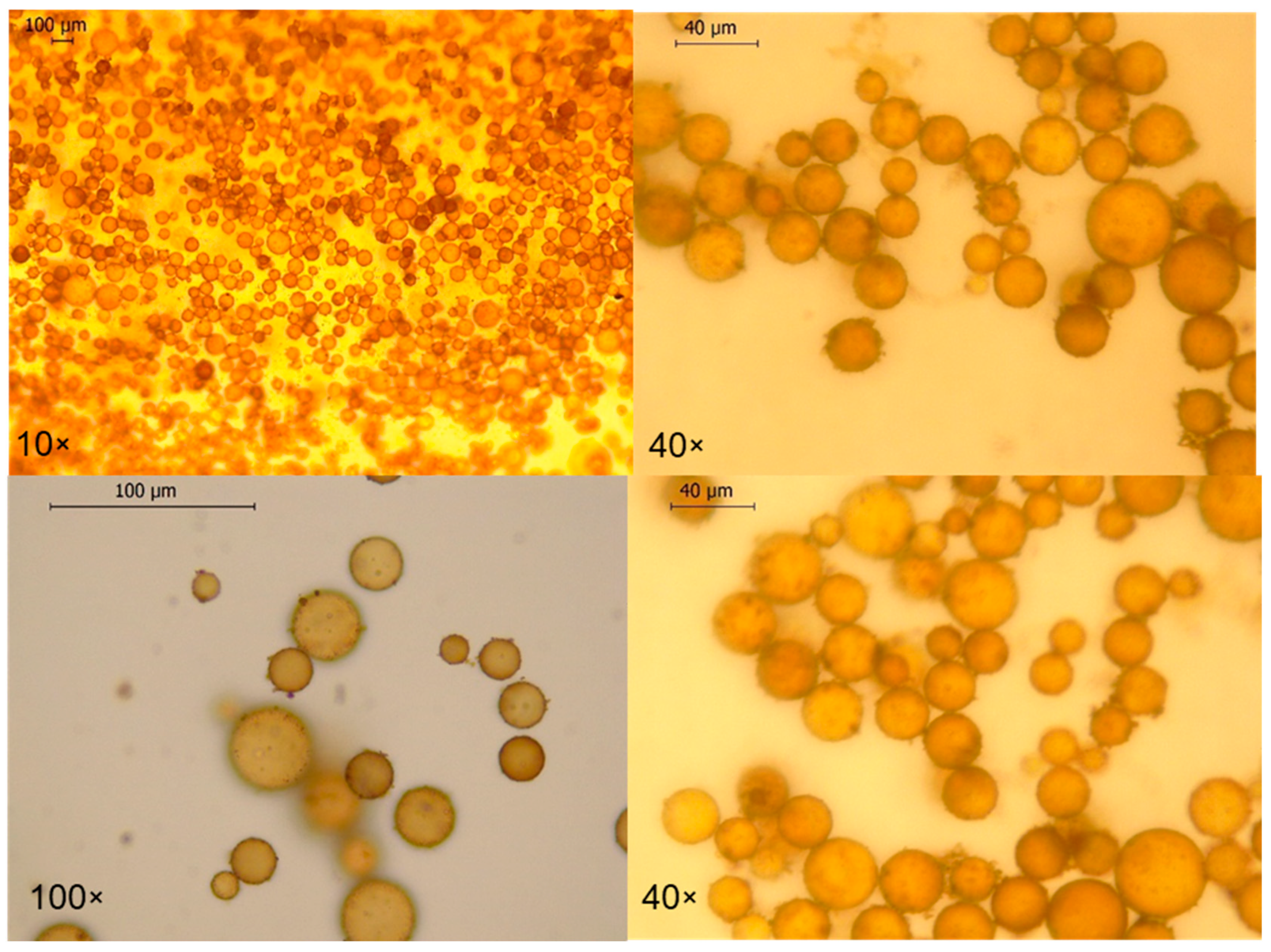
The pH values of the obtained formulations were found to be between 5.537 and 5.716, which are close to the pH of the skin, making them suitable for use. Subsequently, a microscopic examination of the selected PE-1 formulation was conducted. Emulsions were stored at room temperature (25 ± 2 °C) under ambient light and observed for phase separation, creaming, and visual changes (Figure 5). The final emulsion exhibited a characteristic red-brown color due to iron oxide content. This color may pose a challenge for its acceptance in cosmetic or dermatological topical formulations, where product appearance and consumer preference for clear or skin-toned products are often critical for market appeal.

Rheological Properties

Figure 5 shows how viscosity changes with varying shear rates, typical for non-Newtonian fluids. At low shear rates, the viscosity is high, indicating a strong resistance to flow. As the shear rate increases, the viscosity decreases significantly, demonstrating a shear-thinning behavior [42]. According to Figure 5, our formulation shows plastic-type fluid. The study conducted by Ansari et al. investigated the velocity distribution and profile shape of laminar flows for both Newtonian and non-Newtonian fluids in mini-channels, focusing on how these properties are influenced by shear stress and shear rate using the Ostwald-de Waele power law model [43]. This means the fluid becomes less viscous and flows more easily when subjected to higher shear rates. The curve eventually levels off, indicating that further increases in shear rate do not significantly reduce viscosity. This behavior is important for applications in pharmaceuticals, cosmetics, and food products, where understanding how a substance flows under different conditions is crucial for formulation and processing.
According to Figure 6, as shear stress increases, the shear rate also increases in a nonlinear fashion. Initially, at low shear stress values, the shear rate increases slowly. However, as the shear stress continues to rise, the shear rate increases more rapidly [44,45]. This also indicates, once more, a non-Newtonian fluid behavior of emulsion, where the viscosity changes with varying shear stress. This pseudoplastic flow characteristic suggests that the formulation exhibits high viscosity at rest, which progressively decreases under applied shear, thereby enhancing its spreadability and ease of application. The upward curvature of the plot suggests that the fluid exhibits shear-thinning properties, meaning its viscosity decreases with increasing shear stress. This is characteristic of many complex fluids, such as polymer solutions, biological fluids, and certain food products, where the internal structure of the fluid reorganizes under stress, leading to easier flow at higher stresses [46]. Sun et al. reviewed various models used to describe the stress–strain relationship in non-Newtonian fluids, emphasizing their non-linear behavior and the factors influencing this relationship, such as viscosity and shear rate [47]. Understanding this relationship is essential for optimizing processing and application in various industries, ensuring efficient and predictable performance under different mechanical conditions.
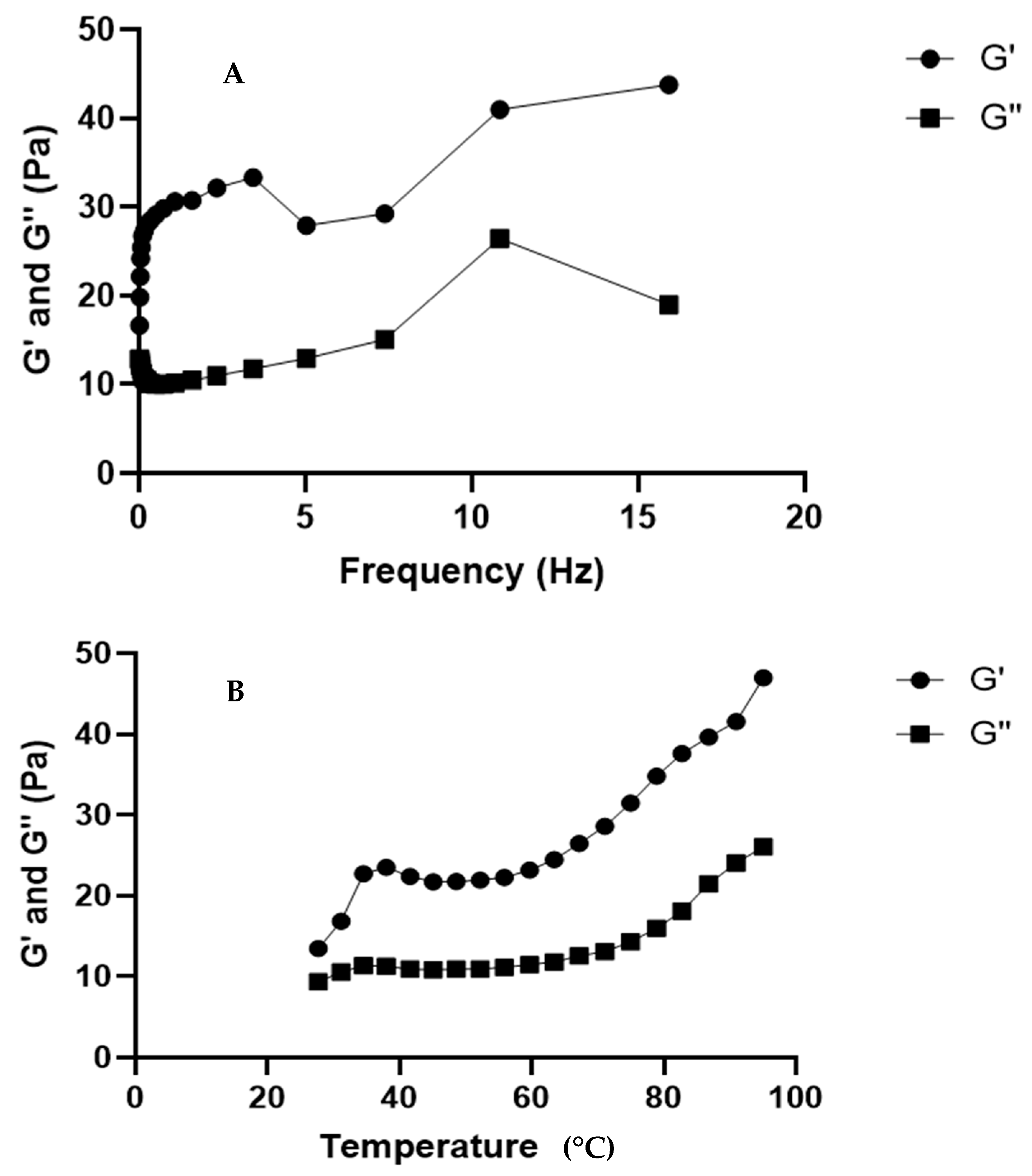
Figure 7A illustrates the oscillatory rheology data, showing the relationship between storage modulus (G’) and loss modulus (G”) across different frequencies. The storage modulus (G’), which reflects elastic or solid-like behavior, initially increases with frequency, peaks, and then continues to rise, indicating enhanced solid-like properties at higher frequencies [48]. In contrast, the loss modulus (G”), representing the viscous or liquid-like behavior, increases more slowly, peaks around 10 Hz, and then slightly decreases, suggesting dominant viscous behavior at lower frequencies. The bottom figure shows the storage and loss moduli as a function of temperature. The storage modulus (G’) increases with temperature after an initial rise and plateau, indicating greater rigidity at higher temperatures. Similarly, the loss modulus (G”) also increases but more gradually, suggesting the material maintains some viscous properties even as it becomes more solid-like. These behaviors, typical of viscoelastic materials, align with recent studies highlighting the impact of frequency and temperature on the rheological properties of complex fluids [49]. These behaviors align with findings from a recent study of Moch et al., which investigated the temperature-dependent rheological properties of glycerol, a glass-forming material. The study employed oscillatory rheology to monitor structural recovery and observed that both the storage and loss moduli exhibited temperature-dependent changes, similar to the trends shown in Figure 7B [50].
Figure 8 illustrates the variation in phase angle (δ) with frequency in an oscillatory rheology experiment. The phase angle, δ, represents the lag between the applied strain and the resulting stress, providing insights into the viscoelastic behavior of the material. At low frequencies, the phase angle is high, indicating a significant viscous response. As the frequency increases, the phase angle decreases sharply, reaching a minimum (~2 Hz), which suggests a transition towards more elastic behavior [47,51]. Beyond this point, the phase angle begins to increase again, indicating a complex interplay between elastic and viscous properties at higher frequencies.
A recent study that was executed by Xu et al. explored the nonlinear oscillatory rheology of cellulose nanocrystal suspensions [52]. The study observed similar trends in phase angle variations with frequency, highlighting the material’s transition from predominantly viscous to more elastic behavior and back to a mixed viscoelastic response at higher frequencies. Additionally, Van Kempen et al. examined the frequency-dependent rheological properties of air/water interfaces stabilized by oligofructose fatty acid esters, also reporting comparable frequency-dependent viscoelastic transitions [49]. These findings underscore the significance of frequency in characterizing the dynamic mechanical properties of complex fluids and gels.

3.3. Antimicrobial Study

The study investigated the antimicrobial effects of iron oxide nanoparticles (IONPs) prepared in different molarities against various microbial strains, including Pseudomonas aeruginosa, Escherichia coli, Staphylococcus aureus, Enterococcus faecalis, Cutibacterium acnes, and Candida albicans. The study investigated the antimicrobial effects of IONP prepared in different molarities against various microbial standard strains. As a result, minimum inhibition concentration of S3 was 0.25 mg/mL concentration of S3 (containing 0.25 M IONP) solution on S. aureus ATCC 29213. MIC values of S1, S2 and S3 solutions on C. acnes ATCC 6919 were determined as 0.25, 0.25 and 0.5 mg/mL, respectively (Table 3). The study investigated the antimicrobial effects of iron nanoparticles (FeNP) prepared in different molarities against various microbial standard strains.
As a result, minimum inhibition concentration of S3 was 0.25 mg/mL concentration of S3 (containing 0.25 M FeNP) solution on S. aureus ATCC 29213, and the MIC values of S1, S2 and S3 solutions on C. acnes ATCC 6919 were determined as 0.25, 0.25 and 0.5 mg/mL, respectively (Table 3).
Because of the high MIC values, IONP was found to be safe. The antimicrobial action of IONPs is multifaceted, primarily involving the generation of reactive oxygen species (ROS) and direct interaction with bacterial cell walls. ROS can induce oxidative stress, leading to damage to bacterial proteins, DNA, and lipid membranes, ultimately resulting in cell death [53,54,55]. In our evaluation against Staphylococcus aureus, our IONPs at 0.5 M and 1.0 M concentrations displayed MIC values of 0.5 mg/mL. The Pickering emulsion formulation achieved a 98.6% reduction in S. aureus ATCC 29213 within 2 h. These results are comparable to or even surpass some concentrations reported in the literature for other IONP formulations. For instance, Fatih et al. reported MIC values ranging from 0.625 to 5 µg/mL for green-synthesized α−Fe2O3 nanoparticles against S. aureus [54]. Tran et al. demonstrated that iron oxide (IO) nanoparticles, synthesized via a polyvinyl alcohol (PVA)-mediated method, inhibited S. aureus growth at a high concentration of 3 mg/mL (equivalent to 3000 µg/mL) over various time points (4, 12, and 24 h) [56]. Furthermore, a study by Flieger et al. found that unmodified IONPs exhibited MIC values against S. aureus ATCC 25923 in the range of 2.5–10 mg/mL, which significantly improved to 0.313–1.25 mg/mL after modification with DMSO extract [55]. MIC values for C. acnes with other iron oxide nanoparticles are not as widely available in the provided literature at directly equivalent concentrations, and reviews indicate that the bacteriostatic concentration range for IONPs against susceptible microorganisms is generally broad, spanning from 25 to 2000 µg/mL (0.025–2 mg/mL). Our observed MIC value of 0.5 mg/mL falls within this reported effective range [53].
Regarding Staphylococcus epidermidis NCTC 11047, our Pickering emulsion showed no antimicrobial effect at 2 h of application; however, a significant microbial reduction (99.9%) was observed after 6 h of exposure at the 1/1 dilution. This suggests that a longer contact time may be required for this particular strain. Flieger et al. also reported that unmodified IONPs had a MIC of 10 mg/mL against S. epidermidis, which improved to 1.25 mg/mL when modified with DMSO extract [55]. These literature findings collectively support the antimicrobial potential of IONPs against S. epidermidis.
Therefore, we evaluated Pickering emulsion formulations and their main component, coconut oil. The results were given in Table 4 and Table 5.
The method applied in this study is quantitative, where the change in the number of microorganisms in fluids added to the sample is proportionally converted into antibacterial activity data. In this method, it is necessary to test control samples that have not undergone any antimicrobial treatment. According to these standards, a reduction of more than 99%, or approximately a 3-log reduction, indicates that antimicrobial activity is sufficient for sanitation. Additionally, the percentage reduction (R%) compared to the initial inoculum reflects the degree of antimicrobial efficacy.
Although a minimum reduction of 99% in the R value is considered sufficient for disinfection, it is believed that even lower reductions in the R value are significant as they indicate the presence of antimicrobial activity. Based on this, it can be concluded that a 2 h application of the formulation shows antimicrobial activity against Cutibacterium acnes ATCC 6919 and S. aureus ATCC 29213. However, antimicrobial effects on S. epidermidis NCTC 11047 were observed only after 4–6 h of application. Based on these findings, it is not possible to evaluate the effect of the product, designed as a sunscreen, on S. epidermidis.
Coconut oil was also tested alone as a raw material, and cases were observed where both the pure oil and its formulated form exhibited antimicrobial effects. However, given the product’s intended use as a sunscreen, the antimicrobial activity results of the formulated product are especially valuable, particularly when considering the application time results.

3.4. Human Keratinocyte (HaCAT) Cell Viability Assay

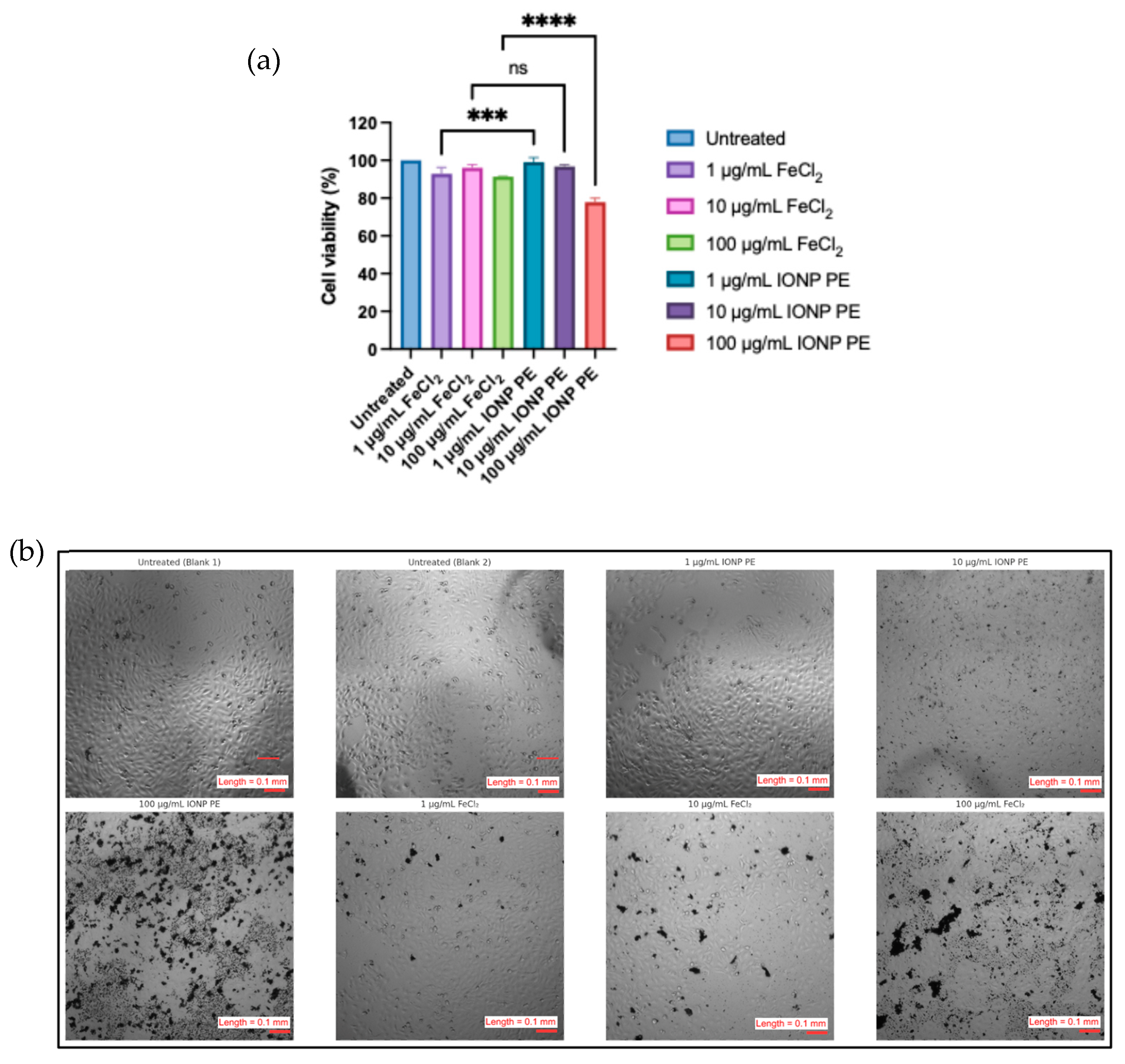
This 24 h in vitro study assessed HaCaT keratinocyte viability following treatment with Fe-containing formulations in PBS and Pickering emulsion, as determined by the Alamar Blue assay and supported by phase-contrast microscopy [48]. Based on the 24 h Alamar Blue assay, the IONP Pickering emulsion (IONP PE1) exhibited a concentration-dependent reduction in HaCaT keratinocyte viability (Figure 9a). While lower Fe concentrations (1–10 µg/mL) maintained high viability (>90%), the reduction observed at 100 µg/mL suggests that the emulsion matrix, particularly pickering emulsion excipients, may contribute to reduced metabolic activity. (Figure 9a).
This reduction is likely due to the excipients rather than iron toxicity. The emulsion formula comprised 10% IONP, 30% coconut oil, and 60% green extract. At the 100 µg/mL FeCl2 dose, the concomitant concentrations are approximately 300 µg/mL coconut oil and 600 µg/mL green extract.
According to Zainodin et al. [49], virgin coconut oil exhibited an IC50 of ~17.8% v/v in HaCaT cells using the MTT assay; cell viability dropped to ~74% at ~11.3% v/v oil. Though 300 µg/mL represents only ~0.03% v/v, this finding suggests that coconut oil at even low concentrations could contribute to metabolic suppression, particularly when combined with other bioactive components such as green extract.
Similarly, Min Jung Kim et al. [49] reported that Camellia sinensis (green tea) water extract began to decrease HaCaT viability at concentrations above 300 µg/mL, with green tea extract reducing viability significantly at 200–300 µg/mL by MTT, although not below. Given the estimated 600 µg/mL green extract in our formulation, it is plausible that this component significantly contributed to the viability decrease observed.
Microscopy images of high-dose IONP-treated cells showed slight morphological changes, such as reduced adherence and lower confluence, though monolayer integrity remained largely intact (Figure 9b).

4. Conclusions

This study highlights the potential of green-synthesized iron oxide nanoparticles (IONPs) and IONP-stabilized Pickering emulsions as multifunctional cosmeceutical agents. The IONPs synthesized using green tea extract demonstrated promising sun protection and antimicrobial properties, effectively inhibiting Cutibacterium acnes and Staphylococcus aureus within a 2 h application period. However, extended contact times were required to observe an effect against Staphylococcus epidermidis, indicating that application time is a critical factor for antimicrobial efficacy.
The formulation’s stability and enhanced performance, attributed to the synergistic properties of green tea extract and coconut oil, support its potential in sunscreen applications. While both coconut oil and its formulation with IONPs exhibited antimicrobial effects, the combined formulation showed enhanced results and added value as a cosmeceutical product. This study contributes to the broader field of natural and sustainable skincare by demonstrating the feasibility of using plant-based synthesis and biocompatible oils to develop dual-function topical agents. The findings align with current trends favoring green nanotechnology and eco-friendly product development.
Nevertheless, the study has certain limitations, including the use of in vitro antimicrobial assays only, a limited number of bacterial strains tested, and the lack of long-term stability and cytotoxicity evaluations.
Future research should focus on in vivo efficacy studies, comprehensive skin compatibility assessments, long-term storage stability, and potential incorporation into commercial sunscreen formulations. Moreover, expanding the antimicrobial testing to include fungal strains or drug-resistant bacteria would provide a more holistic understanding of the formulation’s therapeutic potential.

Author Contributions

A.D.E.: Data Curation, Writing—review & editing, Visualization, Validation, Supervision, Software, Resources, Project administration, Methodology, Investigation, Funding acquisition, Formal analysis, Conceptualization. Z.B.E.: Methodology, Investigation, Formal analysis, Data curation, Conceptualization, M.K.: Methodology, Investigation, Formal analysis, Data curation, Conceptualization, S.Ö.: Writing—original draft, Visualization, Validation, Supervision, Software, Resources, Methodology, Investigation, Formal analysis, Data curation, Conceptualization, G.K.: Data curation, Conceptualization, C.B.: Review & editing, Supervision, Investigation, Funding acquisition, Formal analysis, A.A.B.: Review & editing, Supervision, Investigation, Funding acquisition, Formal analysis. All authors have read and agreed to the published version of the manuscript.

Funding

Trakya University Scientific and Technological Research Council has provided the grant 2023/149, 2023/150 in order to improve students’ scientific knowledge. This research was supported by the funding of the Department of Pharmacy and Pharmaceutical Science, National University of Singapore, WBS number A-8002989-00-00.

Institutional Review Board Statement

Not applicable.

Informed Consent Statement

No human subject studies are involved in this research.

Data Availability Statement

The raw data supporting the conclusions of this article will be made available by the authors on request.

Acknowledgments

The authors would like to thank Ryan Tze Liang Sia (Department of Pharmacy and Pharmaceutical Science, National University of Singapore) and Thong Beng Lu (Electron Microscopy Unit, Yong Loo Lin School of Medicine, National University of Singapore) for their valuable assistance and technical support in acquiring and processing the scanning electron microscopy (SEM) images used in this study.

Conflicts of Interest

The authors declare no conflicts of interest.

References

  1. American Academy of Dermatology. Sunscreen FAQs. Available online: https://www.aad.org/media/stats-sunscreen (accessed on 2 May 2023).
  2. D’Orazio, J.; Jarrett, S.; Amaro-Ortiz, A.; Scott, T. UV radiation and the skin. Int. J. Mol. Sci. 2013, 14, 12222–12248. [Google Scholar] [CrossRef]
  3. Schneider, S.L.; Lim, H.W. Review of environmental effects of oxybenzone and other sunscreen active ingredients. J. Am. Acad. Dermatol. 2019, 80, 266–271. [Google Scholar] [CrossRef] [PubMed]
  4. Altay Benetti, A.; Tarbox, T.; Benetti, C. Current Insights into the Formulation and Delivery of Therapeutic and Cosmeceutical Agents for Aging Skin. Cosmetics 2023, 10, 54. [Google Scholar] [CrossRef]
  5. Petrazzuoli, M. Advances in Sunscreens. Curr. Probl. Dermatol. 2000, 12, 287–290. [Google Scholar] [CrossRef]
  6. Singh, P.; Nanda, A. Enhanced sun protection of nano-sized metal oxide particles over conventional metal oxide particles: An in vitro comparative study. Int. J. Cosmet. Sci. 2014, 36, 273–283. [Google Scholar] [CrossRef] [PubMed]
  7. Krasnikov, I.V.; Seteikin, A.Y.; Popov, A.P. Measurement of sun and heat protecting properties of human skin through addition of titanium dioxide nanoparticles. Opt. Spectrosc. 2010, 109, 298–303. [Google Scholar] [CrossRef]
  8. Afsar, O.; Oltulu, C. Evaluation of the cytotoxic effect of titanium dioxide nanoparticles in human embryonic lung cells. Turk. J. Med. Sci. 2023, 53, 1648–1657. [Google Scholar] [CrossRef]
  9. Zhou, C.; Lee, C.; Salas, J.; Luke, J. Guide to tinted sunscreens in skin of color. Int. J. Dermatol. 2024, 63, 272–276. [Google Scholar] [CrossRef]
  10. Cole, Y.; Ilyas, A.M.; Ilyas, E.N. Availability of Adequate Photoprotection for Skin of Color. Cureus 2023, 15, e42794. [Google Scholar] [CrossRef] [PubMed]
  11. Bernstein, E.F.; Sarkas, H.W.; Boland, P. Iron Oxides in Novel Skin Care Formulations Attenuate Blue Light for Enhanced Protection Against Skin Damage. J. Cosmet. Dermatol. 2020, 20, 532–537. [Google Scholar] [CrossRef]
  12. Castanedo-Cazares, J.P.; Hernandez-Blanco, D.; Carlos-Ortega, B.; Fuentes-Ahumada, C.; Torres-Alvarez, B. Near-visible light and UV photoprotection in the treatment of melasma: A double-blind randomized trial. Photodermatol. Photoimmunol. Photomed. 2014, 30, 35–42. [Google Scholar] [CrossRef]
  13. Dumbuya, H.; Grimes, P.E.; Lynch, S.; Ji, K.; Brahmachary, M.; Zheng, Q.; Bouez, C.; Wangari-Talbot, J. Impact of Iron-Oxide Containing Formulations Against Visible Light-Induced Skin Pigmentation in Skin of Color Individuals. J. Drugs Dermatol. 2020, 19, 712–717. [Google Scholar] [CrossRef]
  14. Tanaka, Y.; Parker, R.; Aganahi, A. Photoprotective Ability of Colored Iron Oxides in Tinted Sunscreens against Ultraviolet, Visible Light and Near-Infrared Radiation. Opt. Photonics J. 2023, 13, 199–208. [Google Scholar] [CrossRef]
  15. Dewi, D.A.R. Sunscreen Protection Against Visible Light: Is It Needed? Malahayati Nurs. J. 2022, 4, 2527–2536. [Google Scholar] [CrossRef]
  16. Shahwan, T.; Sirriah, S.A.; Nairat, M.; Boyacı, E.; Eroğlu, A.E.; Scott, T.B.; Hallam, K. Green Synthesis of Iron Nanoparticles and Their Application as a Fenton-Like Catalyst for the Degradation of Aqueous Cationic and Anionic Dyes. Chem. Eng. J. 2011, 172, 258–266. [Google Scholar] [CrossRef]
  17. Win, T.T.; Khan, S.; Bo, B.; Zada, S.; Fu, P. Green Synthesis and Characterization of Fe3O4 Nanoparticles Using Chlorella-K01 Extract for Potential Enhancement of Plant Growth Stimulating and Antifungal Activity. Sci. Rep. 2021, 11, 21996. [Google Scholar] [CrossRef] [PubMed]
  18. Kürsteiner, R.; Ding, Y.; Ritter, M.; Panzarasa, G. Formation of Iron (Hydr)Oxide Nanoparticles With a pH-Clock. Nanomaterials 2022, 12, 3743. [Google Scholar] [CrossRef] [PubMed]
  19. Sangode, C.M.; Mahant, S.A.; Tidke, P.C.; Umekar, M.J.; Lohiya, R.T. Green Synthesized of Novel Iron Nanoparticles as Promising Antimicrobial Agent: A Review. GSC Biol. Pharm. Sci. 2021, 015, 117–127. [Google Scholar] [CrossRef]
  20. Kuang, Y.; Wang, Q.; Chen, Z.; Megharaj, M.; Naidu, R. Heterogeneous Fenton-Like Oxidation of Monochlorobenzene Using Green Synthesis of Iron Nanoparticles. J. Colloid Interface Sci. 2013, 410, 67–73. [Google Scholar] [CrossRef]
  21. Makarov, V.V.; Makarova, S.S.; Love, A.J.; Синицына, О.В.; Dudnik, A.O.; Яминский, И.B.; Taliansky, M.; Kaлинина, H.B. Biosynthesis of Stable Iron Oxide Nanoparticles in Aqueous Extracts of Hordeum vulgare and Rumex acetosa Plants. Langmuir 2014, 30, 5982–5988. [Google Scholar] [CrossRef]
  22. Kaur, H.; Kumar, S.; Saini, R.; Singh, P.P.; Pugazhendhi, A. One-pot biogenic synthesis of C. limon/TiO2 with dual applications as an advance photocatalyst and antimicrobial agent. Chemosphere 2023, 335, 139106. [Google Scholar] [CrossRef]
  23. Kaur, H.; Kumar, S.; Kaur, G.; Kaur, N.; Badru, R.; Saini, R. An emerging expanse: Novel and eco-friendly-biogenic synthesis of E. cardamomum-wrapped TiO2 nanoparticles for environmental and biological applications. Environ. Res. 2023, 234, 116599. [Google Scholar] [CrossRef]
  24. Yusuf, N.; Irby, C.; Katiyar, S.K.; Elmets, C.A. Photoprotective Effects of Green Tea Polyphenols. Photodermatol. Photoimmunol. Photomed. 2007, 23, 48–56. [Google Scholar] [CrossRef] [PubMed]
  25. Hajihossein, R.; Eslamirad, Z.; Rafiei, F.; Naderi, G.; Assadi, M.K. Anti-Acanthamoeba Effect of Camellia sinensis Extract (Black and Green Tea) in Vitro. J. Med. Plants 2020, 1, 163–169. [Google Scholar] [CrossRef]
  26. Bernegossi, J.; Barbosa, R.M.C.; Rustice, P.M.; Chorilli, M. Green Tea Glycolic Extract-Loaded Liquid Crystal Systems: Development, Characterization and Microbiological Control. Braz. J. Pharm. Sci. 2016, 52, 383–390. [Google Scholar] [CrossRef]
  27. Amrutha, G.S.; Kalpashree, M.M.; Krishna, K. Green Synthesis of Nanoparticles Using Plumbago Auriculata Lam. Floral Extract and Study of Its Antimicrobial Effects. Int. J. Res. Appl. Sci. Eng. Technol. 2022, 10, 1208–1216. [Google Scholar] [CrossRef]
  28. Udoetok, I.A.; Wilson, L.D.; Headley, J.V. Stabilization of Pickering Emulsions by Iron oxide Nano-particles (Fe3O4 NPs). Adv. Mater. Sci. 2016, 1, 24–33. [Google Scholar] [CrossRef]
  29. Bordes, C.; Bolzinger, M.A.; El Achak, M.; Pirot, F.; Arquier, D.; Agusti, G.; Chevalier, Y. Formulation of Pickering emulsions for the development of surfactant-free sunscreen creams. Int. J. Cosmet. Sci. 2021, 43, 432–445. [Google Scholar] [CrossRef]
  30. Kanellis, V.G. Sharing recipes and creating potentially dangerous homemade sunscreens. Australas. J. Dermatol. 2020, 61, 161. [Google Scholar] [CrossRef]
  31. Tiwari, R.; Indu, S.; Gupta, M.; Singh, L.P.; Tiwari, G. Formulation and Evaluation of Herbal Sunscreens: An Assessment Towards Skin Protection From Ultraviolet Radiation. Pharmacophore 2022, 13, 41–49. [Google Scholar] [CrossRef]
  32. Kaur, C.D.; Saraf, S. In vitro sun protection factor determination of herbal oils used in cosmetics. Pharmacogn. Res. 2010, 2, 22–25. [Google Scholar] [CrossRef]
  33. Nareswari, T.L.; Syafitri, E.; Nurjannah, O. Sunscreen lip balm stick formulation containing a combination of virgin coconut oil and crude palm oil. Pharm. Rep. 2022, 2, 22–25. [Google Scholar] [CrossRef]
  34. Banswal, L.K.; Mane, D.S.; Khan, M.S. A Review on Formulation and Evaluation of Herbal Sunscreen Cream. Int. J. Res. Publ. Rev. 2023, 4, 3740–3751. [Google Scholar] [CrossRef]
  35. Bildir, B.; Demirkan, Z.; Kaya, B. Biosynthesis, Characterization and Determination of Sun Protection Factor (Spf) of Iron Nanoparticles with Bee Bread. Türk Doğa Ve Fen Derg. 2022, 11, 110–117. [Google Scholar] [CrossRef]
  36. Sayre, R.M.; Agin, P.P.; LeVee, G.J.; Marlowe, E. A comparison of in vivo and in vitro testing of sunscreening formulas. Photochem. Photobiol. 1979, 29, 559–566. [Google Scholar] [CrossRef] [PubMed]
  37. Mansur, J.S.; Breder, M.N.R.; Mansur, M.C.A.; Azulay, R.D. Determination of Sun Protection Factor by Spectrophotometry. An. Bras. Dermatol. 1986, 61, 121–124. [Google Scholar]
  38. EUCAST—European Committee on Antimicrobial Susceptibility Testing. Breakpoint Tables for Interpretation of MICs and Zone Diameters, version 14.0; EUCAST: Växjö, Sweden, 2024. [Google Scholar]
  39. Kaynak Onurdağ, F.; Yavaş, Ç.; Ökten, S. Are The Disinfectants Used In Hospitals Also Effective On Bacteria That Cause Nosocomial Infections?: A University Hospital Investigation. J. Fac. Pharm. Ank. Univ. 2022, 46, 129–143. [Google Scholar] [CrossRef]
  40. Hussain, A.; Yasar, M.; Ahmad, G.; Ijaz, M.; Aziz, A.; Nawaz, M.G.; Khan, F.A.; Iqbal, H.; Shakeel, W.; Momand, H.; et al. Synthesis, characterization, and applications of iron oxide nanoparticles. Int. J. Health Sci. 2023, 17, 3–10. [Google Scholar] [PubMed]
  41. Katiyar, S.K.; Matsui, M.S.; Elmets, C.A.; Mukhtar, H. Polyphenolic antioxidant (−)-epigallocatechin-3-gallate from green tea reduces UVB-induced inflammatory responses and infiltration of leukocytes in human skin. Photochem. Photobiol. 1999, 69, 148–153. [Google Scholar] [CrossRef]
  42. Alves, T.; Arranca, D.; Martins, A.; Ribeiro, H.; Raposo, S.; Marto, J. Complying with the Guideline for Quality and Equivalence for Topical Semisolid Products: The Case of Clotrimazole Cream. Pharmaceutics 2021, 13, 555. [Google Scholar] [CrossRef]
  43. Ansari, S.; Rashid, M.A.I.; Waghmare, P.R.; Nobes, D.S. Measurement of the flow behavior index of Newtonian and shear-thinning fluids via analysis of the flow velocity characteristics in a mini-channel. SN Appl. Sci. 2020, 2, 1787. [Google Scholar] [CrossRef]
  44. Asadi, A.; Alarifi, I.M.; Nguyen, H.M.; Moayedi, H. Feasibility of least-square support vector machine in predicting the effects of shear rate on the rheological properties and pumping power of MWCNT–MgO/oil hybrid nanofluid based on experimental data. J. Therm. Anal. Calorim. 2021, 143, 1439–1454. [Google Scholar] [CrossRef]
  45. Owen, D.H.; Peters, J.J.; Kieweg, S.L.; Geonnotti, A.R.; Schnaare, R.L.; Katz, D.F. Biophysical analysis of prototype microbicidal gels. J. Pharm. Sci. 2007, 96, 661–669. [Google Scholar] [CrossRef]
  46. Gokce, E.H.; Korkmaz, E.; Tuncay-Tanrıverdi, S.; Dellera, E.; Sandri, G.; Bonferoni, M.C.; Ozer, O. A comparative evaluation of coenzyme Q10-loaded liposomes and solid lipid nanoparticles as dermal antioxidant carriers. Int. J. Nanomed. 2012, 7, 5109–5117. [Google Scholar] [CrossRef] [PubMed][Green Version]
  47. Sun, H.; Jiang, Y.; Zhang, Y.; Jiang, L. A review of constitutive models for non-Newtonian fluids. Fract. Calc. Appl. Anal. 2024, 27, 1483–1526. [Google Scholar] [CrossRef]
  48. Benetti, A.A.; Modh, H.; Panczyk, T.; Badruddoza, A.Z.; Shah, J.; Wacker, M.G.; Pastorin, G. Understanding the Structure-Release Relationships of Sepineo™: Molecular dynamics, physico-chemical characterization, in vitro and ex vivo permeation. J. Drug Deliv. Sci. Technol. 2025, 109, 106957. [Google Scholar] [CrossRef]
  49. Van Kempen, S.E.H.J.; Schols, H.A.; van der Linden, E.; Sagis, L.M.C. Non-linear surface dilatational rheology as a tool for understanding microstructures of air/water interfaces stabilized by oligofructose fatty acid esters. Soft. Matter. 2013, 9, 9579–9592. [Google Scholar] [CrossRef] [PubMed]
  50. Moch, K.; Böhmer, R.; Gainaru, C. Temperature oscillations provide access to high-order physical aging harmonics of a glass forming melt. J. Chem. Phys. 2023, 159, 221102. [Google Scholar] [CrossRef]
  51. Migliozzi, S.; Angeli, P.; Mazzei, L. Gelation kinetics of non-aqueous Carbopol dispersions. Colloids Surf. A Physicochem. Eng. Asp. 2019, 577, 84–95. [Google Scholar] [CrossRef]
  52. Xu, J.; Wang, P.; Zhou, Z.; Yuan, B.; Zhang, H. Nonlinear oscillatory rheology of aqueous suspensions of cellulose nanocrystals and nanofibrils. J. Rheol. 2024, 68, 491–508. [Google Scholar] [CrossRef]
  53. Gudkov, S.V.; Burmistrov, D.E.; Serov, D.A.; Rebezov, M.B.; Semenova, A.A.; Lisitsyn, A.B. Do Iron Oxide Nanoparticles Have Significant Antibacterial Properties? Antibiotics 2021, 10, 884. [Google Scholar] [CrossRef] [PubMed]
  54. Fatih, H.J.; Ashengroph, M.; Sharifi, A.; Zorab, M.M. Green-synthesized alpha-Fe(2)O(3)-nanoparticles as potent antibacterial, anti-biofilm and anti-virulence agent against pathogenic bacteria. BMC Microbiol. 2024, 24, 535. [Google Scholar] [CrossRef]
  55. Flieger, J.; Pasieczna-Patkowska, S.; Zuk, N.; Panek, R.; Korona-Glowniak, I.; Susniak, K.; Pizon, M.; Franus, W. Characteristics and Antimicrobial Activities of Iron Oxide Nanoparticles Obtained via Mixed-Mode Chemical/Biogenic Synthesis Using Spent Hop (Humulus lupulus L.) Extracts. Antibiotics 2024, 13, 111. [Google Scholar] [CrossRef] [PubMed]
  56. Tran, N.; Mir, A.; Mallik, D.; Sinha, A.; Nayar, S.; Webster, T.J. Bactericidal effect of iron oxide nanoparticles on Staphylococcus aureus. Int. J. Nanomed. 2010, 5, 277–283. [Google Scholar] [CrossRef] [PubMed]
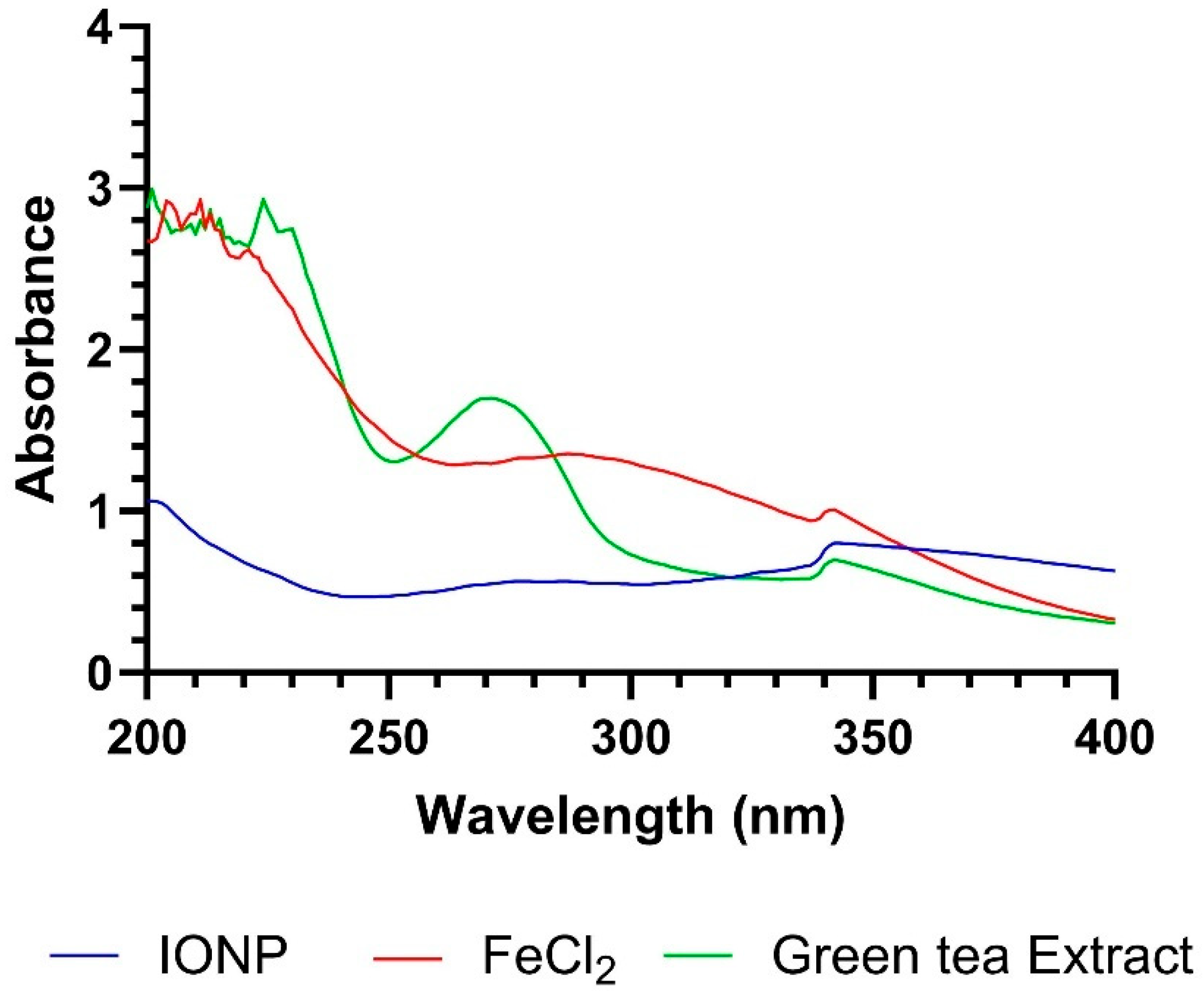
Figure 1. Spectra of 1 M iron nanoparticle (IONP) (blue), iron(II) chloride (red), and Green tea extract (green).
Cosmetics 12 00208 g001
Figure 2. DLS results of iron nanoparticles, (a) the particle size distribution, (b) zeta potential.
Cosmetics 12 00208 g002
Figure 3. (a) SEM images of iron nanoparticles; (ac) IONP; (d,e) IONP Pickering Emulsion.
Cosmetics 12 00208 g003
Figure 4. FT-IR spectra of iron oxide nanoparticle and iron(II) chloride.
Cosmetics 12 00208 g004
Figure 5. Microscope images of iron nanoparticle and iron(II) chloride.
Cosmetics 12 00208 g005
Figure 6. Viscosity–shear rate and shear stress–shear rate plot.
Cosmetics 12 00208 g006
Figure 7. (A) Shear stress–frequency, (B) shear stress–temperature plot.
Cosmetics 12 00208 g007
Figure 8. Variation in phase angle (δ)–Frequency plot.
Cosmetics 12 00208 g008
Figure 9. (a) Alamar Blue cell viability assay on HaCaT cells, (b) Microscopy Images of HaCaT Cells After 24 h Treatment. Statistical analysis was performed using one-way ANOVA; ns: p > 0.05, ***: p ≤ 0.001, ****: p ≤ 0.0001.
Cosmetics 12 00208 g009
Table 1. Formulation table of Pickering emulsions.
FormulationIONP
(w/w)(%)
Coconut Oil (w/w) (%)Green Tea Extract (w/w) (%)
PE-1103060
PE-2102070
PE-3101080
PE-47.53062.5
PE-553065
PE-62.53067.5
Table 2. SPF Values of Iron nanoparticles.
FormulationSPF ± SD
0.1 M IONP0.447 ± 0.002
0.5 M IONP5.185 ± 0.015
1.0 M IONP6.202 ± 0.013
TiO2 NP *3.982 ± 0.006
* TiO2 Np was used for comparison.
Table 3. The MIC (mg/mL) values for different concentrations of iron nanoparticles and antibiotics.
Cutibacterium acnes ATCC 6919S. aureus ATCC 29213
S1 (1 M)
(mg/mL)
S2 (0.5 M) (mg/mL)S3 (0.25 M) (mg/mL)CLR (µg/mL)S1 (1 M) (mg/mL)S2 (0.5 M) (mg/mL)S3 (0.25 M) (mg/mL)GN (µg/mL)CIP (µg/mL)CZ (µg/mL)
0.50.50.58 0.50.50.58 8 8
0.250.250.254 0.250.250.254 4 4
0.1250.1250.1252 0.1250.1250.1252 2 2
0.06250.06250.06251 0.06250.06250.06251 1 1
0.031250.031250.031250.5 0.031250.031250.031250.5 0.5 0.5
0.0156250.0156250.0156250.25 0.0156250.0156250.0156250.25 0.25 0.25
0.00781250.00781250.00781250.125 0.00781250.00781250.00781250.125 0.125 0.125
0.003906250.003906250.003906250.0625 0.003906250.003906250.003906250.0625 0.0625 0.0625
0.001953120.001953120.001953120.0313 0.001953120.001953120.001953120.0313 0.0313 0.0313
0.000976560.000976560.000976560.0156 0.000976560.000976560.000976560.0156 0.0156 0.0156
0.000488280.000488280.00048828 0.000488280.000488280.00048828
0.000244140.000244140.00024414 0.000244140.000244140.00024414
CLR: Clarithromycin; GN: Gentamicine; CIP: Ciprofloxacin; CZ: Ceftazidime. Grey areas showed no inhibition.
Table 4. Colony Counts (CFU/mL) in Formulations Contaminated with Different microorganisms.
Application Timest = 0
(CFU/mL)
2nd Hour
(CFU/mL)
4th Hour
(CFU/mL)
6th Hour
(CFU/mL)
11/21/411/21/411/21/4
C.acnesFormulation0.4 × 1070.1 × 1060.1 × 1060.1 × 106tntctntctntctntctntctntc
Coconut oil0.4 × 107 tntc0.4 × 1070.3 × 1070.1 × 106tntctntc0.1 × 107tntctntc
S. aureusFormulation0.5 × 1070.7 × 1050.5 × 1050.1 × 1060.9 × 1050.6 × 106tntctntctntctntc
Coconut oil0.5 × 107 tntc0.7 × 1060.7 × 1060.1 × 105tntctntc0.1 × 106tntctntc
S. epidermidisFormulation0.4 × 107tntc0.8 × 1070.8 × 108sçk0.4 × 1050.1 × 1060.1 × 1050.2 × 1070.3 × 107
Coconut oil0.4 × 107 tntctntctntc0.3 × 1060.2 × 1050.2 × 106tntctntctntc
tntc: too numerous to count.
Table 5. Percentage Reduction (R%) in Antimicrobial Activity of Formulation and coconut oil Compared to Initial Inoculum.
Cutibacterium acnes ATCC 6919S. aureus ATCC 29213S. epidermidis NCTC 11047
FormulationCoconut OilFormulationCoconut OilFormulationCoconut Oil
1/12nd hour97.50.0098.60.000.000.00
4th hour0.0097.598.299.80.0092.5
6th hour0.0075.00.0098.099.90.00
1/22nd hour97.50.0099.086.00.000.00
4th hour0.000.0088.00.0099.099.5
6th hour0.000.000.000.0050.00.00
1/42nd hour9.5025.098.086.00.000.00
4th hour0.000.000.000.0097.595.0
6th hour0.000.000.000.0025.00.00
Disclaimer/Publisher’s Note: The statements, opinions and data contained in all publications are solely those of the individual author(s) and contributor(s) and not of MDPI and/or the editor(s). MDPI and/or the editor(s) disclaim responsibility for any injury to people or property resulting from any ideas, methods, instructions or products referred to in the content.