Article

A 28-Day Pilot Study of the Effects on Facial Skin Hydration, Elasticity, and Texture of a Centella asiatica Extracellular Vesicle-Based Skin Care Formulation

1
Department of Applied Cosmetology, HungKuang University, Taichung City 433304, Taiwan
2
Schweitzer Biotech Company, Taipei City 114066, Taiwan
3
Department of Medical Laboratory Science and Biotechnology, China Medical University, Taichung City 406040, Taiwan
4
Department of Clinical Application, Center for iPS Cell Research and Application (CiRA), Kyoto University, Kyoto 606-8507, Japan
5
Division of Plastic Surgery, Department of Surgery, University of Pennsylvania, Philadelphia, PA 19010, USA
6
College of Science and Technology, Temple University, Philadelphia, PA 19122, USA
*
Author to whom correspondence should be addressed.
Cosmetics 2025, 12(5), 186; https://doi.org/10.3390/cosmetics12050186
Submission received: 5 June 2025 / Revised: 13 August 2025 / Accepted: 26 August 2025 / Published: 28 August 2025
(This article belongs to the Section Cosmetic Dermatology)

Abstract

Extracellular vesicles (EVs) from the traditional medicinal herb Centella asiatica (Cica) represent a novel category of botanical actives with potential dermatological benefits, yet their clinical effects in cosmetic applications remain unexplored. This pilot study assessed the effects of a skincare serum formulation with Cica EVs as the main active ingredient on facial skin quality in healthy participants. Twenty healthy participants (4 males and 16 females; average age 36.5) were enrolled and asked to apply the formulation twice daily for 28 days. Parameters, including skin hydration, elasticity, melanin content, wrinkles, redness, and pore size, were evaluated using instrumental probes and an imaging system. Facial skin quality assessments were conducted before use and at 7, 14, 21, and 28 days of product application. After 28 days of test product treatment, significant improvements were observed in measured parameters. A post-hoc placebo group of 10 participants received an identical serum without the EVs and underwent the same assessments. The EV-treated group showed statistically significant improvements in all skin parameters (p < 0.001), whereas the placebo group showed no significant changes. These findings have shown the use of a Cica EV-based skincare product in improving skin quality.

1. Introduction

As the largest organ of the human body, skin is the main barrier of entry, which protects the internal body from environmental damage and stress, maintains homeostasis, and provides key immune functions [1]. Over time, skin undergoes aging due to intrinsic factors resulting in cell senescence, characterized by reduced collagen production and decreased elasticity, as well as due to extrinsic factors like UV exposure, pollution, and oxidative stress [2]. To combat the effects of skin aging, modern skincare aims to restore skin hydration and elasticity, reduce pigmentation, and prevent or repair damage caused by aging and environmental factors. According to McKinsey, the global cosmetics market is a multi-billion-dollar industry which grew to $446 billion in 2023 and is estimated to reach $590 billion by 2028. It is characterized by a CAGR of 6%, with skincare representing the largest share of cosmetics, accounting for 44% of the market in 2023 [3]. There has been a renewed interest in natural products for cosmetics, driven by both consumer demand and the industry, due to potential risks associated with synthetic compounds, as well as the perception of benefits from natural products [4].
Centella asiatica, also known as Cica, is commonly used in traditional herbal medicine such as Chinese traditional medicine and Indian Ayurveda therapy [5]. The plant contains a rich assortment of phytochemicals such as triterpenoids, flavonoids, and polyphenols that contribute to its anti-inflammatory, wound-healing, and central nervous system-protective properties [5,6,7]. Due to its broad-spectrum benefits, commercial products containing Cica extracts are available for wound-healing and skin care purposes [8].
In contrast to crude extracts, plant extracellular vesicles (EVs) function as natural delivery systems that can enter mammalian cells and modulate gene expression or inflammatory responses at the molecular level [9,10]. EVs are lipid membrane-bound particles carrying bioactive molecules released by cells into the extracellular environment to modulate the physiological processes of target cells [11]. Recent studies have shown that plant-derived EVs from Cica and other species promote skin barrier repair, hydration, and wound healing more effectively than traditional extracts [10,12,13,14]. In addition, Cica EVs have been reported to carry specific microRNAs that suppress melanogenesis-related pathways, an effect not observed with standard extracts [13]. Thus, plant EVs are actively explored as next-generation bioactive agents in dermatological applications.
Although plant EVs have shown in-vitro skin benefits in the above studies and formulations with Cica EVs have been available on the market, no peer-reviewed clinical trials on Cica EV formulations exist to date. We have previously characterized a proprietary preparation of EVs isolated from tissue culture Cica, and demonstrated their in-vitro antioxidant, anti-melanogenic, and anti-inflammatory properties [15]. Here, we clinically assessed the daily use of a skincare serum product formulated with Cica EVs in its effect on skin quality improvement in healthy adult participants over a period of 28 days.

2. Materials and Methods

2.1. Participants and Study Design

Participants were healthy adults of any gender from the age of 18 to 60 years (inclusive).
Inclusion criteria: 20 healthy adults aged 18–60 years old were recruited through public solicitation. Healthy adults were defined as those who did not have chronic diseases, major diseases or allergies, and were not currently taking any medications or using facial care products.
Exclusion criteria: Pregnant women, breastfeeding women, men or women with chronic diseases, major diseases or allergies were not suitable for participating in this trial. Those who were currently using skin care products were excluded. Subjects with facial wounds, those who had participated in cosmetic tests, and those who had received medical beauty treatments within three months were excluded. During the implementation of this trial, no subject withdrew from the trial.
The post-hoc placebo cohort was selected using identical inclusion/exclusion criteria as the treatment arm. Baseline data were independently reviewed to confirm comparability. Outcome assessors remained blinded to group identity, although participants were not. Sensitivity analyses excluding subjective endpoints were conducted. The CONSORT Flow Diagram of the study participants is shown in Figure 1.
The participants were required to conduct a pre-test screening to confirm the absence of skin irritation or allergic reaction to the test product which consisted of applying a skin patch infused with the test product for 24 h on the inner forearms, based on the Guidelines for Cosmetic Human Skin Patch Test by the Taiwan Food and Drug Administration [16].
After screening, participants were asked to use the test product twice daily (morning and evening) after cleansing their face without using exfoliating products for 28 consecutive days. Each application involved 2 drops (0.1 mL in total volume), evenly applied to the face using fingertips for absorption. Product usage was self-recorded, and the products were stored at room temperature.
The outcome assessments were conducted at the investigation site Hungkuang University (Taichung City, Taiwan), onsite by the principal investigator. Each participant was to allocate 1 h per test (including cleansing and waiting time). The assessments were performed as a baseline test on day 0 (before using the test product), and subsequent tests at 7, 14, 21, and 28 days after test product use.
Outcomes were measured using a C+K Multi Probe Adaptor MPA580 skin analysis device (Courage-Khazaka Electronic GmbH, Koln, Germany) equipped with probes for hydration, pigmentation, and elasticity measurements. Probes were placed 1 cm horizontally and 1 cm vertically from the periorbital region of the eyes and 2 cm above the midpoint of the eyebrows (forehead). Additionally, the VISIA® Skin Analysis system (Canfield Imaging Systems, Fairfield, NJ, USA) was used to assess changes in wrinkle percentage, redness area percentage, and pore percentage by scanning full-face frontal and 45° angles on both sides of the face. Assessors remained blinded to treatment allocation across both groups. All clinical outcome instruments were scored by evaluators who were unaware of whether individuals were in the treatment or placebo cohort. Data files were coded with anonymized IDs, and group assignments were not accessible to assessors during the scoring process.
The tests were conducted in a controlled environment at 20 ± 1 °C and 50 ± 5% relative humidity, without direct sunlight or air conditioning airflow. All assessments were conducted using non-invasive techniques designed for cosmetic ingredient evaluations, ensuring the safety of participants while collecting efficacy data through instrumental analysis. Probes contact the skin’s surface to assess parameters based on electrical resistance, optical principles, and physical changes. Participants’ test results and photographic data were collected. This clinical study was registered at the ClinicalTrials.gov database as NCT06850935 on 26 February 2025 (registered retroactively). This study was conducted in accordance with the International Conference on Harmonization Good Clinical Practice Guidelines and relevant ethical standards for safety and efficacy evaluation of cosmetic products.

2.2. Description of Test Product

The test product, Supreme Rejuvenation Essence, supplied by Schweitzer Biotech Company (Taipei City, Taiwan), was a proprietary water-based serum formulation containing a preparation of EVs derived from tissue culture Centella Asiatica Leaf/Petiole Vesicles (INCI ID: 39425) and Centella Asiatica Callus Vesicles (INCI ID: 40060) as the main active ingredient. The list of ingredients in the test product, in order of decreasing composition by weight, is as follows: Aqua, Propanediol, Polyglycerin-6, Glycolipids, Glycerin, Anhydroxylitol, Ectoin, Epilobium Angustifolium Flower/Leaf/Stem Extract, Xylitylglucoside, 1,2-Hexanediol, Hydrogenated Starch Hydrolysate, Propylene Glycol, Avena Sativa Kernel Extract, Carbomer, Arginine, Caprylyl Glycol, Glucose, Allantoin, Sodium Hyaluronate, Xylitol, Glycosyl Trehalose, Centella Asiatica Extracellular Vesicles (a proprietary mixture of Centella Asiatica Leaf/Petiole Vesicles and Centella Asiatica Callus Vesicles), Chamomilla Recutita Flower Extract, Caprylhydroxamic Acid, Ethylhexylglycerin, PEG-40 Hydrogenated Castor Oil, Phenoxyethanol, Fragrance, Acetyl Hexapeptide-8, Hydroxyl Fullerene, Sodium Benzoate, Crithmum Maritimum Callus Culture Filtrate, Disodium EDTA, and Sodium Metabisulfite. The appearance of the test product is of a transparent gel-like consistency with faint fragrance. The placebo control used in subsequent studies is identical to the above composition but without the C. asiatica EV. The placebo and the test product matched in packaging and dosing appearance.

2.3. Assessment of Outcomes

The study evaluated several skin parameters using the C+K Multi Probe Adaptor MPA580 system (Courage-Khazaka Electronic GmbH, Koln, Germany):
-
Skin hydration: Assessed using the Corneometer® CM825 probe, which measures the dielectric constant related to epidermal moisture at a depth of 60–100 µm.
-
Skin melanin content: Measured with the Mexameter® MX18 probe, which detects melanin levels based on RGB light absorption at 568 nm, 660 nm, and 880 nm.
-
Skin elasticity index (R2 parameter): Evaluated using the Cutometer® Dual MPA580 probe, which analyzes the skin’s suction and stretching properties.
VISIA® Skin Analysis System (Canfield Imaging Systems, Fairfield, NJ, USA) was used to evaluate the following parameters:
-
Wrinkle score: Measured under standard white light, which detects shadow variations to determine wrinkle distribution and count. A higher percentage indicates fewer wrinkles.
-
Redness score: Analyzed using RBX polarized light to identify vascular and inflammatory conditions. A higher percentage corresponds to fewer red zones.
-
Pore score: Measured under white light, detecting shadowed pore depressions and darker areas relative to the surrounding skin. A higher percentage reflects fewer visible pores.
The VISIA skin analysis system evaluates a subject’s skin characteristics by comparing them to VISIA’s own database of individuals in the same age group, using percentile rankings. A higher percentile indicates better skin condition. To assess improvement, the percentage change in percentile for each skin parameter (wrinkles, pores, and redness) was calculated. This was done by subtracting the pre-treatment percentile from the post-treatment percentile (measured at days 7, 14, 21, and 28), dividing the result by the pre-treatment percentile, and then multiplying by 100%.
As the study was open-label and non-randomized, to further reduce observer bias, we implemented the following safeguards:
-
Standardized capture protocol: All VISIA image captures and probe assessments followed strict SOPs (e.g., fixed lighting, angles, probe calibration), ensuring consistency and reducing subjective variability.
-
Automated software extraction: While images were reviewed by humans, primary endpoint data (e.g., lesion counts, texture scores) were derived via automated VISIA and probe software, limiting human interpretation.
-
Inter-rater reliability checks: A subset (~20%) of the images was independently analyzed by a second blinded assessor. Reliability metrics (e.g., intraclass correlation coefficients) were calculated to confirm consistency.
-
Periodic blinding checks: We periodically verified that the assessors remained blind to group assignments. Any deviations were flagged, reviewed, and resolved prior to unblinding.

2.4. Data Presentation and Statistical Analysis

For each of the measured parameters, results are converted to skin improvement rate (%) calculated as follows:
P o s t t r e a t m e n t   i n d e x     P r e t r e a t m e n t   i n d e x P r e t r e a t m e n t   i n d e x   × 100 %
where the pre-treatment index is the score of the parameter examined on Day 0, and the post-treatment index is the score of the parameter examined on Day 7, 14, 21, or 28. In skin hydration and elasticity, a positive skin improvement rate indicates higher hydration and high elasticity, respectively, when compared to Day 0. In skin melanin content, the skin improvement rate is negative because the parameter measures the decrease in melanin content and the lower skin improvement rate indicates a greater decrease in melanin. In parameters measured using the VISIA® Skin Analysis System, a higher skin improvement rate indicates decreased wrinkles, redness, or pores.
Results are presented as the mean of the skin improvement rate with 95% confidence intervals shown as error bars. The analysis package in GraphPad Prism 6.01 (Boston, MA, USA) was used for statistical analysis. A paired Student’s t-test was used to compare the difference between means of skin improvement rate on Days 7, 14, 21, or 28 against Day 0 (baseline), and between placebo and treatment groups on Days 7, 14, 21, or 28 of the study. Cohen’s d was used to evaluate effect size between placebo and treatment groups using the Effect Size Calculator for t-Test at the Social Science Statistics website (https://www.socscistatistics.com/effectsize/default3.aspx (accessed on 11 August 2025)).
VISIA imaging and probe-based measurements were performed by trained technicians blinded to treatment allocation. The data were coded, and analysts had no access to grouping information. Automated software extracted quantitative endpoints. Inter-rater reliability was assessed in a 20% random subset, yielding an ICC > 0.90, confirming high consistency.

3. Results

3.1. Participants

From 4 September 2024 to 4 October 2024, 20 healthy participants were enrolled and all were assessed as negative for irritation or allergic reactions to the test product prior to the study. A post-hoc enrollment of 10 healthy participants was also conducted for the placebo cohort, following the identical inclusion and exclusion criteria to those of the treatment group, and they underwent the same skin test prior to the commencement of the study. The demographic characteristics of the treatment and post-hoc placebo groups are shown in Table 1.

3.2. Assessments of Participants

The participants were assessed for changes in skin parameters, including skin hydration, melanin content, skin elasticity, wrinkles, redness (erythema), and skin pores. The raw data values of the assessment are presented in Tables S1 to S6. The evaluation was performed on three facial regions (left cheek, right cheek, and forehead) every 7 days over a 28-day period. The percentage change of the subject’s skin quality parameters (hydration, melanin, elasticity) was calculated by subtracting the skin quality parameters before using the test product from the skin quality parameters after using the test product for 7, 14, 21, and 28 days, divided by the skin quality parameters before using the test product, and then multiplied by 100%.
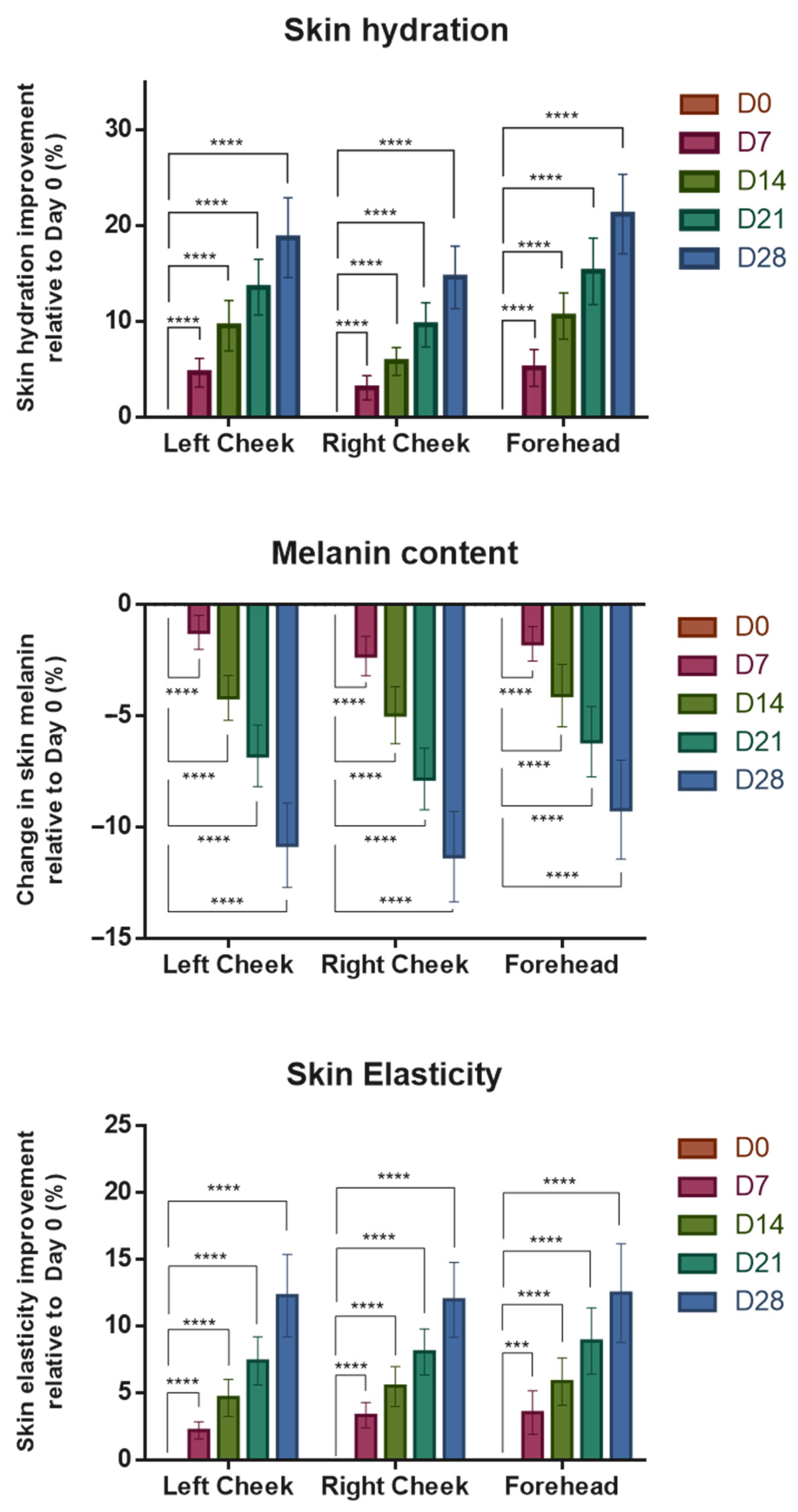
In skin hydration, statistically significant (p < 0.0001) increases in hydration percentage were observed across all facial regions (left cheek, right cheek, and forehead) over the course of 28 days compared to baseline, with the right cheek having slightly less improvement compared to other regions (Figure 2 top, Table S1). Skin hydration improved by 3.1% to 5.1%, 5.8% to 10.6%, 9.6% to 15.2%, and 14.6% to 21.2% on Days 7, 14, 21, and 28, respectively.
As the instrument measures melanin content within the skin, a decrease in melanin content resulted in lower (negative) scores. The test product treatment resulted in significantly (p < 0.0001) lowered melanin content throughout the entire study period with melanin decreasing by 1.2% to 2.3%, 4.1% to 5.0%, 6.2% to 7.8%, and 9.2% to 11.3%, on Days 7, 14, 21, and 28, respectively (Figure 2 center, Table S2).
Similarly, treatment resulted in significant (p < 0.001 or p < 0.0001) improvement in skin elasticity over time uniformly across all regions tested (Figure 2 bottom, Table S3). The improvement in skin elasticity was 2.2% to 3.5%, 4.6% to 5.9%, 7.4% to 8.9%, and 12.0% to 12.5% on Days 7, 14, 21, and 28, respectively. In summary, the physical properties of skin, including hydration, melanin content, and elasticity, all received significant improvement at 28 days of test product use compared to baseline (Day 0).
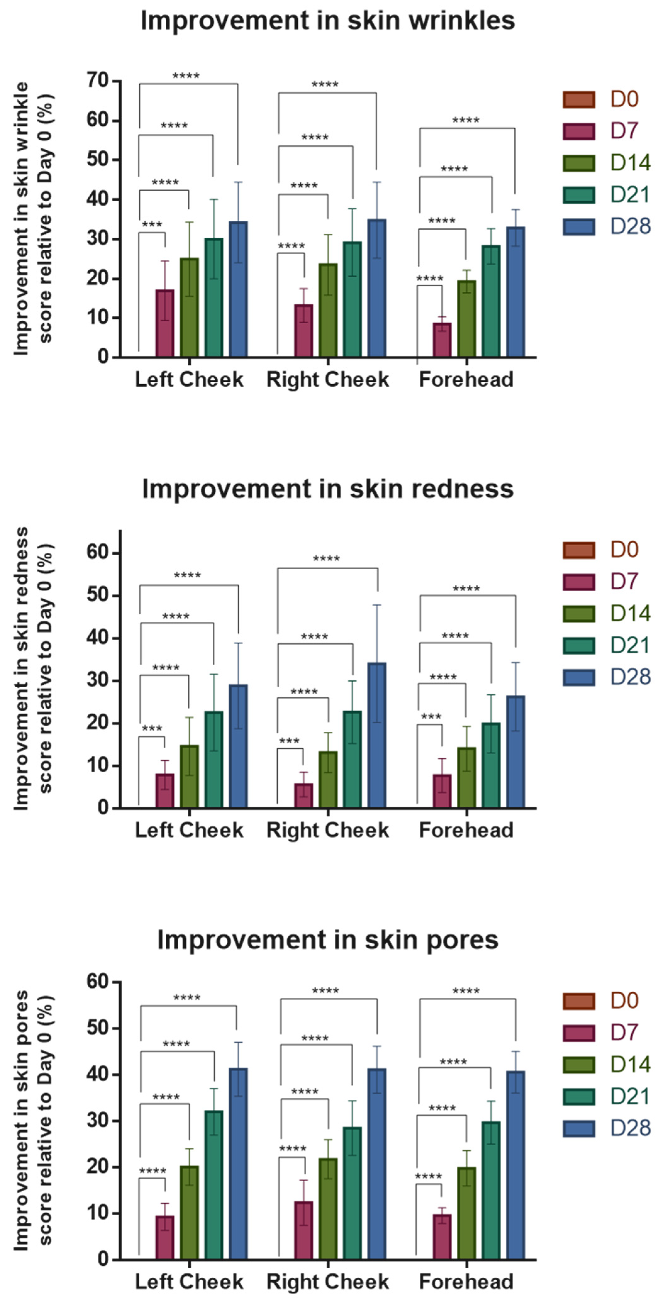
The VISIA® Skin Analysis System was used to image and analyze skin wrinkles, redness, and pore distribution simultaneously by taking photographs of the front and 45° angles on both sides of the face. After the application of the test product, skin wrinkle condition significantly (p < 0.001 or p < 0.0001) improved by 8.6% to 16.9%, 19.3% to 24.9%, 28.2% to 30.0%, and 32.9% to 34.8% on Days 7, 14, 21, and 28, respectively (Figure 3 top, Table S4). The forehead region lagged behind in wrinkle improvement at an earlier stage during the study but increased to approximately equal that of other facial regions by Day 21.
Skin redness was ameliorated significantly (p < 0.001 or p < 0.0001) during the study period by 5.6% to 7.9%, 13.1% to 14.6%, 19.9% to 22.7%, and 26.3% to 34.0% on Days 7, 14, 21, and 28, respectively (Figure 3 center, Table S5).
Finally, test product treatment resulted in significant (p < 0.0001) improvement of skin pore scores by 9.3% to 12.4%, 19.8% to 21.8%, 28.5% to 32.1%, and 40.6% to 41.3% on Days 7, 14, 21, and 28, respectively (Figure 3 bottom, Table S6). Thus, the test product demonstrated significant improvements in skin hydration and elasticity while reducing the distribution and area of melanin, wrinkles, redness, and pores. Figure 4 shows representative examples of before-and-after images of the participant photographed by the VISIA system.
To investigate the effect of treatment in different age groups, the data were stratified according to the age of participants into two groups: >40 years and <40 years. The mean skin improvement rate of each parameter was compared between the two age groups at the three facial regions. We found no significant differences in skin hydration and elasticity between the two age groups (Figure S1). However, those aged above 40 years had significantly less reduction in melanin in both cheek regions compared to participants aged less than 40 years on Day 28 of the study, while there were no significant differences in the forehead region between both age groups (Figure S1). For parameters analyzed with the VISIA® System, there were no significant differences between the two age groups in changes to skin wrinkles and pores (Figure S2). In the skin redness parameter, the overall improvement in the >40 years age group was less than that of the <40 years age group, and the difference between the two age groups was significant on Day 28 at the forehead region (Figure S2).
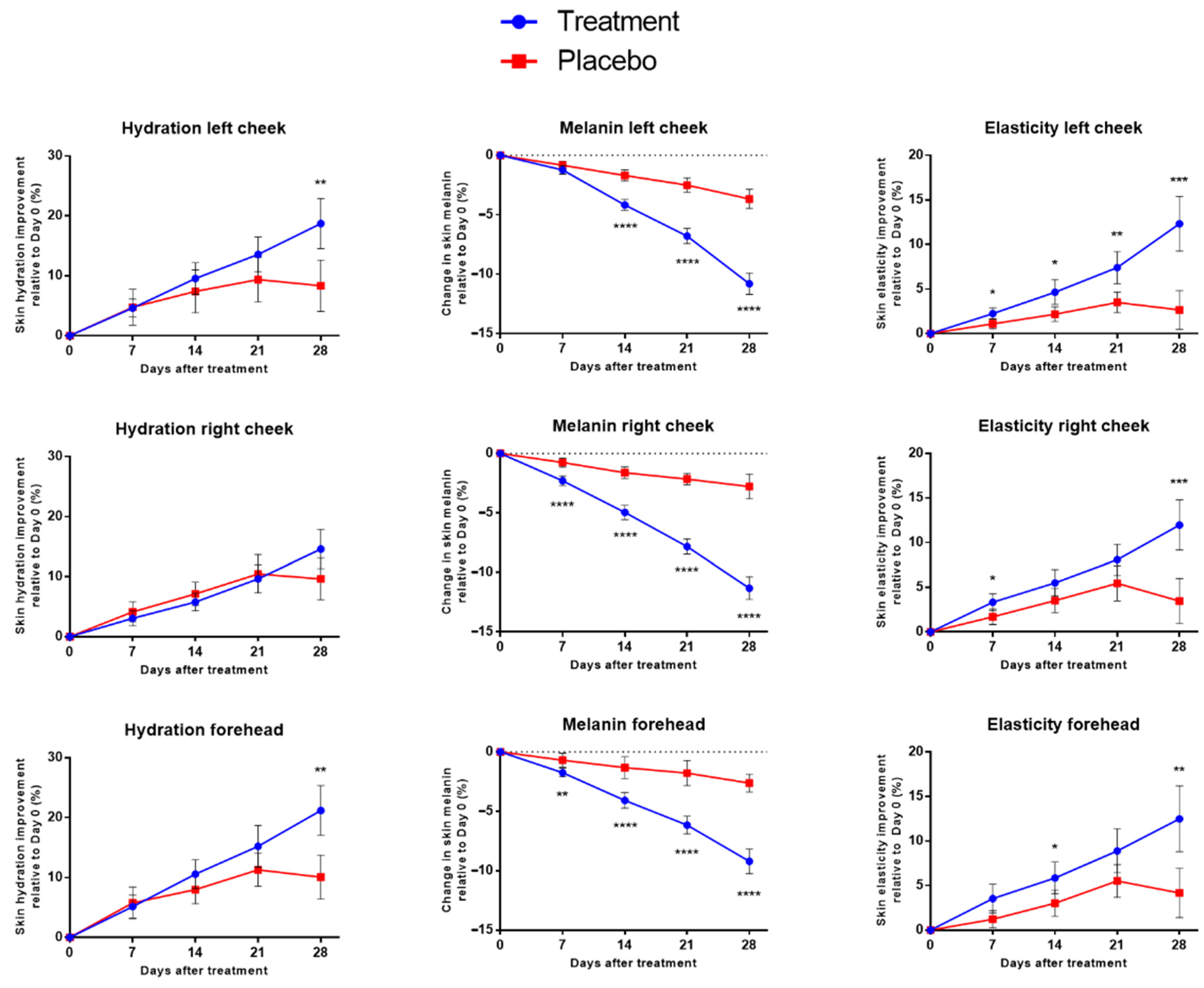
After the initial clinical study was completed, we also enrolled an additional 10 participants (2 males and 8 females; average age 40.1), using a Cica EV-free essence base formula as a placebo control group, following the same application and regimen as the original test product. The results of the placebo group were compared with the above treatment results and on Day 28, except for hydration on the right cheek, all skin parameters were statistically significantly (p < 0.001 or p < 0.0001) improved in the treatment group (Figure 5 and Figure 6, Tables S7–S12). The placebo group participants experienced slightly increased scores during the course of the study but these did not result in any statistically significant changes compared to Day 0 (Figure 5 and Figure 6).

4. Discussion

In this small-scale pilot study, we observed positive improvements in the skin in participants treated with a Cica EV-based test product. Skin integrity parameters, such as hydration and elasticity, increased after treatment with the test product. However, these results raise the question of how the measured changes translate into clinically perceptible outcomes. For example, by Day 28, the mean increase in skin hydration reached 18.7% (left cheek, 95% CI: 14.6–22.9%), 14.6% (right cheek, 11.3–17.8%), and 21.2% (forehead, 17.1–25.3%) (Table S1). Our interpretation is that, while the changes were statistically significant in controlled measurement, they may be subtle or not result in visibly noticeable changes in real-world perception. Moreover, the relatively short duration of the study (28 days) should be taken into account when evaluating the perceptibility of changes.
When comparing our present results with published cosmetic intervention studies, the hydration increases observed in the present trial (mean improvement of 18.1% across regions at Day 28) are comparable to or exceed those reported over similar or longer study periods. For example, a 12-week double-blind, placebo-controlled trial of oral hyaluronan reported significant wrinkle reduction and skin hydration gains in the range of 9 to 14% depending on the measurement site, which are comparable to, and in some cases lower than, the improvements achieved here in just four weeks [17]. Similarly, an 8-week study of a facial cream enriched with C-xyloside showed hydration increases of approximately 22% in hydration alongside 34% decreases in transepidermal water loss (TEWL), as well as a significant increase in elasticity and reduction in wrinkles [18]. In a triple-blind RCT, oral marine collagen supplementation over 12 weeks improved skin elasticity by ~23% and reduced wrinkle scores by up to 35% [19], while our 28-day EV treatment yielded elasticity gains of 12.3, 12.0, and 12.5% in the left cheek, right cheek, and forehead, respectively (Cohen’s d compared to placebo = 1.89, 1.73, and 1.34, respectively). Therefore, our results suggest that Cica EVs can achieve similar magnitudes of hydration improvement within a shorter timeframe and measurable wrinkle score reductions, albeit to a lesser degree due to the shorter intervention duration. We surmise that even more profound and visibly noticeable improvements may be apparent with extended product use or follow-up.
In terms of skin pigmentation, the present EV formulation demonstrated statistically significant reductions in melanin scores, consistent with prior findings that Cica EV-derived miRNAs can downregulate tyrosinase and other melanin synthesis genes [13]. The triterpene madecassoside in Cica has already been studied for its anti-melanogenic effect via anti-inflammatory mechanisms, providing further support that both conventional Cica extract and EV cargo can modulate pigmentation pathways [20]. There has already been evidence in the literature on plant-derived EVs that indicates their ability to deliver bioactive cargo and cargo packaged within, which can influence multiple skin health pathways simultaneously, including melanogenesis and skin extracellular matrix maintenance [9,10,21]. Compared with conventional plant extracts, EVs offer unique advantages such as membrane-protected cargo stability, targeted uptake by recipient cells, and potential cross-kingdom regulation of gene expression [9,21].
Although the present trial did not include in vitro experiments, we have previously characterized tissue-culture Cica EV preparation and demonstrated antioxidant, anti-melanogenic, and anti-inflammatory activity in cultured skin cells [15]. In those studies, Cica EVs also upregulated skin barrier genes (AQP3 and FLG) and enhanced collagen synthesis, mechanisms consistent with the hydration and elasticity improvements observed here. In addition, our in vivo data from mouse UVB skin damage model has shown that Cica EV-based gel can provide skin soothing and reduce inflammation after UV irradiation [15]. In other studies, Cica EV was shown to contain novel miRNAs and downregulate melanogenesis-related and inflammation genes, providing a plausible explanation for the pigmentation and redness reduction recorded in this clinical study [9,10,13]. Skincare products containing Cica EVs or claiming to contain Cica exosomes have been on the market, and one such recent product claimed on its website to provide up to 12 h of skin hydration in a clinical study of 31 people and enhance “skin glow” and skin texture in another clinical study [22]. However, we were not able to find any of the above results published in a scholarly journal, nor any other published clinical studies on Cica EV/exosome for commercial skin care products. Thus, this study provides valuable insight into the effects of Cica EV on the improvement of skin parameters.
While clinical studies on plant-derived EVs are currently scant, clinical and early-human studies using animal- or human-origin exosomes have reported improvements in skin quality and wound repair through angiogenesis, collagen synthesis, and anti-inflammatory activities [21,23]. Kim et al. reported that exosomes derived from human umbilical cord blood mesenchymal stem cells (MSCs) stimulated collagen production and other rejuvenation markers in human skin models [23]. More recent clinical studies using topical MSC-derived or platelet-derived EVs have reported accelerated wound healing, improved texture, reduced erythema, and increased collagen synthesis [24,25]. When comparing with those human/animal-origin exosome studies, our results were of similar direction and magnitude to effects reported with exosome-based interventions, despite a shorter observation window in our study. Other stem cell and platelet-EV clinical reports have demonstrated improved collagen and elasticity measures and reduced redness/texture over several weeks to months, whereas our results here show that a plant-derived EV formulation can produce comparable early improvements within 28 days [26]. In addition, plant-derived EVs offer practical advantages of lower immunogenicity and reduced risk of transmissible pathogens, greater raw-material scalability and cost-effectiveness, and easier sourcing/ethical profile compared with mammalian sources [27]. These are important in broadening consumer cosmetic application use of plant-derived EVs, in particular when there is reluctance toward animal/human-derived exosomes due to ethical and religious concerns, such as vegan, halal, and kosher status.
In addition to Cica EVs, the test product contains other active ingredients which may also work in synergy to contribute to the improvement in skin quality seen in the study. These include plant extracts such as oat (Avena sativa) and chamomile (Chamomilla recutita) extracts, which have anti-inflammatory, anti-oxidant, and skin barrier-stabilizing properties [28,29]. Fireweed (Epilobium angustifolium) and sea fennel (Crithmum Maritimum) extracts also display anti-inflammatory effects and can protect from UV-induced oxidative damage [30,31]. Allantoin and sodium hyaluronate (a salt of hyaluronic acid) are common moisturizing agents used in cosmetics to improve skin hydration and elasticity, as well as reduce skin irritation due to dryness and inflammation [32]. Ectoine is a natural compound derived from several species of bacteria which helps to maintain moisture balance and protect the skin barrier, as well as protect DNA from UV damage [32]. Acetyl hexapeptide-8, a synthetic peptide derived from the N-terminus of synaptosomal-associated protein of 25 kDa (SNAP-25), has anti-wrinkle effects, which makes it a safe and more accessible alternative to botulinum neurotoxin [33]. The synthetic carbon nanoparticle hydroxylated fullerene and trehalose sugar are both free radical scavenging antioxidants with protective effects against UV-induced cellular damage [34,35]. The sugar derivatives xylitylglucoside, anhydroxylitol, and xylitol work in concert to increase skin hydration and elasticity by upregulating skin barrier and water retention gene pathways [36]. However, in our subsequent placebo control, which used a formulation without Cica EVs but retained other active ingredients, we did not observe a significant increase in skin quality on Day 28 (Figure 5 and Figure 6). This may be due to the poor epidermis penetration of plant extracts and other water-soluble compounds [14].
The limitation of the study, which employs a non-concurrent post-hoc placebo group, entails potential temporal bias, residual confounding, and reduced assay sensitivity due to lack of randomization and blinding. These constraints limit causal inference; accordingly, the results should be interpreted as hypothesis-generating rather than confirmatory. This secondary cohort was ethically enrolled under a separate post-study extension after completion of the test product arm and was used solely for exploratory, comparative purposes to help distinguish the contribution of the active ingredient Cica EV. However, both groups followed identical inclusion criteria, product use instructions, and assessment protocols. To mitigate selection bias, an external blinded statistician reviewed baseline characteristics to confirm that the placebo group matched the treatment population on key metrics, reducing selection-driven imbalances. Despite these efforts, residual bias remained possible due to the fact that the two cohorts were not enrolled and randomized concurrently. Thus, direct statistical comparisons of the placebo group with the treatment group should be interpreted with caution.
Another limitation of the study was the small sample size, which may not have included sufficient diversity of demographics, such as different skin types and ethnicities, due to the high homogeneity of the Taiwanese population. The small sample size also made it unfeasible to conduct a more robust analysis of differences between age groups, and stratification into smaller age groups (e.g., 16~25 years, 26~35 years, etc.). The sample population is also heavily gender-skewed toward females (80% of participants), which can also influence the generalizability of the results. Lastly, the study duration was relatively short (28 days). This may not be sufficient to capture the long-term effects of the test product as skin quality can be influenced by numerous factors over time, including aging, UV exposure, and other environmental factors. A longer study period and follow-up would allow a more comprehensive understanding of the product’s sustained efficacy and potential adverse effects. Although imaging and measurement personnel were blinded and automated scoring was used, residual observer bias cannot be fully excluded.

5. Conclusions

The results of this pilot study support the positive effects on skin quality of the test product in improving skin hydration and elasticity as well as reducing melanin, wrinkles, redness, and pores. The differences observed between age groups and the three facial regions were minimal. This pilot study provides initial evidence supporting the potential utility of Cica EVs in skin care, warranting further investigation in larger, controlled studies. Future studies should address the above-mentioned limitations to provide clearer insights into the long-term benefits of skin improvement and application of the test product across a diverse set of populations to address a global market.

Supplementary Materials

The following supporting information can be downloaded at: https://www.mdpi.com/article/10.3390/cosmetics12050186/s1. Figure S1: Change in skin hydration (left column), melanin (middle column), and elasticity (right column) at 7, 14, 21, and 28 days after treatment of test product in left cheek (top row), right cheek (middle row), and forehead (bottom row) facial regions. Figure S2: Change in skin wrinkles (left column), redness (middle column), and pores (right column) at 7, 14, 21, and 28 days after treatment of test product in left cheek (top row), right cheek (middle row), and forehead (bottom row) facial regions. Table S1: Data values for skin hydration during the study period. Table S2: Data values for skin melanin content during the study period. Table S3: Data values for skin elasticity during the study period. Table S4: Wrinkle score during the study period. Table S5: Redness score during the study period. Table S6: Pore score during the study period. Table S7. Percentage of change in skin hydration relative to baseline in the placebo group during the study period. Table S8. Percentage of change in skin melanin content relative to baseline in the placebo group during the study period. Table S9. Percentage of change in skin elasticity relative to baseline in the placebo group during the study period. Table S10. Percentage of change in skin wrinkle score relative to baseline in the placebo group during the study period. Table S11. Percentage of change in skin redness score relative to baseline in the placebo group during the study period. Table S12. Percentage of change in skin pore score relative to baseline in the placebo group during the study period.

Author Contributions

Conceptualization, supervision and project administration: T.-M.C., C.C. and T.-Y.K. Visualization and formal analysis: T.-M.C. and L.T.-C.L. Investigation, data acquisition and curation: T.-M.C., H.-C.H. and J.-Y.L. Methodology: T.-M.C., C.-C.W., H.-C.H., J.-Y.L., C.-H.C., P.-L.K., W.-H.T., W.-Y.Q., I.P. and T.-Y.K. Resources: C.-C.W., C.-H.C., P.-L.K., W.-H.T. and T.-Y.K. Writing—original draft preparation: T.-M.C. and L.T.-C.L. Writing—review and editing: T.-M.C., L.T.-C.L., W.-Y.Q., I.P., C.C. and T.-Y.K. All authors have read and agreed to the published version of the manuscript.

Funding

Schweitzer Biotech Company provided the necessary funding for the study as well as the test product.

Institutional Review Board Statement

The final protocol, case report form, and informed consent form were reviewed and approved by the institutional review board of the Antai Tian-Sheng Memorial Hospital (IRB No. 24-064-A, approved 16 May 2024). All participants provided written informed consent prior to the start of the study. This study was conducted in compliance with the Declaration of Helsinki and in compliance with all International Conference on Harmonization Good Clinical Practice Guidelines (ICH-GCP). This clinical study was registered at the ClinicalTrials.gov database as NCT06850935 on 26 February 2025 (registered retroactively).

Informed Consent Statement

Informed consent for participation was obtained from all subjects involved in the study. Written informed consent has been obtained from all identifiable human participants to publish this paper.

Data Availability Statement

All data generated or analyzed during this study are included in this manuscript.

Acknowledgments

We thank Ji-Ying Liu of HuangKuang University for assisting with the clinical trial and Meei-Yun Lin of Schweitzer Biotech Company for reading and providing feedback for preparation of the manuscript.

Conflicts of Interest

C.-C. W., C.-H. C., P.-L. K., W.-H. T., L. T.-C. L., C. C., and T.-Y. K. are employees of Schweitzer Biotech Company. W.-Y. Q. and I. P. are consultants for Schweitzer Biotech Company. T.-M. C., H.-C. H. and J.-Y. L. have no competing interests to declare. This study and the test product were funded and provided by Schweitzer Biotech Company, but the company otherwise had no role in participant recruitment, data collection, analysis, interpretation, writing of the clinical report, or the decision to submit this article for publication.

References

  1. Rajkumar, J.; Chandan, N.; Lio, P.; Shi, V. The Skin Barrier and Moisturization: Function, Disruption, and Mechanisms of Repair. Skin Pharmacol. Physiol. 2023, 36, 174–185. [Google Scholar] [CrossRef] [PubMed]
  2. Franco, A.C.; Aveleira, C.; Cavadas, C. Skin Senescence: Mechanisms and Impact on Whole-Body Aging. Trends Mol. Med. 2022, 28, 97–109. [Google Scholar] [CrossRef]
  3. The Beauty Boom and Beyond: Can the Industry Maintain Its Growth? Available online: https://www.mckinsey.com/industries/consumer-packaged-goods/our-insights/the-beauty-boom-and-beyond-can-the-industry-maintain-its-growth (accessed on 10 December 2024).
  4. Liu, J.K. Natural Products in Cosmetics. Nat. Prod. Bioprospect. 2022, 12, 40. [Google Scholar] [CrossRef]
  5. Biswas, D.; Mandal, S.; Chatterjee Saha, S.; Tudu, C.K.; Nandy, S.; Batiha, G.E.; Shekhaway, M.S.; Pandey, D.K.; Dey, A. Ethnobotany, Phytochemistry, Pharmacology, and Toxicity of Centella asiatica (L.) Urban: A Comprehensive Review. Phytother. Res. 2021, 35, 6624–6654. [Google Scholar] [CrossRef] [PubMed]
  6. Bandopadhyay, S.; Mandal, S.; Ghorai, M.; Jha, N.K.; Kumar, M.; Radha, N.; Ghosh, A.; Proćków, J.; Perez de la Lastra, J.M.; Dey, A. Therapeutic Properties and Pharmacological Activities of Asiaticoside and Madecassoside: A Review. J. Cell. Mol. Med. 2023, 27, 593–608. [Google Scholar] [CrossRef]
  7. Sun, B.; Wu, L.; Wu, Y.; Zhang, C.; Qin, L.; Hayashi, M.; Kudo, M.; Gao, M.; Liu, T. Therapeutic Potential of Centella asiatica and Its Triterpenes: A Review. Front. Pharmacol. 2020, 11, 568032. [Google Scholar] [CrossRef]
  8. Diniz, L.R.; Calado, L.L.; Duarte, A.B.; de Sousa, D.P. Centella asiatica and Its Metabolite Asiatic Acid: Wound Healing Effects and Therapeutic Potential. Metabolites 2023, 13, 276. [Google Scholar] [CrossRef]
  9. Farley, J.T.; Eldahshoury, M.K.; de Marcos Lousa, C. Unconventional secretion of plant extracellular vesicles and their benefits to human health: A mini review. Front. Cell Dev. Biol. 2022, 10, 883841. [Google Scholar] [CrossRef]
  10. Domaszewska-Szostek, A.; Krzyżanowska, M.; Polak, A.; Puzianowska-Kuźnicka, M. Effectiveness of extracellular vesicle application in skin aging treatment and regeneration: Do we have enough evidence from clinical trials? Int. J. Mol. Sci. 2025, 26, 2354. [Google Scholar] [CrossRef]
  11. Jeppesen, D.K.; Zhang, Q.; Franklin, J.L.; Coffey, R.J. Extracellular Vesicles and Nanoparticles: Emerging Complexities. Trends Cell Biol. 2023, 33, 667–681. [Google Scholar] [CrossRef] [PubMed]
  12. Cho, J.H.; Hong, Y.D.; Kim, D.; Park, S.J.; Kim, J.S.; Kim, H.M.; Yoon, E.J.; Cho, J.S. Confirmation of Plant-Derived Exosomes as Bioactive Substances for Skin Application Through Comparative Analysis of Keratinocyte Transcriptome. Appl. Bio. Chem. 2022, 65, 8. [Google Scholar] [CrossRef]
  13. Kim, M.H.; Yoon, E.J.; Kim, J.S.; Park, S.J.; Lee, H. Comparative Analysis of the Transcriptome and Efficacy of Bioactive Centella asiatica Exosomes on Skin Cells. ResearchSquare 2023. Available online: https://www.researchsquare.com/article/rs-2787704/v1 (accessed on 11 November 2024).
  14. Li, A.; Li, D.; Gu, Y.; Liu, R.; Tang, X.; Zhao, Y.; Qi, F.; Wei, J.; Liu, J. Plant-Derived Nanovesicles: Further Exploration of Biomedical Function and Application Potential. Acta Pharm. Sin. 2023, 13, 3300–3320. [Google Scholar] [CrossRef] [PubMed]
  15. Chang, T.M.; Wu, C.C.; Huang, H.C.; Wang, S.S.; Chuang, C.H.; Kao, P.L.; Tang, W.H.; Liu, L.T.C.; Qiu, W.Y.; Percec, I.; et al. Centella asiatica Tissue Culture-Derived Extracellular Vesicles: A Multifaceted Approach to Skincare Applications. bioRxiv 2024. [Google Scholar] [CrossRef]
  16. Taiwan Food and Drug Administration. Guidelines for Cosmetic Human Skin Patch Test (in Traditional Chinese). November 2019. Available online: https://www.fda.gov.tw/tc/includes/GetFile.ashx?id=f637108864795066124&type=2&cid=32306 (accessed on 7 April 2025).
  17. Hsu, T.F.; Su, Z.R.; Hsieh, Y.H.; Wang, M.F.; Oe, M.; Matsuoka, R.; Masuda, Y. Oral hyaluronan relieves wrinkles and improves dry skin: A 12-week double-blinded, placebo-controlled study. Nutrients 2021, 13, 2220. [Google Scholar] [CrossRef]
  18. Zang, S.; Chen, J.; Chevalier, C.; Zhang, J.; Li, S.; Wang, H.; Li, J.; Chen, Y.; Xu, H.; Sheng, L.; et al. Holistic investigation of the anti-wrinkle and repair efficacy of a facial cream enriched with C-xyloside. J. Cosmet. Dermatol. 2024, 23, 4017–4028. [Google Scholar] [CrossRef]
  19. Evans, M.; Lewis, E.D.; Zakaria, N.; Pelipyagina, T.; Guthrie, N. A randomized, triple-blind, placebo-controlled, parallel study to evaluate the efficacy of a freshwater marine collagen on skin wrinkles and elasticity. J. Cosmet. Dermatol. 2021, 20, 825–834. [Google Scholar] [CrossRef]
  20. Jung, E.; Lee, J.A.; Shin, S.; Roh, K.B.; Kim, J.H.; Park, D. Madecassoside Inhibits Melanin Synthesis by Blocking Ultraviolet-Induced Inflammation. Molecules 2013, 18, 15724–15736. [Google Scholar] [CrossRef]
  21. Zhu, Y.; Zhao, J.; Ding, H.; Qiu, M.; Xue, L.; Ge, D.; Wen, G.; Ren, H.; Li, P.; Wang, J. Applications of Plant-Derived Extracellular Vesicles in Medicine. MedComm 2024, 5, e741. [Google Scholar] [CrossRef] [PubMed]
  22. The Inkey List. Exosome Hydro-Glow Complex. Available online: https://www.theinkeylist.com/products/exosome-hydro-glow-complex (accessed on 10 April 2025).
  23. Kim, Y.J.; Yoo, S.M.; Park, H.H.; Lim, H.J.; Kim, Y.L.; Lee, S.; Seo, K.W.; Kang, K.S. Exosomes Derived from Human Umbilical Cord Blood Mesenchymal Stem Cells Stimulates Rejuvenation of Human Skin. Biochem. Biophys. Res. Commun. 2017, 493, 1102–1108. [Google Scholar] [CrossRef]
  24. Johnson, J.; Law, S.Q.; Shojaee, M.; Hall, A.S.; Bhuiyan, S.; Lim, M.B.; Silva, A.; Kong, K.J.; Schoppet, M.; Blyth, C.; et al. First-in-Human Clinical Trial of Platelet-Derived Extracellular Vesicles for Wound Healing. J. Extracell. Vesicles 2023, 12, e12332. [Google Scholar] [CrossRef] [PubMed]
  25. Peredo, M.; Shivananjappa, S. Topical Human Mesenchymal Stem Cell-Derived Exosomes for Acceleration of Wound Healing Following Tissue Trauma and Aesthetic Procedures: A Case Series. J. Drugs. Dermatol. 2024, 23, 281–284. [Google Scholar] [CrossRef]
  26. Bai, G.; Truong, T.M.; Pathak, G.N.; Benoit, L.; Rao, B. Clinical applications of exosomes in cosmetic dermatology. Skin Health Dis. 2024, 4, e348. [Google Scholar] [CrossRef]
  27. Alzahrani, F.A.; Khan, M.I.; Kameli, N.; Alsahafi, E.; Riza, Y.M. Plant-derived extracellular vesicles and their exciting potential as the future of next-generation drug delivery. Biomolecules 2023, 13, 839. [Google Scholar] [CrossRef]
  28. Kim, H.S.; Hwang, H.J.; Seo, W.D.; Do, S.H. Oat (Avena sativa L.) Sprouts Restore Skin Barrier Function by Modulating the Expression of the Epidermal Differentiation Complex in Models of Skin Irritation. Int. J. Mol. Sci. 2023, 24, 17274. [Google Scholar] [CrossRef]
  29. Sah, A.; Naseef, P.P.; Kuruniyan, M.S.; Jain, G.K.; Zakir, F.; Aggarwal, G. A Comprehensive Study of Therapeutic Applications of Chamomile. Pharmaceuticals 2022, 15, 1284. [Google Scholar] [CrossRef]
  30. Huang, H.; Qian, J.; Zeng, L.; Zhang, J.; Peng, X.; Tang, X.; Tang, Y.; Fu, M.; Liu, Q.; Song, F.; et al. Full-Component Extract of Crithmum maritimum and its Effect on Epidermal Regeneration. RSC Adv. 2025, 15, 1713–1720. [Google Scholar] [CrossRef]
  31. Nowak, A.; Zielonka-Brzezicka, J.; Perużyńska, M.; Klimowicz, A. Epilobium angustifolium L. as a Potential Herbal Component of Topical Products for Skin Care and Treatment-A Review. Molecules 2022, 27, 3536. [Google Scholar] [CrossRef]
  32. Jesus, A.; Sousa, E.; Cidade, H.; Cruz, M.T.; Almeida, I.F. How to Fight Acute Sun Damage? Current Skin Care Strategies. Photochem. Photobiol. Sci. 2024, 23, 1915–1930. [Google Scholar] [CrossRef] [PubMed]
  33. Olsson, S.E.; Sreepad, B.; Lee, T.; Fasih, M.; Fijany, A. Public Interest in Acetyl Hexapeptide-8: Longitudinal Analysis. JMIR Dermatol. 2024, 7, e54217. [Google Scholar] [CrossRef] [PubMed]
  34. Saitoh, Y.; Miyanishi, A.; Mizuno, H.; Kato, S.; Aoshima, H.; Kokubo, K.; Miwa, N. Super-Highly Hydroxylated Fullerene Derivative Protects Human Keratinocytes from UV-Induced Cell Injuries Together with the Decreases in Intracellular ROS Generation and DNA Damages. J. Photochem. Photobiol. B Biol. 2011, 102, 69–76. [Google Scholar] [CrossRef] [PubMed]
  35. Xiao, Z.; Yang, S.; Chen, J.; Li, C.; Zhou, C.; Hong, P.; Sun, S.; Qian, Z.J. Trehalose Against UVB-Induced Skin Photoaging by Suppressing MMP Expression and Enhancing Procollagen I Synthesis in HaCaT cells. J. Funct. Foods 2020, 74, 104198. [Google Scholar] [CrossRef]
  36. Garcia, C.; Valin, E.; Hernandez, E.; Kern, C.; Roso, A.; Garcia, C.; Valin, E.; Hernandez, E.; Kern, C.; Roso, A. Effect of a Simple Sugar-based Ingredient on Skin Moisturization: Biological Mode of Action and Clinical Effects. Asian J. Beauty Cosmetol. 2023, 21, 3–27. [Google Scholar] [CrossRef]
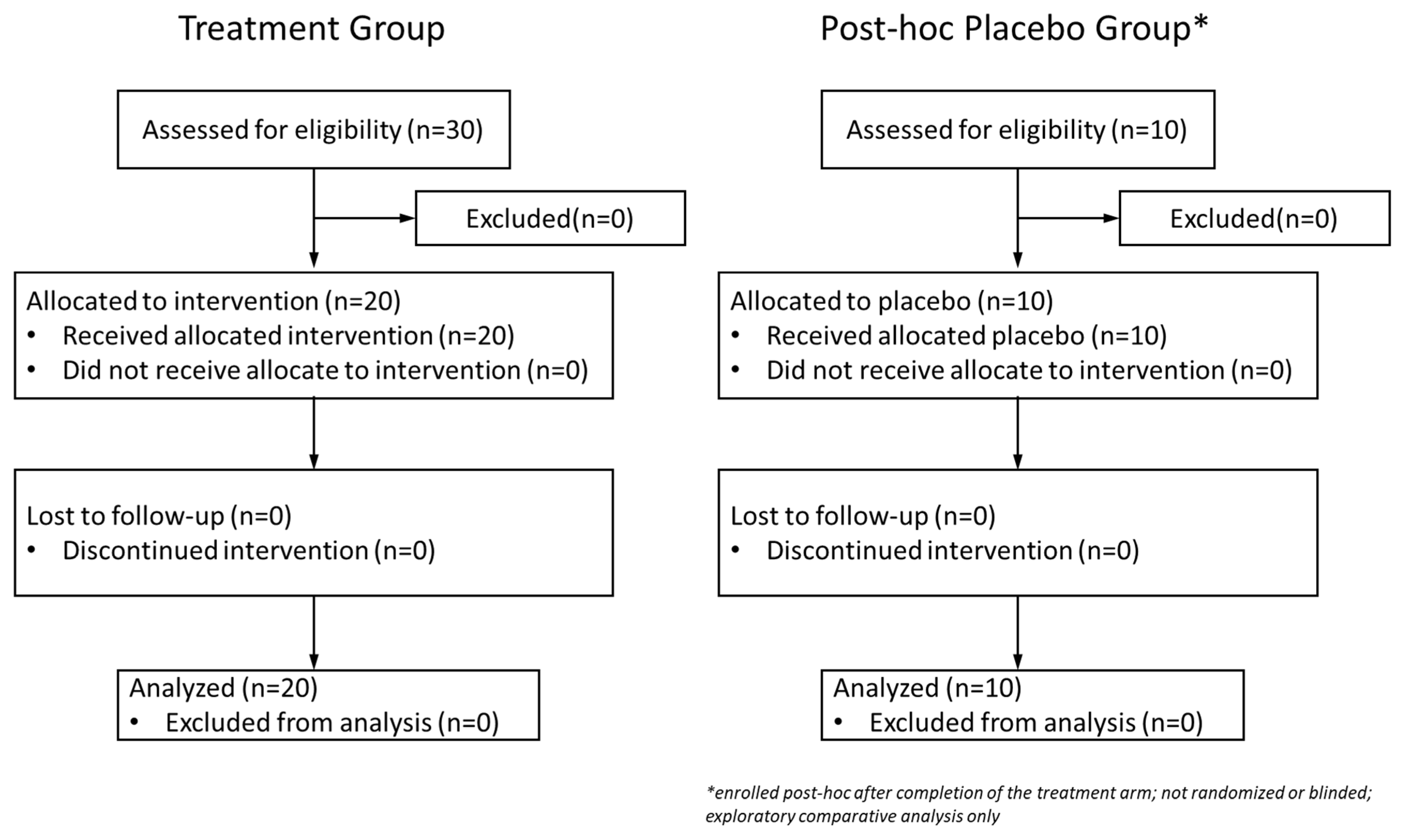
Figure 1. CONSORT flowchart of the study. The left arm represents the original test product cohort (n = 20) enrolled in the initial trial period. The right arm shows the post-hoc placebo cohort (n = 10), recruited and assessed separately following the completion of the treatment group. The placebo group was non-randomized and unblinded, and was added for exploratory comparison only. Identical inclusion/exclusion criteria and outcome assessments were applied for both groups.
Cosmetics 12 00186 g001
Figure 2. Change in skin hydration (top), melanin content (center), and skin elasticity (bottom) at 0, 7, 14, 21, and 28 days after treatment of the test product. For skin hydration and elasticity, higher values indicate more hydrated skin. For melanin content, lower values indicate a decrease in melanin content. The results are presented as bars showing means with error bars representing a 95% confidence interval of the mean. Statistical significances on days 7, 14, 21, and 28 were calculated by a paired t-test compared to the value on day 0 (baseline). *** p < 0.001, **** p < 0.0001.
Cosmetics 12 00186 g002
Figure 3. Change in skin wrinkles (top), skin redness (center), and skin pores (bottom) at 0, 7, 14, 21, and 28 days after treatment with the test product. For skin wrinkles, higher values indicate a decrease in the distribution and number of wrinkles. For skin redness, higher values indicate a decreasing zone of redness. For skin pores, higher values indicate a decrease in the number and area of pores. The results are presented as bars showing means with error bars representing a 95% confidence interval of the mean. Statistical significances on days 7, 14, 21, and 28 were calculated by a paired t-test compared to the value on day 0 (baseline). *** p < 0.001, **** p < 0.0001.
Cosmetics 12 00186 g003
Figure 4. Representative before and after images of a participant photographed by the VISIA system for evaluation of wrinkle, inflammation, and pore reduction.
Cosmetics 12 00186 g004
Figure 5. Change in skin hydration (left column), melanin (middle column), and elasticity (right column) at 7, 14, 21, and 28 days after treatment of test product (n = 20) or placebo (n = 10) in left cheek (top row), right cheek (middle row), and forehead (bottom row) facial regions. Results are shown as means of percentage change in skin parameters for the test product treatment (blue) and placebo (red) with error bars representing a 95% confidence interval of the mean. Statistical significances of means of participants in the two age groups at a single time point were calculated with Student’s t-test. * p < 0.05, ** p < 0.01, *** p < 0.001, **** p < 0.0001.
Cosmetics 12 00186 g005
Figure 6. Change in skin wrinkles (left column), redness (middle column), and pores (right column) at 7, 14, 21, and 28 days after treatment of the test product (n = 20) or placebo (n = 10) in left cheek (top row), right cheek (middle row), and forehead (bottom row) facial regions. Results are shown as means of percentage change in skin parameters for the test product treatment (blue) and the placebo (red), with error bars representing 95% confidence interval of the mean. Statistical significances of means of participants in the two age groups at a single time point were calculated with Student’s t-test. * p < 0.05, ** p < 0.01, *** p < 0.001, **** p < 0.0001.
Cosmetics 12 00186 g006
Table 1. Demographic characteristics of the test product treatment group and post-hoc placebo group.
TreatmentPlacebo
Number of participants2010
Gender
Female16 (80%)8 (80%)
Male4 (20%)2 (20%)
Age (mean ± SD in years)36.5 ± 9.240.1 ± 10.4
EthnicityEast Asian (100%)East Asian (100%)
Fitzpatrick skin typeType II (100%)Type II (100%)
Disclaimer/Publisher’s Note: The statements, opinions and data contained in all publications are solely those of the individual author(s) and contributor(s) and not of MDPI and/or the editor(s). MDPI and/or the editor(s) disclaim responsibility for any injury to people or property resulting from any ideas, methods, instructions or products referred to in the content.