Article

Cosmetic Upgrade of EGF: Genetically Modified Probiotic-Derived Cell-Free Supernatants Containing Human EGF Protein Exhibit Diverse Biological Activities

1
R&D Center, Cell Biotech, Co., Ltd., Gimpo-si 10003, Republic of Korea
2
Department of Dermatology, Chung-Ang University College of Medicine, Seoul 06973, Republic of Korea
*
Authors to whom correspondence should be addressed.
These authors contributed equally to this work.
Cosmetics 2025, 12(4), 176; https://doi.org/10.3390/cosmetics12040176
Submission received: 27 May 2025 / Revised: 29 July 2025 / Accepted: 31 July 2025 / Published: 19 August 2025
(This article belongs to the Section Cosmetic Technology)

Abstract

Although epidermal growth factor (EGF) has potential wide applications in the cosmetic industry, it still has limitations, such as a costly purification process and low stability in the surrounding environment. To overcome these limitations, we developed genetically modified Pediococcus pentosaceus CBT SL4, which can secrete EGF protein in growth media, thereby producing probiotic-derived PP-EGF culture medium supernatant (PP-EGF-SUP). Even at low EGF concentrations, PP-EGF-SUP exhibited EGF activities, such as cell scratch wound healing, tyrosinase inhibition, and improvements in anti-wrinkle factors, similar to or stronger than those of recombinant human EGF (rhEGF), which was used as a positive control. PP-EGF-SUP exhibited strong additional biological activities, such as antioxidant, anti-inflammatory, and anti-microbial activities, even though rhEGF did not have such properties. PP-EGF-SUP could be easily transformed to PP-EGF-SUP dried powder (PP-EGF-DP) using the freeze-drying method, and it could also be well resolved in water up to 20 mg/mL; furthermore, it still maintained its bioactivity after the manufacturing process. To determine melasma improvement efficacy, a human application test was performed using melasma ampoules containing 1% or 5% PP-EGF-DP formulations for four weeks. When comparing the melasma values before and after treatment, it was found that the light melasma value statistically decreased by 3.38% and 3.79% and that the dark melasma value statistically decreased by 1.74% and 2.93% in the test groups applying the 1% and 5% PP-EGF-DP melasma ampoules, respectively. In addition, the melasma area also decreased by 21.21% and 29.1%, while the control group showed no statistical difference. During the study period, no significant adverse skin reactions were observed due to the application of the PP-EGF-DP melasma ampoule. In conclusion, PP-EGF-DP may offer unique advantages in the cosmetic ingredient market, such as safety (as a probiotic derivative), stability (postbiotics protect EGF activity), and diverse bioactivities (activity potentiation and postbiotic-derived biological activities).

1. Introduction

Epidermal growth factor (EGF) was first isolated from mice in 1960 [1]. EGF exerts biological functions, such as promoting cell growth, proliferation, differentiation, and survival, by binding to its receptor (EGFR), which is located in most animal epithelial tissues, including in fibroblasts and endothelial cells. Consequently, EGF can regulate wound healing and maintain tissue integrity [2]. In terms of clinical application, EGF is a molecule with broad pharmacological activity in dermal wound healing [3,4,5]. The healing effects of EGF have been studied in surgical wounds, burn wounds, and diabetic foot ulcers [6]. With advances in recombinant DNA technology, the production of large quantities of recombinant human EGF (rhEGF) has become possible. EGF and EGF-like substances, including insulin-like growth factor (IGF)-1 and fibroblast growth factor (FGF)-1, have demonstrated significant potential as cosmetic ingredients due to their ability to stimulate epidermal cell proliferation, enhance collagen synthesis, and accelerate wound healing processes [7,8,9]. These properties make EGF and EGF-like substances attractive ingredients in anti-aging and skin repair cosmetic products. Therefore, the focus shifted toward their use as cosmetic ingredients for maintaining young skin, and the number of wound healing studies involving EGF began to change.
However, EGF still has some limitations, such as a short half-life and a low absorption ratio, and there is a lack of efficient formulations for its delivery through the transdermal route [10,11,12]. As an unstable protein formulation may impact product appearance, purity, potency, and healing effects, the in vitro stabilization of EGF is an important practical concern. The stabilization of EGF will enhance the control and manufacture of its functional performance in the pharmaceutical and cosmetic industries. With recent developments focusing on enhancing EGF stability for drug delivery, its use in the dermatologic field has also seen a resurgence [13,14,15,16,17].
In addition, the exogenous administration of EGF does not stimulate the malignant transformation or initiation of tumorigenesis in wound bed cells [18,19]. Hence, this study aimed to improve the benefits of EGF as a cosmetic ingredient through the combination of probiotic-derived EGF and useful probiotic-derived metabolites. As is well known, probiotics can produce a wide range of bioactive molecules, such as amino acids, enzymes, vitamins, carbohydrates, oligosaccharides, bacteriocins, immunomodulatory compounds, and short-chain fatty acids [20,21]. Moreover, we established a probiotic drug delivery system (DDS) for colorectal cancer therapy [22]. To this end, we generated a pCBT24-2-pG6Pi-EGF-pG6Pi-EGF plasmid capable of inducing the secretion of large amounts of EGF protein from Pediococcus pentosaceus SL4 (PP). We then found that a PP cell-free supernatant containing PP-EGF (PP-EGF-SUP) exerted diverse bioactivities on mammalian cell lines. This substance exerted proliferative, wound healing, skin whitening, antioxidant, anti-inflammatory, and anti-bacterial activities. To develop a cosmetic ingredient using PP-EGF-SUP, it was subjected to the freeze-drying process for concentration and drying, and then the stability of EGF in a PP-EGF dried powder (PP-EGF-DP) was evaluated after exposure to wide pH and temperature ranges. Finally, we registered PP-EGF-DP as a cosmetic ingredient, and then we confirmed the efficacy of ampoule-type PP-EGF-DP in improving melasma in a human cosmetic application test.
Consequently, our approach may lead to the development of more cost-effective solutions, including cosmetic ingredients with a wide range of bioactivities.

2. Materials and Methods

2.1. Bacterial Strains and Culture

Pediococcus pentosaceus SL4 (PP) KCTC 10297BP, which was isolated from kimchi, was obtained from the culture collection maintained at Cell Biotech Co., Ltd., Gimpo, 10003, Republic of Korea. PP was cultured for 18–24 h in PP-EGF culture media (glucose 2% + Na2HPO4 1.28% + KH2PO4 0.3% NaCl 0.005% + NH4Cl 0.1% yeast extract (Sensient Technologies, Milwaukee, WI, USA) 2% + MgSO4 0.024% + CaCl2 0.00111%, pH 6.0) at 37 °C and 100 rpm. Escherichia coli (E. coli) was cultured for 18–24 h in M9 broth (Difco, Detroit, MI, USA) or LB broth (Difco, Detroit, MI, USA) at 37 °C.

2.2. Construction of Plasmid Harboring DDS-EGF for Use in Pediococcus pentosaceus SL4 System

EGF was cloned into the plasmid pCBT24-2 (KCCM12182P). The dual-promoter system selected for the maximum expression of EGF was ligated to a usp45 secretion signal peptide, thereby enabling the synthesis of DNA fragments (Cosmogenetech Inc., Seoul, Republic of Korea). A portion of each promoter ligated to the signal peptide was digested with NheI/SalI and BamHI/PstI restriction enzymes. DNA fragments encoding PK-usp45-EGF were inserted into the pCBT24-2 expression vector. Finally, the pCBT24-2-PK-EGF-PK-EGF plasmid (accession number: KCCM13348P) was transformed into PP cells.

2.3. Transformation of Pediococcus pentosaceus SL4 (PP-EGF) and Detection of EGF Protein in Culture Supernatant

Transformants grown on De Man, Rogosa, and Sharpe (MRS) agar plates were inoculated into 10 mL of MRS broth containing 10 μg/mL erythromycin and cultured at 37 °C for 15 h (no shaking). Next, 1 mL of pre-culture was inoculated into 10 mL of M9 minimal medium containing 10 μg/mL of erythromycin and cultured at 37 °C for 48 h (no shaking). Next, 5 mL of culture was centrifuged, and the supernatant was collected. The supernatant was concentrated via trichloroacetic acid (TCA) precipitation to isolate the total protein. Finally, EGF protein was detected using Western blotting. Furthermore, the EGF protein in the culture supernatant was identified using LC-MS/MS after concentration using 3 kDa centricon plus-70. The final EGF concentration was determined using an EGF ELISA kit.

2.4. Cell Lines and Culture

Fibroblast (CCD-986sk, 3T3-L1), skin melanoma (SK-MEL-2), and RAW 264.7 cell lines were obtained from the American Type Culture Collection (ATCC; Manassas, VA, USA). The CCD-986sk cell line was maintained at 5% CO2/37 °C in Iscove′s Modified Dulbecco′s Medium (IMDM) (Gibco, Grand Island, NY, USA) containing 10% fetal bovine serum (FBS; Gibco) and 1% penicillin/streptomycin (Gibco). The 3T3-L1 cell line was maintained at 5% CO2/37 °C in Dulbecco′s Modified Eagle′s Medium (DMEM) (Gibco) containing 10% bovine calf serum (BCS; Gibco) and 1% penicillin/streptomycin (Gibco). The SK-MEL-2 cell line was maintained at 5% CO2/37 °C in DMEM (Gibco) containing 10% FBS (Gibco) and 1% penicillin/streptomycin (Gibco). The RAW 264.7 cell line was maintained at 5% CO2/37 °C in DMEM (Gibco) containing 10% FBS (Gibco) and 1% penicillin/streptomycin (Gibco).

2.5. Investigation of Bioactivities of PP-EGF Culture Supernatant

The bioactivities of PP-EGF-SUP were evaluated based on EGF-mediated proliferation/cell scratch wound healing stimulation (cell growth), the negative regulation of tyrosinase expression/inhibition of its enzymatic activity (whitening), and the negative regulation of elastin expression (wrinkles). Additionally, the effects of the metabolites of probiotic-mediated antioxidant, anti-inflammatory, and anti-bacterial activities on harmful skin bacteria were also examined.

2.5.1. Cell Viability (Proliferation) Test

To evaluate the effects on the cell proliferation activation of PP-EGF-SUP, fibroblast (CCD-986sk, 3T3-L1), skin melanoma (SK-MEL-2), and RAW 264.7 cells were seeded in 96-well plates at a density of 1 × 10³ cells per well and incubated at 37 °C. After 24 h, various concentrations of the PP-EGF culture sup. were added to each well and incubated for a further 72 h. Cell viability was determined using a Cell Counting Kit-8 (Dojindo Laboratories, Tokyo, Japan), according to the manufacturer’s protocol. Absorbance was measured using a multifunctional microplate reader (SpectraMax M5; Molecular Devices, Sunnyvale, CA, USA). For cell staining, the cells were fixed for 30 min with 4% paraformaldehyde (PFA; Sigma-Aldrich, Burlington, MA, USA) and then stained with crystal violet for 30 min prior to visualization.

2.5.2. Cell Scratch Wound Healing Test

To evaluate the effects on the cell scratch wound healing activity of PP-EGF-SUP, fibroblast cells were seeded in 6-well plates (5 × 10⁶ cells per well). At 24 h post-seeding, the middle of the plate was scratched using a pipette tip. The cells were then washed three times with phosphate-buffered saline (PBS; Gibco) and incubated at 37 °C for 1 day. Cell scratch wound healing was observed under a microscope (Nikon, Tokyo, Japan).

2.5.3. ELISA Analysis

To evaluate the effects on the enzyme expression of PP-EGF-SUP, the total amount of tyrosinase in SK-MEL-2 cells was calculated using a tyrosinase assay kit (Abcam), and the total amount of elastin in CCD-986sk cells was calculated using a human elastin ELISA kit (Mybiosource, San Diego, CA, USA), according to the manufacturer’s protocol.

2.5.4. Enzymatic Activity Test

To evaluate the enzyme inhibitory properties of PP-EGF-SUP, the enzymatic activities of elastase and collagenase in CCD-986sk cells were determined using a collagenase inhibitor assay kit (Abcam, Cambridge, UK), and the tyrosinase activity in SK-MEL-2 cells was also determined using a tyrosinase inhibitor assay kit (Abcam), according to the manufacturer’s protocol.

2.5.5. Antioxidant Activity Tests (DPPH and ROS Scavenging Assays)

The free radical scavenging ability of PP-EGF-SUP was examined using a 1,1-diphenyl-2-picryl hydrazyl (DPPH; Sigma-Aldrich) antioxidant assay kit, according to the manufacturer’s protocol. Additionally, reactive oxygen species (ROS) scavenging was assessed using a live-cell imaging method and CellROX Deep Red reagent [23]. CCD-986sk cells (1 × 10⁴ cells/well) were seeded in 96-well plates, then CellROX reagent was added to a final concentration of 5 μM in 10% FBS-IMDM media, and the plates were incubated for 30 min at 37 °C. Fluorescence images were acquired using GloMax Explorer (Promega, Madison, WI, USA), with excitation at 627 nm and emission at 660–720 nm.

2.5.6. Quantitative Real-Time PCR (Q-RT-PCR)

The total RNA (1 μg) from each cell line was reverse-transcribed to complementary DNA (cDNA) using hyperscriptTM RT premix (with oligo dTs) (GeneAll, Seoul, Republic of Korea) to a final volume of 20 μL. This mixture was incubated for 1 h at 55 °C and then heated to 95 °C for 10 min to inactivate the RT. The resulting cDNAs were used to amplify the following specific target genes via PCR: GAPDH, tyrosinase, elastin, COX-2, IL-1β, IL-6, and TNF-α. Primers (listed in Supplementary Table S1) were designed such that any genomic DNA product could be distinguished from the target cDNA by size. The PCRs comprised ApONETM Taq premix (GeneAll), 2 μL of cDNA, and 20 pmol of each primer (with a total volume of 20 μL). The PCR conditions were as follows: denaturation at 95 °C for 15 min, followed by 40 cycles of denaturation at 95 °C for 15 s, annealing at 60 °C for 30 s, and extension at 72 °C for 30 s. The results were analyzed using the comparative threshold cycle (CT) method, and the relative expression was calculated using the 2−ΔΔCT method. The qPCR for each sample was run in triplicate, and the results are expressed as the mean ± S.D. Similar data were obtained when repeated using GAPDH for normalization.

2.5.7. Evaluation of Anti-Bacterial Activity Using Disk Diffusion Method

Anti-bacterial activity was evaluated against ten species of harmful skin bacteria using the disk diffusion method [24]. All bacterial strains were inoculated using buffered Listeria (BL) broth (Gibco) and cultured for 1 day. The cultured strains were diluted in 1 McFarland standard equivalent cell number adjusted by approximately 3 × 10⁸ cfu/1 mL, and then they were placed in each dish after mixing in 9 mL of BL agar. Next, 1 mL of PP-EGF-SUP, Pediococcus pentosaceus SL4-empty vector cell-free supernatant (PP-EV-SUP), and M9 medium were concentrated on paper disks and then completely dried. The paper disks were placed on the different bacterial strain plates, and all plates were incubated at 37 °C for 1 day. The inhibition area (clear zone) was measured and compared with that of negative controls (N.T. and M9 media).

2.6. Powdering of PP-EGF Culture Supernatant as a Cosmetic Ingredient

PP-EGF-DP was prepared following the PP-EGF-SUP powdering procedure. First, 3 L of PP-EGF culture was placed in a jar fermenter at 37 °C for 18 h under anaerobic conditions. The supernatant was obtained via centrifugation (6000× g, 10 min, 4 °C). The centrifuged supernatant was passed through a sterile filter unit with a 0.22 µm pore size (Sigma, Steinheim, Germany). The filtrate was collected, and then 5% maltodextrin was added for freeze-drying. The PP-EGF-SUP complex was frozen at −80 °C and then completely dried using the freeze-drying method. The PP-EGF-SUP dried powder (PP-EGF-DP) was used in various experiments.

2.7. Stability Tests of PP-EGF

2.7.1. Thermal Stability

To evaluate the thermal stability of PP-EGF-DP, PP-EGF-DP (containing 164.48 ng/g of EGF protein) in 3T3-L1 culture media (500 μg/mL; actual EGF protein amount was 82.24 pg/mL) was exposed to various temperatures (−20, 25, 37, 40, and 57 °C) for 1 h, and then the media were passed through a sterile filter unit with a 0.22 µm pore size (Sigma-Aldrich). Each medium was treated with 3T3-L1 cells (1 × 10⁴ cells/well) for a cell viability test.

2.7.2. pH Stability

To evaluate the pH stability of PP-EGF-DP, PP-EGF-DP (with 164.48 ng/g of EGF protein) in 3T3-L1 culture media (500 μg/mL; actual PP-EGF amount was 82.24 pg/mL) was titrated at various pHs (4, 6, 8, and 10 pH) for 1 h, and then the media were passed through a sterile filter unit with a 0.22 µm pore size (Sigma-Aldrich). Each medium was treated with 3T3-L1 cells (1 × 10⁴ cells/well) for a cell viability test.

2.8. Statistical Analysis

Each experiment was performed three times independently unless stated otherwise. A statistical analysis was performed using SPSS v18.0 (SPSS, Inc., Chicago, IL, USA) or Prism 6 (GraphPad Software, Inc., Boston, MA, USA). Continuous data were analyzed using Student’s t-test or a one-way analysis of variance, followed by Tukey’s post-hoc test for multiple group comparisons. Data are presented as the mean ± standard deviation. p < 0.05 was considered to indicate statistical significance.

2.9. Cosmetic Human Application Test

2.9.1. Applicants

Twenty Korean adult female volunteers aged 30 to 60 years with freckles and wide melasma pigmentation on the face were enrolled. This study was conducted by the OATC skin clinical trial center (https://oatc-sctc.co.kr/kor/main/, accessed on 15 November 2024) according to the principles of the 1975 Declaration of Helsinki and was approved on 14 November 2024 by the OATC Institutional Review Board of the Human Research Ethics Committee (OATC IRB) (IRB approval no.: 2018071702-2411-HR-487-01). Written informed consent was obtained from all volunteers before conducting the study.

2.9.2. Treatment Protocol

Manufacture of Ampoule Formulation
PP-EGF-DP was used as an active ingredient to manufacture cosmetics (Honest co.kr, Gyeongsan-si, Republic of Korea) for the purpose of removing freckles; additionally, these cosmetics were manufactured in the form of an ampoule with excellent ingredient absorbability (Table S2). The raw materials of phase A were weighed and mixed with Homomixer® (PRIMIX, Tokyo, Japan) at 80–85 °C for 10–15 min. The raw materials corresponding to the moisturizer, functional raw materials, and solvent for phase B were weighed and mixed with Homomixer® (PRIMIX) at 80–85 °C for 10–15 min. Phase C, which included PP-EGF, was weighed and mixed with Homomixer® (PRIMIX) at 80–85 °C for 10–15 min. At this time, two types of ampoule samples of 1% and 5% PP-EGF-DP were prepared (Figure S1). A temperature stability test of the ampoules containing 1% and 5% PP-EGF-DP was conducted at 0 °C, 25 °C, and 45 °C for 30 days, and no changes in appearance, such as discoloration, odor change, separation, or precipitation, were observed. Consequentially, it was confirmed that PP-EGF-DP is a component that can be stably maintained in this formulation.
Ampoule Treatment
An appropriate amount of the 1 or 5% PP-EGF-DP freckle ampoule was applied to the left side of the face twice a day (morning and evening) for 4 weeks (28 days), and the same amount of a placebo ampoule was applied to the right side of the face.

2.9.3. Evaluations

All volunteers were photographed before treatment began and after the last treatment using a digital camera (Nikon D750, Nikon Corp, Tokyo, Japan) and a skin analysis camera (Antera 3D®, Miravex Co., Ltd., Dublin, Ireland). Freckle improvement (light freckles, dark freckles, and freckle area) was evaluated by dermatologists, who also evaluated overall scar improvement, based on the photographs. The analysis was conducted by placing a 31 mm circle on the cheekbone area on both sides of the face. In the light freckle analysis, the minimum (A.U.) value was used, which indicates the minimum pigment deposition concentration per unit area of the selected area; in the dark freckle analysis, the maximum (A.U.) value was used, which indicates the maximum pigment deposition concentration per unit area of the selected area; and, in the freckle area analysis, the affected area (mm2) value was used, which indicates the freckle area of the selected area.

2.9.4. Statistical Methods

Descriptive data are presented as the means, standard deviations, and percentages, as appropriate. A paired t-test was used to determine the statistical differences between the study groups. The Wilcoxon signed-rank test was used to compare two paired comparisons with discrete data. Statistical significance was set to p < 0.05. All statistical analyses were performed using SPSS version 19.0 (SPSS Inc., Chicago, IL, USA).

3. Results

3.1. Development of EGF (PP-EGF) Drug Delivery System Using Probiotics

When EGF is used as a cosmetic ingredient, it is almost always produced using the E. coli expression system; E. coli-derived EGF needs to undergo an additional purification procedure because E. coli also produces endotoxins alongside EGF [25]. This is one of the major reasons why production costs are high and bacterial metabolites with various biological properties cannot be used [26].
In this study, we propose a new strategy for improving the properties of EGF used as a cosmetic ingredient. First, we developed probiotics as a DDS host because their safety has been demonstrated by their consumption as a food for several centuries. Second, we established a probiotic-derived DDS, which can be successfully expressed and then secreted in culture media [22]. The plasmid harboring DDS-EGF for use in the Pediococcus pentosaceus SL4 (PP) system was constructed after EGF was cloned into the plasmid pCBT24-2 (KCCM12182P). The dual-promoter system selected for the maximum expression of EGF was ligated to a usp45 secretion signal peptide, thereby enabling the synthesis of DNA fragments (Cosmogenetech Inc., Seoul, Republic of Korea). A portion of each promoter ligated to the signal peptide was digested with NheI/SalI and BamHI/PstI restriction enzymes. DNA fragments encoding PK-usp45-EGF were inserted into the pCBT24-2 expression vector. Finally, the pCBT24-2-PK-EGF-PK-EGF plasmid (accession number: KCCM13348P) (Figure 1A) was transformed into PP cells. Next, the PP cells were transformed (Figure 1B), and PP-EGF was detected in the culture supernatant after the transformants grown on MRS agar plates were inoculated into 10 mL of MRS broth containing 10 μg/mL erythromycin and cultured at 37 °C for 15 h (no shaking). Next, 1 mL of pre-culture was inoculated into 10 mL of M9 minimal medium containing 10 μg/mL erythromycin and cultured at 37 °C for 48 h (no shaking). Next, 5 mL of culture was centrifuged, and the supernatant was collected. The supernatant was concentrated via TCA precipitation to isolate the total protein. Finally, the EGF protein was detected using Western blotting (Figure 1C). Additionally, the actual amount of EGF protein was calculated using ELISA, and it was determined to be 14.99 ± 0.044 μg/L. However, it was not detected in the Pediococcus pentosaceus SL4-empty vector cell-free supernatant (PP-EV-SUP) (Table 1).

3.2. Investigation of PP-EGF-SUP-Derived Bioactivities

3.2.1. Toxicity Test of PP-EGF-SUP

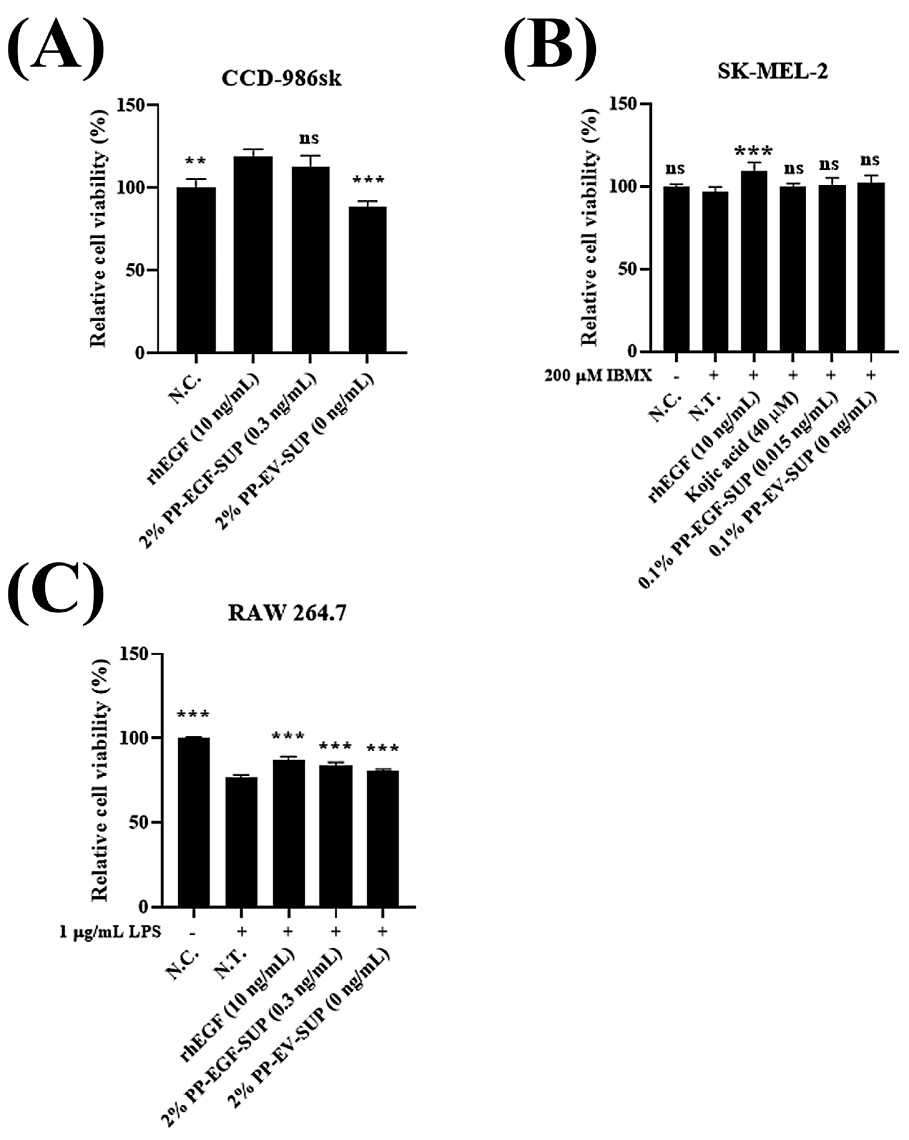
To evaluate the toxicity of PP-EGF-SUP to each cell line used in the bioactivity experiments, we investigated the optimal dose for treatments. Regarding the fibroblast (CCD-986sk) cell line, PP-EV-SUP showed minor toxicity to the cells, whereas both rhEGF and PP-EGF-SUP slightly induced cell proliferation (Figure 2A). This result suggests that the PP-EGF-SUP-treated group (EGF protein: 0.3 ng/mL in 2% treatment) showed similar recovery to the rhEGF-treated group. Regarding the skin melanoma (SK-MEL-2) cell line, although treatments with more than 0.1% PP-EGF-SUP (EGF protein: 15 pg/mL in 0.1% treatment) showed toxicity, this concentration did not support cell proliferation (Figure 2B). Additionally, the positive substance (kojic acid: 40 μM) used in tyrosinase inhibition assays did not affect cell viability. Regarding the RAW 264.7 cell line, all substances (Figure 2C) showed toxicity (~23%), but it did not look serious enough to perform further experiments.

3.2.2. EGF-Derived Bioactivities: Cell Scratch Wound Healing, Tyrosinase Inhibition, and Skin Barrier Improvement (Elastin Expression Stimulation and Collagenase and Elastase Suppression)

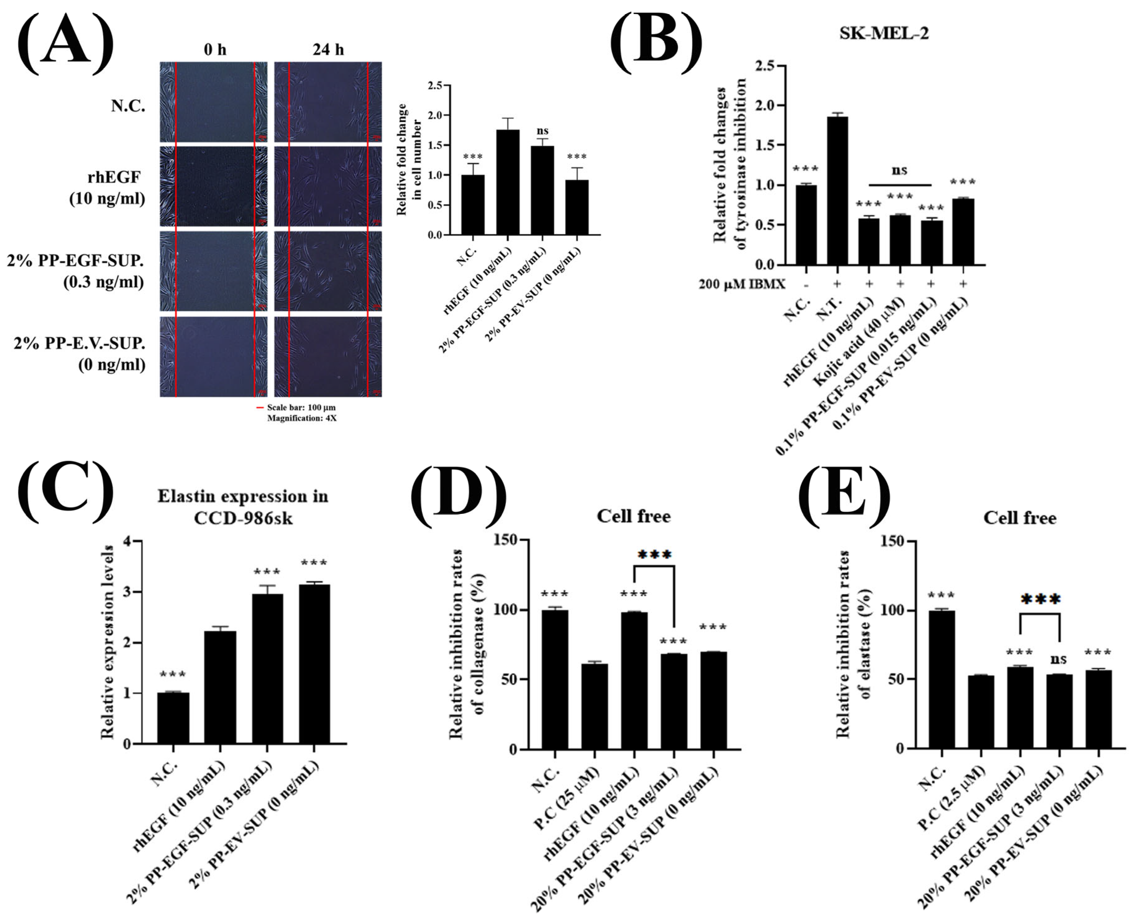
Using the CCD-986sk cell line, the EGF-mediated cell scratch wound healing activity of PP-EGF-SUP was compared with that of rhEGF (10 ng/mL, as a positive control) and PP-EV-SUP (2%, as a negative control). By comparing the cell number in each test group (n = 5 images), it was found that the rhEGF (10 ng/mL)-treated group showed an approximate 1.8-fold increase in the cell regeneration rate compared to that of the untreated group. However, the PP-EGF-SUP (2%) treatment exhibited slightly lower cell scratch wound healing activity (1.5-fold) than the rhEGF treatment, although the actual EGF protein content in PP-EGF-SUP was 33-fold lower than that in rhEGF. Additionally, PP-EV-SUP did not demonstrate cell scratch wound healing activity at the same concentration (Figure 3A).
We next investigated the effect of PP-EGF-SUP on whitening by examining tyrosinase expression in the SK-MEL-2 cell line. Tyrosinase is involved in the synthesis of brown-to-black melanin, which causes skin pigmentation such as spots and freckles, and it is known that suppressing tyrosinase expression is effective in skin whitening [27]. 3-Isobutyl-1-methylxanthine (IBMX; 200 μM), which induces tyrosinase expression in the SK-MEL-2 cell line, was applied as treatment, and it significantly increased tyrosinase levels. Kojic acid (40 μM) was used as a positive control. Both rhEGF (10 ng/mL) and PP-EGF-SUP (0.1%) treatment showed tyrosinase inhibition efficacy similar to that of the kojic acid treatment. Additionally, PP-EV-SUP (0.1%) also slightly reduced tyrosinase activity (Figure 3B). This result indicates that the EGF protein content in PP-EGF-SUP (with an actual EGF protein concentration of 0.0015 ng/mL) was very low compared to that in rhEGF; the actual EGF protein content in PP-EGF-SUP was 666-fold lower than that in rhEGF. PP-EGF-SUP showed tyrosinase inhibition activity similar to that of rhEGF and kojic acid. PP-EV-SUP also inhibited tyrosinase activity. We concluded that the strong whitening effect of PP-EGF-SUP was caused by the combination of EGF and probiotic-mediated metabolites, as well as strong synergistic effects.
We next investigated the effect of PP-EGF-SUP-derived anti-wrinkle factors, such as the elastin expression level, collagenase inhibition rate, and elastase inhibition rate, in the CCD-986sk cell line. Elastin, which is composed of elastic fibers, is involved in the flexibility, elasticity, and resilience of skin tissue. Moreover, elastin is also present between collagen fibers, and it plays a role in binding and supporting collagen to prevent it from escaping. To evaluate the wrinkle-improving efficacy of PP-EGF-SUP, the elastin expression in CCD-986sk cells was investigated using qRT-PCR. Compared to the untreated group, elastin expression significantly increased by ~1.7 times in the rhEGF (10 ng/mL)-treated group. Elastin expression increased by up to 2.95 and 3.14 times in the PP-EGF-SUP (2%)- and PP-EV-SUP (2%)-treated groups, respectively. Although an EGF-mediated increase in elastin expression was confirmed, the probiotics seemed more effective in improving wrinkles because PP-EGF-SUP and PP-EV-SUP showed similar activity, and it was stronger than that of rhEGF (Figure 3C).
Collagen acts as a pillar to firmly support the skin, and it effectively improves skin elasticity and wrinkles, thus determining the mechanical properties of connective tissue. However, collagenase breaks down collagen in the body, thereby reducing the elasticity and moisture of skin tissue and causing wrinkles.
Therefore, an experiment was conducted to confirm collagenase inhibition properties using a cell-free collagenase inhibition assay. Better effects in improving wrinkles by reducing collagenase activity were detected in PP-EGF-SUP (20%); PP-EV-SUP (20%); and 1,10-phenanthroline monohydrate (25 µM), which was used as a positive control. However, rhEGF did not inhibit collagenase activity (Figure 3D). As a result, the positive control was confirmed to reduce collagenase activity, and both PP-EGF-SUP and PP-EV-SUP also decreased collagenase activity in a dose-dependent manner.
Elastase breaks down elastin in elastic fibers. As the inhibition of elastase activity could support improvements in skin elasticity and wrinkles, a cell-free elastase inhibition assay was conducted to confirm the elastase inhibition properties. MeOSuc-AAPV-CMK (2.5 µM), which was used as a positive control, was confirmed to reduce elastase activity by up to ~50%, and rhEGF exhibited up to ~40% inhibition efficacy. Although the EGF protein in PP-EGF-SUP (20%, with an actual EGF protein content of 3 ng/mL) was 3-fold lower than that in rhEGF, PP-EGF-SUP (20% treatment) showed strong activity in a dose-dependent manner similar to that of the positive control and rhEGF (Figure 3E). However, PP-EV-SUP also exhibited similar activity. Thus, the anti-elastase property of PP-EGF-SUP might originate from probiotic bioactivity.

3.2.3. Probiotic-Derived Bioproperties: Antioxidant, Anti-Inflammatory, and Anti-Bacterial Activities

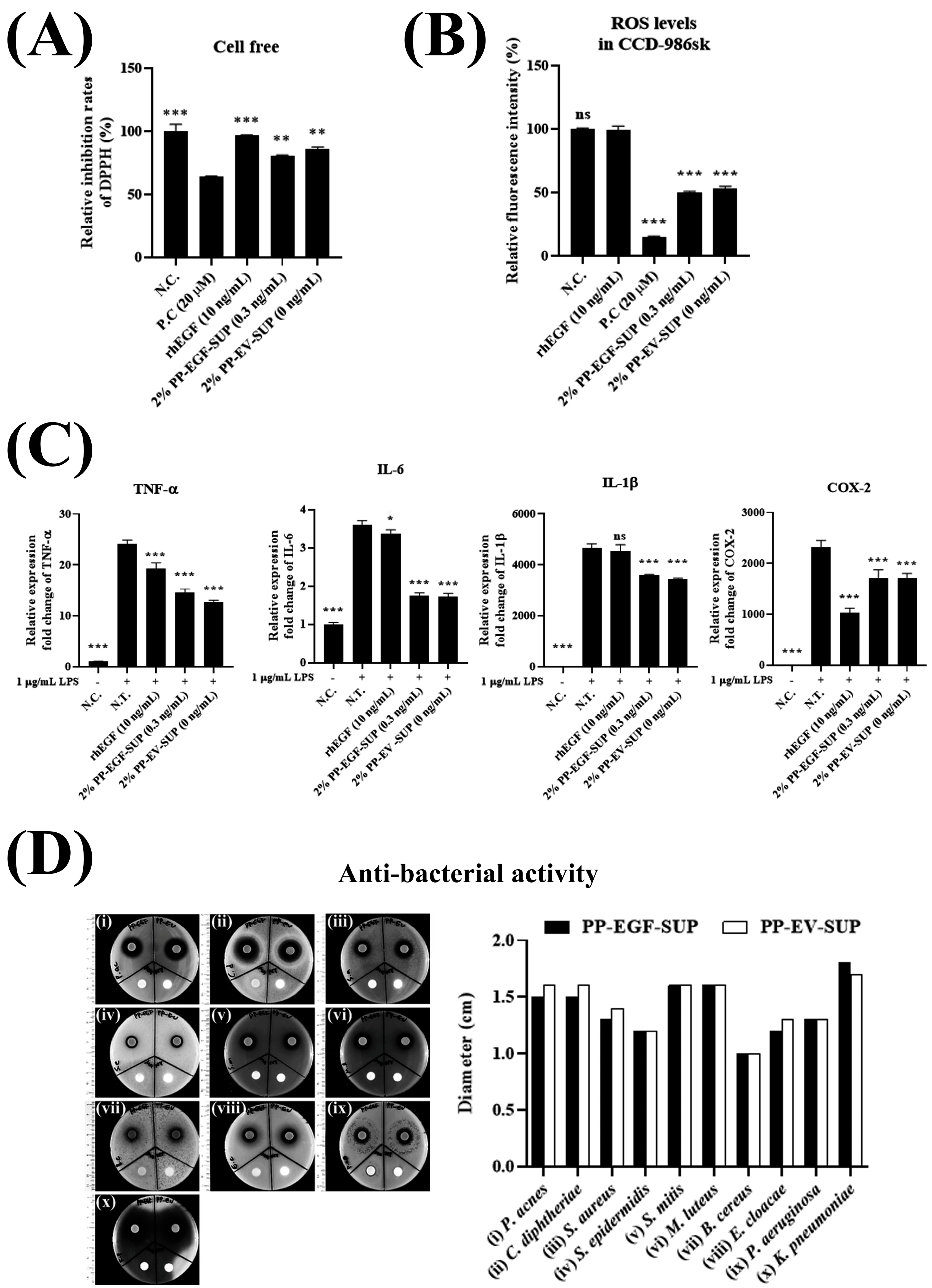
To investigate the probiotic-mediated antioxidant activity of PP-EGF-SUP, the antioxidant efficacy of each sample was evaluated by examining the inhibition of DPPH. Trolox (5, 10, and 20 nmoles/well) was used as a positive control and applied as treatment, and then the DPPH inhibition rate was calculated in a dose-dependent manner. Both PP-EV-SUP and PP-EGF-SUP showed outstanding efficacy, similar to that of the positive control group, in a dose-dependent manner (Figure 4A). However, rhEGF did not show outstanding efficacy. Taken together, the results suggest that the DPPH inhibition activity of both PP-EV-SUP and PP-EGF-SUP originated from the probiotics, and they were thus determined to be a synergistic partnership with excellent antioxidant efficacy that complements the low antioxidant efficacy of EGF.
Furthermore, the reactive oxygen species (ROS) scavenging properties of PP-EGF-SUP were evaluated in the CCD-986sk cell line. The ROS levels in the cells significantly increased following oxidant treatment (1,1-Dimethylethyl hydroperoxide: TBHP). L-Ascorbic acid, which was used as a positive control, was confirmed to reduce ROS activity by up to ~85%, and both PP-EGF-SUP and PP-EV-SUP significantly reduced ROS levels by up to ~50% and ~47%, respectively. As rhEGF alone did not reduce ROS activity, no differences were observed in the ROS scavenging activity between PP-EGF-SUP and PP-EV-SUP (Figure 4B). Taken together, the results in Figure 4A,B suggest that, although the mechanism of the antioxidant activity of PP-EGF-SUP remains unclear, PP-EGF-SUP may contain some postbiotic-derived antioxidant substances.
It is well known that probiotics and their culture sup. exhibit anti-inflammatory activity [28]. Inflammation was successfully induced in RAW 264.7 cells by LPS (1 μg/mL) treatment, with all inflammatory-related markers significantly increasing compared with those in the non-treated group (Figure 4C). After treatment with both PP-EGF-SUP and PP-EV-SUP, the expression of all inflammatory markers significantly reduced in a dose-dependent manner compared with that in the non-treated (N.T.) group. Although high EGF concentrations were used, rhEGF had low efficacy compared with both PP-EGF-SUP and PP-EV-SUP, except for on COX-2. However, PP-EGF-SUP and PP-EV-SUP showed no difference in efficacy. Based on the rhEGF results, we carefully conclude that EGF does not seem to have strong anti-inflammatory activity. Additionally, the EGF protein content in PP-EGF-SUP was not sufficient to change marker protein expression levels (Figure 4C). Therefore, the anti-inflammatory effects might originate from the probiotics.
It is well known that probiotics and their culture supernatant exhibit anti-bacterial activity [29]. The anti-bacterial activity of PP-EGF-SUP was evaluated using ten strains of harmful skin bacteria through the disk diffusion method. PP-EGF-SUP and PP-EV-SUP showed similar anti-bacterial effects on all bacterial strains, although each strain showed different levels of susceptibility (Figure 4D). Strong susceptibility was observed in Klebsiella pneumoniae. Significant efficacy was observed in Propionibacterium acnes, Corynebacterium diphtheria, Streptococcus mitis, and Micrococcus luteus. The other strains showed minor susceptibility. This anti-bacterial activity might originate from the probiotics’ bioproperties. Moreover, PP-EGF-SUP seemed to exhibit broad-spectrum anti-bacterial activity, as susceptibility was observed in some Gram-positive and -negative organisms.

3.2.4. Evaluation of PP-EGF-DP as a Cosmetic Ingredient

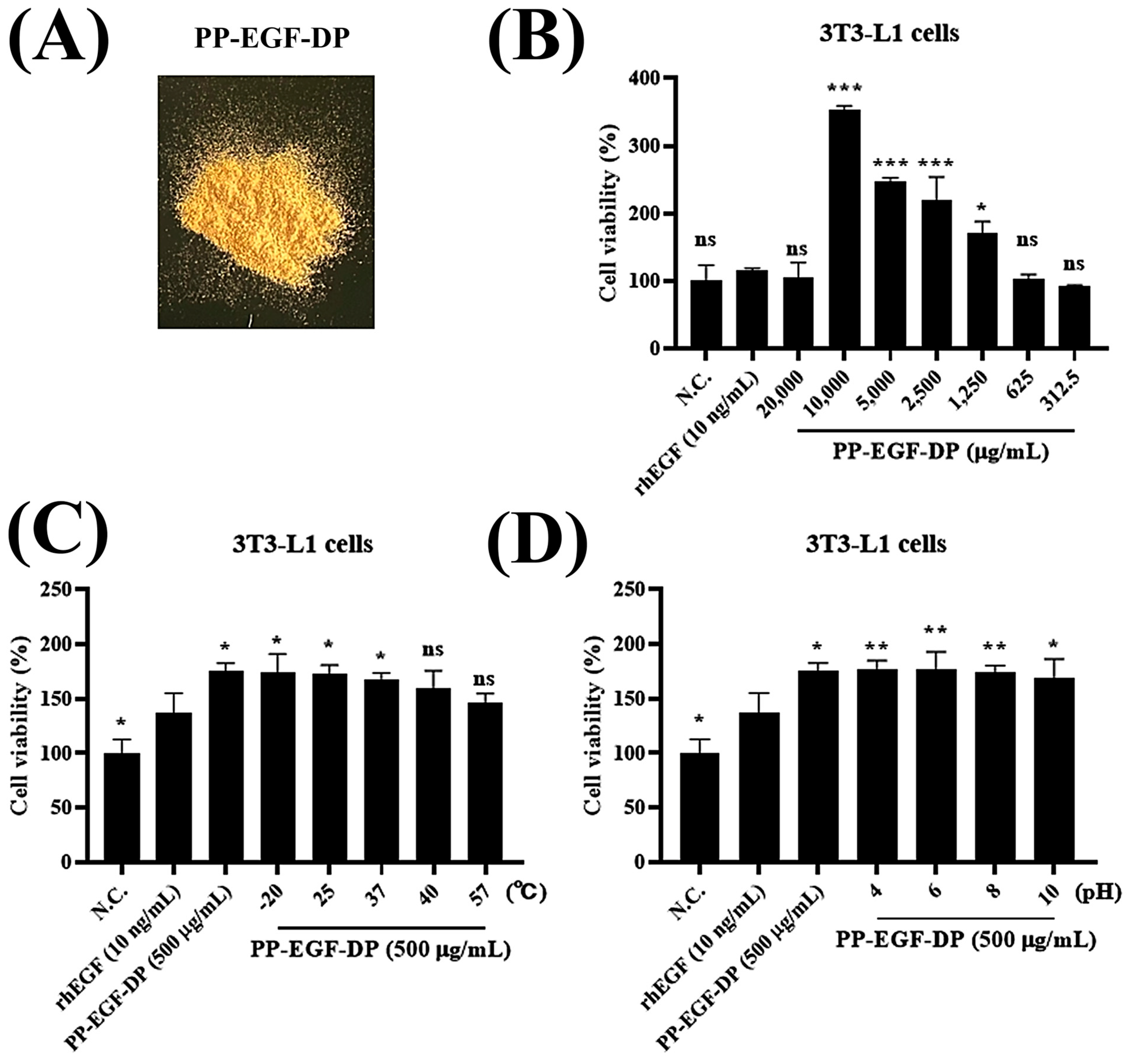
To investigate stabilization methods for PP-EGF-SUP as a cosmetic ingredient exhibiting biological activity, we optimized its immobilization with 5% maltodextrin via freeze-drying. Through the PP-EGF-SUP powdering procedure, a brown powder was obtained (Figure 5A), and then its stimulatory effect on 3T3-L1 cell proliferation was evaluated at various concentrations. The results showed that, compared with the control, 10 mg/mL of PP-EGF-DP (with an actual EGF concentration of 1.6448 ng/mL) increased 3T3-L1 cell proliferation by up to 350%, whereas rhEGF (10 ng/mL) only increased it by up to 20% (Figure 5B). However, at a concentration of 20 mg/mL, PP-EGF-DP lost its stimulatory properties. Next, we evaluated its stability by examining its stimulatory effects on 3T3-L1 cell proliferation after exposure to a wide range of temperatures and pHs. PP-EGF-DP (500 μg/mL) maintained up to ~83% of its activity compared with non-treated PP-EGF-DP, even after exposure to 57 °C for 1 h (Figure 5C). Additionally, PP-EGF-DP (500 μg/mL) maintained up to ~97% of its activity compared with non-treated PP-EGF-DP, even after exposure to pH 10 for 1 h (Figure 5D). PP-EGF-DP still exhibited strong stability under extreme conditions, even when it was resuspended in media.

3.2.5. Evaluation of Triple-Freckle (Light Freckles, Dark Freckles, and Freckle Area) Relief Using Antera 3D®CS

To evaluate the melasma improvement efficacy of the PP-EGF-DP freckle ampoules, clinical changes were assessed using the Antera 3D® CS system over a 4-week application period. Both 1% and 5% PP-EGF-DP ampoules were tested on the left side of the face, while placebo ampoules were applied to the right side of the face.
Light Freckles (Minimum Value): After 4 weeks of use, the minimum value corresponding to light freckles was statistically significantly reduced by 3.38% in the 1% PP-EGF group (p < 0.05) and 3.79% in the 5% PP-EGF group (p < 0.05), whereas no statistically significant changes were observed in the respective placebo groups (Table 2).
Dark Freckles (Maximum Value): The maximum value corresponding to dark freckles was statistically significantly reduced by 1.74% in the 1% PP-EGF-DP group (p < 0.05) and 2.93% in the 5% PP-EGF-DP group (p < 0.05) after 4 weeks. Again, the placebo groups showed no significant differences (Table 2).
Freckle Area (Affected Area): The affected area representing the total freckle coverage significantly decreased by 21.21% in the 1% PP-EGF-DP group and by 29.10% in the 5% PP-EGF-DP group after 4 weeks (p < 0.05). In contrast, the placebo groups showed minimal changes (1%: 2.79%; 5%: 5.93%) and no statistical significance (Table 2).
In a post-study survey, 100% of the participants who used either the 1% or 5% PP-EGF ampoules for 4 weeks reported positive results in all of the following categories: (1) the lightening of light freckles, (2) a reduction in dark freckles, (3) a decrease in overall freckle area, (4) improvements in skin moisture and softness, (4) satisfaction with product spreadability and fragrance, and (5) overall satisfaction with the ampoule. Furthermore, no serious dermatological adverse events related to the ampoules were observed during the test period for either concentration. Both formulations were well tolerated. Therefore, the 1% and 5% PP-EGF-DP freckle ampoules were deemed appropriate for application.

4. Discussion

Growth factors, including EGF, are naturally expressed proteins that stimulate cell proliferation, differentiation, and migration and collagen synthesis in the skin through receptor-mediated signaling cascades [30,31]. These properties accelerate the cell scratch wound healing process and prevent skin aging [32]. However, it should be noted that the proliferative and anti-apoptotic effects of EGF could contribute to enriching the population of genetically damaged cells [33].
The Escherichia coli expression system with recombinant DNA technology is the most commonly used approach for producing recombinant EGF, as it provides it in large amounts [34,35]. However, the high expression rate of the E. coli system often leads to the formation of inclusion bodies by misfolded insoluble aggregates through hydrophobic interactions [36]. Additionally, Gram-negative bacteria host-derived lipopolysaccharide (LPS, known as an endotoxin) can induce a pyrogenic response and ultimately trigger septic shock [37]. LPS must be completely removed from purified EGF before it can be safely used. Other EGF expression systems have been developed using different hosts, including Pichia pastoris [38], Saccharomyces cerevisiae [39], transgenic plants [40], and mammalian cells [41]. As metabolites other than those of EGF are considered contaminants, the purification procedure has been basically considered in previous studies.
However, none of these studies have rejected the notion that the proliferative and anti-apoptotic effects of EGF could contribute to enriching the population of genetically damaged cells.
Probiotics confer various health benefits to the host, and safety issues have not been found; therefore, they have a wide application range and are unique in type, scope, and application [42]. Additionally, it is projected that the cosmetic market for probiotics will grow at a 12% rate in the next ten years [43].
In this study, a novel probiotic-derived potential cosmetic ingredient containing EGF was developed, and its differences to the commercial rhEGF currently being marketed were examined. EGF facilitates cell scratch wound healing by stimulating the resurfacing of the damaged epidermis and inducing granulation tissue outgrowth, angiogenesis, and wound contraction [44,45]. Additionally, elastin and collagen, which are both formed by fibroblasts, are the main fibers that form the extracellular matrix (skin barrier). Collagen is responsible for tensile strength, and elastin provides elasticity to the skin. The production and density of both decrease as a function of age, resulting in sagging and wrinkling [46]. We demonstrated that PP-EGF, synthesized by P. pentosaceus SL4 (probiotics), exhibited EGF-derived bioactivities such as cell scratch wound healing, tyrosinase inhibition, and skin barrier improvement (elastin expression stimulation and collagenase and elastase suppression) (Figure 3). We found that the EGF-derived properties exerted similar efficacy even when the actual EGF protein content in PP-EGF-SUP was lower than that in rhEGF, which was used as a positive control. The results showed that PP-EGF-SUP demonstrated EGF-mediated biological activity similar to that of rhEGF even when its actual EGF protein content was a minimum of 30-fold lower. This outstanding activity might be explained by the synergy between EGF and probiotic-derived metabolites. We believe that probiotic-derived metabolites might contribute to the EGF-derived effects.
Furthermore, we found that the probiotic-derived properties exhibited a higher efficacy even when the actual EGF content in PP-EGF-SUP was lower than that in rhEGF, which was used as a positive control. Additionally, PP-EGF-SUP had not only EGF-derived properties but also probiotic-derived properties, such as antioxidant, anti-inflammatory, and anti-bacterial activities (Figure 4B). In fact, it is well known that probiotic culture supernatant has antioxidant and anti-inflammatory activities [47,48].
Furthermore, the antimicrobial elements secreted by probiotics can be called postbiotics, comprising several bioactive compounds such as organic acids, short-chain fatty acids, carbohydrates, antimicrobial peptides, enzymes, vitamins, cofactors, immune-signaling compounds, and complex agents [49,50]. In fact, PP-EGF-SUP exhibited strong broad-spectrum anti-bacterial properties (Figure 4D). Klebsiella pneumoniae is a Gram-negative anaerobic rod normally found in the oral cavity, skin, and small intestine. It causes infections in various parts of the body, including liver abscesses, urinary tract infections, pneumonia, endophthalmitis, and peritonitis, and it is characterized by frequent bacteremia and metastatic lesions [51,52]. Propionibacterium acnes, a Gram-positive human skin commensal bacterium, is involved in the pathogenesis of acne. Acne is well known to cause various psychosocial issues, such as decreased self-esteem, depression, frustration, and social withdrawal [53]. Corynebacterium diphtheriae, a Gram-positive pathogenic bacterium, causes an epidemic acute bacterial disease named diphtheria. Diphtheria is caused by the adhesion and infiltration of the bacteria into the mucosal layers of the body, primarily affecting the respiratory tract and subsequently releasing an exotoxin that negatively affects components of the upper respiratory tract, such as the tonsils, nasal mucosa, larynx, and pharynx. Less often, it can affect the skin, eye conjunctiva, and vagina [54]. Thus, PP-EGF-SUP also significantly suppresses both properties.
Taken together, the results clearly indicate that PP-EGF-SUP shows promise as a cosmetic ingredient in skin anti-aging agents because it demonstrates bioactivities derived not only from EGF but also from postbiotics. As a purification process is not required for PP-EGF-SUP, it has a competitive production price in the cosmetic industry. Based on these factors, PP-EGF-DP was registered as an International Nomenclature Cosmetic Ingredient (INCI) named “Pediococcus/sh-Oligopeptide-1 Ferment Extract Filtrate (Mono ID: 39676)”.
Although this study has limitations, as only cell line-based in vitro experiments and biochemical evaluations were carried out, the clinical relevance of PP-EGF-DP was validated through a human application test focused on melasma improvement. In an Antera 3D® CS analysis, PP-EGF-DP ampoules at both 1% and 5% concentrations demonstrated statistically significant efficacy in improving light freckles, dark freckles, and freckle area coverage over a 4-week period compared to a placebo. Specifically, the 5% PP-EGF-DP formulation resulted in a 3.79% reduction in light freckle intensity, a 2.93% reduction in dark freckle intensity, and a remarkable 29.10% decrease in the freckle-covered area. Even the lower 1% concentration showed considerable improvements (3.38%, 1.74%, and 21.21%, respectively) (Table 2). These outcomes highlight that PP-EGF-DP not only mimics the biological activity of rhEGF in vitro but also exhibits tangible, dose-dependent skin benefits in clinical settings.
Interestingly, the placebo groups showed minimal to no improvement, indicating that the observed effects are attributable to PP-EGF-DP itself rather than the base formulation. This reinforces the hypothesis that probiotic-derived EGF possesses both EGF- and probiotic/postbiotic-derived bioactivities contributing to skin depigmentation. These include melanogenesis suppression via tyrosinase inhibition, antioxidant protection, and possible anti-inflammatory mechanisms that can mitigate pigment aggravation. Collectively, these data validate PP-EGF-DP’s dual mechanism of action and its potential as a safe, effective, and multifunctional cosmetic ingredient targeting hyperpigmentation.

Supplementary Materials

The following supporting information can be downloaded at: https://www.mdpi.com/article/10.3390/cosmetics12040176/s1. Figure S1: Ampoule manufacturing process; Table S1: List of primers; Table S2: Ingredients of prepared ampoule.

Author Contributions

J.Y.A., S.K. and Y.J.R.: in vitro assay and data analysis; J.H. and Y.R.: construction of PP-EGF plasmid and generation of PP-EGF transformant; M.J.C. and K.Y.P.: conceptualization; B.C.A.: supervisor of the study. All authors have read and agreed to the published version of the manuscript.

Funding

This research was supported by a grant received from the Korea Health Technology R&D Project through the Korea Health Industry Development Institute (KHIDI), funded by the Ministry of Health & Welfare, Republic of Korea (grant number: H12300860).

Institutional Review Board Statement

This study was conducted in accordance with the Declaration of Helsinki and approved by the Institutional Review Board of OATC IRB (IRB approval no.: 2018071702-2411-HR-487-01, dated 14 November 2024).

Informed Consent Statement

Informed consent was obtained from all subjects involved in the study.

Data Availability Statement

No datasets were generated or analyzed during the current study.

Acknowledgments

The authors acknowledge Honest co.kr. for the PP-EGF-DP ampoule.

Conflicts of Interest

Authors J.Y.A., S.K., J.H., Y.R., M.J.C. and B.C.A. were employed by the company Cell Biotech. The remaining authors declare that the research was conducted in the absence of any commercial or financial relationships that could be construed as a potential conflict of interest.

Abbreviations

EGF: epidermal growth factor; rhEGF: recombinant human EGF; PP-EGF-SUP: PP-EGF cell-free supernatant; PP-EGF-DP: PP-EGF-SUP dried power; IGF-1: insulin-like growth factor 1; FGF-1: fibroblast growth factor 1; LPS: lipopolysaccharide; ROS: reactive oxygen species; TCA: trichloroacetic acid; PBS: phosphate-buffered saline; DPPH: 2,2-diphenyl-1-picrylhydrazyl; BL media: blood liver media; cDNA: complementary DNA; PP: Pediococcus pentosaceus SL4; PP-EV-SUP: PP-empty vector cell-free supernatant; MRS media: De Man, Rogosa, and Sharpe media; IMDM: Iscove’s Modified Dulbecco’s Medium; FBS: fetal bovine serum; DMEM: Dulbecco’s Modified Eagle Medium; BCS: Bovine calf serum; DDS: drug delivery system; P.C.: positive control; N.T.: non-treated.

References

  1. Cohen, S. Isolation of a mouse submaxillary gland protein accelerating incisor eruption and eyelid opening in the new-born animal. J. Biol. Chem. 1962, 237, 1555–1562. [Google Scholar] [CrossRef] [PubMed]
  2. Wenczak, B.A.; Lynch, J.B.; Nanney, L.B. Epidermal growth factor receptor distribution in burn wounds. Implications for growth factor-mediated repair. J. Clin. Investig. 1992, 90, 2392–2401. [Google Scholar] [CrossRef] [PubMed]
  3. Brake, A.J.; Merryweather, J.P.; Coit, D.G.; Heberlein, U.A.; Masiarz, F.R.; Mullenbach, G.T.; Urdea, M.S.; Valenzuela, P.; Barr, P.J. Alpha-Factor directed synthesis and secretion of mature foreign proteins in Saccharomyces cerevisiae. Proc. Natl. Acad. Sci. USA 1984, 81, 4642–4646. [Google Scholar] [CrossRef]
  4. Hong, J.P.; Jung, H.D.; Kim, Y.W. Recombinant human epidermal growth factor to enhance healing for diabetic foot ulcers. Ann. Plast. Surg. 2006, 56, 394–398. [Google Scholar] [CrossRef]
  5. Tanaka, A.; Nagate, T.; Matsuda, H. Acceleration of wound healing by gelatin film dressings with epidermal growth factor. J. Vet. Med. Sci. 2005, 67, 909–913. [Google Scholar] [CrossRef]
  6. Yang, S.; Geng, Z.; Ma, K.; Sun, X.; Fu, X. Efficacy of topical recombinant human epidermal growth factor for treatment of diabetic foot ulcer: A systematic review and meta-analysis. Int. J. Low. Extrem. Wounds 2016, 15, 120–125. [Google Scholar] [CrossRef]
  7. Kim, J.; Kim, Y.; Joo, K. Effects of recombinant human epidermal growth factor (rhEGF) on the recovery of skin barrier function after laser resurfacing. Dermatol. Surg. 2020, 46, 252–260. [Google Scholar]
  8. Noordam, R.; Gunn, D.A.; Tomlin, C.C.; Maier, A.B.; Griffiths, T.; Catt, S.D.; Ogden, S.; Slagboom, P.E.; Westendorp, R.G.; Griffiths, C.E.; et al. Leiden Longevity Study group. Serum insulin-like growth factor 1 and facial ageing: High levels associate with reduced skin wrinkling in a cross-sectional study. Br. J. Dermatol. 2013, 168, 533–538. [Google Scholar] [CrossRef] [PubMed]
  9. Schmidt, M.O.; Garman, K.A.; Lee, Y.G.; Zuo, C.; Beck, P.J.; Tan, M.; Agui-lar-Pimentel, J.A.; Ollert, M.; Schmidt-Weber, C.; Fuchs, H.; et al. The Role of Fibro-blast Growth Factor-Binding Protein 1 in Skin Carcinogenesis and Inflammation. J. Investig. Dermatol. 2018, 138, 179–188. [Google Scholar] [CrossRef]
  10. Dogan, S.; Demirer, S.; Kepenekci, I.; Erkek, B.; Kiziltay, A.; Hasirci, N.; Müftüoglu, S.; Nazikoglu, A.; Renda, N.; Dincer, U.D.; et al. Epidermal growth factor-containing wound closure enhances wound healing in non-diabetic and diabetic rats. Int. Wound J. 2009, 6, 107–115. [Google Scholar] [CrossRef]
  11. Krishnamurthy, R.; Manning, M.C. The stability factor: Importance in formulation development. Curr. Pharm. Biotechnol. 2002, 3, 361–371. [Google Scholar] [CrossRef]
  12. Ogiso, H.; Ishitani, R.; Nureki, O.; Fukai, S.; Yamanaka, M.; Kim, J.H.; Saito, K.; Sakamoto, A.; Inoue, M.; Shirouzu, M.; et al. Crystal structure of the complex of human epidermal growth factor and receptor extracellular domains. Cell 2002, 110, 775–787. [Google Scholar] [CrossRef]
  13. Choi, S.M.; Lee, K.M.; Kim, H.J.; Park, I.K.; Kang, H.J.; Shin, H.C.; Baek, D.; Choi, Y.; Park, K.H.; Lee, J.W. Effects of structurally stabilized EGF and bFGF on wound healing in type I and type II diabetic mice. Acta Biomater. 2018, 66, 325–334. [Google Scholar] [CrossRef]
  14. Li, S.; Liu, Y.; Huang, Z.; Kou, Y.; Hu, A. Efficacy and safety of nano-silver dressings combined with recombinant human epidermal growth factor for deep second-degree burns: A meta-analysis. Burns 2021, 47, 643–653. [Google Scholar] [CrossRef]
  15. Koppa Raghu, P.; Bansal, K.K.; Thakor, P.; Bhavana, V.; Madan, J.; Rosenholm, J.M.; Mehra, N.K. Evolution of nanotechnology in delivering drugs to eyes, skin and wounds via topical route. Pharmaceuticals 2020, 13, 167. [Google Scholar] [CrossRef]
  16. Choi, J.H.; Nam, S.H.; Song, Y.S.; Lee, H.W.; Lee, H.J.; Song, K.; Hong, J.W.; Kim, G.C. Treatment with low-temperature atmospheric pressure plasma enhances cutaneous delivery of epidermal growth factor by regulating E-cadherin-mediated cell junctions. Arch. Dermatol. Res. 2014, 306, 635–643. [Google Scholar] [CrossRef]
  17. Kim, J.; Jang, J.H.; Lee, J.H.; Choi, J.K.; Park, W.R.; Bae, I.H.; Bae, J.; Park, J.W. Enhanced topical delivery of small hydrophilic or lipophilic active agents and epidermal growth factor by fractional radiofrequency microporation. Pharm. Res. 2012, 29, 2017–2029. [Google Scholar] [CrossRef]
  18. Berlanga-Acosta, J.; Gavilondo-Cowley, J.; Barco-Herrera, D.G.; Martín-Machado, J.; Guillén-Nieto, G. Epidermal growth factor (EGF) and platelet-derived growth factor (PDGF) as tissue healing agents: Clarifying concerns about their possible role in malignant transformation and tumor progression. J. Carcinogene. Mutagene. 2011, 2, 100–115. [Google Scholar] [CrossRef]
  19. Shin, S.H.; Koh, Y.G.; Lee, W.G.; Seok, J.; Park, K.Y. The use of epidermal growth factor in dermatological practice. Int. Wound J. 2023, 20, 2414–2423. [Google Scholar] [CrossRef]
  20. Indira, M.; Venkateswarulu, T.C.; Abraham Peele, K.; Nazneen Bobby, M.; Krupanidhi, S. Bioactive molecules of probiotic bacteria and their mechanism of action: A review. 3 Biotech 2019, 9, 306. [Google Scholar] [CrossRef]
  21. Kodali, V.P.; Sen, R. Antioxidant and free radical scavenging activities of an exopolysaccharide from a probiotic bacterium. Biotechnol. J. 2008, 3, 245–251. [Google Scholar] [CrossRef]
  22. An, B.C.; Ryu, Y.; Yoon, Y.S.; Choi, O.; Park, H.J.; Kim, T.Y.; Kim, S.I.; Kim, B.K.; Chung, M.J. Colorectal Cancer Therapy Using a Pediococcus pentosaceus SL4 Drug Delivery System Secreting Lactic Acid Bacteria-Derived Protein p8. Mol. Cells 2019, 42, 755–762. [Google Scholar] [PubMed]
  23. Grinberg, I.; West, D.V.; Torres, M.; Gou, G.; Stein, D.M.; Wu, L.; Chen, G.; Gallo, E.M.; Akbashev, A.R.; Davies, P.K.; et al. Perovskite oxides for visible-light-absorbing ferroelectric and photovoltaic materials. Nature 2013, 503, 509–512. [Google Scholar] [CrossRef]
  24. Ngamsurach, P.; Praipipat, P. Antibacterial activities against Staphylococcus aureus and Escherichia coli of extracted Piper betle leaf materials by disc diffusion assay and batch experiments. RSC Adv. 2022, 12, 26435–26454. [Google Scholar] [CrossRef]
  25. Mamat, U.; Wilke, K.; Bramhill, D.; Schromm, A.B.; Lindner, B.; Kohl, T.A.; Corchero, J.L.; Villaverde, A.; Schaffer, L.; Head, S.R.; et al. Detoxifying Escherichia coli for endotoxin-free production of recombinant proteins. Microb. Cell Fact. 2015, 14, 57. [Google Scholar] [CrossRef]
  26. Gasaly, N.; de Vos, P.; Hermoso, M.A. Impact of bacterial metabolites on gut barrier function and host immunity: A focus on bacterial metabolism and its relevance for intestinal inflammation. Front. Immunol. 2021, 12, 658354. [Google Scholar] [CrossRef] [PubMed]
  27. Gillbro, J.M.; Olsson, M.J. The melanogenesis and mechanisms of skin-lightening agents--existing and new approaches. Int. J. Cosmet. Sci. 2011, 33, 210–221. [Google Scholar] [CrossRef] [PubMed]
  28. Cristofori, F.; Dargenio, V.N.; Dargenio, C.; Miniello, V.L.; Barone, M.; Francavilla, R. Anti-inflammatory and immunomodulatory effects of probiotics in gut inflammation: A door to the body. Front. Immunol. 2021, 12, 578386. [Google Scholar] [CrossRef]
  29. Fijan, S. Probiotics and Their Antimicrobial Effect. Microorganisms 2023, 11, 528. [Google Scholar] [CrossRef]
  30. Barrientos, S.; Stojadinovic, O.; Golinko, M.S.; Brem, H.; Tomic-Canic, M. Growth factors and cytokines in wound healing. Wound Repair Regen. 2008, 16, 585–601. [Google Scholar] [CrossRef]
  31. Nanba, D.; Hata, M.; Higashiyama, S. The EGFR ligands and their signaling networks in epithelial cell biology and cancer: Roles in skin homeostasis and therapeutic implications. Cancer Sci. 2013, 104, 419–426. [Google Scholar]
  32. Sriwidodo, S.; Maksum, I.P.; Subroto, T.; Wathoni, N.; Subarnas, A.; Umar, A.K. Activity and effectiveness of recombinant rhEGF excreted by Escherichia coli BL21 on wound healing in induced diabetic mice. J. Exp. Pharmacol. 2020, 12, 339–348. [Google Scholar] [CrossRef] [PubMed]
  33. Berlanga-Acosta, J.; Gavilondo-Cowley, J.; López-Saura, P.; González-López, T.; Castro-Santana, M.D.; López-Mola, E.; Guillén-Nieto, G.; Herrera-Martinez, L. Epidermal growth factor in clinical practice—A review of its biological actions, clinical indications and safety implications. Int. Wound J. 2009, 6, 331–346. [Google Scholar] [CrossRef]
  34. Indriyani, A.; Indrayati, N.; Sriwidodo, S.; Maksum, I. Optimization extracellular secretion of recombinant human epidermal growth factor (rhEGF) in Escherichia coli BL21 (DE3) pD881-OmpA-rhEGF by using response surface method (RSM). Int. J. Res. Pharm. Sci. 2019, 10, 1824–1831. [Google Scholar] [CrossRef]
  35. Shen, W.R.; Liew, M.W.O.; Ong, E. Production of recombinant human epidermal growth factor in Escherichia coli: Strategic upstream and downstream considerations for high protein yield. Process Biochem. 2024, 146, 81–96. [Google Scholar]
  36. de Groot, N.S.; Espargarö, A.; Morell, M.; Ventura, S. Studies on bacterial inclusion bodies. Future Microbiol. 2008, 3, 423–435. [Google Scholar] [CrossRef] [PubMed]
  37. Wakelin, S.J.; Sabroe, I.; Gregory, C.D.; Poxton, I.R.; Forsythe, J.L.; Garden, O.J.; Howie, S.E. “Dirty little secrets”—Endotoxin contamination of recombinant proteins. Immunol. Lett. 2006, 106, 1–7. [Google Scholar] [CrossRef] [PubMed]
  38. Eissazadeh, S.; Moeini, H.; Dezfouli, M.G.; Heidary, S.; Nelofer, R.; Abdullah, M.P. Production of recombinant human epidermal growth factor in Pichia pastoris. Braz. J. Microbiol. 2017, 48, 286–293. [Google Scholar] [CrossRef]
  39. George-Nascimento, C.; Gyenes, A.; Halloran, S.M.; Merryweather, J.; Valenzuela, P.; Steimer, K.S.; Masiarz, F.R.; Randolph, A. Characterization of recombinant human epidermal growth factor produced in yeast. Biochemistry 1988, 27, 797–802. [Google Scholar] [CrossRef]
  40. He, Y.; Schmidt, M.A.; Erwin, C.; Guo, J.; Sun, R.; Pendarvis, K.; Warner, B.W.; Herman, E.M. Transgenic soybean production of bioactive human epidermal growth factor (EGF). PLoS ONE 2016, 11, e0157034. [Google Scholar] [CrossRef]
  41. Negahdari, B.; Shahosseini, Z.; Baniasadi, V. Production of human epidermal growth factor using adenoviral based system. Res. Pharm. Sci. 2016, 11, 43–48. [Google Scholar] [PubMed]
  42. Hill, C.; Guarner, F.; Reid, G.; Gibson, G.R.; Merenstein, D.J.; Pot, B.; Morelli, L.; Canani, R.B.; Flint, H.J.; Salminen, S.; et al. The International Scientific Association for Probiotics and Prebiotics consensus statement on the scope and appropriate use of the term probiotic. Nat. Rev. Gastroenterol. Hepatol. 2014, 11, 506–514. [Google Scholar] [CrossRef] [PubMed]
  43. Probiotic Cosmetic Products Market Forecast, Trend Analysis and Competition Tracking-Global Market Insights 2020 to 2030. Available online: https://www.factmr.com/report/4188/probiotic-cosmetic-products-market (accessed on 20 December 2020).
  44. Park, G.H.; do Rhee, Y.; Moon, H.R.; Won, C.H.; Lee, M.W.; Choi, J.H.; Moon, K.C.; Chang, S.E. Effect of an epidermal growth factor-containing cream on postinflammatory hyperpigmentation after Q-switched 532-nm neodymium-doped yttrium aluminum garnet laser treatment. Dermatol. Surg. 2015, 41, 131–135. [Google Scholar] [CrossRef]
  45. Yun, W.J.; Bang, S.H.; Min, K.H.; Kim, S.W.; Lee, M.W.; Chang, S.E. Epidermal growth factor and epidermal growth factor signaling attenuate laser-induced melanogenesis. Dermatol. Surg. 2013, 39, 1903–1911. [Google Scholar] [CrossRef]
  46. Tzaphlidou, M. The role of collagen and elastin in aged skin: An image processing approach. Micron 2004, 35, 173–177. [Google Scholar] [CrossRef] [PubMed]
  47. De Marco, S.; Sichetti, M.; Muradyan, D.; Piccioni, M.; Traina, G.; Pagiotti, R.; Pietrella, D. Probiotic cell-free supernatants exhibited anti-inflammatory and antioxidant activity on human gut epithelial cells and macrophages stimulated with LPS. Evid. Based Complement. Alternat. Med. 2018, 2018, 1756308. [Google Scholar] [CrossRef]
  48. Lescheid, D.W. Probiotics as regulators of inflammation: A review. Funct. Foods Health Dis. 2014, 4, 299–311. [Google Scholar] [CrossRef]
  49. Noordiana, N. Antibacterial agents produced by lactic acid bacteria isolated from Threadfin Salmon and Grass Shrimp. Int. Food Res. J. 2013, 20, 117–124. [Google Scholar]
  50. Drumond, M.M.; Tapia-Costa, A.P.; Neumann, E.; Nunes, Á.C.; Barbosa, J.W.; Kassuha, D.E.; Mancha-Agresti, P. Cell-free supernatant of probiotic bacteria exerted antibiofilm and antibacterial activities against Pseudomonas aeruginosa: A novel biotic therapy. Front. Pharmacol. 2023, 14, 1152588. [Google Scholar] [CrossRef]
  51. Shon, A.S.; Bajwa, R.P.S.; Russo, T.A. Hypervirulent (hypermucoviscous) Klebsiella pneumoniae: A new and dangerous breed. Virulence 2013, 4, 107–118. [Google Scholar] [CrossRef]
  52. Seo, J.G.; Park, J.C.; Ahn, H.D.; Lee, S.Y.; Kim, J.; Park, Y.S.; Cho, Y.K. Klebsiella pneumoniae multiorgan infection not accompanied by liver abscess: Report of 2 cases. Infect. Chemother. 2008, 40, 346–349. [Google Scholar] [CrossRef]
  53. Xu, J.; Chen, X.; Song, J.; Wang, C.; Xu, W.; Tan, H.; Suo, H. Antibacterial activity and mechanism of cell-free supernatants of Lacticaseibacillus paracasei against Propionibacterium acnes. Microb. Pathog. 2024, 189, 106598. [Google Scholar] [CrossRef] [PubMed]
  54. Hadfield, T.L.; McEvoy, P.; Polotsky, Y.; Tzinserling, V.A.; Yakovlev, A.A. The pathology of diphtheria. J. Infect. Dis. 2000, 181 (Suppl. 1), S116–S120. [Google Scholar] [CrossRef] [PubMed]
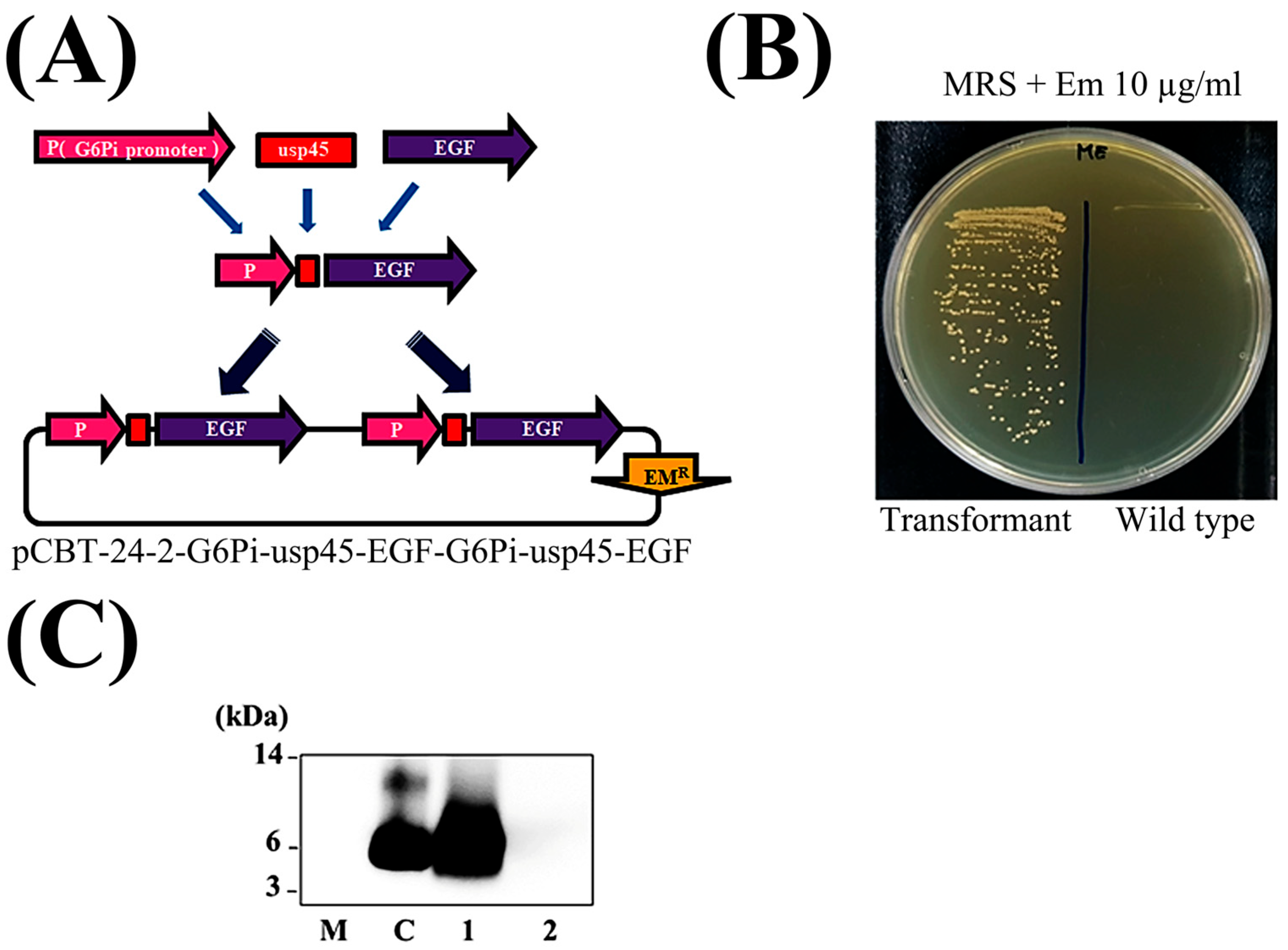
Figure 1. Construction of the plasmid harboring DDS-EGF for use in the Pediococcus pentosaceus SL4 system. (A) Schematic showing the expression vector for PP-EGF. The codon-optimized rhEGF gene was cloned into the pCBT-24-2-G6Pi-usp45-EGF-G6Pi-usp45-EGF expression plasmid. The dual-promoter system selected for the maximum expression of the EGF protein was ligated to a usp45 secretion signal peptide, thereby enabling the synthesis of DNA fragments (Cosmogenetech Inc., Seoul, Republic of Korea). (B) The transformant was placed on an MRS agar plate containing 10 μg/mL erythromycin. (C) The transformant was inoculated into 10 mL of MRS broth containing 10 μg/mL erythromycin and cultured at 37 °C for 15 h (no shaking). Next, 1 mL of pre-culture was inoculated into 10 mL of M9 minimal medium containing 10 μg/mL erythromycin and cultured at 37 °C for 48 h (no shaking). Next, 5 mL of culture was centrifuged, and the supernatant was collected. The supernatant including the EGF protein was concentrated via TCA precipitation to isolate the total protein. Finally, the EGF protein was detected using Western blotting (M: protein size maker; C: rhEGF (100 ng); lane 1: PP-EGF-SUP; lane 2: PP-EV-SUP).
Cosmetics 12 00176 g001
Figure 2. Toxicity test of PP-EGF-SUP. The effect of PP-EGF-SUP on the proliferation of each cell line was evaluated using a cell viability analysis, which was performed using the MTT (WST-8) technique. (A) The fibroblast (CCD-986sk) cell line was treated with various EGF protein concentrations, namely, rhEGF at 10 ng/mL, 2% PP-EGF-SUP, and 2% PP-EV-SUP, for 3 days. The actual EGF content in 2% PP-EGF-SUP was 0.3 ng/mL, and no serious cytotoxicity was confirmed in any of the tests. All data are presented as the mean ± SEM of three independent experiments (n = 3). ns, not significant; ** p < 0.001, *** p < 0.001 vs. rhEGF. (B) The skin melanoma (SK-MEL-2) cell line was treated with various EGF protein concentrations, namely, rhEGF at 10 ng/mL, 0.1% PP-EGF-SUP, and 0.1% PP-EV-SUP, for 1 day. The actual EGF content in 0.1% PP-EGF-SUP was 0.015 ng/mL, and no serious cytotoxicity was confirmed in any of the tests. Additionally, 40 µM kojic acid was used as a positive agent in a tyrosinase inhibition assay, and 200 µM IBMX was used as an inducer of tyrosinase expression. No serious cytotoxicity was confirmed in any of the tests. All data are presented as the mean ± SEM of three independent experiments (n = 3). ns, not significant; *** p < 0.001 vs. N.T. (C) The macrophage (RAW 264.7) cell line was treated with various EGF protein concentrations, namely, rhEGF at 10 ng/mL, 2% PP-EGF-SUP, and 2% PP-EV-SUP, for 1 day. The actual EGF content in 2% PP-EGF-SUP was 0.3 ng/mL, and no serious cytotoxicity was confirmed in any of the tests. All data are presented as the mean ± SEM of three independent experiments (n = 3). *** p < 0.001 vs. rhEGF.
Cosmetics 12 00176 g002
Figure 3. EGF-derived bioactivities: cell scratch wound healing, tyrosinase inhibition, and skin barrier improvement (elastin expression stimulation and collagenase and elastase suppression). To determine whether PP-EGF-SUP demonstrated EGF-derived bioactivities, the wound healing, whitening, and anti-wrinkle properties of PP-EGF-SUP were confirmed using cell scratch wound healing, tyrosinase inhibition, elastin expression, collagenase inhibition, and elastase inhibition assays. (A) In the cell scratch wound healing assay using the CCD-986sk cell line, the cells were treated with various EGF protein concentrations, namely, rhEGF at 10 ng/mL, 2% PP-EGF-SUP, and 2% PP-EV-SUP, after scratching, and then the migrated cell number was counted after 1 day. The actual EGF protein content in 2% PP-EGF-SUP was 0.3 ng/mL. All data are presented as the mean ± SEM of three independent experiments (n = 3). ns, not significant; *** p < 0.001 vs. rhEGF. (B) In the tyrosinase inhibition assay using the SK-MEL-2 cell line, the cells were treated with various EGF concentrations, namely, rhEGF at 10 ng/mL, 40 µM kojic acid, 0.1% PP-EGF-SUP, and 0.1% PP-EV-SUP, for 1 day with 200 µM IBMX, and then tyrosinase activity was measured in each cell lysate. The actual EGF protein content in 0.1% PP-EGF-SUP was 0.015 ng/mL. All data are presented as the mean ± SEM of three independent experiments (n = 3). ns, not significant; *** p < 0.001 vs. N.T. (C) In the elastin expression assay using the CCD-986sk cell line, the cells were treated with various EGF protein concentrations, namely, rhEGF at 10 ng/mL, 2% PP-EGF-SUP, and 2% PP-EV-SUP, for 1 day, and then elastin expression levels were measured using the Q-RT-PCR method. The actual EGF protein content in 2% PP-EGF-SUP was 0.3 ng/mL. All data are presented as the mean ± SEM of three independent experiments (n = 3). *** p < 0.001 vs. rhEGF. (D) In the collagenase inhibition assay conducted using a cell-free assay kit, rhEGF at 10 ng/mL, 10 and 20% PP-EGF-SUP, and 10 and 20% PP-EV-SUP were tested, and then collagenase activity was measured, with 25 µM 1,10-phenanthroline monohydrate used as a positive control. The actual EGF contents in 10 and 20% PP-EGF-SUP were 1.5 and 3 ng/mL, respectively. All data are presented as the mean ± SEM of three independent experiments (n = 3). *** p < 0.001 vs. P.C. (E) In the elastase inhibition assay conducted using a cell-free assay kit, rhEGF at 10 ng/mL, 10 and 20% PP-EGF-SUP, and 10 and 20% PP-EV-SUP were tested, and then collagenase activity was measured, with 2.5 µM MeOSuc-AAPV-CMK used as a positive control. All data are presented as the mean ± SEM of three independent experiments (n = 3). ns, not significant; *** p < 0.001 vs. P.C.
Cosmetics 12 00176 g003
Figure 4. Probiotic-derived bioproperties: antioxidant, anti-inflammatory, and anti-bacterial activities. To determine whether PP-EGF-SUP exhibited probiotic-derived bioactivities, its antioxidant, anti-inflammatory, and anti-bacterial properties were confirmed using a DPPH assay, an ROS scavenging assay, cytokine expression profiling using the Q-RT-PCR method, and an anti-bacterial assay using the disk diffusion method. (A) In the DPPH assay using the cell-free system, rhEGF (10 ng/mL), 2% PP-EGF-SUP, and 2% PP-EV-SUP were tested to measure the efficacy of DPPH inhibition. The actual EGF protein content in 2% PP-EGF-SUP was 0.3 ng/mL, and 10 nmole Trolox was used as a positive control. All data are presented as the mean ± SEM of three independent experiments (n = 3). ** p < 0.001, *** p < 0.001 vs. P.C. (B) In the ROS scavenging activity assay using the CCD-986sk cell line, the cells were treated with various EGF protein concentrations, namely, rhEGF at 10 ng/mL, 2% PP-EGF-SUP, and 2% PP-EV-SUP, for 1 day, and then ROS scavenging activity was measured. The actual EGF protein content in 2% PP-EGF-SUP was 0.3 ng/mL, and 20 mM L-Ascorbic acid was used as a positive control. All data are presented as the mean ± SEM of three independent experiments (n = 3). ns, not significant; *** p < 0.001 vs. rhEGF. (C) In the anti-inflammatory activity assay using the RAW 264.7 cell line, the cells were treated with various EGF concentrations, namely, rhEGF at 10 ng/mL, 2% PP-EGF-SUP, and 2% PP-EV-SUP, for 1 day, and then the gene expression levels of inflammatory markers (COX-2, IL-1β, IL-6, and TNF-α) were measured using the Q-RT-PCR method. Furthermore, 1 g/mL lipopolysaccharide (LPS) was also used as co-treatment to induce an inflammatory reaction in the cells. The actual EGF protein content in 2% PP-EGF-SUP was 0.3 ng/mL. All data are presented as the mean ± SEM of three independent experiments (n = 3). ns, not significant; * p < 0.05, *** p < 0.001 vs. N.T. (D) In the anti-bacterial activity assay, ten species of harmful skin bacteria were treated using the disk diffusion method. The cultured strains were diluted in 1 McFarland standard equivalent cell number adjusted by approximately 3 × 10⁸ cfu/1 mL, and then they were distributed to each dish after mixing in 9 mL of BL agar. Next, 1 mL of PP-EGF-SUP, PP-EV-SUP, and M9 media was concentrated on paper disks and then completely dried. The paper disks were placed on the different bacterial strain plates, and all plates were incubated at 37 °C for 1 day. The inhibition area (clear zone) was measured and compared with that of negative controls (N.T. and M9 media).
Cosmetics 12 00176 g004
Figure 5. Evaluation of PP-EGF-DP as a cosmetic ingredient. (A) PP-EGF-DP was prepared following the PP-EGF-SUP powdering procedure detailed in the Materials and Methods Section. (B) To determine whether PP-EGF-DP had a stimulatory effect on fibroblast cell proliferation due to EGF-derived bioactivity, cell viability was analyzed using the MTT (WST-8) technique, and rhEGF was used as a positive control. All data are presented as the mean ± SEM of three independent experiments (n = 3). ns, not significant; * p < 0.05, *** p < 0.001 vs. rhEGF. (C) To determine the temperature stability of PP-EGF-DP, its stimulatory effects on fibroblast cell proliferation after resuspension in solution was measured using the MTT method. All data are presented as the mean ± SEM of three independent experiments (n = 3). ns, not significant; * p < 0.05 vs. rhEGF. (D) To determine the pH stability of PP-EGF-SUP, the stimulatory effect on fibroblast cell proliferation by PP-EGF-DP solution after resuspension was measured using the MTT method. All data are presented as the mean ± SEM of three independent experiments (n = 3). * p < 0.05, ** p < 0.001 vs. rhEGF.
Cosmetics 12 00176 g005
Table 1. Determination of EGF protein in culture supernatant.
SampleEGF Concentration (μg/L)
PP-EV-SUP0
PP-EGF-SUP14.99 ± 0.044
The EGF protein content in the culture supernatant was determined using the ELISA method. All data are presented as the mean ± SEM of three independent experiments (n = 3).
Table 2. Evaluation of triple-freckle (light freckles, dark freckles, and freckle area) relief using Antera 3D®CS (n = 20).
AmpoulesTime PointsLight Freckles (%)Change Rate (%)Dark Freckles (%)Change Rate (%)Freckle Area (%)Change Rate (%)
1%PlaceboBefore0.474 ± 0.0420.840.777 ± 0.0760.56302.88 ± 78.612.79
After 4 Wks0.478 ± 0.0490.780 ± 0.07286.05 ± 88.77
PP-EGF-DPBefore0.478 ± 0.0373.380.779 ± 0.0661.74320.97 ± 77.821.21
After 4 Wks0.462 ± 0.040.76 ± 0.075251.9 ± 99.06
5%PlaceboBefore0.474 ± 0.0530.080.731 ± 0.1830.55302.38 ± 87.335.93
After 4 Wks0.474 ± 0.0510.726 ± 0.179274.68 ± 108.82
PP-EGF-DPBefore0.475 ± 0.0573.790.745 ± 0.0642.93318.33 ± 97.6829.10
After 4 Wks0.458 ± 0.0590.723 ± 0.061225.07 ± 94.77
Change rate (%) = [(After 4 Wks – Before)/Before] × 100. All data are presented as the mean ± SEM of three independent experiments (n = 3).
Disclaimer/Publisher’s Note: The statements, opinions and data contained in all publications are solely those of the individual author(s) and contributor(s) and not of MDPI and/or the editor(s). MDPI and/or the editor(s) disclaim responsibility for any injury to people or property resulting from any ideas, methods, instructions or products referred to in the content.