Article

Stability, Bioactivity, and Skin Penetration of Prunus Leaf Extracts in Cream Formulations: A Clinical Study on Skin Irritation

by
Lapatrada Mungmai
1,2,
Eakkaluk Wongwad
1,2,
Patcharawan Tanamatayarat
3,
Tammanoon Rungsang
1,2,
Pattavet Vivattanaseth
4,
Nattapol Aunsri
5,6,* and
Weeraya Preedalikit
1,2,*
1
Department of Cosmetic Sciences, School of Pharmaceutical Sciences, University of Phayao, Phayao 56000, Thailand
2
Research and Innovation Center in Cosmetic Sciences and Natural Products, School of Pharmaceutical Sciences, University of Phayao, Phayao 56000, Thailand
3
The College of Herbal Pharmacy of Thailand, Nonthaburi 11000, Thailand
4
Department of Pharmaceutical Sciences, School of Pharmaceutical Sciences, University of Phayao, Phayao 56000, Thailand
5
School of Applied Digital Technology, Mae Fah Luang University, Chiang Rai 57100, Thailand
6
Integrated AgriTech Ecosystem Research Center (IATE), Mae Fah Luang University, Chiang Rai 57100, Thailand
*
Authors to whom correspondence should be addressed.
Cosmetics 2025, 12(4), 146; https://doi.org/10.3390/cosmetics12040146
Submission received: 13 May 2025 / Revised: 25 June 2025 / Accepted: 7 July 2025 / Published: 10 July 2025
(This article belongs to the Section Cosmetic Dermatology)

Abstract

Prunus leaf extracts are rich in phenolic and flavonoid compounds like rutin, and they are known for their antioxidant potential. This study compares the bioactivity and stability of leaf extracts from Prunus domestica L. (EL), Prunus salicina Lindl. (JL), and Prunus cerasifera Ehrh. (CL) and evaluates the dermal safety of a cream containing the extract with the most favorable in vitro properties for potential cosmetic use. Ethanolic extracts were assessed for total phenolic and condensed tannin contents, as well as antioxidants, using DPPH assay and lipid peroxidation inhibitory activities. The CL extract exhibited moderate total phenolic content, the highest condensed tannin content, and strong antioxidant (IC50 = 22.1 ± 3.1 µg/mL) and anti-lipid peroxidation (62.3 ± 1.0%) activities. Based on these results, CL was incorporated into a cream formulation (CCL), which was then evaluated for physicochemical properties, antioxidant retention, and in vitro skin permeation using Franz diffusion cells. The formulation remained physically stable under ambient conditions and retained antioxidant activity above 74.5% under thermal cycling conditions. Rutin from the CCL formulation was retained within the Strat-M™ membrane (4.0 ± 1.1%), which was 5.7-fold higher than that of the control (0.7 ± 0.6%) over 8 h; however, it was not detected in the receptor chamber under these in vitro conditions. A semi-open patch test conducted on 26 healthy volunteers under double-blind conditions revealed no signs of irritation, confirming the formulation’s dermal safety. Overall, the findings support the feasibility of using P. cerasifera extract as a stable antioxidant component in topical skincare formulations.

1. Introduction

In recent years, the demand for natural, plant-derived ingredients in the cosmetics and pharmaceutical industries has increased significantly, driven by consumer preferences for sustainable and safer alternatives to synthetic compounds [1,2]. Plum plants (Prunus spp.), a diverse group of fruit-bearing species belonging to the Rosaceae family, are recognized for their considerable economic value and broad applications in food and health-related products [3]. The genus Prunus includes more than 35 species native to Europe, Asia, and the Americas [4,5]. Commercially cultivated plum species are generally classified into three main types: the European plum (P. domestica L.), the Japanese or Chinese plum (P. salicina Lindl.), and hybrids of the latter, such as the cherry plum (P. cerasifera Ehrh.) [5].
In our previous study [5], we assessed the biological activities of Prunus spp. fruits and leaves, revealing that the leaf extracts demonstrated superior potential compared to the fruit extracts. The leaf extracts demonstrated significant antioxidant, anti-lipid peroxidation, anti-tyrosinase, and anti-inflammatory properties. Ethanolic extract of Prunus leaves is also a rich source of phenolic and flavonoid compounds, especially the bioactive rutin. Additionally, Prunus leaves are a rich source of condensed tannins, which contribute further antioxidant potential through multiple mechanisms such as free radical scavenging, metal chelation, and inhibition of oxidative enzymes [6,7]. These properties support the role of Prunus leaf extracts as multifunctional ingredients in skincare formulations.
Rutin, a strong antioxidant flavonoid, was one of the most frequently identified compounds and associated with a broad range of skin-related benefits. Our previous research identified it as a bioactive marker due to its high yield (ranging from 28.5 ± 0.0 to 43.8 ± 0.1% w/w) and strong antioxidant activity in both DPPH and anti-lipid peroxidation assays, as well as its anti-inflammatory effects through the inhibition of nitric oxide, interleukin-6, and tumor necrosis factor-alpha—key mediators of skin inflammation [5]. It is widely recognized for its effectiveness in reducing oxidative stress; it supports skin protection from environmental damage, reduces lipid peroxidation, and modulates inflammatory mediators, thus helping to alleviate skin irritation, redness, and oxidative damage that contributes to aging signs like wrinkles and dullness [8,9,10]. It also inhibits tyrosinase, aiding in the management of hyperpigmentation and uneven skin tone, making it valuable for formulations aimed at skin brightening and tone correction [11,12,13]. Previous studies have reported that rutin possesses ultraviolet B (UVB) photoprotective properties and can enhance the sun protection factor (SPF) value [14,15,16]. When incorporated into an oil-in-water emulsion, it demonstrated SPF values comparable to those of the standard UV filter homosalate [15]. Furthermore, in vivo studies combining rutin with conventional UV filters support the claim that rutin is an effective and safe bioactive compound for use in sunscreen formulations [16].
However, after fruit harvest, Prunus leaves are often considered agricultural waste and are commonly discarded or burned, contributing to air pollution. Repurposing these by-products offers a sustainable approach to reduce agricultural waste and environmental impact. Utilizing Prunus leaves as a renewable and non-food-competing source of bioactive ingredients not only supports circular economy principles but also promotes the development of eco-friendly skincare formulations. Therefore, further investigations into the bioactivity and stability of Prunus leaf extracts are warranted to maximize their value and support the sustainable valorization of plum leaves in the cosmetic industry [5,17]. Moreover, the development of formulation, assessment of skin permeation profiling, and evaluation of dermal safety for cosmetic products containing Prunus leaf extracts are essential. This information could support eco-conscious formulation strategies and foster innovation in the natural skincare and health products market.
The objective of this study was to investigate three Prunus species cultivated in Southeast Asia, particularly in Thailand—Prunus domestica L., Prunus salicina Lindl., and Prunus cerasifera Ehrh.—for their potential use as natural ingredients in cosmetics and health products. Their total phenolic and condensed tannin contents, antioxidant and anti-lipid peroxidation activities, and stability were determined. A topical cosmetic product incorporating the most promising Prunus leaf extract was then developed, and its stability was assessed, and skin permeation was evaluated using the Franz diffusion cell method. In addition, a double-blind, semi-open patch test was conducted on human volunteers to assess dermal safety.

2. Materials and Methods

2.1. Chemicals and Reagents

Phosphate-buffered saline (PBS, pH 7.4), sodium lauryl sulfate, acetic acid, hydrochloric acid, ethanol, high-performance liquid chromatography (HPLC)-grade acetonitrile, and methanol were purchased from Labscan Asia Co., Ltd. (Bangkok, Thailand). Folin–Ciocalteu reagents were purchased from Merck (Darmstadt, Germany). Ferrous sulfate, thiobarbituric acid, trichloroacetic acid, sodium carbonate, DPPH, linoleic acid, and standard compounds such as Trolox, rutin, gallic acid, catechin, and ascorbic acid were obtained from Sigma-Aldrich (St. Louis, MO, USA). Propylene glycol, dimethicone, cyclopentasiloxane and dimethicone crosspolymer, cyclopentasiloxane and cyclotetrasiloxane, and cyclopentasiloxane and dimethiconol were purchased from Dow Chemical Company (Midland, MI, USA). Cyclopentasiloxane and dimethicone/vinyl dimethicone crosspolymer were obtained from Momentive Performance Materials (Waterford, NY, USA). Cetearyl olivate was obtained from Hallstar (Chicago, IL, USA). Polyacrylamide, C13–14 isoparaffin, and laureth-7 were purchased from Seppic (Paris, France). Ammonium acryloyldimethyltaurate/vinylpyrrolidone copolymer was purchased from Clariant Personal Care (Muttenz, Switzerland). Phenoxyethanol was purchased from Sharon Personal Care (Trezzo sull’Adda (MI), Italy).

2.2. Collection and Identification of Plant Materials

Leaves from three Prunus species, namely, P. domestica, P. salicina, and P. cerasifera, were used in this study. Leaves were collected in June from a garden area in Chiang Rai province, Thailand. The plant specimens were identified by experts from the School of Pharmaceutical Sciences, University of Phayao, Phayao, Thailand. The voucher specimens for these three species were labeled Lapatrada Mungmai001, Lapatrada Mungmai002, and Lapatrada Mungmai003, respectively.

2.3. Preparation of Prunus Species Leaf Extracts

The leaves of three Prunus species were washed with water and air-dried at room temperature for 5 h to remove surface moisture. The plant samples were then dried in a hot-air oven at 50 °C for 2 days, ground using a blender, and 50 g of each sample was extracted with 95% ethanol by maceration for 3 days. Then, the extract solution was evaporated using a rotary evaporator to obtain P. domestica leaf extract (EL), P. salicina leaf extract (JL), and P. cerasifera leaf extract (CL). Subsequently, the percentage yield (% yield) of each extract was calculated, and the rutin content was qualified using HPLC (Shimadzu Prominence, Kyoto, Japan). The analysis was performed under isocratic conditions with solvent A (1% acetonitrile) and solvent B (acetic acid in water) in a 70:30 ratio, a flow rate of 0.8 mL/min, an injection volume of 10 µL, and detection at 280 nm using rutin as the reference standard [5].

2.4. Determination of the Total Phenolic Content (TPC)

The total phenolic contents of each extract were measured using the Folin–Ciocalteu assay with some slight modifications from the method described by Mungmai et al. [18]. Extracts were prepared at a concentration of 1 mg/mL in 95% ethanol. For the assay, 25 μL of the extract was mixed with 80 μL of 10% sodium carbonate in a 96-well plate. Then, 100 μL of 1 M Folin–Ciocalteu reagent was added. The mixture was incubated in the dark at room temperature for 60 min. Absorbance was measured at 765 nm using a microplate reader (BioTek Instruments, Winooski, VT, USA). A standard curve was generated using gallic acid at a concentration ranging from 5.0 to 180 μg/mL. The TPC of the extracts was expressed as milligrams of gallic acid equivalent (GAE) per gram of extract (mg GAE/g extract).

2.5. Determination of the Total Condensed Tannin Content (TTC)

The total condensed tannins in each extract were quantified using a modified method based on Rebaya et al., with catechin as the reference compound [19]. Briefly, 20 µL of the extract was added to a 96-well plate, followed by 120 µL of a 4% vanillin solution in methanol and 60 µL of concentrated hydrochloric acid. The mixture was then incubated in the dark at room temperature for 15 min. After incubation, the absorbance was measured at 500 nm using a microplate reader. The TTC was expressed as milligrams of catechin per gram of extract (mg catechin/g extract).

2.6. Determination of Antioxidant Activity Using DPPH Assay

The free radical scavenging activity of each extract was evaluated using the DPPH assay, following a previously published methodology with minor adjustments [5]. Briefly, 50 μL of the samples and 130 μL of 0.2 mM DPPH in ethanol were added to a 96-well plate. After incubating in the dark at 27 ± 2 °C for 30 min, the absorbance was measured at 515 nm using a microplate reader (BioTek Instruments, Winooski, VT, USA). Trolox and rutin were used as positive controls. The results were expressed as IC50 values, which represent the concentration of the sample required to achieve 50% free radical scavenging activity. IC50 values were calculated using GraphPad Prism 9 software, which was utilized for data analysis, curve fitting, and determination of the IC50 from the dose–response data. The percentage of scavenging activity was calculated using the following formula:
%   S c a v e n g i n g   a c t i v i t y = [ ( A c o n t r o l A s a m p l e ) / A c o n t r o l ] × 100
where A c o n t r o l and A s a m p l e are the absorbances of the control and sample, respectively.

2.7. Determination of Lipid Peroxidation Inhibitory Activity Using theThiobarbituric Acid Reactive Substances (TBARS) Assay

The Lipid peroxidation inhibitory activity of each extract was assessed using a modified TBARS assay, as previously described [20,21]. Briefly, 40 µL of each sample extract, dissolved in ethanol at a concentration of 100 mg/mL, was combined with 140 µL of lipid peroxidation source in a 1.5 mL microcentrifuge tube. The lipid peroxidation source was prepared by emulsifying linoleic acid in phosphate-buffered saline (PBS, pH 7.4) using sonication until a uniform suspension was obtained. The mixture was incubated at 37 °C for 30 min. Next, 20 µL each of 2 mM ferrous sulfate and 2 mM ascorbic acid was added, and the mixture was incubated for an additional 30 min at 37 °C. Following this, 200 µL of TBARS reagent (8% hydrochloric acid, 1.4% thiobarbituric acid, and 40% trichloroacetic acid) was added, and the sample was incubated at 90 °C for 60 min. The samples were then centrifuged at 10,000 rpm for 5 min at 4 °C. 100 µL of the supernatant was measured at 530 nm using a microplate reader. Trolox and rutin were used as positive controls. The results were expressed as the percentage of lipid peroxidation inhibitory activity, calculated using the following formula:
%   L i p i d   p e r o x i d a t i o n   i n h i b i t o r y   a c t i v i t y = [ ( A c o n t r o l A s a m p l e )   /   A c o n t r o l ] × 100
where A c o n t r o l and A s a m p l e are the absorbances of the control and sample, respectively.

2.8. Stability Test of the Prunus Species Leaf Extracts

Prunus species leaf extracts were stored in amber glass bottles with airtight closures to minimize light and air exposure, and samples were maintained under three controlled storage conditions [22]: 4 ± 2 °C, 25 ± 2 °C, and 45 ± 2 °C for 2 months. The pH, odor, color, and bioactivities, including TPC, TTC, and antioxidant activity (measured by the DPPH assay), were assessed at days 0, 7, 14, 28, and 60.
The pH was measured using a calibrated pH meter (Mettler Toledo, SevenCompact™ S210, Columbus, OH, USA) after diluting the sample with deionized water (1:9 ratio). The color changes in the extracts were assessed using ∆E, a quantitative measure of color difference in the CIE Lab color space using a colorimeter (Cortex Technology ApS, Hadsund, Denmark). For each time point and storage condition, triplicate samples were tested to ensure the reproducibility and reliability of the data.

2.9. Developed Cream Formulation and Its Stability Evaluation

The cream formulation containing the most promising Prunus leaf extract was developed, and the ingredients are detailed in Table 1. The formulation was prepared by first dispersing ammonium acryloyldimethyltaurate/VP copolymer in deionized water, adding propylene glycol, and heating to 80 °C. The oil phase, consisting of dimethicone and cetearyl olivate, was heated to 75 °C. Silicone ingredients, including cyclopentasiloxane-based polymers, were mixed until homogeneous and heated to 75 °C. The water phase was gradually added to the oil phase with continuous stirring to form an emulsion. Polyacrylamide (and) C13–14 isoparaffin (and) laureth-7 was then incorporated to stabilize the emulsion. Finally, the selected promising Prunus extract mixed with glycerin was added to the emulsion under gentle stirring to complete the formulation.
To assess its stability, it was packaged in opaque, airless pump bottles to prevent light and oxygen exposure during the stability test. The cream was stored under two conditions: constant ambient temperature (25 ± 2 °C) and heating/cooling stress cycles (45 ± 2 °C for 48 h, alternated with 4 ± 2 °C for 48 h per cycle) [23]. These conditions were maintained for 60 days (sampling every Day 0, 7, 14, 21, 28, and 60), with accelerated stability testing conducted over 24 days (sampling every Day 0, 4, 8, 12, 16, 20, and 24), in accordance with industry-standard practices for assessing cosmetic product stability as recommended by Cosmetics Europe. The stability of the developed cosmetic formulation was then evaluated through organoleptic and physical characteristics, and it retained antioxidant activity.
Organoleptic and physical characteristics, including color, odor, phase separation, homogeneity, pH, and viscosity, were assessed visually and manually by checking the texture and consistency of the cream. Acceptable limits for stability were defined based on established guidelines and included no significant changes in color or odor and the absence of phase separation [24].

2.10. In Vitro Skin Permeation Study of Developed Cream Formulation Using Franz Diffusion Cells

The skin permeation of the developed cream containing the selected most promising Prunus species leaf extract was evaluated using Franz diffusion cells (Logan Instruments, Somerset, NJ, USA) by detecting the bioactive compound rutin present in the cream formulation [25]. Strat-M® membranes (300 μm, EMD Millipore, Burlington, MA, USA) were employed as a simulated skin model, given their ability to mimic the structural and permeability characteristics of human epidermis and dermis [25]. The receptor chamber was filled with 7.5 mL of PBS (pH 7.4) containing 2% w/v sodium lauryl sulfate to maintain sink conditions, kept at 37 ± 2 °C, and stirred continuously at 300 rpm to ensure uniform distribution of the active compound. The Strat-M® membrane was placed between the receptor and donor chambers with an effective exposure area of 3.14 cm2. One gram of the developed cream was applied to the donor chamber, and 1 mL of solution from the receiver chamber was collected at specified intervals (0.5, 1, 2, 4, 6, and 8 h). Each sample withdrawal was immediately replaced with an equal volume of fresh PBS containing 2% w/v sodium lauryl sulfate to maintain consistent test conditions. For this experiment, a 0.3% CL dissolved in deionized water was used as a control to compare the permeation profile of the developed cream formulation.
At the end of the 8 h study, the membrane was removed and gently washed three times with PBS to eliminate unabsorbed cream, then cut into small pieces. The retained rutin in the membrane was extracted with methanol using a sonication bath for 15 min. In addition, both permeated rutin (collected from the receptor chamber) and retained rutin (extracted from the membrane) were quantified using HPLC as described in Section 2.3.

2.11. Clinical Evaluation

2.11.1. Study Design and Methods

The research protocols were approved by the University of Phayao Human Ethics Committee, Thailand (approval number HREC-UP_HSST 1.2/113/66), in compliance with the WHO Guidelines for Good Clinical Practice. The study included twenty-six healthy volunteers aged 30–50 years. The age range was selected to represent a population likely to use skincare products while minimizing age-related variability in skin physiology and sensitivity. All participants received the information protocol and signed a written informed consent form before participating.
Inclusion criteria:
  • Male or female participants aged 30–50 years.
  • Availability for the entire study duration.
  • No use of medication or medical care for at least one month prior to and during the study.
  • Free from any dermatological or systemic disorder that could interfere with test results or increase the risk of adverse reactions. Examples of systemic disorders include autoimmune diseases, diabetes, or severe cardiovascular conditions.
Exclusion criteria:
  • Visible skin diseases at the study site (e.g., eczema, psoriasis) that could interfere with the evaluation.
  • Systemic disorders such as autoimmune diseases, diabetes, or chronic inflammatory conditions that could affect skin response.
  • Use of systemic or topical drugs or medication that could mask or affect study results.
  • History of serious diseases, such as cardiovascular disease or malignancies, or known sensitivity to cosmetics.
  • Diagnosis of chronic skin allergies.
  • Known or suspected intolerance or hypersensitivity to the investigational products or any of their ingredients.
  • Pregnancy or lactation during the study period.

2.11.2. Skin Safety Study in Human Volunteers

The safety assessment was conducted as a double-blind trial, with slight modifications to established protocols to ensure an unbiased and reliable evaluation [26,27]. The study utilized a semi-open test method with a 1.5 cm × 1.5 cm cotton pad to deliver the test substances. The cotton pads were placed under a semi-occlusive adhesive film to ensure controlled exposure to the upper back of each volunteer. The test substances included a cream base, cream containing Prunus species leaf extract, and the extract alone. Additionally, 2% w/v sodium lauryl sulfate served as a positive control, while deionized water was used as a negative control. Prior to application, the test area was cleansed with water, and the patches were applied for 48 h. The use of semi-occlusive adhesive ensured proper contact without complete occlusion, aligning with semi-open test standards.
Erythema and edema formation were observed at 24 h and 48 h after patch removal and were graded by a licensed dermatologist based on the Cosmetic, Toiletry, and Fragrance Association (CTFA) Guideline (Table 2). The average scores of erythema and edema formations were recorded following the Draize scoring system. The Primary Dermal Irritation Index (PDII), representing an average irritation index, was calculated based on these scores to assess skin irritation levels.
In addition, the Dermalab Combo® Series (Cortex Technology ApS, Hadsund, Denmark) was used to measure erythema using a skin color probe.

2.12. Statistical Analysis

All experiments were conducted in triplicate (n = 3), and results were expressed as means ± SD. Statistical analysis was performed using SPSS version 24 for Windows (SPSS, Chicago, IL, USA). One-way ANOVA followed by Tukey’s post hoc test was used for multiple group comparisons, while an independent-samples t-test was applied for pairwise comparisons. Statistical significance was set at p < 0.05. Erythema and edema scores were recorded and analyzed as ordinal data.

3. Results and Discussion

3.1. Extraction of Prunus Species Leaf

In this study, leaf extracts from three Prunus species (EL, JL, and CL) were examined. All extracts displayed a dark green appearance and a characteristic greenish odor, as shown in Figure 1.
The extract yields obtained in this study are shown in Table 3 and ranged from 7.5% to 9.0% w/w. Similarly, a previous study reported that ethanolic extractions of Prunus species leaves yielded between 5.5% and 9.5%, supporting the consistency of these results [5]. Among the extracts, EL exhibited the highest yield, followed by JL and CL, with the observed differences attributed to variations in leaf composition and extractable bioactive compounds. Additionally, variations in the polarity of bioactive compounds, particularly antioxidants, could influence their extractability and contribute to the differences observed in extraction yield and antioxidant activity [28]. Ethanol was employed as the extraction solvent in this study due to its superior ability to extract bioactive compounds such as phenolics and flavonoids, ensuring higher yields of active ingredients. Ethanol’s volatility allows for effective removal, minimizing residual solvent in the final product and adhering to safety standards for cosmetic applications.

3.2. Total Phenolic and Total Condensed Tannin Contents of Prunus Species Leaf Extracts (EL, JL, and CL)

The Prunus species leaf extracts demonstrated notable variation in TPC and TTC across the three species, as shown in Table 4. JL extract exhibited the highest phenolic content, while CL extract had the highest tannin content, significantly surpassing the other species. These findings highlight the differing phytochemical profiles of the extracts, with the CL extract showing a distinct advantage in tannin concentration, which may influence its bioactivity. The TPC and TTC values reflect differences in the phytochemical composition of the extracts, which are strongly associated with their bioactivity [29,30]. Phenolic compounds, including tannins, contribute to the antioxidant potential of the extracts by scavenging free radicals, chelating transition metals, and inhibiting lipid peroxidation [30,31]. Previous studies have shown that higher levels of phenolics correlate with enhanced antioxidant and anti-inflammatory properties in Prunus species [5]. The notable difference in TPC between this study and previous reports, where TPC values were over 10 times higher, may be attributed to variations in harvesting season, environmental factors, and extraction methods, which significantly influence the phenolic profile [32,33].
Condensed tannins, specifically abundant in the CL extract, have been reported to inhibit key inflammatory mediators such as nitric oxide and prostaglandin E2 while also reducing malondialdehyde, a marker of lipid peroxidation. Their antioxidant activity is influenced by the number and degree of polymerization of hydroxyl groups, with higher hydroxyl content leading to increased oxidative scavenging capacity [34]. The elevated TTC in CL extract may explain its stronger bioactivity despite its lower TPC, suggesting that tannins play a critical role in its antioxidant potential [35].

3.3. Antioxidant Activity of Prunus Species Leaf Extracts (EL, JL, and CL) Using DPPH Assay

The results of the DPPH radical scavenging activity of the extracts are presented in Table 4. The EL, JL, and CL extracts demonstrated comparable antioxidant activity, with the CL extract showing slightly stronger activity. However, no statistically significant differences were observed among the three extracts, indicating similar antioxidant potential across the species. Trolox and rutin exhibited much lower IC50 values, indicating higher potency. This finding aligns with the report by Stierlin et al., which highlighted the strong antioxidant activity of plum leaf ethanolic extracts compared to less effective solvents like hexane and ethyl acetate [36]. Despite the higher potency of the standards, the extracts demonstrated promising antioxidant potential, emphasizing their bioactive properties. Notably, rutin, a key phenolic compound present in all extracts, along with condensed tannins, may play a significant role in enhancing their antioxidant activity [37]. The antioxidant activity observed in the DPPH assay aligns with the TPC and TTC of the extracts. While the TPC values were highest in JL and lowest in CL, the TTC was significantly higher in CL compared to EL and JL. This suggests that the antioxidant activity of CL may be driven more by its tannin content rather than its phenolic acid levels.

3.4. Lipid Peroxidation Inhibitory Activity of Prunus Species Leaf Extracts (EL, JL, and CL) Using the TBARS Assay

Lipid peroxidation inhibition involves measuring the damage caused by reactive oxygen species to lipids, particularly polyunsaturated fatty acids, in cell membranes [21]. This process leads to cellular and molecular damage, resulting in various symptoms associated with different pathologies. The TBARS assay revealed that the CL extract exhibited the strongest lipid peroxidation inhibitory activity among the tested samples, significantly outperforming the JL extract and the rutin standard (see Table 4). However, all extracts demonstrated lower potency compared to Trolox, which served as the positive control. The observed differences in lipid peroxidation inhibition among the extracts align with their TTC, highlighting tannins as key contributors to this bioactivity. Tannins are well-known for their ability to scavenge free radicals, inhibit lipid peroxidation, and chelate transition metals, all of which contribute to reducing oxidative damage in lipids [37]. Additionally, the interaction between tannins and phenolic compounds, such as rutin, may have a synergistic effect, further enhancing the antioxidant potential of the extracts [38]. The relatively lower lipid peroxidation inhibition of the JL extract, despite its higher TPC compared to CL, underscores the critical role of tannins in this specific bioactivity.

3.5. Stability Test of the Prunus Species Leaf Extracts (EL, JL, and CL)

3.5.1. Physicochemical Characteristics of the Prunus Species Leaf Extracts

The physicochemical characteristics of the EL, JL, and CL extracts were evaluated after being stored at 4 ± 2 °C, 25 ± 2 °C, and 45 ± 2 °C for 2 months. Assessments included pH, odor, and color (∆E), performed at Days 0, 7, 14, 21, 28, and 60.
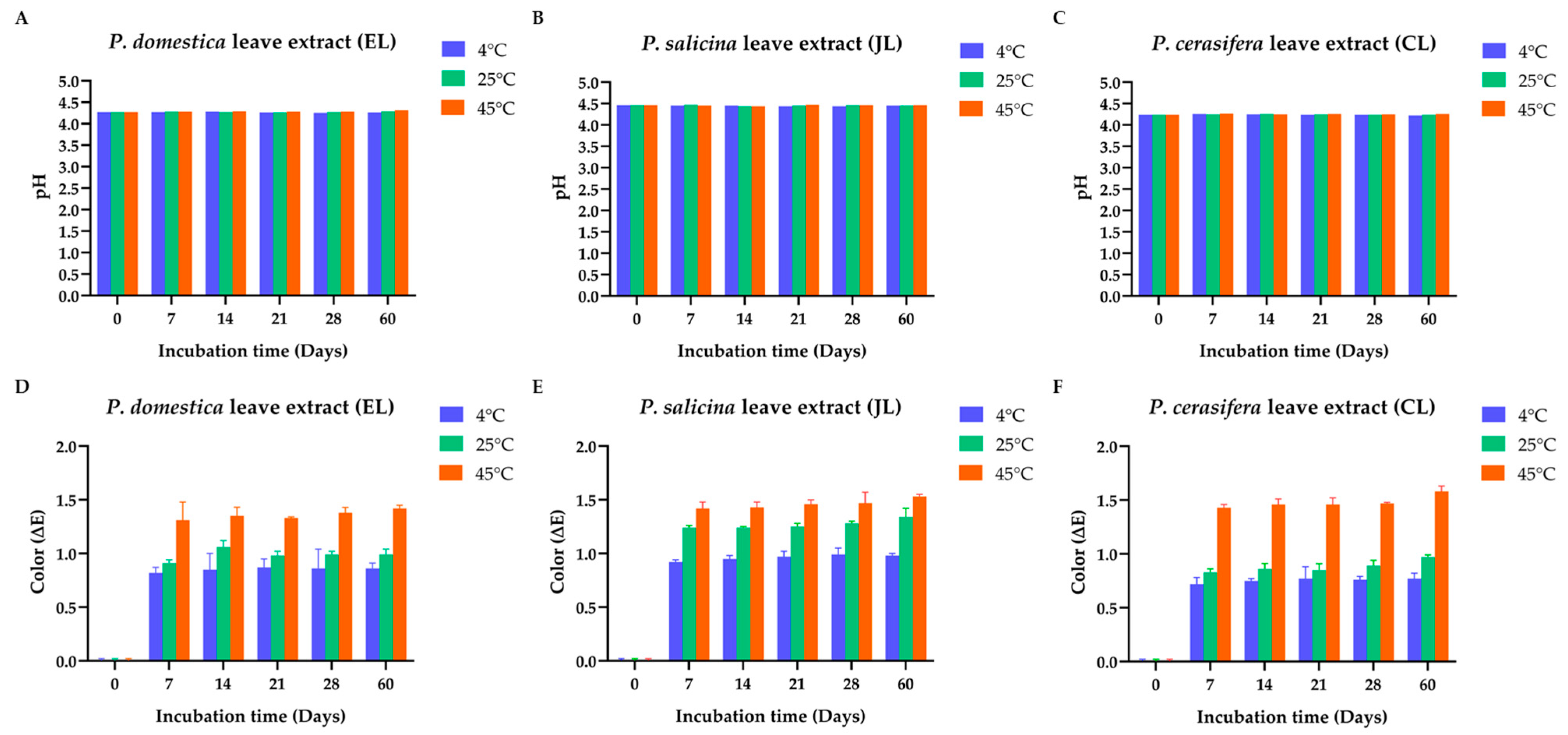
The pH of all extracts ranged from 4.2 to 4.5 (see Figure 2A–C), with only slight increases observed over time. Despite this, values remained within acceptable stability limits, indicating minimal degradation of acidic constituents. The CL extract displayed a consistently stronger odor than EL and JL, which may be attributed to differences in volatile compound profiles specific to each Prunus species [39].
Color changes, evaluated using ∆E values (Figure 2D–F), were more pronounced at elevated temperatures, particularly at 45 ± 2 °C. All extracts showed an increase in ∆E over time, indicating a darkening trend attributed to the oxidation of phenolic compounds. Among the species, JL exhibited the most intense color changes, followed by EL and CL.
Overall, the relative stability of pH and expected changes in color under stress conditions supports the suitability of these extracts.

3.5.2. TPC of the Prunus Species Leaf Extracts (EL, JL, and CL) in Stability Tests

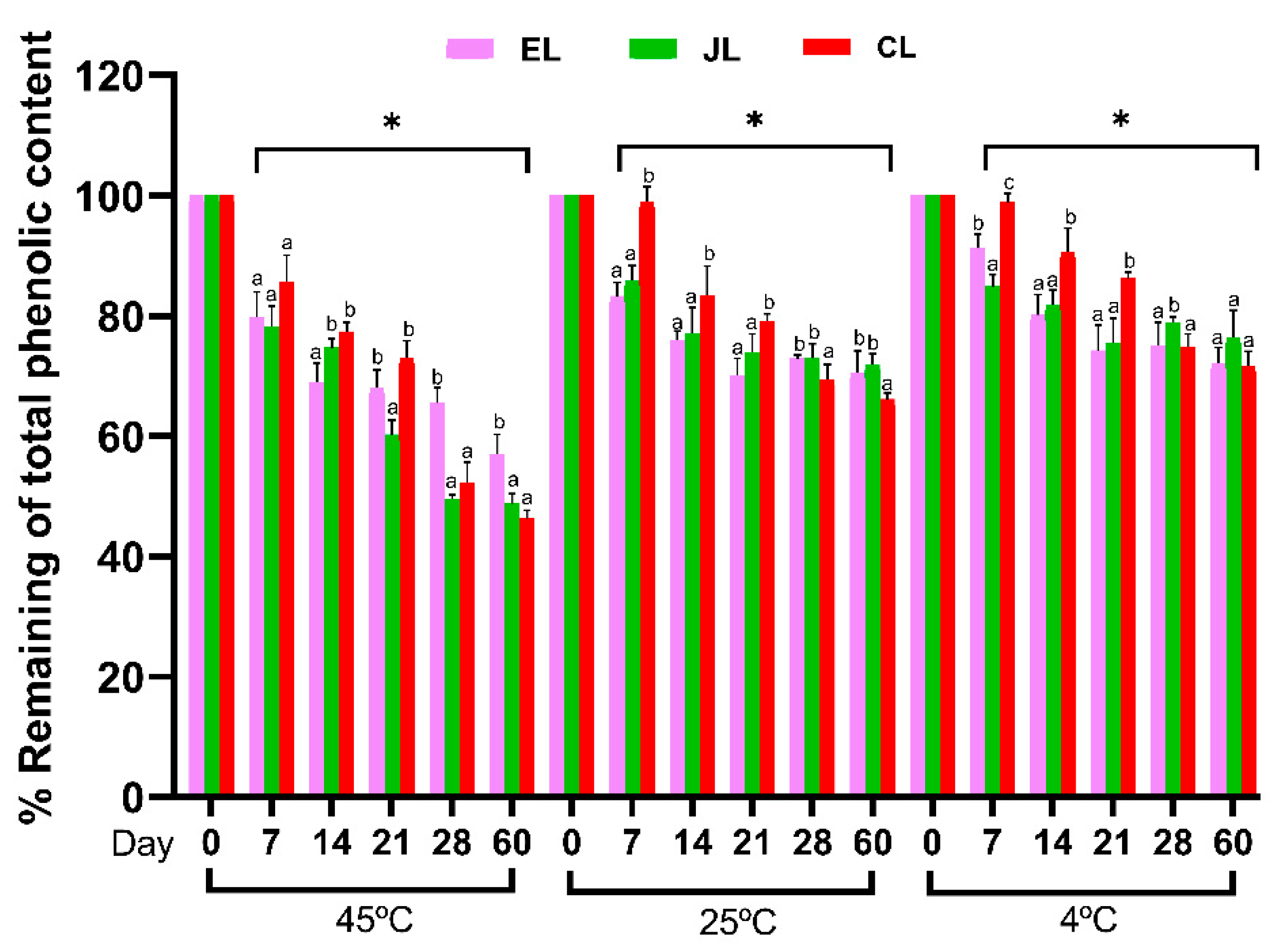
The stability of TPC of the Prunus species extracts was evaluated after storage at 4 ± 2 °C, 25 ± 2 °C, and 45 ± 2 °C for 60 days, as shown in Figure 3. A marked decline in TPC was observed across all extracts when stored at 45 ± 2 °C, beginning from Day 7 and continuing through Day 60. Among the three extracts, the EL extract demonstrated the highest TPC retention under high-temperature storage, maintaining 57.0 ± 3.3% of its initial content after 60 days, followed by the JL extract at 48.8 ± 1.6% and the CL extract at 46.3 ± 1.4%.
At 25 ± 2 °C, the TPC of all extracts showed a slight decrease over 60 days. Interestingly, under refrigerated conditions (4 ± 2 °C), the extracts maintained 70–80% of their initial TPC throughout the storage period. By Day 60, the TPC values across all extracts converged, with no significant variation between species, indicating that lower storage temperatures effectively help preserve phenolic content.
Although the CL extract initially exhibited the highest TPC, it showed greater degradation over time, particularly under thermal stress. This instability is due to the elevated levels of reactive phenolic compounds, such as rutin and condensed tannins, which, despite enhancing antioxidant activity, are more susceptible to oxidative destruction, hydrolysis, and polymerization. Environmental factors, including heat, light, and oxygen, are known to accelerate the breakdown of phenolic compounds, resulting in reduced stability and bioactivity during storage [40,41].

3.5.3. TTC of the Prunus Species Leaf Extracts (EL, JL, and CL) in Stability Tests

The stability of TTC in Prunus species extracts was assessed over 60 days at storage temperatures of 4 ± 2 °C, 25 ± 2 °C, and 45 ± 2 °C as shown in Figure 4. Tannin stability varied notably with temperature, showing a more pronounced decline at 45 ± 2 °C, highlighting the susceptibility of tannins to degradation under elevated thermal conditions. During the early storage period (Days 7–21), the CL extract demonstrated the highest TTC retention across all conditions, with values ranging from 65 to 95%. However, from Day 21 onward, the TTC in all extracts declined sharply, particularly at the 45 ± 2 °C condition. The CL extract, in particular, dropped markedly to 46% by Day 60 at 45 ± 2 °C. These findings are consistent with previous reports indicating that proanthocyanidins, such as procyanidin B2 found in P. domestica leaf extracts, are sensitive to thermal degradation [39]. Similarly, Khanal et al. reported significant anthocyanin losses in grape and blueberry pomace at temperatures exceeding 60 °C, while stability was maintained at moderate temperatures around 40 °C [42].
Conversely, at lower storage temperatures (4 ± 2 °C and 25 ± 2 °C), TTC degradation was substantially slower, demonstrating that cooler conditions help preserve tannin stability. Interestingly, although the CL extract initially exhibited the highest TTC among the three species, it had the lowest TTC retention after 60 days. Nonetheless, even after 60 days, the CL extract maintained higher absolute tannin levels compared to its TPC. These results highlight that, despite its greater susceptibility to degradation, the CL extract retains a substantial number of tannins, which likely contribute to its overall bioactivity and therapeutic potential during storage.

3.5.4. Antioxidant Activity Using DPPH Assay of the Prunus Species Leaf Extracts (EL, JL, and CL) in Stability Tests

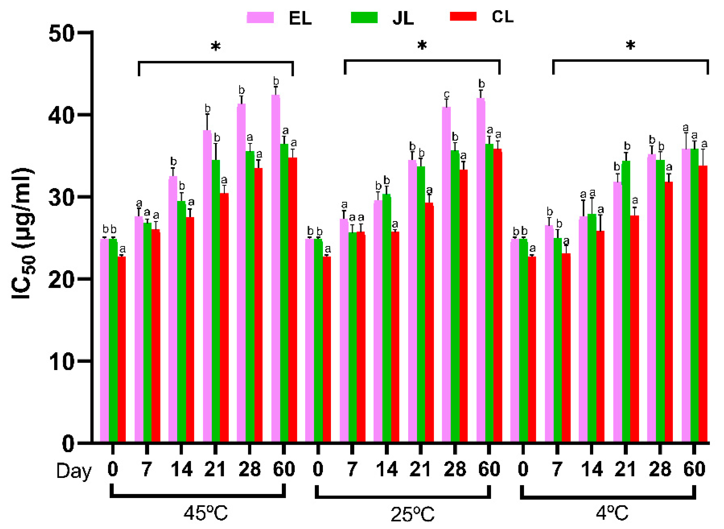
The stability of antioxidant activity, assessed by the DPPH assay, for Prunus species extracts stored at 4 ± 2 °C, 25 ± 2 °C, and 45 ± 2 °C over 60 days is shown in Figure 5. It can be observed that IC50 values of all extracts considerably increase on day 60, indicating a decrease in antioxidant compounds and effects. Notably, the extracts stored at 45 ± 2 °C showed the most significant increases in IC50 values over 60 days, with values ranging from approximately 35 µg/mL at day 0 to nearly 50 µg/mL at day 60. In comparison, extracts stored at 4 ± 2 °C demonstrated the most stable antioxidant activity, with IC50 values increasing only slightly over the storage period (from approximately 20 µg/mL to around 35 µg/mL). At 25 ± 2 °C, the IC50 values showed a moderate increase, indicating an intermediate level of stability between 4 ± 2 °C and 45 ± 2 °C storage conditions. At day 60, the IC50 values were significantly different between storage temperatures, with 4 ± 2 °C showing the lowest IC50 values, followed by 25 ± 2 °C, and the highest values observed at 45 ± 2 °C. The results indicate that the antioxidant activity of Prunus species extracts is affected by both storage temperature and time, with higher temperatures leading to a more pronounced decline in DPPH scavenging activity. The increase in IC50 values over time, particularly at 45 ± 2 °C, suggests that the phenolic and tannin compounds responsible for antioxidant activity are susceptible to thermal degradation [43]. This aligns with the earlier findings on TPC and TTC, where elevated storage temperatures were associated with a more rapid decrease in these compounds. The extracts stored at 4 ± 2 °C showed the most stable antioxidant activity, suggesting that low-temperature storage helps preserve phenolic and tannin stability and, consequently, the antioxidant properties.
Interestingly, the CL extract maintained the highest antioxidant activity across all time points and storage temperatures, exhibiting only slight changes in IC50 values during the stability test. It consistently showed the lowest IC50 values before and after storage, highlighting its strong and stable antioxidant potential. Therefore, the CL extract was selected for incorporation into a cream formulation due to its strong antioxidant potential.

3.6. Stability Test of Cream Formulation

3.6.1. Physicochemical Stability of the Cream Formulation Containing CL (CCL)

A concentration of 0.3% w/w of the CL extract, selected for its strong antioxidant properties, was incorporated into the cream formulation. This concentration was determined based on the extract’s antioxidant activity, measured by the DPPH assay, and its skin permeation profile. Given the low skin permeability of rutin [44], the primary bioactive compound, the concentration was increased approximately 100- to 150-fold relative to its IC50 value to ensure sufficient availability within the formulation to overcome permeability barriers and exert its antioxidant effects during topical application [45].
Throughout the stability study, the cream formulation exhibited no visible changes in color, phase separation, or homogeneity, pH, and viscosity after 60 days at 25 ± 2 °C (Figure 6A) and after 24 days of heating–cooling cycles (Figure 6B), indicating excellent physical stability. The consistent appearance under both ambient and stress conditions suggests that the formulation successfully preserved the structural integrity of its components. These results confirm that the cream containing CL extract remained stable throughout the test period, supporting its suitability for practical cosmetic applications.

3.6.2. DPPH Free Radical Scavenging Activity Determination of CCL

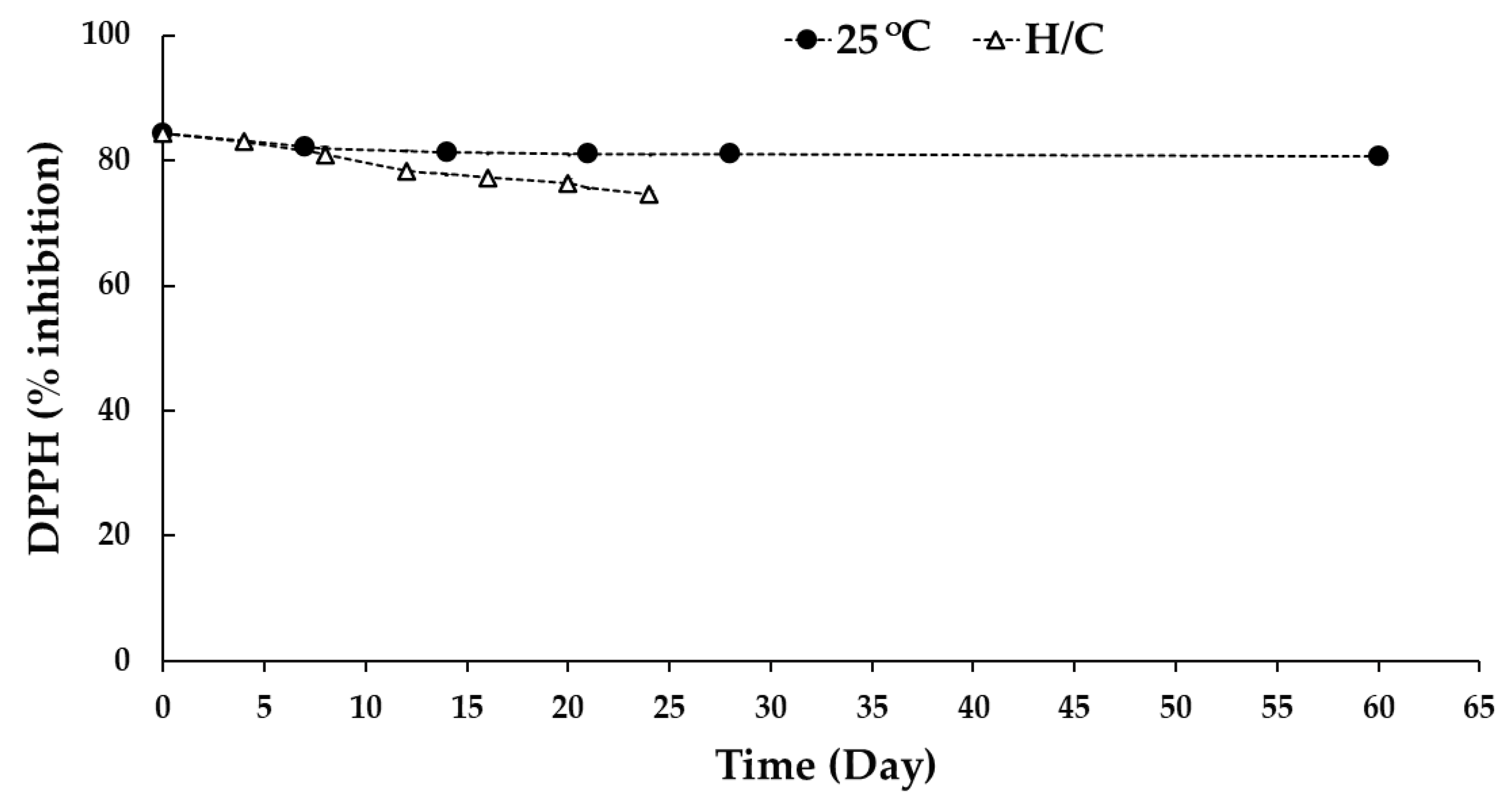
As shown in Figure 7, The DPPH free radical scavenging activity of the CCL was evaluated under two storage conditions. At 25 ± 2 °C, the cream containing 0.3% w/w CL extract exhibited an initial scavenging activity of 84.4 ± 0.2% (Day 0), which gradually declined to 80.7 ± 0.1% by Day 60. This represents a statistically significant reduction of 3.7% in DPPH free radical scavenging activity, demonstrating reasonable stability under ambient storage conditions over the 60-day period. Similarly, previous studies reported a 3.4% reduction in rutin in emulsified systems without soy lecithin stored at room temperature for 90 days [46]. In contrast, under heating/cooling conditions, the initial scavenging activity was also 84.4 ± 0.2%. Still, the decline was more pronounced, with a significant drop to 82.9 ± 0.2% after the first cycle (Day 4), followed by a further decrease to 74.5 ± 0.7% by the sixth cycle (Day 24). The total reduction under heating/cooling conditions was approximately 11.6%, highlighting the significant impact of thermal stress on the stability of the antioxidant compounds in the formulation. A similar trend was observed in a study where rutin degraded significantly under elevated temperature conditions (40.0 ± 0.5 °C) after 90 days, with reductions of 34.2% in rutin concentration in systems without lecithin [22]. These findings highlight the susceptibility of rutin and related antioxidant compounds to thermal degradation, underscoring the need for controlled storage conditions to maintain the bioactivity of formulations. This emphasizes the need for controlled storage environments to preserve the antioxidant efficacy of formulations containing CL extract. For further improvement, stabilization strategies such as encapsulation or the incorporation of additional antioxidants could be explored to enhance the long-term stability of the formulation.

3.7. Skin Permeation Study of CCL

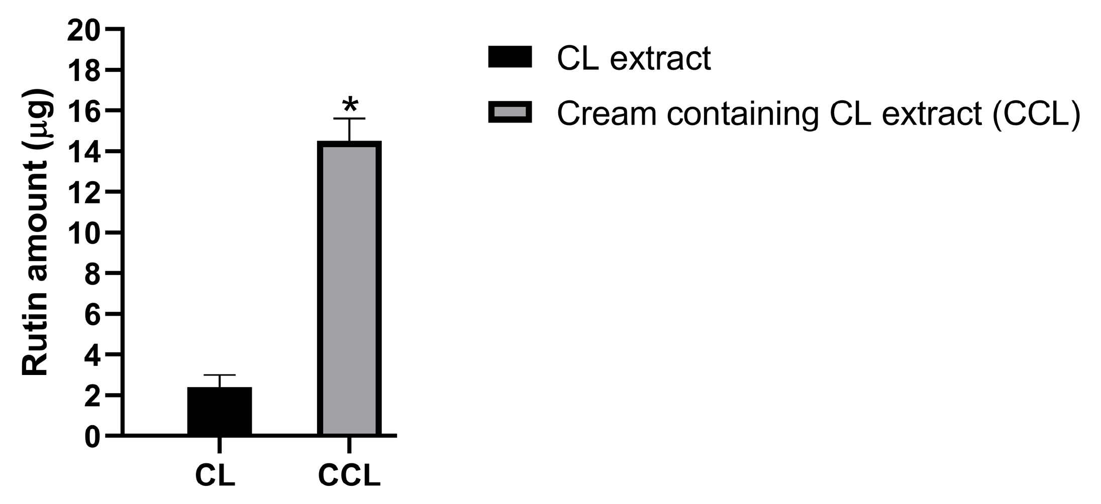
The in vitro permeability of the CCL formulation was assessed using the Franz diffusion cell method with a synthetic Strat-M™ membrane over an 8 h period. A 0.3% w/w aqueous solution of CL was used as a control. Rutin concentrations retained within the Strat-M™ membrane were quantified to assess the formulation’s ability to enhance skin retention (Figure 8).
The selection of 0.3% w/w CL extract was based on preliminary antioxidant screening and formulation optimization. The extract demonstrated an IC50 value of 22.7 ± 3.1 µg/mL in the DPPH assay, suggesting potent antioxidant activity at low concentrations. This level was chosen to ensure both bioactivity and formulation stability.
The results showed that after 8 h of the performed experiment, the CCL formulation achieved an estimated 5.7-fold significantly higher (p < 0.05) rutin retention (14.5 ± 1.1 µg; 4.0 ± 1.1%) compared to the control (2.4 ± 0.6 µg; 0.7 ± 0.6%). However, no rutin was detected penetrating the receiver chamber. When compared to the IC50, the retained amount lies within a biologically relevant range, supporting the effectiveness of this concentration for topical antioxidant delivery.
Rutin’s limited skin permeability is well recognized in the literature, largely due to its high molecular weight, hydrophilicity, and poor lipid solubility. These physicochemical properties hinder its ability to traverse the lipophilic stratum corneum, resulting in its preferential localization at the skin surface [47,48]. Previous studies have consistently reported that rutin accumulates mainly within the upper stratum corneum, with minimal penetration into deeper skin layers [47,49]. The findings of this study further support these observations and demonstrate that the CCL formulation significantly enhances the cutaneous retention of rutin. This enhanced retention is likely due to the presence of functional excipients such as propylene glycol [50], surfactants [51], and silicones [52], which are known to promote delivery and prolong residence time on the skin. In particular, silicones may form a semi-occlusive film on the skin surface, supporting sustained release and reducing transepidermal loss of actives.
No rutin was detected in the receptor chamber at any time point during the 8 h study, indicating minimal transmembrane diffusion under these in vitro conditions. Although the Strat-M™ membrane can permit faster diffusion of some compounds than excised human or animal skin—owing to its synthetic design and less tortuous hydrophilic pathways—it still provides reliable permeability ranking and is widely used as a predictive screening tool [53]. Therefore, the lack of rutin detection in the receptor chamber is not unexpected and further supports its preferential topical retention. The detection of rutin within the membrane further supports this conclusion and highlights the formulation’s capacity to localize active compounds within the skin rather than promote systemic absorption.
Nonetheless, it is important to acknowledge that the Strat-M™ membrane lacks viable cells, vasculature, and metabolic activity. Therefore, the model does not fully replicate the biological complexity of human skin and cannot predict systemic uptake or biotransformation under in vivo conditions [54]. As such, the current findings are limited to the in vitro setting and should not be interpreted as a definitive indicator of in vivo absorption potential.
In summary, the in vitro permeation data demonstrate that the CCL formulation enhances the cutaneous availability of rutin and supports its use in topical antioxidant delivery. Although the limited permeability of rutin is well established, this study contributes new insights into how functional excipients can be leveraged to increase its skin retention. The observed deposition is consistent with the known barrier function of the stratum corneum and underscores the value of formulation strategies aimed at maximizing the local bioavailability of actives for cosmetic or cosmeceutical applications.

3.8. Skin Safety Study

After the application of the test samples on the upper back of the twenty-six healthy volunteers, the results were evaluated. The findings indicated that deionized water, cream base (CB), CL and CCL did not cause any erythema or edema formation 48 h after the patch was removed. Additionally, the results revealed that the Prunus extract and the test creams were non-irritating, with a low PDII value (PDII < 0.5). In contrast, 2% w/v sodium lauryl sulfate was slightly irritating, with a PDII value of 1.83, as shown in Table 5.
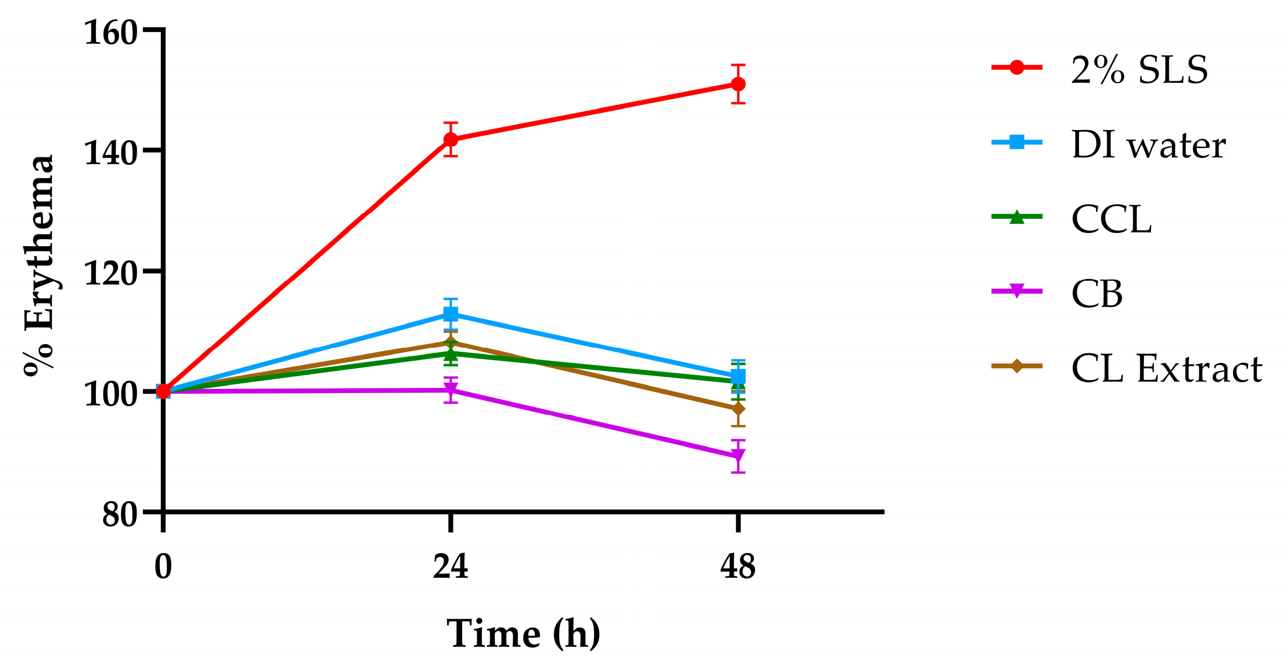
The skin erythema on the upper back of twenty-six healthy volunteers was observed at baseline, 24 h, and 48 h using a skin color probe. The result revealed that the percentages of skin erythema for deionized water, CB, CL, and CCL 24 h after the patch was removed increased significantly compared to baseline (see Figure 9). After 48 h, the percentages of skin erythema decreased and showed no significant difference compared to baseline. These findings suggest that all test substances—including the active-containing formulations—were well tolerated by the skin, with no persistent irritation or adverse reaction.
In contrast, the 2% w/v sodium lauryl sulfate (SLS), used as a positive control, showed significantly elevated erythema at both time points, consistent with its known irritant potential. The absence of visible irritation or positive dermal reactions in the CB, CL, and CCL groups over the 48 h observation period supports the cutaneous compatibility of the formulations. This outcome aligns with the intended use of the products as mild, skin-compatible formulations suitable for cosmetic or cosmeceutical applications.
The skin safety study confirms the safety of the Prunus extract and its formulations, demonstrating their suitability for application on human skin. These findings underscore the potential of CL as a safe ingredient in skincare products designed to reduce inflammation and enhance skin health.

4. Conclusions

This study presents a comparative evaluation of P. cerasifera (CL), P. domestica (EL), and P. salicina (JL) leaf extracts as potential bioactive ingredients for cosmetic applications. Among the three, the CL extract exhibited the highest condensed tannin content and demonstrated strong antioxidant and lipid peroxidation inhibitory activities. Although the extract showed sensitivity to elevated temperatures, it consistently maintained higher antioxidant activity than EL and JL throughout the study period.
A cream formulation containing 0.3% w/w CL extract (CCL) was developed and demonstrated acceptable physicochemical stability under ambient storage conditions for two months. It retained antioxidant activity above 74% under thermal cycling conditions (45 ± 2/4 ± 2), highlighting the stability of the developed cream in terms of both physicochemical and bioactivity.
The in vitro membrane retention study using the Strat-M™ model indicated that the amount of rutin from the CCL formulation that remained within the membrane was 5.7-fold higher than that of the control, with no detection in the receptor chamber. While this synthetic model does not replicate the biological complexity of living skin and cannot predict systemic absorption, the observed retention supports the potential for surface-level delivery, which is consistent with the intended cosmetic application.
Finally, a skin safety assessment in human volunteers showed no signs of irritation, confirming the dermal compatibility of the formulation under the test conditions. Overall, the findings support the feasibility of incorporating CL extract into topical cosmetic formulations aimed at delivering antioxidant activity at the skin surface.

Author Contributions

Conceptualization, W.P., L.M. and E.W.; methodology, L.M., E.W. and W.P.; software, W.P. and N.A.; validation, W.P., L.M., E.W. and P.V.; formal analysis, L.M., E.W., P.V., N.A. and W.P.; investigation, L.M., E.W. and W.P.; resources, L.M., P.T. and E.W.; data curation, L.M., N.A. and W.P.; writing—original draft preparation, L.M. and W.P.; writing—review and editing, L.M., E.W., P.V., P.T., T.R., N.A. and W.P.; visualization, L.M., N.A. and W.P.; supervision, L.M. and W.P.; project administration, L.M.; funding acquisition, L.M. All authors have read and agreed to the published version of the manuscript.

Funding

This research was funded by University of Phayao and Thailand Science Research and Innovation Fund (Fundamental Fund 2024); Grant Number 251/2567. This work was supported in part by Mae Fah Luang University.

Institutional Review Board Statement

The study was conducted in accordance with the Declaration of Helsinki and approved by Ethics Committee of the University of Phayao (HREC-UP_HSST 1.2/113/66) on 11 September 2023.

Informed Consent Statement

Informed consent was obtained from all subjects involved in the study.

Data Availability Statement

The data that support the findings of this study are available from the corresponding author upon reasonable request.

Acknowledgments

The authors are grateful to acknowledge the School of Pharmaceutical Sciences, University of Phayao, for providing space and facilities. The authors also extend their appreciation to the University of Phayao and the Thailand Science Research and Innovation Fund (Fundamental Fund 2024) for their generous support. We also thank Konkanok Saikeawtanakul, Chananchida Srimanee, and Naphatsakorn Jomsri for their work in preparing the extracts and supporting the phytochemical data.

Conflicts of Interest

The authors declare no conflicts of interest.

References

  1. Pauzi, N.A.M.; Cheema, M.S.; Ismail, A.; Ghazali, A.R.; Abdullah, R. Safety assessment of natural products in Malaysia: Current practices, challenges, and new strategies. Rev. Environ. Health 2022, 37, 169–179. [Google Scholar] [CrossRef]
  2. Lee, J.; Hyun, C.-G. Natural Products for Cosmetic Applications. Molecules 2023, 28, 534. [Google Scholar] [CrossRef] [PubMed]
  3. Brar, H.S.; Kaur, P.; Subramanian, J.; Nair, G.R.; Singh, A. Effect of Chemical Pretreatment on Drying Kinetics and Physio-chemical Characteristics of Yellow European Plums. Int. J. Fruit Sci. 2020, 20, S252–S279. [Google Scholar] [CrossRef]
  4. Bioforsk Vest Njøs; Bioforsk Vest Ullensvang; International Society for Horticultural Science; Section Pome and Stone Fruits; International Society for Horticultural Science; Working Group on Plum and Prune Genetics; Breeding and Technology; Norvegian Institute for Agricultural and Environmental Research. Proceedings of the VIIIth International Symposium on Plum and Prune Genetics, Breeding and Pomology: Lofthus, Norway, 5–9 September 2004; Vangdal, E., Sekse, L., Eds.; International Society for Horticultural Science: Leuven, Belgium, 2007.
  5. Wongwad, E.; Preedalikit, W.; Changprasoed, S.; Somsai, S.; Singmee, N.; Srisuksomwong, P.; Srivilai, J.; Rungsang, T.; Mungmai, L. Effects of Different Drying Processes on the Bioactivity and Rutin Content of Prunus spp. (Plums). Int. J. Food Sci. 2024, 2024, 9999731. [Google Scholar] [CrossRef]
  6. Cos, P.; De Bruyne, T.; Hermans, N.; Apers, S.; Berghe, D.V.; Vlietinck, A.J. Proanthocyanidins in health care: Current and new trends. Curr. Med. Chem. 2004, 11, 1345–1359. [Google Scholar] [CrossRef]
  7. Acero, N.; Gradillas, A.; Beltran, M.; García, A.; Muñoz Mingarro, D. Comparison of phenolic compounds profile and antioxidant properties of different sweet cherry (Prunus avium L.) varieties. Food Chem. 2019, 279, 260–271. [Google Scholar] [CrossRef]
  8. Enogieru, A.B.; Haylett, W.; Hiss, D.C.; Bardien, S.; Ekpo, O.E. Rutin as a Potent Antioxidant: Implications for Neurodegenerative Disorders. Oxid. Med. Cell. Longev. 2018, 2018, 6241017. [Google Scholar] [CrossRef] [PubMed]
  9. Gęgotek, A.; Bielawska, K.; Biernacki, M.; Dobrzyńska, I.; Skrzydlewska, E. Time-dependent effect of rutin on skin fibroblasts membrane disruption following UV radiation. Redox Biol. 2017, 12, 733–744. [Google Scholar] [CrossRef]
  10. Gęgotek, A.; Rybałtowska-Kawałko, P.; Skrzydlewska, E. Rutin as a Mediator of Lipid Metabolism and Cellular Signaling Pathways Interactions in Fibroblasts Altered by UVA and UVB Radiation. Oxid. Med. Cell. Longev. 2017, 2017, 4721352. [Google Scholar] [CrossRef]
  11. Coldman, M.F.; Poulsen, B.J.; Higuchi, T. Enhancement of percutaneous absorption by the use of volatile: Nonvolatile systems as vehicles. J. Pharm. Sci. 1969, 58, 1098–1102. [Google Scholar] [CrossRef]
  12. An, R.; Shi, C.; Tang, Y.; Cui, Z.; Li, Y.; Chen, Z.; Xiao, M.; Xu, L. Chitosan/rutin multifunctional hydrogel with tunable adhesion, anti-inflammatory and antibacterial properties for skin wound healing. Carbohydr. Polym. 2024, 343, 122492. [Google Scholar] [CrossRef] [PubMed]
  13. De Tollenaere, M.; Durduret, A.; Chapuis, E.; Lambert, C.; Lemagnen, P.; Tiguemounine, J.; Auriol, D.; Scandolera, A.; Reynaud, R. A highly soluble form of rutin for instant resolution of mask-wearing related disorders. J. Cosmet. Dermatol. 2024, 23, 1734–1744. [Google Scholar] [CrossRef]
  14. Chai, J.; Chen, X.; Jin, C.; Chai, F.; Tian, M. Selective enrichment of Rutin in sunscreen by boronate affinity molecularly imprinted polymer prior to determination by high performance liquid chromatography. Biochem. Eng. J. 2023, 191, 108811. [Google Scholar] [CrossRef]
  15. Choquenet, B.; Couteau, C.; Paparis, E.; Coiffard, L.J. Quercetin and rutin as potential sunscreen agents: Determination of efficacy by an in vitro method. J. Nat. Prod. 2008, 71, 1117–1118. [Google Scholar] [CrossRef] [PubMed]
  16. Tomazelli, L.C.; de Assis Ramos, M.M.; Sauce, R.; Cândido, T.M.; Sarruf, F.D.; de Oliveira Pinto, C.A.S.; de Oliveira, C.A.; Rosado, C.; Velasco, M.V.R.; Baby, A.R. SPF enhancement provided by rutin in a multifunctional sunscreen. Int. J. Pharm. 2018, 552, 401–406. [Google Scholar] [CrossRef]
  17. Plainfossé, H.; Burger, P.; Verger-Dubois, G.; Azoulay, S.; Fernandez, X. Design Methodology for the Development of a New Cosmetic Active Based on Prunus domestica L. Leaves Extract. Cosmetics 2019, 6, 8. [Google Scholar] [CrossRef]
  18. Mungmai, L.; Preedalikit, W.; Rungsang, T.; Sainakham, M. Bioactivity Determination and Development of Oil in Water Emulsion Containing Cassia fistula Bark Extract. Walailak J. Sci. Technol. (WJST) 2021, 18, 8943. [Google Scholar] [CrossRef]
  19. Rebaya, A.; Belghith, S.I.; Baghdikian, B.; Leddet, V.M.; Mabrouki, F.; Olivier, E.; Cherif, J.k.; Ayadi, M.T. Total Phenolic, Total Flavonoid, Tannin Content, and Antioxidant Capacity of Halimium halimifolium (Cistaceae). J. Appl. Pharm. Sci. 2015, 5, 052–057. [Google Scholar]
  20. Wongwad, E.; Pingyod, C.; Saesong, T.; Waranuch, N.; Wisuitiprot, W.; Sritularak, B.; Temkitthawon, P.; Ingkaninan, K. Assessment of the bioactive components, antioxidant, antiglycation and anti-inflammatory properties of Aquilaria crassna Pierre ex Lecomte leaves. Ind. Crops Prod. 2019, 138, 111448. [Google Scholar] [CrossRef]
  21. Saesong, T.; Allard, P.M.; Queiroz, E.F.; Marcourt, L.; Nuengchamnong, N.; Temkitthawon, P.; Khorana, N.; Wolfender, J.L.; Ingkaninan, K. Discovery of Lipid Peroxidation Inhibitors from Bacopa Species Prioritized through Multivariate Data Analysis and Multi-Informative Molecular Networking. Molecules 2019, 24, 2989. [Google Scholar] [CrossRef]
  22. Ariede, M.B.; Gomez Junior, W.A.; Cândido, T.M.; de Aguiar, M.M.G.B.; Rosado, C.; Rangel-Yagui, C.d.O.; Pessoa, F.V.L.S.; Velasco, M.V.R.; Baby, A.R. Would Rutin be a Feasible Strategy for Environmental-Friendly Photoprotective Samples? A Review from Stability to Skin Permeability and Efficacy in Sunscreen Systems. Cosmetics 2024, 11, 141. [Google Scholar] [CrossRef]
  23. Leelapornpisid, P.; Mungmai, L.; Sirithunyalug, B.; Jiranusornkul, S.; Peerapornpisal, Y. A Novel Moisturizer Extracted from Freshwater Macroalga [Rhizoclonium hieroglyphicum (C.Agardh) Kützing] for Skin Care Cosmetic. Chiang Mai J. Sci. 2014, 41, 1195–1207. [Google Scholar]
  24. Sainakham, M.; Promma, B.; Ngernthong, A.; Kiattisin, K.; Boonpisuttinant, K.; Wuttikul, K.; Jantrawut, P.; Ruksiriwanich, W. Preparation and stability investigation of ultrasound-assisted W/O/W multiple nanoemulsions co-loaded with hydrophobic curcumin and hydrophilic arbutin for tyrosinase inhibition. Heliyon 2024, 10, e34665. [Google Scholar] [CrossRef] [PubMed]
  25. Quiñones, R.; Moreno, S.; Smythers, A.L.; Sullins, C.; Pijor, H.; Brown, G.; Trouten, A.; Richards-Waugh, L.L.; Siddig, A. Quantification of Cannabis in Infused Consumer Products and Their Residues on Skin. ACS Pharmacol. Transl. Sci. 2022, 5, 642–651. [Google Scholar] [CrossRef]
  26. Mungmai, L.; Preedalikit, W.; Pintha, K.; Tantipaiboonwong, P.; Aunsri, N. Collagenase and Melanogenesis Inhibitory Effects of Perilla Frutescens Pomace Extract and Its Efficacy in Topical Cosmetic Formulations. Cosmetics 2020, 7, 69. [Google Scholar] [CrossRef]
  27. Poomanee, W.; Yaowiwat, N.; Pattarachaidaecharuch, T.; Leelapornpisid, P. Optimized multiherbal combination and in vivo anti-skin aging potential: A randomized double blind placebo controlled study. Sci. Rep. 2023, 13, 5633. [Google Scholar] [CrossRef]
  28. Naczk, M.; Shahidi, F. Phenolics in cereals, fruits and vegetables: Occurrence, extraction and analysis. J. Pharm. Biomed. Anal. 2006, 41, 1523–1542. [Google Scholar] [CrossRef] [PubMed]
  29. Preedalikit, W.; Chittasupho, C.; Leelapornpisid, P.; Potprommanee, S.; Kiattisin, K. Comparison of Biological Activities and Protective Effects on PAH-Induced Oxidative Damage of Different Coffee Cherry Pulp Extracts. Foods 2023, 12, 4292. [Google Scholar] [CrossRef]
  30. Rodríguez-Pérez, C.; García-Villanova, B.; Guerra-Hernández, E.; Verardo, V. Grape Seeds Proanthocyanidins: An Overview of In Vivo Bioactivity in Animal Models. Nutrients 2019, 11, 2435. [Google Scholar] [CrossRef]
  31. Habib, H.M.; El-Fakharany, E.M.; Kheadr, E.; Ibrahim, W.H. Grape seed proanthocyanidin extract inhibits DNA and protein damage and labile iron, enzyme, and cancer cell activities. Sci. Rep. 2022, 12, 12393. [Google Scholar] [CrossRef]
  32. Bibi, N.; Shah, M.H.; Khan, N.; Al-Hashimi, A.; Elshikh, M.S.; Iqbal, A.; Ahmad, S.; Abbasi, A.M. Variations in Total Phenolic, Total Flavonoid Contents, and Free Radicals’ Scavenging Potential of Onion Varieties Planted under Diverse Environmental Conditions. Plants 2022, 11, 950. [Google Scholar] [CrossRef] [PubMed]
  33. Fratianni, F.; Tucci, M.; Palma, M.D.; Pepe, R.; Nazzaro, F. Polyphenolic composition in different parts of some cultivars of globe artichoke (Cynara cardunculus L. var. scolymus (L.) Fiori). Food Chem. 2007, 104, 1282–1286. [Google Scholar] [CrossRef]
  34. Wijesinghe, W.A.J.P.; Ahn, G.; Lee, W.-W.; Kang, M.-C.; Kim, E.-A.; Jeon, Y.-J. Anti-inflammatory activity of phlorotannin-rich fermented Ecklonia cava processing by-product extract in lipopolysaccharide-stimulated RAW 264.7 macrophages. J. Appl. Phycol. 2013, 25, 1207–1213. [Google Scholar] [CrossRef]
  35. Fraga-Corral, M.; Otero, P.; Echave, J.; Garcia-Oliveira, P.; Carpena, M.; Jarboui, A.; Nuñez-Estevez, B.; Simal-Gandara, J.; Prieto, M.A. By-Products of Agri-Food Industry as Tannin-Rich Sources: A Review of Tannins’ Biological Activities and Their Potential for Valorization. Foods 2021, 10, 137. [Google Scholar] [CrossRef] [PubMed]
  36. Stierlin, E.; Azoulay, S.; Massi, L.; Fernandez, X.; Michel, T. Cosmetic potentials of L. leaves. J. Sci. Food Agric. 2018, 98, 726–736. [Google Scholar] [CrossRef]
  37. Hong, C.Y.; Wang, C.P.; Huang, S.S.; Hsu, F.L. The inhibitory effect of tannins on lipid peroxidation of rat heart mitochondria. J. Pharm. Pharmacol. 1995, 47, 138–142. [Google Scholar] [CrossRef]
  38. Carvalho, R.S.; Carollo, C.A.; de Magalhães, J.C.; Palumbo, J.M.C.; Boaretto, A.G.; Nunes e Sá, I.C.; Ferraz, A.C.; Lima, W.G.; de Siqueira, J.M.; Ferreira, J.M.S. Antibacterial and antifungal activities of phenolic compound-enriched ethyl acetate fraction from Cochlospermum regium (mart. Et. Schr.) Pilger roots: Mechanisms of action and synergism with tannin and gallic acid. South Afr. J. Bot. 2018, 114, 181–187. [Google Scholar] [CrossRef]
  39. Zhang, Q.; Zhu, S.; Lin, X.; Peng, J.; Luo, D.; Wan, X.; Zhang, Y.; Dong, X.; Ma, Y. Analysis of Volatile Compounds in Different Varieties of Plum Fruits Based on Headspace Solid-Phase Microextraction-Gas Chromatography-Mass Spectrometry Technique. Horticulturae 2023, 9, 1069. [Google Scholar] [CrossRef]
  40. Almeida, J.S.; Lima, F.; Ros, S.D.; Bulhões, L.O.S.; de Carvalho, L.M.; Beck, R.C.R. Nanostructured Systems Containing Rutin: In Vitro Antioxidant Activity and Photostability Studies. Nanoscale Res. Lett. 2010, 5, 1603. [Google Scholar] [CrossRef]
  41. Tuominen, A.; Sundman, T. Stability and oxidation products of hydrolysable tannins in basic conditions detected by HPLC/DAD-ESI/QTOF/MS. Phytochem. Anal. 2013, 24, 424–435. [Google Scholar] [CrossRef]
  42. Khanal, R.C.; Howard, L.R.; Prior, R.L. Effect of heating on the stability of grape and blueberry pomace procyanidins and total anthocyanins. Food Res. Int. 2010, 43, 1464–1469. [Google Scholar] [CrossRef]
  43. de Lima Marsiglia, W.I.M.; Oliveira, L.d.S.C.; Lucas Jacinto Almeida, R.; Santos, N.C.; da Silva Neto, J.M.; Santiago, Â.M.; de Melo, B.C.A.; Honorato da Silva, F.L. Thermal stability of total phenolic compounds and antioxidant activities of jaboticaba peel: Effect of solvents and extraction methods. J. Indian Chem. Soc. 2023, 100, 100995. [Google Scholar] [CrossRef]
  44. Baby, A.R.; Migliato, K.F.; Maciel, C.P.M.; Zague, V.; Pinto, C.A.S.D.O.; Salgado, H.R.N.; Kaneko, T.M.; Velasco, M.V.R. Accelerated chemical stability data of O/W fluid emulsions containing the extract of Trichilia catigua Adr. Juss (and) Ptychopetalum olacoides Bentham. Rev. Bras. De Ciências Farm. 2007, 43, 405–412. [Google Scholar] [CrossRef]
  45. Preedalikit, W.; Chittasupho, C.; Leelapornpisid, P.; Qi, S.; Kiattisin, K. Development and Evaluation of Anti-Pollution Film-Forming Facial Spray Containing Coffee Cherry Pulp Extract. Pharmaceutics 2025, 17, 360. [Google Scholar] [CrossRef] [PubMed]
  46. Pyo, S.M.; Meinke, M.; Keck, C.M.; Müller, R.H. Rutin—Increased Antioxidant Activity and Skin Penetration by Nanocrystal Technology (smartCrystals). Cosmetics 2016, 3, 9. [Google Scholar] [CrossRef]
  47. Gullón, B.; Lú-Chau, T.A.; Moreira, M.T.; Lema, J.M.; Eibes, G. Rutin: A review on extraction, identification and purification methods, biological activities and approaches to enhance its bioavailability. Trends Food Sci. Technol. 2017, 67, 220–235. [Google Scholar] [CrossRef]
  48. Cândido, T.M.; De Oliveira, C.A.; Ariede, M.B.; Velasco, M.V.R.; Rosado, C.; Baby, A.R. Safety and Antioxidant Efficacy Profiles of Rutin-Loaded Ethosomes for Topical Application. AAPS PharmSciTech 2018, 19, 1773–1780. [Google Scholar] [CrossRef] [PubMed]
  49. Park, S.N.; Lee, H.J.; Gu, H.A. Enhanced skin delivery and characterization of rutin-loaded ethosomes. Korean J. Chem. Eng. 2014, 31, 485–489. [Google Scholar] [CrossRef]
  50. Som, I.; Bhatia, K.; Yasir, M. Status of surfactants as penetration enhancers in transdermal drug delivery. J. Pharm. Bioallied Sci. 2012, 4, 2–9. [Google Scholar] [CrossRef]
  51. Williams, A.C.; Barry, B.W. Urea analogues in propylene glycol as penetration enhancers in human skin. Int. J. Pharm. 1989, 56, 43–50. [Google Scholar] [CrossRef]
  52. Lee, C.-H.; Bhatt, P.P.; Chien, Y.W. Effect of excipient on drug release and permeation from silicone-based barrier devices. J. Control. Release 1997, 43, 283–290. [Google Scholar] [CrossRef]
  53. Neupane, R.; Boddu, S.H.S.; Renukuntla, J.; Babu, R.J.; Tiwari, A.K. Alternatives to Biological Skin in Permeation Studies: Current Trends and Possibilities. Pharmaceutics 2020, 12, 152. [Google Scholar] [CrossRef] [PubMed]
  54. Haq, A.; Goodyear, B.; Ameen, D.; Joshi, V.; Michniak-Kohn, B. Strat-M® synthetic membrane: Permeability comparison to human cadaver skin. Int. J. Pharm. 2018, 547, 432–437. [Google Scholar] [CrossRef] [PubMed]
Figure 1. Visual appearance of the three types of crude extracts and their corresponding fresh leaves from Prunus species: (A) P. domestica (EL), (B) P. salicina (JL), and (C) P. cerasifera (CL). All extracts displayed a dark green color and were obtained via ethanolic extraction.
Cosmetics 12 00146 g001
Figure 2. Physicochemical characteristics of the Prunus species leaf extracts (EL, JL, and CL) during storage at 4 ± 2 °C, 25 ± 2 °C, and 45 ± 2 °C over 60 days. (AC) show the pH profiles of EL, JL, and CL, respectively. (DF) display the color variation (∆E) for the same extracts under identical conditions. Each bar represents the mean ± SD of triplicate measurements.
Cosmetics 12 00146 g002
Figure 3. Stability of TPC in EL, JL, and CL after storage at different temperatures (4 ± 2 °C, 25 ± 2 °C, and 45 ± 2 °C) over 60 days. The data is expressed as the percentage remaining relative to the initial value (day 0). Different letters (a, b, c) indicate statistically significant differences among extracts at the same time point and storage condition (ANOVA with Tukey’s HSD, p < 0.05). Asterisks (*) denote significant differences compared to day 0 within the same extract and temperature condition (p < 0.05).
Cosmetics 12 00146 g003
Figure 4. Stability of TTC in EL, JL, and CL after storage at different temperatures (4 ± 2 °C, 25 ± 2 °C, and 45 ± 2 °C) over 60 days. The data is expressed as the percentage remaining compared to the initial value (day 0). Different letters (a, b) denote statistically significant differences among extracts at the same time point and temperature (ANOVA with Tukey’s HSD, p < 0.05). Asterisks (*) indicate significant differences compared to day 0 within the same extract and storage condition (p < 0.05).
Cosmetics 12 00146 g004
Figure 5. Stability of antioxidant activity (DPPH assay) in EL, JL, and CL after storage at 4 ± 2 °C, 25 ± 2 °C, and 45 ± 2 °C over a period of 60 days. The data is expressed as IC50 (μg/mL) and different letters (a, b, c) represent statistically significant differences among extracts at the same time point and storage condition (ANOVA with Tukey’s HSD, p < 0.05). Asterisks (*) indicate significant differences compared to day 0 within the same extract and storage condition (p < 0.05).
Cosmetics 12 00146 g005
Figure 6. Physical appearance of the cream formulation containing CL. (A) Stability under ambient storage at 25 ± 2 °C from Day 0 to Day 60. (B) Stability under heating/cooling cycles from Day 0 to Day 24. No visible changes in color, phase separation, or homogeneity were observed under either condition.
Cosmetics 12 00146 g006
Figure 7. Antioxidant activity of CCL during storage. Antioxidant activity, measured as % DPPH inhibition, was evaluated over time under two conditions: (1) ambient storage at 25 ± 2 °C on Days 0, 7, 14, 21, 28, and 60, and (2) thermal stress via a heating/cooling (H/C) cycle consisting of alternating 48 h periods at 45 ± 2 °C and 4 ± 2 °C, assessed on Days 0, 4, 8, 12, 16, 20, and 24.
Cosmetics 12 00146 g007
Figure 8. Comparison of rutin retention within a synthetic membrane after 8 h between the 0.3% w/w CL solution and the CCL. Results are expressed as means ± SD (n = 3). Asterisk (*) indicates a statistically significant difference between the groups (p < 0.05), with the CCL formulation showing significantly higher rutin retention than the aqueous extract alone.
Cosmetics 12 00146 g008
Figure 9. Changes in skin erythema (%) over time after patch removal. The percentage of skin erythema was measured at baseline (day 0) and at 24 h and 48 h post-removal in volunteers exposed to patches containing five different test substances: 2% w/w sodium lauryl sulfate (SLS) as a positive control, deionized water (DI water) as a negative control, cream containing P. cerasifera extract (CCL), cream base (CB), and Prunus cerasifera (CL) extract. Data are expressed as means ± standard deviation (n = 26).
Cosmetics 12 00146 g009
Table 1. Ingredient composition of the developed topical cream formulation containing Prunus leaf extract.
IngredientFunctionConcentration (% w/w)
Deionized WaterSolvent55.0–60.0
Propylene GlycolHumectant/Penetration Enhancer5.0–8.0
Cyclopentasiloxane and Dimethicone/Vinyl Dimethicone CrosspolymerEmollient/Film Former5.0–8.0
Cetearyl OlivateEmulsifier5.0–8.0
Cyclopentasiloxane and Dimethicone CrosspolymerEmollient3.0–5.0
Cyclopentasiloxane and CyclotetrasiloxaneEmollient3.0–5.0
Cyclopentasiloxane and DimethiconolEmollient2.0–3.0
DimethiconeEmollient1.0–2.0
Polyacrylamide (and) C13–14 Isoparaffin (and) Laureth-7Thickener/Stabilizer1.0–2.0
Ammonium Acryloyldimethyltaurate/VP CopolymerThickener/Stabilizer0.5–1.0
PhenoxyethanolPreservative0.5–1.0
Prunus Leaf Extract (CL, Rutin = 12.1 ± 0.1% w/w)Active Ingredient0.3
Table 2. Grading criteria of skin reactions by Cosmetic, Toiletry, and Fragrance Association Guideline.
SymbolGradeClinical Description
-0Negative reaction
+1Slight erythema, either spotty or diffuse
++2Moderate uniform erythema
+++3Intense erythema with edema
++++4Intense erythema with edema and vesicles
Table 3. Percentage yield of extracts from the leaves of three Prunus species (EL, JL, and CL).
Sample% Yield
EL8.5 ± 0.5 c
JL7.8 ± 0.8 b
CL7.6 ± 0.5 a
Note: Data are presented as means ± S.D., with statistical comparisons conducted using Tukey’s HSD test following one-way ANOVA. Superscript letters (a, b, c) in the same column indicate significance; identical letters denote no significant difference, while different letters represent significant differences (p < 0.05).
Table 4. TPC, TTC, DPPH radical scavenging activity, and lipid peroxidation inhibitory activity of ethanolic extracts from the leaves of three Prunus species, positive control (Trolox), and reference standard (Rutin).
SampleTPC
(mgGAE/g Extract)
TTC
(mgCAE/g Extract)
DPPH
IC50 (µg/mL)
Lipid Peroxidation
Inhibitory Activity
(% Inhibition)
EL15.0± 2.2 a28.8 ± 2.0 a24.9 ± 1.9 a52.7 ± 1.9 a, b
JL16.0 ± 2.2 a36.0 ± 1.8 a24.9 ± 3.1 a48.3 ± 2.2 a
CL10.9 ± 1.3 b49.7 ± 3.2 b22.7 ± 3.1 a62.3 ± 1.0 b
Trolox--4.4 ± 0.2 c96.6 ± 2.3 c
Rutin--12.8 ± 0.4 b48.9 ± 1.1 a
Note: Data are presented as means ± S.D., with statistical comparisons conducted using Tukey’s HSD test following one-way ANOVA. Superscript letters (a, b, c) in the same column indicate significance; identical letters denote no significant difference, while different letters represent significant differences (p < 0.05).
Table 5. The primary dermal irritation index (PDII) and skin irritation reaction were observed in twenty-six healthy volunteers.
Tested SubstancesPDII ValueClassification of Skin Reaction
2% w/v sodium lauryl sulfate1.83Slight irritation
Deionized water0.00Non-irritation
CCL0.14Non-irritation
CB0.06Non-irritation
CL extract0.08Non-irritation
Note: CCL = cream containing CL extract, CB = cream base, CL = P. cerasifera leaf extract.
Disclaimer/Publisher’s Note: The statements, opinions and data contained in all publications are solely those of the individual author(s) and contributor(s) and not of MDPI and/or the editor(s). MDPI and/or the editor(s) disclaim responsibility for any injury to people or property resulting from any ideas, methods, instructions or products referred to in the content.