Article

In Vitro Examination of Fungal and Root Extracts Inspired by Traditional Medicine for Potential Periorbital Eye Infrastructure Treatments

1
Vantage, Fairfield, NJ 07004, USA
2
Vantage, Warren, NJ 07059, USA
3
Vantage, Gurnee, IL 60031, USA
4
Bioinnovation Laboratories, Lakewood, CO 80235, USA
*
Author to whom correspondence should be addressed.
Cosmetics 2025, 12(3), 95; https://doi.org/10.3390/cosmetics12030095
Submission received: 13 February 2025 / Revised: 25 March 2025 / Accepted: 30 April 2025 / Published: 8 May 2025
(This article belongs to the Section Cosmetic Dermatology)

Abstract

:
An early indicator of aging may appear around the eyes and the surrounding eye infrastructure. With aging, there come diminishing changes in vascular microcirculation and the accumulation of hemoglobin by-products that gather in the fatty pads beneath the eyes as dark circles, akin to skin bruising. In addition, the extracellular matrix that surrounds the eye is exposed to external threats like UV radiation, weather and pollution, as well as lifestyle choices that create fatigue. This causes the eyes to express wrinkles well before they begin to appear on the rest of the face, particularly in the corners of the eyes called the crow’s feet region. Consumers spend considerable amounts of resources combatting these effects. If consumers could treat some of the sources of these problems, in advance of the inevitable influences of aging, a kind of prejuvenation of the eye infrastructure, then perhaps the inevitable outcomes of aging apparent around the eyes could be slowed. This paper examines the development and in vitro testing of two unique botanical extracts, one based on a traditional medicine mushroom called Phellinus linteus (Huang Sang) and the other based on a traditional medicine root from the plant Angelica polymorpha sinensis (Dong Quai). When combined, these two extracts create a blend called ANGEL-EYE EFX® [INCI: Water (and) Glycerin (and) Phellinus Linteus Extract (and) Angelica polymorpha sinensis Root Extract]. There are several key biomolecules of interest present in this blend, including hispolon, dihydrozingerone, and arginine, as demonstrated using advanced liquid chromatography/mass spectral analyses. The individual extracts were also broadly examined using human genomic microarray assays and then more specifically for their ability to influence several important skin proteins associated with undereye skin aging, including CYGB (Human Cytoglobin), OXSR1 (Oxidative Stress Response Kinase-1), LCE3B (Late Cornified Envelope-3B), EGFR (Epidermal Growth Factor Receptor), VEGFA (Vascular Endothelial Growth Factor-1), and NINJ1 (Ninjurin-1). It was found that the treatment of Normal Human Epidermal Keratinocytes (NHEKs) with increasing concentrations of the active blend between 0.05 and 2.0% showed statistically significant increases in all the proteins noted except VEGFA, which showed a statistically significant decrease in protein expression with the treatment of the Angelica polymorpha sinensis extract at 1.0%.

1. Introduction

In 2023, it was reported that Americans spent approximately USD 422 M just on surgical rejuvenating procedures around the eyes [1]. Ideally, it would be beneficial if topical cosmetic treatments help maintain and improve the skin infrastructure around the eyes, leading to less need for more aggressive surgical and injection procedures. In youth, for both men and women, the skin around the eyes is tight and luminescent, arising partly from the remarkable blood flow that takes place through hundreds of small capillaries that surround the eye [2,3,4]. Driven partially by an individual’s genetics but also by extrinsic aging factors, these tiny capillaries become more fragile and dilated, blood flow slows, and the vascular structure begins to allow leakage of red blood cells and, more importantly, hemoglobin, into the tissues surrounding the undereye. This leaked hemoglobin begins to degrade through normal reactive oxygen species (ROS) oxidative processes and produces hemoglobin by-products, like bilirubin and hemosiderin, that are darker than the original hemoglobin from which they formed, often being associated with skin bruising [5,6,7]. In the area of the eye, this hemoglobin breakdown begins to manifest itself as dark circles. Normal drainage of these by-products through the skin’s lymph system also slows, causing the accumulation of these by-products into fatty areas below the eyes, creating the appearance of dark, baggy, puffy eyes. The measurement of the blood flow around the eyes is one way to assess the damage to the fine capillaries that release blood into the surrounding fatty tissues. Specifically, laser Doppler flowmetry has become a popular, non-invasive method with which to measure microcirculatory blood flow around the eyes [8,9].
In addition, the skin becomes laxer as collagen, elastin, and other extracellular matrix proteins degrade in the thin skin above and below the eyes [10,11]. The breakdown in these critical skin proteins can provide the opportunity for advanced glycation end-products (AGEs) to form, creating a yellow coloring in aging skin [12,13,14]. Additionally, the breakdown of these proteins is driven principally by the thinness of the skin above and below the eyes, which allows for more aggressive oxidative effects and UV damage accumulation. As these oxidative processes progress, the areas around the eyes begin to start looking older, and this is one of the more prominent places where aging becomes visible.
There has been extensive focus on topical treatments that might impact multifactorial sources of eye aging with additional focus on ingredients that are natural and have historic aspects of use. Natural ingredients sourced from traditional medicine, commonly used for years in Asia and India, are often highly sought after [15,16]. The mushroom Phellinus linteus (commonly called Huang Sang mushroom) is a popular Asian fungus known for its medicinal benefits [17,18,19]. Of particular interest regarding eye treatments is the ability of mushroom extracts to stimulate vascular circulation and to modulate skin wrinkles [20,21]. The mushroom contains several active molecules of interest, but one specifically, the polyphenol Hispolon has been studied deeply for its therapeutic effects [22,23]. Angelica polymorpha sinensis root (commonly referred to as Dong Quai) is also a well-studied traditional herb known in the Ayurvedic traditional medicines [24,25]. The root expresses numerous biologically interesting molecules with a polyphenol called Feruloylmethane (commonly referred to as Dihydrozingerone, a derivative of curcurmin), showing potent antioxidant and healing benefits [26].
This paper will review initial analytical and in vitro work conducted on a traditional fungal mushroom extract from Phellinus linteus. and a curcumin-like root extract from Angelica polymorpha sinensis that have been strategically combined and referred to as ANGEL-EYE EFX® [INCI: Water (and) Glycerin (and) Phellinus linteus Extract (and) Angelica polymorpha sinensis Root Extract]. This paper will describe methods to analyze the blend for some strategically important biomolecules using advanced liquid chromatography/mass spectral (LC/MS) analysis. This work will also examine the in vitro benefits of the individual botanical extracts using human genomic microarray assays followed by more in-depth protein assays looking at the influence of the individual extracts to stimulate proteins anticipated to be important for improving the infrastructure around the eye.

2. Materials and Methods

2.1. Extract Preparations

2.1.1. Phellinus linteus Mushroom Extract

The Phellinus linteus mushroom extract was supplied by Vantage [Fairfield, NJ, USA] and was prepared by extracting the mushroom cap powder using a water/glycerin extraction solvent. The exact concentrations of the mushroom powder employed are considered by the company to be proprietary.

2.1.2. Angelica polymorpha sinensis Root Extract

The Angelica polymorpha sinensis root extract was provided by Vantage and was prepared from root powder using the same glycerin/water extraction solvent used previously to create the mushroom extract. The exact concentrations of the root powder used are considered by the company to be proprietary.

2.2. High-Performance Liquid Chromatography/Mass Spectral (HPLC/QTOF-MS) Analysis of Blend

Method Parameters: Chromatographic separation was performed on an Agilent Infinity 1260 HPLC [Agilent Technologies, Santa Clara, CA, USA] system using an Agilent Poroshell 120 EC-C18 (2.7 µm, 3.0 × 100 mm2) reversed-phase column. The column temperature was set to 35 °C with a flow rate of 0.5 mL/min. The mobile phase consisted of methanol (A) and water (B), and the gradient scheme was as follows: 0–5 min, 50% A; 5–10 min 50% A to 90% A; 5 min at 90% A.
Detection was performed using an Agilent QTOF-MS (model G6530C) with dual AJS electrospray ionization in negative ion mode using the following parameters: drying gas (N2) flow rate, 10 L/min; drying gas temperature, 325 °C; nebulizer, 35 psig; sheath gas temperature, 325 °C; sheath gas flow, 10 L/min; capillary, 3000 V; skimmer, 65 V; fragmentor, 175 V. The acquisition was controlled using Agilent MassHunter Acquisition Software Ver. B.08.00, and the data were processed with MassHunter Qualitative software Ver. B.07.00. MS mode measurements were made over a range of m/z 100–3000. For hispolon identity confirmation, targeted MS/MS mode spectra were collected with collision energies of 20 eV with a targeted precursor ion of 219.065 m/z. Accurate mass measurements were obtained by means of reference ion correction using reference masses at m/z 112.9855 and 966.0007 in negative mode.
Chemicals and Extracts: A hispolon standard was purchased from Santa Cruz Biotechnology [Dallas, TX, USA] and diluted in 50:50 methanol/water (0.01 mg/mL). The blend of Phellinus linteaus mushroom and Angelica polymorpha sinensis root extracts was diluted in 50:50 methanol/water (160 mg/mL) prior to HPLC/QTOF-MS analysis. The identification of other specific molecular peaks was partially based on active molecular structures identified in the literature for these botanical extracts. Theoretical monoisotopic masses for those compounds were identified using ChemSpider software Ver B.07.00 as shown www.chemspider.com, last accessed 5 January 25 and compared to the observed masses in the gathered spectra.

2.3. In Vitro Tissue Testing

2.3.1. Phellinus linteus Mushroom Extract Human Microarray Studies

All cell lines used in the in vitro studies described below were adult primary cell cultures taken from Normal Human Epidermal Keratinocytes, which were purchased commercially from Thermo Fisher Scientific [[Waltham, MA, USA] Catalog Number C0055C].
The methods employed to run the human gene microarrays on the Phellinus linteus extract have been reported in an earlier publication [27]. For the human gene microarray, the mushroom extract was evaluated at 1.0% for 24 h on Normal Human Epidermal Keratinocytes (NHEKs) isolated from an adult. From the +19 K genes examined in the Agilent Human DNA Microarray [Agilent Technologies, Santa Clara, CA, USA], a smaller subset of 244 skin-relevant genes was employed to determine how the mushroom extract impacted the genes of the skin [27].

2.3.2. Phellinus linteus Mushroom Extract Protein Studies

Human Keratinocyte Cell Culture: [NHEKs] taken from adult skin were grown using EpiLife Media (60 µM calcium) supplemented with 0.2% v/v bovine pituitary extract, 1 µg/mL recombinant human insulin-like growth factor-I, 0.18 µg/mL hydrocortisone, 5 µg/mL bovine transferrin, and 0.2 ng/mL human epidermal growth factor. The cells were cultured at 37 ± 2 °C and 5 ± 1% CO2. When enough cells had grown, they were seeded into 12-well plates and grown until confluent.
Treatment of Keratinocytes: The cell culture media for the cultured keratinocytes was removed and replaced with media containing the test materials, and the cells were then incubated for 48 h. At the end of the incubation period, the cells were washed once with PBS and then lysed with 100 μl of extraction buffer. The protein concentration of the cell lysates was then determined using a BCA assay.
ELISA Protein Assays: The proteins of interest included the following: CYGB (Human Cytoglobin, Abexxa, Cambridge, UK, catalog# abx151248), OXSR1 (Oxidative Stress Response Kinase-1, Abexxa, catalog# abx382011), LCE3B (Late Cornified Envelope-3B, Abexxa, catalog# abx352190), EGFR (Epidermal Growth Factor Receptor, R&D Systems, Minneapolis, MN, USA catalog# DY231), VEGFA (Vascular Endothelial Growth Factor-1, MyBioSource, San Diego, CA, USA catalog# MBS175951), and NINJ1 (Ninjurin-1, MyBioSource, catalog# MBS915725).
For the ELISA assays, 100 μL of standards or diluted lysates (10 μL lysate diluted with 90 μL of sample buffer) or medial samples was added to the wells of an ELISA plate, and the plate was incubated for 90 min at 37 °C. At the end of the incubation period, the ELISA plate was emptied, and 100 μL of biotinylated detection antibody solution was added to each well. The plate was incubated for 60 min at 37 °C and then washed three times with a wash buffer. After the final wash was removed, 100 μL of an avidin-enzyme solution was added to each well, and the plate was incubated for 30 min at 37 °C. After this final incubation, the plate was washed five times with a wash buffer. Once the last wash was removed, 100 µL of substrate solution was added to each well. When a sufficient level of color development occurred, 50 µL of stop solution was added to each well, and the plate was read at 460 nm.

2.4. Angelica polymorpha sinensis Root Extract Human Microarray and Protein Studies

The human gene microarray was run similarly to the study run with the mushroom except that the treatments were run on Epiderm EFT tissues [Mattek, Ashland, MA, USA]. The treatment level was 0.1% on the tissues for 48 h. The arrays employed were the OneArray platform from Phalanx Biotech (Palo Alto, CA, USA). The results of the arrays were culled with the same set of 244 skin-associated genes noted for the mushroom extract. Protein ELISA assay test methods for the Angelica polymorpha sinensis Root Extract were the same as described above.

2.5. Statistical Analysis

Treatment means were compared using ANOVA, with n = 3 per treatment. Statistical significance was set at p < 0.05. Tukey’s post hoc analysis was conducted after the ANOVA to determine which treatment means were significantly different from the untreated group.

3. Results and Discussion

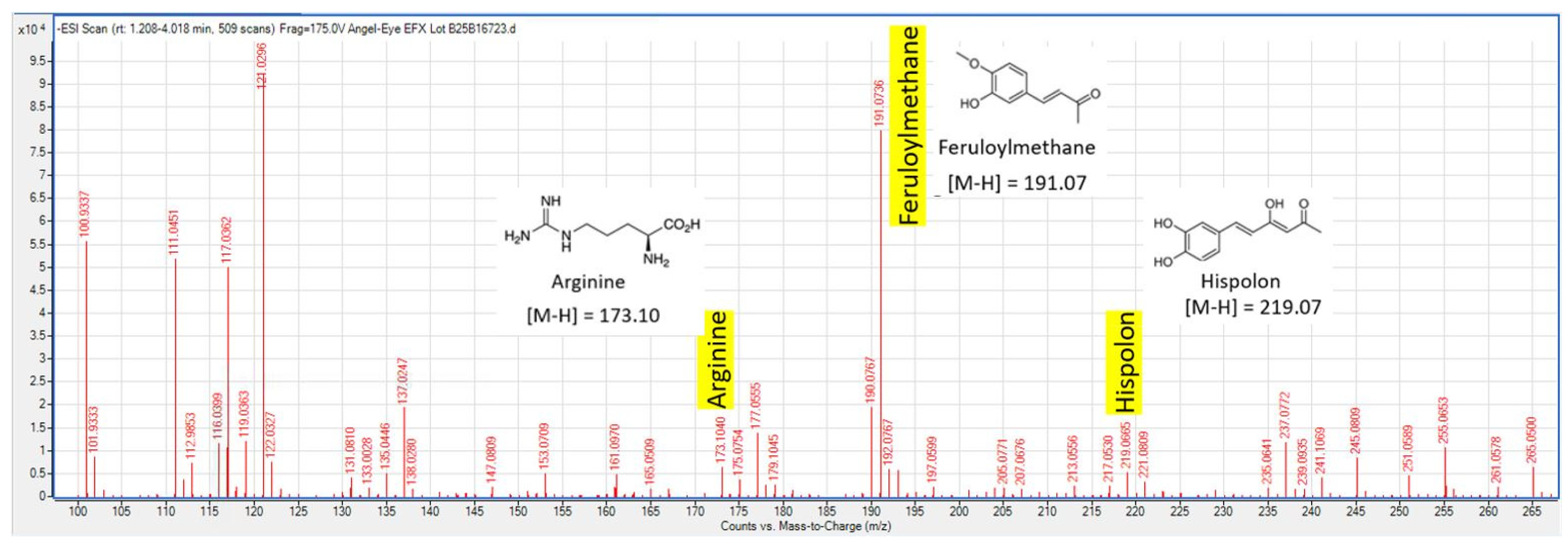
3.1. Liquid Chromatography/Mass Spectral (LC/MS) Analysis of Blended Product

To establish that each botanical source, the Phellinus linteus mushroom and the Angelica polymorpha sinensis root, was contributing key molecular biomarkers to the blend, LC/MS analysis was conducted on the blend. The results are shown below in the image of the LC/MS chromatogram shown in Figure 1.

3.2. Results of In Vitro Testing

To create a product designed to help improve the appearance of the eye infrastructure, six key proteins were identified from the 244 culled skin critical genes found from the human microarray work, Table 1, vida infra. These proteins, shown below in Figure 2, are linked to improvements in the key aspects of eye structure known to change with aging.

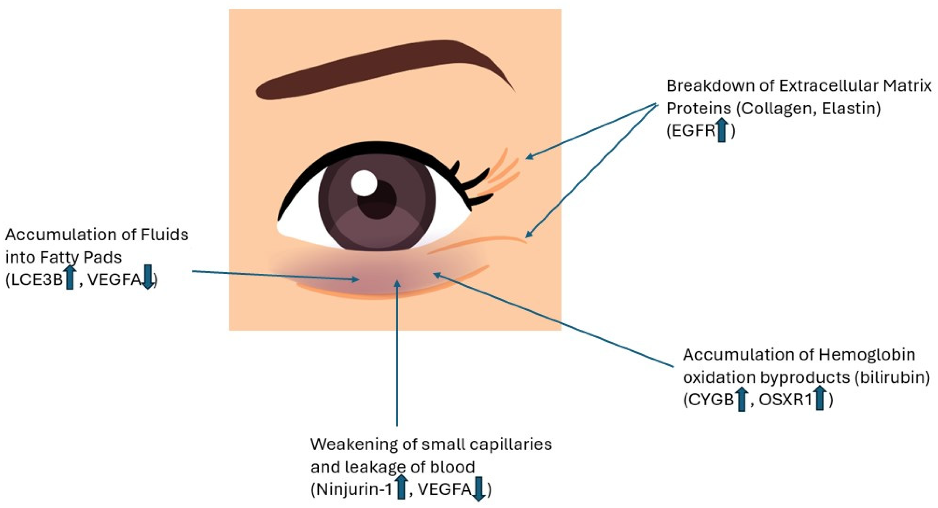
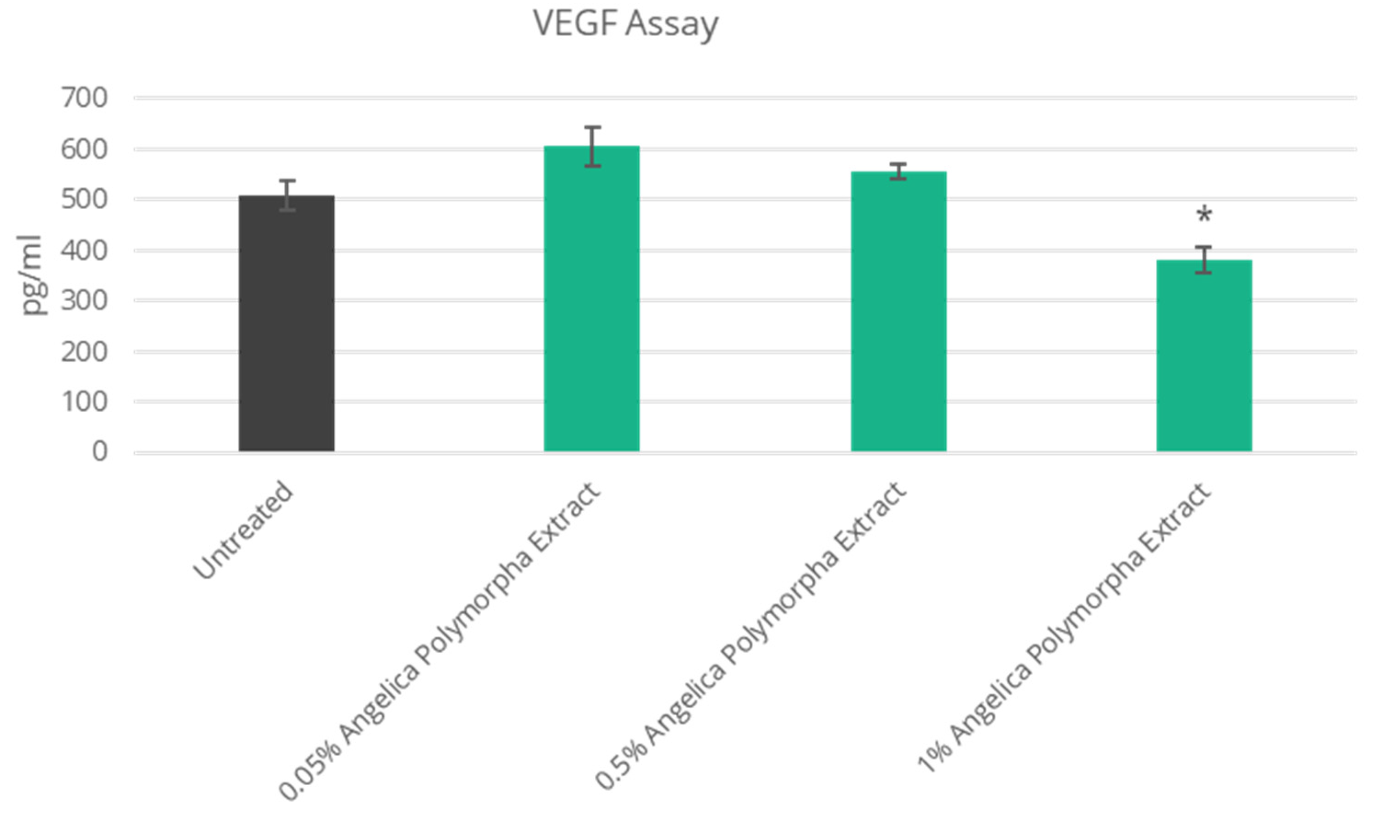
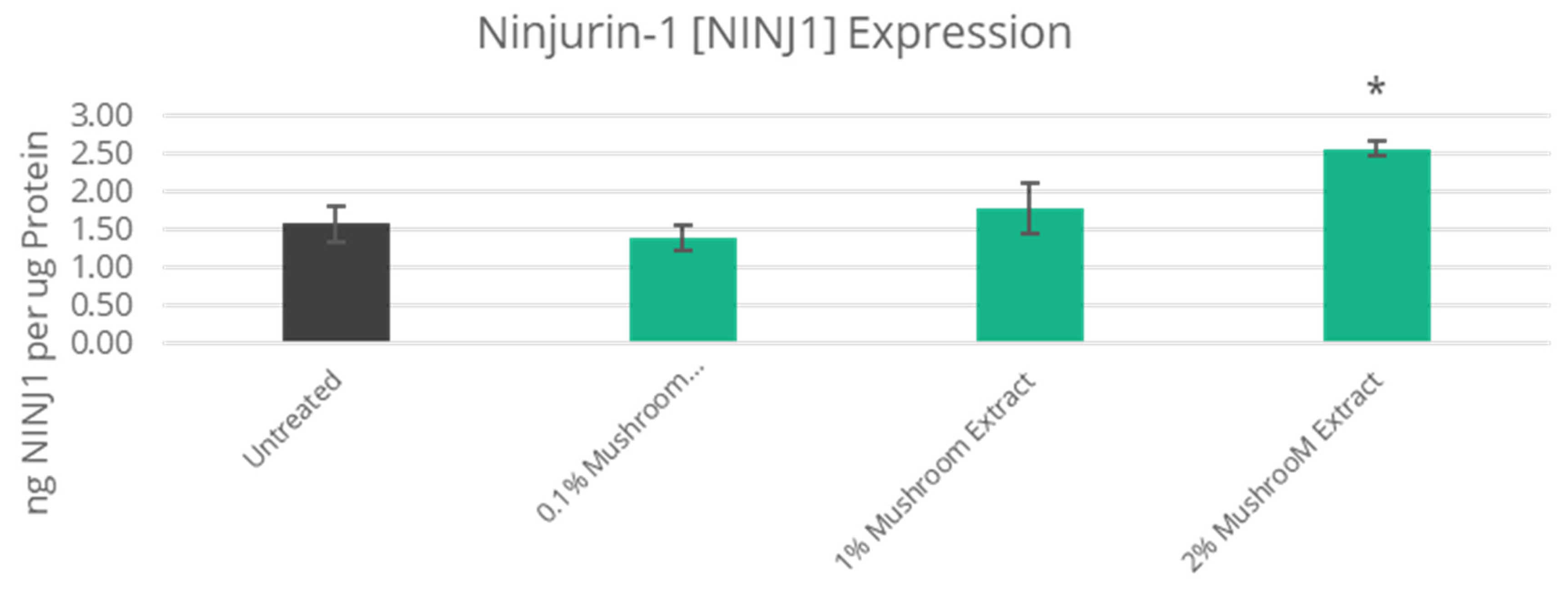
3.3. Improved Skin Vascularization and Fatty Pad Deposits

Because capillary vascularization is critical to the eye’s dark circle formation and improved blood flow, the proteins examined that influence capillary vascularization are Vascular Endothelial Growth Factor-A (VEGFA) and Ninjurin-1 (NINJ1). Overexpression of VEGFA leads to capillary dilation and can result in increased leakage from expanded blood vessels [3,5]. In the protein assays, it was found that the Angelica polymorpha sinensis extract was able to decrease the expression of VEGFA by 25% compared to untreated cells, Figure 3. The expression of Ninjurin-1 has a positive effect on strengthening blood vessels and is critical for new angiogenesis, or the growth of new blood vessels [28,29]. The mushroom extract had a positive effect on the expression of Ninjurin-1 protein, increasing the expression of the protein in skin cells by 62% at a treatment level of 2%, Figure 4.
The improvement of the eyes’ fatty pads and the improvement of the skin’s lymph drainage are also influenced by the function of the Late Cornified Envelop-3B (LCE3B) protein, which is critical for the development of the skin’s barrier integrity [30]. The lack of expression of the LCE3B protein in skin has been linked to poor skin development and inflammation [31]. It was found that both the mushroom and the root extracts had a positive stimulatory effect on the expression of the gene for this protein and, when tested on NHEKs, the mushroom demonstrated a 100% increase in the expression of this protein compared against untreated cells at 2% treatment levels, Figure 5.
The root extract, while also demonstrating a positive gene response for this protein, was not evaluated using ELISA protein assays because of the profound effects seen for the mushroom extract on this protein target.

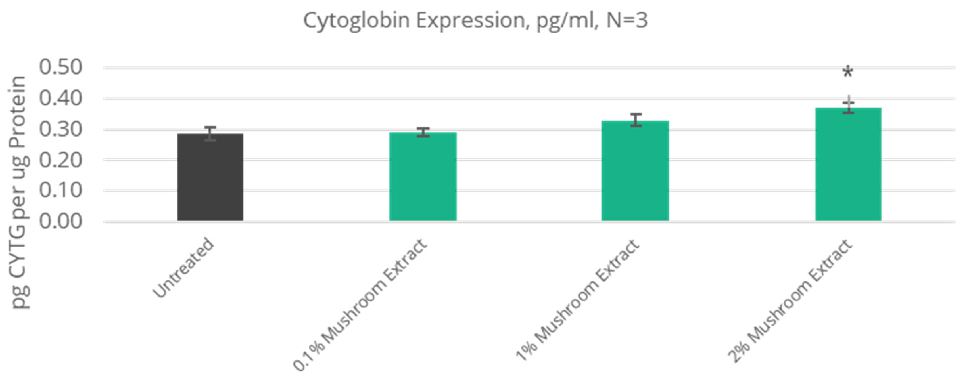
3.4. Improved Antioxidant Defense

Human cytoglobin (CYGB) is a recently discovered fourth form of human globin proteins that also include hemoglobin, myoglobin, and neuroglobin [32,33,34,35]. The structure of human cytoglobin bears a striking resemblance to neuroglobin and has been suggested to be a potent antioxidant protein with superoxide dismutase-type activity that is expressed during times of cellular stress [36]. The protein has been identified to be expressed in Normal Human Epidermal Keratinocytes and Normal Epidermal Melanocytes and therefore is an important antioxidant protein found in skin. Cytoglobin is also well known to be involved in the control of nitric oxide expression in the skin and blood vessels, which is linked closely to vascular health and vascular tone as well as to the control of blood flow in the blood vessels [37]. The protein assay demonstrated a statistically significant 29% increase in the cytoglobin protein expression on NHEKs through treatment with the mushroom extract, Figure 6.
Oxidative Stress Response Kinase-1 (OXSR1) is another key antioxidant protein found expressed in keratinocytes and immune cells that is upregulated during times of oxidative stress via the inflammasome-mediated pathways to inflammation [38]. This protein has been associated with the sleep/wake cycles and interacts with melatonin during these cycles [39]. When evaluated using protein assays, the mushroom extract demonstrated a 68% increase in OXSR1 protein expression in NHEKs at a 2% treatment level, Figure 7.

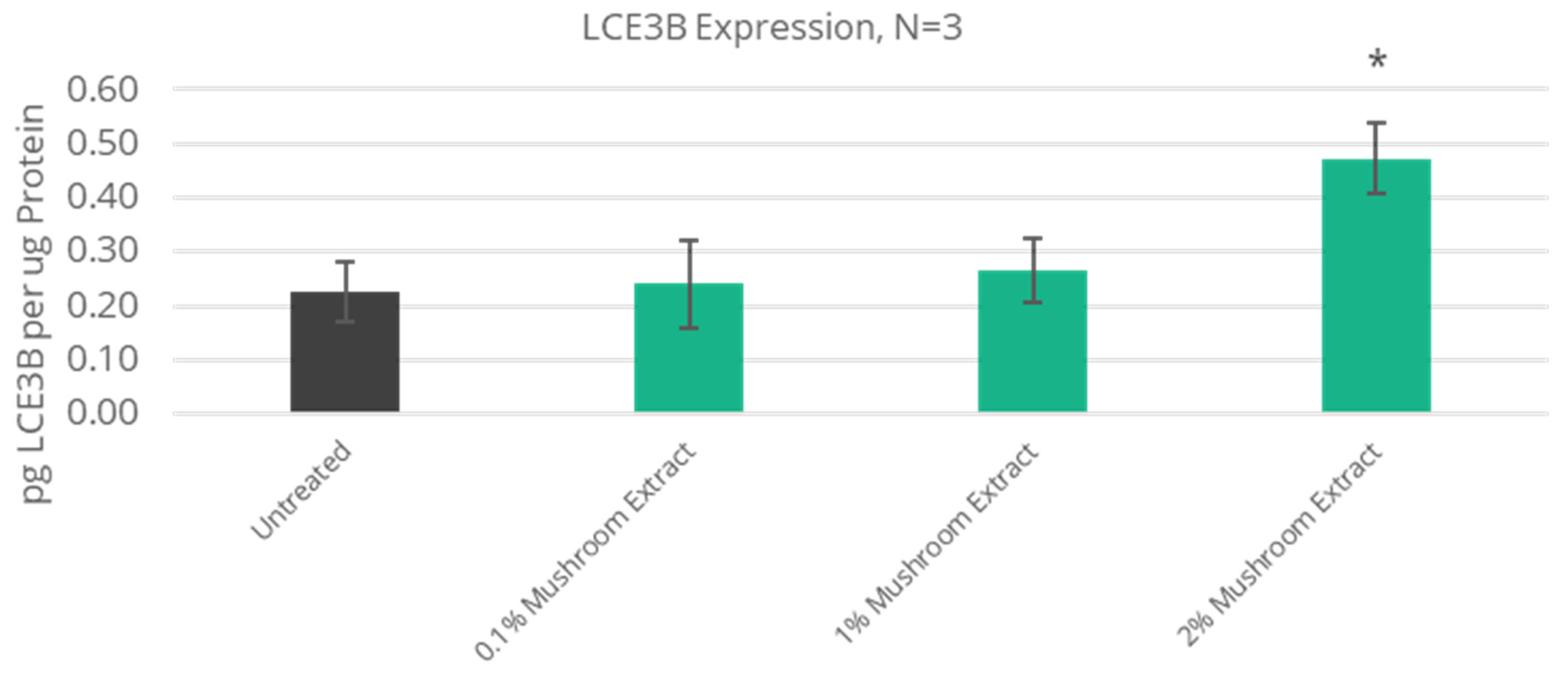
3.5. Improved Extracellular Matrix Protein Expression

The final skin protein target of importance in the rebuilding of the eye infrastructure is the degradation associated with the extracellular matrix (ECM) proteins like collagen, elastin, and filaggrin, among others. The kind of master switch for much of the ECM protein is the Epidermal Growth Factor Receptor (EGFR), which is expressed in keratinocytes and regulates keratinocyte stem cell health but also communicates closely with the fibroblasts found in the dermis through various binding proteins [40,41]. In increasing the expression of this growth factor receptor, improvements in skin structure can commence. It was found that both the mushroom and the root had a stimulatory effect on the expression of EGFR, and the root extract demonstrated a 20% increase in the expression of this protein in NHEKs compared against untreated cells, Figure 8. This target was not assessed for protein expression with the mushroom extract, principally because the root extract demonstrated significant improvement in the expression of this key structural protein.
A complete summary of the human microarray testing and the ELISA protein assays results for each of the individual ingredients is shown below in Table 1.
The work presented here delves into the in vitro influences that extracts of two botanicals known for their use in traditional medicine have on controlling various aspects of periorbital infrastructure in aging eyes. Because the reported work is in vitro, there are limitations on understanding how the individual proteins discussed work in tandem to help improve aging eye conditions. In addition, while the work here focuses on the influence that these strategic proteins have on topical eye improvements, the proteins likely have numerous other impacts in skin function, including influencing wound repair, barrier function, and skin’s innate immune response, which may likely be impacted by the topical application of botanicals. Being of botanical origin, the extracts may even modulate the skin’s microbiota in ways that are not addressed in this paper. Future work and that presently ongoing will look at the human clinical aspects of these botanicals and will help to improve the discussion of the efficacy of botanicals as topical periorbital treatments. However, as is typical of ingredients developed for cosmetic applications, such clinical testing will be non-invasive and will not be able to directly address how these in vitro targets are impacted, regulated, or function directly. This would require the use of skin biopsies with human volunteers, and such testing is not common for ingredients intended for cosmetic applications.

4. Conclusions

Aging eyes are the sign of an aging face. This paper summarizes analytical and in vitro studies conducted on two unique botanical extracts, one from the traditional medicinal mushroom Phellinus linteus and the other from the traditional medicinal root of Angelica polymorpha sinensis. These studies, driven initially by an extensive examination of the genomic influence of each botanical on skin, were followed with specific protein studies demonstrating the benefits of each botanical in key skin proteins associated with improved eye infrastructure correction and repair. The extract blend was further characterized for several key biomolecules using Liquid Chromatography/Mass Spectral analysis to demonstrate the presence of the molecules known to be present in the mushroom and root. This work will be supported by results from an ongoing clinical study that will examine the topical effects of the blended ingredients to improve the appearance of aging eyes. The results of the clinical work will be reported in a future publication.

Author Contributions

Conceptualization, J.V.G., N.T., X.C. and S.M.; methodology, J.V.G., N.T., J.C. and R.H.; software, J.C.; validation, J.V.G., N.T., J.C. and R.H.; formal analysis, J.V.G., J.C. and R.H.; investigation, J.V.G., J.C. and R.H.; resources, X.C.; data curation, J.V.G. and N.T.; writing—original draft preparation, J.V.G.; writing—review and editing, J.V.G., N.T., S.M., J.C. and R.H.; visualization, J.V.G., J.C. and R.H.; supervision, X.C.; project administration, J.V.G. and X.C.; J.V.G., N.T., S.M., X.C. and J.C. work for Vantage. R.H. works for Bioinnovation Laboratory. All authors have read and agreed to the published version of the manuscript.

Funding

This research was fully funded by Vantage.

Institutional Review Board Statement

Not applicable.

Informed Consent Statement

Not applicable.

Data Availability Statement

All the data presented in this study are presented in the present publication.

Conflicts of Interest

The authors declare that this study received funding from Vantage. The funder was not involved in the study design, collection, analysis, interpretation of data, the writing of this article or the decision to submit it for publication. James V. Gruber, Nicole Terpak, Sebastien Massard, Xiang Chen and John Craffey was employed by Vantage. The remaining author declares that the research was conducted in the absence of any commercial or financial relationships that could be construed as a potential conflict of interest.

References

  1. American Society of Plastic Surgeons 2023 Procedural Statistics Release. Available online: https://www.plasticsurgery.org/documents/news/statistics/2023/plastic-surgery-statistics-report-2023.pdf (accessed on 5 February 2025).
  2. Love, L.P.; Farrior, E.H. Periocular anatomy and aging. Facial Plast. Surg. Clin. N. Am. 2010, 18, 411–417. [Google Scholar] [CrossRef] [PubMed]
  3. Vrcek, I.; Ozgur, O.; Nakra, T. Infraorbital dark circles: A review of the pathogenesis, evaluation, and treatment. J. Cutan. Aesthet. Surg. 2016, 9, 65–72. [Google Scholar] [CrossRef] [PubMed]
  4. Sarkar, R.; Ranjan, R.; Garg, S.; Garg, Y.K.; Sonthalia, S.; Bansal, S. Periorbital hyperpigmentation: A comprehensive review. J. Clin. Aesthet. Dermatol. 2016, 9, 49–55. [Google Scholar] [PubMed]
  5. Ryan, T. The ageing of the blood supply and the lymphatic drainage of the skin. Micron 2004, 35, 161–171. [Google Scholar] [CrossRef]
  6. Grishchenko, S.V.; Borkhunova, E.N.; Filatova, A.; Vissarionova, V. Ageing eyelid: Clinical, morphological manifestations and peculiarities of microcirculation. Adv. Gerontol. 2011, 24, 331–339. [Google Scholar]
  7. Berggren, J.V.; Tenland, K.; Bunke, J.; Albinsson, J.; Hult, J.; Merdasa, A.; Sheikh, R.; Lindstedt, S.; Malmsjo, M. Blood perfusion is better in mycutaneous than in cutaneous flaps. Ophthalmic Plast. Reconstr. Surg. 2022, 38, 166–169. [Google Scholar] [CrossRef]
  8. Manner, G.E.; Wardell, K.; Wolfley, D.E.; Nilsson, G.E. Laser Doppler perfusion imaging of eyelid skin. Ophthalmic Plast. Reconstr. Surg. 1996, 12, 178–185. [Google Scholar] [CrossRef]
  9. Figueiras, E.; Campos, R.; Sernedo, S.; Oliveira, R.; Ferreira, L.F.R.; Humeau-Heurtier, A. A new laser Doppler flowmeter prototype for depth dependent monitoring of skin microcirculation. Rev. Sci. Instrum. 2012, 83, 034302. [Google Scholar] [CrossRef]
  10. Brown, D.A.; Canning, M.T.; Nay, S.L.; Pena, A.V.; Yarosh, D.B. Bicyclic monoterpene diols stimulate release of nitric oxide from skin cells, increase microcirculation and elevate skin temperature. Nitric Oxide 2006, 151, 70–76. [Google Scholar] [CrossRef]
  11. Ludwig, P.; Muia, F.; Bennett, S.; Gruber, J.V. Natural extract adds radiance via skin chromophores. Pers. Care 2012, 9, 44–48. [Google Scholar]
  12. Sparavigna, A.; Tenconi, B.; Ponti, I.D.; Guglielmini, G. Evaluation of the activity and tolerability of a cosmetic treatment for the periorbital area on the ageing face: Controlled clinical and instrumental evaluation vs. placebo. Cosmetics 2014, 1, 105–116. [Google Scholar] [CrossRef]
  13. Namkoong, J.; Kern, D.; Knaggs, H.E. Assessment of human skin gene expression by different blends of plant extracts with implications of periorbital skin aging. Int. J. Mol. Sci. 2018, 19, 3349. [Google Scholar] [CrossRef] [PubMed]
  14. Berthon, J.Y.; Cabannes, M.; Bouton, C.; Carre, M.; Bridon, E.; Filaire, E. In vitro, ex vivo and clinical approaches to evaluate the potential effect of Gentiana lutea extract on skin. Int. J. Mol. Sci. 2023, 45, 688–698. [Google Scholar] [CrossRef] [PubMed]
  15. Ibanez, B.; Melero, A.; Montoro, A.; Merino-Torres, J.F.; Soriano, J.M.; Onofre, N.S. A Narrative Review of the Herbal Preparation of Ayurvedic, Traditional Chinese, and Kampo Medicines Applied as Radioprotectors. Antioxidants 2023, 12, 1437. [Google Scholar] [CrossRef]
  16. Leung, P.C.; Ko, E.C.; Siu, W.S.; Pang, E.S.; Lau, C.B. Selected topical agents used in Traditional Chinese Medicine in the treatment of minor injuries-A review. Front. Pharmacol. 2016, 7, 16. [Google Scholar] [CrossRef]
  17. Zhu, T.; Kim, S.H.; Chen, C.Y. A medicinal mushroom: Phellinus Linteus. Curr. Med. Chem. 2008, 15, 1330–1335. [Google Scholar] [CrossRef]
  18. Chen, H.; Tian, T.; Miao, H.; Zhao, Y.Y. Traditional uses, fermentation, phytochemistry and pharmacology of Phellinus linteus: A review. Fitoterpia 2016, 113, 6–26. [Google Scholar] [CrossRef]
  19. Chen, W.; Tan, H.; Liu, Q.; Zheng, X.; Zhang, H.; Liu, Y.; Xu, L. A review: The bioactives and pharmacological applications of Phellinus linteus. Molecules 2019, 24, 1888. [Google Scholar] [CrossRef]
  20. Kwon, Y.; Haam, C.E.; Byeon, S.; Choi, S.J.; Shin, D.H.; Choi, S.K.; Lee, Y.H. Vasodilatory effect of Phellinus linteus extract in rat mesenteric arteries. Molecules 2020, 25, 3160. [Google Scholar] [CrossRef]
  21. Cheon, S.J.; Jang, M.J.; Jang, Y.A.; Choi, E.Y.; Jun, D.H.; Kim, Y.H.; Cho, W.A.; Jeong, Y.S.; Kwon, H.B.; Kim, T.H.; et al. Anti-wrinkle effect of Cambodian Phellinus linteus extracts. J. Life Sci. 2008, 12, 1718–1722. [Google Scholar] [CrossRef]
  22. Sarfraz, A.; Rasul, A.; Sarfraz, I.; Shah, M.A.; Hussain, G.; Shafiq, N.; Masood, M.; Adem, S.; Sarker, S.D.; Li, X. Hispolon: A natural polyphenol and emerging cancer killer by multiple signaling pathways. Environ. Res. 2020, 190, 110017. [Google Scholar] [CrossRef] [PubMed]
  23. Kolla, J.N.; Bollikolla, H.B. Biological activity of hispolon. Caribb. J. Sci. Tech. 2022, 10, 1–8. [Google Scholar] [CrossRef]
  24. Tang, W.; Chen, Y.; Guo, F. Comparative analysis of roots from Vicatia thibetica de Boiss and Angelica sinensis based on chemical composition, antioxidant, nitrite-scavenging and enzyme inhibition activities. Molecules 2023, 28, 1942. [Google Scholar] [CrossRef]
  25. Zhu, T.; Zhang, M.; Su, H.; Li, M.; Wang, Y.; Jin, L.; Li, M. Integrated metabolomic and transcriptomic analysis reveals differential mechanisms of flavonoid biosynthesis in two cultivars of Angelica sinensis. Molecules 2022, 27, 306. [Google Scholar] [CrossRef]
  26. Mapoung, S.; Suzuki, S.; Fuji, S.; Naiki, A.; Kato, H.; Yodkeeree, S.; Sakorn, N.; Ovatlarnporn, C.; Takahashi, S.; Limtrakul, P. Dehydrozingerone, a Curcumin analog, as a potential anti-prostate cancer inhibitor in vitro and in vivo. Molecule 2020, 25, 2737. [Google Scholar] [CrossRef]
  27. Gruber, J.V.; Holtz, R.; Roach, M. Examining the genomic influence of topically applied probiotics in vitro. Int. J. Cosmet. Sci. 2024, 46, 995–1003. [Google Scholar] [CrossRef]
  28. Matsuki, M.M.; Kabara, M.; Saito, Y.; Shimamura, K.; Minoshima, A.; Nishimura, M.; Aonuma, T.; Takehara, N.; Hasebe, N.; Kawabe, J. Ninjurin-1 is a novel factor to regulate angiogenesis through the function of pericytes. Circul. J. 2015, 79, 1363–1376. [Google Scholar] [CrossRef]
  29. Matsuo, R.; Kishibe, M.; Horiuchi, K.; Kano, K.; Tatsukawa, T.; Hayasaka, T.; Kabara, M.; Iinuma, S.; Eguchi, R.; Igawa, G.; et al. Ninjurin1 deletion in NG2-positive pericytes prevents microvessel maturation and delays wound healing. JID Innov. 2022, 2, 100141. [Google Scholar] [CrossRef]
  30. Niehues, H.; van Vlijmen-Willems, I.M.J.J.; Bergboer, J.G.M.; Kersten, F.F.J.; Narita, M.; van den Bogaard, W.J.A.J.E.H.; Zeeuwen, P.L.J.M.; Schalkwijk, J. Late cornified envelope (LCE) proteins: Distinct expression patterns of LCE2 and LCE3 members suggest nonredundant roles in human epidermis and other epithelia. Brit. J. Dermatol. 2016, 174, 795–802. [Google Scholar] [CrossRef]
  31. Bashir, S.; Hassan, I.; Majid, S.; Bhat, Y.J.; Farooq, R. Feasibility of establishing deletion of the late cornified envelope genes LCE3B and LCE3C as a susceptibility factor for psoriasis. Adv. Biomed. Res. 2016, 5, 109. [Google Scholar] [CrossRef]
  32. Hankeln, T.; Wystub, S.; Laufs, T.; Schmidt, M.; Gerlach, F.; Saaler-Reinhardt, S.; Reuss, S.; Burmester, T. The cellular and subcellular localization of neuroglobin and cytoglobin- A clue to their function. IUBMB Life 2004, 56, 671–679. [Google Scholar] [CrossRef] [PubMed]
  33. Mathai, C.; Jourd’heuil, F.L.; Lopez-Soler, R.T.; Jourd’heuil, D. Emerging perspectives on cytoglobin, beyond NO dioxygenase and peroxidase. Redox Biol. 2020, 32, 101468. [Google Scholar] [CrossRef] [PubMed]
  34. Tanaka, Y.; Sato-Matsubara, M.; Tsuruta, D.; Tanaka, H.; Kadono, C.; Sugawara, K.; Kawada, N.; Wakamatsu, K.; To, S.; Yoshizato, K. Cytoglobin functions as a redox regulator of melanogenesis in normal epidermal melanocytes. Pigment. Cell Melanoma Res. 2024, 37, 276–285. [Google Scholar] [CrossRef] [PubMed]
  35. Reeder, B.J. Insights into the function of cytoglobin. Biochem. Soc. Trans. 2023, 51, 1907–1919. [Google Scholar] [CrossRef]
  36. Zweler, J.L.; Hemann, C.; Kundu, T.; Ewees, M.G.; Khaleel, S.A.; Samouilov, A.; Ilangovan, G.; El-Mahdy, M.A. Cytoglobin has potent superoxide dismutase function. Proc. Nat. Acad. Sci. USA 2021, 118, e2105053118. [Google Scholar] [CrossRef]
  37. Zweier, J.L.; Ilangovan, G. Regulation of nitric oxide metabolism and vascular tone by cytoglobin. Antioxid. Redox Signal. 2020, 32, 1172–1187. [Google Scholar] [CrossRef]
  38. Hortle, E.; Tran, V.T.L.; Wright, K.; Fontaine, A.R.M.; Hansbro, P.M.; Britton, W.J.; Oehlers, S.H. OSXR1 inhibits inflammasome activation by limiting potassium efflux during mycobacterial infection. Life Sci. Alliance. 2022, 5, e202201476. [Google Scholar] [CrossRef]
  39. Yang, H.J.; Kim, M.J.; Kim, S.S.; Cho, Y.W. Melatonin modulates nitric oxide regulated WNK-SPAK/OSR1-NKCC1 signaling in dorsal raphe nucleus of rats. Kor. J. Physiol. Pharmacol. 2021, 25, 449–457. [Google Scholar] [CrossRef]
  40. Pastore, S.; Mascia, F.; Mariani, V.; Girolomoni, G. The epidermal growth factor receptor in skin repair and inflammation. J. Investig. Dermatol. 2008, 128, 1365–1374. [Google Scholar] [CrossRef]
  41. Nanba, D.; Toki, F.; Barrandon, Y.; Higashiyama, S. Recent advances in the epidermal growth factor receptor/ligand system biology in skin homeostasis and keratinocyte stem cell regulation. J. Dermatol. Sci. 2013, 72, 81–86. [Google Scholar] [CrossRef]
Figure 1. LC/MS chromatographic spectrum of the blend of Phellinus linteus and Angelica Polymorpha Sinensis. The image indicates the presence of hispolon, known to occur in Phellinus linteus mushroom, and feruloylmethane (also known as dihydrozingerone), known to exist in the Angelica polymorpha sinensis root. Arginine was identified as well in the mixture.
Cosmetics 12 00095 g001
Figure 2. Summary of six key proteins indicating how they might impact the area around the eye and lead to potential aging improvements. Arrows indicate if the protein is up or down-regulated for beneficial effects.
Cosmetics 12 00095 g002
Figure 3. Summary of protein expression results for VEGFA for Angelica polymorpha sinensis extract. Asterisk indicates p ≤ 0.05 against untreated NHEK cells.
Cosmetics 12 00095 g003
Figure 4. Expression for Ninjurin-1 protein from treatment with Phellinus linteus mushroom extract. Asterisk indicates statistical significance at p ≤ 0.05 against untreated NHEK cells.
Cosmetics 12 00095 g004
Figure 5. Protein assay results for Late Cornified Envelop-3B protein from treatment with Phellinus linteus mushroom extract indicating statistically significant protein upregulation at 2% mushroom extract. Asterisk indicates p ≤ 0.05 against untreated control.
Cosmetics 12 00095 g005
Figure 6. Protein assay results for Cytoglobin protein from treatment with Phellinus linteus mushroom extract indicating statistically significant protein upregulation at 2% mushroom extract. Asterisk indicates p ≤ 0.05 against untreated control.
Cosmetics 12 00095 g006
Figure 7. Protein assay results for Oxidative Stress Response Kinase-1 protein from Phellinus linteus mushroom extract indicating statistically significant protein upregulation at 2% mushroom extract. Asterisk indicates p ≤ 0.05 against untreated control.
Cosmetics 12 00095 g007
Figure 8. Protein assay results for Epidermal Growth Factor Receptor protein from treatment with Angelica polymopha sinensis root extract indicating statistically significant protein upregulation at 2% mushroom extract. Asterisk indicates p ≤ 0.05 against untreated control.
Cosmetics 12 00095 g008
Table 1. Genes known to be critical for skin [27]. Values greater than 1.3-fold for gene expression indicate upregulatory effects of a treatment on tissues. ELISA protein testing results shown in Table 1 for each individual extract are statistically significant percentage increases or decreases in the protein expression compared to untreated Normal Human Epidermal Keratinocyte cells. In the table, NT indicates “Not Tested”, NC indicates “No Change”, and N/A indicates “Not Applicable” for a particular genomic target in the array work, indicating that the gene marker was not part of the Agilent or OneArray microarray used.
Phellinus Linteus Extract Angelica Polymorpha Extract
Protein Result (%)Array Result (Fold)Protein SymbolProtein NameArray Result (Fold)Protein Result (%)
28% increase1.4CYGBCytoglobinN/ANT
68% Increase1.9OXSR1Oxidative Stress Response Kinase-1N/ANT
100% Increase1.4LCE3BLate Cornified Envelope-3B1.7NT
NT1.3EGFREpidermal Growth Factor Receptor1.620% Increase
NTNCVEGFAVascular Endothelial Growth Factor-AN/A25% Decrease
62% IncreaseN/ANINJ1Ninjurin-1N/ANT
Disclaimer/Publisher’s Note: The statements, opinions and data contained in all publications are solely those of the individual author(s) and contributor(s) and not of MDPI and/or the editor(s). MDPI and/or the editor(s) disclaim responsibility for any injury to people or property resulting from any ideas, methods, instructions or products referred to in the content.

