Communication

Sun Protection Products Protect Against UV-Induced Mitochondrial DNA Damage and Blue Light-Induced Cell Decline in Human Dermal Fibroblast Skin Cell Viability

1
Skin Life Analytics, Catalyst, Newcastle NE4 5TG, UK
2
Dermatological Sciences, Translational and Clinical Research Institute, Medical School, Newcastle University, Newcastle upon Tyne NE2 4HH, UK
3
SkinCeuticals, 10 Hudson Yards, New York, NY 10001, USA
*
Authors to whom correspondence should be addressed.
Cosmetics 2025, 12(3), 128; https://doi.org/10.3390/cosmetics12030128
Submission received: 2 April 2025 / Revised: 23 May 2025 / Accepted: 5 June 2025 / Published: 19 June 2025
(This article belongs to the Special Issue Feature Papers in Cosmetics in 2025)

Abstract

The first part of the study shows that four commercial sun protection SPF 50 products provide statistically significant (all p < 0.021) protection by reducing the amount of UV-induced mitochondrial (mtDNA) damage in human dermal fibroblast skin cells (i.e., 320% protection). mtDNA damage has been shown to be an effective and reliable biomarker of skin damage and plays a key role in the ageing process. The second part of the study investigates a sub-set, namely two of the four commercial sun protection products. Both products significantly protect (both p < 0.014) against the longer wavelength blue light induced decrease in a different biomarker, namely the viability of human dermal fibroblast skin cells.

1. Introduction

Sunlight is one of the most well studied environmental stressors to the skin [1,2]. Sunlight that reaches skin comprises 6% ultraviolet radiation (UVR), 40% visible light and 54% infrared radiation (IR) [3,4]. Due to the photoaging and skin cancer-related effects of sunlight exposure [5], particularly ultraviolet radiation (UVR) on skin, the application of sunscreens has long been a major part of sun protection strategy [6] and sun protection products have been reported to minimise the rate of skin cancer and reduce signs of photoaging and other effects of sunlight on skin [7,8]. There is a broad variety of sunscreens due to differences in active ingredients with the mineral and organic/chemical based products and different mechanisms of protection [7].
Many studies have been conducted to elucidate the mechanism of ageing, and there is continuing evidence that mitochondria are important in both normal ageing and skin photoaging [6,9], particularly the role of oxidative stress and mitochondrial dysfunction in the process of skin ageing. Mitochondria are reported to be the major site of reactive oxygen species (ROS) production in the cell via the electron transport chain (ETC), and mitochondrial DNA (mtDNA) is found in close proximity to the ROS production and has therefore become an established, major target for damage [10,11,12]. mtDNA has become established as a reliable and sensitive biomarker of UV-induced damage in the skin [6,11,13]. This is due to its absence of protective histones, its limited repair mechanisms, and its presence in multiple copies within a cell. Quantitative real-time PCR (qPCR) is used to measure mtDNA damage [4], based on the principle that qPCR amplification efficiency is decreased in the presence of high levels of UV-induced mtDNA damage.
The deleterious effects of UVR on skin is well known but the reported effects of blue light on skin and skin ageing are becoming more numerous. There is reported increasing daily exposure to blue light from both the sun and electronic devices as part of the effects of lifestyle on skin ageing [14,15,16,17]. The wavelength range of blue light at 400–500 nm is adjacent to those longer wavelengths of the UVR range (UVB and UVA being 280–315 nm and 315–400 nm, respectively). Recent research shows that blue light exposure is associated with hyperpigmentation, altered skin cell morphology and increased matrix metalloproteinase (MMP) expression, the latter causes collagen degradation [18,19]. Importantly for this current study, blue light has also been linked to decreased cell viability and proliferation [20,21].
The two questions to be addressed by the current study are:
  • Do commercial sun protection products provide protection by reducing the amount of UV-induced mtDNA damage in human dermal fibroblast skin cells?
  • Do commercial sun protection products protect against blue light induced decrease in the viability of human dermal fibroblast skin cells?

2. Methods

2.1. UV-Induced Mitochondrial DNA Damage Experiment

The study looked at four different commercial sun protection products (labelled A to D, see Table 1)—all SPF 50. Both mineral and organic/chemical sunscreens were used in the study, with a combination of US and International formulas due to respective regulations on certain filters. Product A is SPF 50 with USA organic/chemical and mineral filters; Product B is SPF 50 with International organic/chemical filters; Product C is SPF 50 with USA mineral filters and Product D is SPF 50 with international organic/chemical and mineral filters (Table 1).
These four products were individually applied to transpore tape, a standard procedure to allow even spreading of the product, on top of a PMMA plate (Unifect, Liss, Hampshire, UK) at a concentration of 2 mg/cm2. This was then suspended between the cultured human skin cells and the UV source. Human skin fibroblasts (HDFn) cells (Invitrogen, Paisley, UK) were then irradiated with a physiological dose, equivalent to two standard erythemal doses (SED) of UVR, which is equivalent to approximately 20–40 min in the sun. SED is not linked to skin type, unlike minimal erythemal dose (MED), and is skin type independent, weighted measurement of sun exposure equivalent to 100 Jm−2, as opposed to MED which is the lowest dose required to produce erythema in an individual. The skin cells used in this study do not exhibit erythema and so MED is not relevant; therefore, SED represents the unit dose [3].
The UV solar lamps used are Cleo performance 100W-R, IsoLde, Stuttgart, Germany as used in our previous studies [22] which show the dynamics of the full solar UVR spectrum that reflects both the UVA and UVB outputs. The full spectrum of the Cleo lamp was used to calculate the two SEDs. The 100% covered dishes were wrapped in aluminium foil to represent complete protection (negative control) and DNA was extracted within 30 min following UV exposure using the manufacturer’s instructions for a QIAamp DNA Mini Kit (Qiagen, Manchester, UK). Total DNA concentration was determined using a NanoDrop ND2000 (ThermoFisher, Loughborough, UK) and the levels of mtDNA damage within the cells were determined via real-time qPCR using a QuantStudio™ 3 Real-Time PCR System (Applied Biosystems, Warrington, UK). mtDNA damage is an established biomarker of UV damage in skin, and the level of UV-induced damage within the mtDNA was determined via real-time qPCR using Skin Life Analytics assays [4]. mtDNA damage is expressed as a Ct value, where a 1 Ct difference is equivalent to a 2-fold difference in damage.
An 83 bp assay was also performed for each condition to determine the relative amount of mtDNA present [4]. This is to ensure that the level of mtDNA analysed was the same across all samples and can therefore be used to determine the overall amount of mtDNA content per sample. For each of the three biological repeats that were performed for each condition, the mtDNA damage qPCR was performed in triplicate for each sample.

2.2. MTS Blue Light Method

Two different commercial sun protection products, one organic/chemical filter based and the other mineral filter based (i.e., products B and C described in the previous section) both at SPF 50 products were applied to transpore tape on top of a PMMA plate at a concentration of 2 mg/cm2. This was then suspended between the cultured human skin cells and the cells were then irradiated using blue light at a dose of 50 J/cm2.
An MTS cell viability assay (Promega, Southampton, UK) was used to determine the viability of the human dermal fibroblast cells (HDFn cell line) following irradiation with blue light. Cell viability was measured via 3-(4,5-dimethylthiazol-2-yl)-5-(3-carboxymethoxyphenyl)-2-(4-sulfophenyl)-2H-tetrazolium (MTS) assays, following blue light irradiation. MTS assays assess cell viability by the reduction in tetrazolium salt (MTS) to formazan via the electron transport chain (ETC) of viable mitochondria [23]. HDFn cells were seeded at 5 × 10³ cells per well in a white 96-well plate and incubated for 24 h at 37 °C, 5% CO2 to allow cells to adhere. Media was aspirated and washed PBS, then replaced with phenol-red free DMEM supplemented with 1% PS, 2% L-glutamine and 1% sodium pyruvate.
Cells were either covered with foil, exposed, or covered with transpore tape with the relevant formulation applied at 2 mg/cm2. Cells were then irradiated using blue light at a dose of 50 J/cm2 which is equivalent to a 1 h dose in noon daily sunlight. Following irradiation, the cells were washed with PBS and the media was replaced with 100 µL complete DMEM and incubated at 37 °C, 5% CO2 for 24 h. The media was removed, the cells were washed with PBS, and replaced with 100 µL of phenol-red free media with 1% PS, 2% L-glutamine, 1% sodium pyruvate and 10% FBS. In limited light, 20 µL MTS (Promega, Southampton, UK) was added per well and the cells were incubated at 37 °C, 5% CO2 for 2 h. After incubation, the absorbance of the cells was read using a SpectroMax 250 Microplate Reader (Molecular Devices, San Jose, CA, USA) at 490 nm.

2.3. Statistical Analysis

Data were analysed in GraphPad Prism statistical software (Version 10, GraphPad Software, Inc., San Diego, CA, USA).

3. Results

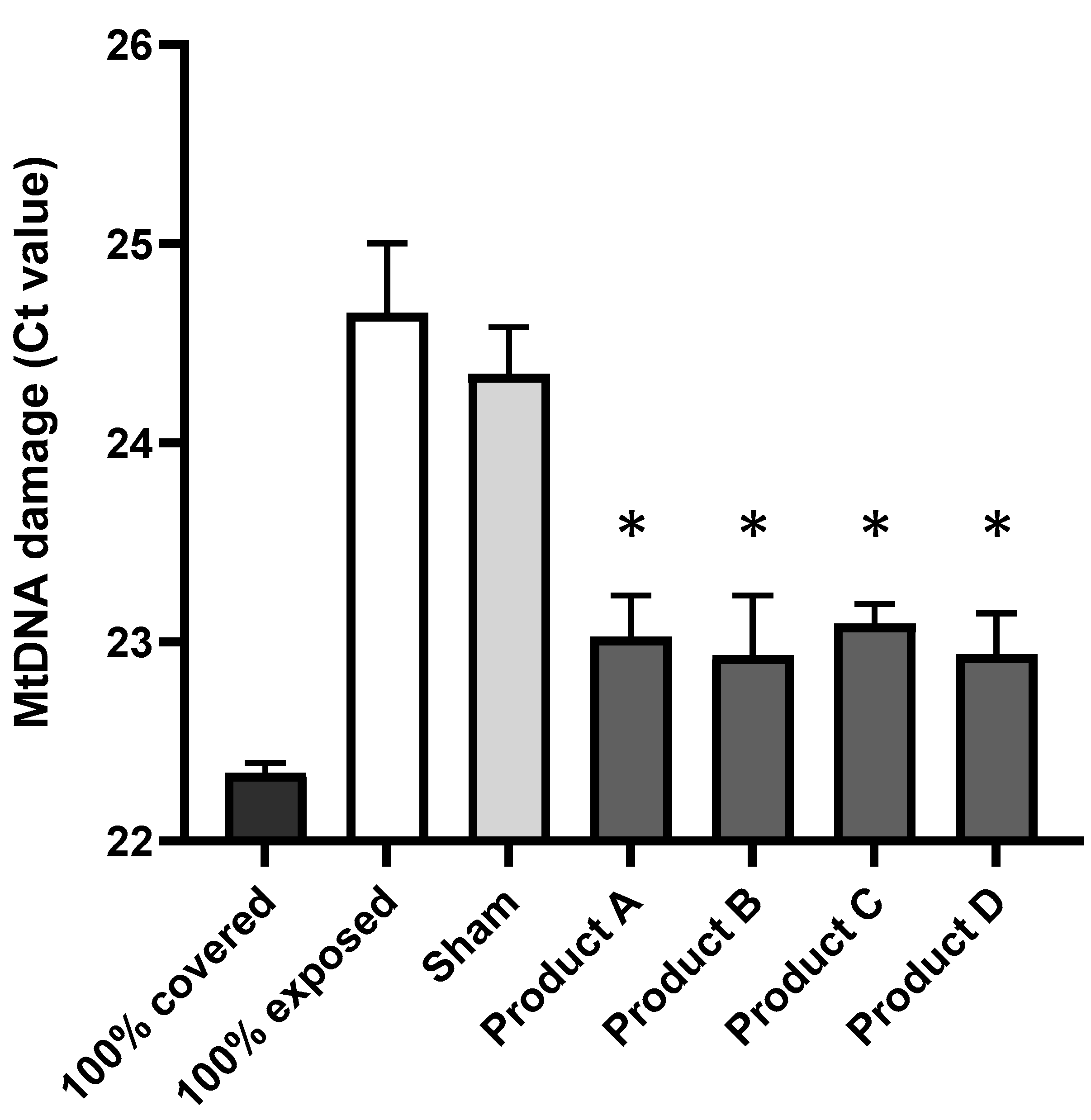
3.1. Commercial Sun Protection Products at SPF 50 Provide Protection by Reducing the Amount of UV-Induced mtDNA Damage in Human Dermal Fibroblast Skin Cells

The 1 kb qPCR results of mtDNA damage are shown in Figure 1. The higher the Ct value on the y-axis, the greater the amount of mtDNA damage. As expected, mtDNA damage was much greater when the cells were exposed to 2 SED solar UV (100% exposed, i.e., positive control) as compared to when the cells were completely foil-covered (100% protected, i.e., negative control). This expected result is highly statistically significant, as seen in Table 1 (p < 0.0028) and confirms that the experiment was successful from a technical viewpoint. It is worth noting that each 1 Ct difference represents a 2-fold difference in damage.
Figure 1 show that all four sun protection products (labelled A to D) provided UV protection of mtDNA in the skin cells compared to the 100% exposed cells, as seen by decreasing the level of mtDNA damage. Therefore, all four SPF products clearly showed statistically significant protection (Table 2) of UV-induced mtDNA damage compared to the 100% exposed cells, as seen by decreasing the level of mtDNA damage by a Ct value of 1.7. This is equivalent to approximately 3.2 fold less damage, or 320% protection, compared to exposed cells. Product D appears to show slightly greater mtDNA protection than the other three products; however, this difference was not statistically significant under the experimental conditions.

3.2. Commercial Sun Protection SPF 50 Products Protect Against Blue Light-Induced Decrease in the Viability of Human Dermal Fibroblast Skin Cells

The higher the value on the y-axis of Figure 2i,ii, the greater the skin cell viability as defined by the MTS assay. As expected, the lowest degree of cell viability (i.e., the highest amount of cell death) was observed when the cells were exposed to blue light in the absence of any products (100% exposed positive control, transpore tape only, no product). Therefore, the data in Figure 2 are presented as cell viability relative to the value of the 100% exposed condition defined as 0% cell viability on the y-axis.
Two products were chosen based upon the results in Figure 1 and ensuring that one product had organic/chemical based filters (product B, international formula) and one had mineral based filters (product C, US formula). As expected, the results in Figure 2 show that the degree of cell viability in the presence of the Sham product was not statistically different from the 100% exposed condition (p = 0.0537 Table 3). The key finding was that both products B and C provided protection against the blue light induced decrease in skin cell viability (Figure 2i (product B) and Figure 2ii (product C)). This is seen by the observed increases in cell viability compared to the value observed in 100% blue light exposed skin cells (i.e., the absence of any product between the blue light source and the skin cells). These results were statistically significant for both products, namely p = 0.0134 and p = 0.0012 for products B and C, respectively (Table 3), compared to the 100% exposed, tape only, no product control).
HDFn cells were irradiated with filtered blue light for a time equivalent to 50 J/cm2, with formulations applied at 2 mg/cm2 onto transpore tape and placed over the relevant cells during irradiation. The MTS assay was used to determine cell viability 24 h after irradiation. The data are presented as cell viability relative to 100% Exposed (Tape only) condition (defined as 0% cell viability). Sham is SPF0 vehicle. Statistical analysis was performed using t-test where (*): p < 0.05, (**): p < 0.01. Data show mean ± SEM, n = 3—three biological repeats were performed for each condition, with eight technical repeats per condition per run (therefore, each column = 24 data points).

4. Discussion

The first part of this study investigated four commercial sun protection products (at SPF 50) in terms of their potency in protecting against UV-induced mtDNA damage with the products in between the light source and the human skin cells. The statistically significant results clearly demonstrate in cultured human skin fibroblasts, the high potency of protection of all four SPF products against UV-induced mtDNA damage. As mtDNA is an established biomarker of UVR damage in human skin ageing [6], these findings show that the SPF products protect against damage to the DNA housed inside the mitochondria of the cell thereby helping to combat skin fatigue and promote increased bioenergy in human skin.
Mitochondria are dynamic, energy producing organelles by the process of oxidative phosphorylation, via the electron transport chain (ETC), generating adenosine triphosphate (ATP). Therefore, maintaining healthy, minimally damaged mitochondria helps to maintain bioenergy production in the form of ATP. The use of mtDNA as a sentinel or biomarker of mitochondrial and broader cellular damage stems from the fact that there are limited DNA repair processes and a lack of protective histone proteins [11,13,24]. For example, there is a lack of nucleotide excision repair (NER) in mitochondria which is used to repair UV-induced photoproducts and NER is severely compromised in the disorder, xeroderma pigmentosum [11] which explains the high incidence of skin cancer in these patients. In addition, the mitochondria are the major site of the primary source of reactive oxygen species (ROS) as electrons leak form the ETC, producing up to 90% of cellular oxidative stress via a cascade process [11]. This in turn affects mitochondrial function which in turn affects mtDNA damage via increased local ROS generation. Together, this cyclical process has been postulated as the vicious cycle theory of ageing [6,25] applicable to many tissues including skin.
The second part of this study investigated two sun protection products (at SPF 50) in terms of their potency in protecting against blue light-induced decrease in cell viability with the products in between the light source and the human skin cells. The results clearly demonstrate in cultured human skin cells, that both sun protection SPF 50 products significantly protected against blue light induced decrease in cell viability. As expected, the product with mineral zinc oxide, US formula (Product C) showed greater protection against blue light induced damage (i.e., increased cell viability) compared to the other organic/chemical, international formula SPF product (Product B). Sunscreen products are generally categorised by chemical/organic and mineral/physical barriers dependent upon the active ingredients in the formulation. Mineral sunscreens often contain filters such as titanium dioxide and zinc oxide which reflect and scatter the UV rays before they penetrate the skin. They are considered to be broad-spectrum filters as they offer protection from UVB, UVA and longer wavelengths (although to a much lesser extent than UVR). Chemical/organic sunscreens contain active ingredients which absorb UV rays and release them as heat energy from the skin. It is also common for sunscreen products to contain a combination of physical and chemical active ingredients which work synergistically to increase protection over longer wavelengths and photostability of the product [26,27,28].
In conclusion, the current study has addressed the two questions posed at the beginning of this manuscript and has provided evidence to show that:
  • Four commercial sun protection SPF 50 products provide significant protection by reducing the amount of UV-induced mtDNA damage in human dermal fibroblast skin cells.
  • Commercial sun protection SPF 50 products protect against blue light induced decrease in the viability of human dermal fibroblast skin cells.

Author Contributions

All authors as a team participated in conceptualization, methodology, software, validation, formal analysis, investigation, resources, data curation, writing—original draft preparation, writing—review and editing, visualization, supervision, project administration, and funding acquisition. All authors have read and agreed to the published version of the manuscript.

Funding

This research was funded by SkinCeuticals.

Institutional Review Board Statement

Not applicable.

Informed Consent Statement

Not applicable.

Data Availability Statement

The data are not publicly available due to privacy or ethical restrictions.

Conflicts of Interest

Jessica Moor and Amy Bowman are employees of Skin Life Analytics, Hina Choudhary is an employee of SkinCeuticals, Jonathan Brookes is a shareholder and co-director of Skin Life Analytics Ltd., Patricia Brieva is an employee of SkinCeuticals, Mark Birch-Machin is a shareholder and co-director of Skin Life Analytics Ltd. The authors declare that this study received funding from SkinCeuticals. The funder (co-authors HC and PB) had the following involvement with the study: study design, interpretation of data, writing and editing of the article, decision to submit and providing the cosmetic products. HC was also involved in the proofreading.

References

  1. Soter, N.A. Acute effects of ultraviolet radiation on the skin. Semin. Dermatol. 1990, 9, 11–15. [Google Scholar] [PubMed]
  2. Parrado, C.; Mercado-Saenz, S.; Perez-Davo, A.; Gilaberte, Y.; Gonzalez, S.; Juarranz, A. Environmental stressors on skin aging. Mech. Insights Front. Pharmacol. 2019, 10, 759. [Google Scholar] [CrossRef]
  3. Diffey, B. Erythema and Acclimatization Following Repeated Sun Exposure: A Modeling Study. Photochem. Photobiol. 2021, 97, 1558–1567. [Google Scholar] [CrossRef] [PubMed]
  4. Birch-Machin, M.A.; Stout, R. Sun Exposure and Pollution: International Patent Application No. PCT/GB2023/050834; A qPCR Method to Analyse mtDNA Mainly in Skin Cells. WO 2023/187386 A1, 30 March 2023. Available online: https://patents.google.com/patent/WO2023187386A1/en?inventor=Mark+Birch-Machin&oq=Mark+Birch-Machin+&page=1 (accessed on 23 December 2024).
  5. Clatici, V.G.; Racoceanu, D.; Dalle, C.; Voicu, C.; Tomas-Aragones, L.; Marron, S.E.; Wollina, U.; Fica, S. Perceived age and life style. The specific contributions of seven factors involved in health and beauty. Maedica 2017, 12, 191. [Google Scholar]
  6. Birch-Machin, M.A.; Moor, J.A. Mitochondria as a Skin Health Biomarker. Aesthet. J. 2024, 11, 32–35. [Google Scholar]
  7. Green, A.C.; Williams, G.M.; Logan, V.; Strutton, G.M. Reduced melanoma after regular sunscreen use: Randomized trial follow-up. J. Clin. Oncol. 2011, 29, 257–263. [Google Scholar] [CrossRef]
  8. Mahendra, C.K.; Ser, H.-L.; Pusparajah, P.; Htar, T.T.; Chuah, L.-H.; Yap, W.H.; Tang, Y.-Q.; Zengin, G.; Tang, S.Y.; Lee, W.L.; et al. Cosmeceutical therapy: Engaging the repercussions of UVR photoaging on the skin’s circadian rhythm. Int. J. Mol. Sci. 2022, 23, 2884. [Google Scholar] [CrossRef]
  9. Trifunovic, A.; Hansson, A.; Wredenberg, A.; Rovio, A.T.; Dufour, E.; Khvorostov, I.; Spelbrink, J.N.; Wibom, R.; Jacobs, H.T.; Larsson, N.-G. Somatic mtDNA mutations cause aging phenotypes without affecting reactive oxygen species production. Proc. Natl. Acad. Sci. USA 2005, 102, 17993–17998. [Google Scholar] [CrossRef] [PubMed]
  10. Yang, J.H.; Lee, H.C.; Lin, K.J.; Wei, Y.H. A specific 4977-bp deletion of mitochondrial DNA in human ageing skin. Arch. Dermatol. Res. 1994, 286, 386–390. [Google Scholar] [CrossRef]
  11. Birch-Machin, M.A. Mitochondria and Skin Disease. Clin. Exp. Dermatol. 2000, 25, 141–146. [Google Scholar] [CrossRef]
  12. Anderson, S.; Bankier, A.T.; Barrell, B.G.; De Bruijn, M.H.L.; Coulson, A.R.; Drouin, J.; Eperon, I.C.; Nierlich, D.P.; Roe, B.A.; Sanger, F.; et al. Sequence and organization of the human mitochondrial genome. Nature 1981, 290, 457–465. [Google Scholar] [CrossRef] [PubMed]
  13. Pang, C.Y.; Lee, H.C.; Yang, J.H.; Wei, Y.H. Human skin mitochondrial DNA deletions associated with light exposure. Arch. Biochem. Biophys. 1994, 312, 534–538. [Google Scholar] [CrossRef]
  14. Addor, F.A.S.A. Beyond photoaging: Additional factors involved in the process of skin aging. Clin. Cosmet. Investig. Dermatol. 2018, 11, 437–443. [Google Scholar] [CrossRef] [PubMed]
  15. Dong, K.; Goyarts, E.; Pelle, E.; Trivero, J.; Pernodet, N. Blue light disrupts the circadian rhythm and create damage in skin cells. Int. J. Cosmet. Sci. 2019, 41, 558–562. [Google Scholar] [CrossRef] [PubMed]
  16. Nabila, Y.A.; Damayanti, D.; Handayani, S.; Setyaningrum, T. The effect of lifestyle on skin aging. Berk. Ilmu Kesehat. Kulit Dan Kelamin 2021, 33, 110–115. [Google Scholar] [CrossRef]
  17. Suitthimeathegorn, O.; Yang, C.; Ma, Y.; Liu, W. Direct and Indirect Effects of Blue Light Exposure on Skin: A Review of Published Literature. Ski. Pharmacol. Physiol. 2022, 35, 305–318. [Google Scholar] [CrossRef]
  18. Nakyai, W.; Tissot, M.; Humbert, P.; Grandmottet, F.; Viyoch, J.; Viennet, C. Effects of Repeated UVA Irradiation on Human Skin Fibroblasts Embedded in 3D Tense Collagen Matrix. Photochem. Photobiol. 2018, 94, 715–724. [Google Scholar] [CrossRef] [PubMed]
  19. Liebel, F.; Kaur, S.; Ruvolo, E.; Kollias, N.; Southall, M.D. Irradiation of skin with visible light induces reactive oxygen species and matrix-degrading enzymes. J. Investig. Dermatol. 2012, 132, 1901–1907. [Google Scholar] [CrossRef]
  20. Opländer, C.; Hidding, S.; Werners, F.B.; Born, M.; Pallua, N.; Suschek, C.V. Effects of blue light irradiation on human dermal fibroblasts. J. Photochem. Photobiol. B Biol. 2011, 103, 118–125. [Google Scholar] [CrossRef]
  21. Liebmann, J.; Born, M.; Kolb-Bachofen, V. Blue-light irradiation regulates proliferation and differentiation in human skin cells. J. Investig. Dermatol. 2010, 130, 259–269. [Google Scholar] [CrossRef]
  22. Latimer, J.; Matts, P.; Diffey, B.; Lloyd, J.; Birch-Machin, M.A. Determination of the action spectrum of UVR-induced mitochondrial DNA damage in human skin cells. J. Investig. Dermatol. 2015, 135, 2512–2518. [Google Scholar] [CrossRef] [PubMed]
  23. Schrand, A.M.; Lin, J.B.; Hussain, S.M. Assessment of cytotoxicity of carbon nanoparticles using 3-(4,5-dimethylthiazol-2-yl)-5-(3-carboxymethoxyphenyl)-2-(4-sulfophenyl)-2H-tetrazolium (MTS) cell viability assay. Methods Mol Biol. 2012, 906, 395–402. [Google Scholar] [PubMed]
  24. Koch, H.; Wittern, K.-P.; Bergemann, J. In human keratinocytes the Common Deletion reflects donor variabilities rather than chronologic aging and can be induced by ultraviolet A irradiation. J. Investig. Dermatol. 2001, 117, 892–897. [Google Scholar] [CrossRef] [PubMed]
  25. Lagouge, M.; Larsson, N.G. The role of mitochondrial DNA mutations and free radicals in disease and ageing. J. Intern. Med. 2013, 273, 529–543. [Google Scholar] [CrossRef]
  26. Smijs, T.G.; Pavel, S. Titanium dioxide and zinc oxide nanoparticles in sunscreens: Focus on their safety and effectiveness. Nanotechnol. Sci. Appl. 2011, 4, 95. [Google Scholar] [CrossRef]
  27. Summers, B.; Summers, R. The prescribing of sunscreen preparations: A guide for the general practitioner. Off. J. S. Afr. Acad. Fam. Pract. Care. 2012, 8, 440–446. [Google Scholar]
  28. Bens, G. Sunscreens. Adv. Exp. Med. Biol. 2014, 810, 429–463. [Google Scholar]
Figure 1. Mitochondrial DNA damage differences following UV irradiation in the presence of the four different SPF products. Cells were irradiated in 35 mm dishes using 2 SED UV light. To represent 100% covered, dishes were wrapped in aluminium foil, and to represent 100% exposed, transpore tape on the dish with no cream was used. The sham was a control cream with no SPF. All four products were each applied to the transpore tape at 2 mg/cm2. Statistical analysis was performed using t-test where (*): p < 0.05. Data show mean ± SEM. Three biological repeats were performed for each condition, with the qPCR run in triplicate for each sample; therefore, each column represents nine data points.
Cosmetics 12 00128 g001
Figure 2. Cell viability of blue light irradiated human skin cell fibroblasts (HDFn cells) in the presence of two different SPF formulations, Products B and C (parts (i) and (ii), respectively). 0% cell viability represents fully uncovered or 100% blue light exposed cells and 100% cell viability represents the control unexposed cells (i.e., foil or fully protected). *: p < 0.05, **: p < 0.01.
Cosmetics 12 00128 g002
Table 1. Composition of Products A to D.
A (Approx 25% Total of Filters Below)B (Approx 21% Total of Filters Below)C (Approx 11% Total of Filters Below)D (Approx 20% Total of Filters Below)
Mineral FiltersZinc Oxide
(UVA + UVB)
Zinc Oxide 5%
(UVA + UVB)
Titanium Dioxide 6% (UVA + UVB)
Titanium Dioxide (UVA + UVB)
Organic FiltersHomosalate (UVB primarily)
Octisalate (UVB primarily)
Octocrylene (UVB, short UVA)
Avobenzone (UVA)
Octisalate (UVB primarily)
Ethylhexyl Triazone (UVB primarily)
Octocrylene (UVB, short UVA)
Drometrizole Trisiloxane (UVA + UVB)
Tinosorb WPGL (UVA + UVB)
Tinosorb S (UVA + UVB)
Octinoxate (UVB primarily)
Mexoryl XL (UVA + UVB)
Diethylamino Hydroxybenzoyl Hexyl Benzoate (UVA)
Bemotrizinol (UVA + UVB)
FDA ApprovedYesNo
Contains: Ethylhexyl Triazone, Drometrizole Trisiloxane, Tinosorb WPGL, Tinosorb S
YesNo
Contains: Mexoryl XL,
Diethylamino Hydroxybenzoyl
Hexyl-Benzoate, Bemotrizinol
Table 2. Statistical Analysis of the data presented in Figure 1. Unpaired t-test to compare to 100% exposed sample where (*): p < 0.05, (**): p < 0.01.
Conditionp-Value
100% exposed vs. 100% covered0.0028 **
100% exposed vs. Sham (SPF0)0.5102
100% exposed vs. Product A0.0161 *
100% exposed vs. Product B0.0203 *
100% exposed vs. Product C0.0157 *
100% exposed vs. Product D0.0133 *
Table 3. Statistical Analysis of the data in Figure 2.
Unpaired t-Test to Compare to 100% Exposed Sample Where (*): p < 0.05, (**): p < 0.01.
Conditionp-Value
100% Exposed (Tape, no product) vs. Sham (SPF0)0.0537
100% Exposed (Tape, no product) vs. Product B0.0134 *
100% Exposed (Tape, no product) vs. Product C0.0012 **
Sham (SPF0) vs. Product C0.0023 **
Sham (SPF0) vs. Product B0.0695
Disclaimer/Publisher’s Note: The statements, opinions and data contained in all publications are solely those of the individual author(s) and contributor(s) and not of MDPI and/or the editor(s). MDPI and/or the editor(s) disclaim responsibility for any injury to people or property resulting from any ideas, methods, instructions or products referred to in the content.

