Article

Marine Algae Extract-Loaded Nanoemulsions: A Spectrophotometric Approach to Broad-Spectrum Photoprotection

by
Julian Tello Quiroz
1,
Ingrid Andrea Rodriguez Martinez
1,
Vanessa Urrea-Victoria
1,2,
Leonardo Castellanos
2 and
Diana Marcela Aragón Novoa
1,*
1
Departamento de Farmacia, Facultad de Ciencias, Universidad Nacional de Colombia, Sede Bogotá, Av. Carrera 30 # 45-03, Bogotá 111321, Colombia
2
Departamento de Química, Facultad de Ciencias, Universidad Nacional de Colombia, Sede Bogotá, Av. Carrera 30 # 45-03, Bogotá 111321, Colombia
*
Author to whom correspondence should be addressed.
Cosmetics 2025, 12(3), 101; https://doi.org/10.3390/cosmetics12030101
Submission received: 1 April 2025 / Revised: 24 April 2025 / Accepted: 6 May 2025 / Published: 15 May 2025

Abstract

:
The depletion of the ozone layer and climate change have increased exposure to ultraviolet (UV) radiation, driving the search for natural photoprotective agents. Marine macroalgae, particularly Gracilaria sp. (Rhodophyta) and Sargassum polyceratium (Ochrophyta), are rich in UV-absorbing bioactives, such as mycosporine-like amino acids (MAAs) and fucoxanthin, offering natural alternatives to synthetic sunscreens. This study aimed to develop and optimize a nanoemulsion incorporating both algal extracts, with MAAs and fucoxanthin strategically distributed in the aqueous and oil phases, respectively, to enhance synergistic broad-spectrum UV protection. MAAs were quantified in Gracilaria sp. using UHPLC-DAD, revealing 8.03 mg/g dry weight, primarily composed of shinorine and porphyra-334. Fucoxanthin was identified in S. polyceratium at 0.98 mg/g dry weight. A Box–Behnken design (BBD) was employed to optimize the nanoemulsion, targeting minimal droplet size and optimal ζ potential. The resulting formulation achieved a droplet size less than 100 nm and a ζ potential less than −25.0 mV. In vitro spectrophotometric analysis demonstrated significant photoprotective potential. The nanoemulsion containing only 375 ppm of algal extracts exhibited a UVA ratio of 1.25 and a critical wavelength of 379 nm, meeting the criteria for broad-spectrum protection and outperforming the commercial natural filter Helioguard®365. These results confirm the efficacy of combining red and brown algae extracts in a nanoemulsion platform to deliver sustainable, low-dose photoprotection. This work presents, for the first time, the incorporation of red and brown algae extracts into a single nanoemulsion system, representing a novel strategy to maximize the combined photoprotective potential of MAAs and fucoxanthin. Ultimately, this investigation contributes to the growing field of marine-derived sunscreens and supports the advancement of “blue beauty” innovations aligned with eco-conscious formulation principles.

1. Introduction

Increasing levels of ultraviolet (UV) radiation—driven by factors such as climate change and stratospheric ozone depletion—have heightened the need for effective and sustainable photoprotective agents. In response, marine algae emerge as a promising source of molecules with unique properties to protect the skin from the damage caused by UV radiation. These organisms, which inhabit aquatic environments exposed to high levels of sunlight, have developed sophisticated biochemical mechanisms throughout their evolution to survive and thrive under conditions of high UV exposure. These mechanisms include the synthesis of photoprotective compounds, such as mycosporine-like amino acids (MAAs), carotenoids, sulfated polysaccharides, and polyphenolic compounds [1].
The genus Gracilaria, comprising red macroalgae (Rhodophyta), is widely distributed in marine and estuarine environments, spanning tropical to temperate regions [2]. Species within this genus are especially valued as a major source of agar—a sulfated polysaccharide extensively utilized in the pharmaceutical, biotechnological, and food industries. Among its bioactive compounds, shinorine and porphyra-334—two mycosporine-like amino acids (MAAs) isolated from Gracilaria cornea Agardh—have demonstrated remarkable photostability under UVB radiation and thermal stress, outperforming conventional pigments such as chlorophyll, carotenoids, and phycocyanin [3]. Additionally, other studies have shown that Gracilaria cornea can modulate the accumulation of UV-absorbing compounds, such as palythine, carotenoids, and phenolic compounds, in response to UVA and UVB radiation, suggesting photoreceptor-mediated photoprotection [4]. Furthermore, Gracilaria gracilis collected from Moroccan coasts has demonstrated significant photoprotective capacity through the presence of MAAs, such as shinorine, palythine–glutamine, and porphyra-334, with in vitro SPF values ranging from 5.55 to 9.34, depending on the extraction solvent, and notable anti-collagenase and anti-elastase activities [5]. In addition to UVB protection, MAAs also exhibit significant absorption in the UVA region [6]. Notably, these compounds are incorporated into commercial photoprotective products, such as Helioguard®365—a natural sunscreen enriched with porphyra-334 and shinorine [7].
On the other hand, the genus Sargassum, a group of marine macroalgae belonging to the class Phaeophyceae (brown algae), is widely distributed along tropical and subtropical coastlines [8]. In recent years, the proliferation of Sargassum blooms has raised growing environmental concerns due to their negative ecological, economic, and social impacts. Consequently, alternative valorization strategies have been explored, including their use as cosmetic ingredients. Notably, organic extracts of S. muticum (Yendo) Fensholt have been shown to reduce reactive oxygen species (ROS) in UVB-irradiated cells, an effect attributed to their antioxidant properties and the upregulation of endogenous enzymes such as superoxide dismutase and catalase [9]. In addition, S. muticum, S. horneri, S. cristaefolium, and S. vulgare have demonstrated potent UV-absorbing and anti-aging properties, further reinforcing the photoprotective potential of the genus. For instance, S. muticum has been shown to accumulate UV-absorbing compounds, enhance non-photochemical quenching through the xanthophyll cycle, and boost antioxidant defenses to mitigate UV-induced photodamage, ultimately restoring photosynthetic performance after UV exposure [10]. Similarly, S. cristaefolium has demonstrated UVA-absorbing capacity and in vivo photoprotection, reducing epidermal thickening and preserving dermal collagen in UV-exposed mice [11]. Recent investigations on S. vulgare have demonstrated that this alga exhibited high photoprotective capacity across multiple UV-induced biological responses, achieving over 96% effective solar absorption radiation (ESAR) for vitamin D3 synthesis, erythema, and immunosuppression at high concentrations and demonstrating strong potential as a cosmeceutical agent due to its antioxidant content and capacity to absorb harmful UV wavelengths [12]. Finally, S. horneri exhibits strong UV-protective activity by producing UV-absorbing compounds, increasing carotenoids, activating antioxidant enzymes, and enhancing photosynthetic recovery mechanisms, effectively mitigating UV-induced damage [13].
Moreover, studies employing electrical impedance spectroscopy (EIS) have demonstrated that formulations containing Sargassum cymosum help preserve the electrical resistance of skin membranes under UVB and oxidative stress conditions, reducing damage by up to 85% compared to untreated controls [14,15]. Additionally, Sargassum species contain bioactive polysaccharides with photoprotective potential—such as alginate and fucoidan—which have shown high in vitro SPF values of 32.218 and 65.709, respectively, depending on the concentration [16]. While these findings support the individual capabilities of red or brown algae in photoprotection, there remains a significant research gap regarding the development of integrated formulations that combine extracts from both Gracilaria and Sargassum to achieve synergistic, broad-spectrum UV protection.
Finally, nanoemulsions have emerged as advanced delivery systems in cosmetic and dermatological formulations due to their unique physicochemical properties and functional versatility. These colloidal dispersions, typically composed of oil, water, and surfactants, exhibit nanometric droplet sizes that confer superior stability, enhanced bioavailability of active ingredients, and improved skin penetration compared to conventional emulsions [17]. In the context of sun protection, nanoemulsions are particularly advantageous, as they enable the effective encapsulation and controlled release of photoprotective compounds, preserving their activity while minimizing degradation under UV exposure [14]. Their small droplet size also enhances the uniformity of topical application, leading to improved coverage and efficacy [18]. Moreover, by integrating natural antioxidant-rich extracts—such as those derived from marine algae—nanoemulsions can offer a dual function: physical protection against UVA and UVB radiation and biological defense via neutralization of reactive oxygen species (ROS) generated by solar exposure [17].
Therefore, considering the complementary photoprotective properties of Gracilaria sp. and Sargassum sp., particularly their content of mycosporine-like amino acids (MAAs) and fucoxanthin, this study aims to develop and optimize an oil-in-water nanoemulsion incorporating both extracts, each strategically allocated to distinct phases of the formulation based on the solubility of the active compounds: Gracilaria extract in the aqueous phase and Sargassum extract in the oil phase. This dual-phase design leverages the unique chemical characteristics and UV-absorbing capabilities of each extract, promoting a synergistic effect for enhanced broad-spectrum protection against both UVA and UVB radiation. By embedding these marine-derived bioactives into a nanometric delivery system, the proposed formulation seeks to provide a sustainable, eco-friendly alternative for next-generation sun care products in the cosmetic industry.

2. Materials and Methods

2.1. Algae Harvesting

Marine macroalgae were collected in Santa Marta (Magdalena, Colombia) in April 2022. The material was stored at −20 °C until use. To prepare the samples, the algae were washed with a saline solution, simulating seawater conditions, to remove fragments of other algae, marine animals, sand, and other debris. Subsequently, the material was evenly distributed into resealable plastic bags with small holes and stored again at −20 °C. Then, the algae were freeze-dried to obtain a dry weight (DW). These procedures were carried out protected from light.
The identification of the red macroalgae Gracilaria sp. (ID: JIW0005027) and Sargassum spp. was carried out by Dr. Briggite Gavio and Dr. Vanessa Urrea-Victoria based on morphological characteristics, following descriptions from the relevant literature. The algal nomenclature and taxonomy followed AlgaeBase [19]. Individual specimens of each sample were deposited at the JIWUKORI Algae Herbarium at Universidad Nacional de Colombia.

2.2. Extract Preparation

The extracts selected for this study were chosen based on previous results from the research group regarding photoprotective activity.
An aqueous extract of Gracilaria sp.: 3 g DW was weighed and placed in a flask with 60 mL of distilled water. Ultrasound-assisted extraction was performed for 15 min while keeping the bath refrigerated (in triplicate). The aqueous extract was then placed in a Falcon tube and frozen at −20 °C. Finally, the extract was freeze-dried to obtain the dry product [20].
A butanol fraction of Sargassum sp.: 3 g DW was weighed and placed in a flask with 60 mL of MeOH:water solution (1:1) (Merck, Darmstadt, Germany). Ultrasound-assisted extraction was performed for 15 min, keeping the bath refrigerated (in triplicate). The initial methanol:water extract was rotary-evaporated to remove the solvent. Then, to obtain the butanol fraction, the extract was dissolved in 30 mL of a BuOH:H2O solution (1:1) (Merck, Darmstadt, Germany) and dried again by rotary evaporation [20].
The choice of extraction solvent was guided by the chemical nature of the target bioactive compounds in each algal species. For Gracilaria sp., an aqueous extraction was employed to isolate mycosporine-like amino acids (MAAs), such as shinorine and porphyra-334, due to their high water solubility and polar structure [21]. In contrast, a butanol fraction was used for Sargassum sp. to selectively extract fucoxanthin, a lipophilic carotenoid that exhibits greater solubility in medium-polarity organic solvents [22]. This solvent selection strategy ensured optimal recovery and preservation of the key photoprotective compounds, aligning with their physicochemical profiles and enhancing their functionality in the nanoemulsion system.

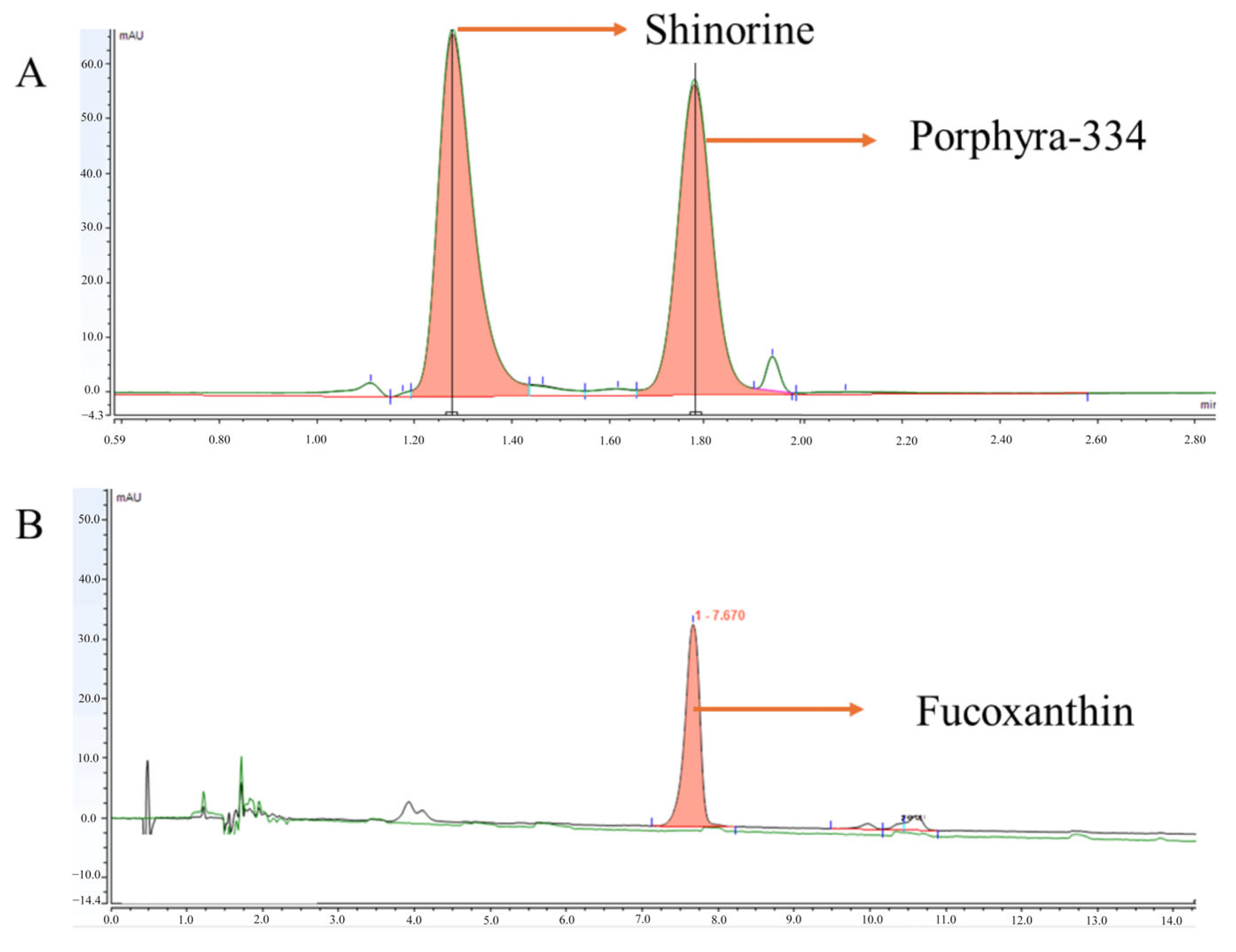
2.3. Extract Characterization

Aqueous extract of Gracilaria sp.: The identification and quantification of MAAs were developed according to the Urrea-Victoria et al. method, with modifications [21]. The analysis was performed using an Ultimate 3000 UHPLC system (Dionex, Thermo Scientific, Waltham, MA, USA). An aliquot of 5 μL of algae extract was injected onto a Synergi Hydro-RP 80A column (4 μm, 150 × 2.0 mm) (Phenomenex, Torrance, CA, USA) protected by a SecurityGuard pre-column. The flow rate was 0.3 mL.min−1 at 22 °C. The gradient system combined an aqueous solution of 0.25% formic acid and 20 mM ammonium formate (solvent A) and acetonitrile (solvent B). It started with 100% A (0–3 min), followed by 5% A (3–7 min) and 50% A (7–8 min). The re-equilibration duration between individual runs was 7 min. Although a scan from 190 to 740 nm was performed, we focused on the set at 310, 320, 334, and 360 nm, as these wavelengths correspond to the peaks of the compounds of interest, such as MAAs. A porphyra-334 standard stock solution (>98% pure), prepared in-house and isolated by one of the authors (V. U-V.), was utilized in the calibration curve, and the results were expressed as mg MAAs.g 1 DW. 2.5. The corrected areas were then used to interpolate the concentrations based on the equation (y = 911918x + 4822.9; R2 = 0.9993). The results were expressed as mg MAAs.g 1 DW. For shinorine (332 nm; 44,668 M1 cm1), the observed area was corrected using the respective molar extinction coefficient ratios, comparing them to those reported for porphyra-334 (334 nm; 42,300 M1 cm1).
Butanol fraction of Sargassum sp.: The fucoxanthin quantification in the butanol extract was determined using an analytical methodology employed by a research group. For this purpose, a Thermo Scientific Dionex Ultimate 3000 chromatograph equipped with a diode array detector (DAD) Dionex Ultimate 3000, a quaternary pump Dionex Ultimate 3000 RS, an online degasser, and an autosampler were used. A Phenomenex Kinetex C8 column (100 mm × 4.6 mm, 2.6 μm) was employed at 40 °C. The gradient system combined water (solvent A) and acetonitrile (solvent B). It started with 65% A (0–8 min), followed by 75% A (8–14 min). The re-equilibration duration between individual runs was 2 min. A 10 μL sample was injected, and detection was performed at a wavelength of 470 nm.

2.4. Sun Protection Factor (SPF), UVA Ratio (UVA-r), and Critical Wavelength (λc)

The absorbance values of the extracts (200 μL) were recorded by a UV–visible spectrophotometer (Varioskan LUX by Thermo Scientific, Waltham, MA, USA) with a spectral window from 290 to 700 nm (1 nm resolution/data point). Spectroscopic data were baseline-corrected.
The SPF of the obtained extracts and the developed product was evaluated using the Mansur method [23]. Stock solutions of the extracts were prepared at a concentration of 2 mg/mL in ethanol absolute pure 99.5% (EtOH; PanReac). Absorbance readings were taken between 290 and 400 nm, using EtOH as a blank. The SPF was calculated using the Mansur equation:
S P F = F C × E E ( λ ) 320290 × I ( λ ) × A b s ( λ )
where FC corresponds to the correction factor value, which has a magnitude of 10; EE is a constant dependent on the wavelength and corresponds to the erythemal effect spectrum; I is the intensity of the solar spectrum at the wavelength of interest; and Abs is the measured absorbance of the solution at the specific wavelength.
Additionally, following the method described by Springsteen et al. [24], the UVA protection factor (UVA-r) and the critical wavelength (λc) were calculated. The UVA-r was determined as the ratio between the integrated intensities of the UVA (320–400 nm) and UVB (290–320 nm) regions. The λc, which indicates the wavelength at which 90% of the area under the absorbance curve (290–400 nm) is reached, was also calculated.

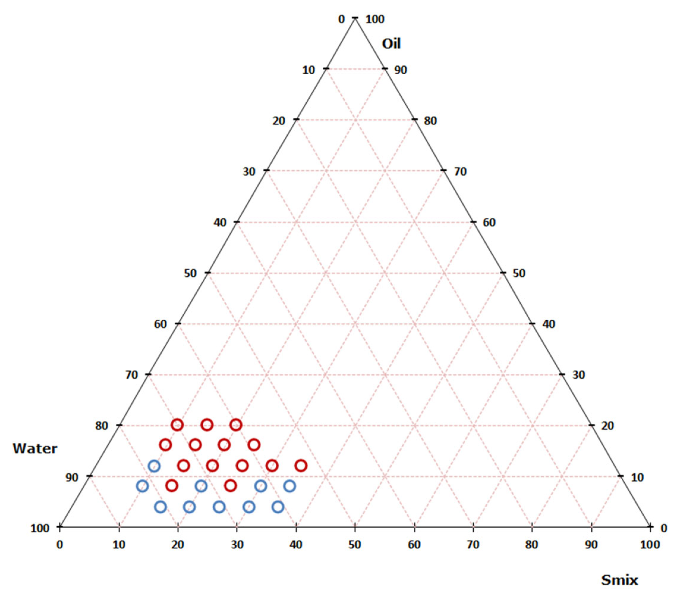
2.5. Nanoemulsion Preparation–Pseudoternary Diagram

Twenty-four nanoemulsions were prepared by varying the proportions of the dispersed phase (Capryol PGMC®) (Gattefossé Co., Saint-Priest, France), the dispersing phase (deionized water), and the surfactant (Kolliphor EL®:propylene glycol (3:2)) (Sigma Chemical Co., St. Louis, MO, USA) to evaluate the influence of composition on the formation of nanoemulsified systems and to identify the nanoemulsion region by a pseudoternary diagram. Sonication (at 80% power) (Q500-SONICATOR, Newton, MA, USA), was used as the preparation method, a high-energy technique suitable for generating emulsions with small droplet sizes. Capryol® PGMC (Figure 1) is composed of propylene glycol monoesters and diesters of saturated caprylic and capric acids, offering amphiphilic properties that aid solubilization of lipophilic compounds. Kolliphor® EL (also known as polyoxyl 35 castor oil) (Figure 1) consists of polyethoxylated castor oil, where 35 ethylene oxide units are added to castor oil, resulting in a nonionic surfactant with both lipophilic and hydrophilic domains. This structural configuration enhances emulsification and interfacial stability. Propylene glycol, a commonly used co-solvent, is included to modulate polarity and improve interfacial compatibility. These ingredients for the nanoemulsion were selected based on sustainable and eco-friendly criteria to develop a new cosmetic product.
All formulations were characterized in terms of droplet size and ζ potential after 1 day and 7 days of preparation using dynamic light scattering (DLS) with a Zetasizer Nano-ZS (scattering angle of 173°) (Malvern Instruments, Worcestershire, U.K). For ζ potential measurements, the nanoemulsions were prepared using 1% (v/v) deionized water. The analysis was performed on the same equipment using disposable folded capillary cells. All measurements were conducted at a temperature of 25 °C. Excel® 16 was used to construct the pseudoternary diagram. MiniTab®19 was used to generate the three-dimensional surface plots.
Day 1 measurements reflect the initial physicochemical characteristics immediately after preparation, including droplet size and ζ potential, which are critical parameters for defining the formation and uniformity of the nanoemulsion. Day 7 measurements were chosen to assess the formulation’s short-term physical stability, particularly to observe potential changes due to surfactant redistribution, interfacial reorganization, or droplet coalescence. This timeframe aligns with previously reported studies on nanoemulsion stabilization dynamics and allows for the detection of early trend behavior [25,26].

2.6. Nanoemulsion Optimization

Once the surfactant and oil levels were selected based on the results of the pseudoternary phase diagram, the optimal emulsification conditions for incorporating the algae extract were determined using a Box–Behnken design (BBD) [14,15]. The response variable for optimization was droplet size.

2.7. Statistical Analysis

The experimental design, data analysis, and nanoemulsion optimization were performed with Minitab® software (version 19, State College, PA, USA).

3. Results and Discussion

3.1. Extract Characterization

Chromatographic analysis by HPLC-DAD confirmed the presence of mycosporine-like amino acids (MAAs) in Gracilaria sp. and fucoxanthin in Sargassum sp. (Figure 2). In Gracilaria sp., the total MAA content was quantified at 8.03 ± 0.009 mg/g dry weight (DW), with shinorine and porphyra-334 contributing 4.23 ± 0.007 mg/g DW and 3.80 ± 0.002 mg/g DW, respectively. These findings are consistent with previously reported MAA levels in red algae [27,28,29,30,31], supporting their recognized potential as natural photoprotective and antioxidant agents.
In Sargassum sp., the butanol extract exhibited a fucoxanthin concentration of 0.98 ± 0.005 mg/g DW—substantially higher than values typically reported for other solvent extracts of this species (0.3–0.7 mg/g). This indicates that the butanol fraction may be particularly effective in concentrating this UV-protective carotenoid [32,33,34].
MAAs, such as shinorine and porphyra-334, are hydrophilic compounds known for their strong UVA absorption, photostability, and antioxidant effects, making them promising candidates for natural sunscreen formulations. These results corroborate earlier studies who demonstrated that topical application of MAAs from red algae could reduce photoaging and perform comparably to synthetic UVA filters [35,36]. Similarly, fucoxanthin in Sargassum sp. contributes to UVB protection and oxidative stress reduction, further validating its utility in marine-derived cosmetic formulations [37,38,39].

3.2. Nanoemulsion Preparation–Pseudoternary Diagram

Nanoemulsions (Table 1) were prepared by varying the proportions of the dispersed phase (Capryol® PGMC), the dispersing phase (deionized water), and the surfactant–cosurfactant mixture (Smix). Emulsification was carried out using an ultrasonicator (Q500-SONICATOR, Newton, MA, USA) operating at 80% amplitude for 4 min.
The droplet sizes of the prepared nanoemulsions ranged from 16.88 ± 0.19 nm (formulated with 4% oil, 35% Smix, and 61% water) to 590.2 ± 4.58 nm. For the construction of the pseudoternary phase diagram, only formulations with droplet sizes below 100 nm—measured at both 1 day and 7 days post-preparation—were considered. This selection ensured that only systems meeting the criteria for true nanoemulsions and demonstrating resistance to coalescence were included. The nanoemulsion region was identified within the compositional space defined by 4–12% oil, 10–35% Smix, and 57–82% water. Based on these data, the pseudoternary diagram was constructed to illustrate the nanoemulsion-forming region (Figure 3).
On the other side, an unexpected yet noteworthy observation in this study was the significant reduction in droplet size of some emulsions after seven days of standing. This phenomenon, although uncommon, may be explained by the progressive reorganization and stabilization of the interfacial film, leading to tighter droplet packing and improved surfactant alignment [24,25]. Such delayed stabilization is characteristic of systems with high HLB surfactants and co-solvents, which continue to redistribute over time, reducing interfacial tension and promoting smaller, more stable droplets [40,41]. This maturation effect enhances the homogeneity of the nanoemulsion and may improve long-term stability, aligning with findings in similar lipid-based formulations [42].

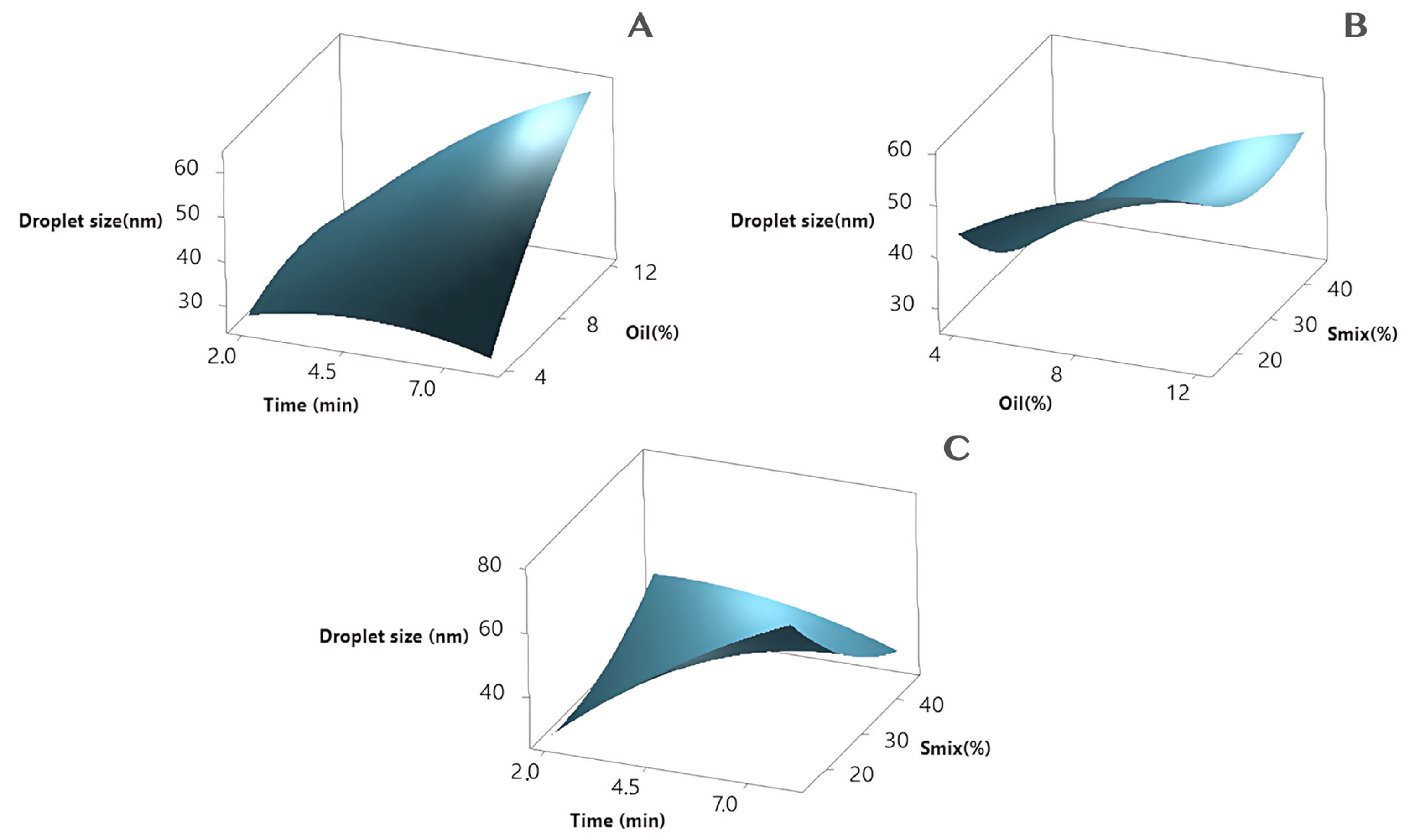
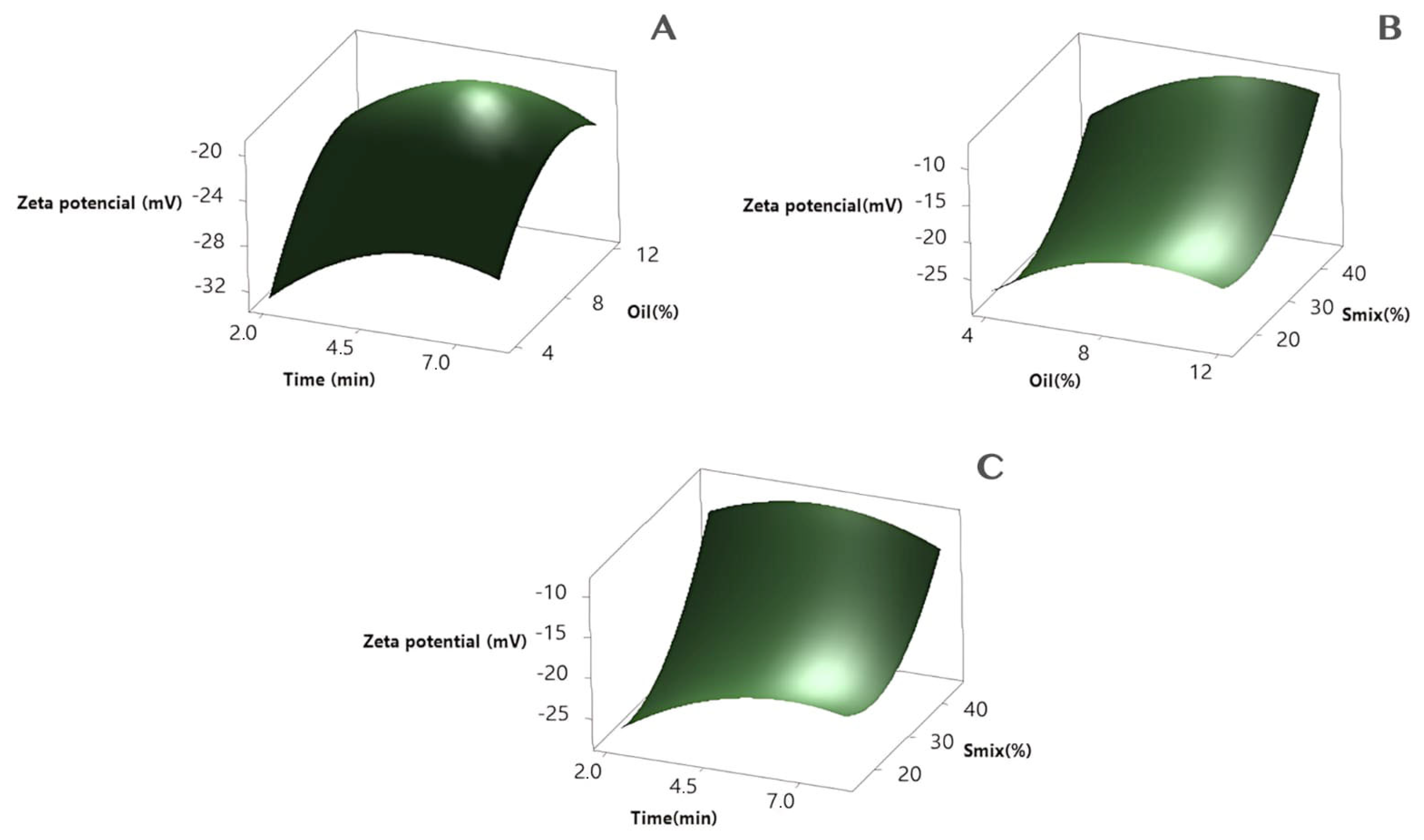
3.3. Nanoemulsion Optimization

Based on the results obtained in the pseudoternary diagram, the levels for the experimental statistical design were selected (Table 2).
To evaluate the variation in droplet size and potential ζ of the nanoemulsions as a function of the formulation variables (%oil, %Smix, and sonication time), the surface models were used. As shown in Table 3, eighteen points of the BBD matrix represent the different combinations of the independent variables and the responses of droplet size and potential ζ.
The experimental data were used to calculate the quadratic polynomial models. The equations are presented below:
Droplet size (nm) = 10.0 + 10.61 Time + 0.87 Oil(%) − 0.51 Smix(%) − 0.407 Time * Time − 0.226 Oil(%) * Oil(%) + 0.0235 Smix(%) * Smix(%) + 0.761 Time * Oil(%) − 0.3250 Time * Smix(%) + 0.0418 Oil(%) * Smix(%)
Potential ζ (nm) = −48.6 + 5.72 Time + 4.57 Oil(%) − 0.989 Smix(%) − 0.351 Time * Time − 0.235 Oil(%) * Oil(%) + 0.02463 Smix(%) * Smix(%) − 0.067 Time * Oil(%) − 0.0378 Time * Smix(%) + 0.0117 Oil(%) * Smix(%)
Accordingly, analysis of variance (ANOVA) was used to determine the statistical significance of the independent variables and their interactions on the response variables—droplet size and ζ potential. The suitability of the models was evaluated using the coefficient of determination (R2) and the p-value for the lack-of-fit test (p-value > 0.05). The statistical analysis of the nanoemulsion optimization is summarized in Table 4. The results indicated that the quadratic model adequately explained the behavior of the system concerning both droplet size and ζ potential. For droplet size, the model demonstrated significant predictive power, with a p-value < 0.05 for the oil concentration and Smix, while sonication time approached significance. Among the interaction terms, the interaction between oil and sonication time (AC) was significant, suggesting that the combined influence of these variables strongly impacts droplet size.
For ζ potential, Smix and sonication time were significant linear contributors (p = 0.049 and p = 0.003, respectively). Additionally, the quadratic term for Smix (C2) showed significance (p = 0.022), highlighting its nonlinear effect on system stability. Again, the lack-of-fit was not significant (p = 0.360), supporting the model’s adequacy.
Overall, these results suggest that oil and Smix concentrations as well as their interactions with sonication time play pivotal roles in defining both the droplet size and the electrostatic stability of the nanoemulsion. The model demonstrated good explanatory power, providing a robust basis for formulation optimization.
Three-dimensional surface plots were generated by MiniTab®19 for the response variables (droplet size and ζ potential) of the nanoemulsions, evaluating the effect of two independent variables and one other variable that remained constant at the center point (Figure 4 and Figure 5).
Finally, the three factors were optimized to obtain the nanoemulsion with the smallest droplet size and ζ potential. Based on the optimization of the nanoemulsion response variables, the emulsion composition is oil 4%, Smix 20.8%, and sonication time 2 min (Table 5).

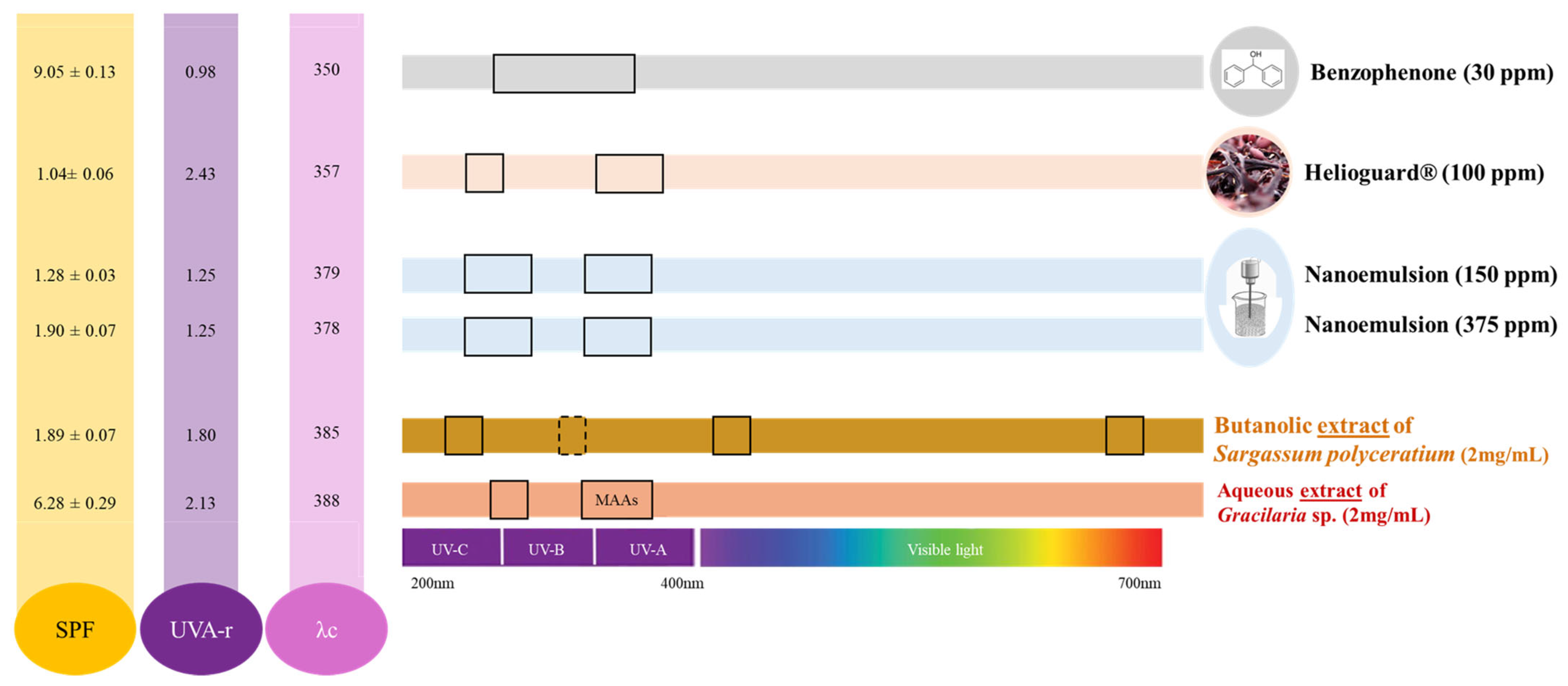
3.4. Sun Protection Factor (SPF), UVA Ratio (UVA-r), and Critical Wavelength (λc)

The photoprotective potential of the formulations was evaluated through SPF, UVA protection factor (UVA-r), and critical wavelength (λc) measurements (Figure 6). As expected, benzophenone-3 exhibited the highest SPF value, reaffirming its well-established efficacy as a UVB filter. Among the algal extracts, Gracilaria sp. demonstrated superior photoprotection compared to Sargassum sp., as reflected in both SPF and UVA-r values. Notably, nanoemulsions containing 150 ppm and 375 ppm of the combined algal extracts outperformed the commercial natural filter Helioguard®365 (100 ppm) (Mibelle Biochemistry, Buchs, Switzerland), indicating a potential synergistic effect within the formulation.
The aqueous extract of Gracilaria sp. exhibited the highest UVA-r value (2.13) and a critical wavelength (388 nm), suggesting strong UVA absorption and photostability. In comparison, the butanolic extract of Sargassum sp. showed a UVA-r of 1.80 and a λc of 385 nm—both within the threshold for broad-spectrum protection (λc ≥ 370 nm). Among the formulations, the nanoemulsion with 375 ppm of bioactives achieved a UVA-r of 1.25 and a λc of 379 nm, confirming its moderate but effective UVA coverage. By contrast, the positive control benzophenone-3 yielded a UVA-r of 0.98 and λc of 350 nm, underscoring its UVB-focused photoprotective action [43].
The combined use of both algal extracts in a single nanoemulsion provided complementary and synergistic photoprotection. MAAs from Gracilaria sp. primarily contributed to UVA absorption and ROS neutralization [7,44], while fucoxanthin from Sargassum sp. extended protection to the UVB range and enhanced antioxidant responses [45,46]. This dual mechanism supports the phase-specific formulation strategy: incorporating Gracilaria extract in the aqueous phase and Sargassum extract in the oil phase.
In vitro spectrophotometric analysis confirmed that, even at a low total concentration (375 ppm), the nanoemulsion delivered broad-spectrum protection (UVA-r > 0.7; λc = 379 nm). These results align with previous studies, such as Galani et al. (2023), which demonstrated that antioxidant-loaded nanoemulsions can enhance the SPF of topical products even at low concentrations [17].
Beyond UV filtration, this formulation is expected to provide biological skin protection. A recent study using Sargassum cymosum-based nanoemulsions showed up to 85% preservation of skin membrane integrity under UVB exposure, as measured by electrical impedance spectroscopy (EIS) [14]. These findings reinforce the potential of algae-derived compounds to mitigate oxidative and structural damage caused by solar radiation.
While the SPF values achieved may be modest compared to synthetic filters, the effectiveness at low concentrations combined with the natural origin, biodegradability, and reef-safe profile of the ingredients represent a valuable trade-off. This approach aligns with current trends in environmentally conscious cosmetics [47]. Moreover, the nanoemulsion platform offers scalability and formulation flexibility, supporting further SPF enhancement through extract concentration adjustment or the incorporation of other natural boosters [48,49].
These results validate the use of Gracilaria sp. and Sargassum sp. extracts in advanced, eco-friendly photoprotective formulations. This study contributes to the growing field of marine-derived sun care technologies by integrating biodiversity, nanotechnology, and sustainable formulation science.
Future work should aim to expand both the biological and physicochemical evaluation of the developed nanoemulsion. In vitro assays using UV-exposed skin cell models are needed to confirm the formulation’s protective effects at the cellular level, including potential anti-inflammatory and antioxidant responses. Additionally, further physicochemical characterization—such as rheological behavior, texture profile analysis, long-term stability studies, and skin permeation assays—will provide a more comprehensive understanding of the formulation’s performance and applicability. These complementary studies will be essential to support the positioning of algae-based nanoemulsions as effective, stable, and sustainable photoprotective alternatives for cosmetic applications.

4. Conclusions

This study demonstrated the development of a nanoemulsion incorporating Gracilaria sp. and Sargassum sp. extracts as a sustainable approach for broad-spectrum UV protection. MAAs and fucoxanthin, identified in the respective algae, were effectively combined in an optimized oil-in-water nanoemulsion with a droplet size of 32.42 nm and ζ potential of −26.0 mV. Photoprotective evaluation showed that the formulation, even at a low concentration (375 ppm), provided broad-spectrum coverage (UVA-r = 1.25; λc = 379 nm), surpassing the performance of a commercial natural filter. These findings highlight the synergistic potential of red and brown algae in nanoformulations for eco-friendly sun care products. This work contributes to the expanding field of marine-derived photoprotectants, offering a promising alternative aligned with “blue beauty” and environmentally conscious formulation strategies. Future studies should focus on increasing extract concentrations and validating in vivo efficacy for cosmetic applications.

Author Contributions

Conceptualization, J.T.Q. and D.M.A.N.; Formal analysis, J.T.Q. and V.U.-V.; Funding acquisition, L.C. and D.M.A.N.; Investigation, J.T.Q. and V.U.-V.; Methodology, J.T.Q., I.A.R.M., V.U.-V. and D.M.A.N.; Project administration, L.C. and D.M.A.N.; Resources, L.C. and D.M.A.N.; Supervision, L.C.; Writing—original draft, J.T.Q., I.A.R.M. and V.U.-V.; Writing—review and editing, I.A.R.M., V.U.-V., L.C. and D.M.A.N. All authors have read and agreed to the published version of the manuscript.

Funding

This research was funded by the MinCiencias with the ‘BALCAR-Q: Bioprospección y Química de Algas del Caribe’ program (code: 1101-852-69964), Contract No. 80740-739-2020.

Institutional Review Board Statement

Not applicable.

Informed Consent Statement

Not applicable.

Data Availability Statement

The original contributions presented in the study are included in the article, further inquiries can be directed to the corresponding author.

Acknowledgments

Thanks to MinCiencias and Universidad Nacional de Colombia for the ‘BALCAR-Q: Bioprospección y Química de Algas del Caribe’ program (code: 1101-852-69964) and also to the Ministerio de Ambiente y Desarrollo Sostenible by Permission No 121 of 22 January 2016 (modification otrosí No 7) for collecting samples.

Conflicts of Interest

The authors declare no conflict of interest. The authors declare that the research was conducted in the absence of any commercial or financial relationships that could be construed as a potential conflict of interest. The funders had no role in the design of the study; in the collection, analyses or interpretation of data; in the writing of the manuscript; or in the decision to publish the results.

References

  1. Pangestuti, R.; Siahaan, E.; Kim, S.-K. Photoprotective Substances Derived from Marine Algae. Mar. Drugs 2018, 16, 399. [Google Scholar] [CrossRef]
  2. Torres, P.; Santos, J.P.; Chow, F.; dos Santos, D.Y. A Comprehensive Review of Traditional Uses, Bioactivity Potential, and Chemical Diversity of the Genus Gracilaria (Gracilariales, Rhodophyta). Algal Res. 2019, 37, 288–306. [Google Scholar] [CrossRef]
  3. Sinha, R.P.; Klisch, M.; Gröniger, A.; Häder, D.-P. Mycosporine-like Amino Acids in the Marine Red Alga Gracilaria cornea—Effects of UV and Heat. Environ. Exp. Bot. 2000, 43, 33–43. [Google Scholar] [CrossRef]
  4. Schneider, G.; Figueroa, F.L.; Vega, J.; Avilés, A.; Horta, P.A.; Korbee, N.; Bonomi-Barufi, J. Effects of UV–Visible Radiation on Growth, Photosynthesis, Pigment Accumulation and UV-Absorbing Compounds in the Red Macroalga Gracilaria cornea (Gracilariales, Rhodophyta). Algal Res. 2022, 64, 102702. [Google Scholar] [CrossRef]
  5. Qoh, A.; Amine, J.; Assobhei, O.; Etahiri, S. Investigation of UV Protection and Antiaging Properties of Mycosporinelike Amino Acids in the Moroccan Red Alga Gracilaria gracilis (Gracilariales, Rhodophyta). J. Appl. Phycol. 2025, 37, 1341–1356. [Google Scholar] [CrossRef]
  6. Ryu, J.; Park, S.-J.; Kim, I.-H.; Choi, Y.H.; Nam, T.-J. Protective Effect of Porphyra-334 on UVA-Induced Photoaging in Human Skin Fibroblasts. Int. J. Mol. Med. 2014, 34, 796–803. [Google Scholar] [CrossRef]
  7. Singh, A.; Čížková, M.; Bišová, K.; Vítová, M. Exploring Mycosporine-like Amino Acids (MAAs) as Safe and Natural Protective Agents against UV-Induced Skin Damage. Antioxidants 2021, 10, 683. [Google Scholar] [CrossRef]
  8. Stiger-Pouvreau, V.; Mattio, L.; De, A.; Uwai, S.; Dominguez, H.; Flórez-Fernández, N.; Connan, S.; Critchley, A.T. A Concise Review of the Highly Diverse Genus Sargassum C. Agardh with Wide Industrial Potential. J. Appl. Phycol. 2023, 35, 1453–1483. [Google Scholar] [CrossRef]
  9. Piao, M.J.; Yoon, W.J.; Kang, H.K.; Yoo, E.S.; Koh, Y.S.; Kim, D.S.; Lee, N.H.; Hyun, J.W. Protective Effect of the Ethyl Acetate Fraction of Sargassum muticum against Ultraviolet B–Irradiated Damage in Human Keratinocytes. Int. J. Mol. Sci. 2011, 12, 8146–8160. [Google Scholar] [CrossRef]
  10. Xu, Z.; Li, B.; Li, L.; Wang, N.; Wang, Y.; Wang, H.; Yan, F.; Bao, M.; Zang, S.; Wu, H.; et al. Effects of UV Radiation on Photosynthesis of Sargassum muticum. J. Exp. Mar. Biol. Ecol. 2023, 569, 151961. [Google Scholar] [CrossRef]
  11. Prasedya, E.S.; Syafitri, S.M.; Geraldine, B.A.F.D.; Hamdin, C.D.; Frediansyah, A.; Miyake, M.; Kobayashi, D.; Hazama, A.; Sunarpi, H. UVA Photoprotective Activity of Brown Macroalgae Sargassum Cristafolium. Biomedicines 2019, 7, 77. [Google Scholar] [CrossRef] [PubMed]
  12. Schneider, G.; Figueroa, F.L.; Vega, J.; Chaves, P.; Álvarez-Gómez, F.; Korbee, N.; Bonomi-Barufi, J. Photoprotection Properties of Marine Photosynthetic Organisms Grown in High Ultraviolet Exposure Areas: Cosmeceutical Applications. Algal Res. 2020, 49, 101956. [Google Scholar] [CrossRef]
  13. Xu, Z.; Li, L.; Jiang, H.; Yan, F.; Liu, L.; Zang, S.; Ma, Y.; Wu, H. Photosynthetic Responses of a Golden Tide Alga (Sargassum horneri) to Ultraviolet Radiation. Front. Mar. Sci. 2022, 9, 978376. [Google Scholar] [CrossRef]
  14. Hernández, A.R.; Sepulveda, L.; Hata, Y.; Castellanos, L.; Björklund, S.; Ruzgas, T.; Aragón, M. Algae Extract-Based Nanoemulsions for Photoprotection against UVB Radiation: An Electrical Impedance Spectroscopy Study. Sci. Rep. 2025, 15, 191. [Google Scholar] [CrossRef] [PubMed]
  15. Guzmán, C.; Rojas, M.A.; Aragón, M. Optimization of Ultrasound-Assisted Emulsification of Emollient Nanoemulsions of Seed Oil of Passiflora edulis var. edulis. Cosmetics 2020, 8, 1. [Google Scholar] [CrossRef]
  16. Panjaitan, M.A.P.; Kasitowati, R.D.; Putri, N.; Yamindago, A.; Asmara, R.; Aliviyanti, D.; Pratiwi, D.C. Polysaccharide Isolation of Brown Seaweed: Sargassum sp and Its Photoprotection Activity. IOP Conf. Ser.Earth Environ. Sci. 2022, 1036, 012039. [Google Scholar] [CrossRef]
  17. Galani, E.; Galatis, D.; Tzoka, K.; Papadimitriou, V.; Sotiroudis, T.G.; Bonos, A.; Xenakis, A.; Chatzidaki, M.D. Natural Antioxidant-Loaded Nanoemulsions for Sun Protection Enhancement. Cosmetics 2023, 10, 102. [Google Scholar] [CrossRef]
  18. Cerqueira-Coutinho, C.; Santos-Oliveira, R.; dos Santos, E.; Mansur, C.R. Development of a Photoprotective and Antioxidant Nanoemulsion Containing Chitosan as an Agent for Improving Skin Retention. Eng. Life Sci. 2015, 15, 593–604. [Google Scholar] [CrossRef]
  19. Guiry, M.D.; Guiry, G.M. AlgaeBase World-Wide Electronic Publication, National University of Ireland, Galway. 2025. Available online: http://www.algaebase.org (accessed on 20 February 2025).
  20. Sepulveda, L. Búsqueda de Compuestos con Posible Actividad Inhibitoria de Enzimas de Interés Cosmético a Partir de Algas del Caribe Colombiano. Repositorio UNAL. Available online: https://repositorio.unal.edu.co/handle/unal/84624 (accessed on 20 February 2025).
  21. Urrea-Victoria, V.; Costa, G.; Gavio, B.; Ramos, F.; Castellanos, L. Mycosporine-like Amino Acids Profile in Red Algae from High UV-Index Geographical Areas (San Andrés Island and La Guajira) of the Colombian Caribbean Coast. Algal Res. 2025, 86, 103927. [Google Scholar] [CrossRef]
  22. Foo, S.C.; Khoo, K.S.; Ooi, C.W.; Show, P.L.; Khong, N.M.H.; Yusoff, F.M. Meeting Sustainable Development Goals: Alternative Extraction Processes for Fucoxanthin in Algae. Front. Bioeng. Biotechnol. 2021, 8, 546067. [Google Scholar] [CrossRef]
  23. Mansur, J.S.; Breder, M.N.; Mansur, M.C.; Azulay, R.D. Determination of Sun Protection Factor by Spectrophotometry. Bras. Dermatol. 1986, 61, 121–124. [Google Scholar]
  24. Springsteen, A.; Yurek, R.; Frazier, M.; Carr, K.F. In Vitro measurement of sun protection factor of sunscreens by diffuse transmittance. Anal. Chim. Acta 1999, 380, 155–164. [Google Scholar] [CrossRef]
  25. Tadros, T.; Izquierdo, P.; Esquena, J.; Solans, C. Formation and Stability of Nano-Emulsions. Adv. Colloid Interface Sci. 2004, 108–109, 303–318. [Google Scholar] [CrossRef] [PubMed]
  26. Lawrence, M.J.; Rees, G.D. Microemulsion-Based Media as Novel Drug Delivery Systems. Adv. Drug Deliv. Rev. 2000, 45, 89–121. [Google Scholar] [CrossRef]
  27. Vaz, B.M.; Leite, M.S.C.; Contieri, L.S.; Mesquita, L.M.d.S.; Conde, A.; Oliveira, J.P.; Pinto, D.C.; Ventura, S.P. Efficient Extraction and Purification of Mycosporineslike Amino Acids (MAAs) Following a Multiproduct Biorefinery Approach. Sep. Purif. Technol. 2025, 363, 132200. [Google Scholar] [CrossRef]
  28. Ashkenazi, D.Y.; Figueroa, F.L.; Korbee, N.; García-Sánchez, M.; Vega, J.; Ben-Valid, S.; Paz, G.; Salomon, E.; Israel, Á.; Abelson, A. Enhancing Bioproducts in Seaweeds via Sustainable Aquaculture: Antioxidant and Sun-Protection Compounds. Mar. Drugs 2022, 20, 767. [Google Scholar] [CrossRef]
  29. Figueroa, F.L.; Álvarez-Gómez, F.; Bonomi-Barufi, J.; Vega, J.; Massocato, T.F.; Gómez-Pinchetti, J.L.; Korbee, N. Interactive Effects of Solar Radiation and Inorganic Nutrients on Biofiltration, Biomass Production, Photosynthetic Activity and the Accumulation of Bioactive Compounds in Gracilaria cornea (Rhodophyta). Algal Res. 2022, 68, 102890. [Google Scholar] [CrossRef]
  30. Torres, P.; Chow, F.; dos Santos, D.Y.A.C. A Chemical Investigation of the Antioxidant Capacity of Extracts from Red Macroalga Gracilaria domingensis. Phycology 2022, 2, 332–343. [Google Scholar] [CrossRef]
  31. Zwerger, M.; Ganzera, M. Fast and Efficient Separation of Eleven Mycosporine-like Amino Acids by UHPLC-DAD and Their Quantification in Diverse Red Algae. Mar. Drugs 2022, 20, 395. [Google Scholar] [CrossRef]
  32. Oliyaei, N.; Moosavi-Nasab, M. Ultrasound-Assisted Extraction of Fucoxanthin from Sargassum Angustifolium and Cystoseira Indica Brown Algae. J. Food Process. Preserv. 2021, 45, e15929. [Google Scholar] [CrossRef]
  33. Hong, D.; Thom, L.T.; Ha, N.C.; Hoai, T.; Minh, T.; Tam, L.T.; Dat, N.M.; Duc, T.M.; Tru, N.V.; Minh, T.; et al. Isolation of Fucoxanthin from Sargassum oligocystum Montagne, 1845 Seaweed in Vietnam and Its Neuroprotective Activity. Biomedicines 2023, 11, 2310. [Google Scholar] [CrossRef]
  34. Molina, G.A.; González-Reyna, M.A.; Loske, A.M.; Fernández, F.; Torres-Ortiz, D.A.; Estevez, M. Weak Shock Wave-Mediated Fucoxanthin Extraction from Sargassum spp. And Its Electrochemical Quantification. Algal Res. 2022, 68, 102891. [Google Scholar] [CrossRef]
  35. Torres, A.; Enk, C.D.; Hochberg, M.; Srebnik, M. Porphyra-334, a Potential Natural Source for UVA Protective Sunscreens. Photochem. Photobiol. Sci. 2006, 5, 432–435. [Google Scholar] [CrossRef]
  36. de la Coba, F.; Aguilera, J.; Korbee, N.; de Gálvez, M.; Herrera-Ceballos, E.; Álvarez-Gómez, F.; Figueroa, F. UVA and UVB Photoprotective Capabilities of Topical Formulations Containing Mycosporine-like Amino Acids (MAAs) through Different Biological Effective Protection Factors (BEPFs). Mar. Drugs 2019, 17, 55. [Google Scholar] [CrossRef] [PubMed]
  37. Heo, S.-J.; Jeon, Y.-J. Protective Effect of Fucoxanthin Isolated from Sargassum Siliquastrum on UV-B Induced Cell Damage. J. Photochem. Photobiol. B Biol. 2009, 95, 101–107. [Google Scholar] [CrossRef]
  38. Chen, S.-J.; Lee, C.-J.; Lin, T.-B.; Peng, H.-Y.; Liu, H.-J.; Chen, Y.-S.; Tseng, K.-W. Protective Effects of Fucoxanthin on Ultraviolet B-Induced Corneal Denervation and Inflammatory Pain in a Rat Model. Mar. Drugs 2019, 17, 152. [Google Scholar] [CrossRef] [PubMed]
  39. Shi, Y.; Ren, J.; Zhao, B.; Zhu, T.; Qi, H. Photoprotective Mechanism of Fucoxanthin in Ultraviolet B Irradiation-Induced Retinal Müller Cells Based on Lipidomics Analysis. J. Agric. Food Chem. 2022, 70, 3181–3193. [Google Scholar] [CrossRef] [PubMed]
  40. Kommuru, T.R.; Gurley, B.; Khan, M.A.; Reddy, I.K. Self-Emulsifying Drug Delivery Systems (SEDDS) of Coenzyme Q10: Formulation Development and Bioavailability Assessment. Int. J. Pharm. 2001, 212, 233–246. [Google Scholar] [CrossRef]
  41. Pouton, C.W. Formulation of Poorly Water-Soluble Drugs for Oral Administration: Physicochemical and Physiological Issues and the Lipid Formulation Classification System. Eur. J. Pharm. Sci. 2006, 29, 278–287. [Google Scholar] [CrossRef]
  42. Badolato, G.G.; Aguilar, F.; Schuchmann, H.P.; Sobisch, T.; Lerche, D. Evaluation of Long Term Stability of Model Emulsions by Multisample Analytical Centrifugation. In Surface and Interfacial Forces–From Fundamentals To Applications; Auernhammer, Günter, K., Butt, H.-J., Vollmer, D., Eds.; Springer: Berlin/Heidelberg, Germany, 2008; pp. 66–73. [Google Scholar]
  43. Coelho, S.G.; Rua, D.; Miller, S.A.; Agrawal, A. Suboptimal UVA Attenuation by Broad Spectrum Sunscreens under Outdoor Solar Conditions Contributes to Lifetime UVA Burden. Photodermatol. Photoimmunol. Photomed. 2020, 36, 42–52. [Google Scholar] [CrossRef]
  44. Rosic, N. Mycosporine-like Amino Acids: Making the Foundation for Organic Personalised Sunscreens. Mar. Drugs 2019, 17, 638. [Google Scholar] [CrossRef] [PubMed]
  45. Urikura, I.; Sugawara, T.; Hirata, T. Protective Effect of Fucoxanthin against UVB-Induced Skin Photoaging in Hairless Mice. Biosci. Biotechnol. Biochem. 2011, 75, 757–760. [Google Scholar] [CrossRef] [PubMed]
  46. Mumu, M.; Das, A.; Emran, T.B.; Mitra, S.; Islam, F.; Roy, A.; Karim, M.M.; Das, R.; Park, M.N.; Chandran, D.; et al. Fucoxanthin: A Promising Phytochemical on Diverse Pharmacological Targets. Front. Pharmacol. 2022, 13, 929442. [Google Scholar] [CrossRef] [PubMed]
  47. Chatzigianni, M.; Pavlou, P.; Siamidi, A.; Vlachou, M.; Varvaresou, A.; Papageorgiou, S. Environmental Impacts due to the Use of Sunscreen Products: A Mini-Review. Ecotoxicology 2022, 31, 1331–1345. [Google Scholar] [CrossRef]
  48. Resende, D.I.S.P.; Jesus, A.; Sousa Lobo, J.M.; Sousa, E.; Cruz, M.T.; Cidade, H.; Almeida, I.F. Up-To-Date Overview of the Use of Natural Ingredients in Sunscreens. Pharmaceuticals 2022, 15, 372. [Google Scholar] [CrossRef]
  49. Chavda, V.P.; Acharya, D.; Hala, V.; Daware, S.; Vora, L.K. Sunscreens: A Comprehensive Review with the Application of Nanotechnology. J. Drug Deliv. Sci. Technol. 2023, 86, 104720. [Google Scholar] [CrossRef]
Figure 1. Chemical structure of (A) Capryol® PGMC and (B) Kolliphor® EL.
Cosmetics 12 00101 g001
Figure 2. Chromatographic profiles of (A) MAAs in Gracilaria sp. and (B) fucoxanthin in Sargassum sp.
Cosmetics 12 00101 g002
Figure 3. Pseudoternary diagram for the determination of the nanoemulsion region for day 1. The blue dots represent the nanoemulsions (sizes below 100 nm), red dots represent emulsions with size above 100 nm. Oil: Capryol PGMC®; Smix: Kolliphor EL®: propylene glycol (3:2).
Cosmetics 12 00101 g003
Figure 4. Surface response diagram showing the effect of the variables on droplet size (nm). (A) Effect of oil(%) and sonication time (min) on droplet size (nm). (B) Effect of Smix(%) and oil(%) on droplet size (nm). (C) Effect of Smix(%) and sonication time (min) on droplet size (nm).
Cosmetics 12 00101 g004
Figure 5. Surface response diagram showing the effect on potential ζ (mV). (A) Effect of oil(%) and sonication time (min) on potential ζ (mV). (B) Effect of Smix(%) and oil(%) on potential ζ (mV). (C) Effect of Smix(%) and sonication time (min) on potential ζ (mV).
Cosmetics 12 00101 g005
Figure 6. SPF, UVA-r, and critical wavelength data (λc) of samples. The chart shows the absorption range of the extracts across the UV regions, indicating the potential for photoprotection. Squares represent metabolites absorbing at specific wavelengths within the electromagnetic spectrum.
Cosmetics 12 00101 g006
Table 1. Droplet size and ζ potential for emulsions without Gracilaria sp. and S. polyceratium extracts.
NoOil (%)Smix (%)Water (%)Day 1Day 7
Droplet Size (nm)Potential ζ (mV)Droplet Size (nm)Potential ζ (mV)
14158117.8 ± 0.7219−24.13 ± 0.8317.46 ± 0.08−23.92 ± 0.24
24207627.36 ± 4.05−24.14 ± 0.2032.38 ± 5.16−23.88 ± 0.36
34257154.27 ± 2.77−22.82 ± 0.3221.70 ± 1.36−22.78 ± 0.31
44306618.62 ± 0.3814−23.56 ± 0.1815.71 ± 0.14−23.84 ± 0.16
54356116.88 ± 0.1943−23.24 ± 0.2815.09 ± 0.09−22.84 ± 0.31
68108217.59 ± 0.4099−22.86 ± 0.1326.72 ± 0.21−22.42 ± 0.19
781577172.5 ± 1.249−23.31 ± 0.10253.40 ± 5.99−23.56 ± 0,07
88207218.15 ± 0.07937−23.99 ± 0.1814.67 ± 0.03−23.68 ± 0.42
982567333.9 ± 13.1−23.32 ± 0.16280.40 ± 2.54−22.88 ± 0.17
108306231.82 ± 0.3584−24.07 ± 0.1721.29 ± 0.03−24.24 ± 0.12
118355778.29 ± 7.053−24.12 ± 0.1322.14 ± 0.03−24.68 ± 0.16
1212107820.75 ± 0.9283−23.38 ± 0.2118.31 ± 0.15−22.70 ± 0.32
13121573322.5 ± 3.584−24.13 ± 0.05289.20 ± 3.70−22.80 ± 0.32
14122068478.1 ± 5.424−24.08 ± 0.10340.50 ± 5.78−24.46 ± 0.06
15122563316.1 ± 2.572−23.75 ± 0.22393.70 ± 1.58−24.58 ± 0.16
16123058359.8 ± 3.786−23.08 ± 0.34124.90 ± 10.54−24.62 ± 0.11
17123553949.9 ± 3.01−24.08 ± 0.06210.30 ± 17.68−24.16 ± 0.13
18161074394.6 ± 2.542−23.4 ± 0.02378.20 ± 11.82−22.44 ± 0.75
19161569380.6 ± 3.11823.05 ± 0.26330.30 ± 2.76−25.60 ± 0.29
20162064439.3 ± 3.464−23.23 ± 0.38399.60 ± 0.93−20.23 ± 0.18
21162559390.8 ± 3.404−23.59 ± 0.38342.50 ± 3.46−22.12 ± 0.09
22201070472.1 ± 5.897−20.09 ± 0.08420.10 ± 1.78−24.44 ± 0.03
23201565258.4 ± 11.17−25.54 ± 0.35262.30 ± 10.50−23.96 ± 0.17
24202060590.2 ± 4.576−24.2 ± 0.33435.30 ± 6.47−23.86 ± 0.12
Oil: Capryol PGMC®; Smix: Kolliphor EL®: propylene glycol (3:2).
Table 2. Matrix of independent variables of the BBD.
Independent VariablesLevels
−10−1
Oil (%)4812
Smix (%)153045
Sonication time (min)258
Oil: Capryol PGMC®; Smix: Kolliphor EL®: propylene glycol (3:2).
Table 3. Factor points, independent factor levels, and experimental values of dependent variables for the Box–Behnken design (BBD).
NoOil (%)Smix (%)Water (%)Sonication Time (min)Day 1Day 7
Droplet Size (nm)Potential ζ (mV)Droplet Size (nm)Potential ζ (mV)
183062545.72 ± 3.39−17.2 ± 2.052.56 ± 6.92−23.0 ± 2.8
283062534.70 ± 8.80−24.0 ± 0.741.50 ± 2.30 −24.5 ± 0.6
3123058830.09 ± 0.47−22.6 ± 3.033.92 ± 1.15−19.9 ± 2.1
444551538.77 ± 0.78−26.1 ± 0.938.32 ± 3.96−25.0 ± 3.1
541581529.33 ± 2.55 −23.8 ± 3.289.80 ± 11.88−13.4 ± 0.6
683062540.12 ± 2.01−24.8 ± 3.223.44 ± 0.51−18.4 ± 3.0
783062555.96 ± 7.25−21.6 ± 1.726.60 ± 0.21−24.1 ± 2.7
884547831.26 ± 2.71−9.3 ± 0.846.17 ± 0.82−11.3 ± 1.7
981577878.01 ± 0.38−20.4 ± 2.0125.70 ± 8.58−24.8 ± 0.9
1084547240.73 ± 3.49−18.0 ± 0.530.95 ± 1.32−15.4 ± 3.5
11123058227.04 ± 0.51−21.4 ± 1.224.25 ± 0.73−16.5 ± 2.4
1283062550.96 ± 2.24−15.2 ± 2.031.56 ± 0.14−22.5 ± 0.4
1343066243.42 ± 9.93−33.4 ± 2.122.21 ± 0.51−18.1 ± 2.7
14124543555.26 ± 0.87−11.8 ± 1.433.09 ± 0.62−11.7 ± 1.6
1583062541.19 ± 1.36−18.9 ± 1.429.99 ± 0.73−18.5 ± 2.3
1681577228.98 ± 0.72−29.9 ± 3.525.41 ± 0.33−20.9 ± 3.2
17121573555.78 ± 0.43−22.3 ± 2.489.94 ± 4.32−19.0 ± 2.3
1843066826.34 ± 1.93−31.4 ± 0.932.14 ± 2.25−14.9 ± 1.3
Oil: Capryol PGMC®; Smix: Kolliphor EL®: propylene glycol (3:2).
Table 4. Analysis of variance of a surface response of quadratic equations for droplet size and potential ζ.
VariableDroplet SizePotential ζ
F-Valuep-ValueF-Valuep-Value
Linear termsA8.610.0191.280.291
B9.840.0145.350.049
C3.810.08716.860.003
Quadratic termsA20.840.3862.630.143
B20.820.3913.730.089
C21.760.2218.110.022
Interaction termsAB4.800.0600.150.704
AC12.300.0080.700.427
BC0.360.5640.120.739
Lack-of-fit0.370.7781.340.360
A: time of sonication (min); B: Oil(%); C: Smix(%). In the model, values of p <0.05 are considered significant.
Table 5. Final characteristics of the optimized formulation.
Response VariableExperimental Value
Droplet size (nm)32.42
Potential ζ−26.0
Final composition
Kolliphor EL®: propylene glycol (%)20.8%
Capryol PGMC (%)4%
Deionized water (%)73.7%
Oil: Capryol PGMC®; Smix: Kolliphor EL®: propylene glycol (3:2).
Disclaimer/Publisher’s Note: The statements, opinions and data contained in all publications are solely those of the individual author(s) and contributor(s) and not of MDPI and/or the editor(s). MDPI and/or the editor(s) disclaim responsibility for any injury to people or property resulting from any ideas, methods, instructions or products referred to in the content.

