Article

Production and Characterization of Semi-Solid Formulations for the Delivery of the Cosmetic Peptide Palmitoyl-GHK

1
Department of Chemical, Pharmaceutical and Agricultural Sciences, University of Ferrara, I-44121 Ferrara, Italy
2
Cosmetology Center, University of Ferrara, I-44121 Ferrara, Italy
3
Department of Life Science and Biotechnology, University of Ferrara, I-44121 Ferrara, Italy
*
Authors to whom correspondence should be addressed.
Cosmetics 2025, 12(2), 50; https://doi.org/10.3390/cosmetics12020050
Submission received: 5 February 2025 / Revised: 3 March 2025 / Accepted: 7 March 2025 / Published: 13 March 2025
(This article belongs to the Special Issue Feature Papers in Cosmetics in 2025)

Abstract

:
In this study, vesicular lipid systems and semi-solid formulations for the skin application of Palmitoyl-GHK were formulated and characterized. Palmitoyl-GHK is a cosmetic peptide with anti-aging action, capable of treating the signs of skin aging by mainly stimulating collagen synthesis in the dermis. The so-called “ethosomes” were evaluated as nanovesicular systems constituted of phosphatidylcholine, organized in vesicles, ethanol, and water. In addition, semi-solid systems were produced and characterized, namely an organogel based on phosphatidylcholine, isopropyl palmitate, and water, a gel based on Poloxamer 407, and the poloxamer organogel, created by combining organogel and Poloxamer gel. To make the ethosomal dispersions suitable for skin application, xanthan gum was added as a gelling agent. Studies were therefore carried out on semi-solid formulations to determine (i) the spreadability, a key factor that influences various aspects of a topical/transdermal formulation, (ii) the occlusive factor, important to guarantee good effectiveness of a dermocosmetic product, and finally, (iii) the hydrating power, to study the effect of a formulation applied to the skin. A formulation study enabled the selection of the most suitable formulations for the incorporation of the active ingredient of interest. Palmitoyl-GHK was found to be soluble both in ethosomes and in the poloxamer organogel. In vitro studies were therefore conducted to evaluate the release kinetics of Palmitoyl-GHK from the formulations, via Franz cells. The qualitative–quantitative analysis, through analytical HPLC, highlighted that the active ingredient is released more slowly from semi-solid formulations compared to vesicular systems; in particular, the presence of poloxamer allows a controlled release of the peptide. Further studies will be necessary to verify the anti-aging efficacy of formulations containing the peptide.

1. Introduction

Recently, polypeptides have been proposed for the treatment of skin aging. Polypeptides or oligopeptides, in fact, can be composed of amino acids that mimic the peptide sequences of molecules, such as collagen or elastin. Through topical application, certain polypeptides can stimulate collagen synthesis and activate dermal metabolism. Generally, peptides are biocompatible: they are considered safe due to their natural presence in the body, they exhibit stability in light and air, and they can be synthesized in laboratory conditions; therefore, they can be utilized in pharmaceutical and dermocosmetic formulations [1]. Some well-established and documented peptides have demonstrated their efficacy through clinical trials, while others remain in the research phase. As a result of biotechnological research in the dermocosmetic field, the so-called ‘biomimetic peptides’ were synthesized, which are active ingredients capable of replicating the function of the protein with which they are structurally and functionally associated. The primary limitation of peptide utilization is related to their hydrophilicity and high molecular weight, which impede skin permeation [2]. Matrikines are low-molecular-weight peptides derived from the proteolysis of ECM proteins, exerting effects similar to growth factors or cytokines, and capable of regulating cellular activity. Certain matrikines can modulate cell proliferation, migration, protease production, or apoptosis [3,4]. The biomimetic characteristics of matrikines ensure a favorable safety profile compared to alpha hydroxy acids and retinoids [5]. GHK (Glycine–Histidine–Lysine), a tripeptide found naturally in human plasma and saliva, is classified as a matrikine. The concentration of GHK in plasma at age 20 is approximately 200 ng/mL, while it decreases to 80 ng/mL by age 60. This decline coincides with the attenuation of an organism’s regenerative capabilities. This tripeptide forms a complex with copper (Cu2+) with a strong affinity, accelerating wound healing, improving the yield of transplanted skin, and possessing anti-inflammatory properties. The molecular weight (lower than 500 Da) and hydrophilic properties of GHK necessitate chemical modifications, such as esterification with alkyl chains, to enhance its permeability through the skin. In this regard, the esterification strategy with palmitic acid results in Palmitoyl-GHK (Palm-GHK), characterized by higher lipophilicity, permeability, and stability compared to GHK. Palm-GHK (Figure 1) is primarily intended to stimulate the replenishment of the degraded extracellular matrix under skin application, with the aim of reducing wrinkles [6,7].
Similarly, additional palmitoyl oligopeptides have been proposed in skin care within certain dermocosmetic formulations as functional ingredients with anti-aging effects, including Palmitoyl tripeptide-5 and Palmitoyl tetrapeptide-7 [8]. Generally, cosmetic products containing palmitoyl oligopeptides can be safely applied to the skin and hair or may come into contact with the eyes and mucous membranes, even with regular use. However, the limitations of utilizing synthetic peptides in cosmetics, related to their poor penetration, stability, and potential long-term effects, necessitate their incorporation into advanced delivery vehicles capable of solubilizing them while enhancing their skin penetration. In this context, phosphatidylcholine (PC) and/or Poloxamer copolymers can serve as suitable excipients for preparing semi-solid formulations for GHK peptide cutaneous delivery [9]. PC is the most prevalent phospholipid in biological systems; it constitutes one of the primary components of the cell membrane and can be isolated and purified from soybeans and/or egg yolk. PC is a natural zwitterionic surfactant widely employed in cosmetics due to its emulsifying properties and its ability to self-assemble in water and/or organic solvents. PC can spontaneously form supramolecular structures suitable for active compound delivery, such as lamellae, vesicles, and cubic and hexagonal phases. Furthermore, it functions as a permeation enhancer due to its structure, which is similar to the lipids that constitute the stratum corneum, creating preferential channels for the penetration of active ingredients [10,11]. The surfactant properties of non-ionic poly(oxyethylene)poly(oxypropylene) (PEO-PPO) block copolymer Poloxamers enable them to self-organize in water, forming micelles capable of increasing the solubility of lipophilic molecules [12,13]. PC dispersion in water in the presence of ethanol leads to the formation of ethosome nanovesicles (ETOs), biocompatible nanocarriers for the dermal and transdermal delivery of drugs. The ETO’s ability to facilitate drug penetration into the deeper layers of the skin is attributed to the presence of ethanol, which acts synergistically with PC as a penetration enhancer. Due to the presence of ethanol, ETOs are more malleable and stable compared to conventional liposomes [14]. As the liquid state of ETOs impedes their retention on the skin, they can be thickened by hydrophilic colloids, such as xanthan gum, resulting in viscous ethosomal gels (ETO-GELs) [15]. Conversely, the dispersion of PC in organic solvents results in the formation of reverse micelles that, with the addition of water, tend to elongate longitudinally until forming tubular structures that entangle in a three-dimensional network characteristic of organogel vehicles (ORG). As the water content increases, an increase in viscosity is observed up to a maximum value, beyond which phase separation occurs. ORG is suitable for topical cutaneous or mucosal application, being composed of biocompatible and therefore non-irritating components. Additionally, the amphiphilic nature of PC allows for the dissolution and transdermal release of both hydrophilic and lipophilic compounds [9,13,16]. Certain Poloxamers, such as 407, exhibit thermo-reversible behavior in aqueous solutions, contingent upon their molecular weight and poly(oxypropylene) (POP) content [17]. Poloxamer 407 solutions (20–30% w/w) demonstrate a thermo-responsive sol–gel transition above 20–30 °C, characterized by the formation of a rigid gel network arising from dense micellar packing [18]. Poloxamer gels (POL) exhibit ‘smart’ behavior, offering significant advantages for cutaneous and mucosal delivery [19]. Their liquid state at ambient temperatures facilitates application, while their transition to a viscous, semi-solid state near body temperature enables controlled drug release [20,21,22]. Poloxamer lecithin organogels (POL-ORGs) represent a novel class of ‘smart’ semi-solid systems. They are generated by combining POL with ORG, involving the incorporation of Poloxamer 407, the gelling agent of POL, into the aqueous phase of the pre-formed ORG [23]. POL-ORG architecture is characterized by a complex biphasic system. In the presence of an organic solvent and water, PC and Poloxamer 407 undergo self-assembly, resulting in the formation of interpenetrating microstructures. Notably, both PC and the poly(propylene oxide) (PPO) segments of Poloxamer 407 have been demonstrated to enhance drug permeation by interacting with and disrupting the lipid packing of cellular membranes [9,24,25].
In the present investigation, PC and Poloxamer-based semi-solid forms suitable for Palm-GHK skin administration, specifically ETO-GEL, ORG, POL, and POL-ORG, were designed, prepared by low-energy consumption procedures, and characterized. Particularly, the spreadability and occlusive factor of semi-solid forms were evaluated in vitro. An in vivo pilot study was conducted to compare the hydration efficacy of the different formulations. The in vitro release kinetics of Palm-GHK from ETO and the semi-solid forms were evaluated using diffusion cells associated with synthetic membranes.

2. Materials and Methods

2.1. Materials

The materials used in this study include soy phosphatidylcholine (Phospholipon 90 G; Lipoid GmbH, Ludwigshafen, Germany), Poloxamer 407 (Sigma Aldrich, BASF ChemTrade GmbH, Ludwigshafen, Germany), isopropyl palmitate (IPP), xanthan gum, Wang resin, Fmoc amino acids (Iris Biotech GmbH, Marktredwitz, Germany), palmitic acid, and dimethylformamide (DMF) (Merck-Sigma Aldrich, Milan, Italy). Palmitoyl-GHK was synthesized following the procedure reported in the literature [26].

2.2. Preparation of the Formulations

2.2.1. Ethosomes

To prepare ETO, PC is first dissolved in ethanol (30 mg/mL) at room temperature. Subsequently, the aqueous phase (70% v/v) is added dropwise to the ethanolic solution under continuous magnetic stirring at approximately 500 rpm using an IKA Eurostar digital stirrer (IKA Labortechnik Janke & Kunkel, Staufen, Germany). Once the entire aqueous phase has been added, magnetic stirring is maintained for 30 min at room temperature. In the case of ethosomes containing Palm-GHK (ETO-GHK), the peptide is dissolved in the PC solution in ethanol (1 mg/mL) prior to the addition of water, using a vortex mixer (ZX, Velp Scientifica, Usmate, Italy) to ensure complete solubilization.

2.2.2. Ethosomal Gel

To increase the viscosity of the ethosomes, xanthan gum (1% w/w) was added directly to the ethosomal dispersion and mixed until the gum was fully dissolved, resulting in an ethosomal gel (ETO-GEL).

2.2.3. Organogel

The organogel (ORG) was prepared from a 200 mM PC solution in IPP, which was obtained by dissolving the PC in IPP under magnetic stirring at 60 °C. Subsequently, water (14.4 µL/mL) was added to the PC solution while maintaining the system under agitation until a viscous gel formed within seconds. The viscosity of the ORG depends on the organic solvent, the concentration of PC, and the water phase content. The latter is expressed as the ratio between the molar concentration of water and PC, or w0 = [W]/[PC]. In the case of organogel containing Palm-GHK (ORG-GHK), the peptide was dissolved (1 mg/mL and 0.5 mg/mL) in the PC solution in IPP before adding the aliquot of water.

2.2.4. Poloxamer Gel

The Poloxamer 407 (POL) gel was prepared using the ‘cold’ method, gradually adding the copolymer (20% w/w) to double-distilled water under magnetic stirring at a temperature of approximately 4°. To obtain the Poloxamer gel containing Palm-GHK (POL-GHK), the peptide was added to the pre-formed gel and solubilized (1 mg/mL) at a cold temperature using a vortex.

2.2.5. Poloxamer Organogel

The poloxamer organogel (POL-ORG) consisted of a lipid phase and an aqueous phase in a 30:70 w/w ratio. The lipid phase was a solution of PC (200 mM) dissolved in IPP at 60 °C under magnetic stirring, while the aqueous phase was a Poloxamer 407 gel (20% w/w) prepared as previously described [27]. The aqueous phase was added to the lipid phase and maintained under magnetic stirring for 30 min. To obtain the poloxamer organogel containing Palm-GHK (POL-ORG-GHK), Palmitoyl-GHK (1 mg/mL) was incorporated into the Poloxamer gel before adding the PC lipid phase.

2.3. Characterization of the Formulations

2.3.1. Size Analysis of Ethosomes

The dimensional analysis of ETO was conducted using Photon Correlation Spectroscopy (PCS) with a Zetasizer 3000 PCS, equipped with a 5 mW helium–neon laser, operating at a wavelength of approximately 633 nm. The samples were diluted with bidistilled water at a ratio of 1:10 (v/v) and introduced into plastic cuvettes. Measurements were performed at 25 °C, with the laser beam passing through the diluted sample at a scattering angle of 90°. Data were analyzed using the cumulant method. Each measurement was repeated three times, and the following parameters were evaluated: the mean particle diameter (Z-Average), the polydispersity index (PDI), and intensity, reflecting the distribution of the nanoparticle population as a percentage.

2.3.2. Spreadability of Semi-Solid Formulations

The spreadability of the semi-solid formulations was assessed by placing a 100 mg aliquot of the preparation at the center of a Petri dish. After positioning a second Petri dish on top, along with a 50-g weight to apply pressure, the diameter of the spread area was measured in centimeters after a predetermined period of 10 s. The measurement of the spread area provides a quantitative assessment of the formulation’s spreadability [27].
From the obtained measurements, the specific spreadability value (S) was calculated using the following equation:
S = l t   ×   m
where
m—is the applied weight (g);
l—is the diameter (cm) occupied by the formulation after applying the weight;
t—is the time (s).

2.3.3. Determination of the Occlusive Factor

The occlusive factor test was used to evaluate the occlusive properties of the formulations applied to the skin in vitro. The procedure involves filling a beaker with water (30 mL), covering the top with a circular filter paper, and sealing it laterally with Parafilm and adhesive tape. An aliquot of the preparation (300 mg) was then deposited on the filter paper and weighed [28].
The assembled system was weighed and placed in an oven at a constant temperature of 32 °C. At regular intervals, weighings were performed to assess the amount of water evaporated, using a similar beaker containing 30 mL of water as a reference.
At the end of the experiment, the weight changes in the beakers from time 0 to 48 h were calculated, and the percentage occlusive factor of each formulation was determined relative to the control, using the following equation:
O F = c t r l f o r m u l a t i o n c t r l   ×   100 %,
where
OF—occlusive factor;
ctrl—weight change in the control sample;
formulation—weight change in the sample with formulation.

2.3.4. Analysis of Palm-GHK Entrapment in Ethosomes

To evaluate the content of Palm-GHK in ETO, a Spectrafuge 24D digital centrifuge was used. The ETO-GHK sample (500 µL) was placed in Amicon Ultra 0.5 mL centrifugal filters, which consisted of two concentric tubes separated by an ultrafiltration membrane (Regenerated Cellulose 3000 NMWL) and ultracentrifuged for 15 min at 4000 rpm [26].
To quantify the peptide in the ETO, the ethosomes were first disrupted by diluting the supernatant in a 1:10 (v/v) ratio with ethanol (EtOH) while maintaining constant magnetic stirring for 30 min on a stir plate. Following disruption, the sample was filtered through nylon filters with a 0.22 µm pore size. The filtered product was then transferred to a microcentrifuge tube for subsequent HPLC analysis.
Entrapment Capacity (EC) was calculated as follows:
EC = D/TD × 100
where D represents the amount of drug retained by the vesicles, and TD denotes the real drug content in the whole formulation.
HPLC analysis was performed using a Beckman System Gold HPLC (Beckman Coulter, Inc., Brea, CA, USA), equipped with 32Karat 8.0 software, a Solvent 126 pump module, and a Beckman 168 UV detector. A Phenomenex Kinetex C8 column (5 micron, 100 Å, 150 × 4.6 mm) was used. Detection occurred at 220 nm, with a linear gradient from 75% solvent A (H2O + 0.1% TFA) to 100% solvent B (CH3CN + 0.1% TFA) over 12 min, at a flow rate of 0.7 mL/min.

2.4. Skin Hydration Test

The measurement of skin hydration was performed using MoistureMeterSC (Delphin Technologies, Bergamo, Italy), which measures the water content in corneocytes in terms of electrical potential. Skin surface moisture is a function of two variables: moisture retained in the stratum corneum and the thickness of the dry layer of the stratum corneum. The MoistureMeterSC uses a precise electromagnetic field (1.25 MHz) to measure the skin’s dielectric constant that accounts for both variables. The measurement principle is based on the resistance that the outer layer of the skin opposes the passage of electric current: a higher value indicates a greater moisture content in the stratum corneum.
In this study, skin hydration was evaluated before and after the application of formulations. The test was conducted on 5 healthy volunteers aged between 24 and 54 years old, not suffering from allergies. The volunteers remained in the same room, with the same temperature (22 °C) and humidity (60%) conditions for the entire duration of the test. Control measurements were first taken before applying the formulations (time 00). Subsequently, the formulations were spread on a marked area (9 cm2) inside the forearm of the volunteers, while an empty area was taken as a control. Skin hydration was measured at specific time intervals, in triplicate, for each formulation for up to 2 h.

2.5. In Vitro Palm-GHK Release Studies

To evaluate the release of Palm-GHK, Franz diffusion cells were employed. The static diffusion cells consist of two vertical compartments: the donor (upper) and the receiver (lower), separated by a circular nylon membrane that was pre-wetted with the receiver phase for 12 h prior to the experiment.
The membrane was placed between the two compartments, and the system was secured with a clamp. The internal area of the cell was 0.78 cm2. Throughout the experiment, the chambers were maintained at a constant temperature of 37 °C to simulate body temperature.
The formulation to be analyzed (approximately 2 mL) was placed in the upper chamber and sealed with a cap, while the receiver compartment was filled with 5 mL of a water–ethanol solution (50:50) and continuously stirred using a magnetic stirrer (300 rpm) to mimic bodily fluids.
The experiment was conducted for 8 h, with samples of approximately 150 µL being withdrawn at regular intervals (0.5, 1, 2, 3, 4, 5, 6, 7, and 8 h) using a syringe with Teflon tubing, taking care to avoid air bubbles. After each withdrawal from the lower compartment, the initial volume was restored by adding more receiving solution.
The concentration of the released active ingredient per unit area over time (J0, µg/cm2/h) was determined using analytical HPLC, with a standard Palm-GHK solution of known concentration (1 mg/mL) as a reference. To evaluate and compare the kinetics of Palm-GHK release, the amount of peptide was plotted against time. The line that fits the data points is defined by the equation y = mx + q, where the slope coefficient m represents J0, and the flux F is calculated dividing J0 by the concentration of Palm-GHK in the vehicle, expressed in mg/mL [29].

2.6. Patch Test

An in vivo irritation test was conducted to evaluate the effect of Palm-GHK loaded or unloaded ETO-GEL, POL-ORG, and POL-ORG applied in a single dose to the intact human skin. The occlusive patch test was carried out at the Cosmetology Center of the University of Ferrara following the basic criteria of the protocols for the skin compatibility testing of potentially cutaneous irritant cosmetic ingredients on human volunteers (SCCNFP/0245/99). The protocol was approved by the Ethics Committee of the University of Ferrara, Italy (study number: 170583). The test was performed on 20 healthy volunteers of both sexes, who gave written consent to the experimentation. We excluded subjects affected by dermatitis, with a history of allergic skin reactions, or under anti-inflammatory drug therapy (either steroidal or non-steroidal). Ten milligrams of formulation were placed into aluminum Finn chambers (Bracco, Milan, Italy) and applied onto the skin of the forearm or the back, protected by self-sticking tape. Particularly, samples were directly applied into the Finn chamber by an insulin syringe and left in contact with the skin surface for 48 h. Skin irritative reactions (erythematous and/or edematous) were evaluated 15 min and 24 h after removing the patch and cleaning the skin from sample residue. Erythematous reactions have been sorted out into three groups, according to the reaction degree: light, clearly visible, and moderate/serious erythema. The average irritation index was calculated as the sum of erythema and edema scores and expressed according to a scale considering 0.5 as the threshold above which the product was classified as slightly irritating, 2.5–5 as moderately irritating, and 5–8 as highly irritating.

3. Results

3.1. Preparation of the Formulations

A pre-formulation study was conducted to develop a semi-solid delivery system for palm-GHK, suitable for topical application to the skin. The optimal formulation should achieve the following: (i) solubilize the lipophilic peptide; (ii) be readily applicable to the skin and maintain localization at the application site; (iii) provide the sustained release of the palm-GHK; (iv) potentially enhance its skin permeation [11]. Based on their amphiphilic nature and transdermal potential, PC and Poloxamer 407 were selected as key components of the formulation. Five distinct formulations were investigated: ETO liquid dispersions, as well as its thickened form, ETO-GEL, a lecithin organogel (ORG), a Poloxamer 407 hydrogel (POL), and a hybrid formulation combining both, termed Poloxamer lecithin organogel (POL-ORG). Table 1 presents the compositions of the produced formulations, while Figure 2 illustrates their macroscopic appearance.

3.1.1. ETO

The bulk approach which we previously employed for ETO preparation was a cold method based on the dropwise addition of water to a PC ethanol solution (30 mg/mL) kept under magnetic stirring [30]. PC 0.9% w/w and ethanol 30% v/v was the composition selected from previous studies, enabling us to obtain vesicles whose Z-Average diameter was around 200 nm and a PDI below 0.2, suggesting a homogeneous size distribution, as reported in Table 2.
Since ETOs have a liquid consistency, it is necessary to add a viscosity agent capable of giving the formulation the consistency suitable for skin application.
In particular, to increase the viscosity of ETOs, xanthan gum 1% w/w was added to the dispersion, resulting in a white and homogeneous ETO-GEL.

3.1.2. ORG

ORG was prepared by gradually adding water to a solution of PC dissolved in IPP. These components were chosen for their biocompatibility and suitability for transdermal delivery. The molar ratio of water to PC ([water]/[PC]) leading to the highest water content that could be achieved in the PC solution before the occurrence of phase separation was 3.5:1, as previously determined [27]. This value corresponded to 14.4 µL/mL, yielding a thick, transparent, yellow, and homogeneous gel (Table 1). Notably, the gel viscosity increased proportionally with the amount of water added. Within the ORG system, PC molecules in IPP initially formed spherical or ellipsoidal reverse micelles. As more water was introduced, these micelles underwent a linear growth process, ultimately forming a three-dimensional network of interconnected, polymer-like structures. This intricate network was stabilized through the formation of hydrogen bonds between the PC molecules and water molecules.

3.1.3. POL

POL was prepared by dissolving 20% (w/w) 407 in cold water under stirring. The 407 exhibits thermo-reversible behavior in aqueous solutions, and indeed at lower temperatures, 407 self-assembles into micelles with a hydrophobic core composed primarily of poly(propylene oxide) (POP) blocks and a hydrophilic corona composed of poly(ethylene oxide) (POE) blocks. Upon heating above its sol–gel transition temperature (Tsol–gel), 407 undergoes a structural transition. The hydrophobic POP blocks dehydrate, while the hydrophilic POE blocks become more hydrated. This leads to the formation of spherical micelles that organize into a highly ordered, three-dimensional paracrystalline lattice. In the case of POL, the transition temperature from a liquid to a semi-solid consistency was 20.6 °C, as determined in a previous study, suggesting its suitability for cutaneous administration [27,31].

3.1.4. POL-ORG

POL-ORG was prepared by mixing an organic phase containing PC in IPP with an aqueous phase consisting of POL 20% (w/w), as previously selected, resulting in final concentrations of PC and POL at 4.68 and 14% (w/w), respectively (Table 1) [27]. This approach yielded a stable, homogeneous, and opaque thick gel. As reported in other studies [20], the presence of PC and IPP significantly increased the gel strength compared to POL alone. It resulted in the formation of a more extensive, interconnected network within the gel, leading to a higher viscosity. This observation can be explained by proposing that the POE groups of 407 interact with both the phosphate groups of neighboring PC molecules and water molecules, ultimately leading to the formation of a complex, three-dimensional structure that contributes to the increased gel strength and viscosity. The presence of PC/IPP induced a slight reduction in the transition temperature to 18 °C, as expected.

3.2. Characterization of Viscous Formulations

To characterize the viscous forms, the spreadability and occlusive factors were evaluated.
Spreadability plays an important role in topical/transdermal formulations as it influences patient compliance, extrusion from the package, uniform application on the skin or mucous membranes, transfer of the dosage, and finally, the therapeutic efficacy of the active ingredient. The spreadability study was conducted on samples of ETO-GEL, POL, POL-ORG, and ORG. For each sample, the test was performed three times, and the average of the values obtained is reported in Table 3.
From the analysis of the reported data, ETO-GEL was the most spreadable formulation, while POL-ORG had a lower spreadability value than ORG. The presence of Poloxamer 407 could, therefore, influence the spreadability of ORG. Furthermore, the spreadability value of POL-ORG was lower than that of POL; this behavior is probably due both to the three-dimensional structure that Poloxamer creates in water and to the supramolecular structure that PC forms with Poloxamer, thus making the formulation less spreadable. The spreadability results were almost superposable to the ones previously obtained, with similar formulations designed for the topical administration of mangiferin [27].
The study of the occlusive factor (reported in Table 3) allowed us to evaluate the occlusive power of the formulations on the skin since the effectiveness of a dermocosmetic formulation also depends on the ability to reduce transcutaneous water loss [32]. For this purpose, the application of the semi-solid formulations on the skin was reproduced in vitro. To simulate untreated skin, the formulations were compared with a control sample without formulation. The evaluation of the occlusive factor was conducted on ETO-GEL, POL, POL-ORG, and ORG three times for each formulation in the conditions described in Section 2. The weight variation corresponding to the water loss was evaluated (Figure 3) and subsequently, the percentage occlusive factor was calculated, applying Equation (2).
As reported in Table 3, ORG exerts a greater occlusive factor than the other formulations. In fact, ORG has a very low water content, which remains retained within the three-dimensional network present within the IPP. On the contrary, the other formulations have a higher water content in the external phase and, therefore, are more available to evaporate. The occlusive factor values were in the same range of lipid nanocolloidal-based formulations evaluated by Iqubal et al., with the same methodology designed for dermal administration [28].

3.3. In Vivo Skin Hydration Test

MoistureMeterSC allows us to obtain important information on the effect of formulations applied on the skin, particularly the water content of corneocytes. The test was performed for ETO-GEL, POL-ORG, and ORG. The results are shown in Figure 4. Double zero corresponds to the skin hydration before the formulation application. The initial value of 15% is the mean of the skin hydration of five volunteers measured in the designated forearm zone.
At time zero, corresponding to the skin immediately after the formulation application, the greatest hydrating power was provided by ETO-GEL, followed by POL-ORG and ORG. However, it is possible to observe that the initial hydration of the stratum corneum following the application of ETO-GEL and POL-ORG is followed by a decrease already after 5 min and continues to decrease up to 2 h from the beginning of the experiment. On the other hand, after 5 min, ORG shows a progressively increasing greater hydrating power, which after 2 h remains superior to the hydrating power provided by the other two formulations. This effect is due to the presence of a greater concentration of IPP compared to POL-ORG. In fact, the oil exerts an emollient and occlusive power that leads to greater skin hydration.

3.4. Preparation of Palm-GHK Formulations

The major obstacle to using peptides in dermocosmetic products is the difficulty in penetrating the stratum corneum. For this purpose, synthetic peptides can be modified to make them more lipophilic and achieve greater penetration through the skin. In this case, palmitic acid, a saturated fatty acid that allows it to permeate from the stratum corneum to the epidermis and the dermis, where it should exert its anti-aging action, was linked to the GHK chain [32]. Therefore, the possibility of loading the Palm-GHK peptide in previously designed PC-based formulations was studied.
Regarding ETO, the solubility of the peptide in the PC solution in ethanol was 3.06 mg/mL. The incorporation of Palm-GHK did not alter the final dispersion appearance. Consequently, the total peptide concentration in ETO-GHK was 0.92 mg/mL. As presented in Table 3, the presence of Palm-GHK resulted in a reduction in the vesicle diameter by approximately 100 nm, indicating that the peptide is positioned at the interface between the phospholipid bilayer and the dispersed aqueous phase, functioning as an emulsifying surfactant capable of reducing the interfacial tension and, subsequently, the size of the vesicular nanostructures. The encapsulation efficiency of Palm-GHK was determined by separating the vesicular lipid phase from the aqueous dispersing phase using ultrafiltration in conjunction with HPLC analysis. Approximately 98% of the Palm-GHK was observed to be entrapped within the vesicles of ETO-GHK, suggesting that the multilamellar lipid system effectively retained the peptide within the vesicles. This outcome was, however, anticipated given the high lipophilicity of the peptide. ETO-GEL-GHK was obtained through the dispersion of xanthan gum in pre-formed ETO-GHK.
To incorporate the peptide into the ORG, thereby obtaining ORG-GHK, Palm-GHK was introduced to the PC solution in IPP (200 mM) at concentrations of 1 and 0.5 mg/mL; however, it exhibited insolubility in both instances. ORG-GHK, in fact, presented an opaque appearance and demonstrated phase separation. Consequently, despite its notable hydration capacity, the ORG-GHK formulation was excluded from further investigation. Subsequently, to integrate the peptide into the POL-ORG, Palm-GHK was initially added to POL (POL-GHK), achieving a solubility value of 1.34 mg/mL. Subsequently, POL-GHK was introduced to PC/IPP, resulting in the formation of POL-ORG-GHK, wherein the final peptide concentration was 0.94 mg/mL. The incorporation of the peptide did not alter the milky appearance of the POL-ORG.

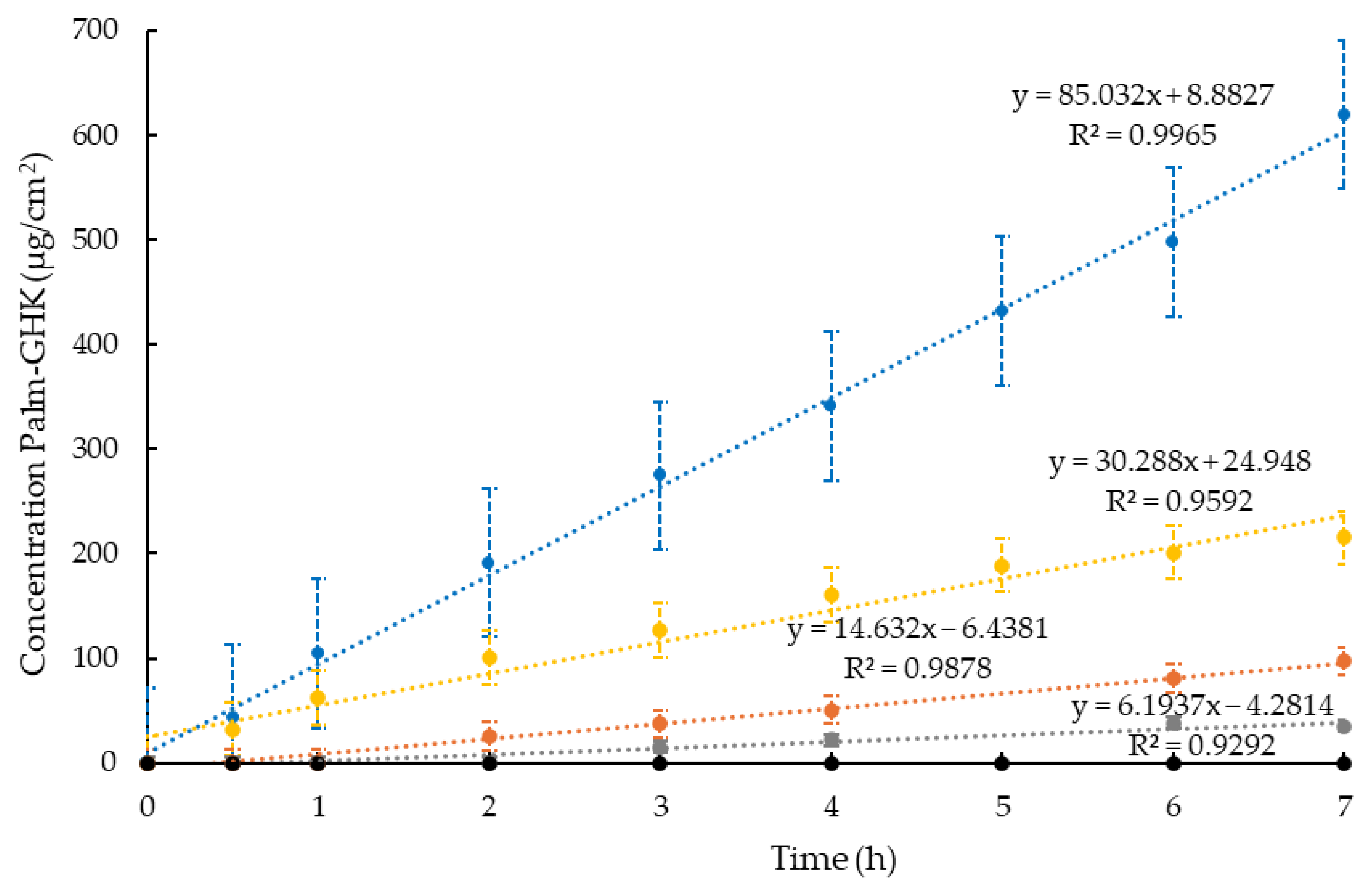
3.5. In Vitro Palm-GHK Release Studies

In vitro release studies were conducted using Franz cells, which allowed the reproduction of the skin application of the formulations, evaluating the kinetics of the active ingredient passing through a membrane that mimics the skin [33]. In particular, the diffusion kinetics of Palm-GHK from ETO, ETO-GEL, POL, and POL-ORG were studied using a solution of the peptide in EtOH/H2O (30:70 v/v) as a control. The amount of Palm-GHK released per unit of time was determined by HPLC analysis using the conditions described in Paragraph 2.5. It should be noted that a synthetic membrane based on nylon was used, instead of using the skin. While natural membranes would give more predictive results, alternatively, they also involve considerable variability, which is why numerous experiments are needed to obtain reliable results. On the other hand, synthetic membranes are easily available, allowing for more reproducible results and comparing different formulations or multiple batches of the same preparation [34]. Figure 5 shows the release profiles of Palm-GHK from the different formulations. As can be seen, the release of the active ingredient in the viscous forms is slower than in the ethanol/water solution since Palm-GHK is free and can cross the membrane more easily.
In the case of ETO-GHK, the Palm-GHK release is faster than in the other formulations. Surprisingly, in the case of ETO-GEL-GHK, the peptide was almost completely retained by the formulation, suggesting that xanthan gum formed a network that hampers the release of the active ingredient. Regarding Palm-GHK release, different aspects should be considered. First of all, in the case of ETO-GEL-GHK, xanthan gum molecules can interact with the peptide through hydrogen bonds, electrostatic, or van der Waals interactions. These interactions can retain the peptide in the gelling matrix, thereby reducing the rate of release. In the absence of a gelling matrix, these interactions are minimal or absent, thus allowing faster release. The xanthan gum gel can form a three-dimensional network that functions as a physical barrier to peptide movement. This implies that the diffusion of the peptide is governed by the density of the network and the molecular size of the peptide itself. On the other hand, in the ethanol/water solution, the peptide diffuses freely without encountering significant physical obstacles. In the case of POL-GHK and POL-ORG-GHK, the presence of the three-dimensional network formed by poloxamer allows the control of the peptide release with respect to the fluid formulation. Particularly, the release from POL-ORG-GHK is slower than from POL-GHK, since the complex supramolecular structure formed by PC and poloxamer in IPP retains Palm-GHK, releasing it more slowly.
Table 4 shows the Jo and F values calculated considering the concentrations of Palm-GHK in the different formulations.
As can be observed, the flux of Palm-GHK in ETO is almost 4-fold higher than POL-ORG and about twice higher than POL, resulting in a 40-fold minor flux with respect to the fluid form. In general, all semi-solid formulations can slow the release of Palm-GHK compared to the active ingredient in the solution or ETO. This preliminary study was needed to compare the influence of the different supramolecular structures on the release profile of Palm-GHK. Nevertheless, to study the transdermal effect of the formulations in vitro, animal or human skin is required instead of synthetic membranes.

3.6. In Vivo Comparative Irritation Test

To evaluate the safety of formulation cutaneous application, a patch test was conducted on 20 healthy volunteers using Palm-GHK unloaded and loaded ETO-GEL, POL, POL-ORG, and POL-ORG. Irritation indexes were recorded at 15 min and 24 h post patch removal. Importantly, all semi-solid forms did not induce any adverse skin reactions. Remarkably, these results demonstrate that all gels are well tolerated and non-irritating to human skin.

4. Discussion

This study highlights the significance of innovative formulation strategies for delivering Palm-GHK, addressing key challenges related to peptide solubility, stability, and controlled release. To obtain biocompatible and safe formulations, PC was chosen as the main component. Using PC, the possibility of producing both ETO dispersions and semi-solid lipid formulations was studied. Additionally, the copolymer POL 407 was employed to form structured networks depending on its concentration and temperature, imparting thermos-reversible behavior to the semi-solid forms. PC and poloxamer block copolymer enabled us to obtain vesicular and semi-solid formulations suitable for skin application. The comparative irritation test confirmed the safety and biocompatibility of all formulations, a critical requirement for consumer products. Despite these promising findings, future studies are essential to further investigate the transdermal penetration and anti-aging efficacy of Palml-GHK formulations in clinical settings. Additional exploration of ex vivo and in vivo models will provide deeper insights into the long-term benefits and potential mechanisms of action of PalmGHK, particularly its ability to stimulate extracellular matrix replenishment and collagen synthesis.
In recent years, the demand for cosmeceutical products able to protect and improve skin health, provided with significant activity, has led industries to look for alternative active ingredients. Peptides have a broad spectrum of bioactivity, making them ideal candidates for developing these products [35,36]. However, a principal concern in the formulation of peptides is bounded with their effective release from the formulation to the upper skin layers. In this study, semi-solid lipid systems for the delivery of Palm-GHK were investigated. To date, there are still few studies on the mechanism of action of Palm-GHK, but it can be hypothesized that it behaves, at least in part, like Cu-GHK.
Upon initial consideration, it appears improbable due to the presumed dependence of copper peptides’ benefits on the copper component. Nevertheless, it is conceivable that the derivatization with the palmitic acid of GHK significantly increases its skin concentration, such that the copper naturally present in the skin (typically in low quantities) suffices to activate it. Furthermore, it is plausible that many of the effects attributed to Cu-GHK are primarily due to the GHK peptide rather than copper and can be replicated using only a highly penetrant version such as PalmGHK [37]. Lintner K. et al. evaluated the anti-wrinkle effect, attributed to increased collagen synthesis, in a blind test on 15 women (aged 44 to 59). Both a cream containing the peptide and a placebo cream were applied around the eye area for four weeks, resulting in a decrease in wrinkle length by 39%, wrinkle depth by 23%, and overall skin roughness by 17%. All the differences between skin treated with the tripeptide versus the placebo cream were statistically significant. Peschard O. et al. applied both a vehicle (unidentified) and Palm-GHK on the skin of 23 volunteers, and it was observed that the thickness of the skin layer treated with the peptide increased by 4% compared to the vehicle [38]. A combination of Palm-GHK and Palm-GQPR is marketed as an anti-wrinkle compound under the trade name Matrixyl™3000 by Sederma S.A. According to the studies conducted, these two peptides produce a very strong synergistic action; the benefits for the skin are greater than the components taken individually. It has been demonstrated that skin matrix synthesis in a tissue culture is increased approximately 2.5–3 times [2]. Wilbur J. et al. conducted a study on the safety of using palmitoyl oligopeptides in cosmetics, finding that these products do not present significant data on acute and repeated dose toxicity, skin irritation, and sensitization data, confirming their favorable safety and efficacy profile [39].

5. Conclusions

Undeniably, cosmetics have become a cornerstone of daily routines. The cosmetic market, especially for performing products, is booming, driven by a strong consumer desire for enhanced appearance. Notably, the development of innovative cosmetic formulations utilizing bioactive compounds (such as antioxidants, proteins, peptides, and growth factors) is rapidly accelerating. In this respect, the present investigation demonstrated the importance of the formulation choice as an efficacious delivery system for Palm-GHK. Indeed, despite scientific evidence supporting bioactive peptide potential cosmetic applications, a limited number of studies underline the impact of the pre-formulative technological design in the development of peptide cosmetic products. Overall, this investigation underscores the importance of tailored formulation strategies in leveraging the full potential of bioactive peptides like Palml-GHK. By addressing critical challenges such as solubility, stability, and controlled release, these advanced formulations pave the way for next-generation cosmeceuticals capable of delivering tangible skin benefits while adhering to safety and sustainability principles.

Author Contributions

Conceptualization, S.M., R.G. and E.E.; Formal Analysis, E.M.; Investigation, E.M., I.D. and R.G.; Methodology, E.M., L.M. and I.D.; Project Administration, E.E.; Supervision, R.G. and E.E.; Validation, L.M. and I.D.; Writing—Original Draft, V.D. and F.S.; Writing—Review and Editing, S.M., R.G. and E.E. All authors have read and agreed to the published version of the manuscript.

Funding

This research was funded by Italian Ministry of University and Research, European Union, Next Generation EU. PRIN 2022MC2SKF, and FAR 2023.

Institutional Review Board Statement

The study was conducted according to the guidelines of the Declaration of Helsinki and approved by the Ethics Committee of the University of Ferrara, Italy (protocol code: 170583; date of approval: 16 November 2017).

Informed Consent Statement

Informed consent was obtained from all subjects involved in the study.

Data Availability Statement

The data presented in this study are available on request from the corresponding author. The data are not publicly available due to privacy restrictions.

Acknowledgments

The authors are grateful to Francesca Ungaro for her experimental support.

Conflicts of Interest

The authors declare no conflicts of interest. The funders had no role in the design of the study; in the collection, analyses, or interpretation of data; in the writing of the manuscript; or in the decision to publish the results.

References

  1. Park, S.-I.; Heo, S.H.; Lee, J.; Cha, H.; Shin, M.S. A Clinical Study on Anti-Wrinkle Efficacy of a Cosmetics Containing Oligoarginine Conjugation of Palmitoyl-GHK Peptide for Skin Penetrating. Turk. J. Comput. Math. Educ. 2021, 12, 401–406. [Google Scholar] [CrossRef]
  2. Schagen, S.K. Topical Peptide Treatments with Effective Anti-Aging Results. Cosmetics 2017, 4, 16. [Google Scholar] [CrossRef]
  3. Maquart, F.X.; Bellon, G.; Pasco, S.; Monboisse, J.C. Matrikines in the Regulation of Extracellular Matrix Degradation. Biochimie 2005, 87, 353–360. [Google Scholar] [CrossRef] [PubMed]
  4. Bonnans, C.; Chou, J.; Werb, Z. Remodelling the Extracellular Matrix in Development and Disease. Nat. Rev. Mol. Cell Biol. 2014, 15, 786–801. [Google Scholar] [CrossRef]
  5. Clinical Evaluation of Matrikynes®: A Novel Cosmetic Ingredient Comprised of Matrikine Peptides. Available online: https://xylyxbio.com/wp-content/uploads/2023/04/white-paper_clinical-evaluation-of-matrikynes.pdf (accessed on 7 March 2025).
  6. Pickart, L.; Vasquez-Soltero, J.M.; Margolina, A. GHK Peptide as a Natural Modulator of Multiple Cellular Pathways in Skin Regeneration. BioMed Res. Int. 2015, 2015, 648108. [Google Scholar] [CrossRef]
  7. Maquart, F.X.; Pickart, L.; Laurent, M.; Gillery, P.; Monboisse, J.C.; Borel, J.P. Stimulation of Collagen Synthesis in Fibroblast Cultures by the Tripeptide-Copper Complex Glycyl-L-Histidyl-L-Lysine-Cu2+. FEBS Lett. 1988, 238, 343–346. [Google Scholar] [CrossRef]
  8. MacLeman, E. Why Are Palmitoyl Oligopeptides Used in Skincare? Available online: https://thedermreview.com/palmitoyl-oligopeptide/ (accessed on 26 August 2024).
  9. Vintiloiu, A.; Leroux, J.C. Organogels and Their Use in Drug Delivery—A Review. J. Control Release 2008, 125, 179–192. [Google Scholar] [CrossRef]
  10. Kim, C.; Shim, J.; Han, S.; Chang, I. The Skin-Permeation-Enhancing Effect of Phosphatidylcholine: Caffeine as a Model Active Ingredient. J. Cosmet. Sci. 2002, 53, 363–374. [Google Scholar]
  11. Touitou, E.; Natsheh, H. Topical Administration of Drugs Incorporated in Carriers Containing Phospholipid Soft Vesicles for the Treatment of Skin Medical Conditions. Pharmaceutics 2021, 13, 2129. [Google Scholar] [CrossRef]
  12. Godin, B.; Touitou, E. Ethosomes: New Prospects in Transdermal Delivery. Crit. Rev. Ther. Drug Carrier Syst. 2003, 20, 63–102. [Google Scholar] [CrossRef]
  13. Natsheh, H.; Vettorato, E.; Touitou, E. Ethosomes for Dermal Administration of Natural Active Molecules. Curr. Pharm. Des. 2019, 25, 2338–2348. [Google Scholar] [CrossRef] [PubMed]
  14. Verma, P.; Pathak, K. Therapeutic and Cosmeceutical Potential of Ethosomes: An Overview. J. Adv. Pharm. Technol. Res. 2010, 1, 274–282. [Google Scholar] [CrossRef] [PubMed]
  15. Nsengiyumva, E.M.; Alexandridis, P. Xanthan Gum in Aqueous Solutions: Fundamentals and Applications. Int. J. Biol. Macromol. 2022, 216, 583–604. [Google Scholar] [CrossRef]
  16. Kumar, R.; Katare, O.P. Lecithin Organogels as a Potential Phospholipid-Structured System for Topical Drug Delivery: A Review. AAPS PharmSciTech 2005, 6, E298–E310. [Google Scholar] [CrossRef]
  17. Martinez, R.M.; Rosado, C.; Velasco, M.V.R.; Lannes, S.C.S.; Baby, A.R. Main Features and Applications of Organogels in Cosmetics. Int. J. Cosmet. Sci. 2019, 41, 109–117. [Google Scholar] [CrossRef]
  18. Esposito, E.; Menegatti, E.; Cortesi, R. Design and Characterization of Fenretinide Containing Organogels. Mater. Sci. Eng. C 2013, 33, 383–389. [Google Scholar] [CrossRef]
  19. Russo, E.; Villa, C. Poloxamer Hydrogels for Biomedical Applications. Pharmaceutics 2019, 11, 671. [Google Scholar] [CrossRef]
  20. Escobar-Chávez, J.J.; López-Cervantes, M.; Naïk, A.; Kalia, Y.; Quintanar-Guerrero, D.; Ganem-Quintanar, A. Applications of Thermo-Reversible Pluronic F-127 Gels in Pharmaceutical Formulations. J. Pharm. Pharm. Sci. 2006, 9, 339–358. [Google Scholar]
  21. Hosa, L.B.; Mira, E.G.; Santana, H.; Folch, J.M.; Masip, M.M.; Prieto, Y.M.; Revuelta, A.; Di Mauro, P.P.; Veciana, J.; Sala, S.; et al. DELOS Nanovesicles-Based Hydrogels: An Advanced Formulation for Topical Use. Pharmaceutics 2022, 14, 199. [Google Scholar] [CrossRef]
  22. Rehman, W.U.; Asim, M.; Hussain, S.; Khan, S.A.; Khan, S.B. Hydrogel: A Promising Material in Pharmaceutics. Curr. Pharm. Des. 2020, 26, 5892–5908. [Google Scholar] [CrossRef]
  23. Almeida, H.; Amaral, M.H.; Lobão, P.; Lobo, J.M.S. Pluronic® F-127 and Pluronic Lecithin Organogel (PLO): Main Features and Their Applications in Topical and Transdermal Administration of Drugs. J. Pharm. Pharm. Sci. 2012, 15, 592–605. [Google Scholar] [CrossRef] [PubMed]
  24. Murdan, S. A Review of Pluronic Lecithin Organogel as a Topical and Transdermal Drug Delivery System. Hosp. Pharm. 2005, 12, 267–270. [Google Scholar]
  25. Saha, S.; Shivarajakumar, R.; Venkata Satyanarayana Reddy Karri, V. Pluronic Lecithin Organogels: An Effective Topical and Transdermal Drug Delivery System. Int. J. Pharm. Sci. Res. 2018, 9, 4540. [Google Scholar] [CrossRef]
  26. Resende, D.I.S.P.; Ferreira, M.S.; Sousa-Lobo, J.M.; Sousa, E.; Almeida, I.F. Usage of Synthetic Peptides in Cosmetics for Sensitive Skin. Pharmaceuticals 2021, 14, 702. [Google Scholar] [CrossRef]
  27. Sicurella, M.; Sguizzato, M.; Cortesi, R.; Huang, N.; Simelière, F.; Montesi, L.; Marconi, P.; Esposito, E. Mangiferin-Loaded Smart Gels for Hsv-1 Treatment. Pharmaceutics 2021, 13, 1323. [Google Scholar] [CrossRef]
  28. Iqubal, M.K.; Iqubal, A.; Anjum, H.; Gupta, M.M.; Ali, J.; Baboota, S. Determination of in Vivo Virtue of Dermal Targeted Combinatorial Lipid Nanocolloidal Based Formulation of 5-Fluorouracil and Resveratrol against Skin Cancer. Int. J. Pharm. 2021, 610, 121179. [Google Scholar] [CrossRef]
  29. Miranda, M.; Pais, A.A.C.C.; Cardoso, C.; Vitorino, C. AQbD as a Platform for IVRT Method Development—A Regulatory Oriented Approach. Int. J. Pharm. 2019, 572, 118695. [Google Scholar] [CrossRef]
  30. Esposito, E.; Calderan, L.; Galvan, A.; Cappellozza, E.; Drechsler, M.; Mariani, P.; Pepe, A.; Sguizzato, M.; Vigato, E.; Dalla Pozza, E.; et al. Ex Vivo Evaluation of Ethosomes and Transethosomes Applied on Human Skin: A Comparative Study. Int. J. Mol. Sci. 2022, 23, 15112. [Google Scholar] [CrossRef]
  31. Dumortier, G.; Grossiord, J.L.; Agnely, F.; Chaumeil, J.C. A Review of Poloxamer 407 Pharmaceutical and Pharmacological Characteristics. Pharm. Res. 2006, 23, 2709–2728. [Google Scholar] [CrossRef]
  32. Zouboulis, C.C.; Ganceviciene, R.; Liakou, A.I.; Theodoridis, A.; Elewa, R.; Makrantonaki, E. Aesthetic Aspects of Skin Aging, Prevention, and Local Treatment. Clin. Dermatol. 2019, 37, 365–372. [Google Scholar] [CrossRef]
  33. Salamanca, C.H.; Barrera-Ocampo, A.; Lasso, J.C.; Camacho, N.; Yarce, C.J. Franz Diffusion Cell Approach for Pre-Formulation Characterisation of Ketoprofen Semi-Solid Dosage Forms. Pharmaceutics 2018, 10, 148. [Google Scholar] [CrossRef] [PubMed]
  34. Ferrara, F.; Benedusi, M.; Sguizzato, M.; Cortesi, R.; Baldisserotto, A.; Buzzi, R.; Valacchi, G.; Esposito, E. Ethosomes and Transethosomes as Cutaneous Delivery Systems for Quercetin: A Preliminary Study on Melanoma Cells. Pharmaceutics 2022, 14, 1038. [Google Scholar] [CrossRef] [PubMed]
  35. Aguilar-Toalá, J.E.; Hernández-Mendoza, A.; González-Córdova, A.F.; Vallejo-Cordoba, B.; Liceaga, A.M. Potential Role of Natural Bioactive Peptides for Development of Cosmeceutical Skin Products. Peptides 2019, 122, 170170. [Google Scholar] [CrossRef] [PubMed]
  36. Ngoc, L.T.N.; Moon, J.Y.; Lee, Y.C. Insights into Bioactive Peptides in Cosmetics. Cosmetics 2023, 10, 111. [Google Scholar] [CrossRef]
  37. Lupo, M.P.; Cole, A.L. Cosmeceutical peptides. Dermatol. Ther. 2007, 20, 343–349. [Google Scholar] [CrossRef]
  38. Safety Assessment of Palmitoyl Oligopeptides Ingredients as Used in Cosmetics. Available online: https://www.cir-safety.org/panelbook/safety-assessment-palmitoyl-oligopeptides-ingredients-used-cosmetics (accessed on 26 August 2024).
  39. Lintner, K.; Peschard, O. Biologically Active Peptides: From a Laboratory Bench Curiosity to a Functional Skin Care Product. Int. J. Cosmet. Sci. 2000, 22, 207–218. [Google Scholar] [CrossRef]
Figure 1. Structural formula of Palmitoyl-GHK in its fully protonated form.
Cosmetics 12 00050 g001
Figure 2. Images of the formulations: (a) ETO, (b) ETO-GEL, (c) POL, (d) ORG, and (e) POL-ORG.
Cosmetics 12 00050 g002
Figure 3. Variation in water loss: Cosmetics 12 00050 i001 control, Cosmetics 12 00050 i002 ETO-GEL, Cosmetics 12 00050 i003 POL-GEL, Cosmetics 12 00050 i004 POL, and Cosmetics 12 00050 i005 ORG.
Cosmetics 12 00050 g003
Figure 4. Comparative analysis of skin moisturizing effect of the formulations: Cosmetics 12 00050 i006control, Cosmetics 12 00050 i007ETO-GEL, Cosmetics 12 00050 i008POL-ORG, and Cosmetics 12 00050 i009ORG; 00 corresponds to the skin hydration before the application of formulation, 0 to the skin immediately after application of formulation. The values are the mean of 3 measurements taken on 5 healthy volunteers before and after the application of the formulations.
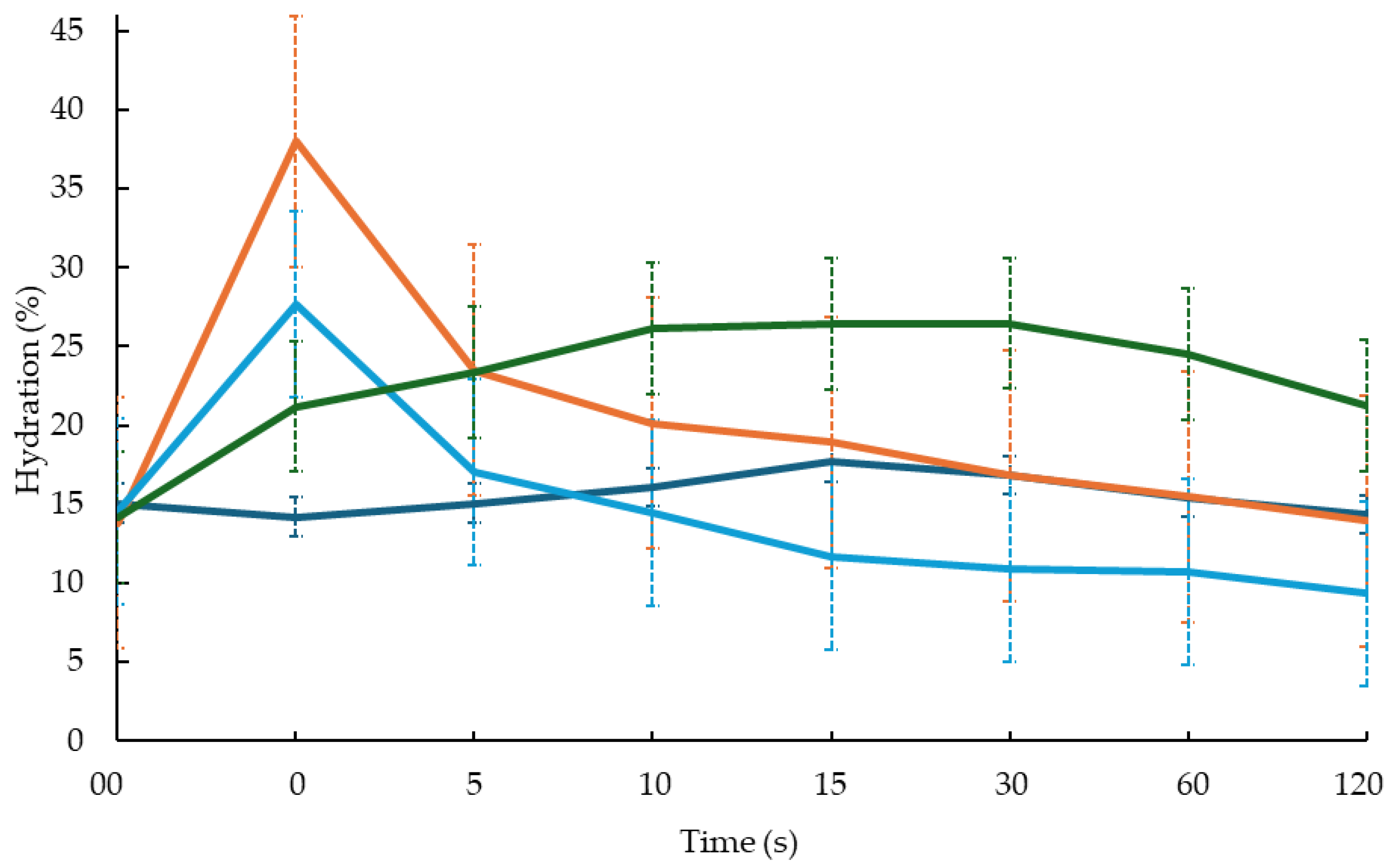
Cosmetics 12 00050 g004
Figure 5. Palm-GHK release kinetics from the different forms: SOL-GHK, ETO-GHK, POL-GHK, POL-ORG-GHK, and • ETO-GEL-GHK.
Cosmetics 12 00050 g005
Table 1. Percentage composition of the formulations.
FormulationPC 1 %w/wEtOH%w/wIPP 2
%w/w
POL 3
%w/w
H2O
%w/w
X-GUM 4
%w/w
ETO0.9029.10--70.00-
ETO-GEL0.9029.10--69.001.00
POL---20.0080.00-
ORG15.60-82.96-1.44-
POL-ORG4.68-25.3214.0056.00-
1 phosphatidylcholine; 2 isopropyl palmitate; 3 Poloxamer 407; 4 xanthan gum.
Table 2. Size distribution of ethosomes.
FormulationZ-Average (nm ± S.D.)Intensity (nm)PDI ± S.D.
ETO206.5 ± 0.0100%0.105 ± 0.00
ETO-GHK102.1 ± 6.4100%0.142 ± 0.02
Table 3. Spreadability and occlusive factor values of semi-solid formulations.
FormulationSpreadability (g·cm·s–1) ± S.D.Occlusive Factor % ± S.D.
ETO-GEL19.17 ± 1.024 ± 0.096
POL-ORG9.17 ± 0.4139 ± 0.076
ORG15.00 ± 0.0063 ± 0.048
POL15.00 ± 0.1032 ± 6.030
Table 4. Release parameters of Palm-GHK from vesicular and semi-solid systems.
FormulationConcentration
Palm-GHK (mg/mL)
Jo
(μg/cm2/h) ± S.D.
Flux
(cm/h) ± S.D.
ETO-GHK0.9227.14 ± 4.2129.50 ± 4.57
ETO-GEL-GHK0.920.68 ± 0.020.74 ± 0.02
POL-GHK0.9418.71 ± 1.9719.90 ± 2.10
POL-ORG-GHK0.947.68 ± 0.688.17 ± 0.72
SOL-GHK1.0077.71 ± 17.8577.71 ± 17.85
Disclaimer/Publisher’s Note: The statements, opinions and data contained in all publications are solely those of the individual author(s) and contributor(s) and not of MDPI and/or the editor(s). MDPI and/or the editor(s) disclaim responsibility for any injury to people or property resulting from any ideas, methods, instructions or products referred to in the content.

