Article

Clinical Efficacy of Endocannabinoid-Mimetic Fatty Acid Amide as a Skin-Soothing Ingredient

1
Department of Dermatology, Chungnam National University Sejong Hospital, Sejong 30099, Republic of Korea
2
Research Team, Incospharm Corp., Daejeon 43000, Republic of Korea
3
R&D Center, Cellapy Bio Inc., Daejeon 34141, Republic of Korea
4
Department of Bio-Cosmetic Science, Seowon University, Cheongju 28674, Republic of Korea
*
Authors to whom correspondence should be addressed.
Cosmetics 2024, 11(6), 225; https://doi.org/10.3390/cosmetics11060225
Submission received: 11 November 2024 / Revised: 3 December 2024 / Accepted: 12 December 2024 / Published: 17 December 2024
(This article belongs to the Section Cosmetic Dermatology)

Abstract

:
Despite the potential benefits of cannabidiol as a skin-soothing ingredient, its regulatory status hampers its general use in cosmetic products in many countries. To develop an alternative to cannabidiol, fatty acid amide molecules mimicking the chemical structure of endocannabinoids were manufactured using a lipase-catalyzed process. A mixture of fatty acid amides from sunflower oil and 1-amino propan-3-ol was synthesized using an immobilized lipase reaction, and the activation of cannabinoid receptor 1 (CB1R) was measured using a cAMP assay. The anti-inflammatory activity of the endocannabimimetic ingredients was evaluated in cultured human monocytes and ex vivo human skin explant models. A clinical study was conducted to address the skin hydration, skin barrier function, and skin redness, and the ratio of the interleukin-1-receptor antagonist (IL1-RA) to IL-1α in corneocytes, as a marker for skin sensitivity, were also measured. As a result, the activation of CB1R by endocannabimimetic ingredients was observed in cAMP assays, and a reduction in inflammatory responses by human monocytes induced by lipopolysaccharide treatment were also observed. External stress-induced inflammatory responses were reduced in ex vivo human skin explants. Improvements in skin hydration and barrier function were observed in a clinical study. A significant decrease in skin redness and the IL-1RA to IL-1α ratio was also observed. Endocannabimimetic ingredients, as alternatives to cannabidiol, can be used in skin-soothing cosmetics to increase skin hydration, improve skin barrier function, and reduce skin sensitivity.

1. Introduction

Recently, cannabidiol (CBD), a major bioactive ingredient in cannabis extract, has received considerable interest from the cosmetics industry, mainly due to its potential benefits as a cosmetic ingredient. However, regulatory issues associated with the use of cannabis or its extracts, known as phytocannabinoids, have impeded its widespread use in personal care products, including cosmetics. Insufficient stability of cannabidiol oil samples under ambient storage condition also poses an important drawback for the practical use in topical formulations [1]. As a major phytocannabinoid, CBD is inactive in the human central nervous system and has various therapeutic and cosmetic benefits, focusing on the inflammatory skin diseases, including acne, atopic dermatitis, and psoriasis [2]. Despite its potential efficacy, many countries still ban the use of CBD in cosmetic products. Considering the great potential of CBD as a bioactive cosmeceutical ingredient, many trials have been conducted to develop alternatives to CBD without regulatory disputes. One approach uses a cellular signaling system, termed the endocannabinoid system (ECS), which mediates phytocannabinoid activity in cells. The ECS comprises endogenous lipid mediators present in virtually all tissues, their G-protein-coupled cannabinoid receptors, biosynthetic and metabolizing enzymes, such as fatty acid amide hydrolase (FAAH) and monoacylglycerol lipase (MAGL), and fatty acid binding proteins (FABPs), which participate in the endocannabinoid trafficking to the degradation pathways [3,4]. Currently, two subtypes of cannabinoid receptors, cannabinoid receptor 1 (CB1R) and receptor 2 (CB2R), have been reported in diverse cell types including keratinocytes, fibroblasts, sebaceous glands, melanocytes, Langerhans cells, and nerve endings [5,6,7,8]. Their expression patterns and biological responses upon cannabinoid binding are context-dependent, and in keratinocytes, CBRs are involved in cell proliferation, differentiation, and inflammatory responses [9]. The recognition of CBRs has resulted in the identification of endogenously synthesized molecules for these receptors in the human body, and these molecules are classified as endocannabinoids. Since the first report of arachidonylethanolamine (AEA) as an endocannabinoid, a series of molecules have been reported as having CBR binding activity. These share a similar chemical structure involving unsaturated long-chain fatty acid amides [10]. As alternatives to cannabis-originated phytocannabinoids, these endocannabinoids can provide CBD-like efficacy without regulatory issues. While there have been a series of studies about the development of cannabidiol-mimetic ingredients or cannabidiol derivatives for therapeutic applications [11], little has been published for endocannabinoid-mimetic compounds. Very recently, Zhang et al. reported a potential application of (R)-methanandamide (methAEA, a metabolically stable form of AEA) for drug addiction treatment [12], but there are no studies reported for the cosmetic application of endocannabinoid-mimetic compounds.
Recently, we developed a new bioprocess for conjugating amino alcohols (monoethanolamine or 3-amino-1-propanol) to plant oils using an immobilized lipase-catalyzed enzyme reaction system. Triglycerides in plant oils serve as fatty acid donors, and the lipase-catalyzed conjugation of amino alcohols produces various fatty acid amide molecules that have almost the same fatty acid composition as the original plant oils. We also showed that different types of amino alcohols, such as monoethanolamine or 3-amino-1-propanol, can be used for the reaction, and the resulting fatty acid amide mixture structurally mimics endocannabinoids and can bind to CBRs in human skin cells, including epidermal keratinocytes. In this study, using a sunflower seed oil propanol amide-based endocannabimimetic ingredient, we investigated the binding properties of cannabimimetic ingredient on CBRs using an in vitro assay system. We also evaluated the biological activity of the ingredient, focusing on its skin-soothing activity, using in vitro, ex vivo, and clinical studies.

2. Materials and Methods

2.1. Enzymatic Synthesis of Endocannabimimetics

Sunflower seed oil-based endocannabimimetic ingredients were synthesized via lipase-catalyzed amidation using monoethanolamine (Duksan, Ansan, Republic of Korea) or 3-amino-1-propanol (TCI, Tokyo, Japan). Briefly, 1 M of sunflower seed oil and 3 M of amino alcohol were mixed in a double-jacket reactor controlled by a water bath at a temperature of 55 °C. The reaction was performed using 1% (w/v) of immobilized lipase (Candida antarctica lipase B variant A [13]) for 72 h. Samples were collected at different times during the reaction and analyzed using a 7890a gas chromatography/flame ionization detector (GC/FID) system equipped with an HP-5 column (30 m length × 0.320 mm diameter × 0.25 µm film thickness, Agilent Technologies, Santa Clara, CA, USA). After reaching > 95% of the reaction volume, the immobilized enzymes were removed by filtration. The glycerol produced from sunflower oil and the unreacted amino alcohol were extracted by hot water treatment, and the final mixture was distilled under vacuum to remove water.

2.2. In Vitro Cell Studies

CBR activation of cannabinoid receptors was measured using the DiscoveX cAMP assay (Eurofins Discover X Corporation, Fremont, CA, USA). DiscoverX is a panel of cell lines that stably express nontagged G protein-coupled receptors (GPCRs) that signal through cAMP. Hit Hunter® cAMP assays monitor the activation of GPCRs via Gi and Gs secondary messenger signaling in a homogenous nonimaging assay format using a technology developed by DiscoverX called Enzyme Fragment Complementation with β-galactosidase (β-Gal) as the functional reporter. The two fatty amides developed in this study were tested in the order of US073-0023751-O. A human monocyte cell line (THP-1) was purchased from Korean Cell Line Bank (Cat. No. 40202) and culture media were purchased from Thermo Fisher Scientific (Waltham, MA, USA). THP-1 cells were maintained in RPMI-1640 medium supplemented with 10% of fetal bovine serum and 1% of antibiotics. Cells were cultured at 37 °C under 5% CO2 and 95% relative humidity.

2.3. Measurement of Cytokines

Expression of tumor necrosis factor-α (TNF-α), interleukin-6 (IL-6), and IL-8 were measured using human TNF-α (R&D Systems, Cat. No. DY 210), IL-6 (Beckton Dickson, Cat. no. 555220), and an IL-8 ELISA kit (R&D Systems, Cat. No. DY208) according to the manufacturer’s instructions. Briefly, the captured antibody was diluted with 0.1 M of sodium carbonate buffer solution and coated onto 96-well plates by incubating overnight at 4 °C. After blocking with 10% of fetal bovine serum in phosphate-buffered saline (PBS) for 1 h at room temperature, culture medium was added to each well and incubated for 2 h at room temperature. After washing with PBS five times, the detection antibody and streptavidin–horseradish peroxidase-conjugated reagent mixture were added to each well and incubated for 1 h at room temperature. Tetramethylbenzidine and hydrogen peroxide solutions were added to each well, and the plates were incubated for 30 min at room temperature in the dark. After adding a stop solution, optical absorbance was measured at 450 nm using an Epoch microplate reader (Biotek, Winooski, VT, USA).

2.4. Ex Vivo Studies

A NativeSkin human skin explant model was purchased from GenoSkin SAS (Toulouse, France). The anonymized human skin samples used in this study were obtained from a healthy female Caucasian donor aged 52 years who underwent an abdominoplasty procedure and provided written informed consent. The donor had no history of allergy or dermatological disorders and did not use corticosteroids. The collection, manufacture, and use of skin models for research purposes were formally authorized by the French Ministry of Research (AC-2017-2897, 12 October 2017) and approved by the French Ethics Committee (Comité de Protection des Personnes). The study was conducted in accordance with the principles of the Declaration of Helsinki. Immediately following surgery, skin samples were collected, and subcutaneous adipose tissue was removed from the skin. Then, 11 mm diameter punch biopsies were excised and embedded in a proprietary biological matrix in transwells (Millicell) according to the patented NativeSkin® procedure developed by GenoSkin. The epidermal surfaces of the skin biopsies were left in contact with air and the dermal compartment was immersed in the matrix. The skin models were cultured in a proprietary and chemically defined hydrocortisone- and serum-free medium in the presence of 100 µg/mL of penicillin and streptomycin, each, in a humidified atmosphere of 5% CO2 at 37 °C. Upon arrival at a laboratory, skin models were acclimatized for 2 h in 12-well plates containing 1 mL of maintenance medium (GenoSkin SAS) in a humidified incubator at 37 °C under 5% CO2. After 2 h, fresh maintenance medium was added to the culture wells and maintained until further experiments.
To induce external stress, a sterilized cotton ball was scraped on the dermal side in a circular mode with constant force and speed for 60 min at 37 °C under 5% CO2 and further exposed to ultraviolet-B (UVB) radiation (50 mJ/cm2). After stress, 25 μL of test sample solution (dissolved in ethanol: polyethylene glycol 400 (70:30) mixture) was applied once a day directly onto the skin surface and further incubated for 48 h. Each treatment was performed in duplicate.

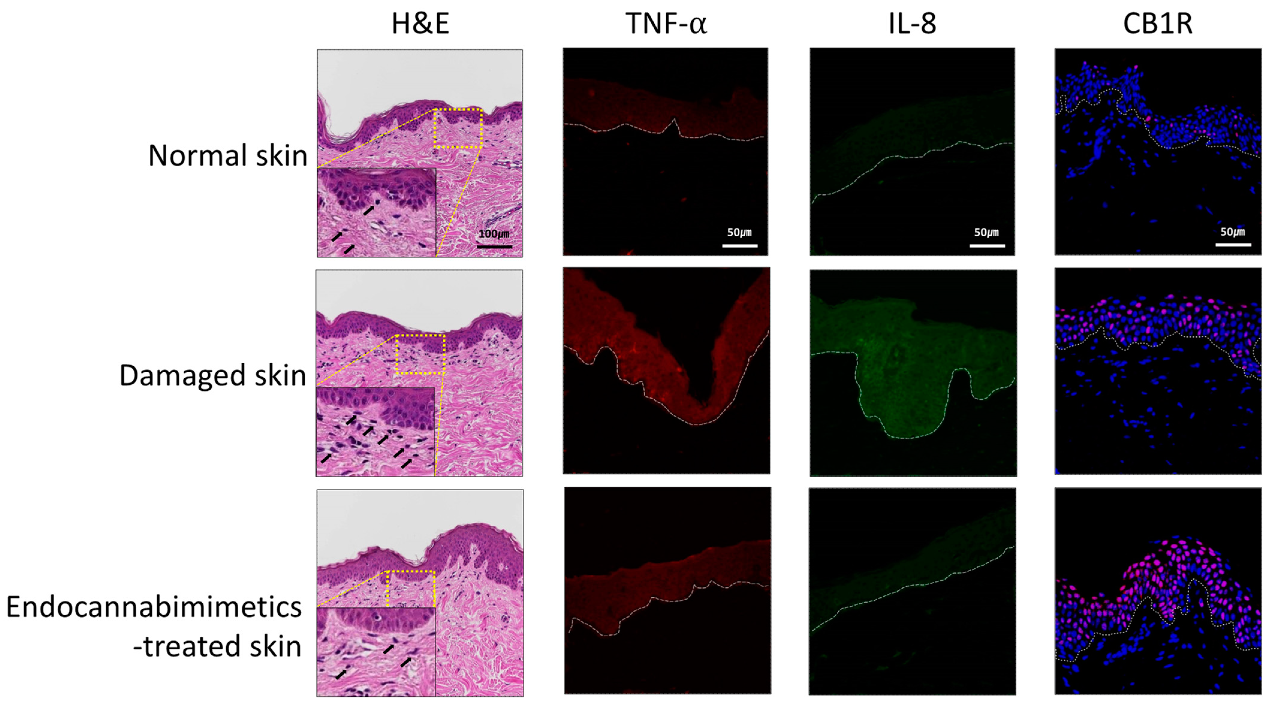
2.5. Immunohistochemical Analysis

At the end of the experiments, skin models were fixed in a 4% neutral buffered formalin (NBF), which was prepared by diluting 10% of strong formalin (40% formaldehyde) with phosphate-buffered saline solution (pH 7.0) overnight at 4 °C and dehydrated across a graded ethanol series prior to paraffin embedding. For immunohistochemical staining, 5 µm thick tissue sections were deparaffinized and rehydrated before heat-induced epitope revival treatment in an antigen retrieval solution (ab937, abcam, Cambridge, MA, USA) at a pH of 6.0 for 10 min at 97 °C, followed by cooling for 10 min in a cooling chamber. After blocking nonspecific antibody binding using a blocking reagent (X0909, Dako, Glostrup, Denmark), mouse anti-human TNF-α antibody (ab9579, abcam), mouse anti-human IL-8 antibody (ab18672, abcam), and rabbit anti-human CB1R antibody (ab23703, abcam) diluted in an antibody dilution solution (S3022) was delivered to the sections and incubated in a humidified chamber overnight at 4 °C. Then, goat anti-rabbit immunoglobulin (IgG) H&L (Alexa Fluor® 488, ab150077, abcam) or goat antimouse IgG H&L (Alexa Fluor® 568, ab175473, abcam) was applied to the tissue samples, respectively, and subsequently analyzed under a fluorescence microscope (Eclipse Ni-U, Intenslight C-HGFI, DS-Ri2, Nikon, Tokyo, Japan) at 400× magnification.

2.6. Clinical Efficacy Study

A clinical efficacy study (CRA22-CT0401) was performed involving 20 Asian female subjects (mean age 29.1 years with standard deviation [SD] of 6.77), who were approved by the Institutional Review Board of CRA Korea Inc. (Approval number: 2020021201-202202-HR-001-01) (4 February 2022). All studies complied with the World Medical Association’s Declaration of Helsinki (2013) concerning biomedical research involving human subjects. Healthy female volunteers without skin or systemic diseases were enrolled in this study. Candidates who had received retinoids or LASER therapy within the previous 6 months or who participated in other clinical studies were excluded. All volunteers signed an informed consent form and participated in the study after being explained its purpose and protocol. Overall, 20 female volunteers were initially enrolled in and completed the study. On the first visit, participants were asked to complete study-related medical record questionnaires to confirm the inclusion and exclusion criteria, and each participant provided written informed consent. The participants were administrated to use the test formulation containing 0.1% of sunflower oil-derived endocannabimimetics-containing cream (Table 1) once a day for 4 weeks on the face and volar arm area.
Before instrumental measurements, participants were asked to rest for at least 30 min in 40–60% relative humidity (RH) and a temperature (20–25 °C)-controlled room. A skin hydration map was acquired on the test area using a capacitance-based epsilon (E100, Biox Systems Ltd., London, UK) Skin Hydration Measurement Instrument and visualized as a three-dimensional image. At each measurement (baseline, 2 and 4 weeks post application), skin surface hydration was measured, and tape stripping was performed 10 times with a D-Squame standard sampling disc (D100, Clinical and Derm LLC., Dallas, TX, USA). Skin redness was assessed as the area of red pixels on photographs acquired using a VISIA-CR dermal scanning camera (Canfield Scientific, Parsippany, NJ, USA) using the image analysis program Image J (NIH, Bethesda, MD, USA).

2.7. Protein Analysis

To measure the protein expression ratio of IL-1 receptor antagonist (IL-1RA) to IL-1α (IL-1α) in corneocytes, D-Squame tape with adhered stratum corneum samples (4 to 6 stripped tapes) were sliced into small pieces and immersed in 0.75 mL of PBS solution, then sonicated for 20 min on ice to extract proteins. Extracted samples were centrifuged at 4 °C and 14,000× g for 10 min and the supernatant was collected for further analysis. Levels of IL-1RA and IL-1α in the supernatant were measured using respective ELISA kits (R&D Systems). The amount of target proteins per total protein content was calculated, and the IL-1RA/IL-1α ratio was calculated as previously described [14].

2.8. Statistical Analysis

Values are expressed as the arithmetic mean ± SD. Parametric, two-tailed, paired Student’s t-tests or nonparametric Wilcoxon signed-rank tests were performed to compare differences before and after application. All statistical analyses were performed using SPSS Statistics (SPSS v.29.0, IBM® SPSS Statistics, Armonk, NY, USA) with a 95% confidence interval, and p values < 0.05 were considered statistically significant.

3. Results

3.1. Enzymatic Synthesis of Fatty Amides from Sunflower Oil

Enzymatic amidation of sunflower oil with two amino alcohols, monoethanolamine and 3-amino-1-propanol, was performed in a 5 L double-jacketed glass reactor. A 1:3 molar ratio of sunflower oil to amine alcohol was mixed and enzymatically amidated in a reactor without solvent. The scheme for each amidation reaction is shown in Figure 1a. The enzymatic conversion of sunflower oil and two amine alcohols to fatty amides was compared. The enzymatic amidation of sunflower oil occurred much faster when monoethanolamine was used than when 3-amino-1-propanol was employed (Figure 1b), possibly because of the different solubility of the two amino alcohols in sunflower oil in the nonsolvent-mediated reaction system. The final fatty amide composition was based on the native fatty acid composition of sunflower oil. Sunflower oil-based endocannabimimetics used in this study were prepared in the form of complexes of different fatty amides (~60% linoleamide, ~30% oleamide, 6% stearamide, and 4% palmitamide).

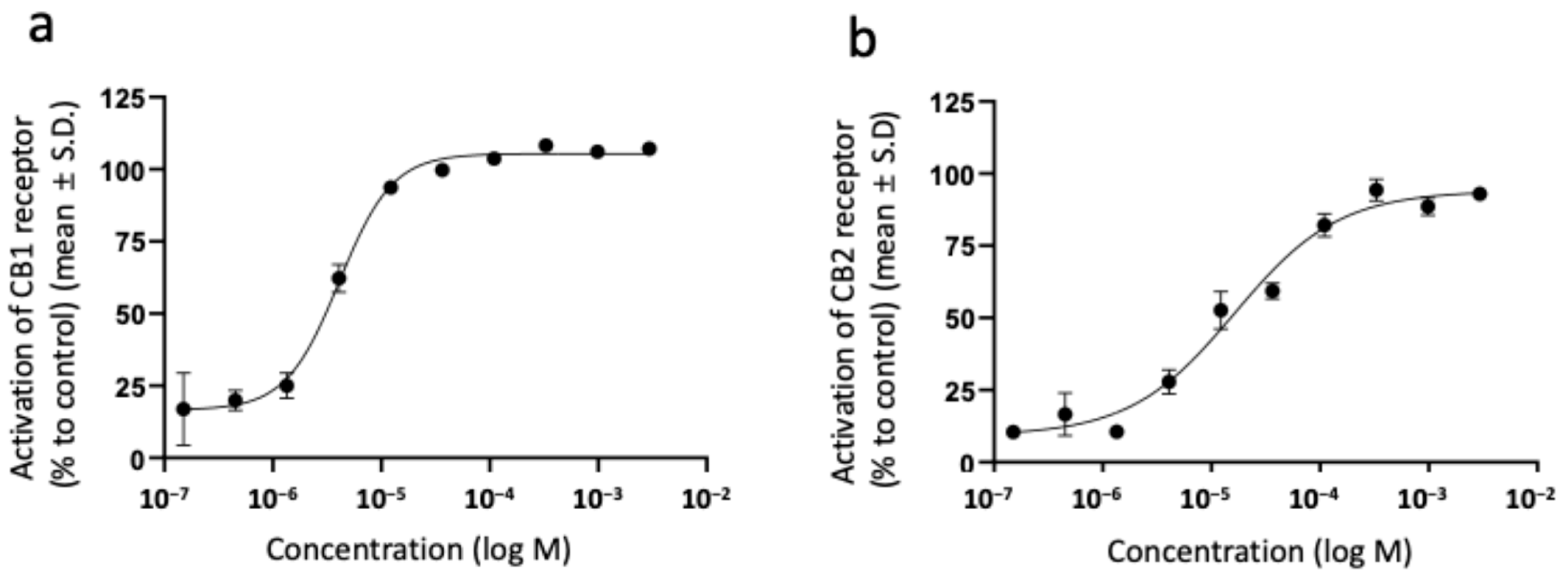
3.2. Activation of Cannabinoid Receptors by Sunflower Oil-Derived Endocannabimimetic Fatty Acid Amides

Cannabinoid receptors are classified as GPCRs that signal mainly through the adenylyl cyclase-inhibiting heterotrimeric G protein Gi. By measuring cAMP (cyclic AMP) generation, the activation of cannabinoid receptors by sunflower oil 1-aminopropanol amide was examined. As shown in Figure 2, the activation of both CB1R and CB2R was observed in sunflower oil-derived endocannabimimetic fatty acid amides. Calculating the EC50 (half-maximal effective concentration) suggested that the potency on the CB1R was approximately four-fold higher than that of CB2R.
Owing to differences in the assay systems, directly comparing the potency of sunflower oil 1-aminopropanol amide with that of AEA or CBD was not possible, and we sought to compare the biological activities through in vitro experiments.

3.3. Anti-Inflammatory and Skin-Soothing Evaluation

The diverse putative biological effects of cannabinoids on the skin, subtle regulation of proliferation, differentiation, and cellular survival, as well as inflammation and immune competence and/or tolerance, suggest that cannabinoids or cannabimimetics can be used not only as therapeutics but also for cosmetic applications (reviewed in [15]). Among those potential benefits, the alleviation of skin inflammation and skin sensitivity are also reported [16]. In this study, the anti-inflammatory activity of endocannabimimetic fatty acid amide was investigated using in vitro and ex vivo human skin explant culture models. The use of a human monocyte cell line (THP-1) to study inflammatory signaling has been previously reported for many cosmetic ingredients [17].
As shown in Figure 3, the induction of the inflammatory cytokines TNF-α, IL-6, and IL-8, by the lipopolysaccharide (LPS) treatment of THP-1 cell was decreased by all the tested materials. Consistent with previous studies [18,19,20], treatments of either CBD or AEA resulted in noticeable anti-inflammatory activity. The treatment of sunflower oil 1-aminopropanol amide also showed nearly comparable activities for the measured cytokines, especially for IL-6 and IL-8. Collectively, these results suggested that the tested endocannabimimetic fatty acid amide showed similar anti-inflammatory activity with those of CBD or AEA.
Further investigation using an ex vivo human skin explant model confirmed the skin-soothing activity of endocannabimimetic molecules against external stresses, which were simulated by physical damage to the skin surface with UV irradiation. Physical damage to the skin surface was induced by continuously scraping the skin tissue surface with a sterile cotton ball, which was set to touch the skin surface and rotated in a circular manner for 1 h. In addition to physical damage, 50 mJ/cm2 of UVB irradiation was used to induce inflammatory responses. After physical stress, 0.1% of the sunflower oil-derived endocannabimimetic fatty acid amides dissolved in PEG400/ethanol (30/70) were topically applied to the skin surface and further incubated for 24 h. After 24 h, skin tissues were harvested, and histological assessments were performed. As shown in Figure 4, the accumulation of inflammatory infiltrates, represented by an increased number of dark purple cells in the reticular dermis layer, was observed in physically stressed tissues (Figure 4, H&E stain, inset). The increased expression of inflammatory cytokines TNF-α and IL-8 was also observed after immunohistochemical staining (Figure 4). Consistent with in vitro testing results, reduced numbers of inflammatory infiltrates and cytokines were observed for sunflower oil-derived endocannabimimetic fatty acid amide-treated skin tissue. Interestingly, the expression of CB1R in the epidermal layer was increased by physical stress, whereas the topical application of endocannabimimetics further upregulated CB1R expression (Figure 4).

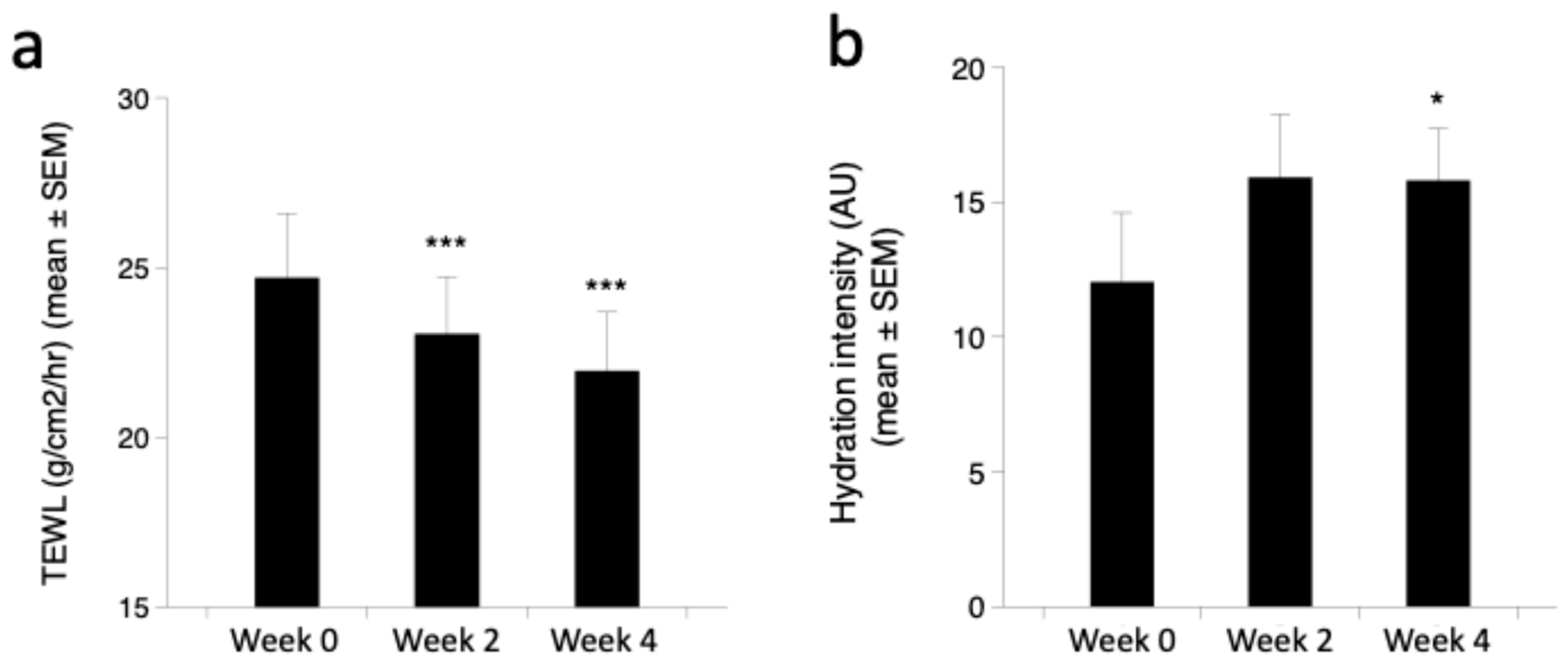
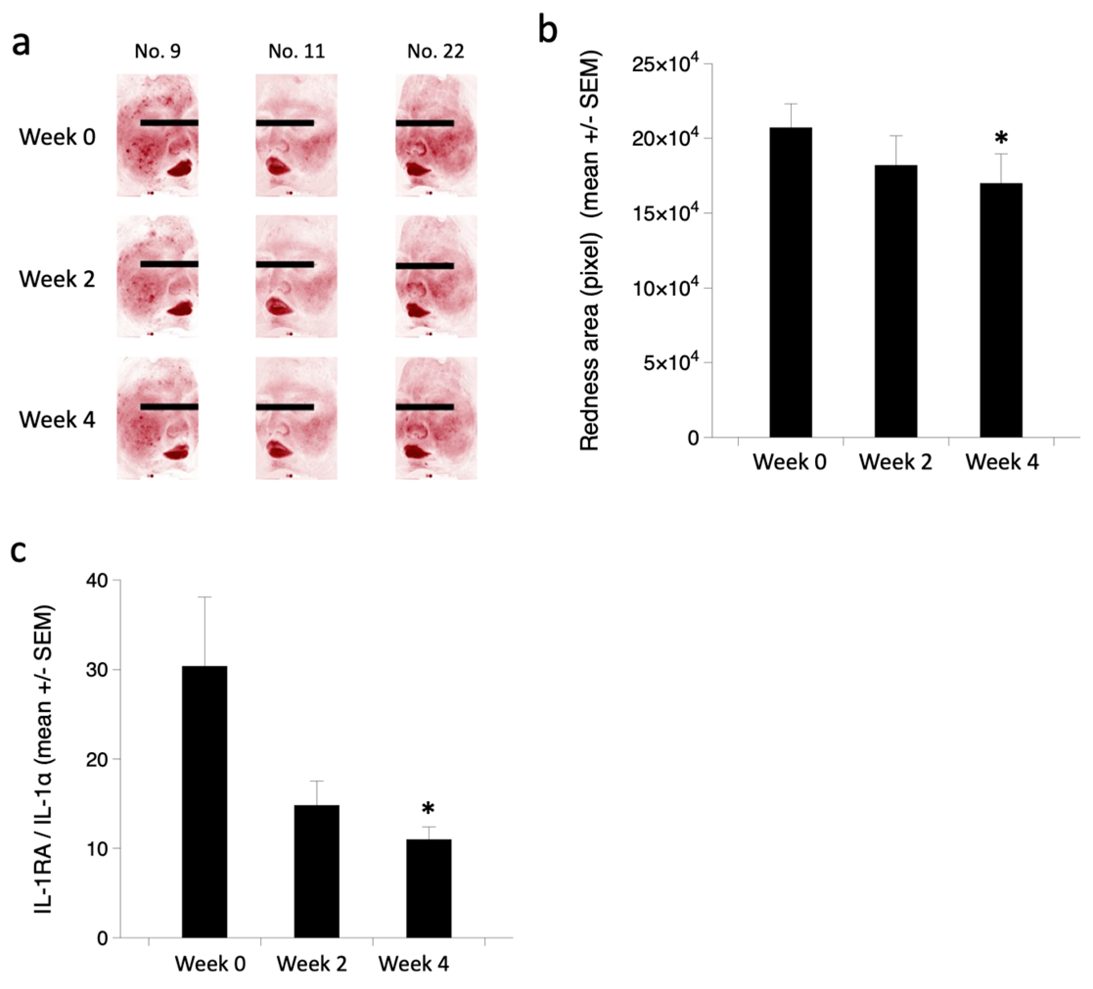
3.4. Clinical Efficacy of Endocannabimimetics

Previous studies have reported the positive effects of CBD or cannabimimetic compounds in terms of skin barrier function and dermal hydration in animal models [21,22]. However, there are few reports concerning the clinical application of endocannabimimetics as cosmetic ingredients. Based on previous studies suggesting the therapeutic benefits of CBD or hemp seed-derived cannabinoids on acne [18,23,24], the clinical efficacy of sunflower oil 1-aminopropanol amide on skin function was assessed in healthy volunteers with acne-prone skin. The participants were administered 0.1% of sunflower oil 1-aminopropanol amide-containing cream product on the facial and volar arm areas once a day for 4 weeks. Trans-epidermal water loss (TEWL), which is the most commonly used parameter for assessing the epidermal permeability barrier function, and skin hydration on volar arm areas were measured before and at 2 and 4 weeks after product application. As shown in Figure 5a, significant reduction in TEWL, a functional parameter of epidermal permeability barrier function, was observed after 4 weeks of application. Increased skin hydration was also observed after 4 weeks of application, as shown in Figure 5b.
Further examination of skin-soothing activity was performed by the direct assessment of skin redness on facial area by photographic analysis using the VISIA-CR instrument, and skin inflammation status was measured using a previously reported biochemical method [14]. As shown in Figure 6a, a reduction in skin redness was observed at 2 and 4 weeks after product application. ImageJ image analysis for the quantification of skin redness further confirmed the decrease in skin redness, and statistical significance was observed at 4 weeks. In a previous study, the application of IL-1RA/IL-1α as a marker for skin damage by UV irradiation was reported [25]. In this study, to collect proteins from skin cells (corneocytes), tape stripping with D-Squame tape was performed on the volar arm area 10 times, and proteins were extracted from three tapes (6–8th stripped tapes). As shown in Figure 6b, a time-dependent decrease in IL-1RA/IL-1α was observed, and statistical significance was shown at 4 weeks after application. No adverse events related to the test products were reported during or after the clinical efficacy evaluation. Collectively, these results suggest skin-calming and skin-soothing effects of endocannabimimetic ingredients.

4. Discussion

In this study, we evaluated the cosmetic benefits of enzymatically manufactured sunflower oil 1-aminopropanol amides as endocannabinoid-mimetic molecules. The recognition of cannabinoid receptors (CBRs), followed by the identification of the endogenous molecules which bind to them (termed as endocannabinoid) provides a biological rationale for developing alternatives to phytocannabinoids. Even with the various proposed efficacies of cannabidiols (CBDs) as cosmetic ingredients, complicated regulatory issues and improper physicochemical properties hamper their practical application for cosmetic products. To overcome these problems, endocannabinoid-mimetic molecules were designed based on the chemical structure of endocannabinoids, and their activities on CBRs were measured. As a result, the activation of CB1R by sunflower oil 1-aminopropanol amides was observed by in vitro assay, and the comparison of biological activities with CBD and arachidonylethanolamine (AEA) further confirmed the endocannabinoid-mimetic properties. The expression of CBRs, as well as other cannabinoid-related biochemical machinery to metabolize endocannabinoids, such as a selective arachidonylethanolamine (AEA) membrane transporter (AMT), a fatty acid amide hydrolase (FAAH), and an AEA-synthesizing phospholipase D (PLD), has also been reported in diverse skin cells, including epidermal keratinocytes [26]. To address whether the tested endocannabimimetic may act on the other ECS pathways, we also investigated the modulation of the enzymatic activity of FAAH, which resulted in a slight increase in enzymatic activity. Considering that FAAH activity should be compromised for enhancing the endocannabinoid system through indirect receptor-mediated mechanism [27], it can be suggested that the direct activation of CB1R was the most plausible mode of action for the tested compounds. The potential modulation of other proteins and enzymes in the endocannabinoid system (ECS) guarantees further investigation.
With the widespread use of CBD as a cosmetic ingredient, little has been reported about its clinical benefits in cosmetics. Based on its anti-inflammatory and sebostatic activities, CBD has been suggested as a potential anti-acne ingredient [28]. In this study, we also observed the improvement of general skin conditions in human participants having acne-prone skin. The improvement of the epidermal permeability barrier function and a reduction in skin redness, as well as the reduction in the biochemical marker of sensitive skin (IL-1RA/IL-1α ratio), were observed after 4 weeks of the test product with statistical significance. In vitro and ex vivo human skin explant model studies supported the observed skin-soothing activity of endocannabimimetic compounds. Recently, it was also reported that the combination of cannabidiol with retinol showed significant activities of improving global skin quality, including static and dynamic wrinkles [29]. Due to the absence of control groups treated with either CBD only or retinol only, it is not clear whether a combination of endocannabinoid with retinol might provide synergistic effects for skin care or not. However, based on the skin-soothing activities of CBD or endocannabimimetic, a combination with other bioactive ingredients with potential skin irritation may be another important application of endocannabimimetic ingredients.
However, several limitations of this study should be noted. While the clinical efficacy testing results showed significant improvements in measured parameters, it was relatively short term (4 weeks) and conducted on a modest sample size (n = 20). Long-term studies with a larger number of participants would be valuable to confirm the clinical efficacies of endocannabimimetic ingredients. Also, while it was observed that the activity of FAAH was not changed by the tested molecules, potential impacts on other ECS pathways, such as AMT or AEA-synthesizing PLD, as well as CBR-independent pathways, such as the peroxisome proliferator-activated receptor (PPAR) or TRPV (transient receptor potential vanilloid) might underlie the bioactivities of fatty acid amides. Further development and investigation about the combination of endocannabimimetic with other bioactive ingredients are also guaranteed.

5. Conclusions

Although there are many reports suggesting the cosmetic benefits of CBD in terms of skin soothing, the development of cannabimimetics for cosmetic applications has been scarcely reported. This study investigated the potential of sunflower oil-derived endocannabimimetic fatty acid amides as a novel cosmetic and potentially therapeutic ingredient with promising skin health benefits. This research demonstrated several significant outcomes including enzymatic synthesis of fatty acid amides from the plant oil, which provides the sustainable production of cosmetic ingredients with a green process. The synthesized endocannabimimetic fatty acid amides showed significant agonistic effects on the cannabinoid receptor 1 (CB1R) and 2 (CB2R), while the specificity on CB1R was quite high compared to CB2R. With its CB1R agonist activity, anti-inflammatory and soothing effects on skin cells and the ex vivo human skin explant model were observed, respectively. Clinical efficacy testing on volunteers with acne-prone skin further demonstrated the beneficial activities of endocannabimimetic, including the improvement of skin barrier function and skin hydration, as well as improvements of skin irritation status, represented by decreased skin redness and a reduced value of skin sensitivity markers (IL-1RA/IL-1α). Importantly, there were no adverse events observed during and after the clinical testing.
These findings suggest that sunflower oil-derived endocannabimimetic fatty acid amides represent a promising new ingredient for cosmetic and potentially therapeutic applications, particularly in skin care. This research highlights the potential of these molecules in managing skin inflammation, improving skin barrier function, and enhancing overall skin health. Future research could further explore the mechanisms of action, long-term effects, and potential applications of these endocannabimimetic compounds in dermatological and cosmetic formulations.

Author Contributions

Conceptualization, S.J. and H.-J.K.; methodology, G.N.; investigation, S.K., Y.K. and J.J.; bioconversion process design and production, H.S., W.-S.R. and J.-H.S.; writing—original draft preparation, H.-J.K., S.J., J.-H.S. and G.N.; writing—review and editing; funding acquisition, H.-J.K. and J.-H.S. All authors have read and agreed to the published version of the manuscript.

Funding

This research was funded by the Cooperative Research Program for Agriculture Science and Technology Development (RS-2020-RD008550) through the Rural Development Administration of Korea, High Value-added Food Technology Development Program through the Korea Institute of Planning and Evaluation for Technology in Food, Agriculture and Forestry (IPET) funded by the Ministry of Agriculture, Food and Rural Affairs (RS-2021-IP321026), and the Korea Health Technology R&D Project through the Korea Health Industry Development Institute (KHIDI), funded by the Ministry of Health and Welfare, Republic of Korea (RS-2023-KH140744).

Institutional Review Board Statement

The study was conducted in accordance with the Declaration of Helsinki, and approved by the Institutional Review Board (or Ethics Committee) of CRA Korea Inc. (Approval number: 2020021201-202202-HR-001-01) (4 February 2022) for studies involving humans. The collection, manufacture, and use of skin models for research purposes were formally authorized by the French Ministry of Research (AC-2017-2897, 12 October 2017) and approved by the French Ethics Committee (Comité de Protection des Personnes).

Informed Consent Statement

Informed consent was obtained from all subjects involved in the study.

Data Availability Statement

Data is contained within the article.

Conflicts of Interest

Author Sungwoo Kim, Yeonjae Kim, Juyeon Jung, Sekyoo Jeong were employed by the company Incospharm Corp. Author Heesung Shin, Wuk-Sang Ryu, Jung-Hoon Sohn were employed by the company Cellapy Bio Inc. The remaining authors declare that the research was conducted in the absence of any commercial or financial relationships that could be construed as a potential conflict of interest.

References

  1. Kosović, E.; Sýkora, D.; Kuchař, M. Stability Study of Cannabidiol in the Form of Solid Powder and Sunflower Oil Solution. Pharmaceutics 2021, 13, 412. [Google Scholar] [CrossRef]
  2. Sheriff, T.; Lin, M.J.; Dubin, D.; Khorasani, H. The Potential Role of Cannabinoids in Dermatology. J. Dermatol. Treat. 2019, 31, 839–845. [Google Scholar] [CrossRef] [PubMed]
  3. Swindell, W.R.; Bojanowski, K.; Singh, P.; Randhawa, M.; Chaudhuri, R.K. Bakuchiol and Ethyl (Linoleate/Oleate) Synergistically Modulate Endocannabinoid Tone in Keratinocytes and Repress Inflammatory Pathway mRNAs. JID Innov. 2023, 3, 100178. [Google Scholar] [CrossRef] [PubMed]
  4. Rezende, B.; Alencar, A.K.N.; De Bem, G.F.; Fontes-Dantas, F.L.; Montes, G.C. Endocannabinoid System: Chemical Characteristics and Biological Activity. Pharmaceuticals 2023, 16, 148. [Google Scholar] [CrossRef]
  5. Baswan, S.M.; Klosner, A.E.; Glynn, K.; Rajgopal, A.; Malik, K.; Yim, S.; Stern, N. Therapeutic Potential of Cannabidiol (CBD) for Skin Health and Disorders. Clin. Cosmet. Investig. Dermatol. 2020, 13, 927–942. [Google Scholar] [CrossRef] [PubMed]
  6. Pucci, M.; Pasquariello, N.; Battista, N.; Di Tommaso, M.; Rapino, C.; Fezza, F.; Zuccolo, M.; Jourdain, R.; Finazzi Agrò, A.; Breton, L.; et al. Endocannabinoids Stimulate Human Melanogenesis via Type-1 Cannabinoid Receptor. J. Biol. Chem. 2012, 287, 15466–15478. [Google Scholar] [CrossRef]
  7. Tóth, B.I.; Dobrosi, N.; Dajnoki, A.; Czifra, G.; Oláh, A.; Szöllősi, A.G.; Juhász, I.; Sugawara, K.; Paus, R.; Bíró, T. Endocannabinoids Modulate Human Epidermal Keratinocyte Proliferation and Survival via the Sequential Engagement of Cannabinoid Receptor-1 and Transient Receptor Potential Vanilloid-1. J. Investig. Dermatol. 2011, 131, 1095–1104. [Google Scholar] [CrossRef] [PubMed]
  8. Zákány, N.; Oláh, A.; Markovics, A.; Takács, E.; Aranyász, A.; Nicolussi, S.; Piscitelli, F.; Allarà, M.; Pór, Á.; Kovács, I.; et al. Endocannabinoid Tone Regulates Human Sebocyte Biology. J. Investig. Dermatol. 2018, 138, 1699–1706. [Google Scholar] [CrossRef] [PubMed]
  9. Chiurchiù, V.; Rapino, C.; Talamonti, E.; Leuti, A.; Lanuti, M.; Gueniche, A.; Jourdain, R.; Breton, L.; Maccarrone, M. Anandamide Suppresses Proinflammatory T Cell Responses In Vitro through Type-1 Cannabinoid Receptor–Mediated mTOR Inhibition in Human Keratinocytes. J. Immunol. 2016, 197, 3545–3553. [Google Scholar] [CrossRef]
  10. Jeong, S.; Kim, M.; Lee, S.; Park, B. Epidermal Endocannabinoid System (EES) and Its Cosmetic Application. Cosmetics 2019, 6, 33. [Google Scholar] [CrossRef]
  11. Wang, X.; Zhang, H.; Liu, Y.; Xu, Y.; Yang, B.; Li, H.; Chen, L. An Overview on Synthetic and Biological Activities of Cannabidiol (CBD) and Its Derivatives. Bioorganic Chem. 2023, 140, 106810. [Google Scholar] [CrossRef]
  12. Zhang, L.-Y.; Zhou, Y.-Q.; Yu, Z.-P.; Zhang, X.-Q.; Shi, J.; Shen, H.-W. Restoring Glutamate Homeostasis in the Nucleus Accumbens via Endocannabinoid-Mimetic Drug Prevents Relapse to Cocaine Seeking Behavior in Rats. Neuropsychopharmacology 2021, 46, 970–981. [Google Scholar] [CrossRef]
  13. Ko, H.; Kim, M.-J.; Kim, H.-J.; Kang, J.; Lee, H.-Y.; Lee, J.H.; Bae, J.-H.; Sung, B.H.; Sohn, J.-H. Efficient Valorization of Food Waste Oils to Renewable Biodiesel by a Candida Antarctica Lipase B Mutant That Catalyzes the Ester Synthesis Reaction in the Presence of Water. J. Clean. Prod. 2023, 428, 139336. [Google Scholar] [CrossRef]
  14. Terui, T.; Hirao, T.; Sato, Y.; Uesugi, T.; Honda, M.; Iguchi, M.; Matsumura, N.; Kudoh, K.; Aiba, S.; Tagami, H. An Increased Ratio of Interleukin-1 Receptor Antagonist to Interleukin-1? In Inflammatory Skin Diseases. Exp. Dermatol. 1998, 7, 327–334. [Google Scholar] [CrossRef] [PubMed]
  15. Gupta, A.K.; Talukder, M. Cannabinoids for Skin Diseases and Hair Regrowth. J. Cosmet. Dermatol. 2021, 20, 2703–2711. [Google Scholar] [CrossRef] [PubMed]
  16. Palmieri, B.; Laurino, B.; Vadalà, M. A Therapeutic Effect of Cbd-Enriched Ointment in Inflammatory Skin Diseases and Cutaneous Scars. Clin. Ter. 2019, 170, 93–99. [Google Scholar] [CrossRef]
  17. Sekiguchi, K.; Koseki, J.; Tsuchiya, K.; Matsubara, Y.; Iizuka, S.; Imamura, S.; Matsumoto, T.; Watanabe, J.; Kaneko, A.; Aiba, S.; et al. Suppression of Propionibacterium Acnes-Induced Dermatitis by a Traditional Japanese Medicine, Jumihaidokuto, Modifying Macrophage Functions. Evid. Based Complement. Altern. Med. 2015, 2015, 1–10. [Google Scholar] [CrossRef]
  18. Peyravian, N.; Deo, S.; Daunert, S.; Jimenez, J.J. The Anti-Inflammatory Effects of Cannabidiol (CBD) on Acne. J. Inflamm. Res. 2022, 15, 2795–2801. [Google Scholar] [CrossRef] [PubMed]
  19. Scheau, C.; Badarau, I.A.; Mihai, L.-G.; Scheau, A.-E.; Costache, D.O.; Constantin, C.; Calina, D.; Caruntu, C.; Costache, R.S.; Caruntu, A. Cannabinoids in the Pathophysiology of Skin Inflammation. Molecules 2020, 25, 652. [Google Scholar] [CrossRef]
  20. Scheau, C.; Caruntu, C.; Badarau, I.A.; Scheau, A.-E.; Docea, A.O.; Calina, D.; Caruntu, A. Cannabinoids and Inflammations of the Gut-Lung-Skin Barrier. J. Pers. Med. 2021, 11, 494. [Google Scholar] [CrossRef]
  21. Roelandt, T.; Heughebaert, C.; Bredif, S.; Giddelo, C.; Baudouin, C.; Msika, P.; Roseeuw, D.; Uchida, Y.; Elias, P.M.; Hachem, J. Cannabinoid Receptors 1 and 2 Oppositely Regulate Epidermal Permeability Barrier Status and Differentiation. Exp. Dermatol. 2012, 21, 688–693. [Google Scholar] [CrossRef]
  22. Kim, H.J.; Kim, B.; Park, B.M.; Jeon, J.E.; Lee, S.H.; Mann, S.; Ahn, S.K.; Hong, S.; Jeong, S.K. Topical Cannabinoid Receptor 1 Agonist Attenuates the Cutaneous Inflammatory Responses in Oxazolone-induced Atopic Dermatitis Model. Int. J. Dermatol. 2015, 54, e401–e408. [Google Scholar] [CrossRef]
  23. Oláh, A.; Tóth, B.I.; Borbíró, I.; Sugawara, K.; Szöllõsi, A.G.; Czifra, G.; Pál, B.; Ambrus, L.; Kloepper, J.; Camera, E.; et al. Cannabidiol Exerts Sebostatic and Antiinflammatory Effects on Human Sebocytes. J. Clin. Investig. 2014, 124, 3713–3724. [Google Scholar] [CrossRef]
  24. Jin, S.; Lee, M.-Y. The Ameliorative Effect of Hemp Seed Hexane Extracts on the Propionibacterium Acnes-Induced Inflammation and Lipogenesis in Sebocytes. PLoS ONE 2018, 13, e0202933. [Google Scholar] [CrossRef]
  25. Fukunaga, A.; Fukushima, S.; Iwata, H.; Nakahara, M.; Sasaki, R.; Baba, N.; Matsunaka, H.; Murakami, Y.; Furue, M.; Nishigori, C. Bioactive Substances in the Stratum Corneum of the Epidermis Found as Indicators of Skin Damage Due to Sun Exposure. Photodermatol. Photoimmunol. Photomed. 2022, 38, 241–249. [Google Scholar] [CrossRef]
  26. Maccarrone, M.; Di Rienzo, M.; Battista, N.; Gasperi, V.; Guerrieri, P.; Rossi, A.; Finazzi-Agrò, A. The Endocannabinoid System in Human Keratinocytes. J. Biol. Chem. 2003, 278, 33896–33903. [Google Scholar] [CrossRef]
  27. Hossain, M.Z.; Ando, H.; Unno, S.; Kitagawa, J. Targeting Peripherally Restricted Cannabinoid Receptor 1, Cannabinoid Receptor 2, and Endocannabinoid-Degrading Enzymes for the Treatment of Neuropathic Pain Including Neuropathic Orofacial Pain. Int. J. Mol. Sci. 2020, 21, 1423. [Google Scholar] [CrossRef] [PubMed]
  28. Ferreira, B.P.; Costa, G.; Mascarenhas-Melo, F.; Pires, P.C.; Heidarizadeh, F.; Giram, P.S.; Mazzola, P.G.; Cabral, C.; Veiga, F.; Paiva-Santos, A.C. Skin Applications of Cannabidiol: Sources, Effects, Delivery Systems, Marketed Formulations and Safety. Phytochem. Rev. 2023, 22, 781–828. [Google Scholar] [CrossRef]
  29. Few, J.; Lee, M.J.; Semersky, A.; Mariscal, E.; Vachon, G. A Single-Center Study Evaluating the Effects of a Novel Retinol and Cannabidiol Combination Topical on Facial Skin. Aesthetic Surg. J. Open Forum 2022, 4, ojac002. [Google Scholar] [CrossRef] [PubMed]
Figure 1. Enzymatic amidation of sunflower oil and amino alcohols. (a) Scheme of reaction, (b) comparison of time course of fatty amide yield involving two amine alcohols.
Cosmetics 11 00225 g001
Figure 2. Activation of cannabinoid receptors by sunflower oil 1-aminopropanol amide. Activation of cannabinoid receptor 1 (CB1R) (a) and CB2R (b) were observed and their EC50 values were calculated as 329.5 nM and 1632 nM, respectively.
Cosmetics 11 00225 g002
Figure 3. Anti-inflammatory activity of endocannabimimetic on lipopolysaccharide (LPS)-induced inflammatory responses. LPS-induced expressions of tumor necrosis factor-α (TNF-α) (a), interleukin-6 (b), and interleukin-8 (c) were reduced by cannabidiol (CBD), endocannabimimetic ingredient, and arachidonylethanolamine (AEA).
Cosmetics 11 00225 g003
Figure 4. Skin-soothing activity of endocannabimimetics on externally stressed ex vivo human skin explant tissue. Hematoxylin and eosin staining showed increased number of inflammatory cells (black arrows) in reticular dermis layer (arrows in inlet), which was decreased by topical application of endocannabimimetics (magnification: 200×) Increased expressions of TNF-α (2nd column, red fluorescence) and interleukin-8 (3rd column, green fluorescence) in damaged skin were also downregulated by endocannabimimetics application (dotted line: dermal epidermal junction). Increased expression of cannabinoid receptor 1 (CB1R) was observed in physically damaged skin, and topical application of endocannabimimetic further increased the expression of CB1R (4th column, red: CB1R, blue: DAPI) (magnification: 400×).
Cosmetics 11 00225 g004
Figure 5. Clinical effects of sunflower oil 1-aminopropanol amide-containing cream on skin functions. Statistically significant reduction in trans-epidermal water loss was observed at 2 weeks and 4 weeks of application (a). Increase in hydration intensity was also observed at 4 weeks of application (b). (*: p < 0.05 vs. week 0; ***: p < 0.001 vs. week 0).
Cosmetics 11 00225 g005
Figure 6. Skin-soothing effects of endocannabimimetic. Representative images showing skin redness changes during clinical evaluation period obtained using VISIA-CR (a), and image analysis results using Image J program showed significant decrease in skin redness after 4 weeks of application (b). Biochemical analysis of proteins expression ratio for interleukin 1 receptor antagonist (IL-1RA) and interleukin-1α (IL-1α) (IL-1RA/IL-1α), representing the skin sensitivity, showed a significant decrease after 4 weeks of application as well (c). (*: p < 0.05 vs. week 0).
Cosmetics 11 00225 g006
Table 1. Clinical efficacy testing formulation.
Aqua
Dimethicone (AND) Dimethicone/Vinyldimethicone Crosspolymer Vinyldimethicone Crosspolymer
Polysorbate 60
Cety Eethylhexanoate
Dimethicone
Panthenol
1,2-Hexanediol
Butylene Glycol
Glycerin
Xanthan Gum
Ammonium Acryloyldimethyltaurate/VP Copolymer
PEG-240/HDI copolymer bis-decyltetradeceth-20 ether
Hydroxyethylacrylate/Sodium Acryloyldimethyltaurate Copolymer &Squalane& Polysorbate 60
Sunflower oil-derived endocannabimimetics
Disclaimer/Publisher’s Note: The statements, opinions and data contained in all publications are solely those of the individual author(s) and contributor(s) and not of MDPI and/or the editor(s). MDPI and/or the editor(s) disclaim responsibility for any injury to people or property resulting from any ideas, methods, instructions or products referred to in the content.

