Abstract
Melasma is a pathology with multifactorial causes that results in hyperpigmentation of sun-exposed areas, particularly facial skin. New treatments targeting the different factors regulating this condition need to be effective with and have limited adverse effects. Here, we describe a novel combination of two natural compounds (apigenin and phloretin) that has synergistic effects regulating melanogenesis in vitro. Both compounds inhibit Wnt-stimulated melanogenesis and induce autophagy in melanocytes. Apigenin induces DKK1, a Wnt pathway inhibitor, and reduces VEGF, a melanogenesis and proangiogenic factor, in fibroblasts. Moreover, apigenin induces miR-675, a melanogenesis inhibitor miRNA that is reduced in melasma skin in melanocytes. Both compounds showed senomorphic effects by regulating extracellular-matrix-related genes in senescent fibroblasts. Topical application of the compounds also showed significant melanin reduction in a reconstructed human epidermis after 7 days. Thus, the combination of apigenin and phloretin shows promising results as an effective topical treatment of skin hyperpigmentation conditions.
Keywords:
skin aging; skin pigmentation; hyperpigmentation; melasma; melanocytes; epigenetics; synergy 1. Introduction
The melanin pigment plays a key role in the photoprotection of skin, the largest organ of the body. However, the abnormal and uneven accumulation of melanin occurs in certain skin conditions, giving rise to skin hyperpigmentation. Melasma is one of these conditions, mainly occurring in facial skin but also in other photo-exposed areas. Although the pathophysiology is not fully understood, several factors can contribute to melanin accumulation in melasma, including hormonal levels, exposure to ultraviolet damage, or genetic factors [1].
At the tissue level, many paracrine factors released by surrounding cells can directly stimulate melanogenesis. Fibroblasts and keratinocytes release promelanogenic factors such as Wnt1, SCF, ET-1, HGF, VEGF, bFGF, or α-MSH, which can directly bind to their respective membrane receptors to activate the melanogenesis process [2]. Interestingly, some of these factors can favor hyperpigmentation through indirect mechanisms. For example, VEGF not only stimulates melanogenesis but also promotes angiogenesis, increasing dermal vascularity, which is another characteristic feature of melasma pathogenesis. On the other hand, neighboring melanocyte cells can also release anti-melanogenic factors, such as IL-6, IL-1β, or DKK1. DKK1 is an antagonist of the Wnt pathway and is highly expressed in human palmoplantar dermal fibroblasts [3]. Thus, the release of this factor is a key approach to limit excessive melanin production [4].
Additionally, hyperpigmented areas in melasma skin share common features with photoaging, including solar elastosis, damaged basement membrane, increased oxidative stress and the aforementioned increase in dermal vascularity [5]. Both melasma and skin photoaging are also characterized by the accumulation of senescent fibroblasts in the dermis [6,7]. These cells have been previously described as contributors to skin hyperpigmentation. Thus, controlling senescent cells’ activity is another interesting factor to be targeted by depigmenting strategies.
At the cellular level, melanocytes from melasma skin show increased intracellular melanin production. Many pathways are dysregulated in these melanocytes, which contribute to this abnormal accumulation, including impaired autophagy or reduced miR-675. Autophagy deficiency favors melanosome retention and the release of proinflammatory mediators in melanocytes [8], while miR-675 downregulates MITF, the main transcription factor that controls the expression of the enzymes involved in melanogenesis (TYR, TYRP1, and TYRP2) [1].
Melasma management is complex due to the chronicity of this condition. Current treatments have either low efficacy or significant side effects such as skin irritation, erythema or post-inflammatory hyperpigmentation [9]. In this context, natural compounds with a good safety profile offer a good alternative to these treatments for melasma and hyperpigmentation management. Both apigenin and phloretin are natural compounds with multiple biological activities. As topical compounds, apigenin and phloretin show UV-protecting, antiaging, and anti-inflammatory effects [10,11], among others. This pleiotropic regulation of different cellular processes by both compounds could result in the synergistic regulation of skin pigmentation.
Here, we describe the synergistic effects of apigenin and phloretin on reducing melanin production in melanocytes. Additionally, we shed more light on the depigmenting mechanism of both compounds by regulating key pathways involved in melasma pathogenesis. Finally, we show that the combination of compounds reduces melanin accumulation in a reconstructed human epidermal 3D model containing phototype VI melanocytes.
2. Materials and Methods
2.1. Cell Culture
Human primary adult melanocytes (Cellsystems GmbH, Troisdorf, Germany) (passage 4–5) were cultured in DermaLife Ma Medium supplemented with Zellshield (Minerva Biolabs GmbH, Berlin, Germany). Human dermal adult fibroblasts (Promocell, Heidelberg, Germany) were cultured in DMEM (Capricorn Scientific GmbH, Ebsdorfergrund, Germany) supplemented with 10% fetal bovine serum (FBS) (Fisher Scientific, Hampton, NH, USA) and Zellshield (Minerva Biolabs GmbH, Berlin, Germany). Both cell types were incubated at 37 °C in 5% CO2. Unless specified, all reagents were obtained from Merck Life Science, Darmstadt, Germany.
2.2. Cell Viability Assay
To test the maximum viable dose of the compounds in each cell type, fibroblasts or melanocytes were treated with the compounds at different concentrations for 24 h. A sulforhodamine B (SRB) colorimetric assay was performed to assess cell viability of the compounds as previously described [12]. The highest dose at which the compounds do not exert cell toxicity was determined to decide the concentrations of compounds used to study the mechanism of action in the following assays.
2.3. Melanin Synthesis Assay for Synergy Evaluation
A synergistic effect exists when the effect of a combination of compounds is greater than the sum of their individual effects. Thus, to study the possible synergy between the two compounds, we compared the effect of adding both compounds separately but successively to the same cells (additive effect) with the effect of adding both compounds at the same time (synergy, or combination effect). For this experiment, melanocytes were treated in different conditions for 2 days:
- -
- Control cells (nontreated);
- -
- Stimulated cells: treated every 24 h with 2 mM l-tyrosine plus 0.5 mM IBMX;
- -
- Additive effect: treated the first 24 h with 2 mM l-tyrosine plus 0.5 mM IBMX plus 10 µM phloretin and treated the next 24 h with 2 mM l-tyrosine plus 0.5 mM IBMX plus 0.5 µM apigenin;
- -
- Combination effect: treated every 24 h with 2 mM l-tyrosine plus 0.5 mM IBMX plus 10 µM phloretin and 0.5 µM apigenin.
After treatment, cells were harvested by trypsinization, and melanin was extracted and quantified as previously described [13]. Melanin content was normalized to cell count number in each condition and is expressed as percentage of control cells.
2.4. Wnt-Induced Melanogenesis Quantification in Melanocytes
To test whether the compounds could inhibit Wnt-stimulated melanogenesis, melanocytes were treated for 24 h with 6-bromoindirubin-3′-oxime (BIO), a Wnt pathway activator [14], plus 0.5 µM apigenin or 10 µM phloretin. Cells were then harvested by trypsinization. RNA was extracted, and retrotranscription and qPCR reaction was performed as previously described [15]. The primers used are detailed in Table S1.
2.5. Melanogenesis-Related miRNA and Autophagy-Related Gene Expression in Melanocytes
To test if the compounds regulate miRNA or autophagy-related gene expression in melanocytes, cells were treated with 0.5 µM apigenin or 10 µM phloretin for 24 h. Cells were harvested by trypsinization. Autophagy-related gene expression was analyzed as described in Section 2.4. The primers used are detailed in Table S1.
For miRNA level quantification, a Mir-XTM miRNA First Strand Synthesis kit (Takara Bio Inc., Kusatsu, Japan) was used to generate cDNA from miRNAs. This kit contains the universal 3′ primer, and the 5′ and 3′ primers for U6 to be used as housekeeping gene. The 5′ primer for miR-675 corresponds to its sequence (5′-CTGTATGCCCTCACCGCTCA-3′). qPCR protocol included one step at 95 °C for 10 s, 40 cycles at 95 °C for 5 s, and 60 °C for 20 s.
2.6. Melanogenesis Paracrine Regulators’ Gene Expression in Fibroblasts
To test if the compounds regulate the gene expression of melanogenesis paracrine regulators, human dermal fibroblasts were treated with 25 µM apigenin or 50 µM phloretin for 24 h. Cells were harvested by trypsinization, and gene expression was analyzed as described in Section 2.4. The primers used are detailed in Table S1.
2.7. Senomorphic Effect in Senescent Fibroblasts
To study the effect of the compounds on senescent fibroblasts, cellular senescence was first induced by repeated UVB damage. For this, human dermal fibroblasts were irradiated with 25 mJ/cm2 of UVB light using a Bio-Link Crosslinker BLX-E365 device (Vilber Lourmat, Marne-la-Vallée, France), incubated for 48 h, irradiated again with 25 mJ/cm2 of UVB light, and incubated for 72 h more. Cells were then treated with 25 µM apigenin or 50 µM phloretin for 48 h, and gene expression was evaluated as described in Section 2.4. The primers used are detailed in Table S1.
2.8. Melanin Accumulation Quantification in 3D Reconstructed Human Epidermis
To test the efficacy of the combination in a 3D skin model, we used SkinEthicTM RHPE/Reconstructed Human Pigmented Epidermis Phototype VI (Episkin, Lyon, France) as a topical treatment with 25 µM apigenin plus 50 µM phloretin once a day for 5 days and harvested on day 7 for MTT and histology analysis. For the MTT assay, tissues were washed with PBS and incubated with MTT 0.5 mg/mL for 3 h at 37 °C. Then, tissues were incubated in isopropanol 100% for 2 h at room temperature. Absorbance of the solution was quantified at 570 nm. For the histology analysis, tissues were washed with PBS, incubated in 4% paraformaldehyde for 12 h, and embedded in paraffin. We created 10 μm sections using a microtome, and Fontana–Masson staining (Abcam, Cambridge, UK) was used to detect the melanin in the samples. All images were obtained by optical microscopy (Evident Scientific, Waltham, MA, USA).
2.9. Statistical Analysis
The experiments were performed three times under the same conditions, and the average and standard deviation calculated for each condition was compared using Student’s t-test (* p < 0.05, ** p < 0.01, *** p < 0.001).
3. Results
3.1. Cell Viability of the Compounds
Human epidermal melanocytes and human dermal fibroblasts were treated with the compounds at different concentrations for 24 h, as shown in Figure 1. The highest viable dose for apigenin in the melanocytes and fibroblasts was 0.5 µM and 25 µM, respectively. The highest viable dose for phloretin in the melanocytes and fibroblasts was 10 µM and 50 µM, respectively. These doses were selected to perform the following experiments where the mechanism of action and synergy of the compounds is studied. Accordingly, the observed outcome of the different assays was produced by the effect of the compounds on the regulation of cellular processes not associated with cell damage or death.
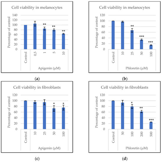
Figure 1.
Cell viability of apigenin and phloretin in melanocytes (a,b) and fibroblasts (c,d) after 24 h. Statistical analysis: Significance of Student’s t-test results is represented by * when p < 0.05, ** when p < 0.01, or *** when p < 0.001.
3.2. Synergistic Effect on Melanin Synthesis Inhibition
To determine if the apigenin and phloretin combination had a synergistic effect on melanin inhibition, melanocytes were stimulated and treated with the compounds separately and successively (additive effect) or treated with both compounds at the same time (combination effect). As shown in Figure 2, the stimulated melanocytes showed a significant increase in melanin accumulation compared to control cells. The additive incubation of the compounds significantly decreased melanin accumulation compared to that in the stimulated cells. Interestingly, both compounds incubated at the same time (combination) induced a larger decrease in melanin production than when the compounds were added separately (additive). This evidence proved that the compounds have a synergistic effect on melanin synthesis inhibition.
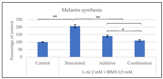
Figure 2.
Melanin production after 48 h in nontreated cells (control), cells stimulated with 2 mM l-tyrosine + 0.5 mM IBMX (stimulated), cells stimulated with 2 mM l-tyrosine + 0.5 mM IBMX and treated for the first 24 h with 10 μM phloretin and the next 24 h with 0.5 μM apigenin (additive), and cells stimulated with 2 mM l-tyrosine + 0.5 mM IBMX and treated with both 10 μM phloretin and 0.5 μM apigenin at the same time (combination). Statistical analysis: significance of -Student’s t-test results is represented by * when p < 0.05 or ** when p < 0.01.
3.3. Inhibition of Wnt-Stimulated Melanogenesis
As the Wnt pathway is upregulated in melasma skin [16], we studied whether the compounds could inhibit Wnt-stimulated melanogenesis. For this, we treated melanocytes with the Wnt activator BIO and the compounds for 24 h. The expression of melanogenesis-related genes was then quantified by qPCR (Figure 3). Apigenin reduced the expressions of DCT and PAX3, while phloretin reduced the expressions of DCT, PAX3, PMEL17, and TYRP1.
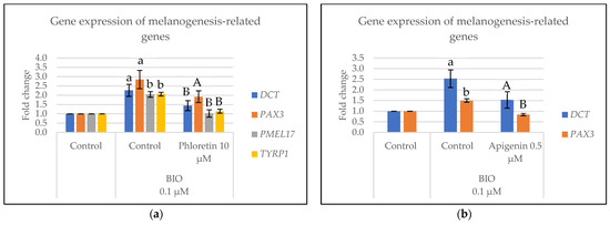
Figure 3.
Inhibition of Wnt-activated melanogenesis in melanocytes treated for 24 h with phloretin (a) or apigenin (b). Statistical analysis: a, Student’s t-test between control and BIO-stimulated control is p < 0.05; b, Student’s t-test between control and BIO-stimulated control is p < 0.01; A, Student’s t-test between BIO-stimulated control and BIO-stimulated cells treated with compound is p < 0.05; and B, Student’s t-test between BIO-stimulated control and BIO-stimulated cells treated with compound is p < 0.01.
3.4. Regulation of miR-675 Levels and Autophagy-Related Gene Expression in Melanocytes
Other characteristic features of melanocytes from melasma skin are impaired autophagy and reduced miR-675 levels [1]. To test whether the compounds could regulate these processes, melanocytes were cultured for 24 h with the compounds, and the markers related to these pathways were quantified by qPCR. As shown in Figure 4, apigenin induced the levels of miR-675, while both apigenin and phloretin induced the expression of autophagy-related genes (ATG5, ATG7, and LC3).
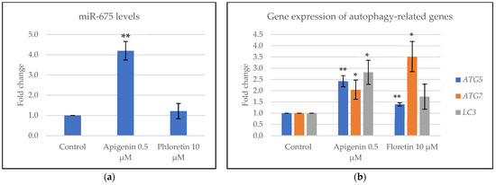
Figure 4.
miR-675 (a) and autophagy-related gene expression (b) levels in melanocytes treated for 24 h with the compounds. Statistical analysis: significance of Student’s t-test results is represented by * when p < 0.05 or ** when p < 0.01.
3.5. Regulation of Melanogenesis Paracrine Regulators in Fibroblasts
To test whether the compounds could regulate key paracrine regulators of melanogenesis [2], human dermal fibroblasts were treated for 24 h with the compounds, and the gene expressions of the paracrine regulators were quantified by qPCR. As shown in Figure 5, apigenin induced the expression of the Wnt pathway inhibitor DKK1 and decreased the expression of the vascular growth factor VEGF, while phloretin did not affect the expression of these genes.
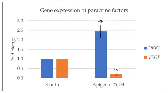
Figure 5.
Gene expression of paracrine factors that regulate melanogenesis in fibroblasts treated with apigenin for 24 h. Statistical analysis: significance of Student’s t-test results is represented by ** when p < 0.01.
3.6. Senomorphic Effect on Senescent Fibroblasts
As senescent fibroblasts have been described to influence skin pigmentation [6], we treated senescent human dermal fibroblasts with the compounds for 48 h and quantified the expression of extracellular matrix-related genes by qPCR to determine the regulation of both compounds in this process. As shown in Figure 6, COL1A1 expression remained unaffected by the compounds. Apigenin significantly reduced MMP1 expression and induced TIMP1 expression, while phloretin significantly reduced MMP1 expression and induced ELN and TIMP1 expressions.
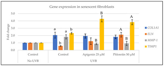
Figure 6.
Gene expression of extracellular-matrix-related genes in senescent fibroblasts treated for 48 h with the compounds. Statistical analysis: a, Student’s t-test between non-UVB control and UVB control is p < 0.05; b, Student’s t-test between non-UVB control and UVB control is p < 0.01; A, Student’s t-test between UVB control and UVB-treated with compound is p < 0.05; and B, Student’s t-test between UVB control and UVB-treated with compound is p < 0.01.
3.7. Depigmenting Effect of the Combination in a 3D Epidermis Model
Finally, to validate the depigmenting efficacy of the compounds in a more complex skin model, a reconstructed human epidermis containing phototype VI was topically treated with the compounds daily for 5 days and melanin accumulation was evaluated by Fontana–Masson staining on day 7. As shown in Figure 7, the combination of apigenin and phloretin reduced the melanin content in the epidermal model after 7 days of treatment.
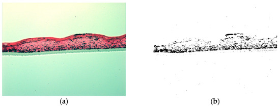
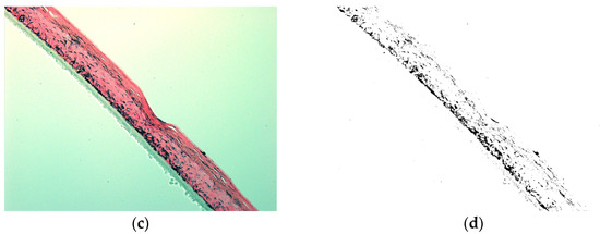
Figure 7.
Representative optical microscopy images, 20× magnification, on day 7 from nontreated reconstructed human epidermis (a) and reconstructed human epidermis treated with 25 µM apigenin + 50 µM phloretin daily for 5 days (c). Fontana–Masson staining was performed to evidence melanin granules. Pictures showing the black pigment staining (b,d) are shown to better distinguish the melanin reduction after treatment.
4. Discussion
Skin hyperpigmentation, particularly melasma, has a complex pathophysiology involving different external and internal factors, such as ultraviolet radiation exposure, hormonal levels, or genetics [17]. Moreover, cells such as keratinocytes and fibroblasts release paracrine factors that result in hyperactivated melanocytes with increased melanogenic activity and impaired autophagy [1]. Here, we show that the combination of apigenin and phloretin is promising for the management of skin hyperpigmentation.
Phloretin was selected based on previous reports about its efficacy as a depigmenting compound, mainly by inhibiting tyrosinase activity [11,18]. Similarly, apigenin has been shown to exert many beneficial effects on skin, including the inhibition of tyrosinase [10]. Here, we demonstrate that the combination of both compounds is promising for decreasing the melanin production I then skin based on the synergistic effect of the compounds in melanogenesis inhibition (Figure 2).
Additionally, we better characterized the depigmenting mechanism of action of both compounds, beyond tyrosinase inhibition. We show that both compounds can inhibit Wnt-stimulated melanogenesis, which has not been reported before as far as we know. Interestingly, other models show that both apigenin and phloretin might have activating or inhibiting effects on the Wnt pathway depending on the cell type studied [19,20,21,22,23,24,25,26]. According to the Wnt regulatory mechanisms in previous research, apigenin and phloretin could be modulating the Wnt pathway at different levels, which could explain the observed synergistic effects of this combination. For example, these compounds might regulate glycogen synthase kinase 3 (GSK3) activity, or b-catenin levels, and/or transcriptional activity over specific melanogenesis genes such as TYR, TYRP1, or DCT.
Our results also show an upregulation of miR-675 by apigenin in melanocytes. This miRNA is reduced in melasma skin and targets MITF, a key transcription factor controlling the melanogenesis pathway [27]. Previous reports have shown that apigenin can influence other miRNAs in different cell models [28,29], but the effect of apigenin on miR-675 has been described for the first time in our research. The exact mechanism through which apigenin induces miR-675 levels might be via direct gene expression or miRNA processing regulation. Regarding autophagy activation, previous research has shown that both apigenin and phloretin can induce autophagy in different cell types, including keratinocytes, macrophages, and different cancer cell types [30,31,32,33,34]. Interestingly, one report showed that apigenin inhibited autophagy in normal keratinocytes but restored autophagy in UVB-damaged keratinocytes, indicating differential effects on the same cell type under different conditions [30]. Here, our results suggest that both apigenin and phloretin activate autophagy signaling in melanocytes. Interestingly, the autophagy pathway can be regulated through different targets, including mTOR, AMPK, or mitogen-activated protein kinase (MAPK) signaling, among others [35]. Thus, apigenin and phloretin could also exert a synergistic depigmenting effect through the modulation of this pathway.
Up to this point, all the described mechanisms that regulate melanogenesis at the melanocyte level could explain the synergy observed by combining apigenin and phloretin. The modulation of Wnt pathway, along with autophagy and miR-675 levels, exerts a multistep regulation of the pigmentation process in melanocytes.
On the other hand, paracrine factors released by surrounding cells play a key role in melasma pathogenesis [2]. Here, we show that apigenin induces DKK1, an antagonist of the Wnt pathway with a proven role in melanogenesis inhibition [36]. No previous reports have described the relationship between apigenin and DKK1. On the other hand, previous research on the effect of apigenin on angiogenesis and VEGF is controversial, with results showing proangiogenic or antiangiogenic effects in different models [37,38,39]. Here, our results indicate that apigenin might have antiangiogenic properties by downregulating the levels of VEGF in human dermal fibroblasts. Regarding phloretin’s effect on these paracrine factors, previous research shows that phloretin increases DKK1 in adipose tissue and upregulates or downregulates VEGF expression depending on the cell type [24,40,41,42]. Here, we observed that both DKK1 and VEGF gene expression remained unchanged in human dermal fibroblasts after phloretin treatment.
Senescent fibroblasts also contribute to increased pigmentation in aged skin [6]. Here, we showed that apigenin and phloretin exert senomorphic effects on senescent fibroblasts by regulating extracellular matrix proteins (Figure 6), which might have a beneficial effect on photoaged skin pigmentation. Interestingly, previous studies showed that apigenin induces collagen I synthesis in dermal fibroblasts while it does not affect MMP1 or TIMP1 expression [43]. According to our research, apigenin does not affect collagen I gene expression but downregulates and upregulates MMP1 and TIMP1 expression, respectively. The differences in these results might be explained by the fact that the previous study tested the effect of apigenin on normal human dermal fibroblasts, while we studied the effect of apigenin on senescent fibroblasts. This suggests different cell behavior in response to apigenin in the same cell type, being beneficial for skin functions in all the studied cases.
Regarding this effect on senescent cells by apigenin and phloretin, previous research already indicated that these compounds have senomorphic effects on senescent cells [44]. Perrott, K. et al. proved the positive effect of Apigenin in modulating the senescence-associated secretory phenotype (SASP) of senescent skin fibroblasts, mainly by regulating inflammatory markers [45]. Other reports identified the senescent-cell-modulating effects of phloretin [46]. Our research confirms the positive effect of apigenin and phloretin on senescent human dermal fibroblasts by regulating extracellular-matrix-related proteins.
Finally, the reduction in melanin accumulation in the reconstructed human epidermis model after 7 days of treatment with the compounds suggests that the combination of both compounds might have promising effects when applied in vivo to hyperpigmented skin. Moreover, this experiment sheds light on the safety of the compounds for topical application, as daily treatment of the 3D reconstructed epidermis for 7 days did not affect tissue viability. Furthermore, both compounds have been used for years in topical products for different chronic dermatological conditions [10,11], which is another proof of the long-term safety of both compounds.
The mechanisms of action of these compounds on melanogenesis inhibition, including Wnt pathway, autophagy, miR-675 levels, and paracrine factors, offer a complementary approach to targeting skin hyperpigmentation. Most natural compounds currently used in the cosmetic market target the activity of tyrosinase, the key and central enzyme involved in the synthesis of melanin [47], and only a few are focused on other key pathways such as inflammation, hormonal, or vascularization pathways [15,48].
Future experiments could include the evaluation of other paracrine factors such as HGF, SCF, or GDF15 on fibroblasts after treatment with apigenin and phloretin. Additionally, the clinical evaluation of a topical formulation including these ingredients is necessary to validate the efficacy of this combination of compounds on melasma skin. This further research would overcome the main limitations of this study. These include the use of in vitro and 3D models, which are useful for mechanistic studies on cellular physiology but do not fully replicate the human skin environment. Interestingly, as solar lentigo and post-inflammatory hyperpigmentation share some features with melasma pathology [49,50], the combination of these compounds might be used to treat other types of skin hyperpigmentation. Given the observed results on the compounds’ safety, their mechanisms of action, and their efficacy in the 3D epidermal model, we expect that the use of this combination of compounds in daily-use creams, masks, or similar topical formulations can boost the efficacy of the current skin whitening treatments.
5. Conclusions
Our research shows that apigenin and phloretin have synergistic effects on the inhibition of melanin accumulation in melanocytes by targeting some of the main features of melasma pathology, including the Wnt pathway, autophagy impairment, miRNA regulation, and paracrine factors released by the surrounding cells. In addition, the topical application of these compounds significantly decreased melanin accumulation in a phototype VI 3D epidermis model. Thus, this combination of compounds shows promising results for the treatment of skin hyperpigmentation and aging. Further studies should be conducted to confirm the depigmenting effect in physiological skin conditions.
Supplementary Materials
The following supporting information can be downloaded at: https://www.mdpi.com/article/10.3390/cosmetics11040128/s1, Table S1: Primer sequences used for qPCR assay.
Author Contributions
Conceptualization, A.M.-G. and M.C.G.; methodology, J.S. and T.N.; software: A.M.-G., J.S. and T.N.; validation, A.M.-G. and M.C.G.; formal analysis, A.M.-G. and J.S.; investigation, A.M.-G., J.S. and T.N.; resources, A.M.-G. and M.C.G.; data curation, A.M.-G. and J.S.; writing—original draft preparation, A.M.-G. writing—review and editing, A.M.-G. and M.C.G.; supervision, A.M.-G. and M.C.G.; project administration, M.C.G.; funding acquisition, M.C.G. All authors have read and agreed to the published version of the manuscript.
Funding
The research has been funded by Mesoestetic Pharma Group S.L.
Data Availability Statement
All data related to this research has been included in this paper.
Acknowledgments
We would like to thank all the collaborators and company departments that supported our research in its different stages.
Conflicts of Interest
The authors are employees of Mesoestetic Pharma Group S.L. The authors declare that the research was conducted in the absence of any commercial or financial relationships that could be construed as a potential conflict of interest. The funders had no role in the design of the study; in the collection, analyses or interpretation of data; in the writing of the manuscript; or in the decision to publish the results.
Correction Statement
The article has been republished with a minor correction to Conflicts of Interest Statement. This change does not affect the scientific content of the article.
References
- Espósito, A.C.C.; Cassiano, D.P.; da Silva, C.N.; Lima, P.B.; Dias, J.A.F.; Hassun, K.; Bagatin, E.; Miot, L.D.B.; Miot, H.A. Update on Melasma—Part I: Pathogenesis. Dermatol. Ther. 2022, 12, 1967–1988. [Google Scholar] [CrossRef] [PubMed]
- Yuan, X.H.; Jin, Z.H. Paracrine regulation of melanogenesis. Br. J. Dermatol. 2018, 178, 632–639. [Google Scholar] [CrossRef]
- Yamaguchi, Y.; Itami, S.; Watabe, H.; Yasumoto, K.; Abdel-Malek, Z.A.; Kubo, T.; Rouzaud, F.; Tanemura, A.; Yoshikawa, K.; Hearing, V.J. Mesenchymal–epithelial interactions in the skin: Increased expression of dickkopf1 by palmoplantar fibroblasts inhibits melanocyte growth and differentiation. J. Cell Biol. 2004, 165, 275–285. [Google Scholar] [CrossRef] [PubMed]
- Yamaguchi, Y.; Passeron, T.; Hoashi, T.; Watabe, H.; Rouzaud, F.; Yasumoto, K.; Hara, T.; Tohyama, C.; Katayama, I.; Miki, T.; et al. Dickkopf 1 (DKK1) regulates skin pigmentation and thickness by affecting Wnt/ β-catenin signaling in keratinocytes. FASEB J. 2008, 22, 1009–1020. [Google Scholar] [CrossRef]
- Passeron, T.; Picardo, M. Melasma, a photoaging disorder. Pigment. Cell Melanoma Res. 2018, 31, 461–465. [Google Scholar] [CrossRef]
- Kim, J.C.; Park, T.J.; Kang, H.Y. Skin-Aging Pigmentation: Who Is the Real Enemy? Cells 2022, 11, 2541. [Google Scholar] [CrossRef]
- Espósito, A.C.C.; Brianezi, G.; Miot, L.D.B.; Miot, H.A. Fibroblast morphology, growth rate and gene expression in facial melasma. An. Bras. Dermatol. 2022, 97, 575–582. [Google Scholar] [CrossRef] [PubMed]
- Ni, C.; Narzt, M.-S.; Nagelreiter, I.-M.; Zhang, C.F.; Larue, L.; Rossiter, H.; Grillari, J.; Tschachler, E.; Gruber, F. Autophagy deficient melanocytes display a senescence associated secretory phenotype that includes oxidized lipid mediators. Int. J. Biochem. Cell Biol. 2016, 81, 375–382. [Google Scholar] [CrossRef]
- Artzi, O.; Horovitz, T.; Bar-Ilan, E.; Shehadeh, W.; Koren, A.; Zusmanovitch, L.; Mehrabi, J.N.; Salameh, F.; Nelkenbaum, G.I.; Zur, E.; et al. The pathogenesis of melasma and implications for treatment. J. Cosmet. Dermatol. 2021, 20, 3432–3445. [Google Scholar] [CrossRef]
- Majma Sanaye, P.; Mojaveri, M.R.; Ahmadian, R.; Jahromi, M.S.; Bahrasoltani, R. Apigenin and its dermatological applications: A comprehensive review. Phytochemistry 2022, 203, 113390. [Google Scholar] [CrossRef]
- Anunciato Casarini, T.P.; Frank, L.A.; Pohlmann, A.R.; Guterres, S.S. Dermatological applications of the flavonoid phloretin. Eur. J. Pharmacol. 2020, 889, 173593. [Google Scholar] [CrossRef] [PubMed]
- Vichai, V.; Kirtikara, K. Sulforhodamine B colorimetric assay for cytotoxicity screening. Nat. Protoc. 2006, 1, 1112–1116. [Google Scholar] [CrossRef] [PubMed]
- Martínez-Gutiérrez, A.; Asensio, J.A.; Aran, B. Effect of the combination of different depigmenting agents in vitro. J. Cosmet. Sci. 2014, 65, 365–375. [Google Scholar] [PubMed]
- Meijer, L.; Skaltsounis, A.-L.; Magiatis, P.; Polychronopoulos, P.; Knockaert, M.; Leost, M.; Ryan, X.P.; Vonica, C.A.; Brivanlou, A.; Dajani, R. GSK-3-Selective Inhibitors Derived from Tyrian Purple Indirubins. Chem. Biol. 2003, 10, 1255–1266. [Google Scholar] [CrossRef] [PubMed]
- Alfredo, M.G.; Maribel, P.M.; Eloy, P.R.; Susana, G.E.; Luis, L.G.S.; Carmen, G.M. Depigmenting topical therapy based on a synergistic combination of compounds targeting the key pathways involved in melasma pathophysiology. Exp. Dermatol. 2023, 32, 611–619. [Google Scholar] [CrossRef] [PubMed]
- Espósito, A.C.C.; Brianezi, G.; de Souza, N.P.; Miot, L.D.B.; Miot, H.A. Exploratory Study of Epidermis, Basement Membrane Zone, Upper Dermis Alterations and Wnt Pathway Activation in Melasma Compared to Adjacent and Retroauricular Skin. Ann. Dermatol. 2020, 32, 101–108. [Google Scholar] [CrossRef] [PubMed]
- Lee, A.Y. Recent progress in melasma pathogenesis. Pigment. Cell Melanoma Res. 2015, 28, 648–660. [Google Scholar] [CrossRef] [PubMed]
- Chen, J.; Li, Q.; Ye, Y.; Huang, Z.; Ruan, Z.; Jin, N. Phloretin as both a substrate and inhibitor of tyrosinase: Inhibitory activity and mechanism. Spectrochim. Acta A Mol. Biomol. Spectrosc. 2020, 226, 117642. [Google Scholar] [CrossRef]
- Li, Y.; Zhao, Z.; Luo, J.; Jiang, Y.; Li, L.; Chen, Y.; Zhang, L.; Huang, Q.; Cao, Y.; Zhou, P.; et al. Apigenin ameliorates hyperuricemic nephropathy by inhibiting URAT1 and GLUT9 and relieving renal fibrosis via the Wnt/β-catenin pathway. Phytomedicine 2021, 87, 153585. [Google Scholar] [CrossRef]
- Ozbey, U.; Attar, R.; Romero, M.A.; Alhewairini, S.S.; Afshar, B.; Sabitaliyevich, U.Y.; Hanna-Wakim, L.; Ozcelik, B.; Farooqi, A.A. Apigenin as an effective anticancer natural product: Spotlight on TRAIL, WNT/β-catenin, JAK-STAT pathways, and microRNAs. J. Cell Biochem. 2019, 120, 1060–1067. [Google Scholar] [CrossRef]
- Pan, F.F.; Shao, J.; Shi, C.J.; Li, Z.P.; Fu, W.M.; Zhang, J.F. Apigenin promotes osteogenic differentiation of mesenchymal stem cells and accelerates bone fracture healing via activating Wnt/β-catenin signaling. Am. J. Physiol. Endocrinol. Metab. 2021, 320, E760–E771. [Google Scholar] [CrossRef]
- Lin, C.M.; Chen, H.H.; Lin, C.A.; Wu, H.C.; Sheu, J.J.; Chen, H.J. Apigenin-induced lysosomal degradation of β-catenin in Wnt/β-catenin signaling. Sci. Rep. 2017, 7, 372. [Google Scholar] [CrossRef]
- Kern, M.; Pahlke, G.; Ngiewih, Y.; Marko, D. Modulation of Key Elements of the Wnt Pathway by Apple Polyphenols. J. Agric. Food Chem. 2006, 54, 7041–7046. [Google Scholar] [CrossRef]
- Casado-Díaz, A.; Rodríguez-Ramos, Á.; Torrecillas-Baena, B.; Dorado, G.; Quesada-Gómez, J.M.; Gálvez-Moreno, M.Á. Flavonoid Phloretin Inhibits Adipogenesis and Increases OPG Expression in Adipocytes Derived from Human Bone-Marrow Mesenchymal Stromal-Cells. Nutrients 2021, 13, 4185. [Google Scholar] [CrossRef]
- Kim, U.; Kim, C.Y.; Lee, J.M.; Oh, H.; Ryu, B.; Kim, J.; Park, J.H. Phloretin Inhibits the Human Prostate Cancer Cells Through the Generation of Reactive Oxygen Species. Pathol. Oncol. Res. 2020, 26, 977–984. [Google Scholar] [CrossRef]
- Kapoor, S.; Padwad, Y.S. Phloretin induces G2/M arrest and apoptosis by suppressing the β-catenin signaling pathway in colorectal carcinoma cells. Apoptosis 2023, 28, 810–829. [Google Scholar] [CrossRef]
- Kim, N.H.; Choi, S.H.; Kim, C.H.; Lee, C.H.; Lee, T.R.; Lee, A.Y. Reduced MiR-675 in Exosome in H19 RNA-Related Melanogenesis via MITF as a Direct Target. J. Investig. Dermatol. 2014, 134, 1075–1082. [Google Scholar] [CrossRef] [PubMed]
- Nimal, S.; Kumbhar, N.; Rathore, S.; Naik, N.; Paymal, S.; Gacche, R.N. Apigenin and its combination with Vorinostat induces apoptotic-mediated cell death in TNBC by modulating the epigenetic and apoptotic regulators and related miRNAs. Sci. Rep. 2024, 14, 9540. [Google Scholar] [CrossRef] [PubMed]
- Husain, K.; Villalobos-Ayala, K.; Laverde, V.; Vazquez, O.A.; Miller, B.; Kazim, S.; Blanck, G.; Hibbs, M.L.; Krystal, G.; Elhussin, I.; et al. Apigenin Targets MicroRNA-155, Enhances SHIP-1 Expression, and Augments Anti-Tumor Responses in Pancreatic Cancer. Cancers 2022, 14, 3613. [Google Scholar] [CrossRef]
- Li, L.; Li, M.; Xu, S.; Chen, H.; Chen, X.; Gu, H. Apigenin restores impairment of autophagy and downregulation of unfolded protein response regulatory proteins in keratinocytes exposed to ultraviolet B radiation. J. Photochem. Photobiol. B 2019, 194, 84–95. [Google Scholar] [CrossRef] [PubMed]
- Yang, J.; Pi, C.; Wang, G. Inhibition of PI3K/Akt/mTOR pathway by apigenin induces apoptosis and autophagy in hepatocellular carcinoma cells. Biomed. Pharmacother. 2018, 103, 699–707. [Google Scholar] [CrossRef] [PubMed]
- Kim, T.W.; Lee, H.G. Apigenin Induces Autophagy and Cell Death by Targeting EZH2 under Hypoxia Conditions in Gastric Cancer Cells. Int. J. Mol. Sci. 2021, 22, 13455. [Google Scholar] [CrossRef] [PubMed]
- Fan, C.; Zhang, Y.; Tian, Y.; Zhao, X.; Teng, J. Phloretin enhances autophagy by impairing AKT activation and inducing JNK-Beclin-1 pathway activation. Exp. Mol. Pathol. 2022, 127, 104814. [Google Scholar] [CrossRef] [PubMed]
- Dierckx, T.; Haidar, M.; Grajchen, E.; Wouters, E.; Vanherle, S.; Loix, M.; Boeykens, A.; Bylemans, D.; Hardonnière, K.; Kerdine-Römer, S.; et al. Phloretin suppresses neuroinflammation by autophagy-mediated Nrf2 activation in macrophages. J. Neuroinflammation 2021, 18, 148. [Google Scholar] [CrossRef]
- He, C.; Klionsky, D.J. Regulation mechanisms and signaling pathways of autophagy. Annu. Rev. Genet. 2009, 43, 67–93. [Google Scholar] [CrossRef]
- Yamaguchi, Y.; Morita, A.; Maeda, A.; Hearing, V.J. Regulation of Skin Pigmentation and Thickness by Dickkopf 1 (DKK1). J. Investig. Dermatol. Symp. Proc. 2009, 14, 73–75. [Google Scholar] [CrossRef]
- Fang, J.; Zhou, Q.; Liu, L.Z.; Xia, C.; Hu, X.; Shi, X.; Jiang, B.H. Apigenin inhibits tumor angiogenesis through decreasing HIF-1α and VEGF expression. Carcinogenesis 2006, 28, 858–864. [Google Scholar] [CrossRef]
- Osada, M.; Imaoka, S.; Funae, Y. Apigenin suppresses the expression of VEGF, an important factor for angiogenesis, in endothelial cells via degradation of HIF-1α protein. FEBS Lett. 2004, 575, 59–63. [Google Scholar] [CrossRef]
- Tu, F.; Pang, Q.; Chen, X.; Huang, T.; Liu, M.; Zhai, Q. Angiogenic effects of apigenin on endothelial cells after hypoxia-reoxygenation via the caveolin-1 pathway. Int. J. Mol. Med. 2017, 40, 1639–1648. [Google Scholar] [CrossRef]
- Tuli, H.S.; Rath, P.; Chauhan, A.; Ramniwas, S.; Vashishth, K.; Varol, M.; Jaswal, V.S.; Haque, S.; Sak, K. Phloretin, as a Potent Anticancer Compound: From Chemistry to Cellular Interactions. Molecules 2022, 27, 8819. [Google Scholar] [CrossRef]
- Hytti, M.; Ruuth, J.; Kanerva, I.; Bhattarai, N.; Pedersen, M.L.; Nielsen, C.U.; Kauppinen, A. Phloretin inhibits glucose transport and reduces inflammation in human retinal pigment epithelial cells. Mol. Cell Biochem. 2023, 478, 215–227. [Google Scholar] [CrossRef]
- Matsugami, H.; Harada, Y.; Kurata, Y.; Yamamoto, Y.; Otsuki, Y.; Yaura, H.; Inoue, Y.; Morikawa, K.; Yoshida, A.; Shirayoshi, Y.; et al. VEGF secretion by adipose tissue-derived regenerative cells is impaired under hyperglycemic conditions via glucose transporter activation and ROS increase. Biomed. Res. 2014, 35, 397–405. [Google Scholar] [CrossRef]
- Zhang, Y.; Wang, J.; Cheng, X.; Yi, B.; Zhang, X.; Li, Q. Apigenin induces dermal collagen synthesis via smad2/3 signaling pathway. Eur. J. Histochem. 2015, 59. [Google Scholar] [CrossRef]
- Martel, J.; Ojcius, D.M.; Wu, C.Y.; Peng, H.H.; Voisin, L.; Perfettini, J.L.; Ko, Y.F.; Young, J.D. Emerging use of senolytics and senomorphics against aging and chronic diseases. Med. Res. Rev. 2020, 40, 2114–2131. [Google Scholar] [CrossRef] [PubMed]
- Perrott, K.M.; Wiley, C.D.; Desprez, P.Y.; Campisi, J. Apigenin suppresses the senescence-associated secretory phenotype and paracrine effects on breast cancer cells. Geroscience 2017, 39, 161–173. [Google Scholar] [CrossRef] [PubMed]
- Malavolta, M.; Bracci, M.; Santarelli, L.; Sayeed, M.A.; Pierpaoli, E.; Giacconi, R.; Costarelli, L.; Piacenza, F.; Basso, A.; Cardelli, M.; et al. Inducers of Senescence, Toxic Compounds, and Senolytics: The Multiple Faces of Nrf2-Activating Phytochemicals in Cancer Adjuvant Therapy. Mediat. Inflamm. 2018, 2018, 1–32. [Google Scholar] [CrossRef]
- Zolghadri, S.; Beygi, M.; Mohammad, T.F.; Alijanianzadeh, M.; Pillaiyar, T.; Garcia-Molina, P.; Garcia-Canovas, F.; Munoz-Munoz, J.; Saboury, A.A. Targeting tyrosinase in hyperpigmentation: Current status, limitations and future promises. Biochem. Pharmacol. 2023, 212, 115574. [Google Scholar] [CrossRef]
- Qian, W.; Liu, W.; Zhu, D.; Cao, Y.; Tang, A.; Gong, G.; Su, H. Natural skin-whitening compounds for the treatment of melanogenesis (Review). Exp. Ther. Med. 2020, 20, 173–185. [Google Scholar] [CrossRef] [PubMed]
- Cardinali, G.; Kovacs, D.; Picardo, M. Mechanisms underlying post-inflammatory hyperpigmentation: Lessons from solar lentigo. Ann. Dermatol. Venereol. 2012, 139, S148–S152. [Google Scholar] [CrossRef]
- Sadick, N.; Pannu, S.; Abidi, Z.; Arruda, S. Topical Treatments for Melasma and Post-inflammatory Hyperpigmentation. J. Drugs Dermatol. 2023, 22, 1118–1123. [Google Scholar] [CrossRef]
Disclaimer/Publisher’s Note: The statements, opinions and data contained in all publications are solely those of the individual author(s) and contributor(s) and not of MDPI and/or the editor(s). MDPI and/or the editor(s) disclaim responsibility for any injury to people or property resulting from any ideas, methods, instructions or products referred to in the content. |
© 2024 by the authors. Licensee MDPI, Basel, Switzerland. This article is an open access article distributed under the terms and conditions of the Creative Commons Attribution (CC BY) license (https://creativecommons.org/licenses/by/4.0/).