Article

Preliminary Experience with a Cleansing Mousse and a Non-Steroidal Emulsion for the Prevention and Treatment of Acute Radiation Dermatitis in Breast Cancer Patients Undergoing Adjuvant Radiotherapy

1
Radiation Oncology Unit, REM Radioterapia, Via Penninazzo 11, 95100 Viagrande, Italy
2
Istituto Oncologico del Mediterraneo, Via Penninazzo 7, 95029 Viagrande, Italy
3
Department of Medicine and Surgery, University Kore of Enna, 94100 Enna, Italy
*
Author to whom correspondence should be addressed.
Cosmetics 2024, 11(4), 117; https://doi.org/10.3390/cosmetics11040117
Submission received: 29 May 2024 / Revised: 29 June 2024 / Accepted: 8 July 2024 / Published: 11 July 2024

Abstract

:
Background: Radiation dermatitis (RD) is the most frequent side effect in patients undergoing adjuvant radiotherapy (RT) for breast cancer. Despite the skin-sparing benefits of new RT techniques, most patients develop RD. There is currently no standard treatment to prevent and soothe RD, which is generally managed with emollients, moisturizers, or corticosteroids. We conducted a prospective observational study to evaluate the rate and grade of RD with the application of a cleansing mousse and a non-steroidal emulsion during the adjuvant RT program in patients with breast cancer submitted to surgery. Materials and Methods: A cleansing mousse containing vegetable glycerin (12%), phytoextract of chamomile (0.5%), yarrow phytoextract (0.5%), sweet almond (0.1%), Oenothera oil (0.1%), and rice protein hydrolyzate (0.1%), and an emulsion containing micronized zinc oxide (3.7%), rapeseed phytosterols (1.7%), aloe (0.5%), 18-beta glycyrrhetinic acid (0.5%), alpha bisabolol (0.5%), and zanthalene (0.5%) were offered to breast cancer patients undergoing adjuvant RT to prevent the onset of RD and mitigate its severity. These specific ingredients were selected for their well-known anti-inflammatory, antioxidant, and moisturizing properties. Skin toxicities were recorded photographically and graded according to the RTOG scoring system. Results: From March 2023 to July 2023, a total of 24 patients with a median age of 59 years (range 42–75) were enrolled. Halfway through the RT treatment, 20 patients (83.3%) had G0 skin toxicity, three (12.5%) G1, one (4.2%) G2. None showed G3–G4 toxicity. At the end of RT, seven patients (29.2%) exhibited G0 skin toxicity, 14 (58.3%) G1, two (8.3%) G2, one (4.2%) G3. No patient developed G4 toxicity. Fifteen days after the end of RT, 13 patients (54.2%) had G0 skin toxicity, 10 (41.1%) G1, one (4.2%) G2, with none showing G3–G4 toxicity. Conclusions: Our data suggest that the tested topicals might be an effective option for preventing and alleviating RD. Further prospective randomized studies are needed to confirm our findings.

1. Introduction

Adjuvant radiotherapy (RT) represents a cornerstone in the treatment path of women undergoing conservative surgery for early breast cancer (BC), having definitively supplanted demolitive mastectomy procedures in the management of small tumors [1]. RT could be omitted only under particular conditions carrying a very low risk of ipsilateral recurrence and cancer-specific death [2]. On the other hand, its administration is warranted even in some post-mastectomy cases depending on unfavorable tumor characteristics [3].
RT to the breast or chest wall is generally well-tolerated, with the risk for late cardiac events being the most feared and attempted to be reduced when treating left-sided BC patients [4,5]. Indeed, radiation dermatitis (RD) is the most common side effect of breast RT. The underlying mechanisms likely responsible for the onset of acute damage are not entirely known. Several hypotheses have been proposed, with the most relevant suggesting that ionizing radiation stimulates cells such as fibroblasts, endothelial cells, and circulating immune cells. In response, these cells release cytokines and chemokines, including IL-6, IL-8, and tumor necrosis factor alpha (TNF-α) [6]. This release occurs within a few hours of radiation exposure. In the following weeks, the hallmark of radiation-induced damage will be represented by the leukocyte infiltration in the irradiated skin [7].
Despite the skin-sparing effect related to the build-up depth of high-energy photon beams, approximately 50–60% of the prescribed dose is still deposited in the skin [8]. As such, approximately 90% of irradiated patients with breast cancer develop moderate to severe RD [9].
Clinically, the first reactive changes, mainly dryness, warmth, and mild erythema, may appear towards the middle of the treatment schedule and progress to moist desquamation and oedema in the following weeks. Additionally, patients may complain of burning, itching, and pain. These events are generally self-limiting and skin integrity is restored within a few weeks after the end of RT. If not, a few patients may develop serious toxicities, such as ulcers or skin necrosis. Most cases recover completely from the acute phase with no or negligible alterations. When this does not happen, a condition of chronic dermatitis can persist indefinitely, even for months or years, showing more fibrotic, drier skin, with telangiectasias and hypo- or hyperpigmentation [10].
Finally, exaggerated skin manifestations can occur quite unexpectedly when combining RT with certain drugs [11,12,13].
From this perspective, although non-life-threatening unlike radiation pneumonitis and ischemic cardiac events, RD emerges, at least, as an injury that can alter women’s quality of life and treatment compliance [14]. Regarding the latter, in case of severe toxicity, it may even be necessary to discontinue the treatment to manage the toxicity and allow for recovery [15]. It is noteworthy to underline that any prolongation of overall treatment time due to unplanned interruptions following difficult-to-manage acute toxicity during RT may have a negative impact on prognosis [16].
Hence the need to effectively prevent and treat such a side effect. To this purpose, a wide variety of topical products exists, including emollients, moisturizers, and corticosteroids. Although several systematic reviews of the literature have attempted to clarify the benefits of one product over another [17,18], none of these preparations has proven to prevail over the others in terms of effectiveness and their choice is mainly based on patient preference and physician experience [19]. Nonetheless, RD is still an active field of research due to its frequency and impact on patients’ lives and natural products are gaining more and more attention, being perceived as better tolerated than drugs [20].
Here we present a single-center prospective observational study on the use of a gentle cleansing soap and an emulsion for the prevention and alleviation of acute RD among BC patients undergoing adjuvant RT, with the aim of serving as a pilot experience eventually stimulating a comparison with other available topical agents.

2. Materials and Methods

From March 2023 to July 2023 women who underwent breast-conserving surgery or mastectomy for non-metastatic breast cancer and required adjuvant RT were offered enrollment in a prospective, observational, single-arm study testing the combination of two non-corticosteroid topicals for the prevention and alleviation of acute RD. Inclusion criteria were: age between 18 and 80 years old, Karnofsky performance status (KPS) ≥80, histopathological diagnosis of breast cancer, need for RT following conservative surgery or mastectomy. Exclusion criteria were: state of pregnancy, suffering from psychiatric disorders or cognitive deficits, or skin diseases (e.g., psoriasis, pemphigus, current sunburn, etc.), skin involvement by cancer (pT4), known hypersensitivity to one of the components of the medications tested, history of RT for previous thoracic malignancies (e.g., lymphoma, etc.). Patients with relapsed BC were eligible for inclusion provided they did not receive any RT at first diagnosis.

2.1. Radiotherapy

A free-breathing computed tomography (CT) simulation with 3 mm thick slices was performed for each patient to plan RT delivery. An additional deep-inspiration breath-hold CT scan was acquired only for left-sided breast cancer patients to identify those benefitting most from respiratory gating for cardiac radiation dose reduction, as per our center’s policy [21]. The target volume delineation on the CT simulation was conducted according to the ESTRO consensus guideline, i.e., cropping the clinical target volume for the residual breast tissue or chest wall (CTV_T) by 5 mm from the body surface. The CTV could also be extended to regional nodal basins (CTV_N) based on the cancer staging and risk of their involvement [22]. Forward treatment planning based on two opposite tangential fields (3D conformal radiation therapy, 3D-CRT) and field-in-field technique (where necessary to improve dose coverage) was used. Two dose prescriptions were allowed: normofractionation (NF) using 50 Gy in 25 fractions over 5 weeks (NF schedule) or hypofractionation (HF) delivering 40.05 Gy in 15 fractions over 3 weeks (HF schedule). A boost to the tumor bed could be planned in three (9 Gy in 3 days) or five fractions (10 Gy in 5 days).
Regarding the dosimetric goals, 95% of the prescribed dose had to cover at least 95% of the CTV. Dose inhomogeneity was kept within the 95–107% range of the prescribed dose according to the ICRU recommendations [23]. Where this was not possible, it was mandatory not to exceed the following dose-volume constraints: V105% < 30 cc (for both NF and HF), V107% < 3 cc (HF), V107% < 9 cc (NF), V107% < 28.6% and V110% < 5.13% (NF), as summarized in [24].
The photon beam energy was 6 MV for the CTV_T and, alternatively, 10 or 15 MV for the CTV_N and boost. No bolus was applied. To verify patient setup, the electronic portal imaging device (EPID) was used once a week for right CTVs and daily for left ones.

2.2. Skin Care

The recruited patients were supported by a gentle cleansing mousse and a non-corticosteroid emulsion for the prevention of acute RD.
The ingredients of the cleansing mousse were:
-
Vegetable glycerin (12%): a natural compound that retains moisture and spread along the stratum corneum of the epidermis, thereby exhibiting moisturizing properties [25];
-
Phytoextract of chamomile (0.5%): a medicinal plant with antioxidant and antibacterial activity [26];
-
Yarrow phytoextract (0.5%): a flowering plant with powerful anti-inflammatory activity [27].
-
Sweet almond (0.1%): an oil widely used for its moisturizing and anti-inflammatory properties [28];
-
Oenothera oil (0.1%): it contains polyphenols, aliphatic alcohols, and fatty acids, which exhibit anti-inflammatory activity [29].
-
Rice protein hydrolyzate (0.1%): known for its ability to hydrate and protect the skin. It works by increasing ceramide production, stimulating collagen production, and repairing cell damage [30].
The ingredients of the emulsion were:
-
Micronized zinc oxide (3.7%): known as a topical protector against UV rays [31];
-
Rapeseed phytosterols (1.7%): a plant with antioxidant and anti-inflammatory activity [32];
-
Aloe (0.5%): known as a topical antibiotic with healing abilities [33].
-
18-beta glycyrrhetinic acid (0.5%): an anti-inflammatory, antibacterial, antioxidant herbal compound [34];
-
Alpha bisabolol (0.5%): a herbal formulation with antimicrobial, antifungal, and analgesic activity [35];
-
Zanthalene (0.5%): an anti-itching ingredient that works by inhibiting synaptic transmissions [36].
Neither cosmetic contained alcohol or perfume.
Patients were given instructions on how to apply the cleansing mousse and emulsion both orally and in writing. The emulsion was applied by the patients themselves on the irradiated skin area twice a day, 4 h before or immediately after RT and in the evening. Patients were required to use the cleansing mousse for daily personal cleansing of the irradiated skin area. The application of the emulsion and cleansing with mousse were started at the beginning of the RT schedule. Patients used the emulsion and cleansing mousse seven days a week during RT and continued the application for another 15 days after its completion or until the resolution of any skin manifestations. Patients were interviewed regularly during the observation period to ensure continued adherence to skin care instructions. They were also asked not to apply other topicals.

2.3. Aim of the Study and Toxicity Assessment

The study aimed to record the rate and grade of RD with the application of the two cosmetics described above among BC women treated with adjuvant RT. Skin toxicity was graded according to the radiation therapy oncology group (RTOG) scale [37] (Table 1) and photographically recorded at the first, mid-treatment, last day of RT program, and 15 and 30 days after its end. We investigated whether the grade of RD correlated with CTV size, type of surgery (conservative vs. mastectomy), boost delivery (yes vs. no), and dose fractionation (NF vs. HF). The flow-chart for the investigation methods is provided in Figure 1.

2.4. Statistical Analysis

Correlation analysis between the five longitudinal toxicity assessments and CTV_T, RT target, dose fractionation, and boost delivery, as well as the longitudinal skin toxicity plot, were conducted using R [38] and RStudio [39]. Additional packages used for the analysis were heatmaply [40] and ggplot2 [41]. Evaluation of the relationship between continuous and categorical variables was performed using Kendall’s rank correlation, whereas the categorical variables were evaluated using Fisher’s Exact Test. The significance level to reject the null hypothesis was set at 5%.

3. Results

Twenty-four patients, aged between 42 and 75 years (median age 59 years), who underwent adjuvant radiotherapy (RT) at REM Radioterapia srl in Viagrande (Catania) participated in the study. Twenty-one and three patients had undergone breast-conserving surgery and mastectomy, respectively. Twelve patients were irradiated to the right side and 12 to the left side. The NF scheme was used for 18 patients, while six were treated with the HF scheme. Boost was delivered in 18 patients (10 Gy/5 fractions in 15 patients and 9 Gy/3 fractions in the remaining three). Six women were also irradiated to the CTV_N, including four on the left side. Median CTV_T size was 541 cc (IQR 406.1–763.5). Patient and treatment characteristics are summarized in Table 2. All treatment plans met the prespecified dosimetric requirements.
Halfway through the radiotherapy program, 20 patients (83.3%) had G0 skin toxicity, three (12.5%) G1, one (4.2%) G2. None of the patients had G3–G4 toxicity.
At the end of the RT course, seven patients (29.2%) showed G0 skin toxicity, 14 (58.3%) G1, two (8.3%) G2, one (4.2%) G3. No patient developed G4 toxicity.
Fifteen days after the end of RT, 13 patients (54.2%) had G0 skin toxicity, 10 (41.1%) G1, one (4.2%) G2, with none showing G3–G4 toxicity.
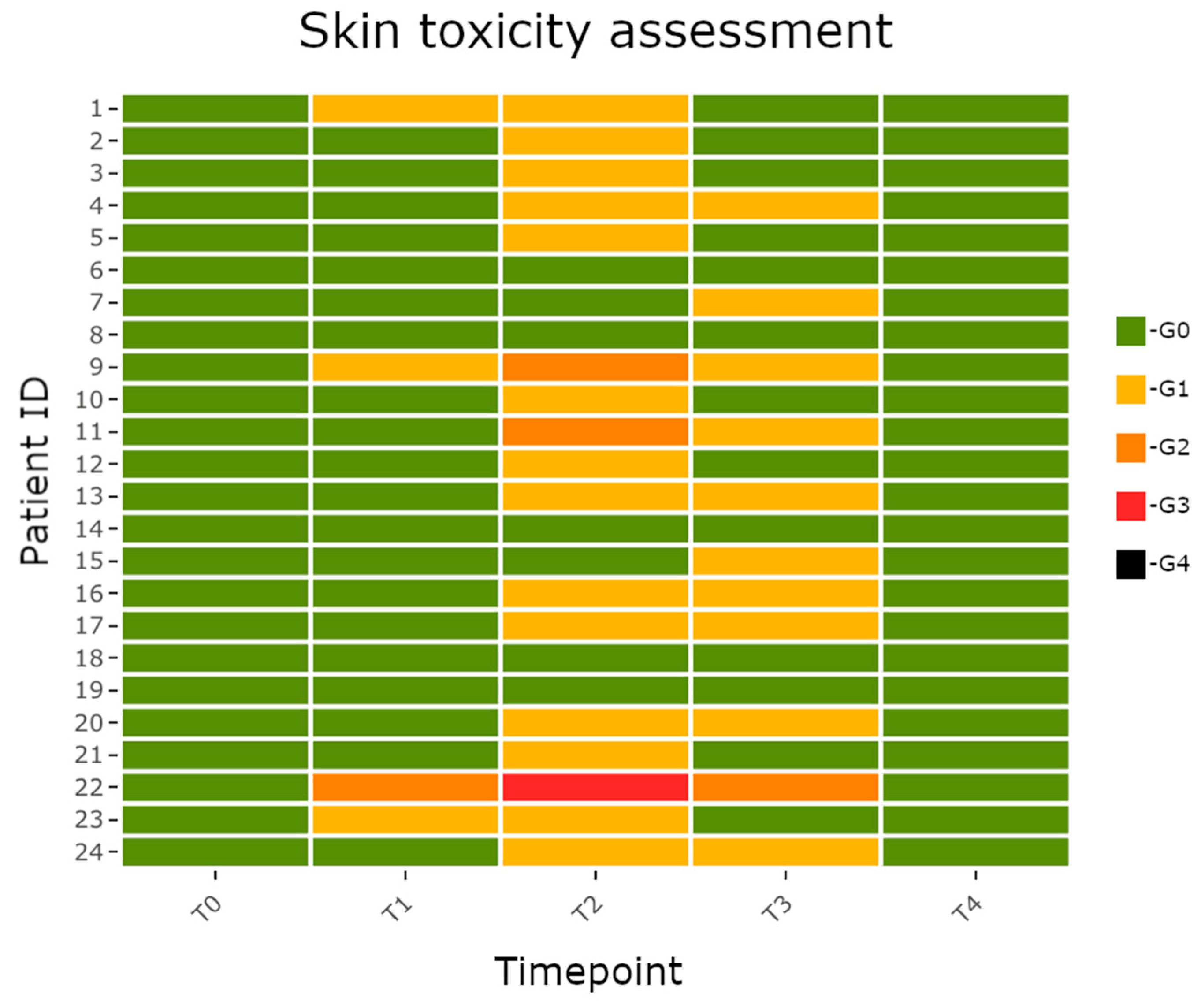
All toxicities resolved at the last assessment 30 days after the end of RT. The evolution of skin toxicities over time in all patients is represented in Figure 2. Figure 3 and Figure 4 represent, respectively, a G1 RD and the only G3 toxicity reported by us.
No patient discontinued either the skin care program due to dissatisfaction with the tested products or RT due to skin toxicity.
The correlation analysis in Table 3 shows a small and statistically significant correlation between CTV_T and skin toxicity at timepoint T3 (after 15 days from the end of RT), T = 0.36, p-value = 0.03. All other toxicity assessments at different timepoints are not correlated with CTV_T. Furthermore, no relationships were found between the toxicity grade observed at each of the five timepoints and RT target (residual breast tissue versus chest wall), dose fractionation (NF versus HF), and boost delivery (yes or no), as shown in Table 3.
No unexpected events were recorded.

4. Discussion

The cosmetics tested in our study were found to be safe and satisfactory in the prevention and treatment of RD among women undergoing adjuvant RT for BC. Nearly 30% of patients did not develop signs of RD and only 12.5% had a G2–G3 exudative dermatitis with moderate oedema at the end of treatment, with no G4 toxicity recorded. Moreover, no toxicity during treatment was severe enough to threaten patient compliance with the RT program. Once RT was completed, skin integrity was rapidly restored over the following 15 days, still supported by the two cosmetics, scoring G0–G1 toxicity in 95.3% of patients. No patient showed unexpected skin reactions or complained about any aspect of the suggested topical treatment (e.g., frequency of administration, smell, consistency, stickiness, and greasiness of the two cosmetics, etc.). Overall, these findings promote our preliminary experience as worthy of further research.
Indeed, RD is still an extremely common side effect of adjuvant RT among patients with BC, although several therapeutic options are available for its prevention [42]. The natural compounds we tested have the advantage over topical corticosteroids of generating fewer concerns regarding possible late cutaneous effects, such as skin atrophy, telangiectasias, or striae [43]. Few studies are specifically dedicated to investigating this possible cause-and-effect relationship and are unable to produce conclusive evidence on the safety of corticosteroids due to small sample sizes [44].
Both the mousse and the emulsion contain a mix of molecules with a soothing, hydrating, anti-inflammatory, and anti-itch action, which replaced the need for real drugs. Our results join those obtained with other non-steroidal topical medications, among which hyaluronic acid stands out for its effectiveness also in other tissues damaged by radiation [45,46,47,48].
There is more and more attention towards the local side effects caused by breast RT due to the growing request from patients to preserve their physical appearance as much as possible [49].
In our study we noticed how good hydration and gentle cleansing of the irradiated area before, during, and after RT had a positive effect on the skin’s tolerability to radiation.
In our clinical practice, we usually prescribe moisturizing creams to all patients with an indication for breast RT based on the experience that preventive therapy for skin side effects is more effective than treating them once they have occurred. This approach shows significant efficacy in reducing the onset of acute skin toxicity [50].
Acute RD can also affect both the quality of life and normal daily activities of patients [51], and, in some cases, can lead to unplanned treatment interruptions, thereby reducing the overall effectiveness of the therapy [16].
Apart from thorough cleansing of the skin and prevention of local trauma, there is no well-defined protocol to prevent radiation toxicity of the skin [52].
From the literature data, we know that there are some risk factors including the use of the bolus, smoking, and previous chemotherapy causing alteration of the small blood vessels [53].
The first pathophysiological stage of RD is determined by an overproduction of free radicals, responsible for damaging the stem cells in the basal layer of the epidermis, resulting in diminished turnover of epidermal cells. These cells, prompted into a state of senescence by ionizing radiation, subsequently release pro-inflammatory cytokines, responsible for initiating the inflammatory cascade. As a consequence of these effects, early RD may be observed clinically. The pathophysiology of moist desquamation involves the loss of keratinized layers and depletion of basal cell population as well. The erythema is the result of epidermal damage to basal cells and dilation of capillaries associated with increased vascular permeability [54].
These effects of radiation on the skin are influenced by several treatment factors, including, besides the total radiation dose, other dosimetric parameters (evaluable on dose–volume histograms), the dose fractionation (normofractionation vs. hypofractionation), the RT technique (3D-CRT vs. intensity-modulated RT, IMRT), and the use of an image-guided protocol for daily setup verification (IGRT). Keenan et al. provided some dose–volume constraints significantly associated with the onset of acute RD, suggesting in particular to minimize the skin volume exceeding 105% of the prescribed dose [24]. The rate of grade 2 or higher acute RD is also reduced by using hypofractionation rather than normofractionation [55]. Compared with 3D-CRT, IMRT decreases the risk of RD by improving dose homogeneity [56]. Furthermore, this reduction is greater the more complex the inverse treatment planning technique is [57]. Lastly, just as with other body targets [58], a daily setup verification by cone beam CT (CBCT) is more accurate than that by EPID and results in a significantly lower rate of acute RD, likely thanks to the more precise radiation delivery that avoids the generation of unplanned hotspots [59]. Large breast size is another known risk factor for skin toxicity [56]. However, our cohort showed no or mild skin toxicity in most cases, regardless of breast size, type of surgery (conservative vs. mastectomy), boost delivery (yes vs. no), and dose fractionation (normofractionation vs. hypofractionation). Although the level of toxicity achieved was mild for most patients, this was sufficient to draw a positive correlation (Table 3) between breast size and skin toxicity 15 days after the end of RT (T = 0.36, p-value = 0.03) highlighting a higher risk of toxicity resulting from increased breast size, according to what Freedman et al. observed [56]. Noteworthy for the same skin toxicity timepoint (T3) is the relationship with the RT target (p-value = 0.06) which is not statistically significant, probably due to the smaller size of the patients undergoing mastectomy, but which sheds light on a possible greater risk of skin toxicity for patients treated with mastectomy. No other relationship was found between the variables of interest in the study cohort; a larger and more diverse sample is mandatory to draw strong conclusions.
As regards our choice of using 3D-CRT, it should be noted that, compared to conventional tangential field irradiation, IMRT increases the integral dose to normal tissues and its potential advantage in reducing skin toxicity should be weighed against a possibly increased risk of more dreaded consequences, such as pneumonitis and secondary malignancies due to a worse exposure of OARs to low radiation doses [60,61,62]. Accordingly, the American Society for Radiation Oncology (ASTRO) does not recommend routine use of IMRT to deliver breast RT [63].
Finally, CBCT-guided IGRT protocols, although effective in reducing skin toxicity, are time-consuming and may hinder the demands of managing high patient loads in RT departments [64].
Radiation dermatitis is still a dynamic area of research, where also the achievements from nanotechnology warrant attention. Indeed, nanocarrier-based dermopharmaceutical formulations penetrate deeper than traditional topicals, potentially resulting in improved efficacy. However, further studies are needed to establish safety and evaluate cost–benefit ratios [65].
This preliminary experience has several limitations. First, the heterogeneity (i.e., for dose fractionation, type of surgery, etc.) and small size of the cohort are underpowered to find any varying effectiveness across different subgroups. Second, the lack of a placebo control group, omitted for ethical reasons in a clinical context of exceptionally frequent skin side effects, prevents quantifying the extent of the benefit deriving from the use of the two cosmetics being tested. In this regard, our pilot study should be understood as a phase I/II trial that can transition to more mature research, hopefully involving the other topical medications available for comparison.

5. Conclusions

The combination of a cleansing mousse containing vegetable glycerin (12%), phytoextract of chamomile (0.5%), yarrow phytoextract (0.5%), sweet almond (0.1%), Oenothera oil (0.1%), rice protein hydrolyzate (0.1%), and an emulsion containing micronized zinc oxide (3.7%), rapeseed phytosterols (1.7%), aloe (0.5%), 18-beta glycyrrhetinic acid (0.5%), alpha bisabolol (0.5%), zanthalene (0.5%) may emerge as an effective option to prevent and treat acute RD among BC patients undergoing adjuvant RT. Larger prospective randomized studies are needed to substantiate this first impression.

Author Contributions

Conceptualization, A.V.; methodology, A.V. and V.Z.; validation, G.F.; investigation, G.F. and E.M.; data curation, A.V. and E.M.; writing—original draft preparation, A.V.; writing—review and editing, G.F.; supervision, V.Z. All authors have read and agreed to the published version of the manuscript.

Funding

This work received no specific funding.

Institutional Review Board Statement

The study was conducted in accordance with the Declaration of Helsinki and approved by Comitato Etico Catania 2 (Approval number: 89/2022/CECT2).

Informed Consent Statement

Informed consent was obtained from all subjects involved in the study.

Data Availability Statement

The data presented in this study are available from the corresponding author upon request. The data are not publicly available due to privacy reasons.

Acknowledgments

The authors thank GD Tecnologie Interdisciplinari Farmaceutiche S.r.l. for selflessly providing patients with the tested topicals free of charge (AVEC® detergente mousse and AVEC® emulsione), through the intermediary of Giuseppe Baratta and Andrea Asero. GD was in no way involved in the study design nor influenced any aspect of the investigation.

Conflicts of Interest

The authors declare no conflicts of interest.

References

  1. Loibl, S.; André, F.; Bachelot, T.; Barrios, C.H.; Bergh, J.; Burstein, H.J.; Cardoso, M.J.; Carey, L.A.; Dawood, S.; Del Mastro, L.; et al. Early breast cancer: ESMO Clinical Practice Guideline for diagnosis, treatment and follow-up. Ann. Oncol. 2024, 35, 159–182. [Google Scholar] [CrossRef] [PubMed]
  2. Early Breast Cancer Trialists’ Collaborative Group (EBCTCG); Darby, S.; McGale, P.; Correa, C.; Taylor, C.; Arriagada, R.; Clarke, M.; Cutter, D.; Davies, C.; Ewertz, M.; et al. Effect of radiotherapy after breast-conserving surgery on 10-year recurrence and 15-year breast cancer death: Meta-analysis of individual patient data for 10,801 women in 17 randomised trials. Lancet 2011, 378, 1707–1716. [Google Scholar] [CrossRef] [PubMed]
  3. EBCTCG (Early Breast Cancer Trialists’ Collaborative Group); McGale, P.; Taylor, C.; Correa, C.; Cutter, D.; Duane, F.; Ewertz, M.; Gray, R.; Mannu, G.; Peto, R.; et al. Effect of radiotherapy after mastectomy and axillary surgery on 10-year recurrence and 20-year breast cancer mortality: Meta-analysis of individual patient data for 8135 women in 22 randomised trials. Lancet 2014, 383, 2127–2135, Erratum in Lancet 2014, 384, 1848. [Google Scholar] [CrossRef] [PubMed]
  4. Darby, S.C.; Ewertz, M.; McGale, P.; Bennet, A.M.; Blom-Goldman, U.; Brønnum, D.; Correa, C.; Cutter, D.; Gagliardi, G.; Gigante, B.; et al. Risk of ischemic heart disease in women after radiotherapy for breast cancer. N. Engl. J. Med. 2013, 368, 987–998. [Google Scholar] [CrossRef] [PubMed]
  5. Ferini, G.; Valenti, V.; Viola, A.; Umana, G.E.; Martorana, E. A Critical Overview of Predictors of Heart Sparing by Deep-Inspiration-Breath-Hold Irradiation in Left-Sided Breast Cancer Patients. Cancers 2022, 14, 3477. [Google Scholar] [CrossRef] [PubMed] [PubMed Central]
  6. Zhang, C.; Liang, Z.; Ma, S.; Liu, X. Radiotherapy and Cytokine Storm: Risk and Mechanism. Front. Oncol. 2021, 11, 670464. [Google Scholar] [CrossRef]
  7. Wei, J.; Meng, L.; Hou, X.; Qu, C.; Wang, B.; Xin, Y.; Jiang, X. Radiation-induced skin reactions: Mechanism and treatment. Cancer Manag. Res. 2018, 11, 167–177. [Google Scholar] [CrossRef] [PubMed]
  8. Vieira, L.A.C.; Menêses, A.G.; Bontempo, P.S.M.; Simino, G.P.R.; Ferreira, E.B.; Guerra, E.N.D.S.; Reis, P.E.D.D. Incidence of radiodermatitis in breast cancer patients during hypofractionated radiotherapy. Rev. Esc. Enferm. USP 2022, 56, e20220173. [Google Scholar] [CrossRef]
  9. Kry, S.F.; Smith, S.A.; Weathers, R.; Stovall, M. Skin dose during radiotherapy: A summary and general estimation technique. J. Appl. Clin. Med. Phys. 2012, 13, 20–34. [Google Scholar] [CrossRef] [PubMed] [PubMed Central]
  10. Singh, M.; Alavi, A.; Wong, R.; Akita, S. Radiodermatitis: A Review of Our Current Understanding. Am. J. Clin. Dermatol. 2016, 17, 277–292. [Google Scholar] [CrossRef] [PubMed]
  11. Sindoni, A.; Severo, C.; Vadala’, R.E.; Ferini, G.; Mazzei, M.M.; Vaccaro, M.; IatÌ, G.; Pontoriero, A.; Pergolizzi, S. Levetiracetam-induced radiation recall dermatitis in a patient undergoing stereotactic radiotherapy. J. Dermatol. 2016, 43, 1440–1441. [Google Scholar] [CrossRef] [PubMed]
  12. Gagliano, A.; Prestifilippo, A.; Cantale, O.; Ferini, G.; Fisichella, G.; Fontana, P.; Sciacca, D.; Giuffrida, D. Role of the Combination of Cyclin-Dependent Kinase Inhibitors (CDKI) and Radiotherapy (RT) in the Treatment of Metastatic Breast Cancer (MBC): Advantages and Risks in Clinical Practice. Front. Oncol. 2021, 11, 643155. [Google Scholar] [CrossRef] [PubMed] [PubMed Central]
  13. Ferini, G.; Zagardo, V.; Strazzanti, A. Acute generalized exanthematous pustulosis induced by exemestane during adjuvant radiotherapy for breast cancer. Med. Clin. 2023, 161, 502–503, (In English, Spanish). [Google Scholar] [CrossRef] [PubMed]
  14. Schnur, J.B.; Ouellette, S.C.; Dilorenzo, T.A.; Green, S.; Montgomery, G.H. A qualitative analysis of acute skin toxicity among breast cancer radiotherapy patients. Psychooncology 2011, 20, 260–268. [Google Scholar] [CrossRef] [PubMed] [PubMed Central]
  15. Martella, S.; Rietjens, M.; Lohsiriwat, V.; Lazzari, R.; Vavassori, A.; Jereczek, B.; Lazzati, V.; Leonardi, M.; Petit, J. Acute radiation dermatitis in breast cancer: Topical therapy with vitamin E acetate in lipophilic gel base. Ecancermedicalscience 2010, 4, 190. [Google Scholar] [CrossRef] [PubMed]
  16. Bese, N.S.; Hendry, J.; Jeremic, B. Effects of prolongation of overall treatment time due to unplanned interruptions during radiotherapy of different tumor sites and practical methods for compensation. Int. J. Radiat. Oncol. Biol. Phys. 2007, 68, 654–661. [Google Scholar] [CrossRef] [PubMed]
  17. Kao, Y.S.; Wu, Y.C.; Wu, M.Y.; Wu, P.L.; Lu, L.Y.; Hung, C.H. Topical Prevention of Radiation Dermatitis in Breast Cancer Patients: A Network Meta-analysis of Randomized Controlled Trials. In Vivo 2023, 37, 1346–1357. [Google Scholar] [CrossRef] [PubMed]
  18. Lee, S.F.; Shariati, S.; Caini, S.; Wong, H.; Chan, A.W.; Gojsevic, M.; Ogita, M.; Ye, J.C.; Chia, D.; Chao, M.; et al. StrataXRT for the prevention of acute radiation dermatitis in breast cancer: A systematic review and meta-analysis of randomized controlled trials. Support. Care Cancer 2023, 31, 515. [Google Scholar] [CrossRef] [PubMed]
  19. Finkelstein, S.; Kanee, L.; Behroozian, T.; Wolf, J.R.; van den Hurk, C.; Chow, E.; Bonomo, P. Comparison of clinical practice guidelines on radiation dermatitis: A narrative review. Support. Care Cancer 2022, 30, 4663–4674. [Google Scholar] [CrossRef] [PubMed]
  20. Robijns, J.; Becherini, C.; Caini, S.; Wolf, J.R.; van den Hurk, C.; Beveridge, M.; Lam, H.; Bonomo, P.; Chow, E.; Behroozian, T. Natural and miscellaneous agents for the prevention of acute radiation dermatitis: A systematic review and meta-analysis. Support. Care Cancer 2023, 31, 195. [Google Scholar] [CrossRef] [PubMed]
  21. Ferini, G.; Molino, L.; Tripoli, A.; Valenti, V.; Illari, S.I.; Marchese, V.A.; Cravagno, I.R.; Borzi, G.R. Anatomical Predictors of Dosimetric Advantages for Deep-inspiration-breath-hold 3D-conformal Radiotherapy among Women with Left Breast Cancer. Anticancer Res. 2021, 41, 1529–1538. [Google Scholar] [CrossRef] [PubMed]
  22. Offersen, B.V.; Boersma, L.J.; Kirkove, C.; Hol, S.; Aznar, M.C.; Biete Sola, A.; Kirova, Y.M.; Pignol, J.P.; Remouchamps, V.; Verhoeven, K.; et al. ESTRO consensus guideline on target volume delineation for elective radiation therapy of early stage breast cancer. Radiother. Oncol. 2015, 114, 3–10. [Google Scholar] [CrossRef] [PubMed]
  23. International Comission on Radiation Unit and Measurment (ICRU). Prescribing, Recording, and Reporting Photon Beam Therapy; ICRU Report No.50; International Comission on Radiation Unit and Measurment (ICRU): Bethesda, MD, USA, 1993; pp. 1–72. [Google Scholar]
  24. Keenan, L.G.; Lavan, N.; Dunne, M.; McArdle, O. Modifiable risk factors for acute skin toxicity in adjuvant breast radiotherapy: Dosimetric analysis and review of the literature. Med. Dosim. 2019, 44, 51–55. [Google Scholar] [CrossRef] [PubMed]
  25. Milani, M.; Sparavigna, A. The 24-h skin hydration and barrier function effects of a hyaluronic 1%, glycerin 5%, and Centella asiatica stem cells extract moisturizing fluid: An intra-subject, randomized, assessor-blinded study. Clin. Cosmet. Investig. Dermatol. 2017, 10, 311–315. [Google Scholar] [CrossRef] [PubMed]
  26. Mailänder, L.K.; Lorenz, P.; Bitterling, H.; Stintzing, F.C.; Daniels, R.; Kammerer, D.R. Phytochemical Characterization of Chamomile (Matricaria recutita L.) Roots and Evaluation of Their Antioxidant and Antibacterial Potential. Molecules 2022, 27, 8508. [Google Scholar] [CrossRef]
  27. Tadić, V.; Arsić, I.; Zvezdanović, J.; Zugić, A.; Cvetković, D.; Pavkov, S. The estimation of the traditionally used yarrow (Achillea millefolium L. Asteraceae) oil extracts with anti-inflamatory potential in topical application. J. Ethnopharmacol. 2017, 199, 138–148. [Google Scholar] [CrossRef] [PubMed]
  28. Zeichner, J.A.; Berson, D.; Donald, A. The Use of an Over-the-Counter Hand Cream with Sweet Almond Oil for the Treatment of Hand Dermatitis. J. Drugs Dermatol. 2018, 17, 78–82. [Google Scholar] [PubMed]
  29. Timoszuk, M.; Bielawska, K.; Skrzydlewska, E. Evening Primrose (Oenothera biennis) Biological Activity Dependent on Chemical Composition. Antioxidants 2018, 7, 108. [Google Scholar] [CrossRef] [PubMed]
  30. Chen, H.J.; Dai, F.J.; Chen, C.Y.; Fan, S.L.; Zheng, J.H.; Huang, Y.C.; Chau, C.F.; Lin, Y.S.; Chen, C.S. Evaluating the Antioxidants, Whitening and Antiaging Properties of Rice Protein Hydrolysates. Molecules 2021, 26, 3605. [Google Scholar] [CrossRef] [PubMed]
  31. Holmes, A.M.; Song, Z.; Moghimi, H.R.; Roberts, M.S. Relative Penetration of Zinc Oxide and Zinc Ions into Human Skin after Application of Different Zinc Oxide Formulations. ACS Nano 2016, 10, 1810–1819. [Google Scholar] [CrossRef]
  32. Tileuberdi, N.; Turgumbayeva, A.; Yeskaliyeva, B.; Sarsenova, L.; Issayeva, R. Extraction, Isolation of Bioactive Compounds and Therapeutic Potential of Rapeseed (Brassica napus L.). Molecules 2022, 27, 8824. [Google Scholar] [CrossRef] [PubMed]
  33. Hekmatpou, D.; Mehrabi, F.; Rahzani, K.; Aminiyan, A. The Effect of Aloe Vera Clinical Trials on Prevention and Healing of Skin Wound: A Systematic Review. Iran. J. Med. Sci. 2019, 44, 1–9. [Google Scholar] [PubMed]
  34. Kowalska, A.; Kalinowska-Lis, U. 18β-Glycyrrhetinic acid: Its core biological properties and dermatological applications. Int. J. Cosmet. Sci. 2019, 41, 325–331. [Google Scholar] [CrossRef] [PubMed]
  35. Eddin, L.B.; Jha, N.K.; Goyal, S.N.; Agrawal, Y.O.; Subramanya, S.B.; Bastaki, S.M.A.; Ojha, S. Health Benefits, Pharmacological Effects, Molecular Mechanisms, and Therapeutic Potential of α-Bisabolol. Nutrients 2022, 14, 1370. [Google Scholar] [CrossRef] [PubMed]
  36. Artaria, C.; Maramaldi, G.; Bonfigli, A.; Rigano, L.; Appendino, G. Lifting properties of the alkamide fraction from the fruit husks of Zanthoxylum bungeanum. Int. J. Cosmet. Sci. 2011, 33, 328–333. [Google Scholar] [CrossRef]
  37. Cox, J.D.; Stetz, J.; Pajak, T.F. Toxicity criteria of the Radiation Therapy Oncology Group (RTOG) and the European Organization for Research and Treatment of Cancer (EORTC). Int. J. Radiat. Oncol. Biol. Phys. 1995, 31, 1341–1346. [Google Scholar] [CrossRef] [PubMed]
  38. R Core Team. A Language and Environment for Statistical Computing; R Foundation for Statistical Computing: Vienna, Austria, 2014. [Google Scholar]
  39. R Studio Team. Integrated Development Environment for R; RStudio, PBC: Boston, MA, USA, 2021; Available online: http://www.rstudio.com/ (accessed on 29 May 2024).
  40. Galili, T.; O’Callaghan, A.; Sidi, J.; Sievert, C. heatmaply: An R package for creating interactive cluster heatmaps for online publishing. Bioinformatics 2018, 34, 1600–1602. [Google Scholar] [CrossRef] [PubMed] [PubMed Central]
  41. Wickham, H. ggplot2: Elegant Graphics for Data Analysis; Springer: New York, NY, USA, 2016; Available online: https://doi.org/10.1007/978-3-319-24277-4./ (accessed on 9 August 2023).
  42. Behroozian, T.; Bonomo, P.; Patel, P.; Kanee, L.; Finkelstein, S.; van den Hurk, C.; Chow, E.; Wolf, J.R.; Multinational Association of Supportive Care in Cancer (MASCC) Oncodermatology Study Group Radiation Dermatitis Guidelines Working Group. Multinational Association of Supportive Care in Cancer (MASCC) clinical practice guidelines for the prevention and management of acute radiation dermatitis: International Delphi consensus-based recommendations. Lancet Oncol. 2023, 24, e172–e185. [Google Scholar] [CrossRef] [PubMed]
  43. Alves, R.O.; Boin, M.F.; Crocco, E.I. Striae after topical corticosteroid: Treatment with nonablative fractional laser 1540 nm. J. Cosmet. Laser Ther. 2015, 17, 143–147. [Google Scholar] [CrossRef] [PubMed]
  44. Ulff, E.; Maroti, M.; Serup, J.; Nilsson, M.; Falkmer, U. Late cutaneous effects of a local potent steroid during adjuvant radiotherapy for breast cancer. Clin. Transl. Radiat. Oncol. 2017, 7, 9–12. [Google Scholar] [CrossRef] [PubMed] [PubMed Central]
  45. Lee, C.J.; Fang, H.F.; Wang, C.Y.; Chou, K.R.; Huang, T.W. Effect of hyaluronic acid on radiodermatitis in patients with breast cancer: A meta-analysis of randomized controlled trials. Support. Care Cancer 2022, 30, 3965–3975. [Google Scholar] [CrossRef] [PubMed]
  46. Tai, R.Z.; Loh, E.W.; Tsai, J.T.; Tam, K.W. Effect of hyaluronic acid on radiotherapy-induced mucocutaneous side effects: A meta-analysis of randomized controlled trials. Support. Care Cancer 2022, 30, 4845–4855. [Google Scholar] [CrossRef] [PubMed]
  47. Shao, Y.; Lu, G.L.; Shen, Z.J. Comparison of intravesical hyaluronic acid instillation and hyperbaric oxygen in the treatment of radiation-induced hemorrhagic cystitis. BJU Int. 2012, 109, 691–694. [Google Scholar] [CrossRef] [PubMed]
  48. Ferini, G.; Tripoli, A.; Umina, V.; Borzì, G.R.; Marchese, V.A.; Illari, S.I.; Cacciola, A.; Lillo, S.; Parisi, S.; Valenti, V. Radiation Proctitis: The Potential Role of Hyaluronic Acid in the Prevention and Restoration of Any Damage to the Rectal Mucosa among Prostate Cancer Patients Submitted to Curative External Beam Radiotherapy. Gastroenterol. Insights 2021, 12, 446–455. [Google Scholar] [CrossRef]
  49. Brunault, P.; Suzanne, I.; Trzepidur-Edom, M.; Garaud, P.; Calais, G.; Toledano, A.; Camus, V. Depression is associated with some patient-perceived cosmetic changes, but not with radiotherapy-induced late toxicity, in long-term breast cancer survivors. Psychooncology 2013, 22, 590–597. [Google Scholar] [CrossRef] [PubMed]
  50. Rose, P. Radiation-induced skin toxicity: Prophylaxis or management? J. Med. Radiat. Sci. 2020, 67, 168–169. [Google Scholar] [CrossRef] [PubMed] [PubMed Central]
  51. Rzepecki, A.; Birnbaum, M.; Ohri, N.; Daily, J.; Fox, J.; Bodner, W.; Kabarriti, R.; Garg, M.; Mehta, K.; Kalnicki, S.; et al. Characterizing the effects of radiation dermatitis on quality of life: A prospective survey-based study. J. Am. Acad. Dermatol. 2022, 86, 161–163. [Google Scholar] [CrossRef] [PubMed]
  52. Chan, D.C.W.; Wong, H.C.Y.; Riad, M.A.; Caini, S.; Wolf, J.R.; van den Hurk, C.; Beveridge, M.; Lam, H.; Bonomo, P.; Chow, E.; et al. Prevention of radiation dermatitis with skin hygiene and washing: A systematic review and meta-analysis. Support. Care Cancer 2023, 31, 294, Erratum in Support. Care Cancer 2024, 32, 107. [Google Scholar] [CrossRef] [PubMed]
  53. Xie, Y.; Wang, Q.; Hu, T.; Chen, R.; Wang, J.; Chang, H.; Cheng, J. Risk Factors Related to Acute Radiation Dermatitis in Breast Cancer Patients after Radiotherapy: A Systematic Review and Meta-Analysis. Front. Oncol. 2021, 11, 738851. [Google Scholar] [CrossRef] [PubMed] [PubMed Central]
  54. Rübe, C.E.; Freyter, B.M.; Tewary, G.; Roemer, K.; Hecht, M.; Rübe, C. Radiation Dermatitis: Radiation-Induced Effects on the Structural and Immunological Barrier Function of the Epidermis. Int. J. Mol. Sci. 2024, 25, 3320. [Google Scholar] [CrossRef] [PubMed] [PubMed Central]
  55. Bruand, M.; Salleron, J.; Guihard, S.; Crety, C.M.; Liem, X.; Pasquier, D.; Lamrani-Ghaouti, A.; Charra-Brunaud, C.; Peiffert, D.; Clavier, J.B.; et al. Acute skin toxicity of conventional fractionated versus hypofractionated radiotherapy in breast cancer patients receiving regional node irradiation: The real-life prospective multicenter HYPOBREAST cohort. BMC Cancer 2022, 22, 1318. [Google Scholar] [CrossRef] [PubMed] [PubMed Central]
  56. Freedman, G.M.; Anderson, P.R.; Li, J.; Eisenberg, D.F.; Hanlon, A.L.; Wang, L.; Nicolaou, N. Intensity modulated radiation therapy (IMRT) decreases acute skin toxicity for women receiving radiation for breast cancer. Am. J. Clin. Oncol. 2006, 29, 66–70. [Google Scholar] [CrossRef] [PubMed]
  57. Joseph, K.; Vos, L.J.; Gabos, Z.; Pervez, N.; Chafe, S.; Tankel, K.; Warkentin, H.; Ghosh, S.; Amanie, J.; Powell, K.; et al. Skin Toxicity in Early Breast Cancer Patients Treated with Field-In-Field Breast Intensity-Modulated Radiotherapy versus Helical Inverse Breast Intensity-Modulated Radiotherapy: Results of a Phase III Randomised Controlled Trial. Clin. Oncol. 2021, 33, 30–39. [Google Scholar] [CrossRef] [PubMed]
  58. Ferini, G.; Tripoli, A.; Molino, L.; Cacciola, A.; Lillo, S.; Parisi, S.; Umina, V.; Illari, S.I.; Marchese, V.A.; Cravagno, I.R.; et al. How Much Daily Image-guided Volumetric Modulated Arc Therapy Is Useful for Proctitis Prevention with Respect to Static Intensity Modulated Radiotherapy Supported by Topical Medications among Localized Prostate Cancer Patients? Anticancer Res. 2021, 41, 2101–2110, Erratum in Anticancer Res. 2022, 42, 5165. [Google Scholar] [CrossRef] [PubMed]
  59. Lin, J.C.; Tsai, J.T.; Chou, Y.C.; Li, M.H.; Liu, W.H. Compared with intensity-modulated radiotherapy, image-guided radiotherapy reduces severity of acute radiation-induced skin toxicity during radiotherapy in patients with breast cancer. Cancer Med. 2018, 7, 3622–3629. [Google Scholar] [CrossRef] [PubMed] [PubMed Central]
  60. Chen, S.N.; Ramachandran, P.; Deb, P. Dosimetric comparative study of 3DCRT, IMRT, VMAT, Ecomp, and Hybrid techniques for breast radiation therapy. Radiat. Oncol. J. 2020, 38, 270–281. [Google Scholar] [CrossRef] [PubMed] [PubMed Central]
  61. Aznar, M.C.; Duane, F.K.; Darby, S.C.; Wang, Z.; Taylor, C.W. Exposure of the lungs in breast cancer radiotherapy: A systematic review of lung doses published 2010–2015. Radiother. Oncol. 2018, 126, 148–154. [Google Scholar] [CrossRef] [PubMed] [PubMed Central]
  62. Hall, E.J.; Wuu, C.S. Radiation-induced second cancers: The impact of 3D-CRT and IMRT. Int. J. Radiat. Oncol. Biol. Phys. 2003, 56, 83–88. [Google Scholar] [CrossRef] [PubMed]
  63. Smith, B.D.; Bellon, J.R.; Blitzblau, R.; Freedman, G.; Haffty, B.; Hahn, C.; Halberg, F.; Hoffman, K.; Horst, K.; Moran, J.; et al. Radiation therapy for the whole breast: Executive summary of an American Society for Radiation Oncology (ASTRO) evidence-based guideline. Pract. Radiat. Oncol. 2018, 8, 145–152. [Google Scholar] [CrossRef] [PubMed]
  64. Petillion, S.; Verhoeven, K.; Weltens, C.; Van den Heuvel, F. Accuracy of a new paired imaging technique for position correction in whole breast radiotherapy. J. Appl. Clin. Med. Phys. 2015, 16, 4796. [Google Scholar] [CrossRef] [PubMed] [PubMed Central]
  65. Cláudia Paiva-Santos, A.; Gama, M.; Peixoto, D.; Sousa-Oliveira, I.; Ferreira-Faria, I.; Zeinali, M.; Abbaspour-Ravasjani, S.; Mascarenhas-Melo, F.; Hamishehkar, H.; Veiga, F. Nanocarrier-based dermopharmaceutical formulations for the topical management of atopic dermatitis. Int. J. Pharm. 2022, 618, 121656, Erratum in Int. J. Pharm. 2022, 627, 122146. [Google Scholar] [CrossRef] [PubMed]
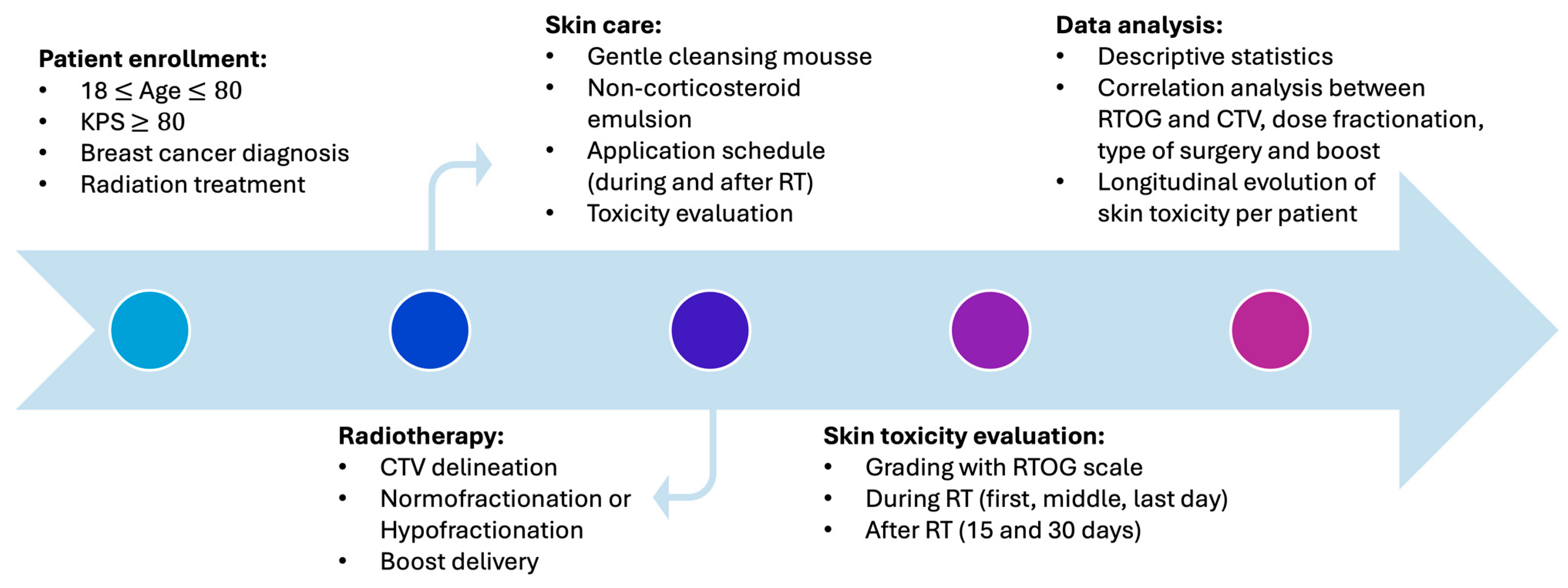
Figure 1. Study flow-chart.
Cosmetics 11 00117 g001
Figure 2. Evolution of skin toxicities over time in all patients. Here T0, T1, T2, T3, and T4 represent, respectively, the toxicity levels at RT start, half RT completed, end of RT, 15 and 30 days after RT completion.
Cosmetics 11 00117 g002
Figure 3. Appearance of the skin breast before (A), on the last day of RT (B), and 15 days after its completion (C) in a patient who experienced G1 RD.
Cosmetics 11 00117 g003
Figure 4. Appearance of the skin breast before (A), on the last day of RT (B), and 15 days after its completion (C) in the patient who experienced G3 RD.
Cosmetics 11 00117 g004
Table 1. RTOG score and corresponding skin changes.
RTOG ScoreSkin Changes
0No change over baseline
1Follicular, faint or dull erythema/epilation/dry desquamation/decreased sweating
2Tender or bright erythema, patchy moist desquamation/moderate edema
3Confluent, moist desquamation other than skin folds, pitting edema
4Ulceration, hemorrhage, necrosis
Table 2. Patient and treatment characteristics.
Patients % (n)
Age, median (range) years59 (42–75)
Sex
      Male0
      Female100 (24)
Surgery treatment before radiotherapy
      Breast-conserving surgery87.5 (21)
      Mastectomy12.5 (3)
Involved side
      Right breast50 (12)
      Left breast50 (12)
CTV_T size, median (range) cc541 (406.1–763.5)
Radiotherapy scheme
      Normofractionation75 (18)
      Hypofractionation25 (6)
Boost delivery75 (18)
      10 Gy in 5 fractions83.3 (15)
      9 Gy in 3 frations16.7 (3)
Table 3. Relationship analysis between RD grade (T1, T2, T3) and CTV_T size, RT target, dose fractionation, and boost delivery.
Correlation AnalysisToxicity T1Toxicity T2Toxicity T3
Kendall’s correlation
T (p-value)
CTV_T (cc)0.26 (0.13)0.29 (0.07)0.36 (0.03)
Fisher’s test
p-value
RT target0.150.170.06
Dose fractionation0.370.430.19
Boost delivery0.220.180.38
Disclaimer/Publisher’s Note: The statements, opinions and data contained in all publications are solely those of the individual author(s) and contributor(s) and not of MDPI and/or the editor(s). MDPI and/or the editor(s) disclaim responsibility for any injury to people or property resulting from any ideas, methods, instructions or products referred to in the content.