Review

An Updated Etiology of Hair Loss and the New Cosmeceutical Paradigm in Therapy: Clearing ‘the Big Eight Strikes’

1
Department of Botany and Plant Biotechnology, Auckland Park Campus, University of Johannesburg, Auckland Park, P.O. Box 524, Johannesburg 2006, South Africa
2
Systems Trichology London Ltd., 17 Blanchard Close, London SE9 4TZ, UK
3
WETHRIVVTM, 3805 Old Easton Road #305, Doylestown, PA 18902, USA
4
Rapaport Hair Institute, Englewood Cliffs, NJ 07632, USA
*
Author to whom correspondence should be addressed.
Cosmetics 2023, 10(4), 106; https://doi.org/10.3390/cosmetics10040106
Submission received: 23 March 2023 / Revised: 12 May 2023 / Accepted: 16 June 2023 / Published: 18 July 2023
(This article belongs to the Special Issue Active Substances and Bioavailability in Cosmetics)

Abstract

:
In this current review, research spanning the last decade (such as transcriptomic studies, phenotypic observations, and confirmed comorbidities) has been synthesized into an updated etiology of hair loss and applied to the new cosmeceutical paradigm of hair rejuvenation. The major etiological components in scalps with hair loss are denoted as the ‘big eight strikes’, which include the following: androgens, prostaglandins, overactive aerobic metabolism of glucose, bacterial or fungal over-colonization, inflammation, fibrosis, metabolism or circulation problems, and malnutrition. The relevance of the ‘big eight’ to nine categories of hair loss is explained. In cases of androgenetic alopecia or female pattern hair loss, both elevated DHT and increased frequency of androgen receptors lead to problems with the metabolism of glucose (sugar), redox imbalance, disruption to the electron transport chain, and PPAR-γ overactivity (the latter is unique to androgenetic alopecia, where the reverse occurs in other types of hair loss). These etiological factors and others from ‘the big eight’ are the focal point of our hypothetical narrative of the attenuative mechanisms of commercial cosmeceutical hair serums. We conclude that cosmeceuticals with the potential to improve all eight strikes (according to published in vitro or clinical data) utilize bioactive peptides and plant compounds that are either flavonoids (isoflavones, procyanidins, flavanols, and flavonols) or sterols/triterpenes. It is noteworthy that many therapeutic interventions are generic to the multiple types of hair loss. Lastly, suggestions are made on how scalp and hair health can be improved by following the cosmeceutical approach.

1. Introduction

A normal healthy hair follicle grows for several years before it is replaced. This process occurs in four major developmental phases, which are anagen, catagen, telogen, and exogen [1]. The anagen phase is when the hair follicle is actively growing. If a person experiences short periods of stress or illness, it is normal for their hair follicles to experience temporary phases of non-growth. If the stress is not too severe, the anagen phase continues for 3–5 years until the club hair is completely formed.
On a healthy scalp, most hairs are in the anagen phase, leaving only 6–8% of hairs that have completed the anagen phase and are undergoing the process of returning to anagen. Immediately as the anagen phase ends, the catagen phase starts, which lasts approximately 10–13 days. During this phase, the hair follicle transforms into a dormant follicle, known as a telogen follicle. The telogen phase can last for up to three months before the follicle releases the hair strand (sheds the hair) and starts creating a new hair strand. The release of the hair strand and reforming of the hair follicle is known as the exogen phase [2]. At the end of the exogen phase, the anagen phase starts once again.
Approximately 100 hairs are shed per day, but at the early stages of hair loss disorders, this number is significantly higher due to the interruption of the normal hair growth cycle. The most significant problem is an interruption to the anagen phase. There are several types of hair loss that may involve premature entry into the telogen phase or complete destruction of the hair follicle, either at the bulb or the bulge region of the pilosebaceous unit.
In the classical view of hair loss, there are nine basic types that are named according to the pattern and cause. These are presented in Table 1. It is noteworthy that many of the symptoms overlap between the types of hair loss, specifically inflammation, scarring, and bacterial/fungal overgrowth [3,4,5,6]. Most hair loss disorders that are chronic have these symptoms.
The information provided in Table 1 is a simplified guideline for the types of hair loss disorders, but scalps with hair loss are often afflicted by multiple types of hair loss. For example, the most common cause of hair loss is androgenetic alopecia (AGA) or female pattern hair loss, but fibrosis occurs at advanced stages, possibly due to a comorbidity, such as a mild form of scarring alopecia.
Scarring alopecia is another major type of hair loss that results in the accumulation of collagenous tissue in the place of hair follicles. It is classified according to its pattern and the dominant type of white blood cell in the bulge region of the hair follicle. For example, primary cicatricial alopecia has no distinct pattern of hair loss over the scalp, but central centrifugal cicatricial alopecia is restricted to the vertex or areas where AGA occurs. In another example, frontal fibrosing alopecia is when hair loss occurs in the frontal scalp or eyebrows [21,22].
Aside from these patterns of hair loss, the types of white blood cells and other factors determine what type of dermatological condition caused the hair follicles to die or enter telogen. Some of the dermatological conditions include lichen planopilaris, follicular decalvans, lichen planus, discoid lupus erythematosus, pseudopelade (patchy scarring alopecia), and pseudopelade of Brocq. As previously mentioned, these conditions sometimes occur together with AGA or other types of hair loss. For example, traction alopecia can occur with scarring alopecia [10] or AGA. Furthermore, AGA can occur with chronic telogen effluvium [23] or with scarring alopecia [24]. Sometimes comorbidities of AGA are caused by the misguided use of dangerous cosmeceuticals, such as oil-dominated serums. Unfortunately, without proper guidelines, people can increase the severity of hair loss and promote scarring alopecia by experimenting with topical serums or oils that are not suited to their pathophysiological status [25].
Aside from hair loss that was caused by external forces leading to injury (i.e., mental stress, chemotherapy, tight hairstyles), most cases involve an autoimmune or inflammatory aspect. But this aspect is less severe in cases of AGA or telogen effluvium, albeit present.
To determine the difference between AGA, telogen effluvium, alopecia areata, or scarring alopecia, diagnostic features on the scalp surface can be used, as well as the character of the epilated hairs. Scalp surface or epilated hairs are photographed (using phototrichoscan or other methods), their features are observed, and the types of hair strands are counted, then a ratio is calculated. Examples of diagnostic features include a high number of vellus hairs (pronounced differences in hair diameter) with formed hair bulbs. This indicates AGA, whereas disfigurement of the hair bulb, or no hair bulb, can indicate alopecia areata [26,27,28].
The closer the examination of hair and scalp, the more complex the diagnosis becomes. However, for the sake of brevity and simplicity, the nine hair loss disorders in Table 1 can be condensed into just the following five categories: (1) reversible autoimmune, (2) androgenetic, (3) autoimmune scarring, (4) fungal/bacterial, and (5) stress/trauma (chemical, psychological, and physical).

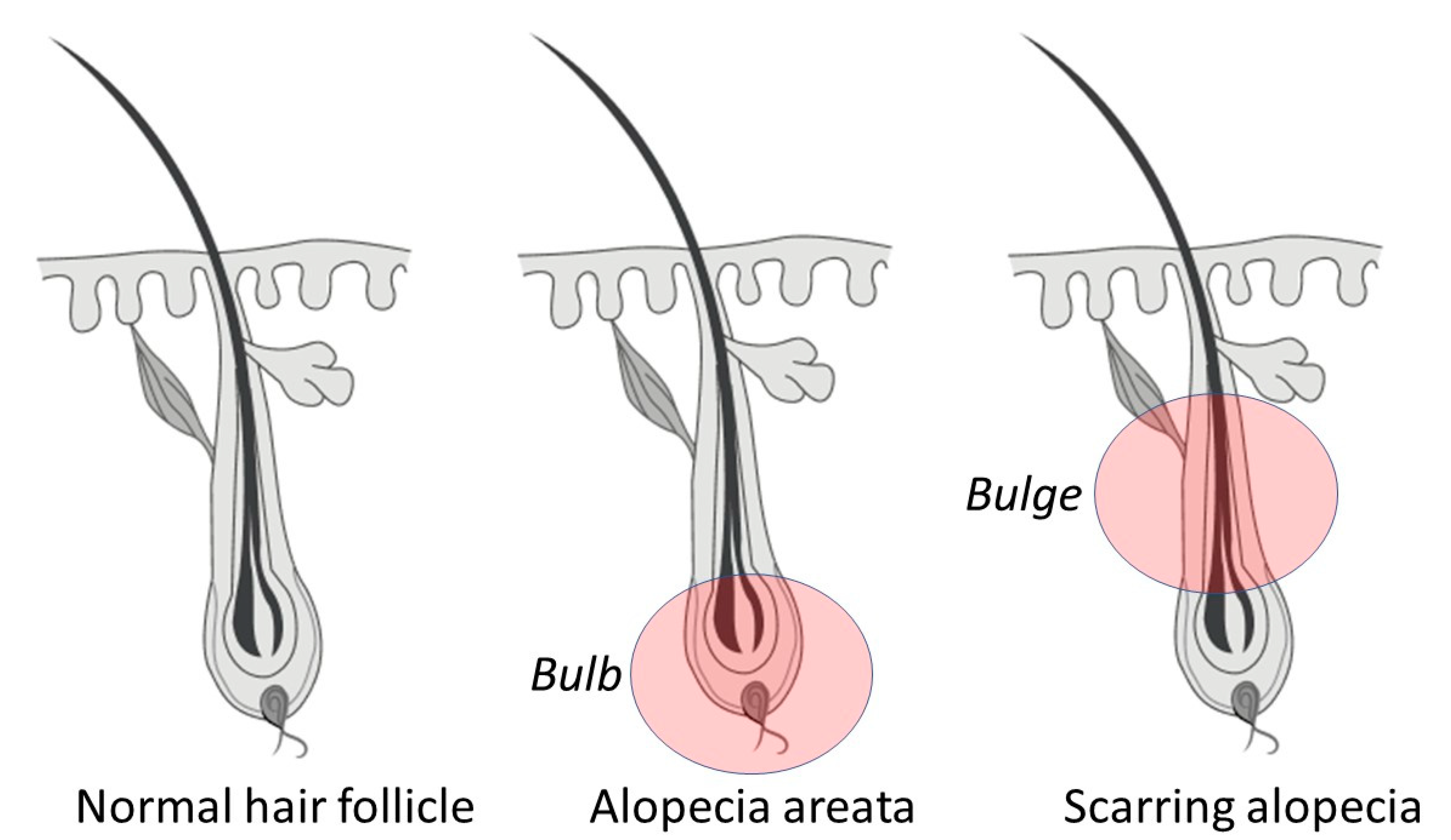
1.1. Reversible Autoimmune: Alopecia Areata

Hair follicles that are affected by alopecia areata (AA) are inflamed due to the increased expression of proinflammatory cytokines, especially inter-feron-c (IFN-c), resulting from activated T cells [29]. The part of the follicle that is affected is the bulb (Figure 1), where dermal papilla cells are differentiated. The reason for this autoimmune attack on hair follicles has not been clarified via research or empirical observation. There are several theories that have strong support from reproducible observations, but reproducibility is far below 100%, creating confusion and controversy over the pathogenesis of AA. However, this may be related to the occurrence of comorbidities, which makes observing the biological aspect of the disease complicated.
To make things more complicated, AA has several different phenotypic presentations, suggesting that it has different causes in different people [29]. Nevertheless, in all cases, anti-inflammatory therapies (and dietary modification) have helped in resolving symptoms. For example, in a study of 60 patients with AA, minoxidil and an anti-inflammatory composition of piperine (from black pepper), capsaicin (from chili), and curcumin (from turmeric) were nearly equally effective in the rejuvenation of hair [30].
While there are several hypotheses on the etiology of AA, most evidence suggests there is a compromise of immune privilege of the hair follicle’s anagen antigens. This is called the ‘immune privilege collapse’ theory. Immune privilege collapse leads to the reversible destruction of the hair follicle in the bulb region by active T cells [31].
The immune privilege collapse theory is supported by the observation of the behavior of CD4 and cytotoxic CD8 T cells that were taken from the scalps of AA patients. These cells attack epitopes from hair follicle keratinocytes and melanocytes. When the same types of T cells are taken from non-AA patients, they are less likely to attack those epitopes [32].
In another study, scalp grafts were taken from AA patients and grafted to immunosuppressed mice. Hair returned to normal growth. T cells were then taken from the same AA patients and activated using melanocyte-peptide epitopes from their scalps. They were then injected into the mice with successful scalp grafts. The hair follicles stopped producing hair strands on the backs of the mice [33].
This observation indicates that the epitopes from AA patients were like autoantigens. In the same study, the authors identified that the HLA-A2-presenting protein is possibly linked to this. While the HLA-A2-presenting protein is present in the hair follicles of half the human population [33], it is believed that AA includes multiple etiological components, with a strong lifestyle aspect. This is because dietary modification and anti-inflammatory therapies have created a moderate to profound recovery in many case studies.

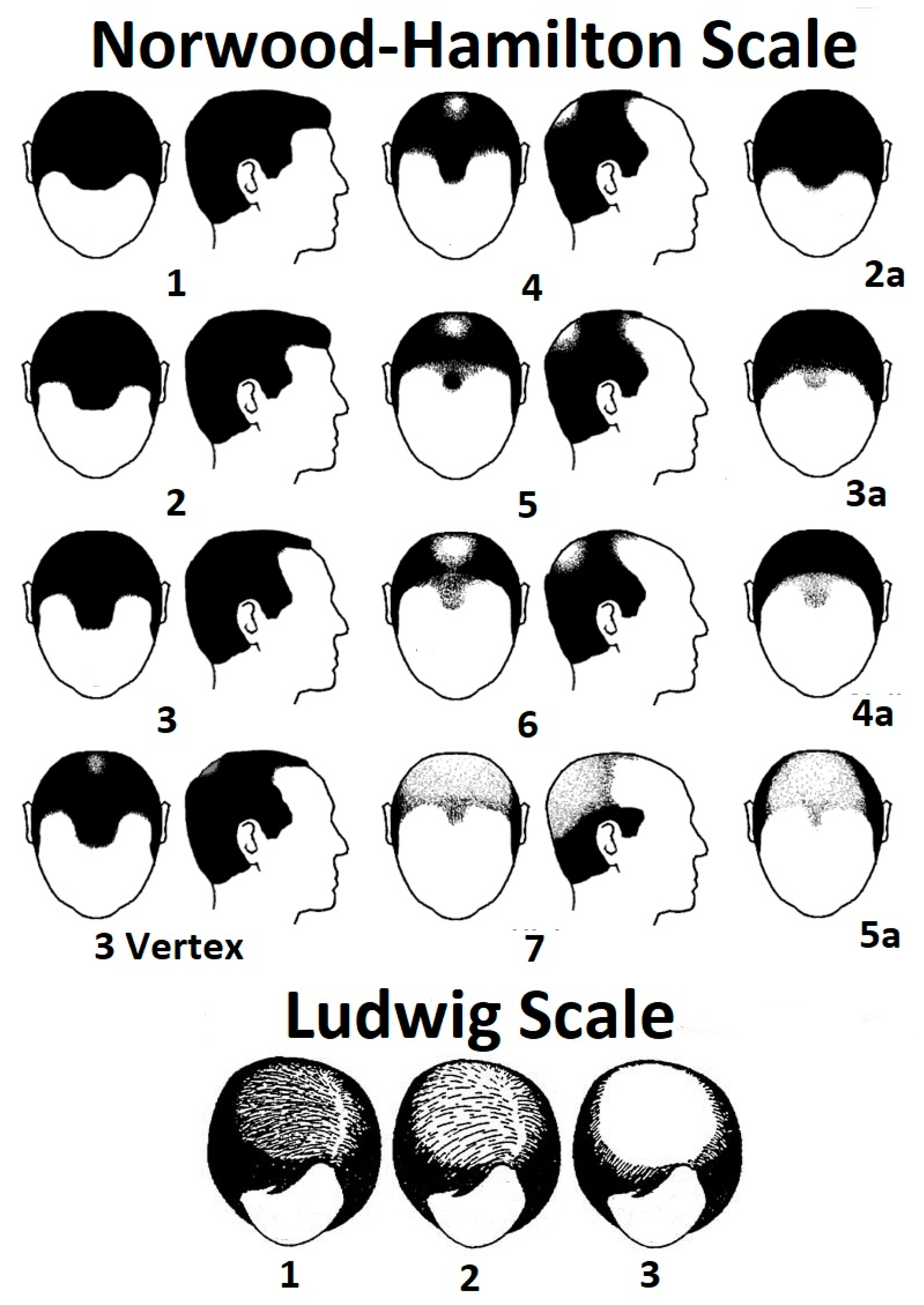
1.2. Androgenetic: Androgenetic Alopecia (AGA: Pattern Hair Loss)

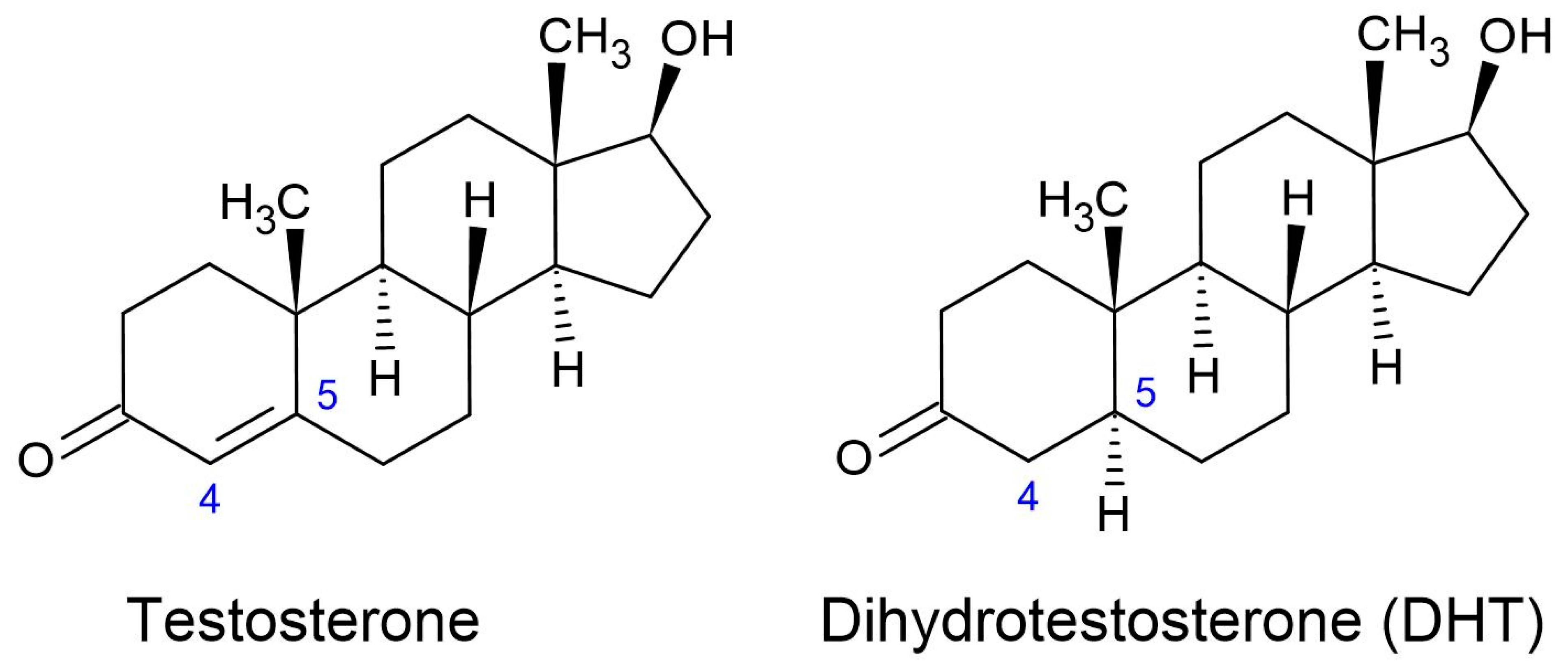
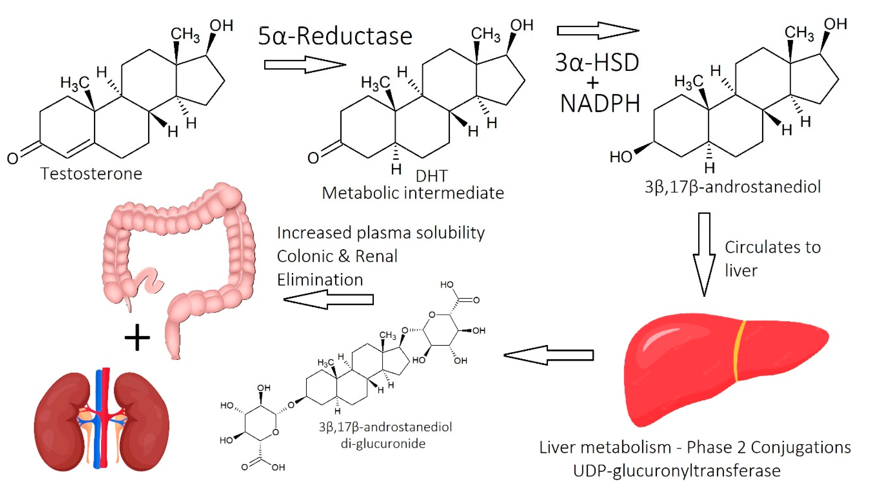
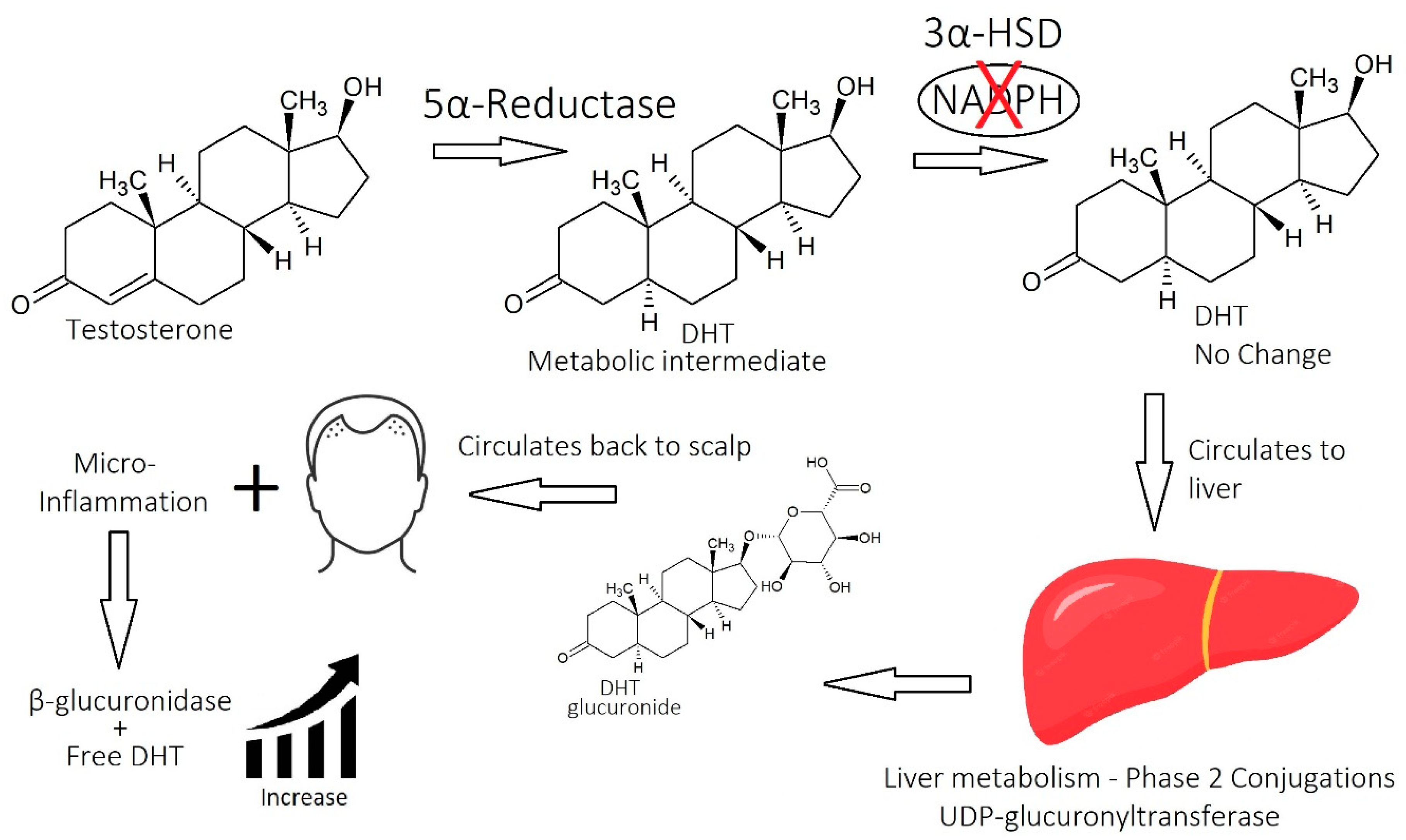
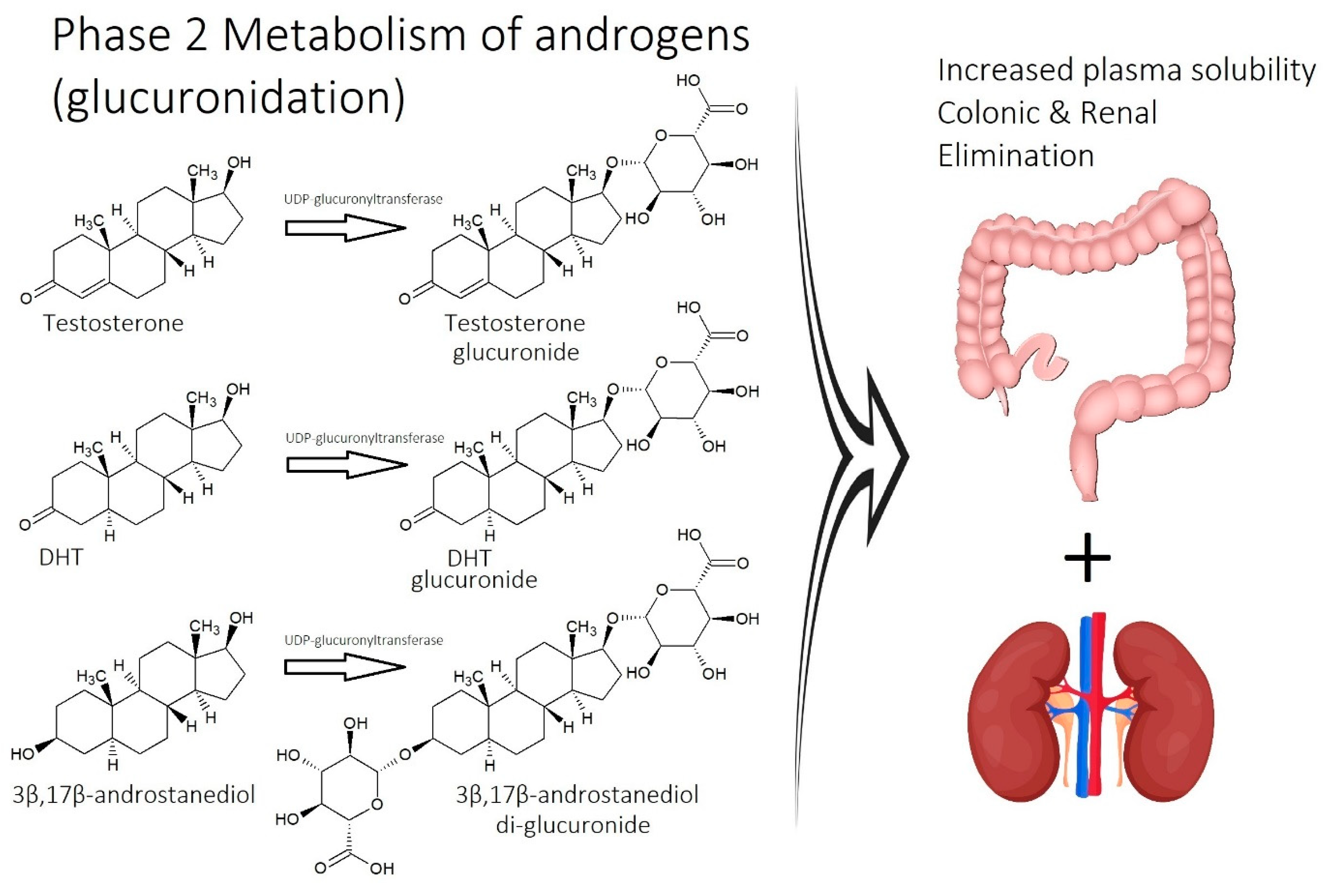
1.2.1. Dihydrotestosterone (DHT)

The only hair loss disorder that can manifest all eight strikes, described in this current commentary (Section 2), is AGA. This is because the first one of the eight strikes is an androgen imbalance that is caused by excess dihydrotestosterone (DHT) in the scalp tissue. While this is more common in males, AGA afflicts both males and females, but in the latter, it is broadly called female pattern hair loss to be inclusive of both androgen-dependent and -independent phenotypes. In both genders, a ‘pattern’ of hair loss manifests that corresponds to the galea aponeurotica section of the scalp (Figure 2). Elevated levels of DHT have been observed in both genders, but the exact etiological role of DHT is poorly understood.
DHT is produced by removing a single double bond from the molecular structure of testosterone in a process known as ‘reduction.’ The reason that there is too much DHT in the balding scalp is because of the excessive reduction in testosterone by an enzyme called 5α-reductase. The enzyme removes the double bond at position 5 of the molecule (Figure 3).
In the new paradigm of AGA, it is recognized that elevated DHT is a significant problem, but the disorder is characterized by multiple factors working together [6,25]. The new theory recognizes and acknowledges that lowering DHT levels in the scalp will help to stop or reverse hair loss because of the success of 5α-reductase blockers in halting hair loss and even restoring hair to a balding scalp [34]. However, the new theory promotes the view that adjuvant therapies can be used to create a multi-modal combination that reduces the pathophysiological changes in the affected scalp as people age. The intended outcome is to prevent the candidate from becoming increasingly dependent on 5α-reductase blockers.

1.2.2. Molecular Mechanisms of DHT

Most of the negative effects of excess DHT on the hair follicle start in the region of the hair follicle dermal papilla cells, the bulb. Androgen-mediated paracrine signaling leads to a regression of blood vessels in the hair follicle bulb region where dermal papilla cells are located [35], making them vulnerable to hypoxia and reactive oxygen species accumulation. Normally, dermal papilla cells are differentiated into cells that belong to the mesenchymal lineage [36]. This differentiation process, and the regulation of hair follicle development, are strongly dependent on crosstalk with other cells, such as keratinocytes and, to a lesser extent, dermal fibroblasts [1,37]. Androgen receptor overactivity by DHT interferes with this process, and as the vascular network diminishes, the effects of DHT are promoted.
Dermal papilla cells that were exposed to a physiologically relevant dose range of 10–100 nM DHT were induced to express interleukin 6 (IL-6). By dosing cultured hair follicles with IL-6, it was confirmed that hair shaft elongation was inhibited in a dose-dependent way. The authors of this study followed this observation up with in vivo testing in mice, and it was confirmed that the anagen phase was shortened with IL-6 treatment [38].
Another study demonstrated that DHT stimulates the expression of dickkopf 1 (DKK-1) from dermal papilla cells. DKK-1 inhibited the growth of outer root sheath keratinocytes [39]. It was realized that DKK-1 is an inhibitor of canonical Wnt signaling by binding to and inactivating low-density lipoprotein receptor-related protein co-receptors. This is detrimental to hair growth because the canonical Wnt (β-catenin) signaling pathway is responsible for gene expression leading to cell proliferation [40]. This pathway is active in the anagen growth of hair follicles, so suppression of this pathway will cause hair loss [41].
Another biochemical factor that was identified as problematic in AGA is transforming growth factor beta (TGF-β) [42]. The isotype TGF-β1 is highly expressed in dermal papilla cells in balding scalps [43]. It is noteworthy that both isotypes (TGF-β1 and TGF-β2) antagonize the canonical Wnt signaling pathway in dermal papilla cells [44] (but not fibroblasts [45]) when above the normal physiological concentration. Furthermore, TGF-β1 induces senescence in keratinocyte stem cells [46].
The cause of elevated TGF-β1 levels and how this relates to DHT is not known in full detail, except that the induction of TGF-β1 by DHT requires reactive oxygen species [43]. In light of this, it is possible that powerful antioxidants can reduce the symptoms of AGA by reducing the expression of TGF-β1 [47].

1.2.3. Finasteride as a 5α-Reductase Inhibitor

The most commonly used drug for AGA is finasteride, which is a selective inhibitor of 5α-reductase type 2. In a long-term study of Japanese patients, 1 mg of daily finasteride was able to reverse hair loss gradually, creating the most noticeable changes in the first year of treatment. During the next nine years, candidates with less severe hair loss experienced a very gradual yearly improvement, but for others, the effects plateaued after their first year [48].
A shortcoming of this 10-year study is that it focused on candidates who responded to finasteride in the first year of use, experienced no side effects, and continued to use it for 10 years. While the study reported no adverse events, this may be an artifact of the selection criteria, i.e., candidates that continued to use finasteride for 10 years [48]. A more balanced study reported that 48% of candidates experienced hair regrowth in the first year, 17% continued to lose hair, and 35% experienced no change [34].
Indeed, finasteride is associated with adverse events, some of which are intolerable. In long-term retrospective studies, side effects are not recorded. This is because participants who stopped using finasteride in the first year are not included in the analysis. This creates the statistical challenge of not knowing the prevalence of side effects in prospective finasteride users. Nevertheless, the formation of ‘The Post Finasteride Syndrome Foundation’ [49] and the several publications describing post-finasteride syndrome [50,51] iterate the severity of the problem. Symptoms of post-finasteride syndrome are commonly irreversible and include sexual dysfunction, brain fogginess, depression, and suicidal ideation [51].
In cases where finasteride is used for benign prostate hyperplasia at the higher dose of 5 mg, candidates are less likely to drop therapy due to side effects. This is possibly because of the importance of continuing treatment, creating a stronger record of side effects. A comprehensive review reported the following observation: at a dose of 5 mg daily, finasteride is associated with moderate to severe erectile dysfunction in 4.9–15.8% of documented cases, difficulty or inability to achieve ejaculation in 2.1–7.7% of cases, and low libido in 3.1–5.4% of cases [52].
While these cases relate to a 5 mg dose of finasteride, only 1 mg is used to target hair loss. Paradoxically, there are more records of adverse events for the 1 mg dose than for the 5 mg dose in the Food and Drug Administration Adverse Event Reporting System [53]. This may be an artifact of the higher numbers of people using finasteride for hair loss, which gives a higher gross value. It also reiterates that the biological effects of finasteride plateau above a 1 mg dose.

1.2.4. Beyond DHT

By targeting DHT, finasteride interrupts one of several potential triggers of alopecia. While this is capable of creating a phenotypic improvement to hair loss, it is the contention of the current narrative that DHT can be activated or exaggerated by other factors to damage hair, factors that are not addressed by monotherapy drugs. These are referred to as ‘potentiators’ of hair loss. They either worsen the effects of DHT, or in cases of non-androgenic conditions, they are the sole drivers of hair loss.
Potentiators of hair loss are known in science, but their significance has been overshadowed by the single-target approach that has dominated the last 30 years of hair loss science. These potentiators are collectively called ‘the big eight strikes’ against hair health.
As previously mentioned, a significant number of individuals who are living with AGA are also experiencing another alopecia comorbidity, such as central centrifugal cicatricial alopecia [24], other mild cases of scarring alopecia, or telogen effluvium. The current narrative suggests that this is the reason why so many individuals fail to obtain satisfactory results by targeting only the androgen in therapy. It is likely that such individuals will be diagnosed with two or more of the eight strikes against hair health, wherein DHT is an equal or lesser etiological component.
Thus, in the new paradigm of hair loss, DHT is part of a more complex orchestration of cascading events that determine the severity and persistence of hair loss. In this new paradigm, individuals who fail to achieve benefit from finasteride will benefit from lateral approaches to hair health or hair health adjuvants, such as topical serums and nutraceutical intervention. Furthermore, those individuals who benefit from finasteride may increase their benefit by targeting some of the other strikes with adjuvant strategies.

1.3. Scarring: Cicatricial/Fibrosing Alopecia

Hair loss that results from perifollicular fibrosis in the dermis, i.e., the building of collagen around hair follicles, is called scarring alopecia in vernacular language. Although most scarring alopecia disease states are characterized by perifollicular scars, some include organized collagen, known as “scleroderma”, not to be confused with the disorder scleroderma. In the context of fibrosing alopecia, the collagenous material is sometimes atypical of scar tissue, so the term “scleroderma” is used loosely to make the distinction [15]. In cases of perifollicular scars, scar tissue infringes on the hair follicle. In cases of “scleroderma”, the follicles themselves are destroyed and replaced by ‘linear circumscribed scleroderma.’ Under the microscope, it looks like a line of ivory- or porcelain-colored thickened skin with elevated collagen. This involves a form of thickening without the destruction of connective tissue (elastin fibers).
In scarring alopecia, hair regrowth is permanently prevented due to damage around the hair bulge (rather than the bulb), which is the site of attachment of the erector pili muscle and the region of the epithelial stem cells [11]. Damaged follicles shed the hairs before they reach the catagen/telogen phase [26]. Scarring alopecia diseases, also known as primary cicatricial alopecia, are either lymphocytic [12], meaning they are caused by adaptive immunity (autoimmunity), or neutrophilic, meaning they are caused by innate immunity (first line of defense).
There are multiple classifications of scarring alopecia, but they generally fall into two major groups: (1) primary and (2) secondary cicatricial alopecia [54]. Primary cicatricial alopecia is recognized in three subgroups: (1) a lymphocytic group (i.e., classic pseudopelade (Brocq), lichen planopilaris, central centrifugal cicatricial alopecia, frontal fibrosing alopecia, and chronic discoid lupus erythematosus); (2) a neutrophilic group (i.e., dissecting cellulitis and follicular decalvans,); and (3) a mixed lymphocytic/neutrophilic group (i.e., folliculitis keloidalis) [4,12,13,55] (Table 1). Secondary scarring alopecia is generally a result of injury [14,15], such as coincident dermal infections, burns, skin trauma via accidents, and exposure to chemical toxicants.
A common scarring alopecia in the African American community is central centrifugal cicatricial alopecia (CCCA). Although the pathogenesis of this is not yet fully understood, it tends to be associated with the use of different hair styling products, such as chemical relaxers, hot combs, and various traction-inducing hairstyles [56,57].
The other types of scarring alopecia are caused by inflammatory dermatological conditions with no known trigger, such as lichen planopilaris, follicular decalvans [58], lichen planus, discoid lupus erythematosus, patchy scarring alopecia (pseudopelade), and pseudopelade of Brocq. Lichen planopilaris and lichen planus are the most common conditions that cause scarring alopecia [11].
Although it is not known what causes scarring alopecia, various theories have been put forth. A popular theory is that the natural bacterial population of the follicular infundibulum loses its immune-privileged status, leading to an immune response that damages the bulge region of the hair follicle [59]. This may involve the migration of bacteria down from the follicular infundibulum into the bulge region, or it may be that the proportion of species is changed, known as species over-colonization. Bacterial overgrowth is not considered a diagnostic criterion for most conditions leading to scarring alopecia. This is possibly because the immune response reduces the number of bacteria.
It is possible that fibrosis is inversely correlated to the expression of peroxisome proliferator-activated receptor gamma (PPAR-γ) in dermal fibroblasts. In contrast with AGA, wherein activity from the PPAR-γ is upregulated in outer and inner root sheath keratinocytes during the miniaturization phase of the follicles, fibrosing alopecia tends to have low PPAR-γ activity, and this is why PPAR-γ agonists are being explored in therapy [60,61]. Because PPAR-γ activity is reciprocal to the expression of TGF-β1 in dermal fibroblasts, high PPAR-γ activity will depress the expression of TGF-β1 [62] (it is not known if this is true for dermal papilla cells, as high PPAR-γ activity and high TGF-β1 are observed together in these cells in cases of AGA).

1.4. Fungal/Bacterial Alopecia: Follicular Decalvans and Tinea Capitis

The only type of alopecia that is known to be entirely a consequence of microbial infection is tinea capitis. This fungal infection is more common in children and occurs via transmission and overgrowth of Trichophyton tonsurans [18]. In early cases, hair loss is reversible, but in chronic infections, it becomes a scarring condition, and the hair follicles are replaced with collagenous tissue. Tinea capitis is treated with antimicrobial therapy.
Follicular decalvans is a type of scarring alopecia that is characterized by an overgrowth of Staphylococcus aureus [58]. Follicular decalvans is classed as the neutrophil form of scarring alopecia (not lymphocytic) with a phenotypic presentation that involves pustules and honey-colored crusting at the periphery of the zone of infection [11]. Antimicrobial therapies are an important strategy to intercept the disease to avoid further damage to hair follicles, but on occasion, a resistant strain is the cause of infection. In such cases, antibiotics are less effective than a plant-based antimicrobial treatment with broad-spectrum activity.
Abnormal bacterial growth is also associated with miniaturized hair follicles from the scalps of candidates with AGA. A recent study demonstrated that the bulge region of miniaturized hair follicles is dominated by Propionibacterium acnes, whereas normal hair follicles are dominated by species from the genus Burkholderia [63]. Even before the publication of this recent study, theories were formed on the potential pathological contribution of P. acnes in the development of AGA. Antimicrobial therapies, either natural or pharmaceutical, may be useful to candidates with AGA in the form of an adjuvant to other lines of treatment.

1.5. Stress/Trauma (Chemical, Psychological, and Physical)

The most common symptom of hair loss disorders caused by psychological stress/trauma is telogen effluvium (TE), which occurs when the hair follicle terminates growth before the life of the follicle has been completed, i.e., premature telogen. Although stress or pre-AGA can trigger short periods of non-growth of the anagen follicle, these are not the same as telogen because, with only minor stress, normal growth continues when the stress period has concluded. While TE is known to occur as a comorbidity of AGA, chronic and acute TE are distinguished from AGA because they are not characterized by miniaturized colorless hairs [23], like those seen in AGA.
TE is more commonly reported in women [64]; however, it also afflicts males, and it is possible that it is misdiagnosed or not recognized as a comorbidity with another hair loss pathology. Premature telogen is theoretically a symptom of most of the cases of hair loss listed in Table 1, but it is dominant in TE or chronic TE [65]. Hair shedding usually occurs 2–3 months after the stress event has passed, sometimes making it difficult for the candidate to identify the cause retrospectively.
TE can be categorized into five types [66]. The first and most common form is immediate anagen release. This is triggered by high cortisol levels from stress [67] or when high fever conditions (i.e., malaria, chronic systemic inflammation) cause systemic cytokine levels to rise, which initiates apoptosis of hair follicle keratinocytes. The second type is delayed anagen release, usually after childbirth, because during gestation, high estrogen levels prevent the normal cycling of anagen hairs into catagen. Then, after childbirth, the sudden lowering of estrogen causes the delayed anagen hairs from the past nine months to enter catagen in synchrony. The third type is immediate telogen release, commonly called the ‘dread shed’, when the three-month-long telogen phase is cut short and the hair strand is released (exogen or teloptosis). This can occur when starting a hair rejuvenation therapy (minoxidil). The fourth type is delayed telogen release, when hairs remain in telogen for longer, and exogen is delayed, sometimes due to winter periods or a lack of light hours in the day cycle. When several telogen hairs resume the cycle into exogen, it results in sudden shedding. The last type is the short anagen phase, a chronic condition involving the inability to grow long hair due to an idiopathic short anagen phase. Some authors agree with these five types [64], but others desire to have it simplified [65].
Regarding the first type, ‘immediate anagen release’, the importance of proinflammatory cytokines in contributing to this condition is not accepted by some scientists [64]. However, a possible risk factor is chronic systemic micro-inflammation [68]. In cases of TE that occur after the recovery of COVID-19 patients, plasma levels of interleukin 1β and c-reactive protein were elevated [69]. In the current narrative, similar studies on chronic TE patients are encouraged.

2. The Big Eight Strikes, in Simple Terms

The big eight strikes define the alienable etiological components or comorbidities of hair loss. They are (1) an imbalance of androgens (DHT, testosterone, and SHBG) in cases of AGA; (2) an imbalance of prostaglandins (PGF2-α and PGD2); (3) overactive sebum production and sugar metabolism; (4) bacterial and fungal overgrowth; (5) micro-inflammation; (6) micro-scarring and collagen; (7) inefficient circulation and metabolism (i.e., cholesterol and scalp tension); and (8) nutrient deficiency (or nutrient metabolism, i.e., vitamin D).
These eight strikes or etiological components may also be described as comorbidities or potentiators of hair loss because the pathophysiological abnormalities often reinforce the disorder. What this means is that when two or more of the strikes occur together, they can create a vicious cycle that makes it harder to break the process of hair follicle deterioration. An example of such a cycle is overactive sebum production that feeds lipophilic bacteria and causes microbial overgrowth. Microbial overgrowth theoretically interferes with the prostaglandin balance, which reinforces sebum production. However, microbial overgrowth also triggers inflammation, which restricts circulation and prevents the reactive oxygen species from being circulated away from the site of infection. Reactive oxygen species work together with DHT to trigger the secretion of transforming growth factor beta, a cytokine that puts the hair follicle into telogen [6]. Thus, the eight strikes are more damaging when they are active simultaneously.
An explanation of the big eight strikes is given in the following subsections.

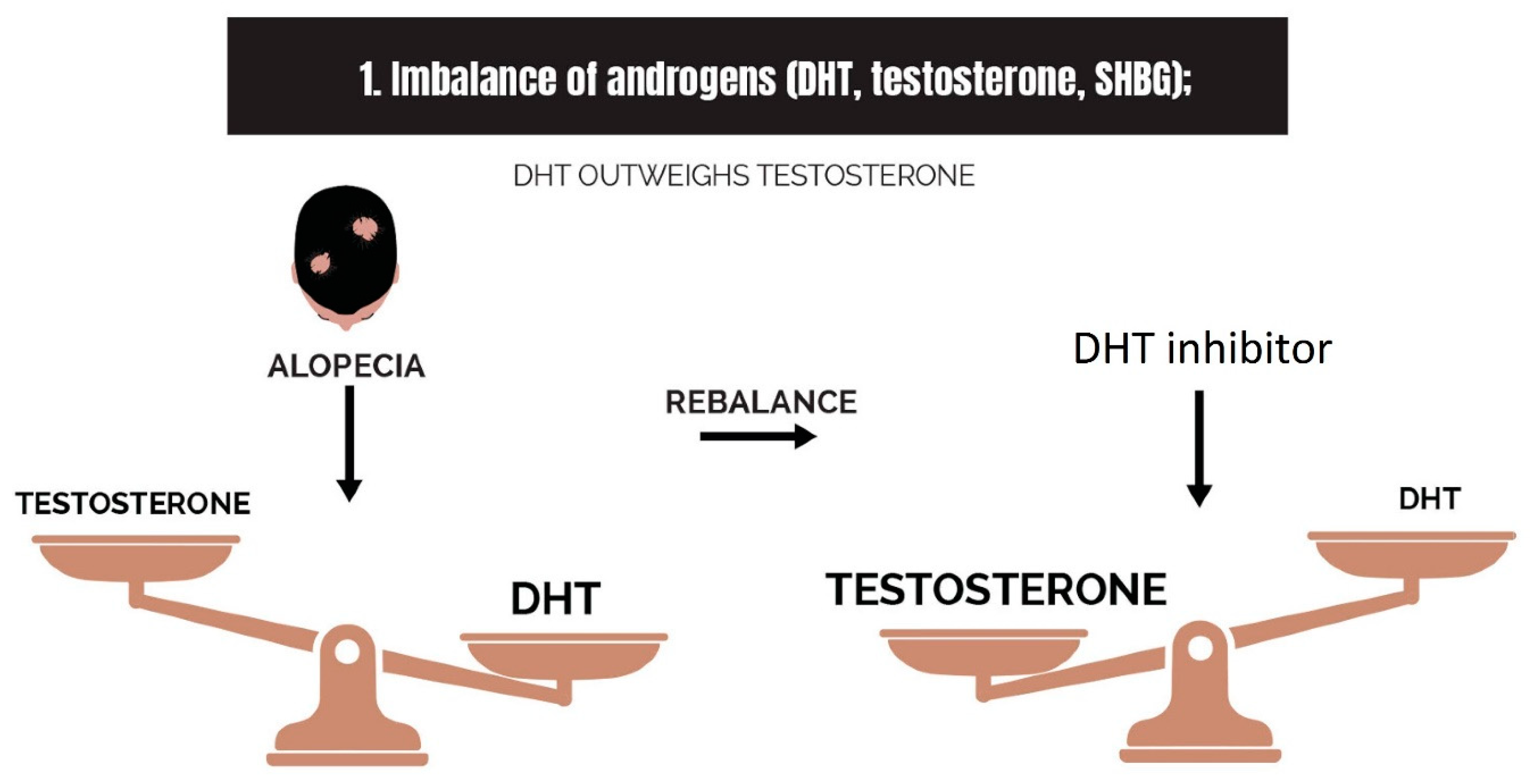
2.1. Imbalance of Androgens (DHT, Testosterone, and SHBG) in Cases of Androgenetic Alopecia

Androgen imbalance is one of the most important abnormalities contributing to AGA (Figure 4). The specific androgen in question is DHT, which is produced from testosterone when the enzyme 5α-reductase reduces it. People who live with AGA have an increased expression of this enzyme in scalp tissue, as well as a higher number of androgen receptors, also known as DHT receptors. DHT binds to the androgen receptor with a five-fold greater affinity than testosterone, making it a more potent androgen. Thus, the androgen imbalance in balding scalps (Figure 5) will cause the over-expression of androgen-responsive genes. Some authors have coined this as ‘intrafollicular androgen overactivity’ [70].
While the cascade of events is complex, the eventual outcome is a shortened anagen phase, an increased number of hairs in the telogen phase, and an increase in the phase between exogen (teloptosis) and the new hair. This middle phase is called the kenogen phase, which does not normally occur in healthy hair. A new theory of hair follicle silencing is that the kenogen phase is dominant in the ‘balder’ regions of the scalp. The hair follicles are not extinct but dormant per se [71]. Gradually fibrosing comorbidities destroy the follicle and cause hair loss to become more permanent.

2.2. Imbalance of Prostaglandins (PGF2-α, PGD2)

Inconsistencies across histopathological studies have interfered with the value of biochemical observations of prostaglandin dysregulation in balding scalps [72]. It was proposed that inconsistencies in reporting occurred because researchers either did not take into consideration the level of hair follicle miniaturization or provided limited details of their methodological selection of hair units or cell types for their studies.
As the hair follicles start to miniaturize in balding scalps, the bulb cells (dermal papilla cells) express higher amounts of prostaglandin D2 (PGD2) [73] relative to prostaglandin F2-α (PGF2-α) (Figure 6). This theoretically occurs prior to the development of fibrosis, a process that becomes more active when TGF-β1 activity from the dermal fibroblasts increases [62].
PGD2 is an agonist of its cognate receptor PPAR-γ, which causes an increase in the expression of androgen receptors and protein kinase B (Akt) signaling [74]. Another co-activator of PPAR-γ, namely the protein PPARGC1α, is also upregulated in the outer and inner root sheath keratinocytes of the pilosebaceous unit [75], where it plays a role in energy metabolism [76]. Aside from their role in energy metabolism, both PGD2 and PPARGC1α increase the number of androgen receptors, the activity of DHT, and Akt signaling [77]. Furthermore, PGD2 is also an agonist of the G protein (heterotrimeric guanine nucleotide)-coupled receptor 44 (GPR44), which inhibits hair growth when activated [73]. Lastly, PGD2 promotes the calcification of human osteoblast cells [78], which may be related to the alleged calcification that occurs in balding scalps [79], but this needs to be properly understood.
In a study of the early stages of AGA in Hispanic men, it was observed that the increased expression of PGD2 was accommodated with a compensatory increase in the enzyme to make PGE2 [80], perhaps as a protective response. However, PGE2 was not measured in that study. Both PGE2 and PGF2-α are known to promote hair growth [81]. During hair follicle miniaturization, there is also a phase when the amount of PGF2-α is diminished [82]. Thus, the severity of hair loss in disorders correlates to the balance of prostaglandins in the early stages; PGD2 is too high, and PGF2-α is too low.
Although it is not known what causes the prostaglandin imbalance to occur in scalps, one theory is that the overgrowth of P. acnes (see Section 1.4) in the follicular infundibulum is an etiological driver. An in vivo study of hamsters demonstrated that the mere application of P. acnes culture supernatant to the skin caused the expression of 15-deoxy-Δ12,14-prostaglandin J2 (15d-PGJ2) to increase [83]. 15d-PGJ2 is an agonist for PPAR-γ [84]. Overactive PPAR-γ causes augmented lipogenesis [85], which is the start of the third strike (see next section).
Because 15d-PGJ2 is produced spontaneously via the double dehydration of PGD2 [86], promoted by the generation of reactive oxygen species [80], then it is clear that the culture supernatant of P. acnes was driving an increase in the expression of PGD2 and 15d-PGJ2 was measured as an artifact. This is supported by a study that demonstrated the reciprocity of 15d-PGJ2 to PGF2-α [87].
However, augmentation of PGD2 by P. acnes is not the only mechanism by which the prostaglandin imbalance can be caused. This is because differences in the expression of prostaglandins can occur in sugar metabolism, specifically the polyol pathway [88]. In a previous study, it was postulated that the polyol pathway is active in the balding scalps of candidates with insulin resistance or metabolic syndrome [6]. In the current narrative, we present the theory that the polyol pathway changes the biosynthesis of prostaglandins to favor PGD2.
Prostaglandin biosynthesis is dependent upon aldo-keto reductase isotypes (AKR1B1, AKR1B3, or AKR1C3) as synthases. Aldo-keto reductase isotypes catalyze the conversion of PGH2 to PGF2-α in the presence of the reducing agent nicotinamide adenine dinucleotide phosphate (Figure 7) (NADPH) [89,90]. In the absence of NADPH, PGD2 is produced instead by using the same synthases [89].
AKR1B1, also known as aldose reductase, is also used as a substrate in the polyol pathway, which consumes NADPH via conversion to NADP+ [91]. In hyperglycemic environments, AKR1B1 is prevented from participating in reactions leading to PGF2-α or PGD2 via the physiologically relevant substrate glucose [92], so other aldo-keto reductases are utilized (possibly AKR1C3), in which case the transient depletion of NADPH by the active polyol pathway directs the synthesis of prostaglandins in favor of PGD2 production.
Alternatively, culture supernatants of P. acnes may be rich in reactive oxygen species, which drives the conversion of PGD2 to 15d-PGJ2 [80]. If this is the case, then P. acnes may also play a role in the depletion of NADPH, which persuades biosynthesis of PGD2 in preference to PGF2-α [89]. It is possible that overgrowth of P. acnes complements the polyol pathway in affected individuals, to exacerbate this problem.
Thus, together with P. acnes overgrowth, the polyol pathway should be considered as a possible cause for the imbalance of prostaglandins in the balding scalp. Strategies that seek to restore this balance should involve the inhibition of P. acnes, the use of a selective AKR1B1 inhibitor, supplementation with magnesium to increase NADPH [93], or the use of plant-based estrogenic compounds that reduce the size of sebaceous glands.

2.3. Overactive Sebum Production and Sugar Metabolism

Numerous observational studies have demonstrated a correlation between AGA and abnormal glucose metabolism in the body, such as metabolic syndrome [94], insulin resistance [95], or both [96]. Another study demonstrated a link between hyperglycemia, sex-hormone-binding globulin, and AGA [97]. Furthermore, a study of postmenopausal women with female pattern alopecia demonstrated a link between the severity of hair loss and body mass index [98], suggestive of metabolic syndrome. Although they were being called comorbidities, there is strong evidence that high blood glucose is directly linked to AGA [6]. This has received support from a study of >1000 participants in China, which demonstrated a link between the severity of AGA and the consumption of sugar-sweetened beverages [99].
The problems with hair follicle glucose metabolism may be due to the decreased expression of HIF-1α, activation of the polyol pathway, and the inability of the pilosebaceous unit to generate glycogen stores and modulate between mitochondrial respiration and anaerobic glycolysis.
Evidence that monosaccharide metabolism is dysregulated comes from transcriptome profiling of a bald scalp. Changes to the expression of genes demonstrated that a redox imbalance in the mitochondria of dermal papilla cells (NADPH < NADP+) [91] may be linked to overactive aerobic respiration [100]. In the same study, it was realized that antioxidant genes are also upregulated, which reinforces the finding that the metabolism of glucose is generating reactive oxygen species (free radicals) at a rate that is damaging to the hair follicle’s cells and cellular organelles.
Another transcriptomic analysis discovered that a co-activator of PPAR-γ (PPARGC1α and syn. PGC1α) is upregulated in the bulge region of the miniaturizing hair follicle [75]. The process of glucose metabolism and lipogenesis is partly modulated by PPAR-γ. The balding scalp is, therefore, a site of both dysregulated glucose metabolism and possibly elevated lipogenesis (Figure 8).
One of the main challenges with the utilization of glucose in aberrant respiration and lipogenesis is that the anagen hair follicles depend on the modulation of energy to support the active growth of the hair fiber. Because so much energy is required for anagen growth, the hair follicle builds a reservoir of glycogen (stored sugar), some at the base near the dermal papilla cells, but most of it is along the outer root sheath [101]. During active growth, the glycogen is gradually converted back to glucose and then to pyruvate, which is either aerobically converted to CO2 and water or lactate, either to generate the ATP required to feed the growing follicle or the lactate required for stem cell activation (Figure 9).
The glycogen store is continuously restocked during the years of anagen growth. It is not clear how this works, but there is evidence that human hair has a circadian rhythm of growth [102] that may correspond to periods of glycogen restoration or utilization. The process of building glycogen stores in hair follicles probably uses lactate by following an internal Cori cycle [103] first via conversion to a triose phosphate derivative (bypassing pyruvate) and then by enacting glycogen synthesis, a process familiar to skeletal muscles [104].
Normally, the cells of the hair follicles preferentially synthesize lactate via glycolysis of glucose to pyruvate, then conversion to lactate via lactate dehydrogenase A [105]. To maintain this process of anaerobic glycolysis, a transcription factor called ‘hypoxia-inducible factor 1 (HIF-1α) is expressed, which normally occurs during hypoxia [106], but is persistent, despite the presence of oxygen, in human scalp tissue [107,108]. The production of lactate downstream of the activity of HIF-1α is required for normal gene expression events in dermal papillae and stem cells [109]. The ability to produce lactate enables the hair cells to modulate the intensity of mitochondrial respiration, creating periods of rest and recovery in synchrony with the various stages of hair follicle growth. It is also possible that the dominance of anaerobic glycolysis is an adaptive mechanism to reduce dependence on oxygen supply, as the oxygen demands for rapid hair growth might not be adequately met in the microvascular network of the scalp dermis, requiring non-synchrony of hair follicle growth phases to distribute resources. In this latter hypothetical case, chronic aerobic respiration will deplete oxygen, creating hypoxia, which is paradoxically prevented by the expression of HIF-1α.
At times of high energy requirement, glycogen stores are accessed via the release of glucose, which is converted to pyruvate and then either lactate, by lactate dehydrogenase A, an enzyme that is associated with hair follicle stem cell activation [110], or the pyruvate is converted to acetyl-CoA via mitochondrial respiration, then either used in lipogenesis or driven onward to produce CO2 + H2O in the course of ATP generation. Thus, lactate dehydrogenase A modulates the conversion of lactate, timing the availability of pyruvate for mitochondrial respiration in the stem cell niche. Thereafter, the pyruvate is not available to be put through the citric acid cycle, as lactate dehydrogenase B is absent from this location, making the conversion to lactate irreversible unless it circulates through the unique internal Cori cycle [110].
Two processes are known to affect glucose metabolism in androgenetic alopecia. First, the mechanism to maintain anaerobic glycolysis to lactate, even in the presence of oxygen, is inhibited. In cases of AGA, enzymes that degrade HIF-1α are expressed, such as the HIF-1α prolyl hydroxylase enzymes EGLN1 and EGLN3, in addition to a potent inhibitor of HIF-1α, pigmentary epithelium-derived factor (PEDF) [107]. The differential expression of these enzymes and factors may be due to the androgenetic abnormality, as DHT was shown to blunt the expression of HIF-1α in hypoxic conditions [111]. Nevertheless, the inability of the hair follicles to follow anaerobic glycolysis to lactate results in a chronic switch to aerobic respiration, occurring in the mitochondria [108]. Thus, with the degradation of HIF-1α, the modulation of mitochondrial respiration is prevented, causing it to persist continuously, eventually creating mitochondrial fatigue and a lack of glycogen stores (Figure 10). In individuals with insulin resistance or metabolic syndrome, the high glucose load is fed into a chronic respiratory chain, creating a destructive hypothetical.
A study of ex situ dermal papilla cells that compared balding and non-balding candidates determined that the electron transport chain is severely interrupted in the former, and the authors speculated that this was due to an increased number of mitochondria in cells from the balding candidates [112]. This theory is in alignment with the observation of increased expression of the PPAR-γ co-activator PPARGC1α [75], which is known to be a promotor of mitochondrial biogenesis [113]. It was also noted that the electron transport chain was failing at complexes 1, 3, and 4 [112]. Complex 1 is an NADH-dependent step, suggesting that NADH is exhausted.
While this higher rate of mitochondrial respiration explains why mitochondrial stress has been observed in miniaturized hair follicles, the cause of the redox imbalance is not entirely explained. In the first instance, lipogenesis in parallel with the pentose phosphate pathway is in a balance regarding NADPH [114] (as previously stated, the expression of genes from PPAR-γ [75] is indicative of increased lipogenesis during the stage of hair follicle miniaturization).
Another hypothetical strain on NADPH levels was proposed recently [6], i.e., when abnormally high amounts of sugar are circulated into scalp tissue, the expression of AKR1B1 increases, hexokinases are saturated, and sugar metabolism spills over to the polyol pathway, which consumes ATP, NAD+, and NADPH and nurtures conditions for lipogenesis [91], creating a redox imbalance [115] (Figure 11). Evidence was found that supports this theory, which is that the same aldo-keto reductase, AKR1B1, which is normally responsible for the synthesis of PGF2-α [92,116], is also responsible for the synthesis of PGD2 in the absence of NADPH [89]. PGD2 is the prostaglandin that is upregulated in balding scalp according to some studies [73].
While NADPH is normally restored via the pentose phosphate pathway (from lactate), the overwhelming deficiency of Mg2+ in the community of AGA phenotypes [117] antagonizes this process due to the dependence of Mg2+ to the pentose phosphate pathway [118] and NADPH rejuvenation [119]. While disruption to the redox balance dysregulates prostaglandin synthesis (as previously stated), it also interferes with the activity of type 3 3α-hydroxysteroid dehydrogenase, the enzyme that metabolizes DHT, since it is an NADPH-dependent reaction [120].
The hair follicle’s internal Cori cycle is unlikely to follow the path of returning lactate to pyruvate, as lactose dehydrogenase B is not present in the hair follicle stem cell niche [110]. The pathway is, therefore, likely to bypass pyruvate and go direct to a triose phosphate intermediate, which is a process that is also dependent on NAD+. However, due to the reduced availability of NAD+, the mechanism of gluconeogenesis (from lactate) is interfered with, interrupting the Cori cycle. Hair follicles become deficient in the energy required for growth, and a high flux of reactive oxygen species damages mitochondria.
A poorly understood anomaly in hair loss disorders is the under-expression of insulin-like growth factor 1 (IGF-1) [121]. While this hormone is involved in the utilization of sugar, like insulin, it is also a growth factor, so it has other significant functions. Research is starting to demonstrate a role for IGF-1 in the building of glycogen stores, either via gluconeogenesis or glucose transport to the cellular machinery for glycogen formation in astrocytes (cells that store glycogen) [122]. It is feasible that the downregulation of IGF-1 in balding dermal papilla cells is another etiological component related to the diminished glycogen stores in this region, and a similar anomaly should also be investigated in the epidermal stem-cell niche of the bulge region. Furthermore, the expression of TGF-β1 may be reciprocal to IGF-1 expression [123], but this requires empirical corroboration in the context of hair follicle cells.
Thus, the expression of IGF-1 can be used as a biomarker of normal activity in the hair follicle. Therapies that are associated with an increase in the expression of IGF-1 in the short term can be expected to lead to hair rejuvenation in the long term, but since this is an early area of research, further studies should go into this anomaly. Nevertheless, the link to sugar metabolism of the hair follicle, particularly the formation of glycogen deposits, has not been properly elucidated.

2.4. Bacterial and Fungal Overgrowth

As previously mentioned, P. acnes overgrowth in the follicular infundibulum is associated with AGA pathogenesis (Section 1.4). However, an unexplained role for the fungus Malassezia furfur has also been identified; this organism is implicated in dandruff [124]. The association of M. furfur with AGA is not widely accepted, but a reliable source identified benefits from the use of ketoconazole shampoo in AGA [125], which is an antifungal drug used in the treatment of dandruff. The benefit may possibly derive from resolving an anti-inflammatory state induced in individuals who respond negatively to the fungi or bacterial overgrowth (Figure 12).

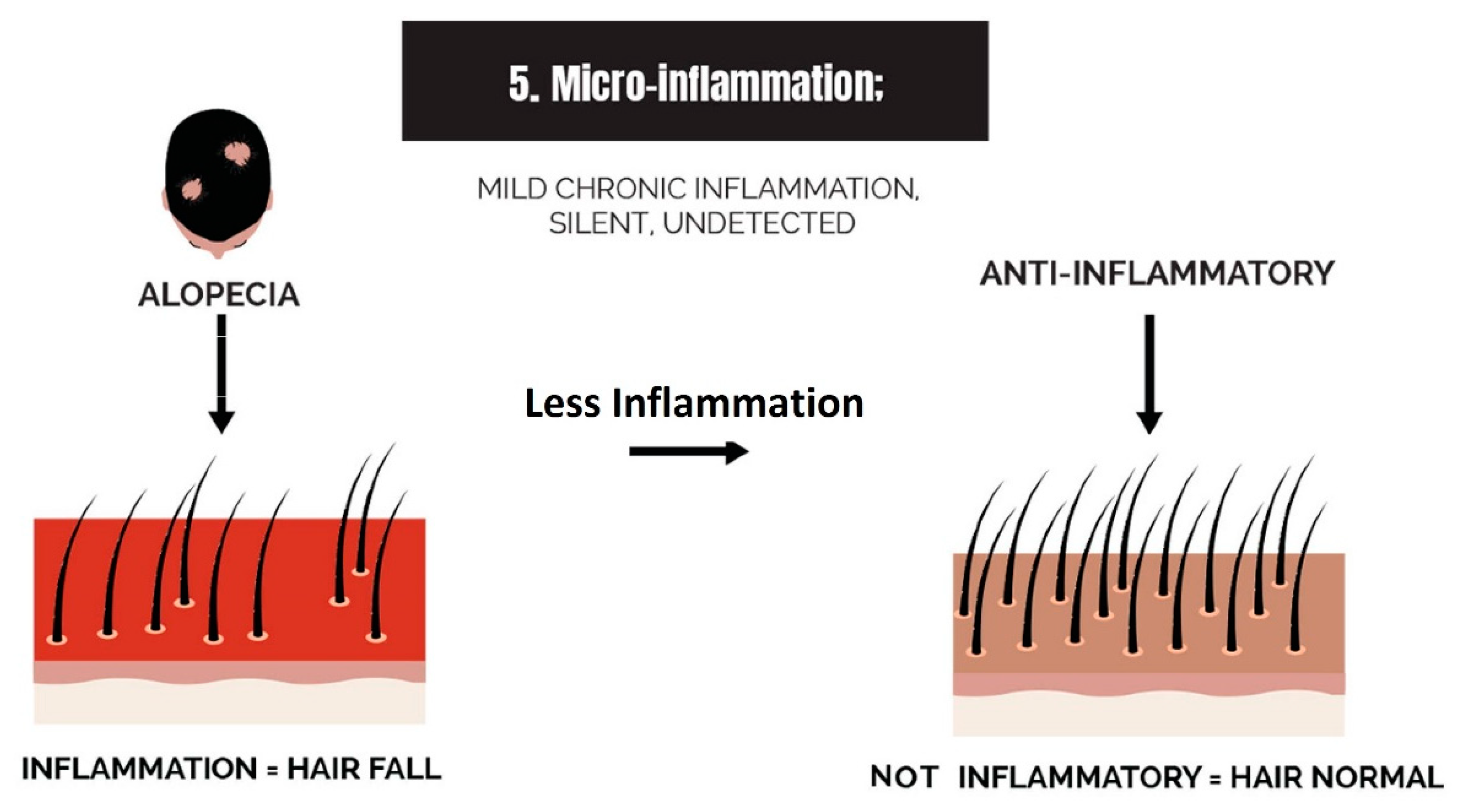
2.5. Micro-Inflammation

As AGA progresses dermatologists frequently observe a reddening of the skin on scalps with baldness (pers. comm). Inflammation is a common feature in hair loss at the stage of hair follicle miniaturization, but as the phenotypic presentation of hair loss becomes more advanced, the inflammatory state is alleviated, and fibrosis becomes more dominant.
The administration of topical hair loss therapies may facilitate a return of the dermis to a normal healthy color if the chosen therapy is effective. Evidence of inflammation during hair follicle miniaturization is corroborated by the observation of perifollicular inflammatory infiltrates, as either lymphocytes or histiocytes, and upregulation of the inflammatory genes, CASP7 and TNF [126]. The occurrence of inflammation in androgenetic alopecia is less perceptible in comparison with other forms of injury or scarring alopecias. For this reason, it is being termed ‘micro-inflammation’ [25] (Figure 13).
Studies of inflammation in AGA are diverse, giving a broad spectrum of incidences of inflammation in AGA ranging from 36 to 70% [25,126]. Because the data demonstrate that lymphocytic infiltration is in the bulge region of the hair follicle, it may be rationalized as a mild form of scarring alopecia, or it could represent the presence of comorbidity. It is the contention of the current narrative that micro-inflammation in AGA is due to the androgenetic effect, which makes the scalp dermis vulnerable to a mild autoimmune disorder in areas of the scalp afflicted by the androgenic problem. This may explain why candidates with AGA demonstrate fibrosis at advanced stages of AGA, at which time the inflammation is less obvious or absent.

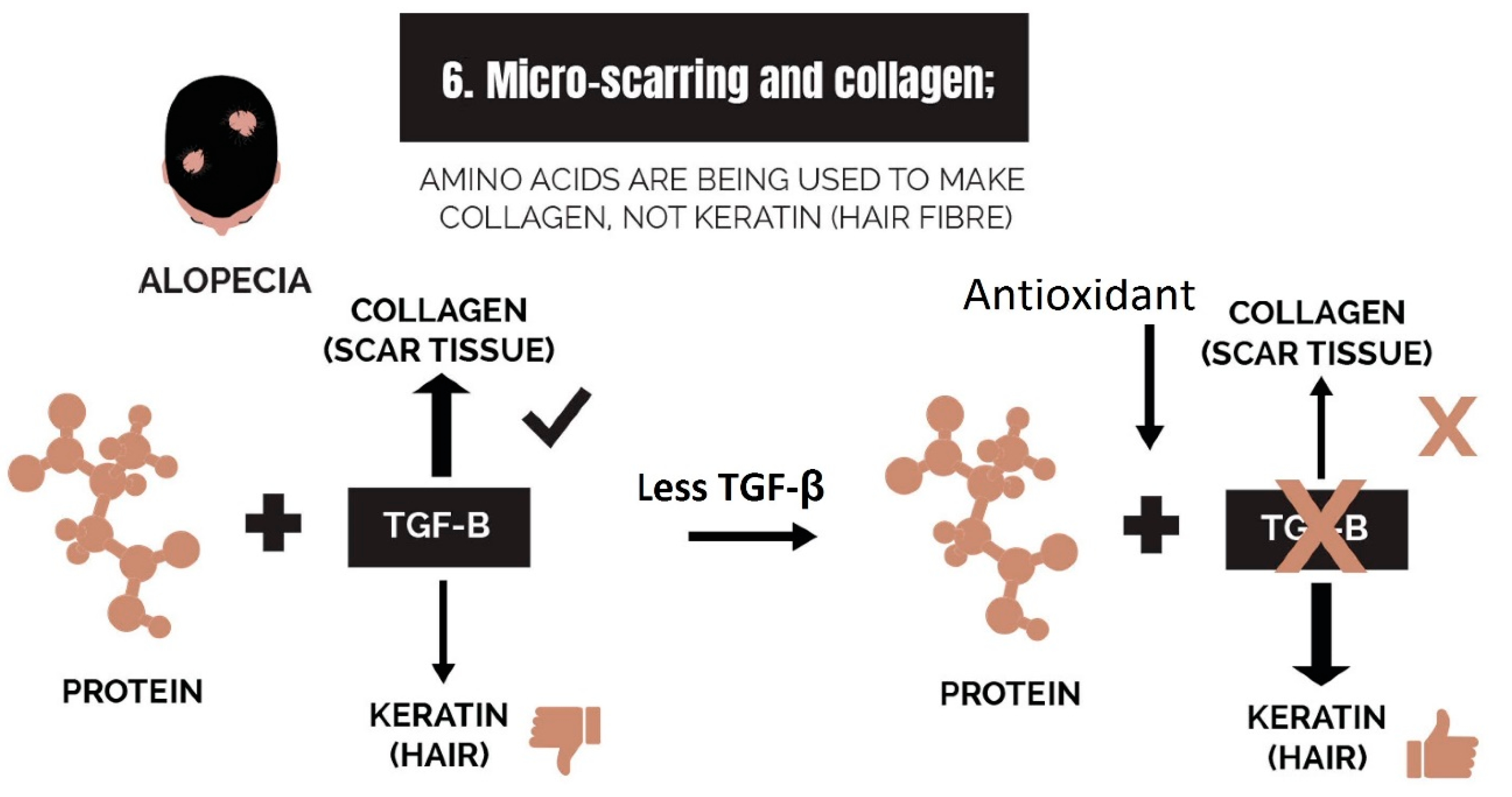
2.6. Micro-Scarring and Collagen

In cases of scarring alopecia, and in the mature stages of AGA, perifollicular fibrosis or “scleroderma” become a pathophysiological character. This creates issues of diminishing vasculature (see next section), and the fibrotic tissue goes on to diminish and replace hair follicles via interferences with stem cell niches [127].
Fibrosis development is potentially a consequence of the increased expression of transforming growth factor beta (TGF-β). The isotype over-expressed in AGA is TGF-β1, and in retinoids-induced hair loss, it is TGF-β2 [42]. Not only do the TGF-β isotypes silence the canonical Wnt signaling cascade, but they stimulate the expression of collagen from dermal fibroblasts. In AGA, DHT induces expression of TGF-β1 from hair follicle dermal papilla cells and the fibroblasts, which are the origin of procollagen. Thereafter, TGF-β1 elicits procollagen from fibroblasts [128] and possibly also from dermal papilla cells [129]. In the latter, the bulb region of the hair follicle is encased in collagen, substantially interfering with the normal differentiation process required for hair growth.
In the scalp dermis with active collagen synthesis, amino acids that are normally utilized in the formation of hair strands are utilized elsewhere. This is the hypothetical case that is put forth in Figure 14.

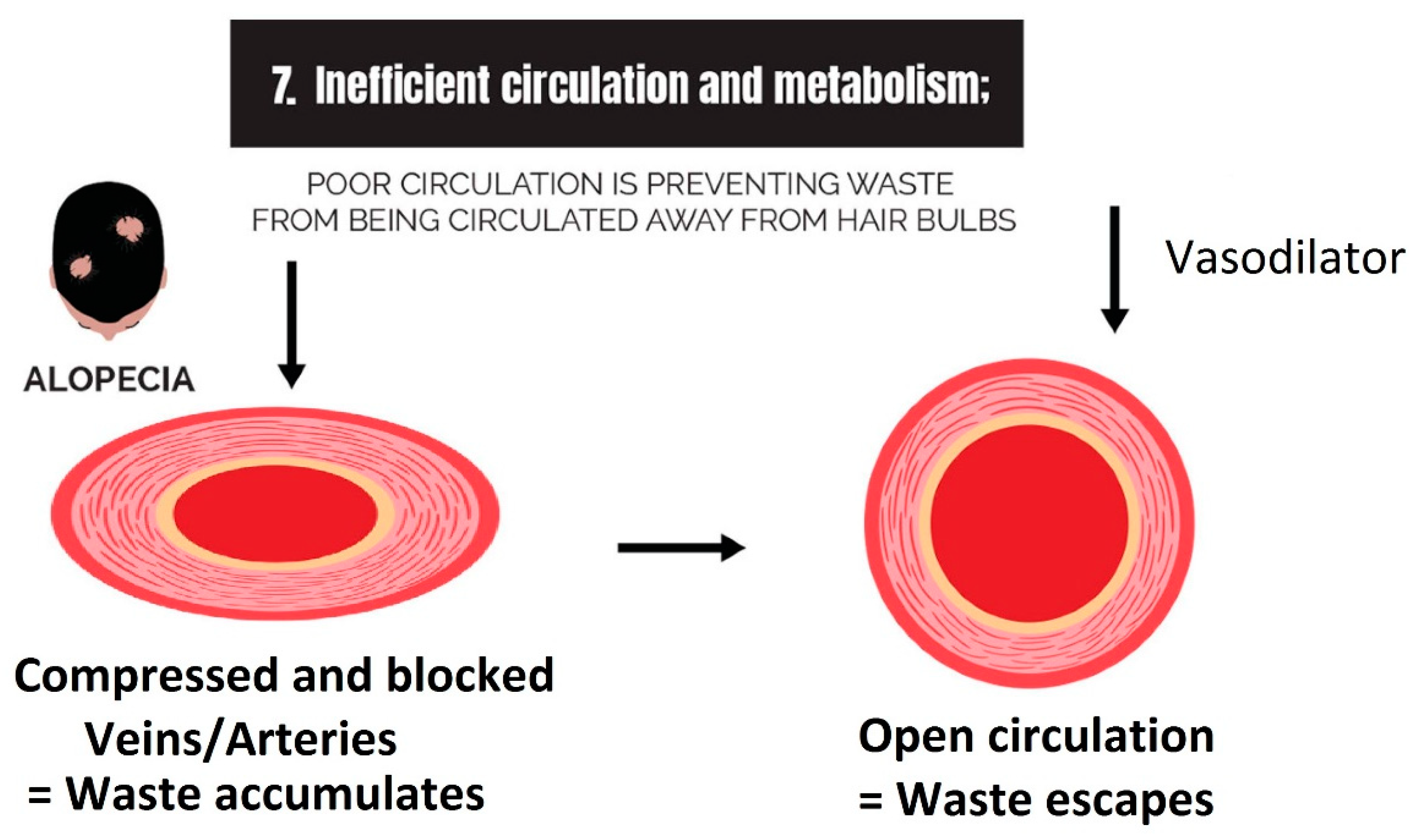
2.7. Inefficient Circulation and Metabolism

Challenges to circulation create a two-fold problem, first by limiting the circulation of nutrients and oxygen to the scalp and second by limiting the circulation of waste away from the scalp. Removal of wastes, such as reactive oxygen species, quenched reactive oxygen species, cholesterol, and purine by-products of glucose metabolism, is a significant part of homeostasis. Metabolism of xenobiotics, such as cholesterol, by cytochrome P450s, is listed in this category because this is also an essential part of the waste removal process.

2.7.1. Circulation

Reduced circulation in hair loss disorders can have several negative effects, including the restriction of nutrient and oxygen supply, the inadequate removal of cellular waste products, and issues with the removal of DHT, causing it to become concentrated locally. Because testosterone is 10 times more soluble than DHT [130], there is a risk that DHT will accumulate locally if the circulation of plasma proteins is restricted.
In hair loss disorders, particularly TE and AGA, there are several mechanisms contributing to the challenges in circulation. The symptom that is mentioned most in informal discussions of hair loss is vasoconstriction, which occurs due to simultaneous inflammation and oxidative stress [131]. Furthermore, androgen-mediated paracrine signaling restricts angiogenesis and induces the regression of the vasculature in hair follicle dermal papilla cells [35]. As mentioned previously, challenges to vasculature may interfere with the removal of waste products. However, the hypothetical consequences of limited oxygen circulation to the scalp are now rationalized by the discovery of the degradation of HIF-1α [107]. In normal scalps, HIF-1α is expressed independently of the presence or absence of oxygen due to the preference for pyruvate conversion to lactate [105]. Thus, healthy hair follicles are not vulnerable to a fluctuating oxygen supply; however, since HIF-1α is degraded in scalps with AGA [107], the utter dependence of cells on the citric acid cycle makes dermal papilla cells vulnerable to hypoxia. Furthermore, the aberrant respiration process promotes the depletion of oxygen, making the balding scalp an environment of hypoxia.
There is also an area of discussion around the negative effects of scalp tension, which are thought to be due to strain in the muscles that pull on the galea aponeurotica, the section of the scalp that corresponds to the Norwood–Hamilton balding pattern. While scalp tension remains an area of contention in the discussion by the scientific community, preliminary evidence is in favor of a contributory role in AGA pathogenesis. This is due to the positive outcome of the use of botulinum toxin to relax the muscles in the occipital and temporal areas of the scalp and ease tension in the vertex [132]. In addition, 325 volunteers who used standard scalp massages self-reported improvement in hair loss pathologies, with greater anecdotal praise in cases of the phenotypic presentation of AGA [133].
A lesser-known contributor to vasoconstriction in the balding scalp is the plaque build-up that reduces the internal diameter of blood vessels (Figure 15). However, there are limited scientific studies that discuss this in the context of hair loss. Reference is made to a letter written in 1942, but calcification of the skull’s sutures and foramens was the focus of the authors’ observations rather than the vasculature in the soft tissue of the dermis [134].
Hence, the association of venereal calcification with AGA needs to be confirmed. One view is that the polyol pathway generates uric acid [135], which has the capacity to form microcrystals, creating a seed upon which calcium oxalate precipitates in arteries or veins, familiar to atherosclerotic oxalosis in coronary arteries [135], but uniquely in scalp dermis. Nevertheless, incidences of oxalosis in scalp tissue have not been confirmed in the published literature. An alternative view is that venereal plaque build-up is a feature of cholesterol-potentiated hair loss [136].
Circulation in scalp tissue is also strained by the degradation of capillaries during fibrosis development. The process for fibrosis development is elucidated in Section 2.6, and with dysmorphia of the scalp, it becomes necessary to consider angiogenesis as a significant outcome of hair rejuvenation therapies. The normal process of angiogenesis is allegedly interrupted due to the degradation of HIF-1α [107] and decreased expression of vascular endothelial growth factor [137] in dermal papilla cells, the two of which normally synergize to regulate angiogenesis [138].

2.7.2. Metabolism

Transcriptome profiling of human scalps with AGA revealed that genes associated with the expression of metabolism enzymes are dysregulated. One of those identified was CYP1B1 [139], an under-expressed gene that codes for a monooxygenase enzyme (CYP450) that performs cholesterol metabolism [140]. CYP1B1 also codes for the proteins that bind and transport cholesterol, i.e., sterol regulatory element-binding proteins (SREBPs). It is possible that cholesterol accumulates in the dermis of the scalp afflicted by AGA. Because cholesterol has a direct antagonistic effect on cultured hair follicles [141], inefficient metabolism and elimination of cholesterol may contribute to cholesterol-potentiated AGA [136].

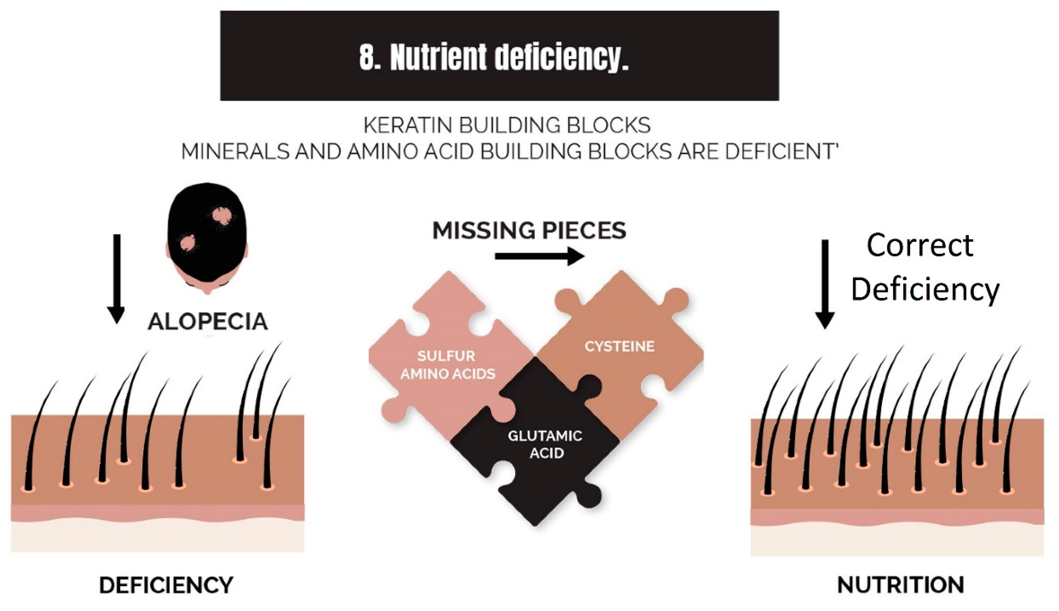
2.8. Nutrient Deficiency

There are several nutrients that become a barrier to recovery from hair loss disorders, such as scarring alopecia, TE, and AGA. These deficiencies are regarded as ‘bottlenecks’ to therapies (Figure 16). A prominent example is iron deficiency, which is a common point of interest at the ‘Cleveland Clinic Foundation’ while addressing hair loss. Practitioners believe that iron supplements should only be used in cases of deficiency, and they purport that the efficacy of their treatment improves when the deficiency is corrected [142].
Another well-known bottleneck is zinc deficiency. In an analysis of a Korean population, it was demonstrated that patients with TE or AA were deficient in zinc, suggesting a possible metabolism barrier to homeostatic levels [143]. Another study of Turkish men with AGA demonstrated that although serum levels of zinc and copper appeared normal, concentrations of both metals were low in excised hair samples from the bald region of the scalp. The authors speculate that local circulation deficiency or metabolism barriers prevented the afflicted follicles from utilizing these elements in hair growth [144].
Studies that focus on B vitamins and amino acids have also corroborated the benefits of supplementation. When medical yeast (for B vitamins), the amino acid cysteine and pantothenic acid (vitamin B5) were supplemented as an adjuvant therapy to minoxidil, results were greater than with minoxidil alone [145].
It is becoming apparent that nutritional deficiencies associated with hair loss disorders are not necessarily a consequence of low dietary intake. Several studies have recognized that breakdown to the function of metabolizing enzymes is occurring in the hair loss patient. For example, a transcriptional study recognized that the gene CYP27B1 is under-expressed in balding hair follicles [146]. This gene codes for the monooxygenase enzyme that metabolizes 25-OH vitamin D into its active form 1,25-OH. Because magnesium supplementation was able to correct serum vitamin D concentrations without the need for changes to vitamin D intake [147], it is feasible that magnesium deficiency is a bottleneck to the function of metabolizing enzymes that rely on NADPH, which is renewed via a magnesium-dependent system [93]. One study confirmed that magnesium deficiency was common in candidates with AGA [117].

3. Clearing the Big Eight Strikes with Cosmeceuticals to Improve Hair

The ideal therapy for the balding scalp will target all eight strikes against hair health. Because candidates have different strikes against their hair, either highly or moderately active, then it is better to avoid designing highly specific single-target drugs to address these problems. For example, by targeting a single enzyme with high specificity, therapy can benefit some individuals who require rebalancing the activity of that enzyme, but when applied to homeostatic individuals, the activity of the same enzyme will be dysregulated. Thus, broad-spectrum natural cosmeceuticals represent a more appealing approach to ensure applicability to a broad spectrum of hair-loss candidates.
The current narrative focuses on natural product interventions as cosmeceuticals, not pharmaceutical drugs or procedures. We only briefly mention several alternative procedures, such as microneedling, platelet-rich plasma, standardized scalp massages, low-level laser light therapy, autologous hair follicle transplants, and botulinum toxin injections [1,132,133]. These procedures and pharmaceutical drugs, such as finasteride, minoxidil, dutasteride, latanoprost, bimatoprost (topical prostaglandin), or cetirizine (PGD2 blocker) have merit [1,34], but such interventions are comprehensively elucidated in other reviews.
While procedural and pharmaceutical interventions have a clear path of success in targeting the eight strikes, this can also be achieved via cosmeceuticals that are entirely plant-based or are mimetic of a human peptide (or matrikine) metabolite. Such ingredients may be derived from sustainable plantations (farming), or they can be manufactured in a green process (i.e., green synthesis). Regarding the ingredients that are identical to human metabolites, such as the peptides [148], they are still natural and not drug-like, provided they are secondary metabolites, not cytokines, enzymes, or hormones. For example, it is possible to derive peptides from plant-based dietary sources, including the well-known tripeptide GHK [149]. But GHK spontaneously chelates with copper (Cu) in human metabolism to become Cu-GHK. Thus, topical application with Cu-GHK is considered reasonably natural.
Lastly, the eight strikes are interdependent. For example, controlling any one of the strikes can improve the other strikes. Thus, by improving each of the strikes, it becomes easier to improve the others, i.e., controlling inflammation can improve prostaglandin balance, controlling DHT can improve inflammation or fibrosis development, controlling bacterial overgrowth can improve inflammation and prostaglandin balance, and so on.

3.1. Strike 1: Imbalance of Androgens

Strategies to correct the androgen imbalance include directly targeting the androgen (DHT), directly targeting 5α-reductase, or blocking the androgen receptor. It is difficult or unfeasible to target the androgen directly with exogenous substances (natural products), but it is feasible to do this indirectly by promoting an increased expression of the enzyme 3α-hydroxysteroid dehydrogenase (3α-HSD) [150]. This enzyme degrades DHT (Figure 17). By increasing the expression of 3α-HSD or enabling its activity by restoring the redox balance, it is conceivable that levels of DHT would decrease [151].
Unfortunately, in scalps afflicted with AGA, the redox balance is impaired, and the activity of 3α-HSD is prevented due to its dependence on NADPH [120] (Figure 18). Consequently, a higher concentration of DHT occurs in the scalp, and because the scalp is one of the primary synthesizers of DHT, systemic DHT is elevated. DHT is circulated to the liver, where it is metabolized to a more polar conjugate, to enable elimination via kidneys or secretion into the colon for microbial deconjugation and elimination.
One of the metabolic conjugates is DHT-glucuronide (Figure 19). Although glucuronides are eliminated from the body, they can also be deconjugated at various sites around the body where β-glucuronidase is expressed, restoring DHT back to its free form. For example, it is known that β-glucuronidase is expressed in the connective tissue sheath of hair follicles that are in the telogen–catagen phase [152], indicating that elevation of free DHT is mechanistically related to the cessation of the anagen phase. Unfortunately, β-glucuronidase is also expressed when macrophages and neutrophils are stimulated [153,154], possibly a mechanism to modulate inflammation [155]. It has also been demonstrated that TGF-β1 stimulates the expression of β-glucuronidase [156]. Thus, a hypothetical schematic for the increase in DHT in the scalp is presented in Figure 20.
Targeting 5α-reductase is feasible with both natural products and synthetic drugs, as previously elucidated, and targeting the androgen receptor can be achieved either by binding directly to it and inactivating it or by changing gene expression patterns to reduce the expression of androgen receptors. The latter is referred to as the degradation of the androgen receptor [157].
Some people who are living with AGA also demonstrate a decrease in sex-hormone-binding globulin (SHBG) [6] (this does not relate to the polycystic ovarian syndrome equivalent in males). This glycoprotein is produced by the liver, and it binds to androgens and carries them around the body. Binding to the androgen changes its activity, which can prevent it from performing its normal function. Interestingly, SHBG binds to DHT with higher affinity than testosterone, like the androgen receptor. This means that higher circulating levels of SHBG will cause a decrease in the amount of free DHT in the body. There are various dietary substances that are associated with normalized SHBG expression in the liver, by increasing levels if low or lowering levels if too high, such as fenugreek [158], oleic acid [159], or equol [160].
Another area of interest is in reducing the damaging effects caused by excess DHT. One group of researchers postulates that the activation of the PI3K/Akt pathway may cascade into the activation of the erythroid 2-related factor 2 (Nrf2) signaling pathway [161], which antagonizes the activity of DHT [162]. This possibly occurs with the consumption of sulforaphane [161]; however, a general improvement in the health of the scalp dermis may also be associated with the control of DHT and its negative effects.

3.2. Strike 2: Imbalance of Prostaglandins

Strategies to correct the imbalance of prostaglandins may involve the use of potent antioxidants to preserve NADPH by reducing reactive oxygen species generated from polyol metabolism. Furthermore, the inhibition of P. acnes via the topical application of antibacterial compounds may also be effective. Lastly, dietary intervention may support the synthesis of hair-growth-promoting prostaglandins by inducing aldo-keto reductase blockers (dihydroberberine) or by adopting a low glycemic index diet.

3.3. Strike 3: Sebum and Sugar Metabolism

In balding scalps, the sugar that circulates to the affected area is metabolized into lipids in a process known as lipogenesis. This process is modulated by the prostaglandins, so the first step to correcting this problem is to rebalance the prostaglandins. However, via inhibition of the aldo-keto reductases responsible for the polyol pathway, both prostaglandin balance and lipid metabolism may be corrected. The specific enzyme to target is AKR1B1, which is the principal reductase responsible for the polyol pathway. There are several plant-based ingredients that inhibit this enzyme, most notably berberine [163].

3.4. Strike 4: Bacterial and Fungal Overgrowth

Bacterial overgrowth can be controlled via plant-based metabolites that are antagonistic to Gram-positive organisms, such as P. acnes or S. aureus. There are many such compounds available from the plant kingdom, such as taxifolin or resveratrol [164].
For fungal overgrowth, there are also several anti-fungal compounds available; however, plant-based compounds that inhibit M. furfur are less common. While ketoconazole is an effective drug, M. furfur can be controlled using a highly concentrated topical application of a broad-spectrum antimicrobial compound from a plant to compensate for the lack of specificity against this fungus (yeast). Some good candidates are the flavan-3-ols from grape seeds, which have moderate activity against M. furfur [165]. To antagonize this fungus, it would require combining a slightly higher amount into the composition.

3.5. Strike 5: Micro-Inflammation

Inflammation can be controlled by reducing microbes in the dermis, quenching free radicals (antioxidants), and via the application of an anti-inflammatory composition. There are several known plant-based anti-inflammatory extracts, such as copaiba [166] or Larix decidua (syn. L. europaea) (Larch) wood extract [167]. Because several plant extracts are antioxidants and confer antimicrobial effects, the anti-inflammatory extract is a multi-modal remedy.

3.6. Strike 6: Micro-Scarring and Collagen

Once fibrosis has occurred in the scalp dermis, it is very difficult to reverse. Interventional therapies generally halt the progression of fibrosis, either via the inhibition of pathogenic bacteria, such as S. aureus, or conferring anti-inflammatory effects. However, the reversal of fibrosis is more difficult because it requires reactivation of the stem cell niche of the hair follicle, which reclaims the dermis and starts the slow process of returning a functional keratinocyte population.
One strategy that may be effective is to trigger the expression of matrix metalloproteinases (MMPs), which degrade collagen and trigger a restructuring of the dermis. Increasing expression of MMPs can be achieved via the use of peptides, particularly Cu-GHK, which signals to the dermis that injury has occurred and that a restructuring process needs to be started. The transdermal penetration of peptides will improve if they are applied in conjunction with microneedling. In such strategies, it is of essence to avoid excessive use of a derma roller to avoid having the opposite effect, which will increase fibrosis rather than reduce it.
Another strategy to prevent scar formation is by antagonizing the differentiation of fibroblasts into myofibroblasts. This modulates collagen formation to avoid fibrotic overgrowth. There are plant extracts that are known to do this, such as the saponin mixture called escin (or aescin) from the chestnut of Aesculus hippocastanum (horse chestnut tree) [168], which is a well-known therapy for skin diseases, controlling the modulation of dermal cells to facilitate normal tissue formation, especially in the context of angiogenesis [169,170].

3.7. Strike 7: Inefficient Circulation and Metabolism

Three strategies to improve circulation are vasodilation, relaxation of muscles at the occipital and temporal regions of the scalp, and angiogenesis. There are plant-based compounds that dilate arteries and veins in a similar way to minoxidil. The most popular example is caffeine, which is a phosphodiesterase inhibitor [171], that typically causes vasodilation via the inhibition of cAMP and cGMP, leading to an increased intracellular concentration of calcium [172]. However, caffeine may not necessarily be a good candidate since it is also an agonist for PPAR-γ [173] and promotes mitochondrial biogenesis [174]. Nevertheless, it is important to know if the benefits outweigh the detriments.
To improve muscle relaxation in the areas at the base of the skull, people can use topical liniments and supplement magnesium, or they can practice standardized scalp massages [133]. For angiogenesis, there are steroidal plant extracts that have been proven to modulate angiogenesis. A good example of a proangiogenic compound is the phytosterol called sitosterol (syn. β-sitosterol), which is found in Aloe vera [175] and Serenoa serrulata [176].

3.8. Strike 8: Nutrient Deficiency

Two primary methods to ameliorate nutrient deficiency are the supply of limited nutrients and the improvement of metabolic processes that are responsible for nutrient neogenesis. While it is of the essence to correct deficiencies caused by dietary insufficiency (magnesium, iron, folic acid, etc.), people who suffer from hair loss disorders usually suffer from comorbidities that prevent the proper utilization of nutrients or nutrient precursors. Some of these deficiencies are localized to the scalp and are not otherwise a problem in the other parts of the body. In such cases, topical application of limiting nutrients can help.
Thus, topical application of sulfur amino acids, glutamic acid, and other keratin building blocks is conceded to be of benefit in hair rejuvenation strategies. Furthermore, copper and zinc can be made into a bioavailable form by fermentation with Saccharomyces cerevisiae, the same yeast used in the fermentation of bread and alcohol. These ferments are commercially available.
The ingestion of collagenous material can increase the absorption of amino acids or peptides after the enzymes in the human digestive tract have reduced them to sizes that can cross the intestinal endothelium. However, if collagenous material is predigested into peptides, they can be absorbed directly across the epidermis of the scalp, making topical application feasible and convenient.
Correction of nutrient neogenesis can be achieved by the supplementation of minerals or vitamins that act as substrates in metabolic processes leading to other types of nutrients. For example, vitamin B12 is necessary for melatonin synthesis [177], and melatonin is good for hair growth [178].

4. Critical Examination of a Selection of Exemplary Cosmeceuticals in the Market

There is a plethora of unregulated claims in the market related to the success of topical therapies that rarely enact the efficacy touted in advertising. The anecdotal success of a hair rejuvenation initiative can persuade the public to expect the generic reproducibility of that outcome. It is common to see such claims made in relation to hair-rejuvenating oils that are manufactured from a mere ‘fixed’ oil, such as olive, coconut, rosehip, or grapeseed oil. On the market, there are numerous topical hair oils that may create aesthetic improvement to the hair strands and possibly improve the growth rate of hair in individuals who are not suffering from a hair loss disorder but have no efficacy in diseased scalps.
Unfortunately, it is difficult to regulate these oil-based therapies in the market. While oils can be used as a base to extract lipophilic components from plant biota, specifically therapeutic phytochemicals, the industry is too focused on the minimization of manufacturing costs. For this reason, there is often minimal presence of biologically active metabolites in these market brands. As such, they almost entirely comprise triglycerides and free fatty acids, giving minimal benefit to individuals who have a true hair loss disorder. When plain oils are applied to scalps with hair loss disorders, they can exaggerate the disease state by promoting the growth of lipophilic bacteria, such as P. acnes [25]. These effects can be countered if the composition includes potently antimicrobial and anti-inflammatory ingredients, but without the proper regulatory control and testing of these oil-based compositions on the market, efficacy is not predictable. Thus, individuals who experience hair loss should be discouraged from using a topical therapy that is based on a ‘fixed’ oil, unless they can verify that the concentration of biologically active phytochemicals is adequate to enact the antimicrobial and anti-inflammatory effects.
Most of the non-oily hair rejuvenation therapies on the market that have clinical backing in the context of AGA and other pathologies are water-based. These serums might have lipophilic ingredients, but they are not dissolved into oils; they are made into emulsions with surfactants or encapsulated with liposomal technology.
There are several ‘business-to-business’ (B2B) proprietary blends (cosmeceuticals) on the market that are used as a base ingredient in water-based serums. These proprietary blends tend to incorporate ethnobotanically significant species in the context of dermal health, vasculature, and hair rejuvenation [179,180]. They often also integrate peptides and/or growth factors to enrich the composition [181].
Examples of cosmeceuticals for hair health are RadensylTM, CapixylTM, Anasensyl®, Procapil®, and AnaGainTM. No published clinical studies were found on these blends that proved efficacy in human volunteers, although white papers are available from the companies with associated claims. However, published data are available from the individual ingredients in these proprietary blends, albeit data obtained in vitro, with no clinical backing in follow-up. There are also numerous studies that demonstrate faster hair shaft elongation out of extracted hair follicles that are grown in a liquid medium (in vitro and ex vivo). Furthermore, some studies focus on results from the treatment of the dorsal of a rodent (in vivo). In such studies, the whole etiological aspect of hair loss disorders has been ignored.
Most hair rejuvenating serums on the market include one or more of the five proprietary blends listed above. Details of these proprietary blends and how they might ameliorate the strikes against hair health are given in the following subsections.

4.1. RedensylTM Ingredients

The active ingredients in RedensylTM include dihydroquercetin-glucoside (taxifolin from Larch wood pulp, <1% wood extract), epigallocatechin gallate glucoside (from Camellia sinensis, the green tea plant, <0.1% leaf extract), and glycine (<1%, amino acid in keratin). Active ingredients are in a base of glycerin (50–55%), water (45–50%), and the preservatives, sodium metabisulphite (<1%) and zinc chloride (<0.1%). The recommended use level is 1–3% in any serum [182].
The strikes cleared by RedensylTM include the following:
-
Strike 1 (androgens)
EGCG glucoside binds to 5α-reductase [183].
-
Strike 2 (prostaglandins);
The antimicrobial properties of taxifolin [184] may indirectly benefit prostaglandin balance.
-
Strike 3 (sebum and sugar);
Taxifolin is an aldose reductase inhibitor [185].
-
Strike 4 (bacteria/fungi);
Taxifolin is antimicrobial against Gram-positive bacteria [184].
-
Strike 5 (micro-inflammation);
Taxifolin is anti-inflammatory [186,187].
-
Strike 8 (nutrients).
Zinc and glycine are nutritional.

4.2. CapixylTM Ingredients

The active ingredients in CapixylTM are acetyl tetrapeptide-3 (0.020–0.035%) and Trifolium pratense (clover) flower extract (0.020–0.030%). The formulation ingredients include butylene glycol (45–55%), water (45–55%), and dextran (0.090–0.150%). The composition is recommended at 2.5–5.0% of a manufactured serum for ‘intensive treatment’ and 0.5–2.5% for ‘preventative care’.
The strikes cleared by CapixylTM include the following:
-
Strike 1 (androgens);
T. pratense extract includes biochanin A, a 5α-reductase inhibitor [188].
-
Strike 6 (collagen/fibrosis).
Acetyl tetrapeptide-3 signals for restructuring of the dermis [189].

4.3. Anasenzyl® Ingredients

The active ingredients in Anasensyl® are ammonium glycyrrhizate, caffeine, zinc gluconate, and Aesculus hippocastanum seed extract. Limited information on concentrations or formulation ingredients was found. The recommended use level ranges from 0.5 to 1.5% in any serum.
The strikes cleared by Anasensyl® include the following:
-
Strike 5 (micro-inflammation);
Ammonium glycyrrhizate is anti-inflammatory [190].
-
Strike 6 (collagen/fibrosis);
Aesculus hippocastanum seed extract controls the differentiation of fibroblasts, reducing fibrosis development [170].
-
Strike 7 (circulation/metabolism);
Caffeine is a vasodilator [171].
-
Strike 8 (nutrients).
Zinc gluconate is a source of zinc.

4.4. Procapil® Ingredients

The active ingredients in Procapil® are apigenin, oleanolic acid, and biotinoyl tripeptide-1 (biotinoyl GHK) (concentrations not found). The formulation ingredients are butylene glycol, water, PPG-26-Buteth-26, and PEG-40 hydrogenated castor oil. The recommended use level is 3% of any serum.
The strikes cleared by Procapil® include the following:
-
Strike 1 (androgens);
Apigenin promotes the proliferation of dermal papilla cells and reduces the expression of TGF-β [191];
Oleanolic acid is an inhibitor of 5α-reductase [192].
-
Strike 5 (micro-inflammation);
Apigenin and oleanolic acid are anti-inflammatory [193,194].
-
Strike 6 (collagen/fibrosis).
Biotinoyl tripeptide-1 is a derivative of GHK for penetration improvement. It signals a restructuring of the dermis, which reduces collagenous material and reactivates stem cell niches [42].

4.5. AnaGainTM Ingredients

The active ingredient in AnaGainTM is an extract of Pisum sativum sprout (0.5% of non-solvent extract), with formulation ingredients of phenoxyethanol (1%), sodium benzoate (0.5%), and water. The recommended use level is 2–4% of any serum.
The strikes cleared by AnaGainTM include the following:
-
Strike 6 (collagen/fibrosis).
Pisum sativum sprout extract at 2% applied topically upregulates noggin [195], which is antagonistic to bone morphogenetic growth factor 4, a member of the transforming growth factor beta superfamily [196].

4.6. Concentration, Clinical Efficacy, and Combining the Proprietary Blends

While clinical or in vitro efficacy is demonstrated for most of the raw ingredients of the five proprietary blends in the context of hair, the concentration of active ingredients must be matched to those in clinical studies to replicate positive outcomes. For example, clinical studies of the topical application of the P. sativum sprout extract used a 2% concentration to achieve hair-rejuvenating effects greater than the placebo [195]. However, if manufacturers of serums dilute AnaGainTM down to its highest recommended concentration of 4%, the active extract is diluted 25 times down to 0.02%, a 100-fold difference compared to the clinical study. This may be a consequence of a lack of market viability, requiring the costs of manufacture to be lowered drastically to the inevitable loss of product efficacy.
There are a number of other ingredients that have high potency, permitting lower concentrations that are inevitable in industry. For example, taxifolin is anti-inflammatory at concentrations >75 µM [187], equivalent to 0.009%. If the concentration of taxifolin in the wood extract of larch is at a minimum of 90% [197], then a 1% solution of RedensylTM gives 0.09% taxifolin, 10-fold higher than required for anti-inflammatory effects. The pharmacokinetics of taxifolin in the dermis determines the saturated tissue concentration. However, >80% permeation was seen using a human skin model [198]. While the elimination half-life of taxifolin is short in human plasma [199], saturation in human skin is expected to preserve the molecule for longer, as it is renal elimination that dominates metabolic removal from human plasma. Thus, RedensylTM may be able to enact the effects observed in vitro. Unfortunately, no peer-reviewed published clinical study on human volunteers was found to corroborate this.
The five most common proprietary blends on the market do not clear the big eight strikes unless they are combined. The most comprehensive blend is RedensylTM, which clears six of the eight strikes. Nevertheless, these proprietary blends are generally not sold on their own in the ‘business to consumer’ market, as they are used to formulate commercial brands. Only two serums (brands) were identified on the market (although there may be more) that included enough diversity of ingredients to cater to all eight strikes. One of the two serums is REVIVV® from WETHRIVVTM, and the other serum is RevivHair™ Max Hair Stimulating Serum. Generally, such compositions use three or four of the above-mentioned proprietary blends and additional ingredients to improve transdermal absorption or the various forms of GHK, including biotinoyl-GHK and palmitoyl-GHK. Penetration enhancers are also often used, such as menthol and amino acids (bifunctional ingredients). Lastly, proprietary blends may not include an adequate amount of a specific metabolite, such as epigallocatechin gallate in RedensylTM, so manufacturers of serums sometimes elect to raise their concentration.

5. Conclusions

Hair loss disorders are multifactorial, which requires multimodal initiatives to achieve improvement. There are eight alienable etiological components in hair loss disorders, and the higher number of strikes (one strike, two strikes, three strikes, …) will correlate to the severity of hair loss.
The eight strikes are as follows:
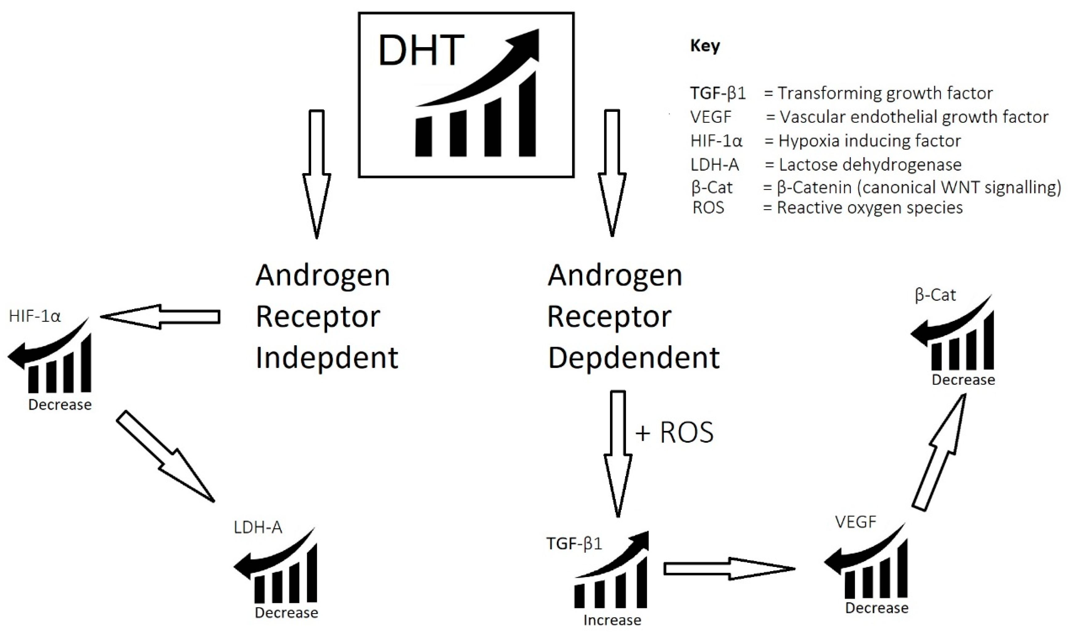
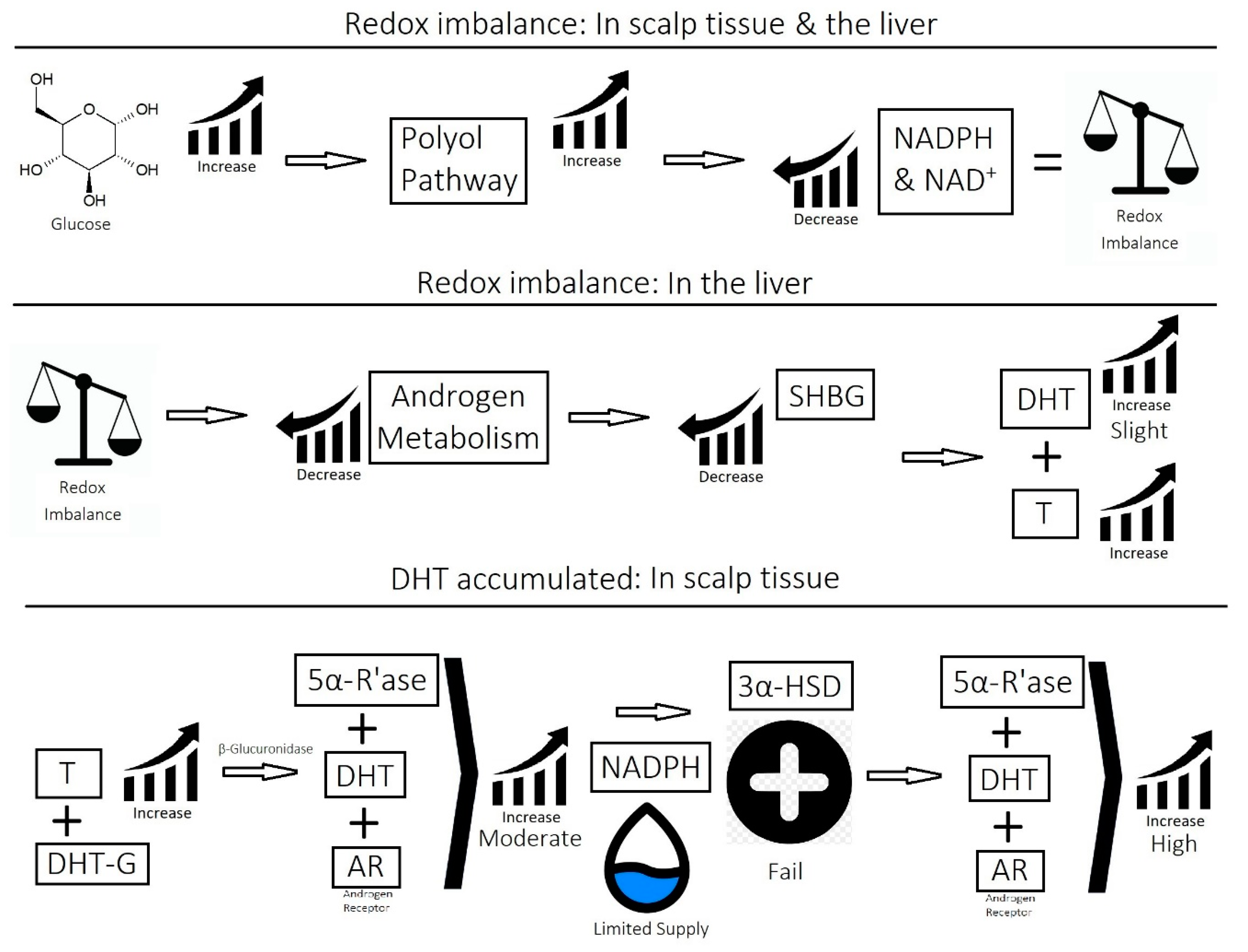
  • Imbalance of androgens (DHT, testosterone, and SHBG) in cases of androgenetic alopecia: DHT triggers the expression of TGF-β1, which is reciprocal to the canonical Wnt signaling pathway.
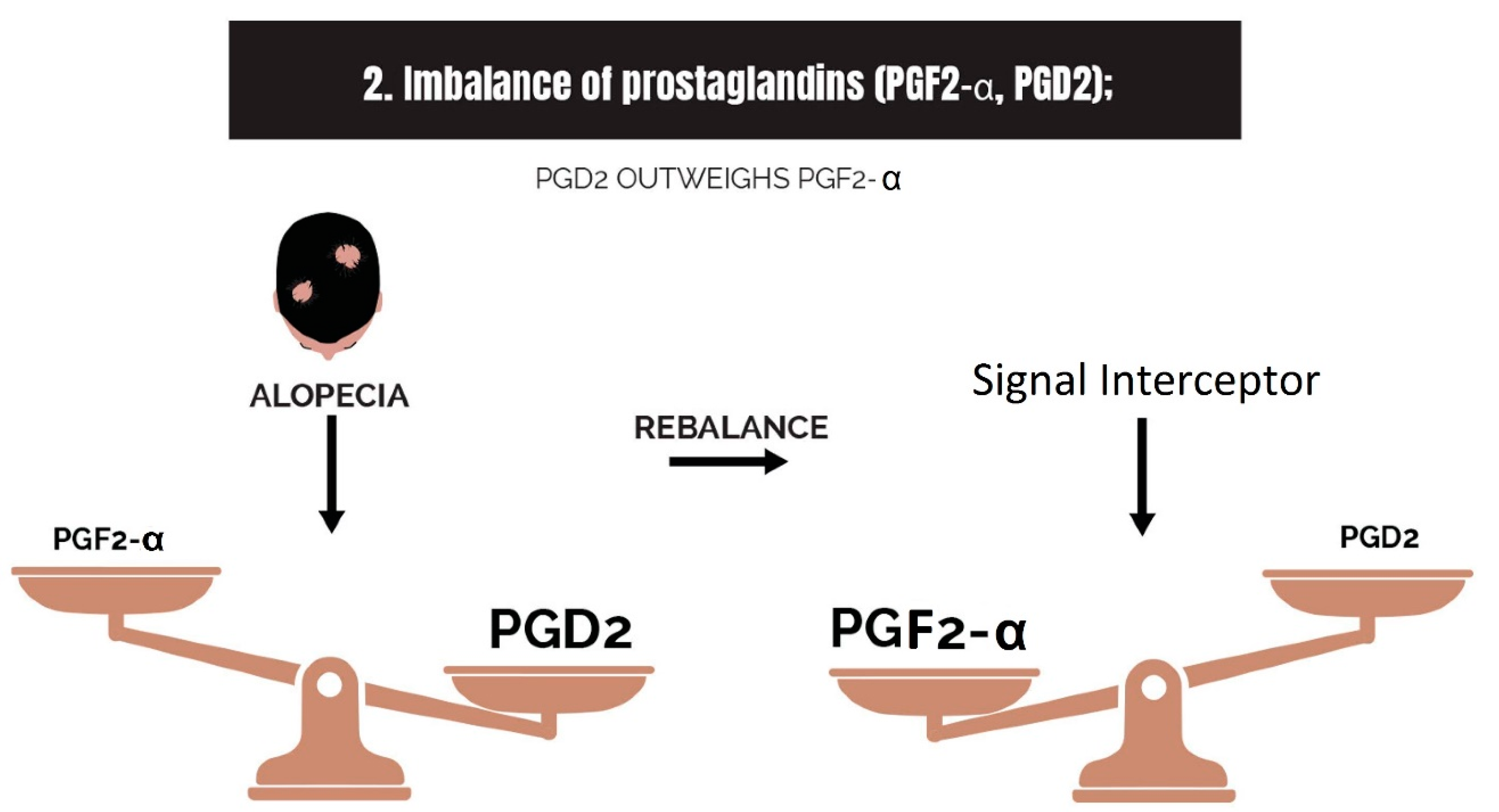
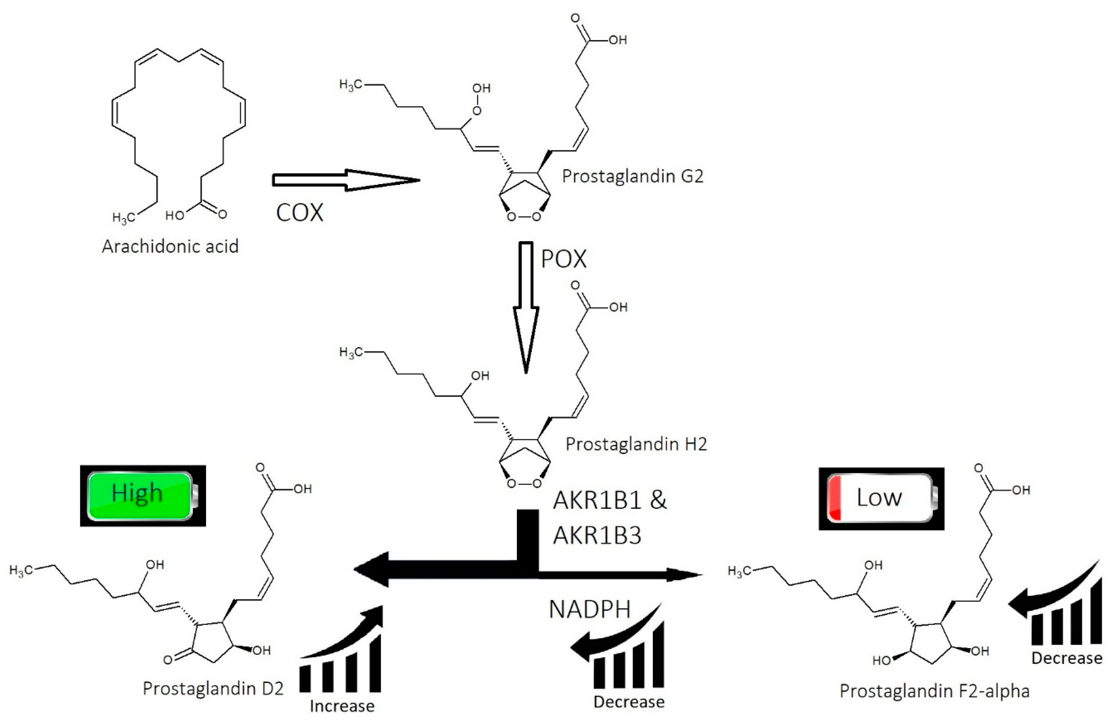
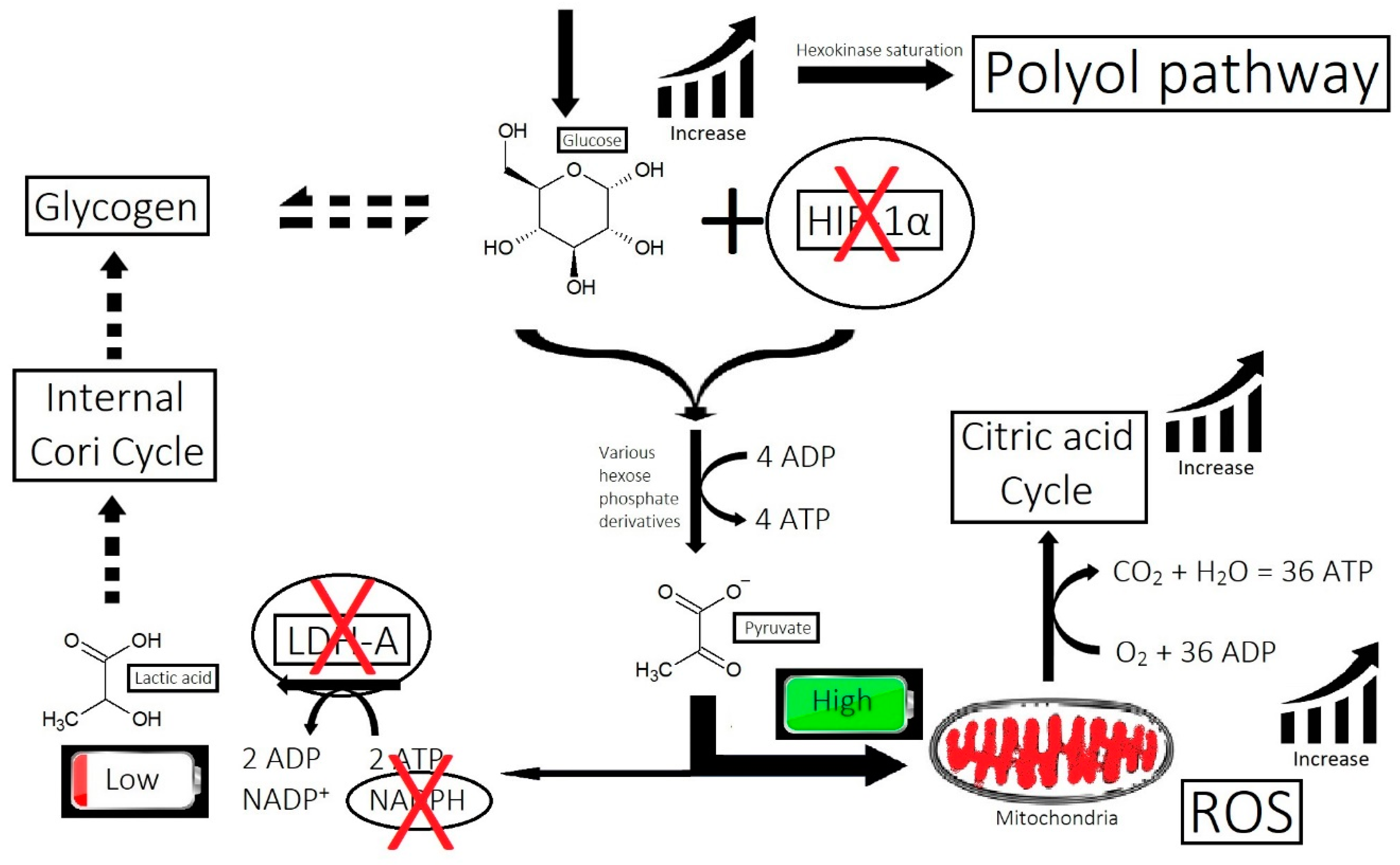
  • Imbalance of prostaglandins (PGF2-α, PGD2): Depletion of NADPH redirects the biosynthesis of prostaglandins toward PGD2. This may be jointly caused by bacterial overgrowth of P. acnes and the polyol pathway.
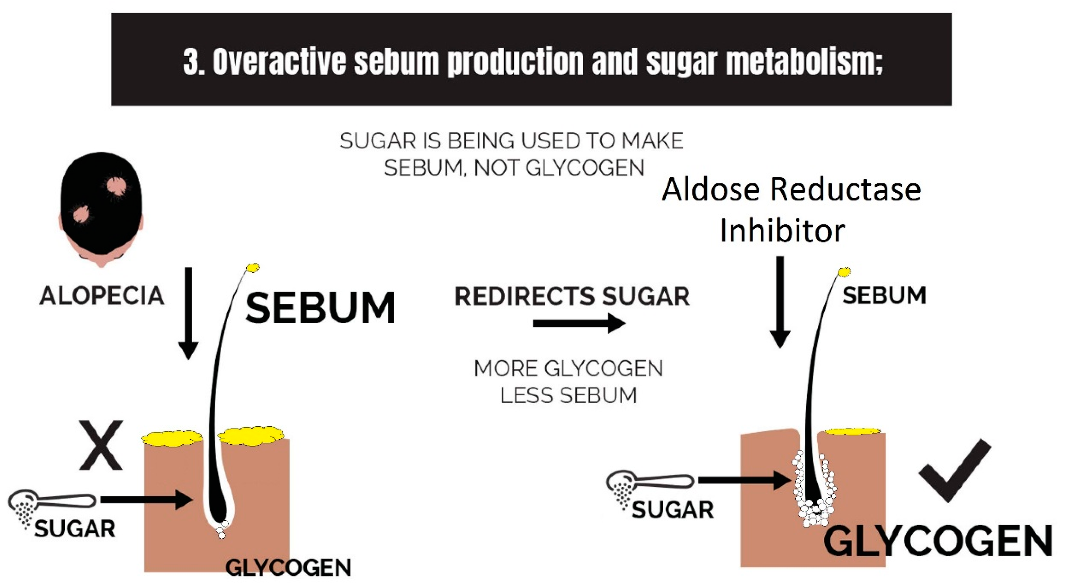
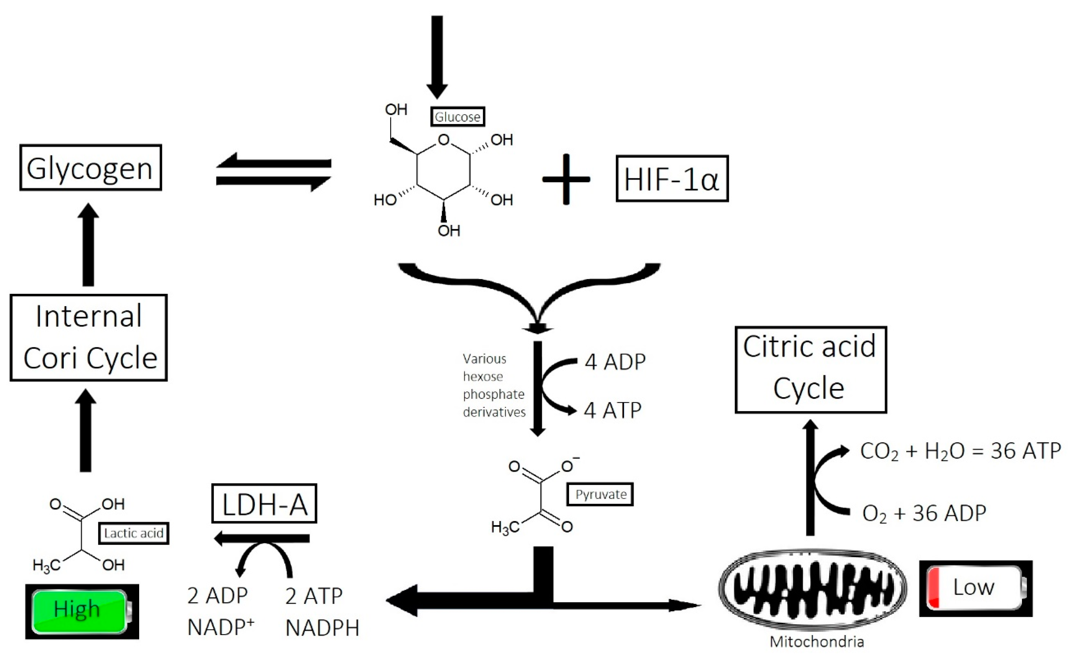
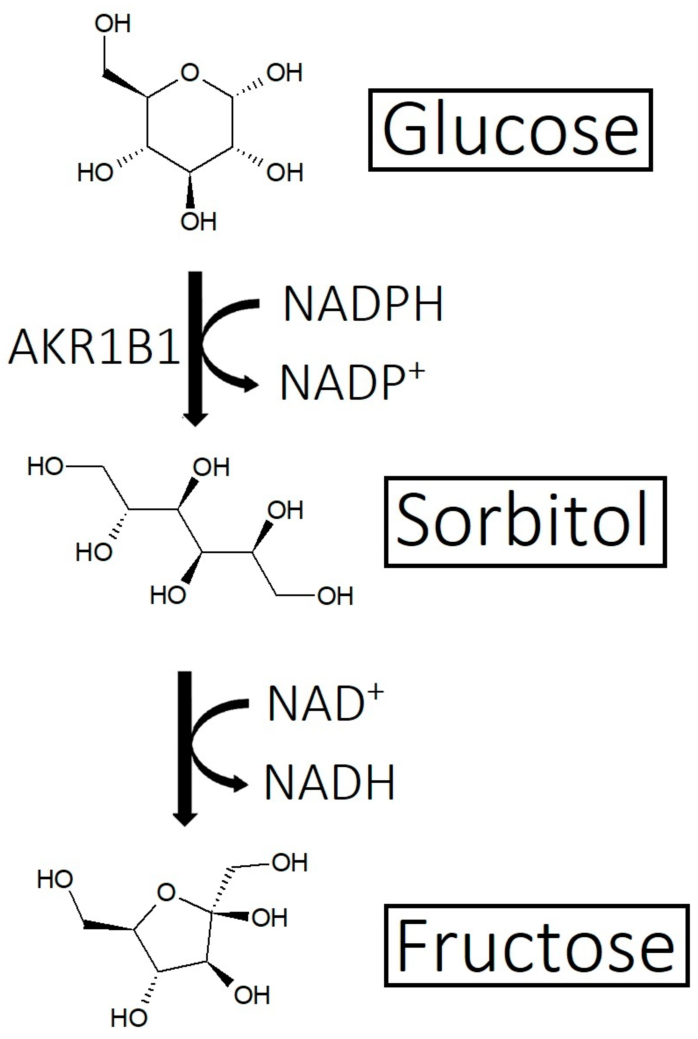
  • Overactive sebum production and sugar metabolism: Prostaglandins and the polyol pathway change the metabolism of sugar so that lipid is produced in favor of glycogen stores; this may be due to increased expression of AKR1B1, a substrate for prostaglandin synthesis, and a trigger of the polyol pathway; insulin-like growth factor 1 is under-expressed in the balding scalp, and this may be why the glycogen stores are depleted.
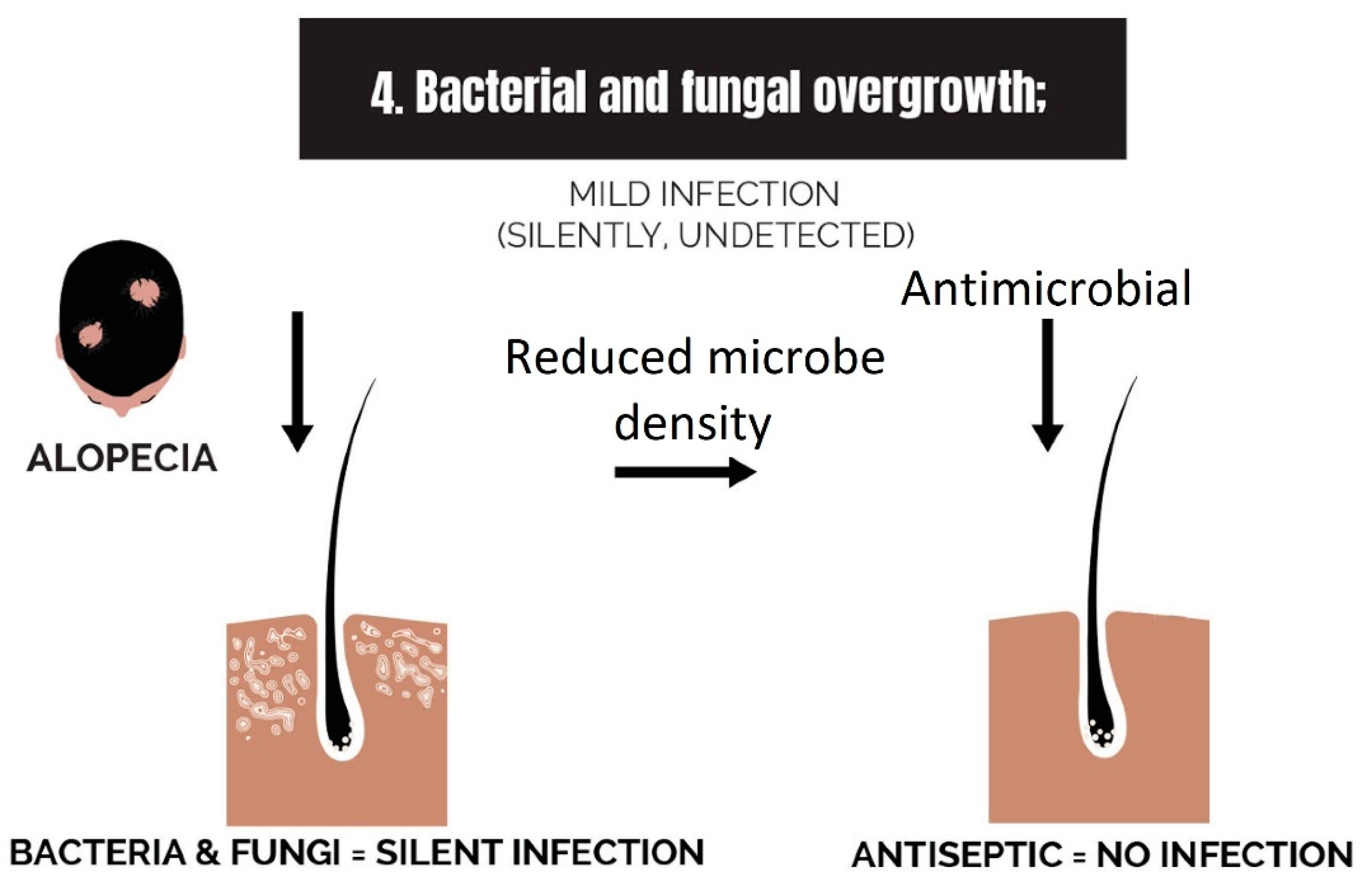
  • Bacterial and fungal overgrowth: Scalps afflicted with hair loss generally have bacterial overgrowth or inflammatory infiltrates as byproducts of clearing microbes. The bacteria can be P. acnes or S. aureus (in cases of scarring alopecia). The fungus is M. furfur.
  • Micro-inflammation: Inflammation in the balding scalp can be severe, such as in scarring alopecia, or it can be low-grade chronic, such as in AGA. The term micro-inflammation was coined because candidates are unaware of the inflammation in AGA; it is subtle.
  • Micro-scarring and collagen: While therapies can halt the progression of fibrosis, it is difficult to reverse it. The best approach is via the use of biomimetic peptides that signal a restructuring of the dermis and promote a return of the stem cell niche.
  • Inefficient circulation and metabolism (i.e., cholesterol and scalp tension): Circulation is two-fold. It involves the transport of nutrients to the scalp, and it is equally as important to transport waste out. The elimination of waste can be antagonized by the inactivity of metabolizing enzymes that are responsible for converting waste into soluble forms, such as the metabolism of cholesterol.
  • Nutrient deficiency (or nutrient synthesis via metabolism, i.e., vitamin D): Nutrient deficiencies can be corrected with supplementation, but the benefit can also be experienced by supplementing an item in the absence of a deficiency of that item, such as by the addition of amino acids to the diet. Furthermore, a deficiency can be caused by a failure of local metabolic processes that create nutrients, such as vitamin D, which can be adequate in terms of dietary intake but is not being utilized by hair follicles in balding scalps.
Cosmeceuticals that target all eight of these etiological components are likely to be more effective than therapies that target a small number of strikes. It is rare for a hair rejuvenating serum that targets all eight strikes to be available in the market.

Author Contributions

N.S. conceptualized and wrote the manuscript. J.R. contributed to the conceptualization and revision of the manuscript. S.B. and D.B. provided suggestions for revision. All authors have read and agreed to the published version of the manuscript.

Funding

This research received no external funding.

Institutional Review Board Statement

Not applicable.

Informed Consent Statement

Not applicable.

Data Availability Statement

Not applicable.

Acknowledgments

The authors would like to acknowledge Creative Klick for assisting with some of the images.

Conflicts of Interest

The authors declare no conflict of interest. The ideas presented in this review and the propounded theories are independent of any financial bias.

References

  1. Wall, D.; Meah, N.; Fagan, N.; York, K.; Sinclair, R. Advances in hair growth. Fac. Rev. 2022, 11, 1. [Google Scholar] [CrossRef] [PubMed]
  2. Houschyar, K.S.; Borrelli, M.R.; Tapking, C.; Popp, D.; Puladi, B.; Ooms, M.; Chelliah, M.P.; Rein, S.; Pförringer, D.; Thor, D.; et al. Molecular Mechanisms of Hair Growth and Regeneration: Current Understanding and Novel Paradigms. Dermatology 2020, 236, 271–280. [Google Scholar] [CrossRef] [PubMed]
  3. Pinto, D.; Sorbellini, E.; Marzani, B.; Rucco, M.; Giuliani, G.; Rinaldi, F. Scalp bacterial shift in Alopecia areata. PLoS ONE 2019, 14, e0215206. [Google Scholar] [CrossRef] [Green Version]
  4. Bolduc, C.; Sperling, L.C.; Shapiro, J. Primary cicatricial alopecia: Other lymphocytic primary cicatricial alopecias and neutrophilic and mixed primary cicatricial alopecias. J. Am. Acad. Dermatol. 2016, 75, 1101–1117. [Google Scholar] [CrossRef] [PubMed]
  5. Gupta, A.K.; Summerbell, R.C. Tinea capitis. Med. Mycol. 2000, 38, 255–287. [Google Scholar] [CrossRef] [PubMed]
  6. Sadgrove, N.J. The ‘bald’ phenotype (androgenetic alopecia) is caused by the high glycaemic, high cholesterol and low mineral ‘western diet’. Trends Food Sci. Technol. 2021, 116, 1170–1178. [Google Scholar] [CrossRef]
  7. Pratt, C.H.; King, L.E.; Messenger, A.G.; Christiano, A.M.; Sundberg, J.P. Alopecia areata. Nat. Rev. Dis. Prim. 2017, 3, 17011. [Google Scholar] [CrossRef] [Green Version]
  8. Randall, V.A. Molecular Basis of Androgenetic Alopecia. In Aging Hair; Trüeb, R.M., Tobin, D.J., Eds.; Springer: Berlin/Heidelberg, Germany, 2010; pp. 9–24. [Google Scholar] [CrossRef]
  9. Thuangtong, R. Vertex Accentuation in Female Pattern Hair Loss in Asians. Siriraj Med. J. 2016, 68, 155–159. [Google Scholar]
  10. Lawson, C.N.; Bakayoko, A.; Callender, V.D. Central Centrifugal Cicatricial Alopecia: Challenges and Treatments. Dermatol. Clin. 2021, 39, 389–405. [Google Scholar] [CrossRef]
  11. Filbrandt, R.; Rufaut, N.; Jones, L.; Sinclair, R. Primary cicatricial alopecia: Diagnosis and treatment. Can. Med. Assoc. J. 2013, 185, 1579. [Google Scholar] [CrossRef] [Green Version]
  12. Oremović, L.; Lugović, L.; Vucić, M.; Buljan, M.; Ozanić-Bulić, S. Cicatricial alopecia as a manifestation of different dermatoses. Acta Dermatovenerol. Croat. ADC 2006, 14, 246–252. [Google Scholar]
  13. Rigopoulos, D.; Stamatios, G.; Ioannides, D. Primary Scarring Alopecias. In Alopecias-Practical Evaluation and Management; Ioannides, D., Tosti, A., Eds.; Current Problems in Dermatology; Karger Medical and Scientific Publishers: Basel, Switzerland, 2015; Volume 47, pp. 76–86. [Google Scholar]
  14. Harries, M.J.; Trueb, R.M.; Tosti, A.; Messenger, A.G.; Chaudhry, I.; Whiting, D.A.; Sinclair, R.; Griffiths, C.E.; Paus, R. How not to get scar(r)ed: Pointers to the correct diagnosis in patients with suspected primary cicatricial alopecia. Br. J. Dermatol. 2009, 160, 482–501. [Google Scholar] [CrossRef]
  15. Whiting, D.A. Cicatricial alopecia: Clinico-pathological findings and treatment. Clin. Dermatol. 2001, 19, 211–225. [Google Scholar] [CrossRef] [PubMed]
  16. Paus, R.; Haslam, I.S.; Sharov, A.A.; Botchkarev, V.A. Pathobiology of chemotherapy-induced hair loss. Lancet Oncol. 2013, 14, e50–e59. [Google Scholar] [CrossRef] [PubMed]
  17. Daunton, A.; Harries, M.; Sinclair, R.; Paus, R.; Tosti, A.; Messenger, A. Chronic Telogen Effluvium: Is it a Distinct Condition? A Systematic Review. Am. J. Clin. Dermatol. 2023. [Google Scholar] [CrossRef]
  18. Hay, R.J. Tinea Capitis: Current Status. Mycopathologia 2017, 182, 87–93. [Google Scholar] [CrossRef] [PubMed] [Green Version]
  19. Billero, V.; Miteva, M. Traction alopecia: The root of the problem. Clin. Cosmet. Investig. Dermatol. 2018, 11, 149–159. [Google Scholar] [CrossRef] [PubMed] [Green Version]
  20. Grant, J.E.; Dougherty, D.D.; Chamberlain, S.R. Prevalence, gender correlates, and co-morbidity of trichotillomania. Psychiatry Res. 2020, 288, 112948. [Google Scholar] [CrossRef]
  21. Su, H.-J.; Cheng, A.-Y.; Liu, C.-H.; Chu, C.-B.; Lee, C.-N.; Hsu, C.-K.; Lee, J.Y.-Y.; Yang, C.-C. Primary scarring alopecia: A retrospective study of 89 patients in Taiwan. J. Dermatol. 2018, 45, 450–455. [Google Scholar] [CrossRef]
  22. Villablanca, S.; Fischer, C.; García-García, S.C.; Mascaró-Galy, J.M.; Ferrando, J. Primary Scarring Alopecia: Clinical-Pathological Review of 72 Cases and Review of the Literature. Ski. Appendage Disord. 2017, 3, 132–143. [Google Scholar] [CrossRef]
  23. Rebora, A.; Guarrera, M.; Baldari, M.; Vecchio, F. Distinguishing Androgenetic Alopecia from Chronic Telogen Effluvium When Associated in the Same Patient: A Simple Noninvasive Method. Arch. Dermatol. 2005, 141, 1243–1245. [Google Scholar] [CrossRef] [PubMed] [Green Version]
  24. Zinkernagel, M.S.; Med, C.; Trüeb, R.M. Fibrosing Alopecia in a Pattern Distribution: Patterned Lichen Planopilaris or Androgenetic Alopecia with a Lichenoid Tissue Reaction Pattern? Arch. Dermatol. 2000, 136, 205–211. [Google Scholar] [CrossRef] [PubMed] [Green Version]
  25. Sadgrove, N.J. The new paradigm for androgenetic alopecia and plant-based folk remedies: 5α-reductase inhibition, reversal of secondary microinflammation and improving insulin resistance. J. Ethnopharmacol. 2018, 227, 206–236. [Google Scholar] [CrossRef]
  26. Habashi-Daniel, A.; Roberts, J.L.; Desai, N.; Thompson, C.T. Absence of catagen/telogen phase and loss of cytokeratin 15 expression in hair follicles in lichen planopilaris. J. Am. Acad. Dermatol. 2014, 71, 969–972. [Google Scholar] [CrossRef] [PubMed]
  27. Jain, N.; Doshi, B.; Khopkar, U. Trichoscopy in alopecias: Diagnosis simplified. Int. J. Trichol. 2013, 5, 170–178. [Google Scholar] [CrossRef] [Green Version]
  28. Park, S.H.; Seol, J.E.; Kim, D.H.; Kim, H. Analysis of Microscopic Examination of Pulled out Hair in Telogen Effluvium Patients. Ann. Dermatol. 2020, 32, 141–145. [Google Scholar] [CrossRef]
  29. McElwee, K.J.; Gilhar, A.; Tobin, D.J.; Ramot, Y.; Sundberg, J.P.; Nakamura, M.; Bertolini, M.; Inui, S.; Tokura, Y.; King, L.E.; et al. What causes alopecia areata? Exp. Dermatol. 2013, 22, 609–626. [Google Scholar] [CrossRef]
  30. Mao, Y.; Xu, Z.; Song, J.; Xie, Y.; Mei, X.; Shi, W. Efficacy of a mixed preparation containing piperine, capsaicin and curcumin in the treatment of alopecia areata. J. Cosmet. Dermatol. 2022, 21, 4510–4514. [Google Scholar] [CrossRef]
  31. Żeberkiewicz, M.; Rudnicka, L.; Malejczyk, J. Immunology of alopecia areata. Cent. Eur. J. Nbsp Immunol. 2020, 45, 325–333. [Google Scholar] [CrossRef]
  32. Wang, E.H.C.; Yu, M.; Breitkopf, T.; Akhoundsadegh, N.; Wang, X.; Shi, F.T.; Leung, G.; Dutz, J.P.; Shapiro, J.; McElwee, K.J. Identification of Autoantigen Epitopes in Alopecia Areata. J. Investig. Dermatol. 2016, 136, 1617–1626. [Google Scholar] [CrossRef] [Green Version]
  33. Gilhar, A.; Landau, M.; Assy, B.; Shalaginov, R.; Serafimovich, S.; Kalish, R.S. Melanocyte-associated T cell epitopes can function as autoantigens for transfer of alopecia areata to human scalp explants on Prkdc(scid) mice. J. Investig. Dermatol. 2001, 117, 1357–1362. [Google Scholar] [CrossRef] [Green Version]
  34. Shapiro, J.; Kaufman, K.D. Use of Finasteride in the Treatment of Men with Androgenetic Alopecia (Male Pattern Hair Loss). J. Investig. Dermatol. Symp. Proc. 2003, 8, 20–23. [Google Scholar] [CrossRef] [PubMed]
  35. Deng, Z.; Chen, M.; Liu, F.; Wang, Y.; Xu, S.; Sha, K.; Peng, Q.; Wu, Z.; Xiao, W.; Liu, T.; et al. Androgen Receptor–Mediated Paracrine Signaling Induces Regression of Blood Vessels in the Dermal Papilla in Androgenetic Alopecia. J. Investig. Dermatol. 2022, 142, 2088–2099.e2089. [Google Scholar] [CrossRef] [PubMed]
  36. Betriu, N.; Jarrosson-Moral, C.; Semino, C.E. Culture and Differentiation of Human Hair Follicle Dermal Papilla Cells in a Soft 3D Self-Assembling Peptide Scaffold. Biomolecules 2020, 10, 684. [Google Scholar] [CrossRef] [PubMed]
  37. Morgan, B.A. The Dermal Papilla: An Instructive Niche for Epithelial Stem and Progenitor Cells in Development and Regeneration of the Hair Follicle. Cold Spring Harb. Perspect. Med. 2014, 4, a015180. [Google Scholar] [CrossRef] [Green Version]
  38. Kwack, M.H.; Ahn, J.S.; Kim, M.K.; Kim, J.C.; Sung, Y.K. Dihydrotestosterone-Inducible IL-6 Inhibits Elongation of Human Hair Shafts by Suppressing Matrix Cell Proliferation and Promotes Regression of Hair Follicles in Mice. J. Investig. Dermatol. 2012, 132, 43–49. [Google Scholar] [CrossRef] [Green Version]
  39. Kwack, M.H.; Sung, Y.K.; Chung, E.J.; Im, S.U.; Ahn, J.S.; Kim, M.K.; Kim, J.C. Dihydrotestosterone-Inducible Dickkopf 1 from Balding Dermal Papilla Cells Causes Apoptosis in Follicular Keratinocytes. J. Investig. Dermatol. 2008, 128, 262–269. [Google Scholar] [CrossRef] [Green Version]
  40. Millar, S.E. Molecular mechanisms regulating hair follicle development. J. Investig. Dermatol. 2002, 118, 216–225. [Google Scholar] [CrossRef]
  41. Heilmann, S.; Kiefer, A.K.; Fricker, N.; Drichel, D.; Hillmer, A.M.; Herold, C.; Tung, J.Y.; Eriksson, N.; Redler, S.; Betz, R.C.; et al. Androgenetic Alopecia: Identification of Four Genetic Risk Loci and Evidence for the Contribution of WNT Signaling to Its Etiology. J. Investig. Dermatol. 2013, 133, 1489–1496. [Google Scholar] [CrossRef] [Green Version]
  42. Sadgrove, N.J.; Simmonds, M.S.J. Topical and nutricosmetic products for healthy hair and dermal anti-aging using ‘dual-acting’ (2 for 1) plant-based peptides, hormones, and cannabinoids. FASEB BioAdv. 2021, 3, 601. [Google Scholar] [CrossRef]
  43. Shin, H.; Yoo, H.G.; Inui, S.; Itami, S.; Kim, I.G.; Cho, A.-R.; Lee, D.H.; Park, W.S.; Kwon, O.; Cho, K.H.; et al. Induction of transforming growth factor-beta 1 by androgen is mediated by reactive oxygen species in hair follicle dermal papilla cells. BMB Rep. 2013, 46, 460–464. [Google Scholar] [CrossRef]
  44. Lu, G.-Q.; Wu, Z.-B.; Chu, X.-Y.; Bi, Z.-G.; Fan, W.-X. An investigation of crosstalk between Wnt/β-catenin and transforming growth factor-β signaling in androgenetic alopecia. Medicine 2016, 95, e4297. [Google Scholar] [CrossRef] [PubMed]
  45. Akhmetshina, A.; Palumbo, K.; Dees, C.; Bergmann, C.; Venalis, P.; Zerr, P.; Horn, A.; Kireva, T.; Beyer, C.; Zwerina, J.; et al. Activation of canonical Wnt signalling is required for TGF-β-mediated fibrosis. Nat. Commun. 2012, 3, 735. [Google Scholar] [CrossRef] [Green Version]
  46. Philips, N.; Auler, S.; Hugo, R.; Gonzales, S. Beneficial regulation of matrix metalloproteinases for skin health. Enzym. Res. 2011, 2011, 427286. [Google Scholar] [CrossRef] [Green Version]
  47. Beoy, L.A.; Woei, W.J.; Hay, Y.K. Effects of tocotrienol supplementation on hair growth in human volunteers. Trop. Life Sci. Res. 2010, 21, 91–98. [Google Scholar] [PubMed]
  48. Yanagisawa, M.; Fujimaki, H.; Takeda, A.; Nemoto, M.; Sugimoto, T.; Sato, A. Long-term (10-year) efficacy of finasteride in 523 Japanese men with androgenetic alopecia. Clin. Res. Trials 2019, 5, 1–5. [Google Scholar] [CrossRef] [Green Version]
  49. PFS. Post-Finasteride Syndrome Foundation. Available online: https://www.pfsfoundation.org/ (accessed on 8 July 2022).
  50. Diviccaro, S.; Melcangi, R.C.; Giatti, S. Post-finasteride syndrome: An emerging clinical problem. Neurobiol. Stress 2020, 12, 100209. [Google Scholar] [CrossRef]
  51. Traish, A.M. Post-finasteride syndrome: A surmountable challenge for clinicians. Fertil. Steril. 2020, 113, 21–50. [Google Scholar] [CrossRef] [PubMed]
  52. Carbone, D.J.; Hodges, S. Medical therapy for benign prostatic hyperplasia: Sexual dysfunction and impact on quality of life. Int. J. Impot. Res. 2003, 15, 299–306. [Google Scholar] [CrossRef] [Green Version]
  53. Baas, W.R.; Butcher, M.J.; Lwin, A.; Holland, B.; Herberts, M.; Clemons, J.; Delfino, K.; Althof, S.; Kohler, T.S.; McVary, K.T. A Review of the FAERS Data on 5-Alpha Reductase Inhibitors: Implications for Postfinasteride Syndrome. Male Sex. Dysfunct. 2018, 120, 143–149. [Google Scholar] [CrossRef]
  54. Harries, M.J.; Paus, R. The Pathogenesis of Primary Cicatricial Alopecias. Am. J. Pathol. 2010, 177, 2152–2162. [Google Scholar] [CrossRef] [PubMed]
  55. Somani, N.; Bergfeld, W.F. Cicatricial alopecia: Classification and histopathology. Dermatol. Therapy. 2008, 21, 221–237. [Google Scholar] [CrossRef]
  56. Khumalo, N.P.; Pillay, K.; Ngwanya, R.M. Acute ‘relaxer’-associated scarring alopecia: A report of five cases. Br. J. Dermatol. 2007, 156, 1394–1396. [Google Scholar] [CrossRef]
  57. Subash, J.; Alexander, T.; Beamer, V.; McMichael, A. A proposed mechanism for central centrifugal cicatricial alopecia. Exp. Dermatol. 2020, 29, 190–195. [Google Scholar] [CrossRef] [PubMed] [Green Version]
  58. Harries, M.J.; Sinclair, R.D.; MacDonald-Hull, S.; Whiting, D.A.; Griffiths, C.E.M.; Paus, R. Management of primary cicatricial alopecias: Options for treatment. Br. J. Dermatol. 2008, 159, 1–22. [Google Scholar] [CrossRef] [PubMed]
  59. Polak-Witka, K.; Rudnicka, L.; Blume-Peytavi, U.; Vogt, A. The role of the microbiome in scalp hair follicle biology and disease. Exp. Dermatol. 2020, 29, 286–294. [Google Scholar] [CrossRef] [Green Version]
  60. Ho, A.; Shapiro, J. Medical therapy for frontal fibrosing alopecia: A review and clinical approach. J. Am. Acad. Dermatol. 2019, 81, 568–580. [Google Scholar] [CrossRef]
  61. Harnchoowong, S.; Suchonwanit, P. PPAR-γ Agonists and Their Role in Primary Cicatricial Alopecia. PPAR Res. 2017, 2017, 2501248. [Google Scholar] [CrossRef] [Green Version]
  62. Zhang, G.-Y.; Cheng, T.; Zheng, M.-H.; Yi, C.-G.; Pan, H.; Li, Z.-J.; Chen, X.-L.; Yu, Q.; Jiang, L.-F.; Zhou, F.-Y.; et al. Peroxisome proliferator-activated receptor-γ (PPAR-γ) agonist inhibits transforming growth factor-beta1 and matrix production in human dermal fibroblasts. J. Plast. Reconstr. Aesthet. Surg. 2010, 63, 1209–1216. [Google Scholar] [CrossRef]
  63. Ho, B.S.-Y.; Ho, E.X.P.; Chu, C.W.; Ramasamy, S.; Bigliardi-Qi, M.; de Sessions, P.F.; Bigliardi, P.L. Microbiome in the hair follicle of androgenetic alopecia patients. PLoS ONE 2019, 14, e0216330. [Google Scholar] [CrossRef] [Green Version]
  64. Trüeb, R.M. Telogen Effluvium: Is There a Need for a New Classification? Ski. Appendage Disord. 2016, 2, 39–44. [Google Scholar] [CrossRef] [PubMed] [Green Version]
  65. Rebora, A. Proposing a Simpler Classification of Telogen Effluvium. Ski. Appendage Disord. 2016, 2, 35–38. [Google Scholar] [CrossRef] [Green Version]
  66. Malkud, S. Telogen Effluvium: A Review. J. Clin. Diagn. Res. 2015, 9, We01–We03. [Google Scholar] [CrossRef] [PubMed]
  67. Thom, E. Stress and the Hair Growth Cycle: Cortisol-Induced Hair Growth Disruption. J. Drugs Dermatol. 2016, 15, 1001–1004. [Google Scholar] [PubMed]
  68. Sorriento, D.; Iaccarino, G. Inflammation and Cardiovascular Diseases: The Most Recent Findings. Int. J. Mol. Sci. 2019, 20, 3879. [Google Scholar] [CrossRef] [PubMed] [Green Version]
  69. Guarnieri, G.; Bertagna De Marchi, L.; Marcon, A.; Panunzi, S.; Batani, V.; Caminati, M.; Furci, F.; Senna, G.; Alaibac, M.; Vianello, A. Relationship between hair shedding and systemic inflammation in COVID-19 pneumonia. Ann. Med. 2022, 54, 869–874. [Google Scholar] [CrossRef]
  70. Sinclair, R. Male pattern androgenetic alopecia. BMJ 1998, 317, 865. [Google Scholar] [CrossRef]
  71. Guarrera, M.; Rebora, A. The Higher Number and Longer Duration of Kenogen Hairs Are the Main Cause of the Hair Rarefaction in Androgenetic Alopecia. Ski. Appendage Disord. 2019, 5, 152–154. [Google Scholar] [CrossRef]
  72. English, R.; Ruiz, S. Conflicting Reports Regarding the Histopathological Features of Androgenic Alopecia: Are Biopsy Location, Hair Diameter Diversity, and Relative Hair Follicle Miniaturization Partly to Blame? Clin. Cosmet. Investig. Dermatol. 2021, 14, 357–365. [Google Scholar] [CrossRef]
  73. Garza, L.A.; Liu, Y.; Yang, Z.; Alagesan, B.; Lawson, J.A.; Norberg, S.M.; Loy, D.E.; Zhao, T.; Blatt, H.B.; Stanton, D.C.; et al. Prostaglandin D2 inhibits hair growth and is elevated in bald scalp of men with androgenetic alopecia. Sci. Transl. Med. 2012, 4, 126ra134. [Google Scholar] [CrossRef] [Green Version]
  74. Jeong, K.H.; Jung, J.H.; Kim, J.E.; Kang, H. Prostaglandin D2-mediated DP2 and AKT signal regulate the activation of androgen receptors in human dermal papilla cells. Int. J. Mol. Sci. 2018, 19, 556. [Google Scholar] [CrossRef] [Green Version]
  75. Ho, B.S.-Y.; Vaz, C.; Ramasamy, S.; Chew, E.G.Y.; Mohamed, J.S.; Jaffar, H.; Hillmer, A.; Tanavde, V.; Bigliardi-Qi, M.; Bigliardi, P.L. Progressive expression of PPARGC1α is associated with hair miniaturization in androgenetic alopecia. Sci. Rep. 2019, 9, 8771. [Google Scholar] [CrossRef] [Green Version]
  76. Martínez-Redondo, V.; Pettersson, A.T.; Ruas, J.L. The hitchhiker’s guide to PGC-1α isoform structure and biological functions. Diabetologia 2015, 58, 1969–1977. [Google Scholar] [CrossRef] [PubMed] [Green Version]
  77. Horii, N.; Hasegawa, N.; Fujie, S.; Uchida, M.; Iemitsu, M. Resistance exercise-induced increase in muscle 5α-dihydrotestosterone contributes to the activation of muscle Akt/mTOR/p70S6K- and Akt/AS160/GLUT4-signaling pathways in type 2 diabertic rats. FASEB J. 2020, 34, 11047–11057. [Google Scholar] [CrossRef] [PubMed]
  78. Koshihara, Y.; Kawamura, M. Prostaglandin D2 stimulates calcification of human osteoblast cells. Biochem. Biophys. Res. Commun. 1989, 159, 1206–1212. [Google Scholar] [CrossRef]
  79. English, R.S. A hypothetical pathogenesis model for androgenic alopecia: Clarifying the dihydrotestosterone paradox and rate-limiting recovery factors. Med. Hypotheses 2018, 111, 73–81. [Google Scholar] [CrossRef]
  80. Villarreal-Villarreal, C.D.; Sinclair, R.D.; Martínez-Jacobo, L.; Garza-Rodríguez, V.; Rodríguez-León, S.A.; Lamadrid-Zertuche, A.C.; Rodríguez-Gutierrez, R.; Ortiz-Lopez, R.; Rojas-Martinez, A.; Ocampo-Candiani, J. Prostaglandins in androgenetic alopecia in 12 men and four female. J. Eur. Acad. Dermatol. Venereol. 2019, 33, e214–e215. [Google Scholar] [CrossRef] [PubMed]
  81. Sasaki, S.; Hozumi, Y.; Kondo, S. Influence of prostaglandin F2α and its analogues on hair regrowth and follicular melanogenesis in a murine model. Exp. Dermatol. 2005, 14, 323–328. [Google Scholar] [CrossRef] [PubMed]
  82. Chovarda, E.; Sotiriou, E.; Lazaridou, E.; Vakirlis, E.; Ioannides, D. The role of prostaglandins in androgenetic alopecia. Int. J. Dermatol. 2021, 60, 730–735. [Google Scholar] [CrossRef]
  83. Iinuma, K.; Sato, T.; Akimoto, N.; Noguchi, N.; Sasatsu, M.; Nishijima, S.; Kurokawa, I.; Ito, A. Involvement of Priopionibacterium acnes in the augmentation of lipogenesis in hamster sebaceous glans in vivo and in vitro. J. Investig. Dermatol. 2009, 129, 2113–2119. [Google Scholar] [CrossRef] [Green Version]
  84. Harris, S.G.; Phipps, R.P. Prostaglandin D2, its metabolite 15-d-PGJ2, and peroxisome proliferator activated receptor-γ agonists induce apoptosis in transformed, but not normal, human T lineage cells. Immunology 2002, 105, 23–34. [Google Scholar] [CrossRef]
  85. Schadinger, S.E.; Bucher, N.L.R.; Schreiber, B.M.; Farmer, S.R. PPARγ2 regulates lipogenesis and lipid accumulation in steatotic hepatocytes. Am. J. Physiol. Endocrinol. Metab. 2005, 288, E1195–E1205. [Google Scholar] [CrossRef]
  86. Bie, Q.; Dong, H.; Jin, C.; Zhang, H.; Zhang, B. 15d-PGJ2 is a new hope for controlling tumor growth. Am. J. Transl. Res. 2018, 10, 648–658. [Google Scholar]
  87. Iwata, C.; Akimoto, N.; Sato, T.; Morokuma, Y.; Ito, A. Augmentation of Lipogenesis by 15-Deoxy-Δ12,14-Prostaglandin J2 in Hamster Sebaceous Glands: Identification of Cytochrome P-450-mediated 15-Deoxy-Δ12,14-Prostaglandin J2 Production. J. Investig. Dermatol. 2005, 125, 865–872. [Google Scholar] [CrossRef] [PubMed] [Green Version]
  88. Johnson, R.J.; Sánchez-Lozada, L.G.; Andrews, P.; Lanaspa, M.A. Perspective: A historical and scientific perspective of sugar and its relation with obesity and diabetes. Adv. Nutr. 2017, 8, 412–422. [Google Scholar] [CrossRef] [PubMed] [Green Version]
  89. Nagata, N.; Kusakari, Y.; Fukunishi, Y.; Inoue, T.; Urade, Y. Catalytic mechanism of the primary human prostaglandin F2α synthase, aldo-keto reductase 1B1--prostaglandin D2 synthase activity in the absence of NADP(H). FEBS J. 2011, 278, 1288–1298. [Google Scholar] [CrossRef] [PubMed]
  90. Kabututu, Z.; Manin, M.; Pointud, J.C.; Maruyama, T.; Nagata, N.; Lambert, S.; Lefrançois-Martinez, A.M.; Martinez, A.; Urade, Y. Prostaglandin F2alpha synthase activities of aldo-keto reductase 1B1, 1B3 and 1B7. J. Biochem. 2009, 145, 161–168. [Google Scholar] [CrossRef]
  91. Yan, L.J. Redox imbalance stress in diabetes mellitus: Role of the polyol pathway. Anim. Model. Exp. Med. 2018, 1, 7–13. [Google Scholar] [CrossRef]
  92. Lacroix Pépin, N.; Chapdelaine, P.; Fortier, M.A. Evaluation of the prostaglandin F synthase activity of human and bovine aldo-keto reductases: AKR1A1s complement AKR1B1s as potent PGF synthases. Prostaglandins Other Lipid Mediat. 2013, 106, 124–132. [Google Scholar] [CrossRef]
  93. Mansmann, H.C. Consider magnesium homeostasis: III: Cytochrome P450 enzymes and drug toxicity. Padiatr. ASthma Allergy Immunol. 1994, 8, 7–28. [Google Scholar] [CrossRef]
  94. Dharam Kumar, K.C.; Kishan Kumar, Y.H.; Neladimmanahally, V. Association of Androgenetic Alopecia with Metabolic Syndrome: A Case-control Study on 100 Patients in a Tertiary Care Hospital in South India. Indian J. Endocrinol. Metab. 2018, 22, 196–199. [Google Scholar] [CrossRef] [PubMed]
  95. Matilainen, V.; Koskela, P.; Keinänen-Kiukaanniemi, S. Early androgenetic alopecia as a marker of insulin resistance. Lancet 2000, 356, 1165–1166. [Google Scholar] [CrossRef]
  96. Bakry, O.A.; Shoeib, M.A.M.; El Shafiee, M.K.; Hassan, A. Androgenetic alopecia, metabolic syndrome, and insulin resistance: Is there any association? A case-control study. Indian Dermatol. Online J. 2014, 5, 276–281. [Google Scholar] [CrossRef]
  97. Arias-Santiago, S.; Gutiérrez-Salmerón, M.T.; Buendía-Eisman, A.; Girón-Prieto, M.S.; Naranjo-Sintes, R. Sex hormone-binding globulin and risk of hyperglycemia in patients with androgenetic alopecia. J. Am. Acad. Dermatol. 2011, 65, 48–53. [Google Scholar] [CrossRef] [PubMed]
  98. Chaikittisilpa, S.; Rattanasirisin, N.; Panchaprateep, R.; Orprayoon, N.; Phutrakul, P.; Suwan, A.; Jaisamrarn, U. Prevalence of female pattern hair loss in postmenopausal women: A cross-sectional study. Menopause 2022, 29, 415–420. [Google Scholar] [CrossRef] [PubMed]
  99. Shi, X.; Tuan, H.; Na, X.; Yang, H.; Yang, Y.; Zhang, Y.; Xi, M.; Tan, Y.; Yang, C.; Zhang, J.; et al. The Association between Sugar-Sweetened Beverages and Male Pattern Hair Loss in Young Men. Nutrients 2023, 15, 214. [Google Scholar] [CrossRef] [PubMed]
  100. Chew, E.G.Y.; Ho, B.S.-Y.; Ramasamy, S.; Dawson, T.; Tennakoon, C.; Liu, X.; Leong, W.M.S.; Yang, W.Y.S.; Lim, S.Y.D.; Jaffar, H.; et al. Comparative transcriptome profiling provides new insights into mechanisms of androgenetic alopecia progression: Whole transcriptome discovery study identifies altered oxidation-reduction state of hair follicles of androgenetic alopecia patients. Br. J. Dermatol. 2017, 176, 265. [Google Scholar] [CrossRef] [PubMed]
  101. Figlak, K.; Paus, R.; Williams, G.; Philpott, M. 597 Outer root sheath is able to synthesise glycogen from lactate-investigating glycogen metabolism in human hair follicles. J. Investig. Dermatol. 2019, 139, S317. [Google Scholar] [CrossRef] [Green Version]
  102. Plikus, M.V.; Vollmers, C.; Cruz, D.d.l.; Chaix, A.; Ramos, R.; Panda, S.; Chuong, C.-M. Local circadian clock gates cell cycle progression of transient amplifying cells during regenerative hair cycling. Proc. Natl. Acad. Sci. USA 2013, 110, E2106–E2115. [Google Scholar] [CrossRef]
  103. Figlak, K.; Williams, G.; Bertolini, M.; Paus, R.; Philpott, M.P. Human hair follicles operate an internal Cori cycle and modulate their growth via glycogen phosphorylase. Sci. Rep. 2021, 11, 20761. [Google Scholar] [CrossRef]
  104. Lund, J.; Aas, V.; Tingstad, R.H.; Van Hees, A.; Nikolić, N. Utilization of lactic acid in human myotubes and interplay with glucose and fatty acid metabolism. Sci. Rep. 2018, 8, 9814. [Google Scholar] [CrossRef] [PubMed] [Green Version]
  105. Williams, R.; Philpott, M.P.; Kealey, T. Metabolism of Freshly Isolated Human Hair Follicles Capable of Hair Elongation: A Glutaminolytic, Aerobic Glycolytic Tissue. J. Investig. Dermatol. 1993, 100, 834–840. [Google Scholar] [CrossRef] [PubMed]
  106. Kierans, S.J.; Taylor, C.T. Regulation of glycolysis by the hypoxia-inducible factor (HIF): Implications for cellular physiology. J. Physiol. 2021, 599, 23–37. [Google Scholar] [CrossRef] [PubMed]
  107. Liu, Q.; Tang, Y.; Huang, Y.; Wang, J.a.; Yang, K.; Zhang, Y.; Pu, W.; Liu, J.; Shi, X.; Ma, Y.; et al. Insights into male androgenetic alopecia using comparative transcriptome profiling: Hypoxia-inducible factor-1 and Wnt/β-catenin signalling pathways. Br. J. Dermatol. 2022, 187, 936–947. [Google Scholar] [CrossRef]
  108. Philpott, M. Transcriptomic analysis identifies regulators of the Wnt signalling and hypoxia-inducible factor pathways as possible mediators of androgenetic alopecia. Br. J. Dermatol. 2022, 187, 845. [Google Scholar] [CrossRef]
  109. Seo, J.; Yan, L.; Kageyama, T.; Nanmo, A.; Chun, Y.-S. Hypoxia inducible factor-1a promotes trichogenic gene expression in human dermal papilla cells. Sci. Rep. 2023, in press. [CrossRef]
  110. Flores, A.; Schell, J.; Krall, A.S.; Jelinek, D.; Miranda, M.; Grigorian, M.; Braas, D.; White, A.C.; Zhou, J.L.; Graham, N.A.; et al. Lactate dehydrogenase activity drives hair follicle stem cell activation. Nat. Cell Biol. 2017, 19, 1017–1026. [Google Scholar] [CrossRef] [Green Version]
  111. Zuloaga, K.L.; Gonzales, R.J. Dihydrotestosterone attenuates hypoxia inducible factor-1α and cyclooxygenase-2 in cerebral arteries during hypoxia or hypoxia with glucose deprivation. Am. J. Physiol. Heart Circ. Physiol. 2011, 301, H1882–H1890. [Google Scholar] [CrossRef]
  112. Chew, E.G.Y.; Lim, T.C.; Leong, M.F.; Liu, X.; Sia, Y.Y.; Leong, S.T.; Yan-Jiang, B.C.; Stoecklin, C.; Borhan, R.; Heilmann-Heimbach, S.; et al. Observations that suggest a contribution of altered dermal papilla mitochondrial function to androgenetic alopecia. Exp. Dermatol. 2022, 31, 906–917. [Google Scholar] [CrossRef]
  113. Bogacka, I.; Ukropcova, B.; McNeil, M.; Gimble, J.M.; Smith, S.R. Structural and functional consequences of mitochondrial biogenesis in human adipocytes in vitro. J. Clin. Endocrinol. Metab. 2005, 90, 6650–6656. [Google Scholar] [CrossRef] [Green Version]
  114. Jin, E.S.; Lee, M.H.; Murphy, R.E.; Malloy, C.R. Pentose phosphate pathway activity parallels lipogenesis but not antioxidant processes in rat liver. Am. J. Physiol. Endocrinol. Metab. 2018, 314, E543–E551. [Google Scholar] [CrossRef] [Green Version]
  115. Garg, S.S.; Gupta, J. Polyol pathway and redox balance in diabetes. Pharmacol. Res. 2022, 182, 106326. [Google Scholar] [CrossRef]
  116. Fujimori, K.; Ueno, T.; Nagata, N.; Kashiwagi, K.; Aritake, K.; Amano, F.; Urade, Y. (2010). Suppression of adipocyte differentiation by aldo-keto reductase 1B3 acting as prostaglandin F2α synthase. J. Biol. Chem. 2010, 285, 8880–8886. [Google Scholar] [CrossRef] [Green Version]
  117. Kondrakhina, I.N.; Verbenko, D.A.; Zatevalov, A.M.; Gatiatulina, E.R.; Nikonorov, A.A.; Deryabin, D.G.; Kubanov, A.A. A Cross-sectional Study of Plasma Trace Elements and Vitamins Content in Androgenetic Alopecia in Men. Biol. Trace Elem. Res. 2021, 199, 3232–3241. [Google Scholar] [CrossRef] [PubMed]
  118. Page, S.; Salem, M.; Laughlin, M.R. Intracellular Mg2+ regulates ADP phosphorylation and adenine nucleotide synthesis in human erythrocytes. Am. J. Physiol. 1998, 274, E920–E927. [Google Scholar] [CrossRef]
  119. Villa-Bellosta, R. Dietary magnesium supplementation improves lifespan in a mouse model of progeria. EMBO Mol. Med. 2020, 12, e12423. [Google Scholar] [CrossRef]
  120. Rizžner, T.L.n.; Lin, H.K.; Peehl, D.M.; Steckelbroeck, S.; Bauman, D.R.; Penning, T.M. Human Type 3 3α-Hydroxysteroid Dehydrogenase (Aldo-Keto Reductase 1C2) and Androgen Metabolism in Prostate Cells. Endocrinology 2003, 144, 2922–2932. [Google Scholar] [CrossRef] [Green Version]
  121. Panchaprateep, R.; Asawanonda, P. Insulin-like growth factor-1: Roles in androgenetic alopecia. Exp. Dermatol. 2014, 23, 216–218. [Google Scholar] [CrossRef]
  122. Muhič, M.; Vardjan, N.; Chowdhury, H.H.; Zorec, R.; Kreft, M. Insulin and Insulin-like Growth Factor 1 (IGF-1) Modulate Cytoplasmic Glucose and Glycogen Levels but Not Glucose Transport across the Membrane in Astrocytes. J. Biol. Chem. 2015, 290, 11167–11176. [Google Scholar] [CrossRef] [Green Version]
  123. Perlino, E.; Tommasi, S.; Moro, L.; Bellizzi, A.; Marra, E.; Casavola, V.; Reshkin, S.J. TGF-beta1 and IGF-1 expression are differently regulated by serum in metastatic and non-metastatic human breast cancer cells. Int. J. Oncol. 2000, 16, 155–160. [Google Scholar] [CrossRef]
  124. Rudramurthy, S.M.; Honnavar, P.; Dogra, S.; Yegneswaran, P.P.; Handa, S.; Chakrabarti, A. Association of Malassezia species with dandruff. Indian J. Med. Res. 2014, 139, 431–437. [Google Scholar]
  125. Piérard-Franchimont, C.; De Doncker, P.; Cauwenbergh, G.; Piérard, G.E. Ketoconazole Shampoo: Effect of Long-Term Use in Androgenic Alopecia. Dermatology 1998, 196, 474–477. [Google Scholar] [CrossRef] [PubMed]
  126. Peyravian, N.; Deo, S.; Daunert, S.; Jimenez, J.J. The Inflammatory Aspect of Male and Female Pattern Hair Loss. J. Inflamm. Res. 2020, 13, 879–881. [Google Scholar] [CrossRef]
  127. Halley-Stott, R.P.; Adeola, H.A.; Khumalo, N.P. Destruction of the stem cell Niche, Pathogenesis and Promising Treatment Targets for Primary Scarring Alopecias. Stem Cell Rev. Rep. 2020, 16, 1105–1120. [Google Scholar] [CrossRef]
  128. Yoo, H.G.; Kim, J.S.; Lee, S.R.; Pyo, H.K.; Moon, H.I.; Lee, J.H.; Kwon, O.S.; Chung, J.H.; Kim, K.H.; Eun, H.C.; et al. Perifollicular Fibrosis: Pathogenetic Role in Androgenetic Alopecia. Biol. Pharm. Bull. 2006, 29, 1246–1250. [Google Scholar] [CrossRef] [Green Version]
  129. Katsuoka, K.; Mauch, C.; Schell, H.; Hornstein, O.P.; Krieg, T. Collagen-type synthesis in human-hair papilla cells in culture. Arch. Dermatol. Res. 1988, 280, 140–144. [Google Scholar] [CrossRef] [PubMed]
  130. Askew, E.B.; Gampe, R.T.; Stanley, T.B.; Faggart, J.L.; Wilson, E.M. Modulation of Androgen Receptor Activation Function 2 by Testosterone and Dihydrotestosterone. J. Biol. Chem. 2007, 282, 25801–25816. [Google Scholar] [CrossRef] [PubMed] [Green Version]
  131. Pleiner, J.; Mittermayer, F.; Schaller, G.; Marsik, C.; MacAllister, R.J.; Wolzt, M. Inflammation-induced vasoconstrictor hyporeactivity is caused by oxidative stress. J. Am. Coll. Cardiol. 2003, 42, 1656–1662. [Google Scholar] [CrossRef] [Green Version]
  132. Freund, B.J.; Schwartz, M. Treatment of male pattern baldness with botulinum toxin: A pilot study. Plast. Reconstr. Surg. 2010, 126, 246e–248e. [Google Scholar] [CrossRef]
  133. English, R.S., Jr.; Barazesh, J.M. Self-Assessments of Standardized Scalp Massages for Androgenic Alopecia: Survey Results. Dermatol. Ther. 2019, 9, 167–178. [Google Scholar] [CrossRef] [Green Version]
  134. Hoelzel, F. Baldness and calcification of the “Ivory Dome”. J. Am. Med. Assoc. 1942, 119, 968. [Google Scholar] [CrossRef]
  135. Hotta, N.; Kawamura, T.; Umemura, T. Are the polyol pathway and hyperuricemia partners in the development of non-alcoholic fatty liver disease in diabetes? J. Diabetes Investig. 2020, 11, 786–788. [Google Scholar] [CrossRef] [PubMed]
  136. Trevisan, M.; Farinaro, E.; Krogh, V.; Jossa, F.; Giumetti, D.; Fusco, G.; Panico, S.; Mellone, C.; Frascatore, S.; Scottoni, A.; et al. Baldness and coronary heart disease risk factors. J. Clin. Epidemiol. 1993, 46, P1213–P1218. [Google Scholar] [CrossRef]
  137. Goldman, C.K.; Tsai, J.C.; Soroceanu, L.; Gillespie, G.Y. Loss of vascular endothelial growth factor in human alopecia hair follicles. J. Investig. Dermatol. 1995, 104, 18s–20s. [Google Scholar] [CrossRef] [PubMed]
  138. Zimna, A.; Kurpisz, M. Hypoxia-Inducible Factor-1 in Physiological and Pathophysiological Angiogenesis: Applications and Therapies. Biomed. Res. Int. 2015, 2015, 549412. [Google Scholar] [CrossRef] [Green Version]
  139. Dey-Rao, R.; Sinha, A.A. A genomic approach to susceptibility and pathogenesis leads to identifying potential novel therapeutic targets in androgenetic alopecia. Genomics 2017, 109, 165–176. [Google Scholar] [CrossRef] [PubMed]
  140. Maguire, M.; Larsen, M.C.; Vezina, C.M.; Quadro, L.; Kim, Y.-K.; Tanumihardjo, S.A.; Jefcoate, C.R. Cyp1b1 directs Srebp-mediated cholesterol and retinoid synthesis in perinatal liver; Association with retinoic acid activity during fetal development. PLoS ONE 2020, 15, e0228436. [Google Scholar] [CrossRef]
  141. Zhang, D.; Tomisato, W.; Su, L.; Sun, L.; Choi, J.H.; Zhang, Z.; Wang, K.-W.; Zhan, X.; Choi, M.; Li, X.; et al. Skin-specific regulation of SREBP processing and lipid biosynthesis by glycerol kinase 5. Proc. Natl. Acad. Sci. USA 2017, 114, E5197–E5206. [Google Scholar] [CrossRef]
  142. Trost, L.B.; Bergfeld, W.F.; Calogeras, E. The diagnosis and treatment of iron deficiency and its potential relationship to hair loss. J. Am. Acad. Dermatol. 2006, 54, 824–844. [Google Scholar] [CrossRef]
  143. Kil, M.S.; Kim, C.W.; Kim, S.S. Analysis of Serum Zinc and Copper Concentrations in Hair Loss. Ann. Dermatol. 2013, 25, 405–409. [Google Scholar] [CrossRef] [Green Version]
  144. Ozturk, P.; Kurutas, E.; Ataseven, A.; Dokur, N.; Gumusalan, Y.; Gorur, A.; Tamer, L.; Inaloz, S. BMI and levels of zinc, copper in hair, serum and urine of Turkish male patients with androgenetic alopecia. J. Trace Elem. Med. Biol. 2014, 28, 266–270. [Google Scholar] [CrossRef] [PubMed]
  145. Trüeb, R.M. The Difficult Hair Loss Patient: Guide to Successful Management of Alopecia and Related Conditions; Springer: Cham, Switzerland, 2015. [Google Scholar]
  146. Michel, L.; Reygagne, P.; Benech, P.; Jean-Louis, F.; Scalvino, S.; So, S.L.K.; Hamidou, Z.; Bianoici, S.; Pouch, J.; Ducos, B.; et al. Study of gene expression alteration in male androgenetic alopecia: Evidence of predominant molecular signalling pathways. Br. J. Dermatol. 2017, 177, 1322–1336. [Google Scholar] [CrossRef] [PubMed] [Green Version]
  147. Dai, Q.; Zhu, X.; Manson, J.E.; Song, Y.; Li, X.; Franke, A.A.; Costello, R.B.; Rosanoff, A.; Nian, H.; Fan, L.; et al. Magnesium status and supplementation influence vitamin D status and metabolism: Results from a randomized trial. Am. J. Clin. Nutr. 2018, 108, 1249–1258. [Google Scholar] [CrossRef] [PubMed] [Green Version]
  148. Pickart, L. The human tri-peptide GHK and tissue remodeling. J. Biomater. Sci. Polym. Ed. 2008, 19, 969–988. [Google Scholar] [CrossRef] [PubMed] [Green Version]
  149. Pickart, L.; Margolina, A. Regenerative and protective actions of the GHK-Cu peptide in the light of the new gene data. Int. J. Mol. Biosci. 2018, 19, 1987. [Google Scholar] [CrossRef] [Green Version]
  150. Sasaki, M.; Shinozaki, S.; Shimokado, K. Sulforaphane promotes murine hair growth by accelerating the degradation of dihydrotestosterone. Biochem. Biophys. Res. Commun. 2016, 472, 250–254. [Google Scholar] [CrossRef]
  151. Park, Y.; Choi, K.; Kim, H.; Lee, J.; Park, G.; Kim, J. Sulforaphane, L-Menthol, and Dexpanthenol as a Novel Active Cosmetic Ingredient Composition for Relieving Hair Loss Symptoms. Cosmetics 2021, 8, 63. [Google Scholar] [CrossRef]
  152. Lattanand, A.; Johnson, W.C. Male Pattern Alopecia A Histopathologic and Histochemical Study. J. Cutan. Pathol. 1975, 2, 58–70. [Google Scholar] [CrossRef]
  153. Shimoi, K.; Nakayama, T. Glucuronidase deconjugation in inflammation. Methods Enzym. 2005, 400, 263–272. [Google Scholar] [CrossRef]
  154. Kawai, Y. β-Glucuronidase activity and mitochondrial dysfunction: The sites where flavonoid glucuronides act as anti-inflammatory agents. J. Clin. Biochem. Nutr. 2014, 54, 145–150. [Google Scholar] [CrossRef]
  155. Bianchi, V.E. The Anti-Inflammatory Effects of Testosterone. J. Endocr. Soc. 2019, 3, 91–107. [Google Scholar] [CrossRef] [Green Version]
  156. Yue, H.; Yang, B.; Zhang, H.; Zhu, S.D.; Du, X.J.; Feng, X.L.; Yu, Z.; Xia, Y.T.; Yu, J.P. Clinical significance of TGF- beta1 and beta-glucuronidase synchronous detection in human pancreatic cancer. Hepatobiliary Pancreat. Dis. Int. 2002, 1, 309–311. [Google Scholar]
  157. Itsumi, M.; Shiota, M.; Takeuchi, A.; Kashiwagi, E.; Inokuchi, J.; Tatsugami, K.; Kajioka, S.; Uchiumi, T.; Naito, S.; Eto, M.; et al. Equol inhibits prostate cancer growth through degradation of androgen receptor by S-phase kinase-associated protein 2. Cancer Sci. 2016, 107, 1022–1028. [Google Scholar] [CrossRef]
  158. Rao, A.; Steels, E.; Beccaria, G.; Inder, W.J.; Vitetta, L. Influence of a Specialized Trigonella foenum-graecum Seed Extract (Libifem), on Testosterone, Estradiol and Sexual Function in Healthy Menstruating Women, a Randomised Placebo Controlled Study. Phytother. Res. 2015, 29, 1123–1130. [Google Scholar] [CrossRef]
  159. Sáez-López, C.; Soriguer, F.; Hernandez, C.; Rojo-Martinez, G.; Rubio-Martín, E.; Simó, R.; Selva, D.M. Oleic acid increases hepatic sex hormone binding globulin production in men. Mol. Nutr. Food Res. 2014, 58, 760–767. [Google Scholar] [CrossRef]
  160. Duncan, A.M.; Merz-Demlow, B.E.; Xu, X.; Phipps, W.R.; Kurzer, M.S. Premenopausal Equol Excretors Show Plasma Hormone Profiles Associated with Lowered Risk of Breast Cancer1. Cancer Epidemiol. Biomark. Prev. 2000, 9, 581–586. [Google Scholar]
  161. Leoncini, E.; Malaguti, M.; Angeloni, C.; Motori, E.; Fabbri, D.; Hrelia, S. Cruciferous Vegetable Phytochemical Sulforaphane Affects Phase II Enzyme Expression and Activity in Rat Cardiomyocytes through Modulation of Akt Signaling Pathway. J. Food Sci. 2011, 76, H175–H181. [Google Scholar] [CrossRef]
  162. Ji, R.; Jia, F.-Y.; Chen, X.; Wang, Z.-H.; Jin, W.-Y.; Yang, J. Salidroside alleviates oxidative stress and apoptosis via AMPK/Nrf2 pathway in DHT-induced human granulosa cell line KGN. Arch. Biochem. Biophys. 2022, 715, 109094. [Google Scholar] [CrossRef] [PubMed]
  163. Liu, W.; Liu, P.; Tao, S.; Deng, Y.; Li, X.; Lan, T.; Zhang, X.; Guo, F.; Huang, W.; Chen, F.; et al. Berberine inhibits aldose reductase and oxidative stress in rat mesangial cells cultured under high glucose. Arch. Biochem. Biophys. 2008, 475, 128–134. [Google Scholar] [CrossRef] [PubMed]
  164. Shevelev, A.B.; La Porta, N.; Isakova, E.P.; Martens, S.; Biryukova, Y.K.; Belous, A.S.; Sivokhin, D.A.; Trubnikova, E.V.; Zylkova, M.V.; Belyakova, A.V.; et al. In Vivo Antimicrobial and Wound-Healing Activity of Resveratrol, Dihydroquercetin, and Dihydromyricetin against Staphylococcus aureus, Pseudomonas aeruginosa, and Candida albicans. Pathogens 2020, 9, 296. [Google Scholar] [CrossRef] [Green Version]
  165. Simonetti, G.; D’Auria, F.D.; Mulinacci, N.; Innocenti, M.; Antonacci, D.; Angiolella, L.; Santamaria, A.R.; Valletta, A.; Donati, L.; Pasqua, G. Anti-Dermatophyte and Anti-Malassezia Activity of Extracts Rich in Polymeric Flavan-3-ols Obtained from Vitis vinifera Seeds. Phytother. Res. 2017, 31, 124–131. [Google Scholar] [CrossRef]
  166. Hebert, P.; Barice, E.J.; Park, J.; Dyess, S.M.; McCaffrey, R.; Hennekens, C.H. Treatments for Inflammatory Arthritis: Potential But Unproven Role of Topical Copaiba. Integr. Med. 2017, 16, 40–42. [Google Scholar]
  167. Pferschy-Wenzig, E.M.; Kunert, O.; Presser, A.; Bauer, R. In vitro anti-inflammatory activity of larch (Larix decidua L. ) sawdust. J. Agric. Food Chem. 2008, 56, 11688–11693. [Google Scholar] [CrossRef]
  168. Kovac, I.; Melegova, N.; Coma, M.; Takac, P.; Kovacova, K.; Holly, M.; Durkac, J.; Urban, L.; Gurbalova, M.; Svajdlenka, E.; et al. Aesculus hippocastanum L. Extract Does Not Induce Fibroblast to Myofibroblast Conversion but Increases Extracellular Matrix Production In Vitro Leading to Increased Wound Tensile Strength in Rats. Molecules 2020, 25, 1917. [Google Scholar] [CrossRef] [Green Version]
  169. Green, A.; Padilla-Gonzalez, G.F.; Phumthum, M.; Simmonds, M.S.J.; Sadgrove, N.J. Comparative metabolomics of reproductive organs in the genus Aesculus (Sapindaceae) reveals that immature fruits are a key organ of procyanidin accumulation and bioactivity. Plants 2021, 10, 2695. [Google Scholar] [CrossRef]
  170. Gallelli, L. Escin: A review of its anti-edematous, anti-inflammatory, and venotonic properties. Drug Des. Develop. Ther. 2019, 13, 3425–3437. [Google Scholar] [CrossRef] [PubMed] [Green Version]
  171. Bansal, M.; Manchandra, K.; Pandey, S.S. Role of caffeine in the management of androgenetic alopecia. Int. J. Trichol. 2012, 4, 185–186. [Google Scholar] [CrossRef] [PubMed] [Green Version]
  172. Juhász, M.L.W.; Mesinkovska, N.A. The use of phosphodiesterase inhibitors for the treatment of alopecia. J. Dermatol. Treat. 2020, 31, 245–253. [Google Scholar] [CrossRef]
  173. Velickovic, K.; Wayne, D.; Leija, H.A.L.; Bloor, I.; Morris, D.E.; Law, J.; Budge, H.; Sacks, H.; Symonds, M.E. Caffeine exposure induces browning features in adipose tissue in vitro and in vivo. Sci. Rep. 2019, 9, 9104. [Google Scholar] [CrossRef] [Green Version]
  174. Ojuka, E.O.; Jones, T.E.; Han, D.H.; Chen, M.; Holloszy, J.O. Raising Ca2+ in L6 myotubes mimics effects of exercise on mitochondrial biogenesis in muscle. FASEB J. Off. Publ. Fed. Am. Soc. Exp. Biol. 2003, 17, 675–681. [Google Scholar] [CrossRef]
  175. Moon, E.J.; Lee, Y.M.; Lee, O.H.; Lee, M.J.; Lee, S.K.; Chung, M.H.; Park, Y.I.; Sung, C.K.; Choi, J.S.; Kim, K.W. A novel angiogenic factor derived from Aloe vera gel: Beta-sitosterol, a plant sterol. Angiogenesis 1999, 3, 117–123. [Google Scholar] [CrossRef]
  176. Sorenson, W.R.; Sullivan, D. Determination of Campesterol, Stigmasterol, and beta-Sitosterol in Saw Palmetto RawMaterials and Dietary Supplements by Gas Chromatography: Single-Laboratory Validation. J. AOAC Int. 2019, 89, 22–34. [Google Scholar] [CrossRef]
  177. Ikeda, M.; Asai, M.; Moriya, T.; Sagara, M.; Inoué, S.; Shibata, S. Methylcobalamin amplifies melatonin-induced circadian phase shifts by facilitation of melatonin synthesis in the rat pineal gland. Brain Res. 1998, 795, 98–104. [Google Scholar] [CrossRef]
  178. Fischer, T.W.; Trüeb, R.M.; Hänggi, G.; Innocenti, M.; Elsner, P. Topical melatonin for treatment of androgenetic alopecia. Int. J. Trichol. 2012, 4, 236–245. [Google Scholar] [CrossRef] [PubMed] [Green Version]
  179. De Maria, R.P. Hair Sustaining Formulation. U.S. Patent 8,580,236, 12 November 2013. [Google Scholar]
  180. Lieberman, C. Use a Plant Extract to Enhance Hair Growth and Hair Restoration for Damaged Hair. U.S. Patent 10/934,700, 10 March 2005. [Google Scholar]
  181. Karaca, N.; Akpolat, N.D. A Comparative Study between Topical 5% Minoxidil and Topical “Redensyl, Capixyl, and Procapil” Combination in Men with Androgenetic Alopecia. J. Cosmetol. Trichol. 2019, 5, 150370987. [Google Scholar]
  182. Givaudan. Redensyl® The Hair Growth Galvanizer Reactivates Hair Follicle Stem Cells for an Outstanding Hair Growth. Available online: https://agerahealth.co.za/wp-content/uploads/2019/06/TDS-Redensyl-04.pdf (accessed on 12 January 2023).
  183. Liao, S.S.; Hiipakka, R.A. Selective-Inhibition of Steroid 5 α-Reductase Isozymes by Tea Epicatechin-3-Gallate and Epigallocatechin-3-Gallate. Biochem. Biophys. Res. Commun. 1995, 214, 833–838. [Google Scholar] [CrossRef] [PubMed]
  184. Batubara, I.; Kuspradini, H.; Mitsunaga, T. Anti-acne and Tyrosinase Inhibition Properties of Taxifolin and Some Flavanonol Rhamnosides from Kempas (Koompassia malaccensis). Wood Res. J. 2017, 1, 5. [Google Scholar] [CrossRef]
  185. Haraguchi, H.; Ohmi, I.; Fukuda, A.; Tamura, Y.; Mizutani, K.; Tanaka, O.; Chou, W.-H. Inhibition of Aldose Reductase and Sorbitol Accumulation by Astilbin and Taxifolin Dihydroflavonols in Engelhardtia chrysolepis. Biosci. Biotechnol. Biochem. 1997, 61, 651–654. [Google Scholar] [CrossRef]
  186. Gupta, M.B.; Bhalla, T.N.; Gupta, G.P.; Mitra, C.R.; Bhargava, K.P. Anti-inflammatory activity of taxifolin. Jpn. J. Pharmacol. 1971, 21, 377–382. [Google Scholar] [CrossRef]
  187. Muramatsu, D.; Uchiyama, H.; Kida, H.; Iwai, A. In vitro anti-inflammatory and anti-lipid accumulation properties of taxifolin-rich extract from the Japanese larch, Larix kaempferi. Heliyon 2020, 6, e05505. [Google Scholar] [CrossRef]
  188. Evans, B.A.J.; Griffiths, K.; Morton, M.S. Inhibition of 5α-reductase in genital skin fibroblasts and prostate tissue by dietary lignans and isoflavonoids. J. Endocrinol. 1995, 147, 295–302. [Google Scholar] [CrossRef] [PubMed]
  189. Tian, L.-W.; Luo, D.; Chen, D.; Zhou, H.; Zhang, X.-C.; Yang, X.-L.; Wang, Y.-L.; Liu, W. Co-delivery of bioactive peptides by nanoliposomes for promotion of hair growth. J. Drug Deliv. Sci. Technol. 2022, 72, 103381. [Google Scholar] [CrossRef]
  190. Barone, A.; Cristiano, M.C.; Cilurzo, F.; Locatelli, M.; Iannotta, D.; Di Marzio, L.; Celia, C.; Paolino, D. Ammonium glycyrrhizate skin delivery from ultradeformable liposomes: A novel use as an anti-inflammatory agent in topical drug delivery. Colloids Surf. B Biointerfaces 2020, 193, 111152. [Google Scholar] [CrossRef] [PubMed]
  191. Huh, S.; Lee, J.; Jung, E.; Kim, S.-C.; Kang, J.-I.; Lee, J.; Kim, Y.-W.; Sung, Y.K.; Kang, H.-K.; Park, D. A cell-based system for screening hair growth-promoting agents. Arch. Dermatol. Res. 2009, 301, 381. [Google Scholar] [CrossRef]
  192. Cheon, S.-Y.; Jin, B.-R.; Kim, H.-J.; An, H.-J. Oleanolic Acid Ameliorates Benign Prostatic Hyperplasia by Regulating PCNA-Dependent Cell Cycle Progression In Vivo and In Vitro. J. Nat. Prod. 2020, 83, 1183–1189. [Google Scholar] [CrossRef]
  193. Singh, G.B.; Singh, S.; Bani, S.; Gupta, B.D.; Banerjee, S.K. Anti-inflammatory activity of oleanolic acid in rats and mice. J. Pharm. Pharmacol. 1992, 44, 456–458. [Google Scholar] [CrossRef]
  194. Ginwala, R.; Bhavsar, R.; Chigbu, D.G.I.; Jain, P.; Khan, Z.K. Potential Role of Flavonoids in Treating Chronic Inflammatory Diseases with a Special Focus on the Anti-Inflammatory Activity of Apigenin. Antioxidants 2019, 8, 35. [Google Scholar] [CrossRef] [Green Version]
  195. Grothe, T.; Wandrey, F.; Schuerch, C. Short communication: Clinical evaluation of pea sprout extract in the treatment of hair loss. Phytother. Res. 2020, 34, 428–431. [Google Scholar] [CrossRef]
  196. Moon, B.-S.; Yoon, J.-Y.; Kim, M.-Y.; Lee, S.-H.; Choi, T.; Choi, K.-Y. Bone morphogenetic protein 4 stimulates neuronal differentiation of neuronal stem cells through the ERK pathway. Exp. Mol. Med. 2009, 41, 116–125. [Google Scholar] [CrossRef] [Green Version]
  197. Turck, D.; Bresson, J.L.; Burlingame, B.; Dean, T.; Fairweather-Tait, S.; Heinonen, M.; Hirsch-Ernst, K.I.; Mangelsdorf, I.; McArdle, H.J.; Naska, A.; et al. Scientific Opinion on taxifolin-rich extract from Dahurian Larch (Larix gmelinii). EFSA J. Eur. Food Saf. Auth. 2017, 15, e04682. [Google Scholar] [CrossRef] [Green Version]
  198. Alves, C.M.; de Almeida, A.P.; Polonini, C.H.; Bhering, A.P.C.; de Ferreira, A.O.; Brandao, A.F.M.; Raposo, R.B.N. Taxifolin: Evaluation through Ex vivo Permeations on Human Skin and Porcine Vaginal Mucosa. Curr. Drug Deliv. 2018, 15, 1123–1134. [Google Scholar] [CrossRef] [PubMed]
  199. Saito, S.; Tanaka, M.; Satoh-Asahara, N.; Carare, R.O.; Ihara, M. Taxifolin: A Potential Therapeutic Agent for Cerebral Amyloid Angiopathy. Front. Pharmacol. 2021, 12, 643357. [Google Scholar] [CrossRef] [PubMed]
Figure 1. In cases of alopecia areata, the site of immune privilege collapse is in the bulb area of the hair follicle, whereas in scarring alopecia, the site of immune attack is at the bulge, the site of the stem cell niche.
Cosmetics 10 00106 g001
Figure 2. The gradings of androgenetic alopecia in both men and women. Numbers correspond to grading of baldness.
Cosmetics 10 00106 g002
Figure 3. The structures of testosterone versus dihydrotestosterone (DHT).
Cosmetics 10 00106 g003
Figure 4. Imbalance of DHT to testosterone is a significant factor in the hair loss disorder known as pattern baldness or androgenetic alopecia.
Cosmetics 10 00106 g004
Figure 5. DHT is an agonist of the androgen receptor. It independently promotes the expression of genes that code for factors and enzymes that inhibit and degrade HIF-1α. Furthermore, the increased expression of TGF-β1 is dependent on the presence of reactive oxygen species.
Cosmetics 10 00106 g005
Figure 6. In hair loss disorders, particularly pattern hair loss, prostaglandin D2 (PGD2) is upregulated, causing the balance between PGD2 and prostaglandin F2-α (PGF2-α) to change. PGF2-α is a hair growth enhancer, but excess PGD2 antagonizes hair growth.
Cosmetics 10 00106 g006
Figure 7. With disruption to the redox balance, prostaglandin synthesis is redirected toward favoring the production of prostaglandin D2.
Cosmetics 10 00106 g007
Figure 8. In AGA, sugar that circulates to the scalp is utilized to produce oil (lipid), and minimal is used to feed the hair follicle or to generate glycogen.
Cosmetics 10 00106 g008
Figure 9. Normal anaerobic metabolism of glucose in healthy scalp, where lactate is generated and put through an internal Cori cycle, which modulates the amount of aerobic metabolism occurring. This protects the hair follicle cells against mitochondrial fatigue. The process is dependent upon lactate dehydrogenase, NADPH, and HIF-1α.
Cosmetics 10 00106 g009
Figure 10. In the scalp that is afflicted with AGA, depletion of NADPH and HIF-1α interferes with the anaerobic metabolism of glucose to lactate, thereby interrupting the internal Cor cycle and siphoning pyruvate via aerobic metabolism, generating high levels of ROS and causing mitochondrial fatigue. The red X corresponds to where the expression levels or availability of a specific substrate is significantly reduced.
Cosmetics 10 00106 g010
Figure 11. The polyol pathway is thought to be a significant mechanism in the disruption of the redox balance in hair follicles.
Cosmetics 10 00106 g011
Figure 12. Scalps afflicted with hair loss conditions often demonstrate bacterial or fungal overgrowth. This may be contributing to the issue of inflammation.
Cosmetics 10 00106 g012
Figure 13. Inflammation in scalps afflicted with hair loss pathologies often appears slightly tinged red, which is resolved with anti-inflammatory therapy. Inflammation is a significant etiological component of this type of hair loss.
Cosmetics 10 00106 g013
Figure 14. Development of fibrosis in scalp causes a switching off of the genes that propagate hair follicle cells and produce keratin fibers for hair strand development. Amino acid building blocks are diverted from keratin synthesis and used in collagen growth.
Cosmetics 10 00106 g014
Figure 15. Arteries and veins in the scalp dermis of candidates with AGA are compressed and narrow, and the complexity of vasculature is reduced as fibrotic tissue encroaches upon capillaries.
Cosmetics 10 00106 g015
Figure 16. Deficiencies of amino acids or minerals can act as bottlenecks to hair rejuvenation therapies.
Cosmetics 10 00106 g016
Figure 17. Metabolism of DHT in a healthy scalp with no signs of alopecia.
Cosmetics 10 00106 g017
Figure 18. Interrupted metabolism of DHT in scalp with androgenetic alopecia. Due to a redox imbalance, the enzyme 3α-HSD is unable to perform a reduction on the keto group of DHT, then with glucuronidation, it becomes systemic DHT-G. Thereafter, on its path to elimination, it circulates back to scalp tissue. Inflammation hair follicle cells express β-glucuronidase, which returns DHT back to its free form. The red X corresponds to a significant reduction of a known substrate.
Cosmetics 10 00106 g018
Figure 19. Phase 2 metabolism of androgens in the liver to produce glucuronides, one of the types of conjugates generated in metabolism to increase polarity for renal or colonic elimination.
Cosmetics 10 00106 g019
Figure 20. A hypothetical schematic for the increase in scalp DHT, enacting a vicious cycle (positive feedback loop). Depletion of NADPH and increased expression of β-glucuronidase in scalp tissue interferes with DHT metabolism and deconjugates DHT g as it is circulated to the scalp on its path to renal and colonic elimination.
Cosmetics 10 00106 g020
Table 1. The nine major types of alopecia as recognized in the medical community.
Type and VariationsVernacularDescription
Alopecia areata, and phenotypic variations.Patchy hair loss. Phenotypic variations include alopecia totalis, alopecia universalis, diffuse AA, and ophiasis AA. This is characterized by either patchy, diffuse, or complete hair loss that is thought to relate to an autoimmune disorder that attacks the base of the follicle (the bulb). This does not have a distinct pattern; it can occur anywhere on the scalp or the body [7].
Androgenetic alopecia (AGA) or female pattern hair loss.Pattern hair loss, male pattern baldness, and hereditary baldness. This is the most common cause of hair loss, characterized by hair miniaturization and inactivity triggered by over-expression of dihydrotestosterone in scalp tissue [8] and an orchestration of other factors [6]. Female pattern hair loss can be diagnosed in males. It is more diffuse than the male phenotype and tends to be concentrated on the vertex or mid-scalp, rather than the frontal region [9].
Primary cicatricial alopecia.Primary or general scarring alopecia.This is a group of hair follicle disorders in which the bulge region is irreversibly destroyed, and follicles are replaced by fibrous tissue. Three subgroups include (1) the lymphocytic group (i.e., classic pseudopelade (Brocq), lichen planopilaris, central centrifugal cicatricial alopecia [10], frontal fibrosing alopecia [11], and chronic discoid lupus erythematosus); (2) the neutrophilic group (i.e., dissecting cellulitis and follicular decalvans,); and (3) the lymphocytic/neutrophilic group (i.e., folliculitis keloidalis) [4,12,13].
Secondary cicatricial alopecia. Injury alopecia.This involves irreversible destruction of the hair follicle by injuries, such as burns, deep skin infection, trauma, metastatic cancer, or radiation [14,15].
Chemotherapy-induced alopecia. Anagen effluvium.Chemotherapy causes hair loss via P53-dependent apoptosis of hair-matrix keratinocytes. This is reinforced by the dystrophic anagen or dystrophic catagen pathway leading to chemotherapy-induced hair-cycle abnormalities [16].
Chronic telogen effluvium.Stress alopecia.This occurs when chemical or mental stress causes hair to stop growing and stay at rest, then shed. It is commonly diffuse but can be restricted to specific regions that correlate to an area of a comorbidity, such as androgenetic alopecia. While generally characterized by a lack of miniaturized hairs, it often occurs as a comorbidity of androgenetic alopecia, both of which can be difficult to recognize or diagnose. Sometimes, the cause of telogen effluvium is not identified [17].
Tinea capitis.Fungal alopecia.Characterized by patches of hair loss caused by a fungal infection wherein hair bulbs are sometimes inflamed severely, sometimes not, yet hairs are frequently broken rather than shed [18].
Traction alopecia.Injury alopecia.Caused by strain against the hair follicles, typically via tight hairstyles. This causes terminal hairs to be replaced with vellus hairs, creating a marginal or non-marginal patchy phenotype that may involve the development of fibrotic tissue that replaces the capillary network if hair styling practices persist without intervention or treatment [19].
Trichotillomania.Hair pulling.A psychosomatic disorder involving compulsive plucking of hairs from one’s scalp, eyelashes, or eyebrows often in relation to a psychological comorbidity, such as obsessive–compulsive disorder, post-traumatic stress disorder, or attention deficit hyperactivity disorder [20].
Disclaimer/Publisher’s Note: The statements, opinions and data contained in all publications are solely those of the individual author(s) and contributor(s) and not of MDPI and/or the editor(s). MDPI and/or the editor(s) disclaim responsibility for any injury to people or property resulting from any ideas, methods, instructions or products referred to in the content.

