Review

COVID-19 in Light of Seasonal Respiratory Infections

Federal State Budgetary Scientific Institution “Institute of Experimental Medicine”, 197376 Saint Petersburg, Russia
*
Author to whom correspondence should be addressed.
Biology 2020, 9(9), 240; https://doi.org/10.3390/biology9090240
Submission received: 30 June 2020 / Revised: 12 August 2020 / Accepted: 19 August 2020 / Published: 20 August 2020
(This article belongs to the Special Issue Theories and Models on COVID-19 Epidemics)

Abstract

:
A wide diversity of zoonotic viruses that are capable of overcoming host range barriers facilitate the emergence of new potentially pandemic viruses in the human population. When faced with a new virus that is rapidly emerging in the human population, we have a limited knowledge base to work with. The pandemic invasion of the new SARS-CoV-2 virus in 2019 provided a unique possibility to quickly learn more about the pathogenesis of respiratory viruses. In this review, the impact of pandemics on the circulation of seasonal respiratory viruses is considered. The emergence of novel respiratory viruses has often been accompanied by the disappearance of existing circulating strains. Some issues arising from the spread of pandemic viruses and underlying the choices of a strategy to fight the coronavirus infection are discussed.

Graphical Abstract

1. Introduction

With a wide diversity of zoonotic viruses capable of overcoming host range restrictions and acquiring the ability to spread in an immunologically naive population, the risk of the emergence of a new potentially pandemic virus in the human population is always present [1].
For many years, the term “pandemic” has been associated with influenza. A claim of the pandemic potential of an influenza virus rests on three premises. For a strain of influenza virus to cause an epidemic or pandemic, it should be (i) antigenically novel to the human immune system; (ii) virulent to the human host; (iii) person-to-person transmissible [2]. Taken separately, each of these prerequisites is necessary, but not sufficient, for the virus to become a pandemic strain. Only the combination of all three qualities provides the influenza virus with pandemic potential.
All of this fully applies to any pandemic virus of a different origin. As the tragic history of recent months has shown, another virus can also act as a pandemic virus that is not less, but rather significantly more dangerous than influenza viruses.
At the end of December 2019, China reported a cluster of pneumonia cases of unknown causes in Wuhan, Hubei province. Soon after, Chinese scientists identified a novel beta-coronavirus as the most likely causative agent. The virus is now referred to as severe acute respiratory syndrome coronavirus 2 (SARS-CoV-2), and the disease is called COVID-19 (coronavirus disease 2019) [3]. SARS-CoV-2 properties satisfy the three basic requirements that enable a strain to become a pandemic: it is new to the human immune system, highly virulent to infected persons, and highly transmissible—less transmissible than measles but more contagious than influenza.
Globally, since 31 December 2019 and as of 12 August 2020, there have been 20,162,474 confirmed cases of COVID-19, including 737,417 deaths, reported to the World Health Organization (WHO) [4]. The country with the highest mortality (14.9%) is the United Kingdom. The mortality rate in the majority of countries worldwide is much lower.

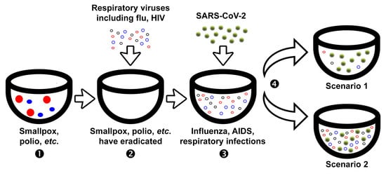
2. Impact of Influenza Pandemics on the Circulation of Seasonal Respiratory Viruses

Wars, stresses, and prolonged force majeure situations upset the delicate balance in host-pathogen interactions. Nevertheless, not only wars but also global outbreaks are stressful for society. What happens to other infectious diseases during global disasters such as pandemics?
There is a wide range of respiratory pathogens associated with human infections. To better understand the threat that these viruses pose to humankind, it is often valuable to study newly identified viruses, not in isolation, but in comparison with other related viruses [1]. In this section, we will briefly discuss the relationships between newly emerged pandemic viruses and circulating seasonal strains.
The first virologically documented pandemic, the Spanish flu, occurred in 1918–1919 [5]. It was caused by the influenza A (H1N1) virus. According to various estimates, this pandemic, unprecedented in its severity, killed between 40 and 50 million people and caused a huge number of severe forms of the disease. Thirty percent of the world’s population became sick. In subsequent years, the Spanish influenza A (H1N1) moved into the category of seasonal pathogens. The virus acquired mutations that allowed it to escape the population’s immunity and cause influenza epidemics annually.
Unfortunately, we were unable to find accessible literature on the prevalence of other respiratory viral infections during the most destructive pandemic of all, the 1918 influenza pandemic. No documentary evidence regarding the pro et contra spread of other respiratory illnesses could be found. However, a huge range of publications describing the situation with the spread of common respiratory infections during the following pandemics, in 1957, 1968, and 2009, are available.
In 1957, a new pandemic began, caused by the Asian influenza A (H2N2) virus [6,7], killing 1.5 million people. Asian influenza viruses circulated for 11 years, causing annual epidemics.
A new influenza A (H3N2) pandemic virus, called the Hong Kong virus, replaced the Asian influenza virus in 1968 [6,8]. It killed 1 million people. Drift variants of the influenza A/Hong Kong (H3N2) virus continue to circulate to date.
In 1977, the subtype influenza A (H1N1) virus again caused morbidity among young, immunologically naive people [8]. Studies have shown that this virus was not genetically different from the A (H1N1) virus circulating in the 1950s.
It is noteworthy that the emergence of each new pandemic virus was regularly marked by the disappearance of its pandemic predecessor, drifting in seasonal epidemics. There is an explanation of the mysterious phenomenon of the displacement of a previously circulating virus by a new pandemic virus, according to which the mechanism leading to the elimination of the previous pathogen is an increase in the production of human antibodies to the conserved stem of viral hemagglutinin protein. These antibodies are enough to displace an obsolete virus [9,10].
An exception is the joint circulation of viruses A (H3N2) and A (H1N1) after 1977 when virus A (H1N1) returned to the community, and was unable to displace virus A (H3N2). Both subtypes alternately or simultaneously circulated among people until 2009.
In 2009, the serotype A (H1N1) viruses that drifted since 1977 were supplanted by a completely new virus of the same serotype A (H1N1)pdm09, which caused a new moderate pandemic [11,12]. Virus A (H1N1)pdm09 has moved into the category of seasonal viruses and continues to circulate with viruses A (H3N2) to date.
The evolution of influenza viruses is related to all of the above. What happens to seasonal respiratory viruses in the interpandemic and pandemic periods? As an example, we consider the situation covering the period before and after the last influenza pandemic of 2009 (2007–2012). In sum, influenza A (H1N1)pdm09 pandemic viruses had distinct effects on the circulation of other respiratory viruses when they first appeared. On the contrary, no effect was seen when novel A (H1N1)pdm09 strains evolved from pandemic to seasonal viruses. When pandemic influenza virus strains just replaced the circulating seasonal influenza virus strains, seasonal influenza A (H3N2) (0.3%) and B (2.0%) infections were hardly detected. However, the circulation of other respiratory pathogens remained unchanged [13].
Globally, seasonal influenza has demonstrated wide variations in incidence depending on the epidemic season and the antigenic properties of the circulating strains. In contrast, the yearly incidence rates for such noninfluenza pathogens, such as respiratory syncytial virus, are fairly stable. At the same time, influenza virus detection had always been significantly below that of the respiratory syncytial virus in influenza seasons before 2009 [14]. The 2009 pandemic significantly altered the detection rates of respiratory viruses, establishing influenza viruses as the major respiratory pathogens worldwide [14]. As an example, a study of the circulation of respiratory viruses other than influenza during the 2009 pandemic in Spain revealed that apart from influenza A (H1N1)pdm09 viruses, other respiratory viruses were detected in 39.9% of patients. Co-infection of two or more respiratory viruses was observed in approximately 15% of cases. The highest detection rates were obtained for human rhinovirus and respiratory syncytial virus, representing 43.7% and 31.4% of positive noninfluenza samples [14].
It has been hypothesized that at least in some European countries, a viral interference of human rhinovirus could have delayed the epidemic evolution of the pandemic influenza A (H1N1)pdm09 virus in the fall of 2009. Similarly, the influenza A (H1N1)pdm09 virus could interfere with another respiratory pathogen, a respiratory syncytial virus, in the same period [14].
Some authors have postulated that a possible interference of other respiratory viruses such as human rhinovirus and respiratory syncytial virus with influenza A (H1N1)pdm09 virus during the pandemic could change the epidemic wave of pandemic influenza [15,16,17].

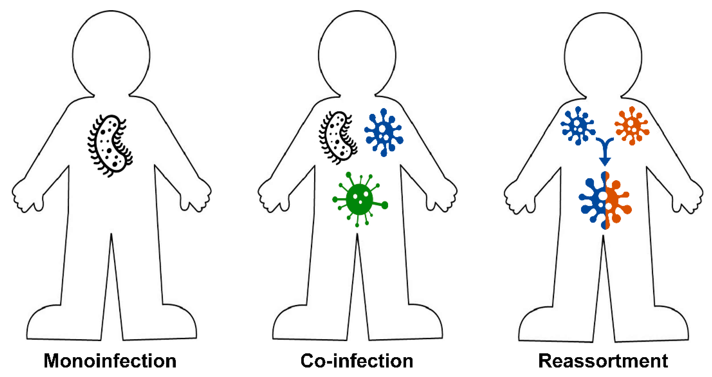
3. The Role of Mixed Infections in Etiology of Pulmonary Diseases

Mono-infections are not the only infections that can be found in nature. The co-infection of more than one pathogen in one host is common across viruses and occurs more commonly than would be expected by chance in nature [18,19]. Different variations of interactions of circulating pathogens exist in nature. It could be a (i) single infection (one pathogen, one host) [14,20]; (ii) mixed infection (co-infection), when two or more antigenically distinct pathogens infect one host [21,22,23,24,25,26]; (iii) reassortment of two or more viruses in one host, when an individual is co-infected with two or more of the same type of virus, for example, influenza A viruses, resulting in reassortant progeny [27,28,29] (Figure 1). The second variant of host-pathogen interactions—co-infection—is most closely related to the focus of this paper.
The mixed etiology of pulmonary infections is frequent, with co-infections of pathogenic respiratory influenza-like viruses and bacteria in respiratory secretions. The clinical significance of these viral-bacterial co-infections has been a controversial topic for a long time [30]. Viruses can alter the host’s susceptibility to bacterial infections both by altering the environmental conditions in the lung to favor bacterial replication and by suppressing the host’s defense mechanisms to prevent the clearance of the bacteria [31]. Understanding the synergy or antagonism between viruses and bacteria will facilitate the design of novel therapeutic approaches to prevent the mortality associated with bacterial co-infections [32].
The human respiratory tract hosts a diverse community of cocirculating viruses that are responsible for acute respiratory infections. In the respiratory airways, the ecological niche contains viruses and bacteria that periodically circulate and enter interspecific interactions [33,34,35]. An example of the interaction between viruses and bacteria is the relationship between influenza virus and pneumococcus, leading to about a 100-fold increase in susceptibility to pneumococcal pneumonia after influenza infection [36]. This shared niche also facilitates virus-virus interactions [33]. During a seasonal rise in the incidence of influenza, a few antigenically distinct viruses circulate, the ratio of which varies annually. Seasonal influenza consists of variable mixes of influenza A (H3N2), A (H1N1)pdm09, and the two B virus lineages.
Evidence of virus-virus interactions between 11 human respiratory viruses has recently been presented [33]. The experimental models of respiratory virus co-infections have demonstrated several interaction-induced effects, from an enhanced or reduced viral growth to the attenuation of disease. In the case of recirculation, seasonal influenza-like viruses, and a ubiquitous common-cold-like virus, a short-lived protective effect, such as that induced by interferon, was demonstrated [37,38]. It has also been shown that cell fusion induced by certain viruses may enhance the replication of others in co-infections. However, despite the epidemiological, clinical, and experimental indications of interactions among respiratory viruses, quantitatively robust evidence is still lacking [33].
The cocirculation of different respiratory viruses can lead not only to cooperative but also to competitive forms of virus-virus interactions. It is believed that such interactions occur, for instance, among influenza and rhinoviruses [33]. It can also be assumed that competitive viruses are built between viruses, where an epidemically stronger rival wins. In such cases, the circulation of one viral pathogen may reduce the likelihood of or delay infection by another. Thus, the pandemic A (H1N1)pdm09 influenza virus delayed infection with respiratory syncytial virus. The 2009 respiratory syncytial virus infections were first noticed only when the infection with the pandemic swine influenza began to decline. Interestingly, in 2010 to 2011, infections with respiratory syncytial virus were observed together with the pandemic swine influenza infection and may reflect the conversion of the pandemic influenza virus from a pandemic virus to a regular seasonal influenza virus [39]. In other cases, such interactions can have large consequences, aggravating the course of diseases. Therefore, there is a clinical need for a robust investigation into co-infection in patients with COVID-19.
As noted above, pandemic influenza viruses displace seasonal influenza strains; however, the circulation of other respiratory pathogens such as rhinovirus and the respiratory syncytial virus remains at the interpandemic level. What happens to these seasonal respiratory viruses in the coronavirus pandemic?

4. Impact of the COVID-19 Pandemic on the Circulation of Seasonal Respiratory Viruses

Humans are constantly being inoculated with various microorganisms resident in the upper respiratory tract and by inhaled aerosols [31]. Mixed infections significantly contribute to the insufficient control of respiratory infections. A single infection may be caused by the simultaneous synergistic interaction of a variety of pathogens. In the context of SARS-CoV-2, in the COVID-19 pandemic, individuals may be co-infected with (i) influenza virus; (ii) another respiratory virus; (iii) other pathogens such as nonrespiratory viruses, bacteria including Mycobacterium tuberculosis, fungi, and mycoplasma (Figure 2). Each of these variants may negatively influence COVID-19 treatments. An important question raised by many scientists is the following: should we expect an increasing or decreasing number of co-infections between SARS-CoV-2 and other respiratory pathogens, and what do we observe now?
COVID-19 can occur with other viral infections. Some of these co-infections can be effectively treated, while only supportive treatment is the mainstay of treatment for others [40].
Most publications describing co-occurring infections in which COVID-19 is involved focus on influenza and influenza-like diseases. There are only a few papers on other infections. In this review, we have strictly confined ourselves to influenza and other viral respiratory infections.

4.1. Co-Infection of SARS-CoV-2 with Influenza Viruses

According to the WHO, as of 22 June 2020, global influenza activity is at lower levels than expected for this time of the year. In the Northern Hemisphere, influenza activity returned to inter-seasonal levels, while in the Southern Hemisphere, the influenza season has not started yet [41]. Most countries in the Caribbean and Central America reported to the WHO sporadic influenza detections, though many areas are experiencing rises in COVID-19 cases [41]. The WHO warns that its information should be interpreted with caution because of the possible influences of both COVID-19 activity and distance measures. During the epidemic of COVID-19 in Beijing (China), influenza viruses, especially influenza type A viruses, accounted for a large proportion of respiratory virus infections [42].
A recent study revealed that Wuhan citizens with documented mixed infections of SARS-CoV-2 and influenza A or B did not appear to show a more severe clinical condition; they displayed similar clinical characteristics to those patients with SARS-CoV-2 infections only [43].
Some believe that SARS-CoV-2 and influenza virus co-infection is rare [23]. Globally, there has been a limited number of COVID-19-influenza co-infected patients reported from different countries around the world. For instance, a study performed in Istanbul, Turkey revealed that only 0.54% out of 1103 COVID-19 patients were diagnosed as co-infected with influenza A or B viruses [23]. Co-infections with influenza A virus were reported in the USA, Turkey, Germany, Iran, China, Japan, and Spain [21,23,40,43,44,45,46,47,48,49,50]; co-infections with influenza B virus were reported in Turkey, China, and Spain [23,43,44]; simultaneous influenza A and B and COVID-19 infections was detected in China and Spain [44,51].
In any situation, influenza preparedness measures are crucial, especially for high-risk groups, and the influenza vaccination coverage rate must be at least 70% [52]. COVID-19 measures may reduce the number of cases of influenza infections. Seasonal influenza cases in the Northern Hemisphere usually peak in February and tail off by the end of May. Unusually, this year, lab-confirmed cases of influenza dropped dramatically in early April, soon after the WHO declared the COVID-19 pandemic. It has been hypothesized that the influenza season was cut short and that influenza incidence was substantially reduced by control measures aimed at interrupting SARS-CoV-2 transmission [53].
In a WHO press conference on 15 June 2020, the WHO Director-General Dr. Tedros A. Ghebreyesus said, “Despite the ongoing global response to the COVID-19 pandemic, we cannot lose sight of other significant public health issues including influenza. Influenza affects every country every year and takes its deadly toll. As we enter the Southern Hemisphere influenza season and begin planning for the Northern Hemisphere season, we must ensure that influenza remains a top priority” [54]. Dr. Tedros A. Ghebreyesus noted that today we face a dramatic decrease in the number of specimens tested for influenza globally. These disruptions may have short- and long-term effects such as loss of capacities to detect and report new influenza viruses with pandemic potential.
Thus, SARS-CoV-2 may significantly affect infections with other respiratory viruses including influenza viruses. However, it is difficult to distinguish its direct impact on reducing respiratory infections because of the effect of declining influenza surveillance as a result of the COVID-19 pandemic.

4.2. Co-Infection of SARS-CoV-2 with Influenza-like Viruses

Besides influenza viruses, other respiratory viruses play a significant role in epidemiological processes. Recently, respiratory viruses showed different levels of mixed infections with SARS-CoV-2 [55]. In light of the foregoing information, the COVID-19 pandemic will probably develop according to one of two scenarios: (i) SARS-CoV-2 viruses will cocirculate with other viral pathogens, or (ii) SARS-CoV-2 viruses will displace other respiratory viruses.
The latest evidence suggests that unlike co-infection rates with influenza viruses, which demonstrated some reduction in activity, the co-infection rates between SARS-CoV-2 and other respiratory pathogens are higher than expected [56]. A study performed in California (USA) revealed that, of patients with confirmed SARS-CoV-2 infections, 20.7% were positive for one or more other pathogens; the most common were rhinovirus, enterovirus, respiratory syncytial virus, and seasonal coronavirus [57]. In a study performed in China, among 32 confirmed COVID-19 cases, 14 patients were infected with other pathogens, including viral, bacterial, and fungal infections. Four of them were infected with respiratory viruses-respiratory syncytial virus, human parainfluenza virus, human metapneumovirus, and rhinovirus [58].
During the beginning of the COVID-19 spread in Japan, influenza and human metapneumovirus were prevalent among children (the study was performed in the Furano region). Three children were diagnosed with COVID-19, two of whom were co-infected with influenza and human metapneumovirus [59].
Some believe that co-infection with two respiratory pathogens is uncommon in adults and mainly inherent in children [26]. Nonetheless, a mixed infection of metapneumovirus and SARS-CoV-2 was detected in an adult patient in Providence (RI, USA) [26].
In sum, among the concomitants of SARS-CoV-2 pathogens, respiratory syncytial virus and rhinovirus prevail. Respiratory syncytial virus and rhinovirus are the most frequent causes of acute respiratory infections, with especially severe diseases in young children and the elderly. Primary respiratory syncytial virus infections may lead to severe bronchiolitis and pneumonia [60,61]. Rhinovirus usually causes rhinopharyngitis but may also cause illness of the lower respiratory tract and asthma exacerbations and has been associated with severe acute lower respiratory tract infections [60,61]. The post-infection immunity to these infections is short-lasting, which leads to the repeated circulation of viruses in the same population. The co-infection of such seasonal pathogens, along with influenza viruses, with the new pandemic SARS-CoV-2 can aggravate the course of the disease and represents a challenge for its diagnosis and treatment [52,62,63].

4.3. Co-Infection of SARS-CoV-2 with Other Viral and Nonviral Pathogens

Current evidence does not support a high rate of bacterial co-infections in patients with COVID-19 [24]. On the contrary, a low proportion of COVID-19 patients have bacterial co-infections [55]. Bengoechea and Bamford [3] noted that the limited reference to nonviral co-infections in COVID-19 is surprising. We found that only a few publications described co-infections with nonviral pathogens. In particular, there is limited information available regarding the mixed infections of SARS-COV-2 with bacterial and fungal infectious agents, including Mycoplasma pneumonia [64,65], Mycobacterium tuberculosis [66,67,68], Legionella pneumophila [69], and Aspergillus fumigatus [70,71]. Nonrespiratory viral pathogens, such as cytomegalovirus [72] and HIV [73], also did not play a crucial role in viral-viral mixed infections. Therefore, there is a paucity of data to support the association of SARS-CoV-2 and these pathogens with the severity of co-infection.

5. On the Way to the Anti-COVID-19 Vaccine

It is strongly hoped that this effort in developing an anti-COVID-19 vaccine will be successful. Almost 150 teams all over the world are working hard with a diverse set of strategies and platforms to develop a potent and harmless vaccine against COVID-19. As of 10 August 2020, 139 vaccines are in preclinical evaluation, and 28 are in phase 1, 2, or 3 clinical trials [74].
However, some caution is prudent, and the gains that have been recently made in understanding the interactions between the host immune system and viruses should be fully exploited [75]. Most vaccines go through years of tests before they appear on the market. Some authors suggest that the first vaccines may not prevent COVID-19 infection because, generally, vaccines aim to protect people against disease, not necessarily the infection itself [76]. Such a vaccine will prevent people from getting sick or dying but will not stop them from catching the coronavirus. Taking into account the fact that the vaccine may not prevent infection with the SARS-CoV-2 virus, we have to be warned that some coronavirus vaccinations might harm more than help those who are desperate for protection. A coronavirus vaccination could lead to negative consequences, including enhancing the negative effect of the infection in a person instead of mitigating it [77]. The “cytokine storm,” an uncontrolled over-production of soluble markers of inflammation, appears to be one of the most dangerous and potentially life-threatening events related to COVID-19 [78]. Infection with the wild-type SARS-CoV-2 virus followed by vaccination may induce a severe disease [79].
Safety is a primary goal for any vaccine; potential risks associated with vaccine development for COVID-19 are described in several publications [77,79,80,81,82,83]. Some authors suggest that vaccinations could increase the severity of subsequent SARS-CoV-2 infections. This has happened with vaccines against feline coronavirus [77]. There are two syndromes described for vaccine-enhanced diseases, antibody-dependent enhancements (a process in which a virus leverages antibodies to aid infection), and the vaccine-associated enhanced respiratory disease (cell-based enhancement that includes allergic inflammation).
Immunological priming could involve autoimmunity in lung tissues due to previous exposure to the coronavirus spike protein. Immunogenic viral peptides that match human proteins are good candidates for pathogenic priming peptides by analogy with the immune system reaction known as an immune enhancement. The homology between human and viral proteins is an established factor in viral- or vaccine-induced autoimmunity [81,83].
Thus, researchers must prove whether or not their vaccines cause the same types of immune system malfunctions that have been observed in the past.
Unfortunately, no specific antiviral therapy for COVID-19 exists; no licensed vaccine is available yet. Therefore, scientists put forward a wide variety of theories regarding which of the available chemical and immunobiological preparations may be used in the fight against this disease.
Since the early stages of the COVID-19 pandemic, a possible role of vaccination with a live attenuated vaccine against tuberculosis (bacille Calmette-Guérin vaccine, BCG) in preventing novel coronavirus infections has been discussed in numerous publications [84,85,86,87]. Based on the hypothesis that BCG can enhance the reactivity of the innate immune system, some researchers have speculated that the BCG vaccine may be used as a preventive and/or therapeutic measure against COVID-19.
Nonetheless, the results were conflicting, and later studies did not confirm a correlation between COVID-19 occurrence and BCG vaccination rates. After a detailed analysis of all available data, it was concluded that there is no reasonable evidence to recommend BCG vaccination for the prevention of COVID-19 [88,89]. The official position of the WHO is that there is no evidence that the BCG vaccine protects people against infection from the COVID-19 virus [90]. However, some clinical trials aiming to determine whether a BCG vaccination could protect against COVID-19 infections are in progress [91,92].
We also must remember that influenza remains an ever-present cause of disease and death globally [93]. Patients co-infected with SARS-CoV-2 and influenza virus have a higher risk of poor outcomes. Given the potential for influenza-COVID-19 co-infections globally [23,94], especially during the influenza season, it has been suggested that influenza vaccinations could be used to indirectly control COVID-19 [50,52,95]. Of course, the influenza vaccine will not fully protect against coronavirus infection, but a vaccination with a proven and safe influenza vaccine, especially a live one, will affect the incidence of mixed influenza-COVID-19 infections and thus reduce the severity of coronavirus infections.
Some authors suggested that this fall, the influenza vaccine for larger groups of the population should be recommended to simplify clinicians’ work when the second wave of COVID-19 is expected in parallel with the start of the influenza season [23,96]. Other researchers are not optimistic [80,97,98]. In any case, understanding the relationship between COVID-19 and influenza is very important.
Salem and El-Hennawy [95] hypothesized that immunity against influenza infections may foster immunity against SARS-CoV-2. This suggestion is supported by studies showing a cross-reactivity of immunity between influenza virus and coronavirus due to the similarity in their structures. The influenza vaccination itself would also generate sustained immunity that may enhance immunity against SARS-CoV-2. The authors suggest that the immunity induced by the influenza virus has more beneficial effects for COVID-19 than those suggested for BCG or MMR (measles, mumps, and rubella) vaccines. More than 500 million people are vaccinated against the flu every year based on recommendations from the WHO on the composition of influenza virus vaccines [54,99]. According to [95], given the high safety of the influenza vaccine confirmed by decades of vaccination of millions of vaccinees, influenza vaccines can be used to minimize the severity of the COVID-19 disease.

6. Conclusions

The pandemic invasion of a new SARS-CoV-2 virus in 2019 provided a unique possibility to learn more about the pathogenesis of respiratory viruses. When faced with a new virus that is rapidly emerging in the human population, we have a limited knowledge base to work with. The clock is ticking, and we have to learn quickly about this new virus while simultaneously using our knowledge of previous influenza outbreaks that have much in common with the recent COVID-19 outbreak [93,100]. Ozawa and Kawaoka [101] have suggested that the “complete control of influenza viruses seems impossible.” It seems that it is not futile to think that complete influenza eradication is possible in the near future. As for today, the COVID-19 pandemic is far from over, and as with influenza, we are far from defeating it. Meanwhile, nobody can predict anything. We are just at the beginning of a long and winding road.

Author Contributions

Conceptualization, I.K. and E.G.; writing—original draft preparation, I.K., N.L., E.G. and M.A.F.; I.K. and M.A.F. made the figures; writing—review and editing, I.K. and L.R.; supervision, L.R. All authors have read and agreed to the published version of the manuscript.

Funding

This research received no external funding.

Acknowledgments

The authors gratefully acknowledge the Institute of Experimental Medicine Administration for the administrative and technical support to fulfill this research.

Conflicts of Interest

The author declares no conflict of interest.

References

  1. Belser, J.A. Assessment of SARS-CoV-2 replication in the context of other respiratory viruses. Lancet. Respir. Med. 2020. [Google Scholar] [CrossRef]
  2. Lowen, A.C.; Mubareka, S.; Tumpey, T.M.; Garcia-Sastre, A.; Palese, P. The guinea pig as a transmission model for human influenza viruses. Proc. Natl. Acad. Sci. USA 2006, 103, 9988–9992. [Google Scholar] [CrossRef] [PubMed] [Green Version]
  3. Bengoechea, J.A.; Bamford, C.G.G. SARS-CoV-2, bacterial co-infections, and AMR: The deadly trio in COVID-19? EMBO Mol. Med. 2020. [Google Scholar] [CrossRef] [PubMed]
  4. WHO. Coronavirus Disease (COVID-19) Dashboard. Available online: https://covid19.who.int/ (accessed on 12 August 2020).
  5. Neumann, G.; Noda, T.; Kawaoka, Y. Emergence and pandemic potential of swine-origin H1N1 influenza virus. Nature 2009, 459, 931–939. [Google Scholar] [CrossRef] [Green Version]
  6. Flahault, A.; Zylberman, P. Influenza pandemics: Past, present and future challenges. Public Health Rev. 2010, 32, 319–340. [Google Scholar] [CrossRef] [Green Version]
  7. Potter, C.W. A history of influenza. J. Appl. Microbiol. 2001, 91, 572–579. [Google Scholar] [CrossRef]
  8. Krammer, F.; Smith, G.J.D.; Fouchier, R.A.M.; Peiris, M.; Kedzierska, K.; Doherty, P.C.; Palese, P.; Shaw, M.L.; Treanor, J.; Webster, R.G.; et al. Influenza. Nat. Rev. Dis. Primers 2018, 4, 3. [Google Scholar] [CrossRef]
  9. Krammer, F.; Palese, P. Influenza virus hemagglutinin stalk-based antibodies and vaccines. Curr Opin Virol 2013, 3, 521–530. [Google Scholar] [CrossRef] [Green Version]
  10. Palese, P.; Wang, T.T. Why do influenza virus subtypes die out? A hypothesis. mBio 2011, 2. [Google Scholar] [CrossRef] [Green Version]
  11. Dawood, F.S.; Iuliano, A.D.; Reed, C.; Meltzer, M.I.; Shay, D.K.; Cheng, P.Y.; Bandaranayake, D.; Breiman, R.F.; Brooks, W.A.; Buchy, P.; et al. Estimated global mortality associated with the first 12 months of 2009 pandemic influenza A H1N1 virus circulation: A modelling study. Lancet. Infect. Dis. 2012, 12, 687–695. [Google Scholar] [CrossRef] [Green Version]
  12. Simonsen, L.; Spreeuwenberg, P.; Lustig, R.; Taylor, R.J.; Fleming, D.M.; Kroneman, M.; Van Kerkhove, M.D.; Mounts, A.W.; Paget, W.J. Global mortality estimates for the 2009 influenza pandemic from the GLaMOR project: A modeling study. PLoS Med. 2013, 10, e1001558. [Google Scholar] [CrossRef] [PubMed] [Green Version]
  13. Meningher, T.; Hindiyeh, M.; Regev, L.; Sherbany, H.; Mendelson, E.; Mandelboim, M. Relationships between A(H1N1)pdm09 influenza infection and infections with other respiratory viruses. Influenza Other Respir. Viruses 2014, 8, 422–430. [Google Scholar] [CrossRef] [PubMed]
  14. Navarro-Marí, J.M.; Pérez-Ruiz, M.; Galán Montemayor, J.C.; Marcos Maeso, M.; Reina, J.; de Oña Navarro, M.; Cilla Eguiluz, C.G. Circulation of other respiratory viruses and viral co-infection during the 2009 pandemic influenza. Enferm. Infecc. Y Microbiol. Clin. 2012, 30 (Suppl. 4), 25–31. [Google Scholar] [CrossRef]
  15. Casalegno, J.S.; Ottmann, M.; Bouscambert-Duchamp, M.; Valette, M.; Morfin, F.; Lina, B. Impact of the 2009 influenza A(H1N1) pandemic wave on the pattern of hibernal respiratory virus epidemics, France, 2009. Eur. Commun. Dis. Bull. 2010, 15. [Google Scholar]
  16. Mak, G.C.; Wong, A.H.; Ho, W.Y.; Lim, W. The impact of pandemic influenza A (H1N1) 2009 on the circulation of respiratory viruses 2009-2011. Influenza Other Respir. Viruses 2012, 6, e6–e10. [Google Scholar] [CrossRef] [PubMed] [Green Version]
  17. Ånestad, G.; Nordbø, S.A. Virus interference. Did rhinoviruses activity hamper the progress of the 2009 influenza A (H1N1) pandemic in Norway? Med Hypotheses 2011, 77, 1132–1134. [Google Scholar] [CrossRef]
  18. Diaz-Munoz, S.L. Viral coinfection is shaped by host ecology and virus-virus interactions across diverse microbial taxa and environments. Virus Evol. 2017, 3, vex011. [Google Scholar] [CrossRef] [Green Version]
  19. Alcaide, C.; Rabadan, M.P.; Moreno-Perez, M.G.; Gomez, P. Implications of mixed viral infections on plant disease ecology and evolution. Adv. Virus Res. 2020, 106, 145–169. [Google Scholar] [CrossRef]
  20. Maurice, J.B.; Patel, A.; Scott, A.J.; Patel, K.; Thursz, M.; Lemoine, M. Prevalence and risk factors of nonalcoholic fatty liver disease in HIV-monoinfection. Aids 2017, 31, 1621–1632. [Google Scholar] [CrossRef]
  21. Nowak, M.D.; Sordillo, E.M.; Gitman, M.R.; Paniz Mondolfi, A.E. Co-infection in SARS-CoV-2 infected patients: Where are influenza virus and rhinovirus/enterovirus? J. Med Virol. 2020. [Google Scholar] [CrossRef]
  22. Ou, X.; Zhou, L.; Huang, H.; Lin, Y.; Pan, X.; Chen, D. A severe case with co-infection of SARS-CoV-2 and common respiratory pathogens. Travel Med. Infect. Dis. 2020, 101672. [Google Scholar] [CrossRef] [PubMed]
  23. Ozaras, R.; Cirpin, R.; Duran, A.; Duman, H.; Arslan, O.; Bakcan, Y.; Kaya, M.; Mutlu, H.; Isayeva, L.; Kebanlı, F.; et al. Influenza and COVID-19 co-infection: Report of 6 cases and review of the Literature. J. Med Virol. 2020. [Google Scholar] [CrossRef] [PubMed]
  24. Rawson, T.M.; Moore, L.S.P.; Zhu, N.; Ranganathan, N.; Skolimowska, K.; Gilchrist, M.; Satta, G.; Cooke, G.; Holmes, A. Bacterial and fungal co-infection in individuals with coronavirus: A rapid review to support COVID-19 antimicrobial prescribing. Clin. Infect. Dis. 2020. [Google Scholar] [CrossRef]
  25. Rodriguez, J.A.; Rubio-Gomez, H.; Roa, A.A.; Miller, N.; Eckardt, P.A. Co-Infection with SARS-COV-2 and parainfluenza in a young adult patient with pneumonia: Case report. IDCases 2020, 20, e00762. [Google Scholar] [CrossRef] [PubMed]
  26. Touzard-Romo, F.; Tapé, C.; Lonks, J.R. Co-infection with SARS-CoV-2 and human metapneumovirus. Rhode Island Med. J. 2020, 103, 75–76. [Google Scholar]
  27. Steel, J.; Lowen, A.C. Influenza A virus reassortment. Curr. Top. Microbiol. Immunol. 2014, 385, 377–401. [Google Scholar] [CrossRef]
  28. Klempa, B. Reassortment events in the evolution of hantaviruses. Virus Genes 2018, 54, 638–646. [Google Scholar] [CrossRef] [Green Version]
  29. Matsuzaki, Y.; Sugawara, K.; Takashita, E.; Muraki, Y.; Hongo, S.; Katsushima, N.; Mizuta, K.; Nishimura, H. Genetic diversity of influenza B virus: The frequent reassortment and cocirculation of the genetically distinct reassortant viruses in a community. J. Med. Virol. 2004, 74, 132–140. [Google Scholar] [CrossRef]
  30. Brealey, J.C.; Sly, P.D.; Young, P.R.; Chappell, K.J. Viral bacterial co-infection of the respiratory tract during early childhood. FEMS Microbiol. Lett. 2015, 362. [Google Scholar] [CrossRef] [Green Version]
  31. Babiuk, L.A.; Lawman, M.J.; Ohmann, H.B. Viral-bacterial synergistic interaction in respiratory disease. Adv. Virus Res. 1988, 35, 219–249. [Google Scholar] [CrossRef]
  32. Jia, L.; Xie, J.; Zhao, J.; Cao, D.; Liang, Y.; Hou, X.; Wang, L.; Li, Z. Mechanisms of severe mortality-associated bacterial co-infections following influenza virus infection. Front. Cell. Infect. Microbiol. 2017, 7, 338. [Google Scholar] [CrossRef] [PubMed]
  33. Nickbakhsh, S.; Mair, C.; Matthews, L.; Reeve, R.; Johnson, P.C.D.; Thorburn, F.; von Wissmann, B.; Reynolds, A.; McMenamin, J.; Gunson, R.N.; et al. Virus-virus interactions impact the population dynamics of influenza and the common cold. Proc. Natl. Acad. Sci. USA 2019, 116, 27142–27150. [Google Scholar] [CrossRef] [PubMed] [Green Version]
  34. Lloyd-Smith, J.O. Vacated niches, competitive release and the community ecology of pathogen eradication. Philos. Trans. R. Soc. Lond. Ser. BBiol. Sci. 2013, 368, 20120150. [Google Scholar] [CrossRef] [PubMed] [Green Version]
  35. Bosch, A.A.; Biesbroek, G.; Trzcinski, K.; Sanders, E.A.; Bogaert, D. Viral and bacterial interactions in the upper respiratory tract. PLoS Pathog. 2013, 9, e1003057. [Google Scholar] [CrossRef] [Green Version]
  36. Shrestha, S.; Foxman, B.; Weinberger, D.M.; Steiner, C.; Viboud, C.; Rohani, P. Identifying the interaction between influenza and pneumococcal pneumonia using incidence data. Sci. Transl. Med. 2013, 5, 191ra184. [Google Scholar] [CrossRef] [Green Version]
  37. Isaacs, A.; Burke, D.C. Viral interference and interferon. Br. Med Bull. 1959, 15, 185–188. [Google Scholar] [CrossRef]
  38. Dianzani, F. Viral interference and interferon. La Ric. Clin. E Lab. 1975, 5, 196–213. [Google Scholar] [CrossRef]
  39. Hirsh, S.; Hindiyeh, M.; Kolet, L.; Regev, L.; Sherbany, H.; Yaary, K.; Mendelson, E.; Mandelboim, M. Epidemiological changes of respiratory syncytial virus (RSV) infections in Israel. PLoS ONE 2014, 9, e90515. [Google Scholar] [CrossRef]
  40. Konala, V.M.; Adapa, S.; Gayam, V.; Naramala, S.; Daggubati, S.R.; Kammari, C.B.; Chenna, A. Co-infection with influenza A and COVID-19. Eur. J. Case Rep. Intern. Med. 2020, 7, 001656. [Google Scholar] [CrossRef]
  41. WHO. Overview of Influenza Activity Globally. Influenza Update N° 373 of 3 August 2020. Available online: http://www.who.int/influenza/surveillance_monitoring/updates/latest_update_GIP_surveillance (accessed on 12 August 2020).
  42. Li, Y.; Wang, J.; Wang, C.; Yang, Q.; Xu, Y.; Xu, J.; Li, Y.; Yu, X.; Zhu, H.; Liu, J. Characteristics of respiratory virus infection during the outbreak of 2019 novel coronavirus in Beijing. Int. J. Infect. Dis. 2020, 96, 266–269. [Google Scholar] [CrossRef] [PubMed]
  43. Ding, Q.; Lu, P.; Fan, Y.; Xia, Y.; Liu, M. The clinical characteristics of pneumonia patients coinfected with 2019 novel coronavirus and influenza virus in Wuhan, China. J. Med Virol. 2020. [Google Scholar] [CrossRef] [PubMed]
  44. Cuadrado-Payán, E.; Montagud-Marrahi, E.; Torres-Elorza, M.; Bodro, M.; Blasco, M.; Poch, E.; Soriano, A.; Piñeiro, G.J. SARS-CoV-2 and influenza virus co-infection. Lancet 2020, 395, e84. [Google Scholar] [CrossRef]
  45. Wu, D.; Lu, J.; Ma, X.; Liu, Q.; Wang, D.; Gu, Y.; Li, Y.; He, W. Coinfection of influenza virus and severe acute respiratory syndrome coronavirus 2 (SARS-COV-2). Pediatric Infect. Dis. J. 2020, 39, e79. [Google Scholar] [CrossRef]
  46. Wu, X.; Cai, Y.; Huang, X.; Yu, X.; Zhao, L.; Wang, F.; Li, Q.; Gu, S.; Xu, T.; Li, Y.; et al. Co-infection with SARS-CoV-2 and influenza A virus in patient with pneumonia, China. Emerg. Infect. Dis. 2020, 26, 1324–1326. [Google Scholar] [CrossRef]
  47. Azekawa, S.; Namkoong, H.; Mitamura, K.; Kawaoka, Y.; Saito, F. Co-infection with SARS-CoV-2 and influenza A virus. IDCases 2020, 20, e00775. [Google Scholar] [CrossRef] [PubMed]
  48. Hashemi, S.A.; Safamanesh, S.; Ghafouri, M.; Taghavi, M.R.; Mohajer Zadeh Heydari, M.S.; Namdar Ahmadabad, H.; Ghasem Zadeh-Moghaddam, H.; Azimian, A. Co-infection with COVID-19 and influenza A virus in two died patients with acute respiratory syndrome, Bojnurd, Iran. J. Med Virol. 2020. [Google Scholar] [CrossRef] [PubMed]
  49. Khodamoradi, Z.; Moghadami, M.; Lotfi, M. Co-infection of coronavirus disease 2019 and influenza A: A report from Iran. Arch. Iran. Med. 2020, 23, 239–243. [Google Scholar] [CrossRef] [Green Version]
  50. Wehl, G.; Laible, M.; Rauchenzauner, M. Co-infection of SARS CoV-2 and influenza A in a pediatric patient in Germany. Klin. Padiatr. 2020. [Google Scholar] [CrossRef]
  51. Wu, Q.; Xing, Y.; Shi, L.; Li, W.; Gao, Y.; Pan, S.; Wang, Y.; Wang, W.; Xing, Q. Coinfection and other clinical characteristics of COVID-19 in children. Pediatrics 2020. [Google Scholar] [CrossRef]
  52. Osterhaus, A.D.M.E. Influenza and RSV in a COVID-19 world. The First ESWI Webinar 11 June 2020. Available online: https://eswi.org/eswi-tv/influenza-and-rsv-in-a-covid-19-world-full-webinar/ (accessed on 25 June 2020).
  53. Jones, N. How Coronavirus Lockdowns Stopped Flu in its Tracks. Nature News. Available online: https://www.nature.com/articles/d41586-020-01538-8 (accessed on 18 June 2020).
  54. WHO. Coronavirus Disease (COVID-19) Pandemic. Press Conference of 15 June 2020. Available online: https://www.who.int/emergencies/diseases/novel-coronavirus-2019 (accessed on 1 August 2020).
  55. Lansbury, L.; Lim, B.; Baskaran, V.; Lim, W.S. Co-infections in people with COVID-19: A systematic review and meta-analysis. J. Infect. 2020. [Google Scholar] [CrossRef]
  56. Sánchez-Duque, J.A.; Orozco-Hernández, J.P.; Marín-Medina, D.S.; Cvetkovic-Vega, A.; Aveiro-Róbalo, T.R.; Mondragon-Cardona, A.; Failoc-Rojas, V.E.; Gutiérrez-Ocampo, E.; Villamizar-Peña, R.; Henao-Martínez, J.F.; et al. Are we now observing an increasing number of coinfections between SARS-CoV-2 and other respiratory pathogens? J. Med. Virol. 2020. [Google Scholar] [CrossRef]
  57. Kim, D.; Quinn, J.; Pinsky, B.; Shah, N.H.; Brown, I. Rates of co-infection between SARS-CoV-2 and other respiratory pathogens. JAMA 2020, 323, 2085–2086. [Google Scholar] [CrossRef] [Green Version]
  58. Li, Z.T.; Chen, Z.M.; Chen, L.D.; Zhan, Y.Q.; Li, S.Q.; Cheng, J.; Zhu, A.R.; Chen, L.Y.; Zhong, N.S.; Li, S.Y.; et al. Coinfection with SARS-CoV-2 and other respiratory pathogens in COVID-19 patients in Guangzhou, China. J. Med. Virol. 2020. [Google Scholar] [CrossRef]
  59. Kakuya, F.; Okubo, H.; Fujiyasu, H.; Wakabayashi, I.; Syouji, M.; Kinebuchi, T. The first pediatric patients with coronavirus disease 2019 (COVID-19) in Japan; The risk of co-infection with other respiratory viruses. Jpn. J. Infect. Dis. 2020. [Google Scholar] [CrossRef]
  60. Costa, L.F.; Queiróz, D.A.; Lopes da Silveira, H.; Bernardino Neto, M.; de Paula, N.T.; Oliveira, T.F.; Tolardo, A.L.; Yokosawa, J. Human rhinovirus and disease severity in children. Pediatrics 2014, 133, e312–e321. [Google Scholar] [CrossRef] [Green Version]
  61. Li, Y.; Pillai, P.; Miyake, F.; Nair, H. The role of viral co-infections in the severity of acute respiratory infections among children infected with respiratory syncytial virus (RSV): A systematic review and meta-analysis. J. Glob. Health 2020, 10. [Google Scholar] [CrossRef]
  62. Osterhaus, A.D.M.E. Clinical Manifestations of COVID-19, Influenza and RSV. The Second ESWI Webinar 18 June 2020. Available online: https://eswi.org/eswi-tv/clinical-manifestations-of-covid-19-influenza-and-rsv-full-webinar/ (accessed on 20 June 2020).
  63. Osterhaus, A.D.M.E. Intervention Strategies for COVID-19, Influenza and RSV. The Third ESWI Webinar 23 June 2020. Available online: https://player.vimeo.com/video/431944051 (accessed on 30 June 2020).
  64. Fan, B.E.; Lim, K.G.E.; Chong, V.C.L.; Chan, S.S.W.; Ong, K.H.; Kuperan, P. COVID-19 and mycoplasma pneumoniae coinfection. Am. J. Hematol. 2020, 95, 723–724. [Google Scholar] [CrossRef]
  65. Gayam, V.; Konala, V.M.; Naramala, S.; Garlapati, P.R.; Merghani, M.A.; Regmi, N.; Balla, M.; Adapa, S. Presenting characteristics, comorbidities, and outcomes of patients coinfected with COVID-19 and Mycoplasma pneumoniae in the USA. J. Med. Virol. 2020. [Google Scholar] [CrossRef]
  66. Stochino, C.; Villa, S.; Zucchi, P.; Parravicini, P.; Gori, A.; Raviglione, M.C. Clinical characteristics of COVID-19 and active tuberculosis co-infection in an Italian reference hospital. Eur. Respir. J. 2020. [Google Scholar] [CrossRef]
  67. Tadolini, M.; Codecasa, L.R.; García-García, J.M.; Blanc, F.X.; Borisov, S.; Alffenaar, J.W.; Andréjak, C.; Bachez, P.; Bart, P.A.; Belilovski, E.; et al. Active tuberculosis, sequelae and COVID-19 co-infection: First cohort of 49 cases. Eur. Respir. J. 2020. [Google Scholar] [CrossRef]
  68. Togun, T.; Kampmann, B.; Stoker, N.G.; Lipman, M. Anticipating the impact of the COVID-19 pandemic on TB patients and TB control programmes. Ann. Clin. Microbiol. Antimicrob. 2020, 19, 21. [Google Scholar] [CrossRef]
  69. Arashiro, T.; Nakamura, S.; Asami, T.; Mikuni, H.; Fujiwara, E.; Sakamoto, S.; Miura, R.; Shionoya, Y.; Honda, R.; Furukawa, K.; et al. SARS-CoV-2 and Legionella co-infection in a person returning from a Nile cruise. J. Travel Med. 2020, 27. [Google Scholar] [CrossRef] [Green Version]
  70. van Arkel, A.L.E.; Rijpstra, T.A.; Belderbos, H.N.A.; van Wijngaarden, P.; Verweij, P.E.; Bentvelsen, R.G. COVID-19 associated pulmonary aspergillosis. Am. J. Respir. Crit. Care Med. 2020. [Google Scholar] [CrossRef]
  71. Blaize, M.; Mayaux, J.; Nabet, C.; Lampros, A.; Marcelin, A.G.; Thellier, M.; Piarroux, R.; Demoule, A.; Fekkar, A. Fatal invasive aspergillosis and coronavirus disease in an immunocompetent patient. Emerg. Infect. Dis. 2020, 26. [Google Scholar] [CrossRef]
  72. D’Ardes, D.; Boccatonda, A.; Schiavone, C.; Santilli, F.; Guagnano, M.T.; Bucci, M.; Cipollone, F. A case of coinfection with SARS-COV-2 and cytomegalovirus in the era of COVID-19. Eur. J. Case Rep. Intern. Med. 2020, 7, 001652. [Google Scholar] [CrossRef]
  73. Benkovic, S.; Kim, M.; Sin, E. 4 Cases: HIV and SARS-CoV-2 Co-infection in patients from Long Island, New York. J. Med. Virol. 2020. [Google Scholar] [CrossRef]
  74. WHO. Draft Landscape of COVID-19 Candidate Vaccines. Update of 10 August 2020. Available online: https://www.who.int/publications/m/item/draft-landscape-of-covid-19-candidate-vaccines (accessed on 12 August 2020).
  75. Burton, D.R.; Walker, L.M. Rational vaccine design in the time of COVID-19. Cell Host Microbe 2020, 27, 695–698. [Google Scholar] [CrossRef]
  76. Lauerman, J.; Paton, J. The First COVID Vaccines May Not Prevent COVID Infection. Prognosis of 15 June 2020. Available online: https://www.bloomberg.com/news/articles/2020-06-15/the-first-covid-vaccines-may-not-prevent-you-from-getting-covid (accessed on 2 August 2020).
  77. Graham, B.S. Rapid COVID-19 vaccine development. Science 2020, 368, 945–946. [Google Scholar] [CrossRef]
  78. Coperchini, F.; Chiovato, L.; Croce, L.; Magri, F.; Rotondi, M. The Cytokine Storm in COVID-19: An Overview of the Involvement of the Chemokine/Chemokine-Receptor System. Cytokine Growth Factor Rev. 2020, 53, 25–32. [Google Scholar] [CrossRef]
  79. Eyal, N.; Lipsitch, M.; Smith, P.G. Human challenge studies to accelerate coronavirus vaccine licensure. J. Infect. Dis. 2020, 221, 1752–1756. [Google Scholar] [CrossRef] [Green Version]
  80. Morris, K.V. The improbability of the rapid development of a vaccine for SARS-CoV-2. Mol. Ther. J. Am. Soc. Gene Ther. 2020. [Google Scholar] [CrossRef]
  81. Peeples, L. News feature: Avoiding pitfalls in the pursuit of a COVID-19 vaccine. Proc. Natl. Acad. Sci. USA 2020, 117, 8218–8221. [Google Scholar] [CrossRef] [Green Version]
  82. Hotez, P.J.; Corry, D.B.; Bottazzi, M.E. COVID-19 vaccine design: The Janus face of immune enhancement. Nat. Rev. Immunol. 2020, 20, 347–348. [Google Scholar] [CrossRef]
  83. Lyons-Weiler, J. Pathogenic priming likely contributes to serious and critical illness and mortality in COVID-19 via autoimmunity. J. Transl. Autoimmun. 2020, 3, 100051. [Google Scholar] [CrossRef]
  84. Ozdemir, C.; Kucuksezer, U.C.; Tamay, Z.U. Is BCG vaccination affecting the spread and severity of COVID-19? Allergy 2020. [Google Scholar] [CrossRef]
  85. Curtis, N.; Sparrow, A.; Ghebreyesus, T.A.; Netea, M.G. Considering BCG vaccination to reduce the impact of COVID-19. Lancet 2020, 395, 1545–1546. [Google Scholar] [CrossRef]
  86. Meena, J.; Yadav, A.; Kumar, J. BCG vaccination policy and protection against COVID-19. Indian J. Pediatrics 2020, 1. [Google Scholar] [CrossRef]
  87. Macedo, A.; Febra, C. Relation between BCG coverage rate and COVID-19 infection worldwide. Med. Hypotheses 2020, 142, 109816. [Google Scholar] [CrossRef]
  88. Kumar, J.; Meena, J. Demystifying BCG vaccine and COVID-19 relationship. Indian Pediatrics 2020, 57, 588–589. [Google Scholar] [CrossRef]
  89. Riccò, M.; Gualerzi, G.; Ranzieri, S.; Bragazzi, N.L. Stop playing with data: There is no sound evidence that Bacille Calmette-Guérin may avoid SARS-CoV-2 infection (for now). Acta Bio-Med. Atenei Parm. 2020, 91, 207–213. [Google Scholar] [CrossRef]
  90. WHO. Bacille Calmette-Guérin (BCG) Vaccination and COVID-19. Scientific Brief of 12 April 2020. Available online: https://www.who.int/news-room/commentaries/detail/bacille-calmette-gu%C3%A9rin-(bcg)-vaccination-and-covid-19 (accessed on 1 August 2020).
  91. O’Connor, E.; Teh, J.; Kamat, A.M.; Lawrentschuk, N. Bacillus Calmette Guérin (BCG) Vaccination Use in the Fight Against COVID-19—What’s Old is New Again? Future Oncol. 2020. [Google Scholar] [CrossRef]
  92. Redelman-Sidi, G. Could BCG be used to protect against COVID-19? Nat. Rev. Urol. 2020, 17, 316–317. [Google Scholar] [CrossRef]
  93. Dunning, J.; Thwaites, R.S.; Openshaw, P.J.M. Seasonal and pandemic influenza: 100 years of progress, still much to learn. Mucosal Immunol. 2020, 1–8. [Google Scholar] [CrossRef]
  94. Antony, S.J.; Almaghlouth, N.K.; Heydemann, E.L. Are co-infections with COVID-19 and Influenza low or underreported? An observational study examining current published literature including three new unpublished cases. J. Med. Virol. 2020. [Google Scholar] [CrossRef]
  95. Salem, M.L.; El-Hennawy, D. The possible beneficial adjuvant effect of influenza vaccine to minimize the severity of COVID-19. Med Hypotheses 2020, 140, 109752. [Google Scholar] [CrossRef]
  96. Hanney, S.R.; Wooding, S.; Sussex, J.; Grant, J. From COVID-19 research to vaccine application: Why might it take 17 months not 17 years and what are the wider lessons? Health Res. Policy Syst. 2020, 18, 61. [Google Scholar] [CrossRef]
  97. Mukherjee, R. Global efforts on vaccines for COVID-19: Since, sooner or later, we all will catch the coronavirus. J. Biosci. 2020, 45. [Google Scholar] [CrossRef]
  98. Merante, D. SARS-CoV-2, from its current highly contagious spreading toward the global development of an effective and safe vaccine: Challenges and uncertainties. Expert Opin. Drug Saf. 2020, 1–3. [Google Scholar] [CrossRef]
  99. WHO. Recommendations on the Composition of Influenza Virus Vaccines. Available online: https://www.who.int/influenza/vaccines/virus/recommendations/en/ (accessed on 1 August 2020).
  100. Bogaert, D.; Dockrell, D.H. 100 years of influenza research seen through the lens of Covid-19. Mucosal Immunol. 2020, 1–2. [Google Scholar] [CrossRef]
  101. Ozawa, M.; Kawaoka, Y. Cross talk between animal and human influenza viruses. Annu Rev Anim Biosci 2013, 1, 21–42. [Google Scholar] [CrossRef] [PubMed] [Green Version]
Figure 1. Host-pathogen interactions.
Biology 09 00240 g001
Figure 2. The nature of mixed infections of SARS-CoV-2 with other pathogens in the COVID-19 pandemic.
Biology 09 00240 g002

