Article

Helleborus odorus subsp. cyclophyllus: An Unexploited Source of Antioxidant, Antimicrobial, and Cytotoxic Bioactivity


by
Panagiotis Sarametidis
1,2,
Evgenia-Anna Papadopoulou
3,
Panagiotis Katsoris
2,
Konstantinos A. Aliferis
3,
Keith D. Rochfort
1 and
Konstantinos Grintzalis
1,*
1
School of Biotechnology, Dublin City University, D09 N920 Dublin, Ireland
2
Department of Biology, University of Patras, 265 04 Patras, Greece
3
Laboratory of Pesticide Science, Agricultural University of Athens, Iera Odos 75, 118 55 Athens, Greece
*
Author to whom correspondence should be addressed.
Biology 2026, 15(11), 852; https://doi.org/10.3390/biology15110852 (registering DOI)
Submission received: 20 April 2026 / Revised: 23 May 2026 / Accepted: 25 May 2026 / Published: 29 May 2026
(This article belongs to the Section Toxicology)

Simple Summary

In the present day, plant-derived remedies are routinely used within traditional medicine. Furthermore, many commercially available compounds were initially isolated from plant sources. Hellebores are plants commonly found in Europe, and research conducted on their extracts has indicated inherent physiological effects on cells of the immune and vascular systems, amongst others. In this study, we characterised the chemical composition and antioxidant properties of the extract of the root from Helleborus sp. Following this, we assessed its impact on toxicity, oxidative stress, and angiogenesis using a human aortic endothelial cell model. Combining metabolomics with in vitro assays, our study aims to identify targets and therapies in relation to unexplored sources from hellebores.

Abstract

Helleborus sp. are small, perennial herbs with a wide distribution across central and southern Europe and Asia. Historically, extracts of these plants have been used in remedies exhibiting cathartic and anthelmintic effects; however, their adverse and toxic potential have also been reported. As such, Helleborus extracts have been trialled as alternative approaches to cancer treatment in rare cases. In this study, a detailed phytochemical characterisation of root extracts from Helleborus odorus subsp. cyclophyllus was performed in parallel with an in vitro assessment of their cytotoxicity and effect on cell phenotype. GC/EI/MS metabolite profiling revealed the complexity of the extract, which exhibited a high relative content of fatty acids (17.98%), with the most abundant group being the unsaturated fatty acids, followed by saturated and hydroxy fatty acids. Bioassays indicated the extract had antioxidant capabilities, while in vitro analysis demonstrated the extract induced a pro-oxidant phenotype that reduced cell viability, inhibited growth in bacterial (E. coli) and mammalian (human aortic endothelial cells) cell types, and exacerbated pro-inflammatory indices. Helleborus odorus subsp. cyclophyllus contains bioactive molecules that elicit a number of cellular responses, and the therapeutic potential of these molecules warrants further investigation.

1. Introduction

The interest in the therapeutic potential of bioactive molecules derived from medicinal plants has increased significantly over recent years. There are numerous references in the literature to the diverse properties of plant extracts in the context of health and disease, with many demonstrating their therapeutic effects through antioxidant, antibacterial, and antiviral means [1,2]. Moreover, individuals are less likely to develop resistance to plant-derived medicines compared to conventional drugs. As such, in line with the increasing interest in plant-derived alternative medicines, a correlative increase in the exploitation and adoption of plant extracts in health sciences has been observed, with many citing their use to treat infections, cancer, and circulatory disease.
Helleborus sp. belongs to the Ranunculaceae family and includes small, perennial herbs commonly found in central and southern Europe and Asia. Their distribution in Europe has elevated their significance in therapeutic remedies since antiquity [3]. In the literature, Helleborus niger is the most widely reported species used in a medicinal context, followed by Helleborus odorus, albeit to a lesser extent. Owing to the potency and complexity of their root extracts, hellebores have been routinely used in traditional and folk remedies, with subsequent preparations exhibiting cathartic, anthelmintic, and other beneficial properties; however, adverse and toxic potential have been reported [3]. Mass spectrometry and nuclear magnetic resonance profiling of different Helleborus species indicate that this genus is a rich source of bioactive compounds: glycosides, saponins, ecdysteroids, and lactones, amongst others [4,5,6,7,8]. Over time, several studies have demonstrated that these hellebore-derived compounds exert a high biological activity on numerous cell types, atypically exerting immunostimulatory and anti-inflammatory effects [9,10,11,12,13,14].
Of particular interest, Helleborus extracts have been shown to influence the phenotype of the cells associated with the vascular system, with lasting vasoactive effects [15,16]. In vivo trials have reported the safety profile of administered Helleborus-derived compounds to the vascular system, in addition to showing treatment-influenced vascular health. In vivo studies have observed a reduction in atherosclerotic plaque size following treatment with hellebore-derived compounds [17], though the exact mechanism is currently unknown. Atherosclerosis is a multifactorial disease driven by the interplay of several cellular activation states within the vascular wall, of which, Helleborus extracts have been shown to influence. For example, several species of the Helleborus genus have been reported to influence the activity of several enzymes central to atherogenesis; for example, cyclooxygenase-1 and -2, and 5-lipooxygenase [13]. Moreover, Helleborus-derived extracts have also been shown to reduce the expression of cell surface receptors responsible for immune cell recruitment on endothelial cells, effectively reducing immune cell activity and migration [17]. In addition, several species of Helleborus have been shown to demonstrate radical scavenging abilities. Owing to their influence in cardiovascular physiology, the ability of Helleborus to target reactive oxygen species (ROS) and interfere in ROS-driven cellular behaviours offers further credence to its application and efficacy in treating vascular-associated disease states. Several other studies have highlighted this antioxidant activity in other genera [18,19,20], and as such, the multifactorial ability of Helleborus extracts to exert a therapeutic effect on cells of the vascular system has garnered attention for treating not only disorders of the vascular system, but also vascular-related chronic diseases such as diabetes, cancer, and age-associated degenerative disorders [21].
Despite this body of evidence supporting the use of Helleborus-derived treatments in vascular disease states, there are several contrasting reports highlighting the significant risks that Helleborus-derived extracts pose as treatments, owing to their purported adverse and toxic properties. Studies have shown preparations of specific species of the Helleborus genus to exert potent cytotoxic effects at particular concentration ranges [8,22], with a number of case studies detailing the toxicity and side effects of Helleborus administration in experimental setups utilising human biopsies as models of disease [23]. These effects have been shown to extend to and include cells of the vascular system, with Helleborus-derived extracts shown to induce anti-proliferative, anti-angiogenic, and pro-apoptotic effects within certain concentration ranges [24].
Interestingly, the aforementioned cytotoxic activities of Hellebores were seen to be potentiated in cells which are oncogenic in nature, with primary cells demonstrating increased tolerance to these effects [22]. As such, these results have only broadened their application, with them being utilised as an alternative approach to cancer treatment. For example, the anti-angiogenic effects of particular Helleborus extracts may explain their success in treating certain forms of cancer, with studies demonstrating that Helleborus treatment inhibits the action of the pro-tumorigenic VEGF-A [24]. Moreover, different genera at different concentrations have shown the ability of Helleborus to interfere with the cell-cycle and microtubule dynamics in experimental models of cancer. These anti-proliferative and anti-migratory properties have also been observed in cell models of renal, brain, and breast cancers, amongst many others [19,24,25,26]. Moreover, the translational value of these anti-oncogenic effects is corroborated in clinical data in sporadic patient cases in the literature [9,27]. Taken together, further research on the contrasting biological effects of Helleborus and its compounds on models of human physiology is required in order to advance the understanding of its true therapeutic potential.
In this study, we focused on the phytochemical characterisation of Helleborus odorus subsp. cyclophyllus, combining biochemical assays and chromatographic characterisation to profile the bioactive properties of the extracts derived from the species. The toxicity profile of the extract was then examined in bacterial and mammalian cell cultures, with a focus on a selected number of cell characteristics examined in primary mammalian cultures. Finally, the therapeutic potential of the extract was assessed in a model of the human vasculature in a number of pro-inflammatory disease contexts.

2. Materials and Methods

2.1. Preparation and Extraction of Helleborus odorus Plant Tissues

H. odorus subsp. cyclophyllus (A. Brawn Maire and Petitm) mother plants were harvested from the Greek flora (39°22.199′ N, 021°14.109′–110′ E, Altitude: 861–862 m) in spring and subsequently submitted to clonisation at the Holly and Great Monastery of Vatopaidi in Mount Athos, Greece. As a result, certified H. odorus subsp. cyclophyllus pharmaceutical plant clones were developed. The H. odorus subsp. cyclophyllus ‘Vatopaidi’ (Italia Herbarium depository number: 2017.012 Helleborus οdorus Waldst. & Kit. subsp. cyclophyllus A. Brawn Maire and Petitm) was used as experimental material. Air-dried roots from H. odorus subsp. cyclophyllus were ground using an electrical grinder. Samples (25 mg) were extracted in 1 mL sterile phosphate-buffered saline (PBS) or water on a rocker plate overnight. The extracts were purified by centrifugation (14,000× g, room temperature for 5 min) or sterile filtration (for cell and microbial culturing experiments), and the resulting homogenate was collected and analysed immediately.

2.2. Phytochemical and Antioxidant Characterisation of Helleborus Extract

The phytochemical content of aqueous Helleborus extracts was assessed with biochemical assays following our novel multiparametric protocol [28]. As blanks, PBS diluted with water was used in place of extracts, and each extract was assessed with three replicates.
Polyphenols were quantified by the Folin reagent. Briefly, 100 μL of appropriately diluted extract was mixed with 100 μL of Folin reagent and 100 μL of Na2CO3 (1.89 M). The mixtures were incubated for 40 min at room temperature, and the absorbance was measured at 765 nm and expressed as equivalents of gallic acid.
Flavonoids were quantified by their reaction with aluminium trichloride. Appropriately diluted extract (100 μL) was mixed with 50 μL of aqueous NaNO2 (2%, w/v) and incubated at room temperature for 10 min. Subsequently, 50 μL of aqueous AlCl3 (7.5%, w/v) and 50 μL of NaOH (3.5 N) were added, and then the mixtures were incubated for 10 min at room temperature. The absorbance was measured at 500 nm and expressed as equivalents of catechin.
Tannins were quantified by their reaction with vanillin under acidic conditions [29]. An amount of 100 μL of appropriately diluted extract was mixed with 100 μL of 4% vanillin in methanol and 50 μL of 100% H2SO4 and incubated at room temperature for 10 min. Absorbances were measured at 500 nm and expressed as equivalents of catechin.
The metal chelating potential of Helleborus extract was assessed for iron and copper ions. The ferric reducing power (FeRP) was determined by the reduction of ferric to ferrous ions, which then react with 2,4,6-tri-pyridyl-s-triazine (TPTZ) to form an absorbing complex at 595 nm. An amount of 100 μL of appropriately diluted extract was mixed with 100 μL of FeRP reagent (250 mM of acetic acid, 1 mM of TPTZ, 0.054% of FeCl3·6H2O), and then prepared and incubated at room temperature for 40 min. The absorbance was measured at 595 nm and converted to equivalents of gallic acid. The cupric reducing power (CuRP) was determined by the complex of copper ions with neocuproine reagent [bis(neocuproine)copper(II)], which, when reduced by phytochemicals, absorbs at 450 nm. An amount of 125 μL of appropriately diluted extract was mixed with 125 μL of Cu–neocuproine–ammonium acetate reagent (3.33 mM of CuSO4·5H2O, 2 mM of neocuproine, and 0.33 M of ammonium acetate) and incubated for 40 min at room temperature. The absorbance was measured at 450 nm and converted to equivalents of gallic acid.
The radical scavenging potential of Helleborus extracts was assessed for ABTS•+, DPPH and galvinoxyl radicals. ABTS•+ radical was synthesised by the reaction of 14 mM ABTS and 5 mM potassium persulfate overnight. For the assay, 125 μL of appropriately diluted extract was mixed with 125 μL of ABTS radical cation (ABTS•+), which was appropriately diluted in ddH2O and incubated at room temperature for 40 min, and the absorbance was then measured at 734 nm. For the DPPH radical, the reagent was diluted in methanol, and 200 μL of DPPH radical reagent was mixed with 50 μL of appropriately diluted extract and incubated in the dark at room temperature for 10 min, and the absorbance was then measured at 515 nm. For galvinoxyl radical, the reagent was diluted in methanol, and 200 μL of galvinoxyl radical reagent was mixed with 50 μL of appropriately diluted extract and incubated in the dark at room temperature for 10 min, and the absorbance was then measured at 435 nm. The % radical scavenging was calculated based on the equation 100 × (ARadical − ASample)/ARadical and expressed as nmoles of equivalents of gallic acid from a linear standard curve.

2.3. Metabolite Profiling of the Helleborus sp. Extract Employing Gas Chromatography–Electron Impact–Mass Spectrometry (GC/EI/MS)

Complementary to the abovementioned assays, the deconvolution of the metabolite composition of the extract was performed by employing a gas chromatography–electron impact–mass spectrometry analyser [GC/EI/MS; Agilent 6890 N GC platform (Agilent Technologies Inc., Santa Clara, CA, USA), 5973 T mass selective detector (MSD), 7683 autosampler]. The preparation of the dry samples (15 mg) for the GC/MS analysis was performed in a two-step process using a vacuum concentrator (Eppendorf Concentrator Plus, Eppendorf, Hamburg, Germany), as previously described [30,31], following minor modifications. Briefly, for the methoxymation of the samples, an amount of 80 μL of a methoxylamine hydrochloride solution (20 mg mL−1 in pyridine) was added, and the resulting solution was thoroughly vortexed and incubated for 2 h at 30 °C in a water bath (Daihan Labtech, Namyangju-Si, Gyeonggi-do, Korea). In the second step, 80 μL of N-Methyl-N-trimethylsilyl-trifluoroacetamide (MSTFA) was added for silylation, and then the samples were incubated for 1.5 h at 37 °C in the water bath. Prior to their transfer to 200 μL glass microinserters (Macherey-Nagel, Dueren, Germany) into 2 mL glass autosampler vials, the resulting solutions were centrifuged using a benchtop centrifuge (5000 rpm, 10 min) to remove any possible debris that could interfere with the analysis. In total, four replications were prepared and, additionally, experimental blanks that had been prepared following the exact same protocol as the samples were analysed, in order to detect features originating from possible contamination. Also, an aliquot (20 μL) of a methanolic ribitol solution (0.2 mg mL−1) served as the internal standard for quality control purposes. The annotation of the metabolites was based on mass fragmentation patterns, using the mass spectra library of the National Institute of Standards and Technology, 2024 (NIST 24, Gaithersburg, MD, USA), and/or analytical standards, where available. The analysis of the derivatised extract was performed under previously described analytical conditions [30,31]. Briefly, the extract (1 μL) was injected on a 30 m long column (HP-5MS, Agilent Technologies Inc.) having a diameter of 0.25 mm and film thickness of 0.25 μm, applying a split ratio of 5:1. Helium was used as the carrier gas at a flow rate of 1 mL min−1. Full scan mass spectra were acquired over the range of 50–800 Da (4 scans s−1), applying positive electron ionisation (70 eV). The injector’s temperature was set at 230 °C, and the oven temperature program was as follows: initial temperature of 70 °C, stable for 5 min, increase to 310 °C (°C min−1), and then stable for 1 min. The MS source temperature was 230 °C, and that of the quadrupole was 150 °C.

2.4. Antibacterial Activity of Helleborus odorus subsp. cyclophyllus Extracts

The antibacterial activity of Helleborus odorus subsp. cyclophyllus extracts was assessed based on the calculation of their minimum inhibitory concentration (MIC) in liquid cultures of the model species Escherichia coli. E. coli was incubated at 37 °C under continuous agitation at 150 rpm in LB broth medium. The bacterial culture was centrifuged at 3000× g for 3 min, and the supernatant was removed. The pelleted bacterial cells were re-suspended in sterile PBS and pelleted again by centrifugation at 3000× g for 3 min. The supernatant was discarded and the bacteria were re-suspended in sterile PBS, and then the bacteria concentration was measured based on their absorbance at 600 nm (1 A is equal to 8 × 10⁸ cells/mL). For the assessment of the minimum inhibitory concentration, 100 μL of Helleborus odorus subsp. cyclophyllus extract (prepared in sterile PBS or water and appropriately diluted in sterile PBS or water) was incubated with 100 μL of LB broth containing 104 cells/mL at 37 °C for 24 h. The MIC was evaluated as the minimum concentration of plant extract that inhibits bacterial growth.

2.5. Cell Culture Bioassay

Human Aortic Endothelial Cells (HAECs) (Cat. No. C12271, PromoCell, Heidelberg, Germany) obtained from a 23-year-old Caucasian male were cultured in Endothelial Cell Growth Medium MV (Cat. No. C22020, PromoCell, Heidelberg, Germany) containing the following supplements: fetal calf serum (0.05 mL/mL), endothelial cell growth supplement (0.004 mL/mL), epidermal growth factor (10 ng/mL), heparin (90 µg/mL), and hydrocortisone (1 µg/mL). The culture medium was also supplemented with penicillin (100 IU/mL) and streptomycin (100 µg/mL) (Cat. No. P0781, Merck, Dublin, Ireland). HAEC cultures were maintained in a humidified incubator at 37 °C, 5% CO2, and 95% humidity. Passages 6–12 were used for experimental purposes. For experiments, unless otherwise stated, HAECs were grown to confluence in the specified culture dishes before treatment with Helleborus odorus subsp. cyclophyllus extracts for up to 48 h. HAEC cultures were treated with a Helleborus odorus subsp. cyclophyllus extract across a concentration range spanning from 50 ng/mL to 1000 ng/mL. In certain experiments, cultures were also treated with vascular endothelial growth factor (VEGF) (50 ng/mL) (Cat. No. SRP3182, Merck, Dublin, Ireland), platelet-derived growth factor (PDGF) (100 ng/mL) (Cat. No. SRP3138, Merck, Dublin, Ireland), D-glucose (30 mM) (Cat. No. G8644, Merck, Dublin, Ireland), or TNF-α (100 ng/mL) (Cat. No. GF023, Merck, Dublin, Ireland). Additional parallel experiments involving HAEC treatments with superoxide dismutase (SOD) (100 U/mL) (Cat. No. S5395, Merck, Dublin, Ireland) or apocynin (APO) (10 mM) (Cat. No. 178385, Merck Millipore, Dublin, Ireland) were pre-treated with the respective treatment at the final concentration 1 h prior to the addition of Helleborus odorus subsp. cyclophyllus extract.

2.6. Crystal Violet Assay

The crystal violet assay was implemented to measure cell viability and cell proliferation. HAECs were seeded at a density of 15,000 cells/well and 3000 cells/well on a 96-well plate for cell viability and cell proliferation, respectively. The following day, the wells were gently washed with 200 µL/well of PBS before fresh culture medium containing the experimental conditions of interest was added. Following treatment, the plate was once again gently washed with 200 µL of PBS before the cells were fixed with 50 µL of 3.7% formaldehyde for 15 min. After removing the formaldehyde, the cells were gently washed with 200 µL of distilled water before 50 µL of a 0.5% crystal violet solution was added to each well. The cells were incubated with the stain for 30 min before being subjected to six washes with distilled water to remove excess dye and then left to air dry overnight. The following day, the crystal violet was extracted from the cells by adding 50 µL of a 2% SDS solution. Following a 30 min incubation on an orbital rotator to assist the extraction process, the absorbance of the plate was read at 570 nm using a Tecan Infinite M200 (Tecan, Manneford, Switzerland).

2.7. Dihydroethidium Assay

Cell-derived reactive oxygen species (ROS) levels were measured using dihydroethidium staining as adapted from a previous protocol [32]. Briefly, HAECs were seeded at a density of 15,000 cells/well on a white 96-well plate 24 h prior to the addition of Helleborus odorus subsp. cyclophyllus extracts. HAECs were labelled with 3 µM DHE (Cat. No. 37291, Merck, Dublin, Ireland) 30 min prior to the completion of each specific time point. Post-treatment, the 96-well plate was analysed using a Tecan Infinite M200 (Tecan, Manneford, Switzerland) with excitation and emission wavelengths set at 560 and 590 nm, respectively. For normalisation purposes, the DHE signal was baseline corrected with respect to viability levels.

2.8. Microscopy

Briefly, HAECs were seeded in 6-well dishes and grown to confluence before being treated ± Helleborus odorus subsp. cyclophyllus extract. An amount of 3 µM of DHE was added to the HAEC cultures 30 min prior to the cessation of the experimental time point. The culture medium was then removed, and the cells were washed three times with PBS before the cells were fixed for 15 min with 3.7% paraformaldehyde. The fixed cells were washed a further three times before a thin layer of distilled water was added to ensure the monolayer remained hydrated. Stained HAECs were imaged on a Nikon Eclipse Ti fluorescent microscope (Nikon, Tokyo, Japan) at 40× magnification, with the exposure time kept constant across all experimental samples. An unstained control was included for all microscopy work.

2.9. Permeability Assay

The analysis of HAEC monolayer permeability was performed using a modified version of the transwell method [33]. Briefly, HAECs were seeded at a high density (1 × 10⁶ cells/insert) into hanging transwell cell culture inserts placed within 6-well dishes (Sarstedt AG & Co, Munich, Germany). Cell culture medium was added to the upper (2 mL) and lower (4 mL) chambers of the transwell insert, and the cells were allowed to adhere overnight. The following day, the HAECs within the transwell were then treated with Helleborus odorus subsp. cyclophyllus extract ± SOD or APO. Untreated and PBS-treated controls were included for all permeability work. Post-treatment, the medium in the upper and lower chambers was replaced with fresh medium, and fluorescein isothiocyanate (FITC)-labelled 40 kDa dextran was added to the upper chamber at a final concentration of 250 µg/mL. Samples from the lower chamber (28 µL) were taken every 30 min, and after 3 h, all samples were diluted to a final volume of 400 µL with medium before being plated in 100 µL volumes in a 96-well white plate. A Tecan Infinite F200 (Tecan, Mannedorf, Switzerland) was used with excitation and emission wavelengths set at 490 nm and 520 nm, respectively. Permeability is presented as the rate of FITC-dextran 40 kDa transendothelial exchange per hour.

3. Results and Discussion

3.1. Phytochemical and Antioxidant Properties of Helleborus odorus subsp. cyclophyllus Extract

The initial characterisation of the major categories of phytochemicals and the antioxidant properties of Helleborus odorus subsp. cyclophyllus was assessed in water and PBS extracts for comparisons (Table 1). Although there are differences in the two preparations, each extract contains several forms of flavonoids, polyphenols, and tannins that are responsible for the radical scavenging potential for ABTS, DPPH, and galvinoxyl radicals and their metal reducing power against iron and copper ions.

3.2. GC/EI/MS Metabolite Profiling Revealed the High Content of Bioactive Fatty Acids

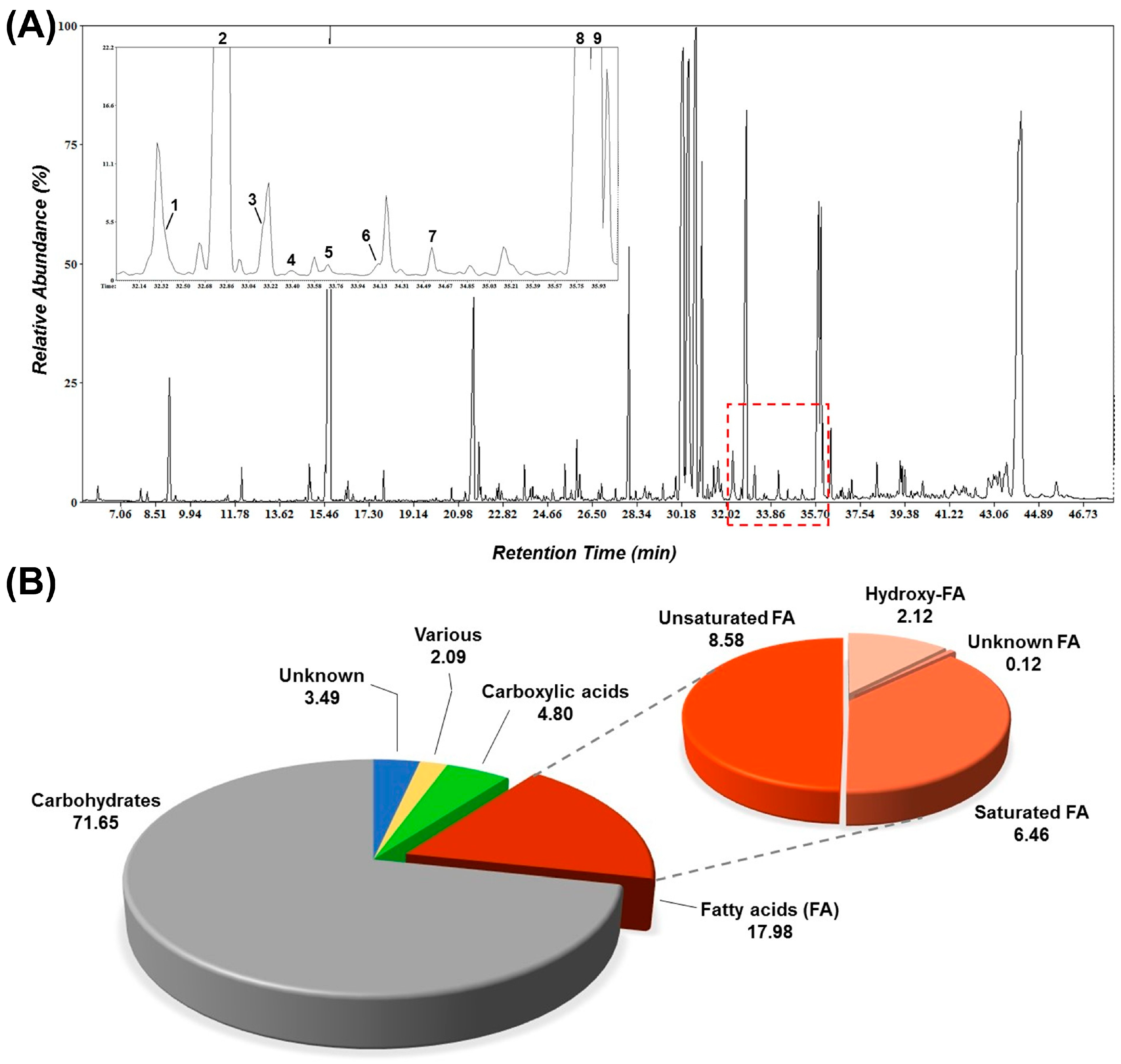
The applied GC/EI/MS metabolite profiling protocol resulted in an improved chromatographic separation, which was confirmed by the quality (e.g., number and shape of peaks, baseline) of the obtained total ion chromatograms (Figure 1A). A total of 169 metabolite features, identified at different identification levels, were recorded. The majority of the annotated metabolite features belong to carbohydrates, carboxylic acids, and fatty acids (FAs) (Figure 1B). Interestingly, the extracts had a high relative content in FAs (17.98%), with the most abundant group being the unsaturated FAs, followed by the saturated and hydroxy FAs (Figure 1B and Figure 2 and Supplementary Table S1). Since the phenolic content of the plant has been studied [34], here, the main focus was the deconvolution of its FA content and the correlation to its bioactivity.
FAs are abundant metabolites in plant tissues exhibiting well-established bioactivities, including, among others, antimicrobial, antioxidant, and plant defence-inducing capacity [35,36]. Additionally, they are metabolites of high energetic status that can regulate redox homeostasis via their interference with ROS metabolism [27]. Early evidence has supported the antioxidant capacities of several of the annotated FAs in the analysed Helleborus odorus subsp. cyclophyllus extract (e.g., myristic acid, palmitic acid), suggesting a positive correlation between the antioxidant activity of the saturated FAs and the increasing chain length [36]. The results also confirmed the superior antioxidant capacity of the majority of the unsaturated FAs being evaluated. Oleic and linoleic acids were the two most abundant unsaturated FAs of the extract (3.59% and 3.30%, respectively), which exhibit superior antioxidant and anticancer profiles [37]. They are also well-known for their anticancer, anti-inflammatory, and immune system-regulating properties [38].
Hydroxy FAs are also metabolites with well-studied bioactivities [27,39]. Among the hydroxy FAs of the extract, azelaic acid was the most abundant (3.44%). The metabolite has established anti-inflammatory and antioxidant activities, as well as antimicrobial activity against Gram-negative and Gram-positive bacteria [39,40,41]. The latter has been attributed to alterations in the intracellular pH [40]. Additionally, azelaic acid exhibits cytotoxicity and anti-proliferative activity against the human malignant melanocyte [42]. Traumatic acid is a metabolite involved in plant responses to stresses that has been reported to protect cells from peroxidation and ROS-induced toxicity [27].
Based on the obtained evidence and the information from the literature, the observed bioactivities of the extract cannot be attributed to a single component. In conclusion, it is plausible to suggest that the recorded bioactivity of the analysed extract can be, at least partially, attributed to its FA content as a whole, an observation that is in line with previous studies on similar FA-rich sources and their potential for applications in the agrochemical, food, and pharmaceutical industries [43,44].

3.3. Antimicrobial Properties of Helleborus odorus subsp. cyclophyllus Extract

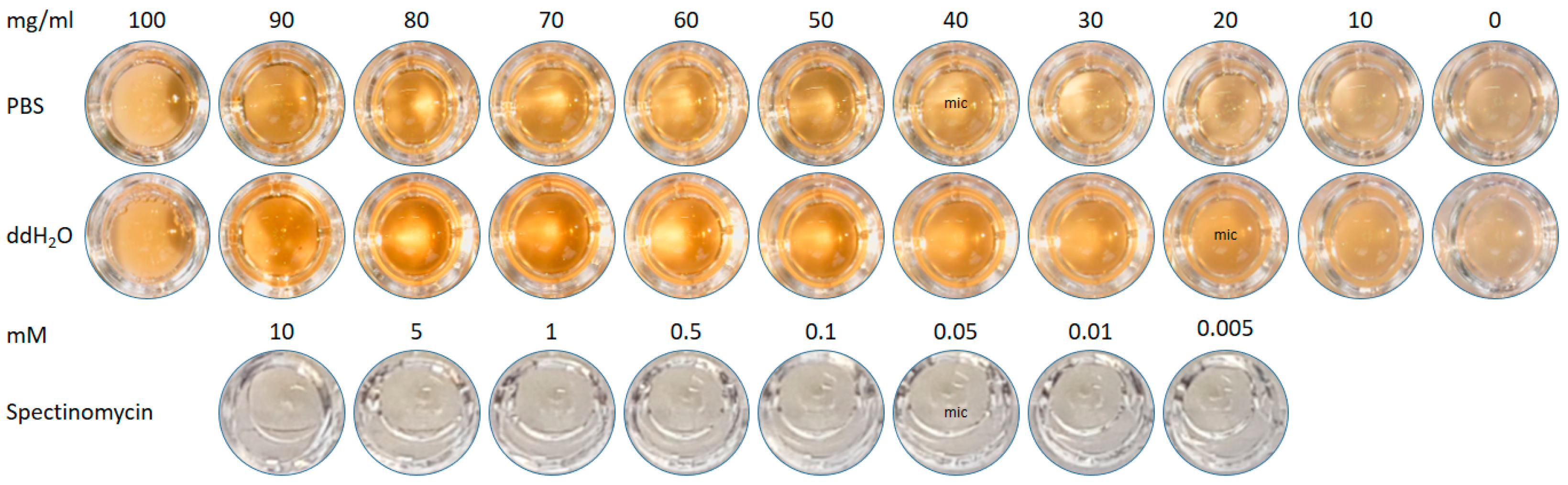
Helleborus extracts in sterile PBS or water were assessed by the minimum inhibitory concentration (MIC) in E. coli cultures (Figure 2). The MIC was verified by growth as 40 mg/mL and 20 mg/mL in PBS and water, respectively, and compared with spectinomycin as a positive control with a MIC value of 0.05 mM. The use of Helleborus preparations to treat infectious diseases is an established practise [10,15], and while research has examined the impact of Helleborus preparations on physiological systems, the data on the specific antimicrobial activity of Helleborus in the context of these infectious diseases is limited in comparison. Rosselli [45] demonstrated the antibacterial influence of Helleborus bocconei extracts against several strains of pathophysiological microorganisms, with later work by Puglisi [46] demonstrating similar effects with a specific focus on strains known to promote respiratory infections. Interestingly, Rosselli reported an MIC in the µg/mL range for Helleborus bocconei in all microorganisms examined, including E. coli. However, Puglisi reported an MIC in the mg/mL range for extracts from the same Helleborus species against E. coli. Our data agrees with Puglisi in that Helleborus odorus displays an MIC in the mg/mL range, although the extraction methods of the plant materials differ.

3.4. Helleborus Concentrations Greater than 50 ng/mL Dose- and Time-Dependently Reduce HAEC Viability

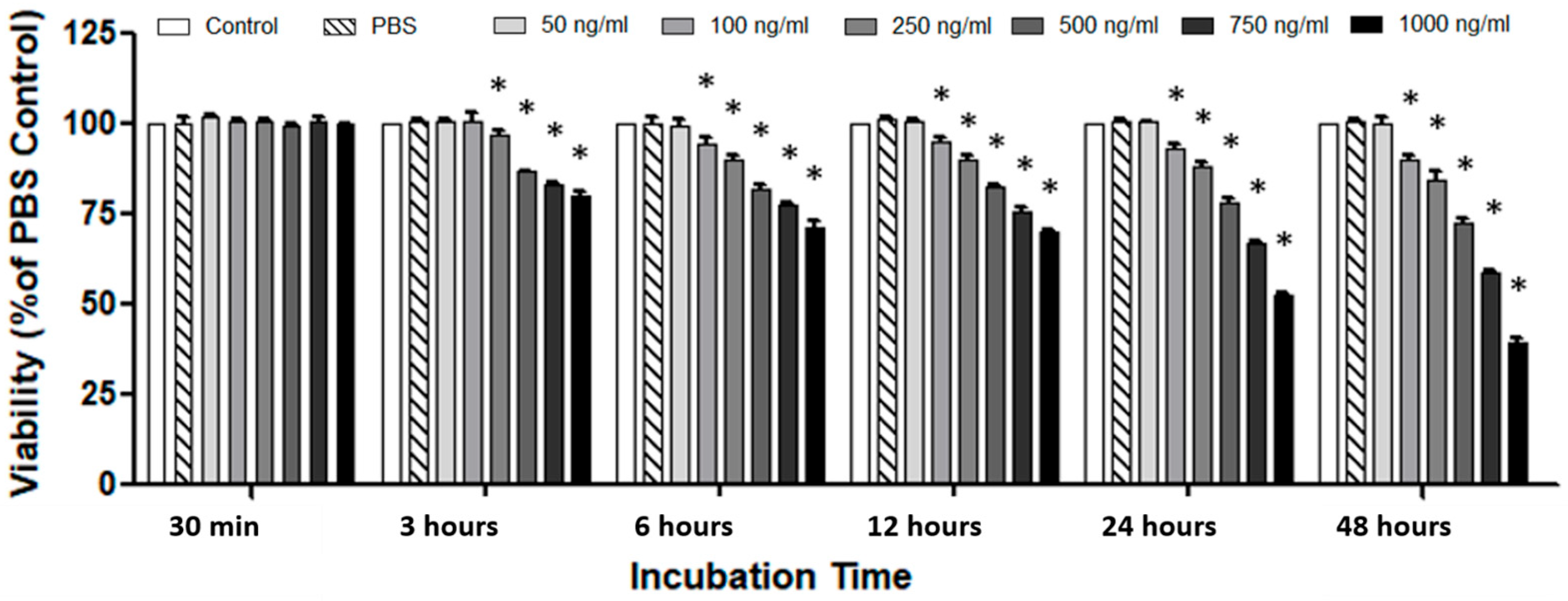
In order to investigate the potential physiological impact of Helleborus odorus subsp. cyclophyllus, HAECs were used as a model of the vascular system, and the impact of dose and treatment time on cell viability was first examined by crystal violet assay. Treatment of HAECs with Helleborus extracts (50 ng/mL–1000 ng/mL) dose- and time-dependently reduced HAEC viability up to 48 h. Concentrations of ≤50 ng/mL had no significant impact on cell viability (Figure 3).
Several studies on Helleborus-derived extracts have revealed that they, or metabolites from such, demonstrate cytotoxic activity. Lindholm [47] found in a screening of 100 plant extracts that Helleborus cyclophyllus and Helleborus caucasicus both demonstrated potent antitumoral activity in vitro. Similar studies of Helleborus niger [23], Helleborus bocconei [48], Helleborus multifidus [19], and Helleborus purpurascens [49,50] demonstrated similar cytotoxic potential in cancer cell lines, the efficacy of which has seen patents granted for use of Helleborus extracts as cytotoxic agents, with specific application of Helleborus derivative as an anticancer agent, owing to the reported antitumor activity in the nanomolar range [51].

3.5. Helleborus-Dependent ROS Induction Influences HAEC Viability

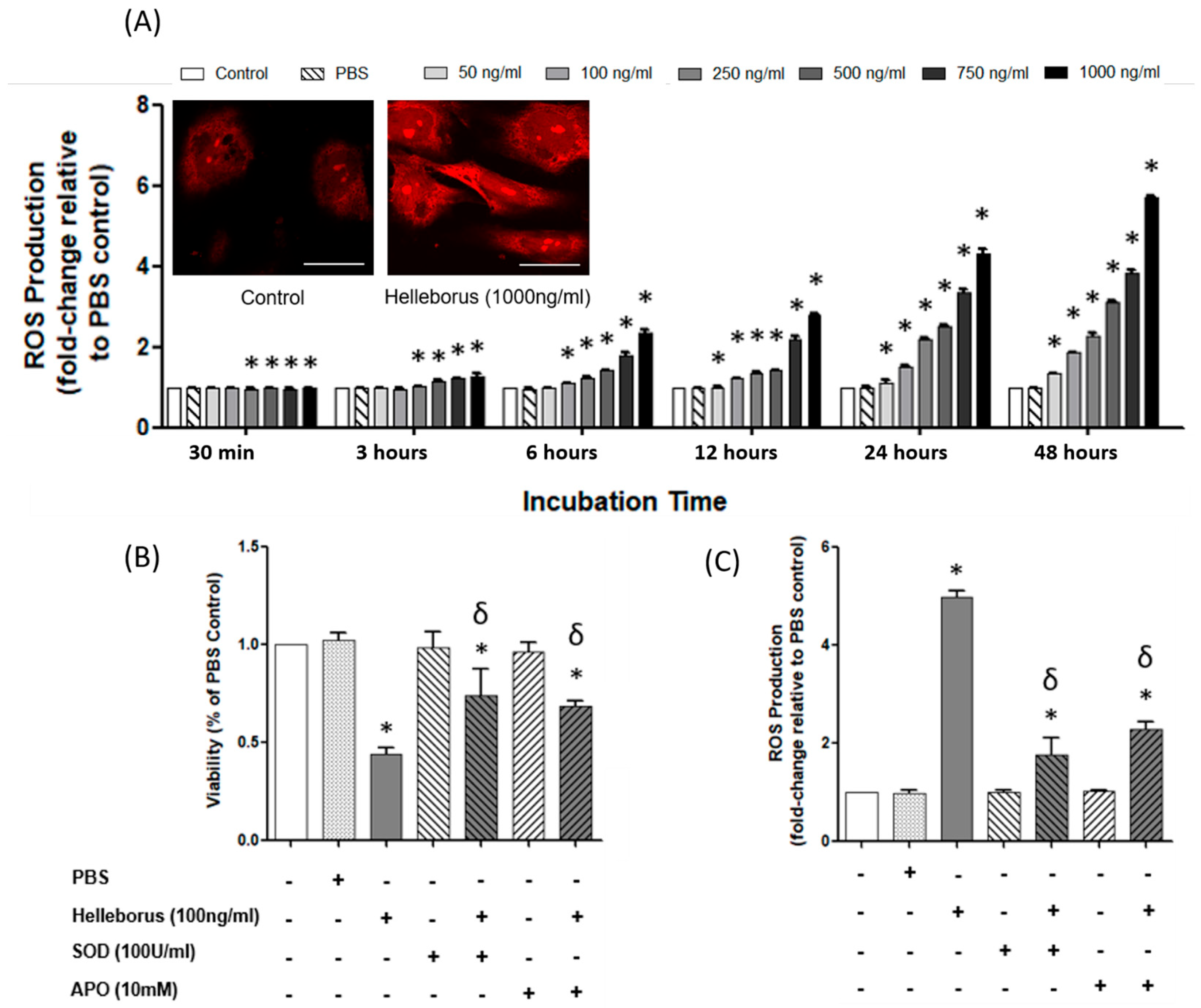
The impact of Helleborus treatment of HAECs on ROS induction was investigated quantitatively using a dihydroethidium assay. Treatment of HAECs with Helleborus extracts (50 ng/mL–1000 ng/mL) dose- and time-dependently significantly induced ROS production (Figure 4A). The conversion of dihydroethidium to ethidium bromide courtesy of ROS induction was confirmed by fluorescence microscopy (Figure 4 insert). To focus on the potential non-lethal effects of Helleborus odorus subsp. cyclophyllus, all subsequent experiments focused on a narrow range of concentrations (50/100/250 ng/mL). Co-treatment of HAECs treated with Helleborus (50/100/250 ng/mL) with SOD (100 U/mL) or APO (10 mM) was shown to significantly attenuate the effects of Helleborus treatment on cell viability (Figure 4B) through the significant amelioration of ROS (Figure 4C).
Previous studies have reported contrasting data on the antioxidant potential of Helleborus species. Work by Apetrei et al. [18] and Păun-Roman et al. [20] revealed that extracts of Helleborus purpurascens demonstrated potent antioxidant potential in vitro, with other works on material/s isolated from Helleborus multifidus, Helleborus hercegovinus, and Helleborus odorus demonstrating similar abilities. A study by Malik [13], however, reported that Helleborus purpurascens had negligible antioxidant activity, with similar results reported for Helleborus niger and Helleborus odorus. While our biochemical data suggests that extracts of Helleborus odorus do contain compounds of antioxidant potential (Figure 1), other compounds that are extracted appear to induce pro-oxidant pathways and/or interfere with the ability of the antioxidant compounds to exert their effect on HAEC cultures. Activation of NADPH oxidase has previously been shown to be responsible for pro-inflammatory induction of ROS in endothelial cells [52]. Ablation of ROS using APO in our data indicates that extracts of Helleborus odorus promote assembly and activation of the NADPH oxidase complex, promoting a pro-inflammatory phenotype in HAECs as a result.

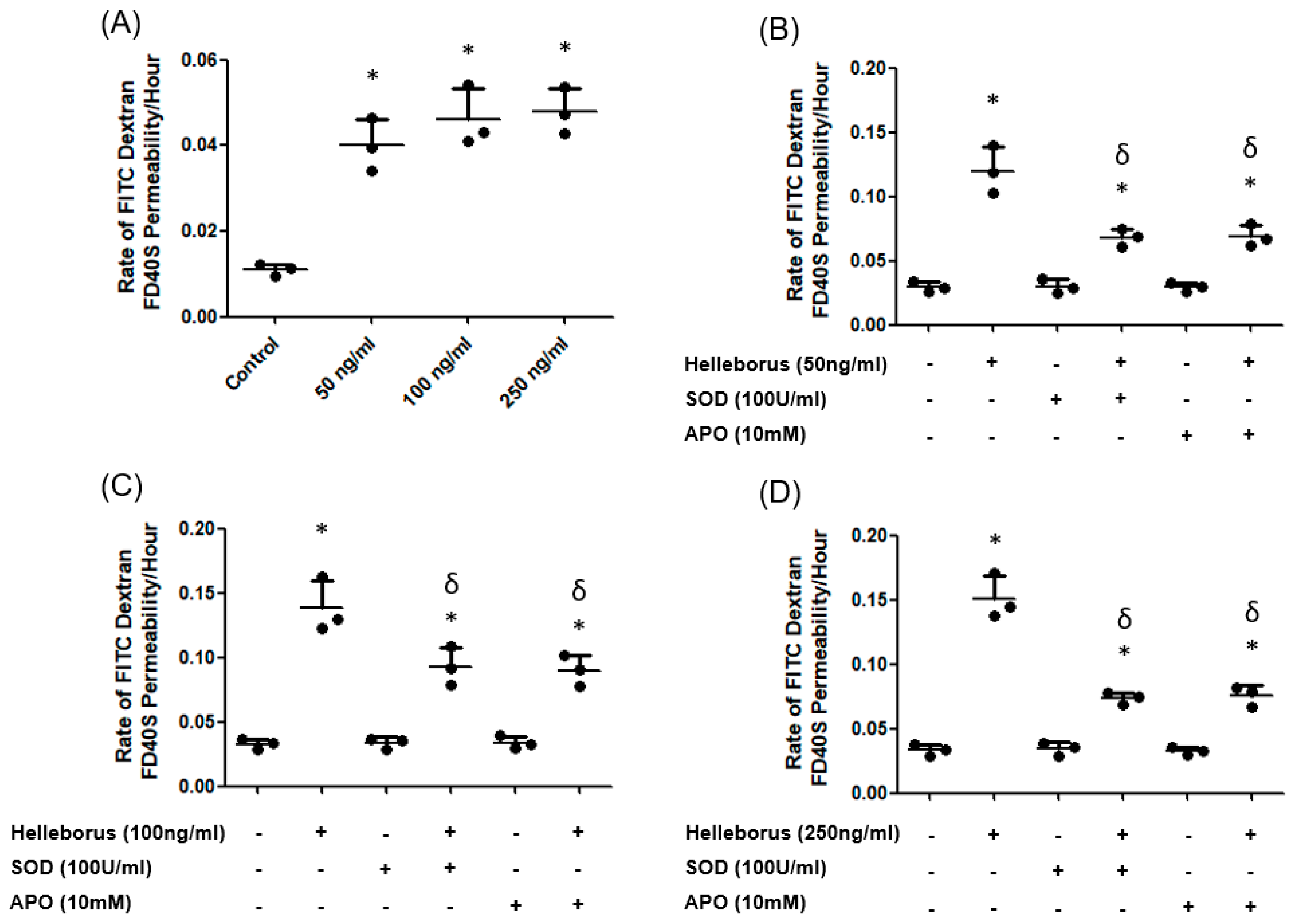
3.6. Helleborus-Dependent ROS Induction Influences HAEC Permeability

The impact of Helleborus treatment (50/100/250 ng/mL) on HAEC permeability was examined using a transwell permeability assay. Treatment of HAECs with Helleborus (50/100/250 ng/mL) saw a significant increase in monolayer permeability (Figure 5A). Co-treatment of Helleborus-treated HAECs with SOD (100 U/mL) or APO (10 mM) was shown to significantly attenuate the Helleborus-induced increase in monolayer permeability for 50 (Figure 5B), 100 (Figure 5C), and 250 ng/mL (Figure 5D) of Helleborus extract up to 48 h of treatment.
Previous data has shown compounds derived from Helleborus to be cardioprotective, owing to its ability to directly influence the endothelium. Protoanemonin, a γ-lactone that can be isolated from members of the Helleborus family, has been shown to modulate endothelial function and promote vascular integrity. Work by Hu [53] and Duan [54] showed that anemonin, a secondary metabolite of protoanemonin, downregulates the expression of the key signalling molecules inducible nitric oxide synthase (iNOS) and endothelin-1 (ET-1) in microvascular endothelial cells. In nature, pleiotropic molecules decrease the expression of both proteins, in parallel with intercellular adhesion molecule-1 (ICAM-1), which promotes increased endothelial barrier function, in addition to an anti-inflammatory phenotype. Our contrasting data suggests that extracts of Helleborus odorus subsp. cyclophyllus disrupt endothelial barrier integrity, partly through a ROS-mediated pro-inflammatory mechanism. Previous data from our laboratory has demonstrated that NADPH oxidase plays an influential role in pro-inflammatory disruption of the endothelial barrier [33,55], with the protective effects of APO indicating a similar trend in response to Helleborus odorus treatment.

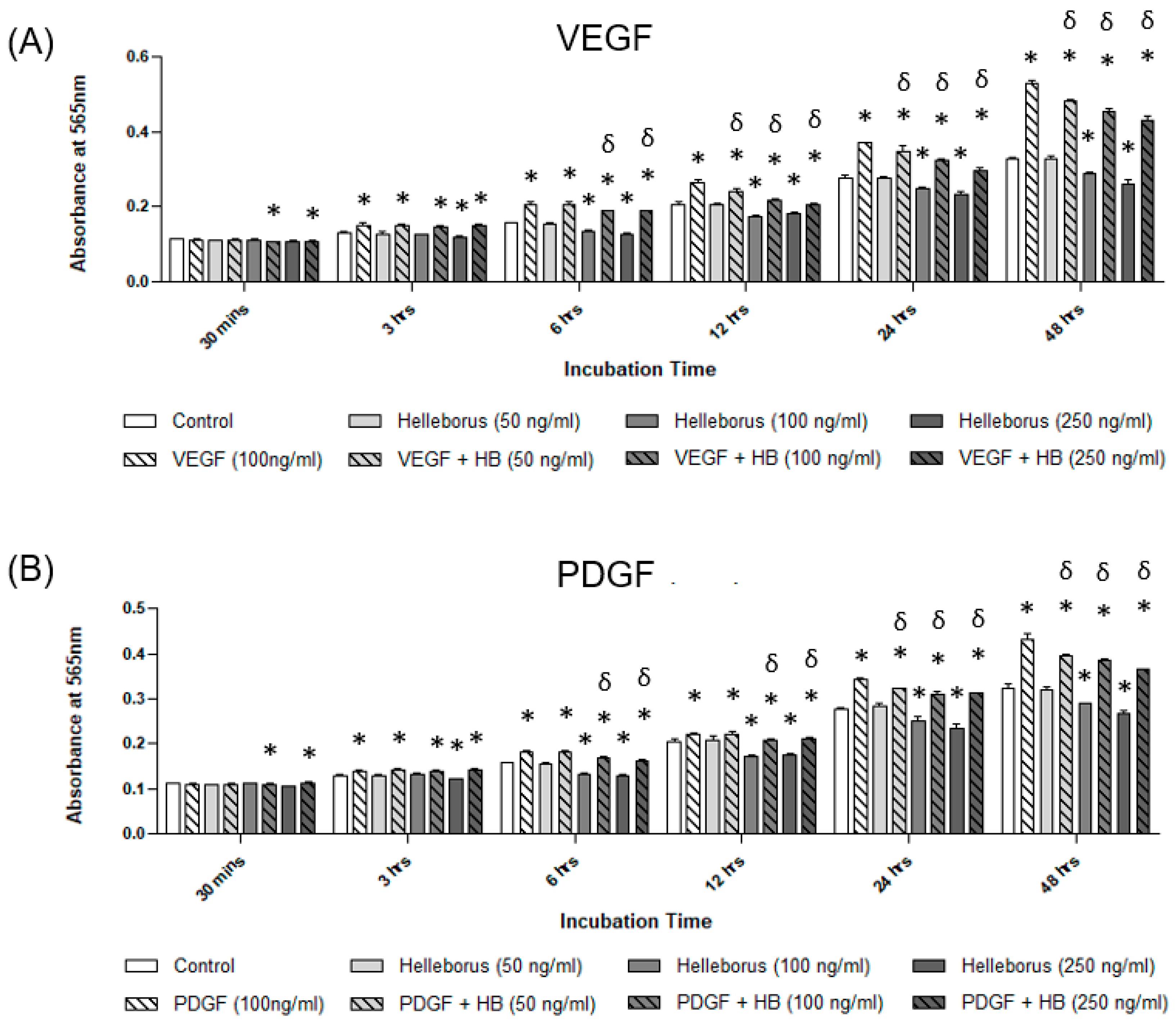
3.7. Helleborus Treatment Attenuates VEGF and PDGF Pro-Angiogenic Effects

The impact of Helleborus treatment (50/100/250 ng/mL) on pro-angiogenic conditions was examined using a crystal violet assay adapted for measuring proliferation. Treatment of HAECS with VEGF (50 ng/mL) (Figure 6A) or PDGF (100 ng/mL) (Figure 6B) saw a significant increase in cell number when compared to untreated controls. Co-treatment of VEGF- or PDGF-treated HAECs with Helleborus odorus subsp. cyclophyllus extract showed a dose-dependent reduction in the proliferative effects of both VEGF and PDGF, with significant effects observed across all concentrations (50/100/250 ng/mL) from 12 h for VEGF and 24 h for PDGF.

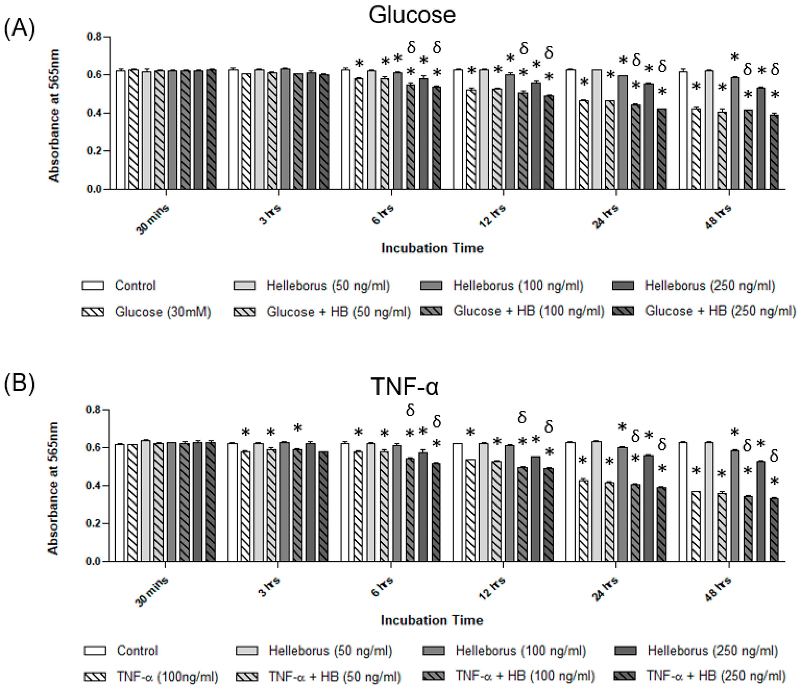
3.8. Helleborus Treatment Exacerbates Hyperglycaemic and TNF-α Pro-Apoptotic Effects

The impact of Helleborus treatment (50/100/250 ng/mL) on pro-apoptotic conditions was examined using a crystal violet assay. Treatment of HAECs with D-glucose (30 mM) (Figure 7A) or TNF-α (100 ng/mL) (Figure 7A) showed a decrease in cell number when compared to untreated controls. Co-treatment of hyperglycaemic- or TNF-α-treated HAECs with Helleborus extract saw a dose-dependent exacerbation in the pro-apoptotic effects of both D-glucose and TNF-α, with significant effects observed for 100 ng/mL and 250 ng/mL of Helleborus treatment from 6 h of treatment.
In a screening of 25 medicinal plant extracts, Malik [13] reported that Helleborus purpurascens was the best at inhibiting the pro-inflammatory enzymes COX-1, COX-2, and 5-LOX, with Helleborus niger and Helleborus odorus also exhibiting similar, albeit less potent, functionality. Subsequent GC-MS analysis of Helleborus purpurascens fractions revealed that the fraction is comprised of a complex of fatty acids, of which α-linoleic acid was the predominant constituent, demonstrating the greatest inhibitory action towards these enzymes. In addition to our findings of antioxidant potential (Table 1), our GC/EI/MS analysis reported relatively high levels of linoleic acid in the Helleborus odorus subsp. cyclophyllus extracts used in this manuscript and should therefore promote an anti-inflammatory phenotype. To evaluate the therapeutic efficacy of extracts of Helleborus odorus subsp. cyclophyllus, our data firmly suggests that the extracts exacerbate the pro-inflammatory effects induced by the agonists employed.
Previous examinations of the therapeutic potential of Helleborus extracts agree with these findings. In addition to reporting negligible antioxidant activity, Čakar et al. [19] also found the Helleborus extracts to exhibit anti-proliferative properties, which is a finding that several other screenings of Helleborus-derived compounds have corroborated, in addition to cytotoxic activity. Data pertaining to the vasculature is limited; however, a study by Felenda [24] also reported anti-proliferative and anti-angiogenic effects in Helleborus niger-treated HUVECs.
Similar extracts from other species (Helleborus niger) exhibit cytotoxicity to cancer and leukaemia cell lines and primary cells of patients with childhood ALL and AML, as they showed inhibition of proliferation by induction of apoptosis [23]. In addition, extracts have been shown to have differential cytotoxicity towards tumour cell lines and healthy human T- and NK-cells by inhibiting proliferation and inducing apoptosis [56]. Recently, another species (Helleborus cyclophyllus Boiss) induced apoptotic cell death and vesicular formations on A549 human bronchial epithelial adenocarcinoma cells selectively and in comparison to normal cells, which were not affected [22]. Although these are in vitro studies and can reach as far as they are limited to a translational model, single-patient case studies have been reported in which extracts of hellebores have been used instead of, or in conjunction with, cancer treatment. In a recent case, a minor regression and long-time survival were observed in a cancer patient who did not receive any other cancer treatment against malignant pleural mesothelioma. The patient remained in good health, and death occurred 56 months after the initial diagnosis [57].

4. Conclusions

The genus Helleborus has demonstrated a diverse range of physiological abilities, many of which have proven beneficial therapeutic actions that are unique and/or contrasting with respect to each species and subspecies. However, Helleborus species remain relatively unexplored, particularly at the cellular level, and investigation of each species and its inherent profile of chemical compounds could potentially identify bioactive molecules of therapeutic potential. As such, the examination of species of the Helleborus genus warrants further investigation, and in this study, Helleborus extracts were found to contain several phytochemical compounds and exhibited antioxidant, antimicrobial, and cytotoxic potential. Given their cytotoxic and antibacterial action, Helleborus extracts have potential as alternative approaches to chemotherapy, as evidenced in case studies highlighting their importance in medicine. Further examination with in vitro and in vivo models would provide important insight into the compound-specific properties of the constituents of the extracts and potentially yield new bioactive molecules to be isolated and investigated further.

Supplementary Materials

The following supporting information can be downloaded at: https://www.mdpi.com/article/10.3390/biology15110852/s1. Supplementary Table S1: Fatty acid content of the analysed Helleborus sp. extract. Information was retrieved from the repositories of NIST, PubChem, KEGG, and LipidMaps.

Author Contributions

Conceptualisation, K.G.; methodology, K.G., K.D.R. and K.A.A.; investigation, formal analysis, visualisation, P.S., E.-A.P., P.K., K.G., K.D.R. and K.A.A.; writing—original draft preparation, P.K., K.G., K.D.R. and K.A.A.; writing—review and editing, P.K., K.G., K.D.R. and K.A.A.; supervision, funding acquisition. All authors have read and agreed to the published version of the manuscript.

Funding

P.S. thanks the Erasmus+ programme for their financial support. This study was partially supported by the internal funds of Dublin City University and the project “PlantUp: Upgrading the Plant Capital. Exploitation of Natural Products of the Hellenic plant biodiversity”, Research Infrastructure, RI (Grant No. MIS, 5002803).

Institutional Review Board Statement

Ethical review and approval were waived for this study due to the fact that the model systems used do not require authorisation from the Health Products Regulatory Authority (HPRA).

Informed Consent Statement

Not applicable.

Data Availability Statement

The datasets used and/or analysed during the current study are available from the corresponding author upon reasonable request. The original contributions presented in this study are included in the Supplementary Materials.

Conflicts of Interest

The authors declare no conflicts of interest. The funders had no role in the design of the study; in the collection, analyses, or interpretation of data; and in the writing of the manuscript.

Abbreviations

The following abbreviations are used in this manuscript:
ALLAcute lymphocytic leukaemia
AMLAcute myeloid leukaemia
APOApocynin
COXCyclooxygenase
DHEDihydroethidium
ET-1Endothelin-1
FAFatty acid
FITCFluorescein isothiocyanate
HAECsHuman aortic endothelial cells
ICAM-1Intercellular adhesion molecule-1
iNOSInducible nitric oxide synthase
LOXLipoxygenase
MICMinimum inhibitory concentration
PBSPhosphate-buffered saline
PDGFPlatelet-derived growth factor
ROSReactive oxygen species
SODSuperoxide dismutase
TNF-αTumour necrosis factor α
VEGFVascular endothelial growth factor

References

  1. Garber, A.; Barnard, L.; Pickrell, C. Review of Whole Plant Extracts With Activity Against Herpes Simplex Viruses In Vitro and In Vivo. J. Evid.-Based Integr. Med. 2021, 26, 2515690X20978394. [Google Scholar] [CrossRef]
  2. Oulahal, N.; Degraeve, P. Phenolic-Rich Plant Extracts With Antimicrobial Activity: An Alternative to Food Preservatives and Biocides? Front. Microbiol. 2021, 12, 753518. [Google Scholar] [CrossRef]
  3. Balazs, V.L.; Filep, R.; Ambrus, T.; Kocsis, M.; Farkas, A.; Stranczinger, S.; Papp, N. Ethnobotanical, historical and histological evaluation of Helleborus L. genetic resources used in veterinary and human ethnomedicine. Genet. Resour. Crop Evol. 2020, 67, 781–797. [Google Scholar] [CrossRef]
  4. Karrer, W. Über Hellebrin, ein krystallisiertes glykosid aus radix Hellebori nigri. Helv. Chim. Acta 1943, 26, 1353–1367. [Google Scholar] [CrossRef]
  5. Wissner, W.; Kating, H. Botanical and phytochemical investigations of species of the genus Helleborus growing in Europe and Asian Minor. II. Comparative phytochemical investigations of the cardio active glycosides and saponins (author’s transl). Planta Medica 1974, 26, 228–249. [Google Scholar] [CrossRef] [PubMed]
  6. Wissner, W.; Kating, H. Botanical and phytochemical investigations of species of the genus Helleborus growing in Europe and Asia Minor. I. Distribution, morphology and cultivation of Helleborus species (author’s transl). Planta Medica 1974, 26, 128–143. [Google Scholar] [CrossRef] [PubMed]
  7. Yang, J.; Zhang, Y.H.; Miao, F.; Zhou, L.; Sun, W. Two new bufadienolides from the rhizomes of Helleborus thibetanus Franch. Fitoterapia 2010, 81, 636–639. [Google Scholar] [CrossRef] [PubMed]
  8. Yokosuka, A.; Inomata, M.; Yoshizawa, Y.; Iguchi, T.; Mimaki, Y. Bufadienolides and ecdysteroids from the whole plants of Helleborus niger and their cytotoxicity. J. Nat. Med. 2021, 75, 393–402. [Google Scholar] [CrossRef]
  9. Bussing, A.; Schweizer, K. Effects of a phytopreparation from Helleborus niger on immunocompetent cells in vitro. J. Ethnopharmacol. 1998, 59, 139–146. [Google Scholar] [CrossRef]
  10. Erdemoglu, N.; Kupeli, E.; Yesilada, E. Anti-inflammatory and antinociceptive activity assessment of plants used as remedy in Turkish folk medicine. J. Ethnopharmacol. 2003, 89, 123–129. [Google Scholar] [CrossRef]
  11. Fujita, T.; Tabata, M.; Yesilada, E.; Honda, G.; Takeda, Y.; Tanaka, T.; Takaishi, Y. Traditional medicine in Turkey VII. Folk medicine in middle and west Black Sea regions. Econ. Bot. 1995, 49, 406. [Google Scholar] [CrossRef]
  12. Linke, S.; Dufter, C.; Kerek, F.; Jung, M.; Watzlik, A.; Jung, T.; Opelz, G.; Terness, P. Functional characterization of HP12: A novel immunosuppressant purified from Helleborus species. Transplant. Proc. 1998, 30, 4106–4107. [Google Scholar] [CrossRef]
  13. Malik, J.; Tauchen, J.; Landa, P.; Kutil, Z.; Marsik, P.; Kloucek, P.; Havlik, J.; Kokoska, L. In vitro antiinflammatory and antioxidant potential of root extracts from Ranunculaceae species. S. Afr. J. Bot. 2017, 109, 128–137. [Google Scholar] [CrossRef]
  14. Olinescu, A.; Hristescu, S.; Poliopol, M.; Agache, F. Effects of Boicil on some immunocompetent cells. I. In vitro modulation of the human peripheral blood lymphocyte functions. Arch. Roum. Pathol. Exp. Microbiol. 1986, 45, 33–45. [Google Scholar] [PubMed]
  15. Kerek, F. Boicil, a new and very efficient antialgic, spasmolytic, and blood vessel regulating drug obtained from the plant Helleborus. In Proceedings of the First International Conference on Chemistry and Biochemistry of Biologically Active Natural Compounds, Varna, Bulgaria, 3 September 1981; pp. 22–37. [Google Scholar]
  16. Maior, M.C.; Dobrota, C.I. Natural compounds with important medical potential found in Helleborus sp. Cent. Eur. J. Biol. 2013, 8, 272–285. [Google Scholar] [CrossRef]
  17. Kuehn, C.; Tauchi, M.; Stumpf, C.; Daniel, C.; Bauerle, T.; Schwarz, M.; Kerek, F.; Steinkasserer, A.; Zinser, E.; Achenbach, S.; et al. Suppression of proatherogenic leukocyte interactions by MCS-18--Impact on advanced atherosclerosis in ApoE-deficient mice. Atherosclerosis 2016, 245, 101–110. [Google Scholar] [CrossRef]
  18. Apetrei, N.S.; Lupu, A.R.; Calugaru, A.; Kerek, F.; Szegli, G.; Cremer, L. The antioxidant effects of some progressively purified fractions from Helleborus purpurascens. Rom. Biotechnol. Lett. 2011, 16, 6673–6682. [Google Scholar]
  19. Cakar, J.; Paric, A.; Vidic, D.; Haveric, A.; Haveric, S.; Maksimovic, M.; Bajrovic, K. Antioxidant and antiproliferative activities of Helleborus odorus Waldst. & Kit, H. multifidus Vis. and H. hercegovinus Martinis. Nat. Prod. Res. 2011, 25, 1969–1974. [Google Scholar] [CrossRef]
  20. Paun Roman, G.; Neagu, E.; Radu, G.L. Membrane processes for the purification and concentration of Helleborus purpurascens extracts and evaluation of antioxidant activity. Rev. Chim. 2010, 61, 877–881. [Google Scholar]
  21. Tesoriere, L.; Butera, D.; Gentile, C.; Livrea, M.A. Bioactive components of caper (Capparis spinosa L.) from Sicily and antioxidant effects in a red meat simulated gastric digestion. J. Agric. Food Chem. 2007, 55, 8465–8471. [Google Scholar] [CrossRef]
  22. Yfanti, P.; Karkabounas, A.; Batistatou, A.; Tsapinou, A.; Leneti, E.; Manos, G.; Lekka, M.E. Study of potent cytotoxic activity of Helleborus cyclophyllus Boiss against a human adenocarcinoma cell line. Cytotechnology 2020, 72, 819–831. [Google Scholar] [CrossRef] [PubMed]
  23. Jesse, P.; Mottke, G.; Eberle, J.; Seifert, G.; Henze, G.; Prokop, A. Apoptosis-inducing activity of Helleborus niger in ALL and AML. Pediatr. Blood Cancer 2009, 52, 464–469. [Google Scholar] [CrossRef]
  24. Felenda, J.E.; Turek, C.; Morbt, N.; Herrick, A.; Muller, M.B.; Stintzing, F.C. Preclinical evaluation of safety and potential of black hellebore extracts for cancer treatment. BMC Complement. Altern. Med. 2019, 19, 105. [Google Scholar] [CrossRef]
  25. Martucciello, S.; Paolella, G.; Muzashvili, T.; Skhirtladze, A.; Pizza, C.; Caputo, I.; Piacente, S. Steroids from Helleborus caucasicus reduce cancer cell viability inducing apoptosis and GRP78 down-regulation. Chem.-Biol. Interact. 2018, 279, 43–50. [Google Scholar] [CrossRef]
  26. Segneanu, A.E.; Grozescu, I.; Cziple, F.; Berki, D.; Damian, D.; Niculite, C.M.; Florea, A.; Leabu, M. Helleborus purpurascens-Amino Acid and Peptide Analysis Linked to the Chemical and Antiproliferative Properties of the Extracted Compounds. Molecules 2015, 20, 22170–22187. [Google Scholar] [CrossRef]
  27. Jablonska-Trypuc, A.; Pankiewicz, W.; Czerpak, R. Traumatic Acid Reduces Oxidative Stress and Enhances Collagen Biosynthesis in Cultured Human Skin Fibroblasts. Lipids 2016, 51, 1021–1035. [Google Scholar] [CrossRef]
  28. Michalaki, A.; Grintzalis, K. A Multiparametric Protocol for the Detailed Phytochemical and Antioxidant Characterisation of Plant Extracts. Methods Protoc. 2023, 6, 40. [Google Scholar] [CrossRef]
  29. Price, M.L.; van Scoyoc, S.; Butler, L.G. A critical evaluation of the vanillin reaction as an assay for tannin in sorghum grain. J. Agric. Food Chem. 1978, 26, 1214–1218. [Google Scholar] [CrossRef]
  30. Kalampokis, I.F.; Erban, A.; Amillis, S.; Diallinas, G.; Kopka, J.; Aliferis, K.A. Untargeted metabolomics as a hypothesis-generation tool in plant protection product discovery: Highlighting the potential of trehalose and glycerol metabolism of fungal conidiospores as novel targets. Metabolomics 2020, 16, 79. [Google Scholar] [CrossRef] [PubMed]
  31. Kostopoulou, S.; Ntatsi, G.; Arapis, G.; Aliferis, K.A. Assessment of the effects of metribuzin, glyphosate, and their mixtures on the metabolism of the model plant Lemna minor L. applying metabolomics. Chemosphere 2020, 239, 124582. [Google Scholar] [CrossRef] [PubMed]
  32. Forde, H.; Harper, E.; Rochfort, K.D.; Wallace, R.G.; Davenport, C.; Smith, D.; Cummins, P.M. TRAIL inhibits oxidative stress in human aortic endothelial cells exposed to pro-inflammatory stimuli. Physiol. Rep. 2020, 8, e14612. [Google Scholar] [CrossRef]
  33. Rochfort, K.D.; Collins, L.E.; Murphy, R.P.; Cummins, P.M. Downregulation of blood-brain barrier phenotype by proinflammatory cytokines involves NADPH oxidase-dependent ROS generation: Consequences for interendothelial adherens and tight junctions. PLoS ONE 2014, 9, e101815. [Google Scholar] [CrossRef]
  34. Tsiftsoglou, O.S.; Stefanakis, M.K.; Kalpourtzi, E.N.; Hadjipavlou-Litina, D.I.; Lazari, D.M. Chemical constituents isolated from the aerial parts of Helleborus cyclophyllus (A. Braun) Boiss. (Ranunculaceae), evaluation of their antioxidant and anti-inflammatory activity in vitro and virtual screening of molecular properties and bioactivity score. Nat. Prod. Res. 2022, 36, 6031–6038. [Google Scholar] [CrossRef]
  35. Djami-Tchatchou, A.T.; Ncube, E.N.; Steenkamp, P.A.; Dubery, I.A. Similar, but different: Structurally related azelaic acid and hexanoic acid trigger differential metabolomic and transcriptomic responses in tobacco cells. BMC Plant Biol. 2017, 17, 227. [Google Scholar] [CrossRef]
  36. Henry, G.E.; Momin, R.A.; Nair, M.G.; Dewitt, D.L. Antioxidant and cyclooxygenase activities of fatty acids found in food. J. Agric. Food Chem. 2002, 50, 2231–2234. [Google Scholar] [CrossRef]
  37. Menendez, J.A.; Papadimitropoulou, A.; Vellon, L.; Lupu, R. A genomic explanation connecting “Mediterranean diet”, olive oil and cancer: Oleic acid, the main monounsaturated fatty acid of olive oil, induces formation of inhibitory “PEA3 transcription factor-PEA3 DNA binding site” complexes at the Her-2/neu (erbB-2) oncogene promoter in breast, ovarian and stomach cancer cells. Eur. J. Cancer 2006, 42, 2425–2432. [Google Scholar] [CrossRef] [PubMed]
  38. Sales-Campos, H.; Souza, P.R.; Peghini, B.C.; da Silva, J.S.; Cardoso, C.R. An overview of the modulatory effects of oleic acid in health and disease. Mini Rev. Med. Chem. 2013, 13, 201–210. [Google Scholar] [PubMed]
  39. Blaskovich, M.A.T.; Elliott, A.G.; Kavanagh, A.M.; Ramu, S.; Cooper, M.A. In vitro Antimicrobial Activity of Acne Drugs Against Skin-Associated Bacteria. Sci. Rep. 2019, 9, 14658. [Google Scholar] [CrossRef] [PubMed]
  40. Bojar, R.A.; Cunliffe, W.J.; Holland, K.T. Disruption of the transmembrane pH gradient—A possible mechanism for the antibacterial action of azelaic acid in Propionibacterium acnes and Staphylococcus epidermidis. J. Antimicrob. Chemother. 1994, 34, 321–330. [Google Scholar] [CrossRef]
  41. Sieber, M.A.; Hegel, J.K. Azelaic acid: Properties and mode of action. Ski. Pharmacol. Physiol. 2014, 27, 9–17. [Google Scholar] [CrossRef]
  42. Fitton, A.; Goa, K.L. Azelaic acid. A review of its pharmacological properties and therapeutic efficacy in acne and hyperpigmentary skin disorders. Drugs 1991, 41, 780–798. [Google Scholar] [CrossRef]
  43. Medeiros de Azevedo, W.; Ferreira Ribeiro de Oliveira, L.; Alves Alcantara, M.; Tribuzy de Magalhaes Cordeiro, A.M.; Florentino da Silva Chaves Damasceno, K.S.; Kelly de Araujo, N.; Fernandes de Assis, C.; Sousa Junior, F.C. Physicochemical characterization, fatty acid profile, antioxidant activity and antibacterial potential of cacay oil, coconut oil and cacay butter. PLoS ONE 2020, 15, e0232224. [Google Scholar] [CrossRef]
  44. Pohl, C.H.; Kock, J.L.; Thibane, V.S. Antifungal Free Fatty Acids: A Review. In Science Against Microbial Pathogens: Communicating Current Research and Technological Advances; Formatex Research Center: Badajoz, Spain, 2011. [Google Scholar]
  45. Rosselli, S.; Maggio, A.; Formisano, C.; Napolitano, F.; Senatore, F.; Spadaro, V.; Bruno, M. Chemical composition and antibacterial activity of extracts of Helleborus bocconei Ten. subsp. intermedius. Nat. Prod. Commun. 2007, 2, 675–679. [Google Scholar] [CrossRef]
  46. Puglisi, S.; Speciale, A.; Acquaviva, R.; Ferlito, G.; Ragusa, S.; De Pasquale, R.; Iauk, L. Antibacterial activity of Helleborus bocconei Ten. subsp. siculus root extracts. J. Ethnopharmacol. 2009, 125, 175–177. [Google Scholar] [CrossRef] [PubMed]
  47. Lindholm, P.; Gullbo, J.; Claeson, P.; Goransson, U.; Johansson, S.; Backlund, A.; Larsson, R.; Bohlin, L. Selective cytotoxicity evaluation in anticancer drug screening of fractionated plant extracts. J. Biomol. Screen. 2002, 7, 333–340. [Google Scholar] [CrossRef]
  48. Rosselli, S.; Maggio, A.; Bruno, M.; Spadaro, V.; Formisano, C.; Irace, C.; Maffettone, C.; Mascolo, N. Furostanol saponins and ecdysones with cytotoxic activity from Helleborus bocconei ssp. intermedius. Phytother. Res. 2009, 23, 1243–1249. [Google Scholar] [CrossRef] [PubMed]
  49. Pilut, C.N.; Manea, A.; Macasoi, I.; Dobrescu, A.; Georgescu, D.; Buzatu, R.; Faur, A.; Dinu, S.; Chioran, D.; Pinzaru, I.; et al. Comparative Evaluation of the Potential Antitumor of Helleborus purpurascens in Skin and Breast Cancer. Plants 2022, 11, 194. [Google Scholar] [CrossRef]
  50. Vochita, G.; Mihai, C.T.; Gherghel, D.; Iurea, D.; Roman, G.; Radu, G.L.; Rotinberg, P. New potential antitumoral agents of polyphenolic nature obtained from Helleborus purpurascens by membranary micro- and ultrafiltration techniques. Analele Ştiinţifice Ale Univ. “Ovidius” Constanţa 2011, 12, 41–51. [Google Scholar]
  51. Dewelle, J.; Yazidi, M.E.L.; Van Quaquebeke, E.; Neve, D.N.; Mijatovic, T.; Ingrassia, L.; Kiss, R.; Van Vynckt, F.; Heureux, N. Hellebrin and Hellebrigenin Derivatives. WO2010102673A1, 16 September 2010. [Google Scholar]
  52. Guinan, A.F.; Rochfort, K.D.; Fitzpatrick, P.A.; Walsh, T.G.; Pierotti, A.R.; Phelan, S.; Murphy, R.P.; Cummins, P.M. Shear stress is a positive regulator of thimet oligopeptidase (EC3.4.24.15) in vascular endothelial cells: Consequences for MHC1 levels. Cardiovasc. Res. 2013, 99, 545–554. [Google Scholar] [CrossRef]
  53. Hu, Y.; Chen, X.; Duan, H.; Hu, Y.; Mu, X. Pulsatilla decoction and its active ingredients inhibit secretion of NO, ET-1, TNF-alpha, and IL-1 alpha in LPS-induced rat intestinal microvascular endothelial cells. Cell Biochem. Funct. 2009, 27, 284–288. [Google Scholar] [CrossRef]
  54. Duan, H.; Zhang, Y.; Xu, J.; Qiao, J.; Suo, Z.; Hu, G.; Mu, X. Effect of anemonin on NO, ET-1 and ICAM-1 production in rat intestinal microvascular endothelial cells. J. Ethnopharmacol. 2006, 104, 362–366. [Google Scholar] [CrossRef]
  55. Rochfort, K.D.; Cummins, P.M. Cytokine-mediated dysregulation of zonula occludens-1 properties in human brain microvascular endothelium. Microvasc. Res. 2015, 100, 48–53. [Google Scholar] [CrossRef]
  56. Schink, M.; Garcia-Kaufer, M.; Bertrams, J.; Duckstein, S.M.; Muller, M.B.; Huber, R.; Stintzing, F.C.; Grundemann, C. Differential cytotoxic properties of Helleborus niger L. on tumour and immunocompetent cells. J. Ethnopharmacol. 2015, 159, 129–136. [Google Scholar] [CrossRef]
  57. Werthmann, P.G.; Saltzwedel, G.; Kienle, G.S. Minor regression and long-time survival (56 months) in a patient with malignant pleural mesothelioma under Viscum album and Helleborus niger extracts-a case report. J. Thorac. Dis. 2017, 9, E1064–E1070. [Google Scholar] [CrossRef]
Figure 1. Representative GC/EI/MS total ion chromatogram (TIC) of the analysed Helleborus odorus subsp. cyclophyllus extract. Annotations for the selected fatty acids are displayed: 1. palmitoleic acid; 2. palmitic acid; 3. 2-hydroxysebacic acid; 4. traumatic acid; 5. methyl oleate; 6. cis-10-heptadecenoic acid; 7. margaric acid; 8. linoleic acid; and 9. oleic acid (A). Pie charts displaying the relative abundance (%) of the major chemical groups of the annotated metabolites (B).
Biology 15 00852 g001
Figure 2. Antimicrobial properties of Helleborus extracts in PBS or water against E. coli defined by the minimum inhibitory concentration.
Biology 15 00852 g002
Figure 3. The effect of Helleborus concentration on HAEC viability (N = 3). HAECs were treated with Helleborus (50 ng/mL–1000 ng/mL) for up to 48 h. Crystal violet stain was added at the end of the incubation period in order to quantify cell density of the HAECs following Helleborus treatment. * p ≤ 0.05 versus the untreated control.
Biology 15 00852 g003
Figure 4. The effect of Helleborus concentration on ROS induction in HAECs (N = 3). HAECs were treated with Helleborus odorus subsp. cyclophyllus extract (50 ng/mL–1000 ng/mL) for up to 48 h. DHE stain (3 µM) was added 30 min prior to the end of the incubation period to quantify the production of ROS in HAECs. (A) The time- and dose-dependent impact of Helleborus odorus subsp. cyclophyllus on ROS production in HAECs. All values are baseline corrected to cell viability. Microscopic visualisation of ROS in HAECs after 48 h of treatment ± 1000 ng/mL of Helleborus odorus subsp. cyclophyllus extract (insert). Red: DHE staining for ROS. Scale bar is 30 µm. Images are at 40× and are representative. (B) The effect of SOD and APO individually on Helleborus-induced ROS. (C) The effect of SOD and APO individually on Helleborus-induced cell death. * p ≤ 0.05 versus the untreated control; δ p ≤ 0.05 versus the Helleborus-treated cultures (100 ng/mL).
Biology 15 00852 g004
Figure 5. The effect of Helleborus concentration on HAEC permeability (N = 3). HAECs were treated for up to 48 h with Helleborus (50, 100 or 250 ng/mL) in the absence (A) and presence (BD) of either superoxide dismutase (SOD, 100 U/mL) or apocynin (APO, 10 mM). HAEC barrier integrity was then monitored by transendothelial permeability assay with data represented as the mean rate of FITC-dextran diffusion: %FD40 TEE.hour−1. * p ≤ 0.05 versus the untreated control; δ p ≤ 0.05 versus the Helleborus-treated cultures.
Biology 15 00852 g005
Figure 6. The effect of Helleborus treatment on pro-angiogenic stimulation (N = 3). HAECs were treated for up to 48 h with VEGF (50 ng/mL) (A) or PDGF (100 ng/mL) (B). The HAEC cell number was then monitored by crystal violet staining, with data represented as the absorbance at 565 nm. * p ≤ 0.05 versus the untreated control; δ p ≤ 0.05 versus the (A) VEGF- or (B) PDGF-treated cultures.
Biology 15 00852 g006
Figure 7. The effect of Helleborus treatment on pro-apoptotic stimulation (N = 3). HAECs were treated for up to 48 h with glucose (30 mM) (A) or TNF-α (100 ng/mL) (B). The HAEC cell number was then monitored by crystal violet staining, with data represented as the absorbance at 565 nm. * p ≤ 0.05 versus the untreated control; δ p ≤ 0.05 versus the (A) glucose- or (B) TNF-α treated cultures.
Biology 15 00852 g007
Table 1. Phytochemical and antioxidant properties of Helleborus odorus subsp. cyclophyllus extracts.
ExtractPolyphenolsFlavonoidsTanninsFeRPCuRPABTS•+DPPHGalvinoxyl
PBS 3.58 ± 0.250.715 ± 0.028.55 ± 0.8227.8 ± 0.821.2 ± 0.88.05 ± 0.81.05 ± 0.0287.94 ± 0.64
Water 2.96 ± 0.470.135 ± 0.0339.97 ± 0.8742.8 ± 1.717.8 ± 0.94.3 ± 0.160.995 ± 0.01653.62 ± 0.78
Data represent average ± SD (N = 3) and were considered statistically significant (bold font) by Student’s t-test between extractions. Polyphenols, metal chelation potential, and the % of radical scavenging were expressed as equivalents of gallic acid per g of extract, whereas flavonoids and tannins were expressed as equivalents of catechin per g of Helleborus dried root sample.
Disclaimer/Publisher’s Note: The statements, opinions and data contained in all publications are solely those of the individual author(s) and contributor(s) and not of MDPI and/or the editor(s). MDPI and/or the editor(s) disclaim responsibility for any injury to people or property resulting from any ideas, methods, instructions or products referred to in the content.