Review

Decoding Stress Responses in Farmed Crustaceans: Comparative Insights for Sustainable Aquaculture Management

by
Fitriska Hapsari
1,2,
Muhammad Agus Suprayudi
1,
Dean M. Akiyama
3,
Julie Ekasari
1,*,
Parisa Norouzitallab
4 and
Kartik Baruah
4,*
1
Department of Aquaculture, Faculty of Fisheries and Marine Sciences, IPB University, Bogor 16680, Indonesia
2
Department of Technical Aquaculture, Jakarta Technical University of Fisheries, Jakarta 12520, Indonesia
3
PT. Central Proteina Prima, Jakarta 12190, Indonesia
4
Department of Applied Animal Science and Welfare, Faculty of Veterinary, Medicine and Animal Sciences, Swedish University of Agricultural Sciences, 75007 Uppsala, Sweden
*
Authors to whom correspondence should be addressed.
Biology 2025, 14(8), 920; https://doi.org/10.3390/biology14080920
Submission received: 30 April 2025 / Revised: 30 June 2025 / Accepted: 16 July 2025 / Published: 23 July 2025
(This article belongs to the Section Marine Biology)

Simple Summary

Aquaculture is vital for providing essential nutrients to the growing human population, but it faces challenges such as limited water quality and space. To address these, aquaculture systems have become more intensive, but this also introduces stress risks to cultured organisms, such as overcrowding, waste buildup, and water quality deterioration, which can harm their growth, health, and immunity. Climate change and human activities further increase environmental stress, especially for crustaceans like shrimp. Shrimp have a simpler physiology compared to fish, leading to less complex stress responses. Although stress is known to impact shrimp growth, health, and immunity, research on their stress responses is limited. Understanding these responses at both the organismal and cellular levels is crucial for identifying effective biomarkers and developing targeted strategies to reduce stress. This review summarizes the physiological changes in crustaceans under stress and aims to improve crustacean health management by filling knowledge gaps in stress response mechanisms.

Abstract

Aquaculture is a crucial food-producing sector that can supply more essential nutrients to nourish the growing human population. However, it faces challenges, including limited water quality and space competition. These constraints have led to the intensification of culture systems for more efficient resource use while maintaining or increasing production levels. However, intensification introduces stress risks to cultured organisms by, for instance, overcrowding, waste accumulation, and water quality deterioration, which can negatively affect the growth, health, and immunity of animals and cause diseases. Additionally, environmental changes due to climate and anthropogenic activities further intensify the environmental stress for aquaculture organisms, including crustaceans. Shrimp are one of the most widely cultured and consumed farmed crustacea. Relative to aquatic vertebrates such as fish, the physiology of crustaceans has simpler physiological structures, as they lack a spinal cord. Consequently, their stress response mechanisms follow a single pathway, resulting in less complex responses to stress exposure compared to those of fish. While stress is considered a primary factor influencing the growth, health, and immunity of shrimp, comprehensive research on crustacean stress responses remains limited. Understanding the stress response at the organismal and cellular levels is essential to identify sensitive and effective stress biomarkers which can inform the development of targeted intervention strategies to mitigate stress. This review provides a comprehensive overview of the physiological changes that occur in crustaceans under stress, including hormonal, metabolic, hematological, hydromineral, and phenotypic alterations. By synthesizing current knowledge, this article aims to bridge existing gaps and provide insights into the stress response mechanisms, paving the way for advancements in crustacean health management.

1. Introduction

Driven by the increasing demand for aquatic products, the aquaculture sector has expanded rapidly over the past few decades. Over the past six decades, the share of feed consumption attributable to aquaculture has risen significantly, from just 6% in the 1960s to 56% in 2021 [1]. In 2020, aquaculture accounted for 56% of the aquatic products consumed by humans [2]. Shellfish consumption has also shown a notable increase over the same period, rising from 14% to 26%. Within this group, crustaceans, including shrimp, lobster, and crab, have demonstrated strong growth in both consumption and production. Crustaceans are the second most cultured aquatic organisms after finfish, and their production has increased from about 5.5 million tons in 2016 to about 7.5 million tons in 2022 (36% increase) [1]. While many crustacean species are cultivated for human consumption, others, such as copepods, play an important indirect role in aquaculture as live feed for the larval stages of fish and shrimp, thus supporting the production of higher-trophic-level species. Over the last two decades, shrimp production in China has seen the greatest increase, rising by 194% (from 1.11 million MT to 3.27 million MT), followed by Ecuador (12.69%), Argentina (3.33%), and Vietnam (3.14%). Shrimp production in India and Indonesia has also tripled during the same period. This growth is largely attributed to the intensification of shrimp aquaculture systems in these regions [3]. A meta-analysis examining the relationship between shrimp production intensity and land use in these countries revealed that Vietnam has the highest production intensity (36.66 t/ha/year), followed by Indonesia (22.85 t/ha/year) [4]. Despite the potential benefits, production intensification faces some major challenges, such as environmental changes and disease outbreaks.
Climate change imposes additional environmental pressures on aquaculture, including extreme weather events, temperature fluctuations, changes in sea surface salinity, shifting rainfall patterns, rising sea levels, and increased anthropogenic pollution. These factors have direct and indirect adverse effects on aquaculture production [2,5]. Although aquaculture is generally practiced in controlled environments, many operations, particularly pond-based and coastal systems, remain exposed to climatic variability. For instance, excessive rainfall or prolonged drought can alter pond salinity and temperature beyond optimal thresholds, disrupting water quality and animal health. These climate-driven stressors can compromise the effectiveness of control measures, especially in open or semi-enclosed systems such as earthen shrimp ponds. Multiple studies have demonstrated the detrimental consequences of climate change on the aquaculture industry, including total stock loss, increased mortality, decreased productivity, pond/tank damage, increased operational expenses, and reduced profitability [6,7,8], as well as a negative impact on the seafood supply chain [9]. The increase in global population significantly exacerbates the anthropogenic environmental footprint, such as pollution and eutrophication, resulting in the deterioration of water sources available for aquaculture activities. This further induces physiological responses such as metabolic stress and toxicity responses that lead to growth retardation, reduced immunity, or even mortality.
In an aquaculture system, disease outbreaks are closely linked to environmental quality, the animals’ physiological condition, the presence of pathogenic organisms, and their interrelations. When situated in a suboptimum environmental condition, most aquatic organisms will try to adapt and develop a series of metabolic responses in response to stress to overcome the changes and immediately return to homeostatic conditions. These processes will alter the overall bioenergetics in the animal’s body, where energy will mainly be allocated for metabolic adjustment, thus reducing the energy available for other physiological functions, such as the defense system and growth. Previous studies have demonstrated the adverse impacts of environmental changes, such as oxygen levels [10,11,12], temperature [13,14,15,16,17,18], salinity [19,20,21,22,23], pH [24,25,26], and ammonia concentrations [27,28,29,30,31], on crustacean species, especially shrimp. In those studies, environmental stressors were shown to cause a series of physiological dysfunctions, including a weakening of immunity, resulting in an increased susceptibility of the organisms to diseases. Accumulating evidence [11,12,16,32] suggests that stress strongly corresponds to disease susceptibility by both compromising the immunity of organisms and increasing the virulence factors of pathogens. Therefore, stress mitigation is crucial for improving the resilience of farmed crustaceans, particularly in the face of environmental challenges and increasing disease threats. To achieve effective stress mitigation strategies, a better understanding of stress responses in crustaceans is essential. However, only limited information in this area is available, highlighting an urgent need for comprehensive research to bridge this critical gap.
This literature review focuses on providing a thorough overview of stress response in crustaceans, highlighting stress biomarkers and potential strategies for mitigation. The structure of the review includes an overview of stress in aquaculture systems, covering the types and sources of stress, general stress responses in aquatic organisms, the physiology of stress in crustaceans starting from the neuroendocrine system to the immune response, differences in stress responses between crustaceans and fish (vertebrates), stress indicators, and mitigation strategies, as well as identifying gaps and future research directions.

2. Scope of Stress in Aquaculture

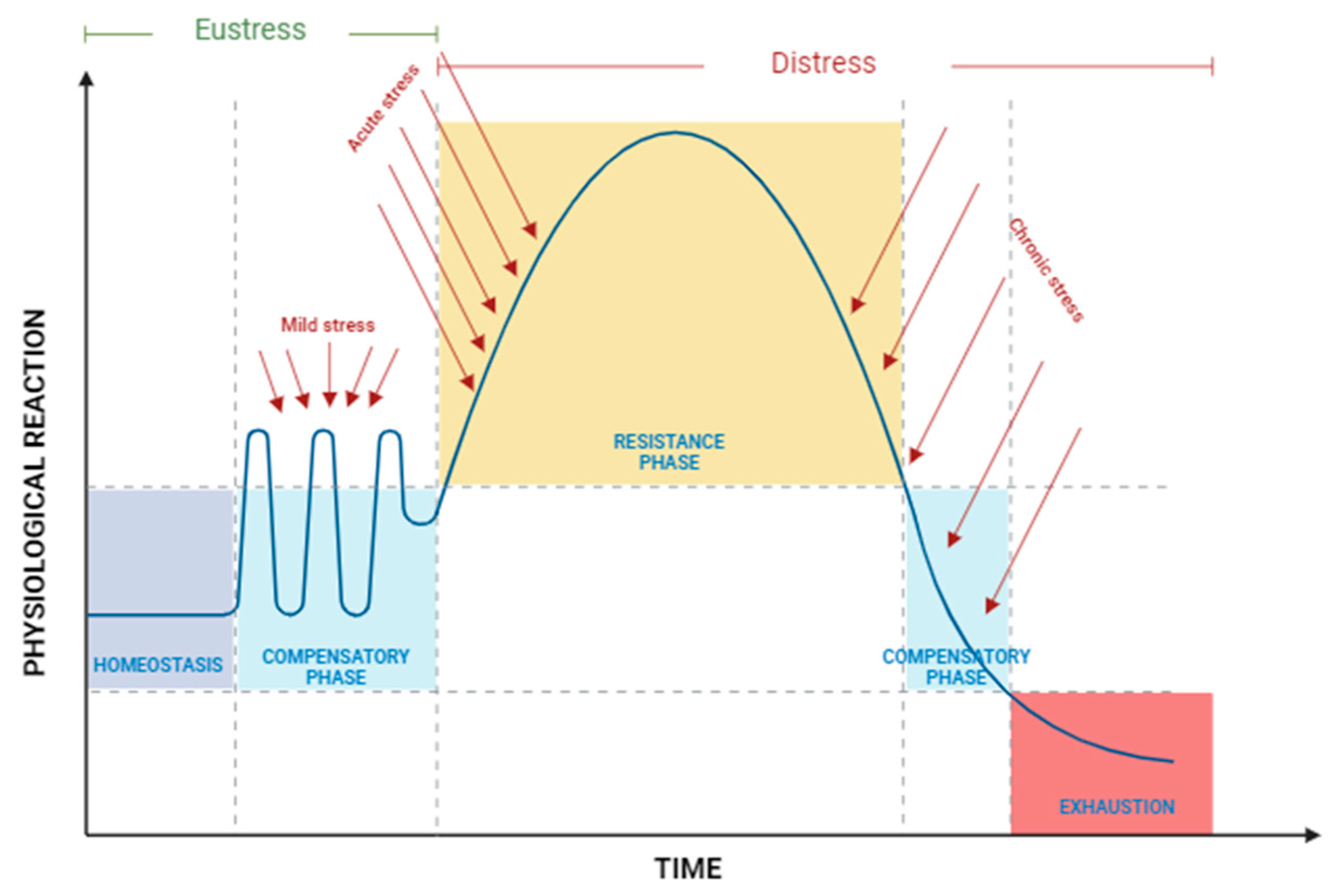
2.1. Definition of Stress

The concept of “stress” has been well defined in previous studies [33,34,35,36,37]. Stress is an essential mechanism in the physiological adaptation to stressors; it includes molecular and biochemical modifications in restoring homeostasis or behavioral responses such as escaping [38]. Despite the lack of a universally accepted definition, it is reasonable to define stress as a state generated by excessive exogenous stimuli (stressors) that induces adaptive responses of altered neuroendocrine, physiological, and behavioral states in an organism.
The stress level of an organism can be classified into two categories: eustress, which represents a low level of stress, and distress, which signifies a higher level of stress [34]. Eustress can be advantageous and can improve performance and have a favorable effect. Conversely, distress or chronic stress may result in both beneficial and detrimental behaviors, decreasing performance and threatening the well-being and overall health of the animal [34,36,39,40]. Dealing with long-term stress requires significant energy and resources [40], which suggests that the body’s response to stress is influenced by the type and intensity of the stressor [38].
Stressful conditions in cultured organisms typically arise from multiple interacting stressors. Various stressors can hinder growth, weaken immunity, diminish disease resistance, and possibly lead to mortality [35], which is related to the energy required to restore the organism to the equilibrium state. Therefore, the ability of an organism to survive a stressor will depend on its capability to return to initial homeostatic condition [34]. Figure 1 illustrates the physiological reactions that occur in the body when exposed to multiple stressors. Homeostasis is a state in which the body’s metabolic condition remains stable and balanced. Because of this function, energy homeostasis can be used as a tool to assess how tolerant an organism is to stress in the environment [41]. In response to mild stress, such as minor fluctuations in an unstable aquatic environment, the body’s metabolism may shift slightly. However, its regulatory mechanisms generally manage these conditions within an adaptive range, entering what is known as the compensatory phase. This form of mild stress, often classified as eustress, allows the organism to adapt without significant disruption (Figure 1).
When the intensity of the stressor increases, acute stress arises, resulting in pronounced physiological imbalances, including hormonal shifts and metabolic alterations [40,42]. This level of stress, along with chronic stress, is categorized as distress, as it exceeds the body’s adaptive capacity and can lead to adverse effects. In the acute stress state, the body enters the resistance phase, where it attempts to re-establish equilibrium or homeostasis [34]. However, with prolonged or chronic exposure to stressors, the adaptive mechanisms of the organism become exhausted, ultimately reaching an exhaustion phase that may lead to decline or even death [33,42,43].

2.2. Types and Sources of Stress

Stressors can be categorized based on the exposure duration and the source. Accordingly, stress response will also depend on the characteristics and intensity of the stressor as well as the duration of exposure, ranging from rapid recovery to severe imbalances and death [40,42].

2.2.1. Stressors According to Exposure Duration and Intensity

Stress can manifest as either acute or chronic, with chronic stress, such as prolonged exposure to anthropogenic noise, potentially leading to significant physiological disruptions in fish (e.g., elevated cortisol levels, increased heart rate, and accelerated yolk sac depletion) and behavioral alterations (e.g., anxiety-like responses and impaired exploratory behavior) [34,43,44,45]. Most stressors in nature are considered acute because they occur due to demanding situations like predation or fighting. These stressors typically occur over a short period with a high level of intensity. If the outcome is successful, it can lead to a valuable learning experience for future situations [43]. Chronic situations, in contrast, occur when the intensity of the stressor is consistently low but continuous [40]. These typically involves extended durations [42,43] and a shift in energetic metabolism to fulfill the requirements of the stressors [36,40]. Prolonged and intense stress exposure can cause the stress response to lose its ability to adapt effectively, leading to growth suppression, reproductive failure, and reduced susceptibility to infections [33,42,43].
In farmed crustaceans, several external stressors can have acute effects. For instance, temperature changes in Macrobranchium rosenbergii and hypoxia in Pacific white shrimp Litopenaeus vannamei can lead to an increase in norepinephrine levels 30 and 120 min after exposure to stress, respectively [10,15]. In contrast, stressors such as ammonia-N exposure and salinity changes in L. vannamei result in a delayed, chronic response, with elevated norepinephrine levels observed after 6 and 12 h, respectively [20,21]. These findings suggest that the classification of a stressor as acute or chronic should be based on its physiological effects over time, not merely on the duration or intensity of the stressor itself.

2.2.2. Stressors According to Source

In the context of an aquaculture system, stress can also be classified based on its source (Table 1). Environmental factors are the most common stressors in aquaculture [35,38,40,46] that may lead to physiological dysfunction in the animal [40,46,47]. These stressors can be categorized into physical, chemical, and biological stressors, while non-environmental stressors are considered procedural stressors (Table 1). Interestingly, although the physiological response and biochemistry processes pathways upon exposure may generally be similar, different stressors can result in different and specific responses and therefore different mitigation strategies.
Physical stressors, such as temperature, light, and noise, are considered the most common physical stressors that could disrupt the physiological and biochemistry processes of aquatic organisms, especially through oxidative stress pathways. Temperature stress, whether cold or heat, disrupts metabolism and energy allocation (ATP) in crustaceans. For example, prolonged cold exposure (10 °C for 72 h) in Kuruma shrimp (Marsupenaeus japonicus) [13] can result in cellular damage, while heat stress (an increase of 3 °C to 7 °C) in copepods triggers oxidative stress and eventually affects the egg production rate [48,49]. Photoperiods, light intensity, and light spectrum have varying effects across crustacean species and seems to be related to the circadian rhythm, which is regulated by melatonin, the light perception hormone [50]. Continuous light exposure could induce oxidative stress and immune suppression in crabs [51,52]. In addition to oxidative stress, prolonged darkness could also induce photoreceptor damage, which eventually suppresses animals’ visual sensitivity [53]. A light intensity that is too low or too high can not only lead to oxidative damage but also to the suppression of molting behavior through the downregulation of ecdysteroid hormone synthesis and the upregulation of mol-inhibition hormone (MIH) in Scylla paramamosain [54] and Portunus trituberculatus [55]. Interestingly, light spectrum can also trigger a similar stress response in S. paramamosain, as shown by Chen et al. [54], who reported that violet light increased cortisol levels and the upregulation of MIH. Noise pollution is another environmental factor capable of inducing stress responses in organisms. For example, studies on L. vannamei revealed that noise pollution led to elevated stress hormone levels. However, the increase was minimal, as no significant differences were observed compared to control groups maintained under normal conditions [56,57]. Furthermore, turbidity levels above 30 NTU in both L. vannamei and delta smelt (Hypomesus transpacificus) increased stress markers, compromised immune function, and increased mortality rates [58].
Chemical stressors, including dissolved oxygen (DO), ammonia, nitrite, nitrate, hardness, CO2, pH, alkalinity, salinity, feed, pollutants, xenobiotics, and toxins, significantly affect the physiological condition of most aquatic species. Under hypoxic conditions, both invertebrates and vertebrates exhibit similar adaptive responses aimed at conserving energy, marked by changes in Hypoxia Inducible Factor 1-α (HIF-1α) gene expression, along with carbohydrate and lipid metabolism [10,11,59].
In L. vannamei, the differences in biogenic amine responses in the hemolymph and eyestalk suggest that biogenic amines play a key role in mediating the body’s stress response and energy mobilization [10]. Exposure to ammonia, nitrite, and nitrate stress increased oxidative stress-related enzymes like catalase (CAT), malondialdehyde (MDA), superoxide dismutase (SOD), and glutathione (GSH), impacting growth and immunity [29,60,61,62,63,64]. Among these stressors, L. vannamei exhibited a stronger stress response to nitrite than ammonia [64]. Additionally, factors like water hardness and pH also contribute to stress in aquatic species. In contrast, pH stress reveals species-specific responses: shrimp show lower levels of glutathione s-transferase (GST), SOD, glutathione peroxidase (GPx), and MDA compared to controls [24].
Alkalinity stress leads to gill damage and deformation, along with the downregulation of genes associated with ion transport after prolonged exposure to alkalinity (48 h) in shrimp [65]. In L. vannamei, high CO2 concentrations (>88.0 mg/L) result in sharp mortality rates within the first 24 h [66]. Although shrimp are euryhaline, low salinity (3 g/L) triggers stress responses in the hepatopancreas, indicated by the differential expression of proteins involved in energy metabolism, detoxification, and lipid and protein metabolism [67]. Similarly, the freshwater prawn M. nipponense exhibits comparable responses under low-salinity conditions [68]. To cope with such stress, L. vannamei adjust the osmotic concentration of their hemolymph [69]. Other crustaceans, such as copepods, also experience alterations in isosmotic intracellular regulation (IIR) when exposed to both low- and high-salinity changes [70].
Exposure to heavy metal pollutants damages the gills, kidneys, and liver of crustaceans. L. vannamei exposed to cadmium and lead demonstrated significant elevations in oxidative stress markers such as SOD, MDA, and GSH activities [32]. Toxins from bacterial or fungal species can also induce lethal stress, as shown by differential gene expression analyses, which revealed alterations in antioxidant activity, lipid and carbohydrate metabolism, and protein synthesis in affected organisms [71], as well as disruptions in the molting process in copepods [72]. Dietary nutrient imbalance also induces stress. For instance, L. vannamei fed with varying carbohydrate/protein, lipid/protein, and energy/protein ratios exhibited altered growth performance and increased SOD activity in the digestive gland [73].
Biological factors, including parasitic pathogens, predator presence, and overcrowding, serve as significant stressors for aquatic organisms. Pathogen attacks—both viral and bacterial—on shrimp have been linked to heightened oxidative stress, marked by increased peroxidase enzymes in Penaeus monodon [74]. In oriental river prawns (Macrobrachium nipponense) exposed to the parasite Tachaea chinensis, oxidative stress indicators such as MDA, CAT, GST, and alkaline phosphatase (AKP) were notably elevated [75]. Predator threats may also be another biological stressor. For example, the presence of predators decreased the routine metabolic rate (RMR) as an adaptive mechanism to evade predation in spiny lobster due to their tendency to hide when predators are nearby [76]. Overcrowding is a biological stressor known to induce oxidative stress across aquatic species. A density of more than 45 PL/m2 in L. vannamei led to elevated SOD, CAT, GPx, blood glucose levels, and stress gene expression [77,78].
Procedural stressors in aquaculture systems include handling, confinement, disease treatment, feeding, and transportation. Handling is one of the most immediate stressors, rapidly elevating stress hormone levels. In shrimp, stress indicators appear in metabolic responses, such as fluctuations in hemolymph glucose, lactate, total lipid, and total protein levels [79]. A tenfold surge in dopamine is observed within the first 30 min following acute handling stress, along with increased glucose and lactate levels in the hemolymph of L. vannamei [80]. Additionally, nauplii of the copepod Acartia tonsa exhibit a higher survival rate than adults when subjected to handling [81]. Confinement of L. vannamei influences blood glucose levels and magnesium ions in the blood [82].
Transportation is one of the most common procedural stressors. Numerous studies have been conducted to identify the most efficient and low-stress transportation methods. One such method is water-free transport. A previous study [83] showed that the water-free transport of shrimp led to increased antioxidant enzyme activity in the hemolymph and muscle tissues. Conversely, a 100% survival rate was recorded for turbot fish transported without water for 24 h [84]. The key difference between these two studies lies in the temperature used during transport: L. vannamei shrimp were subjected to a temperature shock of 13 °C, whereas turbot fish were exposed to 2 °C. The higher temperature prevented the shrimp from entering a fully dormant state, leading to heightened stress during transportation.
Table 1. Common stressors encountered in crustacean aquaculture practice.
Type of StressorStressorsRangeSpeciesReference
PhysicalTemperature10 ± 0.5 °CMarsupenaeus japonicus[13]
Light intensity and photoperiod12 L:12 D–24 L:0 DEriocheir sinensis[52]
Sound128 dB re 1 µPaLitopenaeus vannamei[57]
Turbidity30–120 NTULitopenaeus vannamei[58]
ChemicalDissolved oxygen0.8–3.5 mg/LLitopenaeus vannamei[11]
Ammonia60.21 mg/LLitopenaeus vannamei[29]
Nitrite5–15 mg/LScylla paramamosain[61]
Nitrate35–910 ppmLitopenaeus vannamei[63]
Hardness25–1000 mg/LMacrobrachium rosenbergii[85]
pH9.5Litopenaeus vannamei[24]
Alkalinity10 mmol/LExopalaemon carinicauda[65]
Dissolved CO14.5–175.0 mg/LLitopenaeus vannamei[66]
Salinity3 pptLitopenaeus vannamei[67]
FeedCBH:P 0.6–2.1; L:P 0.2–0.36Litopenaeus vannamei[73]
Pollutants500 μg/L Cd and 500 μg/L PbLitopenaeus vannamei[32]
Toxins0 and 4 mg/kgEriocheir sinensis[71]
BiologicalPathogens100 µL of a 1 × 10¹¹ CFU/LPenaeus monodon[86]
ParasitesNot statedMacrobrachium nipponense[75]
PredatorsOpen-flow kairomonesJasus edwardsii[76]
Overcrowding 30–60 PL8/m2Litopenaeus vannamei[78]
ProceduralHandling and haulingEvery morning for 4 weeksLitopenaeus vannamei[79]
ConfinementBottom pond for 48 hLitopenaeus stylirostris[82]
Disease treatment1 mg/L for 1 weekLitopenaeus vannamei[87]
Feeding1–5 times/dayProcambarus clarkia[88]
TransportationWater-free for 10 hLitopenaeus vannamei[83]

3. Stress Response

In response to stress, an organism employs an adaptive mechanism to maintain homeostasis [33,35,36,37,40]. Chronic stress, however, impairs these adaptive mechanisms and negatively affects the immune system, growth, and reproduction [33]. The extent of response varies based on the type and duration of the stressor, the species, the stage of life, nutritional status, and overall physiological condition [34,35,36,89]. This is because the stress response involves multiple physiological mechanisms, including neuroendocrine regulation, gene and protein changes, energy metabolism, immune function, and metabolic adjustments [35,40].
The general adaptive syndrome (GAS) was identified as an organism’s adaptive physiological response to a stressor. It comprises three stages: first, an alarm reaction that triggers the secretion of stress hormones, such as catecholamines and corticosteroids; second, a resistance stage, that is, a period of struggle during adjustment or adaptation; and third, a stage of exhaustion, that is, a phase of collapse when adaptation is lost due to strong and long-lasting stress [90]. In animals, the GAS can vary among individuals and is strongly influenced by the types and intensities of stressors [89].
The stress response cascade has been classified into three stages: primary response, secondary response, and tertiary response [34,35,38,47,91]. The primary response includes the initial perception of a stressor, which leads to the activation of a neuroendocrine cascade alarming the mobilization of biological systems [34,38,39]. The neuroendocrine response results in the activation of various physiological processes to help the body to adapt to and resist against the stressor. The tertiary response is characterized by the loss of adaptive capacity and an exhaustion of biological systems.
There are distinctions between the primary responses of invertebrates (i.e., crustaceans) and vertebrates (i.e., fish) (Table 2). The distinction between aquatic vertebrates’ and invertebrates’ responses to stress is mainly distinguished by the presence of the vertebrae, hormones, and bioamine products. In crustaceans, the primary response is the rapid and consistent release of crustacean hyperglycemic hormones (CHHs), which have similar effects as cortisol and corticosterone in vertebrates, from the sinus gland in the eyestalks [38,92], which is different from the response in other Arthropods, such as insects. Insects synthesize adepokinetic hormone (AKH) and octopamine (OA) [42] as neurohormones in the brain; these are similar to the neurotransmitter norepinephrine [88]. In vertebrates, such as fish, the primary response involves the secretion and synthesis of corticosteroid hormone (cortisol) and catecholamines (mainly adrenaline and noradrenaline, also known as epinephrine and norepinephrine) in both the hypothalamus and pituitary [34,39].
Invertebrates and vertebrates show relatively similar secondary and tertiary responses upon exposure to stress. Secondary responses to stress include alterations in the cardiovascular system, hydromineral balance, respiratory responses, and immune system function. These responses can affect blood constituent concentrations, such as ions at the cellular level, and the expression of heat shock proteins [33,34,35,36,47,91]. Additionally, secondary responses involve the mobilization of intracellular glycogen and lactate, leading to an increase in hemolymph glucose levels and resulting in metabolic acidosis due to elevated glucose metabolism and lactate accumulation in the bloodstream [38,93]. Tertiary responses encompass changes in behavior, growth, the immune system, and reproduction [34,36,38,47,91].

3.1. Primary Response

The primary response to stress is the initial response that occurs when an animal perceives the stressor and triggers an alert in the body. This alert involves a hormonal release that activates various pathways to maintain homeostasis. Invertebrates and vertebrates differ in their stress response systems (Figure 2).
In vertebrates, the endocrine system includes the hypothalamus, pituitary, and various endocrine glands. Conversely, invertebrates like crustaceans lack these organs and instead have an integrated neuroendocrine system [46]. Crustaceans, such as shrimp, have a central nervous system with a double ventral nerve cord linked by ganglia, primarily located at the anterior end and acting as the brain [88]. When crustaceans perceive stress, their central nervous system (CNS) or ganglia are activated [38,46] and release stress hormones, such as corticotrophin-releasing hormone (CRH) and adrenocorticotropic hormone (ACTH), as well as neurotransmitters like GABA/ɣ-aminobutyric acid and enkephalin [21,46]. Research indicated that crustacean neurotransmitters, including biogenic amines such as catecholamines (epinephrine, norepinephrine, and dopamine) and serotonin (5-HT) [10,21,46,94], also act as second messengers in response to stressors such as escape behavior, locomotion, and aggression [94] and are associated with various immune responses [46]. Biogenic amines are produced in the central nervous system and eyestalk, although the exact production site in the eye remains unidentified [10].
In crustaceans, the primary neuroendocrine organs are the pericardial organs and the X-organ–sinus gland (XO-SG) complex located in the eyestalk [46], with the XO-SG serving as the central neuroendocrine system [95]. These structures are functionally analogous to the hypothalamus–pituitary system in vertebrates and are crucial for producing important hormones for crustaceans [20,38,43]. The XO-SG system produces several hormones, including the crustacean hyperglycemic hormone (CHH), molt-inhibiting hormone (MIH), the vitellogenesis-inhibiting hormone (VIH), also known as the gonad-inhibiting hormone (GIH), and the mandibular organ-inhibiting hormone (MOIH) [95]. The sinus gland nerve connects the X-organ (XO) to the sinus gland (SG), and, when necessary, the SG receives neuropeptide hormones produced by the XO, stores them, and releases them via the axon. This implies that the XO-SG system plays a critical role in regulating various physiological processes, including molting, reproductive maturation, metabolic adaptation, and the immune response, primarily through peptide hormones in response to environmental changes [20,46].
One of the hormones synthesized and secreted by the X-organ–sinus gland (XO-SG) system is the crustacean hyperglycemic hormone (CHH), which is a neuropeptide composed of amino acids. This hormone plays multiple vital roles in crustacean physiology, including the regulation of energy metabolism, maintenance of ionic homeostasis, control of ammonia excretion and metabolism, and mediation of stress responses [23,96,97,98]. The release of CHH from the XO-SG system is regulated by CRH and ACTH, along with biogenic amines [38,42,46,94]. CHH, produced in the medulla terminalis ganglionic organ X and released by sinus glands in the eyestalk, functions similarly to cortisol in fish [38,42]. Despite its relatively straightforward primary response pathway in crustaceans, research on this topic is still limited.
In contrast to crustaceans, vertebrate stress pathways are more complex due to their advanced neuroendocrine system (Figure 2B). Stress response pathways across vertebrates, from fish to humans, exhibit remarkable similarities [33]. These pathways involve catecholaminergic and steroidal stress hormones, which facilitate oxygen uptake and transfer, mobilize energy, redistribute energy away from growth and reproduction, and cause immunosuppression. When the central nervous system (CNS) detects a stressor, it initiates a physiological stress response by activating the neural alarm system, which includes the brain–sympathetic–chromaffin (BSC) axis and the hypothalamus–pituitary–interrenal (HPI) axis [33,99]. The BSC axis activates within seconds [91], rapidly releasing catecholamines into the blood, which complicates studies in vertebrates [34]. The BSC axis influences the cardiorespiratory system by increasing ventilatory and heart rates, cardiac output, and blood flow, with glucose and adrenaline serving as primary mediators in the gills and muscles [39].
Stress signals from the BSC axis travel through the spinal cord and sympathetic ganglia, leading to catecholamine release from chromaffin cells in the head kidney via sympathetic nerve fibers. This process initiates the release of adrenaline (A) and noradrenaline (NA) into the bloodstream. The HPI axis is activated after longer exposure, typically within minutes [91]. Activation of the HPI axis stimulates the release of corticotropin-releasing hormone (CRH) and adrenocorticotropic hormone (ACTH) into the bloodstream. ACTH then prompts the interrenal cells of the head kidney to synthesize and secrete cortisol into the bloodstream [34,100,101,102]. The HPI axis helps reorganize resources by increasing catabolic pathways, supplying glucidic sources, processing fatty acids for energy, and suppressing high-cost energy processes such as immune responses, with plasmatic cortisol levels acting as a major mediator [39].

3.2. Secondary Response

The secondary response of stress in both vertebrates and invertebrates involves hormonal changes that circulate through the bloodstream or hemolymph, impacting several signaling pathways and leading to metabolic alterations. This response is characterized by the release of stress hormones, such as cortisol, adrenaline (epinephrine), non-adrenaline (norepinephrine), and crustacean hyperglycemic hormone. These hormones target organs like the hepatopancreas, hemocytes, and muscles, influencing physiological processes [94,102].
In invertebrates, the stress hormone and bioamines increase rapidly, with the timing of cortisol elevation varying with species, life stage, and stressor characteristics [34,36]. Under hypoxia conditions, stress indicators in Kuruma and L. vannamei emerge at different times. In Kuruma shrimp, MDA levels spike after 3 h of exposure, while in L. vannamei, MDA levels reach their peak around 6 h after exposure, which indicates a slightly delayed oxidative stress response compared to Kuruma shrimp, and these levels generally decline during the reoxygenation phase, reflecting partial recovery from lipid peroxidation damage. These stress indicators increased significantly during prolonged hypoxia, suggesting that the physiological coping capacity of the species may begin to decline beyond acute exposure durations [12,103].
Under ammonia stress, similar patterns emerge, but with different dynamics. In L. vannamei exposed to ammonia stress (20 mg/L), biogenic amine levels increased 6 h after exposure, followed by a decrease in the subsequent hour [20]. Meanwhile, in Kuruma shrimp exposed to ammonia stress (42 mg/L), catalase and glutathione S-transferase increased steadily from 6 to 24 h after exposure, before declining at 48 and 96 h [27]. Additionally, a previous study showed that L. vannamei exposed to low-pH stress (pH 6.5, near-normal conditions) showed increased enzyme activity (SOD, GPx dan Carboxypeptidase B-like) 48 h post-exposure. However, exposure to high-pH stress (pH 9.7) triggered an earlier increase in hemolymph clottable protein-like, serine proteinase inhibitor B3, and calcium-activated chloride channel regulator 1-like at 24 h [104].
The release of stress hormones induces physiological changes, such as alterations in blood pressure, heart rate [101,102,105], glucose and lactate concentrations [101,102,106], and behavior [34]. These changes reflect the organism’s attempt to reorganize the energy resources [34,101,102,107]. Secondary stress responses also include hematological imbalances, the production of molecular chaperones or heat shock proteins (HSP), and other cellular responses [47]. In fish, cortisol reduces ghrelin production in the stomach and stimulates leptin production in the liver, which can lead to reduced food intake and slower swimming speeds [100,108,109]. In crustaceans, stress reduces the utilization of feed as an energy source for growth, as seen in crayfish [88]. In L. vannamei, exposure to heavy metal stress damages the intestinal wall, impairing nutrient absorption [32]. However, the pathway linking stress, gut wall damage, and appetite regulation remains unclear. Overall, metabolic alterations, cardiovascular changes, cellular stress responses, redox regulation, osmoregulatory or hydromineral disturbance, and immune function adjustments are all classified as secondary responses to stress.

3.2.1. Metabolic Alterations

The liver in fish or the hepatopancreas in crustaceans is the primary organ targeted by hormones that enable animals to metabolically adapt to stress. In response to stress, metabolic alteration primarily aims to mobilize immediate energy, mainly in the form of glucose, to restore homeostasis following a stressor. Hyperglycemia is one of the most common metabolic responses upon exposure to stress in many crustacean species [94]. Glucose, the primary energy source, is synthesized in the liver or hepatopancreas through glycogenolysis or gluconeogenesis and is stored as glycogen. During stress or when glycogen reserves are depleted, the body shifts to utilizing lipids and proteins to maintain blood glucose homeostasis. This metabolic adjustment is supported by previous studies showing that stress exposure induces significant changes in gene expression and metabolite profiles associated with glycolysis, gluconeogenesis, fatty acid and amino acid metabolism, and antioxidative pathways [110,111,112]. The regulation of glucose level in the hemolymph is mainly controlled by CHH, which is stimulated by serotonin [113].
Free fatty acids (FAAs) are stored in the body in the form of triacylglycerol (TAG), which can be mobilized when the animal is exposed to stressors, requiring energy to restore homeostasis. However, the exact mechanism of FFA utilization in the form of TAG remains poorly understood [114]. Similarly, the mechanisms related to the utilization of free amino acids (FAAs) for overcoming stress are also unclear [115]. A review by Lee et al. [114] concluded that exposure to different environmental stressors leads to changes in lipid metabolism, which include obesity, changes in fatty acid, TAG, and sterol compositions, and changes in the area of neutral lipids.
Although the molecular interaction of metabolic and energy changes in the crustacean body remains unclear, several studies in L. vannamei highlighted energy allocation under stress. Under heat stress, genes involved in ATP synthesis and utilization were downregulated in the hepatopancreas and gills, while protein turnover is almost completely halted. Conversely, genes involved in ATP synthesis and energy-consuming processes are upregulated in muscle, reallocating energy to heat protection and antioxidant system [116]. Similar findings by Nguyen et al. [117] showed an increase in TCA cycle intermediates and a decrease in amino acid and fatty acid metabolism under hypoxia stress, leading to hyperglycemia via a Wargburg-like effect. The process enhances ATP production for stress response through the induction of HSPs.
Furthermore, findings from some studies [62,118] demonstrated that exposure to stressors such as ammonia, nitrite, or hypoxia alters the expression of genes and activities of enzymes related to protein, lipid, and carbohydrate metabolism. For instance, energy-related genes such as peroxisomal acyl-coenzyme A oxidase 1-like were downregulated under salinity, high-pH, and nitrite stress in the hepatopancreas of L. vannamei, indicating that multiple stressors impact specific molecular pathways and biological processes [104].
Notably, there were distinct stress responses between the hepatopancreas and muscle: metabolic activity in the muscle increases temporarily, while gene expression in the hepatopancreas tends to decrease more stably. This suggests that the shrimp exhibit differences in energy regulation when adapting to stress, with the hepatopancreas reducing energy expenditure and muscle increasing energy expenditure to protect the body and maintain homeostasis [116,118,119].

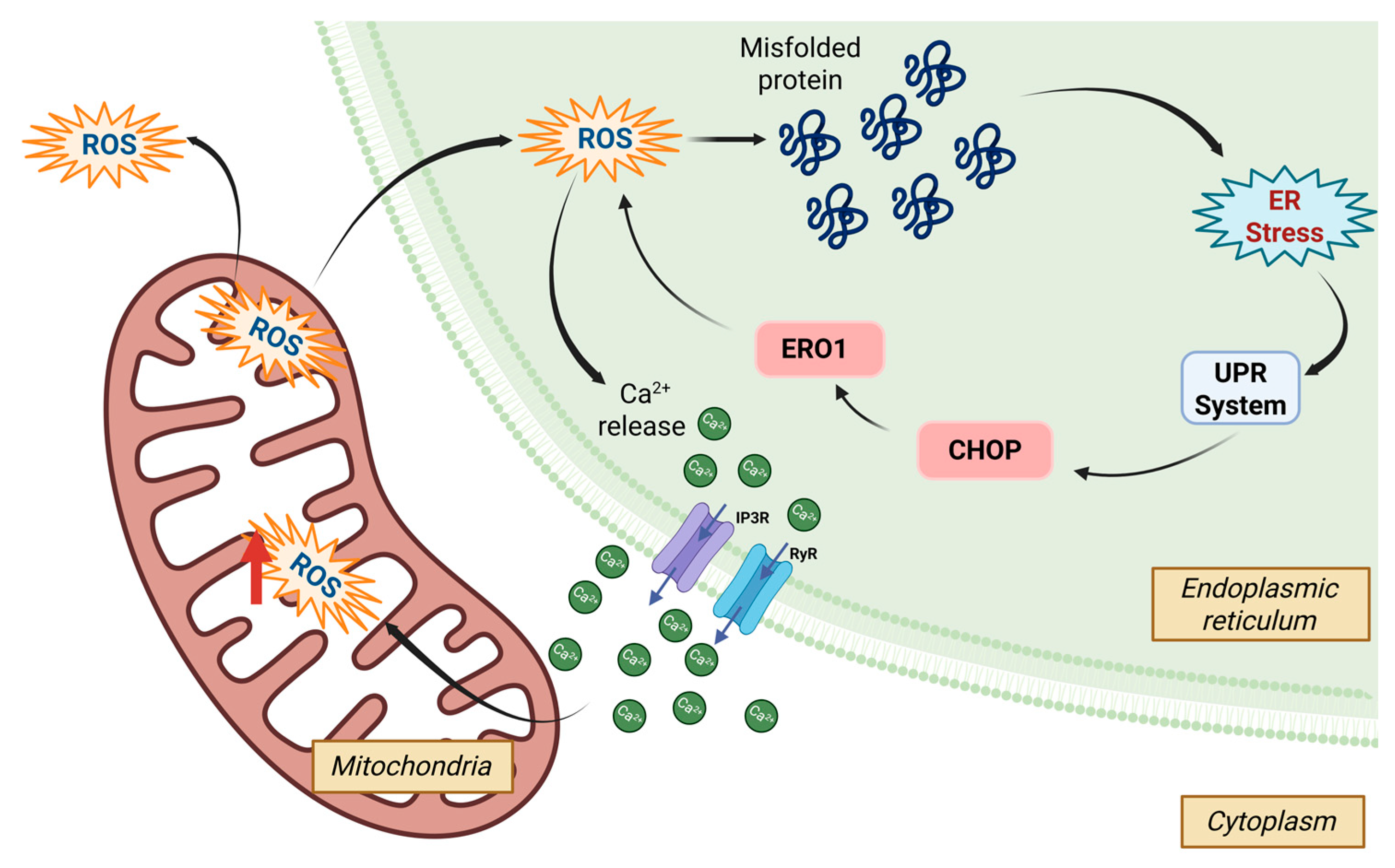
3.2.2. Unfolded Protein Mechanism

Stress response at the cellular level involves multiple mechanisms, one of which is unfolded protein (UPR), leading to endoplasmic reticulum (ER) stress. The ER is a large organelle responsible for lipid and steroid synthesis, Ca2+ homeostasis and storage, carbohydrate metabolism, and protein synthesis. ER stress causes defects in lipid metabolism, apoptosis, autophagy, reduced respiratory activity, and oxidative phosphorylation. As the cell’s primary Ca2+ storage site and the location for protein biosynthesis, folding, and assembly, the ER plays a crucial role in cellular function [120,121].
When stress disrupts cellular balance, eukaryotic cells respond by downregulating protein synthesis, increasing the expression of chaperone-encoding genes and other proteins that prevent polypeptide aggregation and degrading misfolded proteins. This coordinated response is triggered by an intracellular signaling cascade known as the “Unfolded Protein Response” (UPR) [120]. Research on the UPR system in invertebrates is limited compared to vertebrates. However, existing evidence has revealed marked differences in the UPR system between the two groups. Invertebrates possess only one activating transcription factor 6 (ATF6) gene, whereas vertebrates have ATF6⍺ and ATF6β, GADD34 (Growth arrest and DNA-damage-inducible 34), and CHOP (C/EBP-homologous protein). Although many studies have used transcriptional analysis to provide a general framework for understanding shrimp stress mechanisms at the cellular level, certain aspects remain unclear [122].
Several UPR-related genes have been detected in crustaceans in response to different stressors, including Bip (binding immunoglobulin protein), XBP1 (X-box binding protein 1), IRE1 (inositol-requiring transmembrane kinase/endoribonuclease), eIF2α (eukaryotic translation initiation factor α), PERK (PKR-like ER kinase), ATF6, ATF4 (activating transcription factor 4), calnexin, calreticulin, HSP10, HSP21, HSP37, HSP40, HSP60, HSP70, HSC70, HSP90, and PD1 (Protein disulfide isomerase) [123]. The UPR is controlled by three signaling pathways, that is, IRE1, Protein Kinase RNA-like ER kinase (PERK), and ATF6. During ER stress, the upstream non-folding protein recognition factor IRE1 dimerizes and auto-phosphorylates, enhancing endonuclease activity. IRE1 then specifically detects the XBP1 precursor mRNA and excises a particular intron, resulting in the splicing of the XBP1 precursor mRNA. This splicing event combines two open reading frames (ORFs) to encode an active transcription factor: XBP1s [122,123]. XBP1s are translocated to the nucleus and initiate the transcription of downstream genes involved in UPR. These genes encode protein chaperones that promote protein folding, maturation, and trafficking, as well as components of the ER-associated degradation (ERAD) pathway that remove accumulated misfolded proteins in the ER. However, if the chaperones fail to restore homeostasis in the ER, apoptosis is triggered to eliminate the stressed cells [123].
The PERK signaling pathway is implicated in ER stress. PERK activation induces the phosphorylation of eIF2, which globally decreases protein synthesis while promoting the translation of ATF4. ATF4 regulates apoptosis-related genes, such as CHOP, and increases the expression of ER chaperones (HSP) and the oxidative response [120,122,124].
During ER stress, ATF6 is transported from the ER to Golgi, where it is cleaved by the site-1 protease (S1P) and site-2 protease (S2P). The active ATF6 is translocated to the nucleus and binds to promoters of various genes involved in the UPR, regulating the transcription of downstream effectors such as CHOP, BiP, and XBP1 [120,122]. ER stress also activates the ERAD pathway, the calcium signaling pathway, and additional signaling pathways, including the mitogen-activated protein kinase (MAPK) signaling pathway [122]. Similarly to IRE1 pathway, ATF6 initiates the transcription of HSP and ERAD components [124] (Figure 3).
Heat shock proteins (HSPs) are molecular chaperones that play a crucial role in mitigating stress. Their expression can triple in response to various forms of stress, including physical, chemical, and biological [125]. HSP activation is regulated by Heat Shock Factors (HSFs), which upregulate HSP production to protect cells from damaged or misfolded proteins [110,111,126] by binding to the Heat Shock Element (HSE), which mediates the transcription of heat shock genes [111]. A previous study conducted by Zhou et al. [123] showed that HSF gene expression is highest in the lymphoid organs of stressed L. vannamei, with the lowest levels found in the eyestalk. Additionally, stress induces an increase in HSF gene expression in Penaeus monodon hemocytes [110].
Although the exact mechanism by which HSF influences HSP function remains unclear, studies have suggested that HSF inhibits the NF-κB and Jak/Stat pathways during ammonia stress, leading to a weakened immune response and reduced antiviral activity [126]. Commonly studied HSPs in shrimp stress research include HSP70, HSP90, HSP60, HSP40, and HSP10 [111,126]. Among these, HSP70 showed the most sensitive and tissue-specific gene expression in response to stress, playing a crucial role in protecting cells during adverse conditions [111]. Research on temperature-related stress indicated that HSP70 expression increases significantly in L. vannamei and P. monodon shrimp when exposed to extreme temperatures of 20 °C and 34 °C, respectively, but stabilizes at near-normal temperatures [69,112]. A previous study [127] found that HSP70 and HSP90 gene expression increased under high-temperature stress but decreased under hypoxic conditions, highlighting how different stressors affect gene regulation associated with stress proteins. Moreover, Niu et al. [72] reported that when the marine copepod Tigriopus japonicus was exposed to saxitoxins for 48 h, the HSP70 level increased more than fourfold, indicating cellular stress and disruption of signal transduction pathways. Multi-transcriptomic analysis reveals that HSP gene expression varies by tissue type, with each tissue responding uniquely to environmental stressors [128].

3.2.3. Redox Regulation (Antioxidant)

Various stressors may disrupt the redox state of an organism, including crustaceans, leading to oxidative stress, which is characterized by an imbalance between an excessive production of reactive oxygen species (ROS) and insufficient antioxidant defenses [129,130]. Oxidative stress results in the production of ROS and reactive nitrogen species (RNS), causing damage to DNA and proteins, lipid peroxidation, apoptosis, and overall cell damage [122,130]. Organisms counteract oxidative stress through several signaling pathways and enzymes, including superoxide dismutase (SOD) and glutathione reductase (GR). In crustaceans, various enzymes related to redox reactions have been studied, such as SOD, MnSOD, MDA, GPx, thioredoxin, peroxidase, catalase, NADPH-oxidase, and aldehydeoxidase [122].
The maintenance of the redox environment in vivo relies on the reduced glutathione (GSH)-oxidized glutathione (GSSG) cycle [117,122]. GSH, containing an active sulfhydryl (SH) group, is easily oxidized and dehydrogenated. GSH is reduced to H2O by GPx, which then oxidizes to GSSG. GSSG is subsequently reduced to GSH by GR, maintaining the scavenging of free radicals in vivo [122].
There are several molecular signaling processes related to the redox state in aquatic organism. This includes Nrf2 signaling, Notch signaling, PPAR signaling, MAPK signaling, NF-kB signaling, and dTLR2-MyD88-NF-kB signaling [131]. Since the redox process requires oxygen, the body suppresses the GSH pathway in ROS regulation under hypoxic conditions. A previous study demonstrated that when exposed to hypoxia, Penaeid shrimp downregulates the synthesis of metabolites associated with the glutathione pathway (cysteine, glutathione, glutamic acid, and methionine) [117].
Oxidative stress and endoplasmic reticulum (ER) stress are strongly connected (Figure 4), although the underlying mechanisms of this interaction remain incompletely understood in crustaceans. In crustaceans, these stress responses are believed to converge on pathways involving calcium signaling, unfolded protein response (UPR), and antioxidant defense systems, which are critical in maintaining cellular homeostasis under environmental and pathogenic stress. While mitochondria are the primary site of ROS generation, the ER contributes approximately 25% to overall ROS production, mainly through the protein-folding process. ER stress activates ROS-sensitive Ca2+ channels, such as inositol-1,4,5-triphosphate receptor (IP3R) and ryanodine receptor (RyR), on the ER membrane, leading to the release of Ca2+ into the mitochondria. This release further stimulates ROS production. Elevated mitochondrial ROS levels can escape into the ER, exacerbating ER stress. Additionally, components of the UPR contribute directly to oxidative stress; for example, the transcription factor CHOP can increase the expression of ERO1, enhancing the conversion of oxygen into hydrogen peroxide during oxidative folding [121].

3.2.4. Hydromineral Regulation

The secondary response to stress aids in restoring osmotic balance and suppressing the immune system. Upon exposure to a stressor, hydromineral dysfunction occurs due to adrenaline (epinephrine) altering gill blood flow patterns and gill permeability. This alteration favors water movement along its osmotic gradient, either into or out of the body, depending on environmental salinity. Consequently, the role of stress hormones in this context is the restoration of osmotic balance [47,132].
Osmoregulation involves managing ionic fluxes, primarily Na+ and Cl ions, through limiting and compensatory processes such as membrane permeability, epithelial leaks, and active pumping. Maintaining ion gradients is one of the most ATP-intensive activities, making osmoregulation energetically costly. Stress exposure alters the expression of channels or active membrane carriers, including Na+/K+-ATPase (NKA) or Na+/K+/Cl cotransporters or carbonic anhydrase. Environmental stressors can influence the relationship between osmoregulation and oxidative stress. When the organism experiences stress, it reallocates energy to mitigate stress. Mitochondria, the main energy producers in eukaryotic cells, also generate the majority of ROS and RNS. Elevated levels of ROS and RNS deactivate enzymes involved in osmolyte synthesis and membrane-bound transporters, thus inhibiting osmoregulation [133,134]. In an earlier study, Pinto et al. [28] demonstrated that high ammonia stress not only increased (Na+, K+)- and V(H+)-ATPase activity but also altered the activity of oxidative stress enzymes (SOD, GST, GR, and G6PDH) and V(H+)-ATPase genes.

3.2.5. Alternation in Immune Responses

Crustaceans exhibit simpler endocrine/neuroendocrine and immune systems compared to vertebrates; however, their stress responses, including the secretion of stress hormones/neurohormones, are comparable to those of vertebrates. During stress exposure, biogenic amine receptors can modulate the immune response by altering second messengers such as adenylyl cyclase (AC), phospholipase C (PLC), cyclic adenosine monophosphate (cAMP), and cyclic guanosine monophosphate (cGMP) [21,135]. Crustaceans primarily rely on a non-specific immune system comprising circulating hemocytes and various active substances released into the hemolymph, including the prophenoloxidase (proPO) system, antibacterial peptides, lectins, and proteinase inhibitors. The neuroendocrine–immunoregulatory network involves physiological and biochemical interactions between the neuroendocrine system and immune system, protecting organisms from stress and diseases [21].
Shrimp have an innate immune system encompassing humoral and cellular responses. The humoral immune response is mainly influenced by the TLR/IMD-NF-κB, JAK-STAT, and RNAi signaling pathways [122]. TLR receptors are activated by pathogen-associated molecular patterns (PAMPs), damage-associated molecular patterns (DAMPs), or homeostasis-altering molecular processes (HAMPs). This activation allows nuclear factor kappa B (NF-κB) to translocate to the nucleus and enhance the transcription of the inflammasome components, pro-IL-1β and pro-IL-18. Concurrently, ROS induce the oligomerization of the NLRP3 inflammasome and activate caspase1, converting pro-IL-1β and pro-IL-18 into IL-1β and IL-18, which promote inflammation and pyroptosis [136].
DAMPs act as an alarm in the innate immune system, promoting pro-inflammatory responses [136]. Besides PAMPs, such as lipopolysaccharide, -1,3-glucan, and peptidoglycan, DAMPs are released upon cell injury caused by biotic and abiotic stresses [137]. There are more than eight types of DAMPs, such as HSPs, high-mobility group protein Box 1 (HMGB1), ATP, uric acid crystals, extracellular DNA, mitochondrial DNA, fibronectin, free fatty acids, and short fragments of free hyaluronan [136]. Peroxiredoxins are also reported as DAMPs [138]. Supplementation with peroxiredoxins can enhance arginine and proline metabolism, suggesting their potential as antibiotics for bacterial diseases in shrimp [137].

3.3. Tertiary Response

The tertiary response in both vertebrates and invertebrates occurs when the system becomes dysregulated and fails to return to homeostasis, impairing the metabolic and immune system function [35,47,139]. Although stress hormones have beneficial effects by mobilizing energy during stress exposure, many tertiary responses at the whole-organism level are maladaptive. These include impaired health and disease resistance [140], reproduction [141], growth, learning, and other behaviors such as predator avoidance [38]. In crustaceans, the tertiary response inhibits growth, reproduction, molting, and metamorphosis [38]. For example, when exposed to stressors like high temperature and low pH, crustaceans, particularly L. vannamei, experience an increase in metabolic load characterized by the mobilization of glycogen and lipids. This leads to an increase in feed intake without corresponding growth. The metabolic load is further evidenced by a doubling of ammonia excretion and a nearly 50% increase in metabolic oxygen uptake [142]. In both freshwater and seawater decapods, changes in water temperature and salinity do not initially impact growth. However, as the duration and intensity of these stressors increase, survival rates, egg production, and hatching success can significantly decline [48,49,143,144].

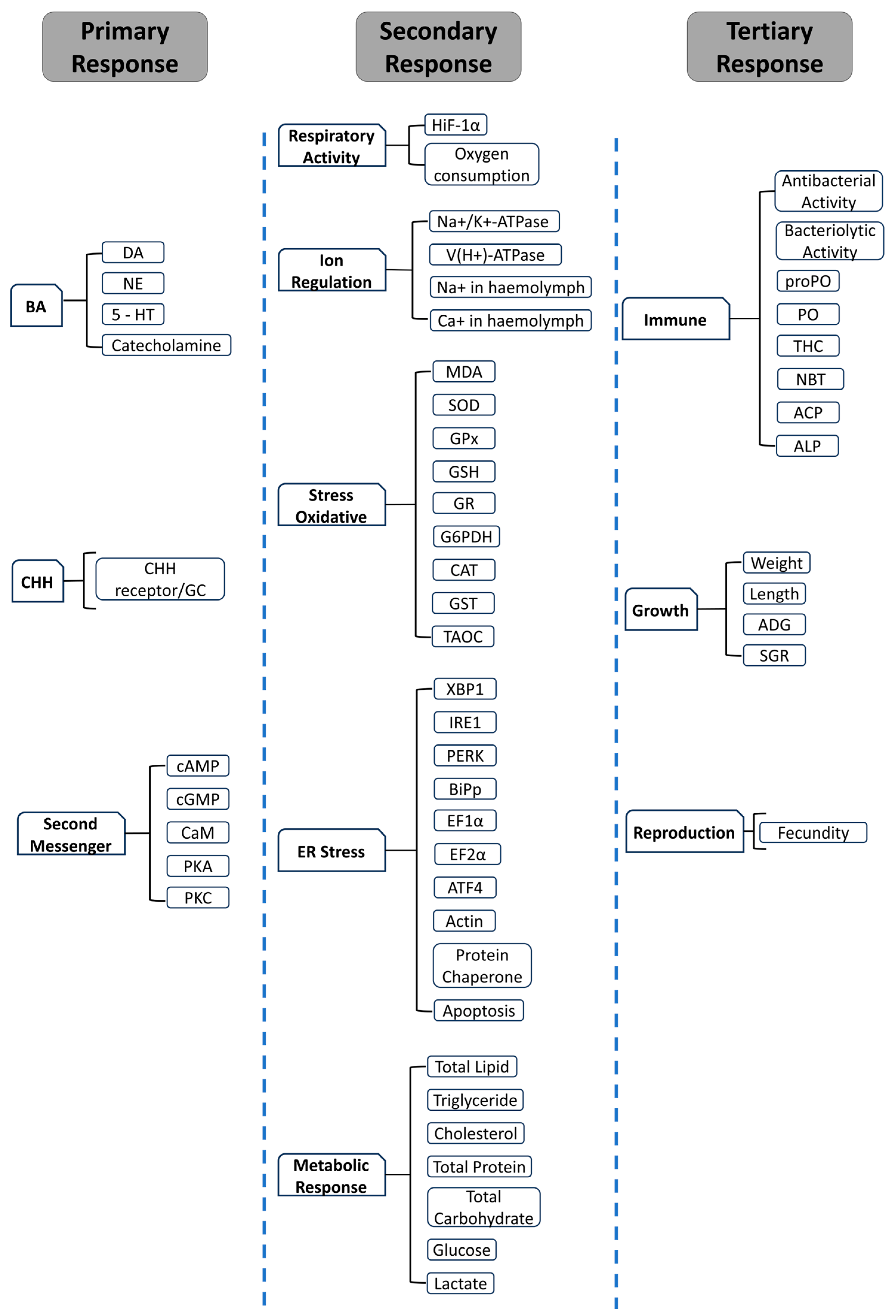
4. Bioindicators for Stress

Stress significantly impacts not only the metabolism and growth of aquatic animals but also their immunity and disease resistance, making it a crucial factor in disease transmission among aquaculture animals [43]. Early and effective stress mitigation can help the animals immediately return to homeostasis, improve recovery rates, and reduce the risk of chronic stress impact. Therefore, reliable, sensitive, and unbiased stress bioindicators are essential for an early warning system and as parameters to evaluate the efficacy of stress management strategies. To date, the most commonly measured stress biomarkers in crustacea include hormonal, metabolic and cellular, and phenotypic indicators, representing the primary, secondary, and tertiary responses to stress (Figure 5).

4.1. Hormonal Indicators

Stress responses in both fish and shrimp are primarily mediated by the neuroendocrine system, involving hormonal and biogenic amine changes. Second messengers facilitate intracellular signaling and activate downstream pathways. In fish, catecholamines and cortisol, produced in the head kidney, are key stress hormones. Conversely, in shrimp or other crustaceans, the crustacean hyperglycemic hormone (CHH), synthesized in the eyestalk, functions analogously to cortisol. Hormonal assays are valuable for detecting stress due to their rapid response to stimuli; however, CHH measurements have traditionally relied on HPLC-based bioassays [98,145], PCR [96,146,147], or ELISA [23,148].
Several studies have utilized hormones as indicators of stress in crustaceans. CHH levels, for instance, increased in response to environmental changes or behavioral stressors, such as predation or migration, signaling stress within the organism. These elevations may occur within 5 [97], 10 [98,145], or even 120 min [149] of exposure to a stressor, depending on its intensity. Furthermore, the amount of CHH secreted varies by crustacean species, type of stressor, and developmental stage. Cortisol levels, commonly used in other taxa, can be influenced by the stress of blood collection itself and are further affected by factors such as diet, season, maturation, and concurrent stressors, complicating their interpretation [150,151].
In addition to its rapid stress response, CHH appears to interact with biogenic amines, adding complexity to its regulatory role. The relationship between CHH and biogenic amines such as serotonin (5-HT), dopamine (DA), norepinephrine (NE), and epinephrine (E) is not fully understood. Some studies indicated a mutually influential relationship. For example, it was demonstrated that 5-HT enhances the release of CHH [148], while silencing the CHH gene leads to a decrease in the levels of DA and other biogenic amines, including NE and 5-HT [96,146,147].
Much evidence showed that alterations in CHH, biogenic amines, and second messengers, such as cAMP, cGMP, calmodulin, protein kinase A (PKA), and protein kinase C (PKC), influence secondary stress response indicators. These include changes in glucose metabolism [147] and also blood glucose [106], ion transport (e.g., Na+/K+-ATPase activity) [96], and HSP expression [23]. Hormonal indicators are highly sensitive to stress, making them useful for early detection. However, their fast dynamics may lead to challenges in capturing the precise moment of primary stress response, as the organism may have already transitioned to secondary pathways by the time measurements are performed.

4.2. Metabolic and Cellular Indicators

After CHH activates second messengers, such as cAMP or Ca2+, intracellular signaling cascades are triggered, leading to modifications in various metabolic and cellular pathways. These alterations depend on the type of stressor affecting the organism and can be categorized into five major groups: pathways related to respiratory processes, ion regulation, oxidative stress, ER stress, or energy regulation required to maintain homeostasis.

4.2.1. Respiratory Process

In response to stress, animals experience increased adrenaline, which triggers vasoconstriction and elevates cardiac output. These changes enhance gill diffusing capacity and ion transport, affecting oxygen intake, plasma osmolality, and circulating ion concentrations. Prolonged stress, such as oxygen deprivation, leads to hypoxia, a condition that disrupts the hypoxia-inducible factor (HIF) pathway. This pathway plays a critical role in cellular adaptation to low oxygen levels, primarily through the stabilization and activation of HIFα, a biomarker for hypoxic stress. Interestingly, the presence of HIFα varies across invertebrates. Graham et al. [152] found that among 70 invertebrate species, decapods commonly have the HIFα gene, while only some copepods exhibit the expression of this gene. This suggests that hypoxia adaptation mechanisms differ significantly among invertebrates, depending on their ecological and physiological traits.
In vertebrates, HIFα expression under hypoxic conditions is influenced by the intensity and duration of the stressor [153,154]. Chronic hypoxia resulted in the highest expression of HIFα in the liver, followed by the brain, muscle, heart, and kidneys [153]. In another study, Li et al. [59] demonstrated that hypoxia primarily affects energy metabolism pathways, followed by pathways related to immunity, growth, signal transduction, and hypoxia-specific adaptations. These findings align with other studies showing that hypoxia, or alterations in HIFα, impact glucose metabolism [10,118], glucose transport [154], lactate production [155], oxidative stress [103,118], and the expression of HSPs [127].

4.2.2. Ion Regulation

In both vertebrates and invertebrates, hypoxia-induced stress often disrupts ion homeostasis, emphasizing the critical role of ion-motive ATPase enzymes in maintaining physiological balance under stress conditions. These energetically driven enzymes regulate ion gradients across cell membranes, which are essential in various metabolic pathways. These ions, such as Na+, Ca2+, and Cl, are abundant in the hemolymph and contribute to critical physiological processes [70]. The regulation of these ions is mediated by ion-motive ATPase enzymes, including Na+/K+-ATPase, Ca2+-ATPase, V-ATPase, and H+-ATPase. Furthermore, in both seawater and freshwater fish, cortisol or stress hormones regulate chloride (Cl), potassium (K), and sodium (Na) ions, increasing sodium–potassium adenosine triphosphatase (Na+-K+-ATPase) activity in the gills, enhancing salinity tolerance [156]. These enzymes utilize energy from ATP to maintain ion homeostasis by driving ion exchange across cell membranes [70,157].
Previous studies demonstrated that both crustacean hyperglycemic hormone (CHH) [148] and cortisol [156], triggered by biogenic amines, increase the activity of ion-motive enzymes such as Na+/K+-ATPase and H+-ATPase through second messengers, including cAMP, PKA, and PKC. This activation enhances ion regulation, such as Na+ uptake, in the gills of blue crabs [98]. In addition to affecting Na+ uptake, this mechanism alters the concentrations of other ions, including Ca2+, Mg2+, and K+, in the blood of both vertebrates and invertebrates [156,158]. Similar regulatory mechanisms occur in vertebrates, as seen in brown trout and delta smelt, where exposure to environmental stressors like acidification and low salinity influences ion-motive ATPase activity, ensuring ionic balance [159,160].
Ions regulate fluid balance and facilitate the absorption, metabolism, and utilization of many metabolites (fatty acids, amino acids, glucose, monocarboxylate) [157,161]. Na+-K+-ATPase and V-H+-ATPase are indicators of transport imbalance, with evidence indicating increased activity in response to stress. For instance, tilapia transferred from freshwater (salinity 0) to seawater (salinity 25 ppt) exhibited significant increases in Na+-K+-ATPase activity in the gills and a reduction in glycogen content [162]. Similarly, L. vannamei adapted to low salinity showed higher Na+-K+-ATPase activity compared to the control group [158]. These findings indicate that organisms generate energy as an adaptive response to stressors. Ion concentrations in the hemolymph often serve as physiological indicators, where deviations may signify stress and disruption of homeostatic mechanisms. However, the mechanisms underlying ion regulation in crustaceans remain poorly understood, particularly in response to environmental stressors. Despite the crucial role of ion balance in survival and aquaculture performance, recent studies in this topic are scarce, leaving significant gaps in our understanding of crustacean homeostasis and stress resilience.

4.2.3. Cellular Stress

Alterations in homeostasis affect cellular processes, leading to oxidative and endoplasmic reticulum stress. Stress triggers the production of ROS, which are counteracted by antioxidants from the body or diet. An imbalance between ROS and antioxidants results in oxidative stress, disrupting cellular function [130,133].
The activity of antioxidant enzymes, such as alkaline phosphatase (AKP), acid phosphatase (ACP), total antioxidant capacity (T-AOC), glutathione S-transferase (GST), glutathione peroxidase (GPX), glutathione (GSH), malondialdehyde (MDA), and protein carbonyl (PC), is used as an indicator of oxidative stress [29,130,134]. Their reliability, however, varies with developmental stage and diet, which influence antioxidant efficiency [130]. Studies have shown that in L. vannamei, antioxidant enzyme activity can differ between tissues, such as the hepatopancreas and gills, with some authors reporting asynchronous spikes, while others observe simultaneous responses [11,103]. These variations highlight the complexity of antioxidant regulation under stress.
When antioxidant defenses fail to neutralize the excess ROS generated by stress, cellular imbalance intensifies, leading to ER stress. In response, the ER activates the unfolded protein response (UPR), a signaling pathway designed to alleviate the accumulation of misfolded proteins. This process involves intricate intracellular mechanisms that help restore cellular homeostasis [120,121,163]. However, the precise relationship between oxidative stress and ER stress, particularly in crustaceans, remains poorly understood. Recent studies have begun to explore this connection by identifying genes involved in the UPR and ER stress, offering insights into the underlying cellular mechanisms [104,164,165].
The most commonly used indicators of ER stress are Damage-Associated Molecular Patterns (DAMPs), such as HSPs. Among these, HSP70 is the most frequently used, due to its abundance and conservation across species. Other key HSPs, including HSP40, HSP90, HSP60, and Heat Shock Factor 1 (HSF-1), are also utilized as indicators of cellular stress [17,166,167,168,169]. He et al. [138] identified peroxidoxin-1 as a novel DAMP involved in acute liver injury (ALI) in mice. In ALI models induced by acetaminophen (APAP) and carbon tetrachloride (CCl4), serum Prdx1 levels increased progressively, correlating with macrophage infiltration and elevated expression of IL-1β, IL-6, and TNF-α. Prdx1-deficient (Prdx1/) mice exhibited reduced inflammation and liver damage, while reintroducing recombinant Prdx1 restored these effects, confirming its role in ALI pathogenesis via NF-κB activation and NLRP3 inflammasome signaling. Despite extensive research on DAMPs in vertebrates, their role in invertebrate stress physiology, particularly in crustaceans, remains largely unexplored.

4.2.4. Metabolic Indicators

Plasma metabolites such as glucose and lactate are frequently used to assess stress conditions in aquatic organisms. Cortisol modulates glucose production through processes like gluconeogenesis and glycogenolysis, while lactate is generated from glucose via anaerobic glycolysis to meet the increased cellular energy demands. Despite their utility, glucose measurements often exhibit variability and are less reliable than cortisol for stress assessment, thus serving primarily as supplementary indicators [150,170]. In addition to glucose and lactate levels in the blood, other metabolic indicators used in previous studies include total lipids, total proteins, triglyceride [79], blood cholesterol, free fatty acids (FFAs) [171], and free amino acids (FAAs) [115]. However, a meta-analysis study by Conneley et al. [172] showed that lactate concentration is a particularly accurate stress marker, although it may be less suitable for chronic stress assessment [172].
Hyperglycemia, characterized by elevated blood glucose and lactate levels, occurs in response to stress and affects blood components such as leukocytes, erythrocytes, hemoglobin, and hematocrit values in fish [151]. Blood profiling is essential for assessing the overall health status of an animal. In crustaceans, similar alterations occur, impacting the total hemocyte count (THC) and differential hemocyte count (DHC) [94]. Consequently, these enzymes are frequently used as biomarkers to assess the degree of stress in fish.
In shrimp, stress-induced cellular damage is often reflected by changes in the activities of enzymes in hemolymph, such as alanine aminotransferase (ALT), aspartate aminotransferase (AST), phenoloxidase (PO), lysozyme (LZM), lactate dehydrogenase (LDH), and creatine kinase (CK). These enzymes are released into the hemolymph following cellular injury or death, and their elevated levels can indicate stress, especially when tissue damage is minimal [135]. Consequently, these enzymes are frequently used as biomarkers to assess the degree of stress in shrimps.
Specifically, ALT and AST are aminotransferases linked to tissue and cellular damage. For example, a study by Li et al. [173] demonstrated that in L. vannamei, reduced salinity led to decreased ALT and AST activity, while PO activity increased, indicating damage to hepatopancreas cells. Elevated levels of these enzymes often correlate with cellular damage, oxidative stress, and tissue inflammation, making them useful indicators of shrimp health and well-being. Monitoring the levels of these key enzymes in shrimp populations can thus serve as an effective tool for assessing environmental stress and optimizing aquaculture management practices. However, further research is necessary to better understand the specific mechanisms behind their release and to establish standardized thresholds for their use as reliable biomarkers in different stages of shrimp cultured under different environmental conditions.

4.3. Phenotypic Indicators

Stress significantly affects the performance of both fish and crustaceans, including growth, disease resistance, and metabolism [108]. Although easy to measure, these phenotypic traits or physiological indicators have limited specificity. Key indicators of welfare include growth and fitness, condition factor (length–weight relationship), feed consumption, metabolic rate, and behavioral changes, such as aggressiveness, swimming performance (speed, intensity, and duration), gill ventilation rate, color changes, predator–prey dynamics, malformations, and pathological conditions. Various organosomatic indicators also reflect general health [151].
In certain situations, traditional stress biomarkers are difficult to interpret and may lead to challenging diagnoses. Therefore, identifying and comprehending alternative tools is crucial for aquatic organism welfare [35]. Recent studies have combined traditional stress biomarkers with genomic tools to understand the entire process of stress responses in both acute and chronic stages [47] and to identify specific and early indicators of stressors [35]. In aquaculture, technologies such as genomics, transcriptomics, proteomics, and metabolomics have been employed to enhance knowledge and understanding of safety, production, quality, and health. The application of various omics approaches—genomics (the study of DNA variation), transcriptomics (the analysis of genome characterization by gene expression, mRNA), proteomics (the study of cell and tissue protein expression), and metabolomics (the study of chemical processes involving small molecules known as metabolites)—is necessary for aquaculture research. By integrating multiple omics platforms, it is possible to comprehend the complexity of biological interactions and understand how cells and organisms adapt, function, grow, and develop.

5. Stress Mitigation Strategy

Stress mitigation strategies for the aquaculture industry may be implemented following two approaches: first, by system modification to reduce the magnitude of stress, and second, by improving the robustness of the cultured organisms to stress. System modification includes efforts to reduce culture intensity (density per unit area) [174], maintaining good water quality [175], and improving biosecurity [176], as well as reducing the impact of climate on the culture by performing the culture in a closed indoor system [177]. The second approach includes genetic improvement and dietary interventions to improve the animals’ robustness against stress.
Selective breeding programs targeting genetic traits that are related to stress robustness have been developed to maintain a sustainable aquaculture production [178]. In crustaceans, such programs are primarily applied to shrimp, with a strong emphasis on enhancing disease resistance [179], which remains a major challenge in shrimp farming. However, the implementation of selective breeding is often limited for small- and medium-scale farmers, as it requires substantial financial investment, long-term commitment, and access to sophisticated breeding facilities.
This limitation becomes even more pronounced when considering advanced genomic and omics-based approaches for stress mitigation, which are currently not feasible for direct application in most farm-level operations. Nevertheless, these approaches play a crucial role in identifying molecular biomarkers associated with stress responses. Such biomarkers can serve as early indicators of stress exposure and allow for rapid diagnostics, thereby supporting more timely and precise farm management interventions.
Several studies have investigated the heritability of stress tolerance traits and their underlying molecular mechanisms. For example, Anastasiadi et al. [180] demonstrated that DNA methylation patterns in European seabass can be influenced by early-life thermal exposure, potentially affecting the stress phenotype of the offspring. Similarly, Fellous et al. [181] found that a 2 °C temperature increase altered the expression of DNA methylation-related genes in seabass larvae. In marine sticklebacks, adaptation to elevated temperatures induced dynamic, temperature-sensitive methylation changes throughout development [182]. However, similar studies in shrimp and other crustaceans are still lacking, pointing to a significant gap in our understanding of how stress resilience may be inherited in these taxa.
Apart from providing balanced and optimum nutrients for normal growth through diet, some research demonstrated that stress mitigation can be achieved through the addition of some specific nutritive compounds, like amino acids, vitamins, and minerals or non-nutritive compounds such as probiotics and organic acids [47]. Tryptophan, for instance, has been considered to be a promising stress mitigator in fish. This amino acid is a precursor of serotonin (5-hydroxytryptamine, 5HT) and melatonin, which play an important role in stress-coping ability and behavior in several fish and crustacea [183]. Increased dietary tryptophan leads to an increased availability of this amino acid for uptake into the brain, where it can later be converted to 5-HT with the facilitation of tryptophan hydroxylase and aromatic L-amino acid decarboxylase in the presence of vitamin B6 [184]. Dietary tryptophan has been shown to reduce the levels of stress indicators in some fish species, such as Channa punctatus [185], Lates calcarifer [186], Oreochromis niloticus [187], Argyrosomus regius [188], Chirrhinus mrigala [189], and brown trout [190]. On the other hand, the role of tryptophan in mitigating stress in crustaceans remains underexplored.
While extensive studies have demonstrated tryptophan’s modulatory effects on serotonergic activity, stress responses, immune regulation, and antioxidant capacity in various teleost species [191], similar investigations in crustaceans are scarce. This highlights a critical research gap and the need for species-specific studies to better understand the physiological pathways involved. Nonetheless, emerging studies provide promising evidence. For instance, in crayfish, anxiety-like behavior is influenced by the intensity of stress stimuli, with levels of anxiety increasing as brain 5-HT concentrations rise [192]. In L. vannamei, dietary tryptophan supplementation at 0.36% has been shown to significantly elevate 5-HT levels in the hepatopancreas, eyestalk, and hemolymph, suggesting enhanced neurochemical resilience to stress [193]. Additionally, Jin et al. [194] demonstrated that dietary tryptophan at 3.61 g/kg optimized growth performance and feed conversion efficiency in L. vannamei reared under low-salinity conditions, while also improving protein efficiency, reducing stress-related enzymes (AST and ALT), and enhancing metabolic profiles, including hemolymph amino acid concentrations. In juvenile mud crabs, tryptophan supplementation at 0.75–1% reduced aggressive behavior and improved survival, correlating with increased 5-HT levels after social conflict [195]. These findings underscore the potential of tryptophan as a dietary intervention for stress mitigation in crustaceans, warranting further investigation into species-specific dose responses and underlying molecular pathways.

6. Future Directions

Environmental stressors are currently evaluated by measuring their effects on various biological indicators, including population density, growth, reproduction, behavior, physiology, and immune responses. Environmental stress may trigger some sublethal effects, such as mutations and epigenetic changes, that can be transgenerationally inherited, influencing phenotypic plasticity and increasing vulnerability to diseases, tissue pathologies, and changes in social behavior and biological invasions [196]. In the future, stress monitoring should be performed in real time with high precision, allowing for early mitigation and thus reducing the risk of loss. However, compared to terrestrial animal husbandry, research and technological advances in stress mitigation in aquaculture, especially crustaceans, are relatively far behind.
Internet of things (IoT)-based technology has been successfully implemented in cattle production to monitor stress levels. In aquaculture, real-time stress monitoring in fish has advanced using biosensors that track biomarkers such as blood glucose [197,198] or visualized behavior [199,200]. This advancement may not be applicable to crustacean aquaculture yet. There remains insufficient knowledge of crustaceans’ stress responses, which limits the development of biosensors. Previous research has mainly focused on secondary physiological responses, such as changes in intestinal tissue structure and appetite [201] and energy allocation [13,22,116,117,202]. However, little attention has been given to the molecular alterations that drive these changes. The development of multi-omics tools presents an opportunity for a comprehensive understanding of the responses of aquatic animals to stressors at both primary (molecular and cellular) and secondary (physiological and behavioral) levels. However, several critical gaps remain unaddressed. First, there is a lack of integrated studies linking molecular, physiological, and behavioral stress responses, particularly in crustaceans. Second, while biosensors have proven effective in fish, their application in crustaceans is hindered by the absence of validated, quantifiable biomarkers. Third, the long-term and transgenerational impacts of sublethal stressors—such as epigenetic modifications—are still poorly understood.
Future research should prioritize the identification and validation of crustacean-specific stress genes and pathways, the development of non-invasive biomonitoring techniques, and the application of real-time stress tracking technologies tailored to aquatic invertebrates. Exploring these gaps will be essential to advance precision aquaculture and enhance resilience against environmental challenges.
In addition, feed supplementation to mitigate stress still has a lot of room for exploration; examples include the use of alternative ingredients like quinoa husk [203], galacto oligosaccharides (GOSs) [204], or essential oils in feed [205]. In particular, the use of essential oils remains underexplored, with limited studies elucidating their mechanisms of action, especially their antioxidant capacity and role in stress mitigation, highlighting a promising avenue for future research in aquatic animal nutrition.

7. Conclusions

In essence, stressors in aquaculture are inevitable and can have a significant impact on sustainable production. Thus, it is vital to explore effective strategies for mitigating stress in cultured species. A comprehensive understanding of the stress responses in these species, particularly crustaceans, is crucial before implementing any interventions, whether through changes in rearing practices, technological monitoring, or dietary modification.
This review highlights the major physiological pathways involved in stress responses in crustaceans and emphasizes their relevance to aquaculture. It also identifies critical knowledge gaps—such as the limited integration of molecular, physiological, and behavioral data; the absence of validated, crustacean-specific stress biomarkers; and the lack of tools for real-time, non-invasive monitoring. Furthermore, the long-term and transgenerational effects of sublethal stressors remain poorly understood.
Addressing these gaps through multi-omics approaches, advanced sensor technologies, and tailored nutritional strategies will be essential to developing precise, adaptive stress management frameworks. Encouraging research in these directions is key to enhancing crustacean resilience and ensuring the sustainability of aquaculture under accelerating environmental change.

Author Contributions

Conceptualization, F.H., M.A.S., D.M.A., J.E., P.N., and K.B.; investigation: F.H. and J.E.; writing—original draft preparation, F.H. and J.E.; writing—review and editing, F.H., M.A.S., D.M.A., J.E., P.N., and K.B.; funding acquisition, F.H., J.E., and P.N. All authors have read and agreed to the published version of the manuscript.

Funding

The work was supported by the Ministry of Marine Affairs and Fisheries of Indonesia through a scholarship (grant no. 55/KEPMEN/KP.532/V/2023) granted to the first author, FORMAS (Sweden) grants awarded to Kartik Baruah (BioTrout project; 2019-01159) and Parisa Norouzitallab (EpiTrout project; 2022-00605), and grants awarded by the Swedish Board of Agriculture and European Union to Kartik Baruah (ResiChar project; 2023-1338).

Institutional Review Board Statement

Not applicable.

Informed Consent Statement

Not applicable.

Data Availability Statement

The data underlying this study’s findings are accessible from the corresponding author upon a reasonable request.

Acknowledgments

We would like to thank Ministry of Marine Affairs and Fisheries of Indonesia, FORMAS (Sweden) and Swedish Board of Agriculture and European Union for their financial support.

Conflicts of Interest

Author Dean Akiyama was employed by the company Central Proteina Prima, Indonesia. The remaining authors declare that the research was conducted in the absence of any commercial or financial relationships that could be construed as a potential conflict of interest.

References

  1. FAO. The State of World Fisheries and Aquaculture 2024—Blue Transformation in Action; FAO: Rome, Italy, 2024. [Google Scholar]
  2. FAO. The State of World Fisheries and Aquaculture 2022. Towards Blue Transformation; FAO: Rome, Italy, 2022. [Google Scholar]
  3. N’Souvi, K.; Sun, C.; Che, B.; Vodounon, A. Shrimp Industry in China: Overview of the Trends in the Production, Imports and Exports during the Last Two Decades, Challenges, and Outlook. Front. Sustain. Food Syst. 2023, 7, 1287034. [Google Scholar] [CrossRef]
  4. Davis, R.; Abebe, A.; Boyd, C.; McNevin, A. Exploring the Relationship between Production Intensity and Land Use: A Meta-Analytic Approach with Shrimp Aquaculture. J. Environ. Manag. 2021, 300, 113719. [Google Scholar] [CrossRef] [PubMed]
  5. Maulu, S.; Hasimuna, O.J.; Haambiya, L.H.; Monde, C.; Musuka, C.G.; Makorwa, T.H.; Munganga, B.P.; Phiri, K.J.; Nsekanabo, J.D.M. Climate Change Effects on Aquaculture Production: Sustainability Implications, Mitigation, and Adaptations. Front. Sustain. Food Syst. 2021, 5, 609097. [Google Scholar] [CrossRef]
  6. Asiedu, B.; Malcolm, D.; Iddrisu, S. Assessing the Economic Impact of Climate Change in the Small-Scale Aquaculture Industry of Ghana, West Africa. AAS Open Res. 2018, 1, 26. [Google Scholar] [CrossRef] [PubMed]
  7. Hamdan, R.; Othman, A.; Kari, F. Climate Change Effects on Aquaculture Production Performance in Malaysia: An Environmental Performance Analysis. Int. J. Bus. Soc. 2017, 16, 364–385. [Google Scholar] [CrossRef]
  8. Muhala, V.; Chicombo, T.F.; Macate, I.E.; Guimarães-Costa, A.; Gundana, H.; Malichocho, C.; Hasimuna, O.J.; Remédio, A.; Maulu, S.; Cuamba, L.; et al. Climate Change in Fisheries and Aquaculture: Analysis of the Impact Caused by Idai and Kenneth Cyclones in Mozambique. Front. Sustain. Food Syst. 2021, 5, 714187. [Google Scholar] [CrossRef]
  9. Fleming, A.; Hobday, A.J.; Farmery, A.; van Putten, E.I.; Pecl, G.T.; Green, B.S.; Lim-Camacho, L. Climate Change Risks and Adaptation Options across Australian Seafood Supply Chains—A Preliminary Assessment. Clim. Risk Manag. 2014, 1, 39–50. [Google Scholar] [CrossRef]
  10. Aparicio-Simón, B.; Piñón, M.; Racotta, R.; Racotta, I.S. Neuroendocrine and Metabolic Responses of Pacific Whiteleg Shrimp Penaeus Vannamei Exposed to Hypoxia Stress. Lat. Am. J. Aquat. Res. 2018, 46, 364–376. [Google Scholar] [CrossRef]
  11. Han, S.Y.; Wang, M.Q.; Liu, M.; Wang, B.J.; Jiang, K.Y.; Wang, L. Comparative Sensitivity of the Hepatopancreas and Midgut in the White Shrimp Litopenaeus vannamei to Oxidative Stress under Cyclic Serious/Medium Hypoxia. Aquaculture 2018, 490, 44–52. [Google Scholar] [CrossRef]
  12. Wang, P.; Liu, H.; Zhao, S.; Yu, S.; Xie, S.; Hua, S.; Yan, B.; Xing, C.; Gao, H. Hypoxia Stress Affects the Physiological Responses, Apoptosis and Innate Immunity of Kuruma shrimp, Marsupenaeus japonicus. Fish Shellfish Immunol. 2022, 122, 206–214. [Google Scholar] [CrossRef] [PubMed]
  13. Ren, X.; Yu, Z.; Xu, Y.; Zhang, Y.; Mu, C.; Liu, P.; Li, J. Integrated Transcriptomic and Metabolomic Responses in the Hepatopancreas of Kuruma shrimp (Marsupenaeus japonicus) under Cold Stress. Ecotoxicol. Environ. Saf. 2020, 206, 111360. [Google Scholar] [CrossRef] [PubMed]
  14. Fan, L.; Wang, A.; Miao, Y.; Liao, S.; Ye, C.; Lin, Q. Comparative Proteomic Identification of the Hepatopancreas Response to Cold Stress in White Shrimp, Litopenaeus vannamei. Aquaculture 2016, 454, 27–34. [Google Scholar] [CrossRef]
  15. Chang, C.C.; Jiang, J.R.; Cheng, W. A First Insight into Temperature Stress-Induced Neuroendocrine and Immunological Changes in Giant Freshwater Prawn, Macrobrachium rosenbergii. Fish Shellfish Immunol. 2015, 47, 528–534. [Google Scholar] [CrossRef] [PubMed]
  16. Zhang, C.; Zhang, Q.; Song, X.; Pang, Y.; Song, Y.; Cheng, Y.; Yang, X. Dietary L-Tryptophan Modulates the Hematological Immune and Antibacterial Ability of the Chinese Mitten Crab, Eriocheir sinensis, Under Cheliped Autotomy Stress. Front. Immunol. 2018, 9, 2744. [Google Scholar] [CrossRef] [PubMed]
  17. Sung, Y.Y.; Rahman, N.A.; Shazili, N.A.M.; Chen, S.; Lv, A.; Sun, J.; Shi, H.; MacRae, T.H. Non-Lethal Heat Shock Induces Hsp70 Synthesis and Promotes Tolerance against Heat, Ammonia and Metals in Post-Larvae of the White Leg Shrimp Penaeus Vannamei (Boone, 1931). Aquaculture 2018, 483, 21–26. [Google Scholar] [CrossRef]
  18. Matozzo, V.; Gallo, C.; Marin, M.G. Effects of Temperature on Cellular and Biochemical Parameters in the Crab Carcinus Aestuarii (Crustacea, Decapoda). Mar. Environ. Res. 2011, 71, 351–356. [Google Scholar] [CrossRef] [PubMed]
  19. Van Horn, J.; Malhoe, V.; Delvina, M.; Thies, M.; Tolley, S.G.; Ueda, T. Molecular Cloning and Expression of a 2-Cys Peroxiredoxin Gene in the Crustacean Eurypanopeus Depressus Induced by Acute Hypo-Osmotic Stress. Comp. Biochem. Physiol.—B Biochem. Mol. Biol. 2010, 155, 309–315. [Google Scholar] [CrossRef] [PubMed]
  20. Zhang, L.; Pan, L.; Xu, L.; Si, L. Effects of Ammonia-N Exposure on the Concentrations of Neurotransmitters, Hemocyte Intracellular Signaling Pathways and Immune Responses in White Shrimp Litopenaeus vannamei. Fish Shellfish Immunol. 2018, 75, 48–57. [Google Scholar] [CrossRef] [PubMed]
  21. Zhao, Q.; Pan, L.; Ren, Q.; Wang, L.; Miao, J. Effect of Salinity on Regulation Mechanism of Neuroendocrine-Immunoregulatory Network in Litopenaeus vannamei. Fish Shellfish Immunol. 2016, 49, 396–406. [Google Scholar] [CrossRef] [PubMed]
  22. Liu, H.Y.; Sun, W.W.; Tan, B.P.; Chi, S.Y.; Dong, X.H.; Yang, Q.H. Molecular Cloning and Expression of Hepatopancreas Glutamine Synthetase in the Pacific White Shrimp, Litopenaeus vannamei, Induced by Acute Hypo-Osmotic Stress. Aquaculture 2012, 362–363, 80–87. [Google Scholar] [CrossRef]
  23. Chang, E.S. Stressed-Out Lobsters: Crustacean Hyperglycemic Hormone and Stress Proteins. Integr. Comp. Biol. 2005, 45, 43–50. [Google Scholar] [CrossRef] [PubMed]
  24. Yu, Q.; Xie, J.; Huang, M.; Chen, C.; Qian, D.; Qin, J.G.; Chen, L.; Jia, Y.; Li, E. Growth and Health Responses to a Long-Term PH Stress in Pacific White Shrimp Litopenaeus vannamei. Aquac. Rep. 2020, 16, 100280. [Google Scholar] [CrossRef]
  25. Wang, W.N.; Li, B.S.; Liu, J.J.; Shi, L.; Alam, M.J.; Su, S.J.; Wu, J.; Wang, L.; Wang, A.L. The Respiratory Burst Activity and Expression of Catalase in White Shrimp, Litopenaeus vannamei, during Long-Term Exposure to PH Stress. Ecotoxicology 2012, 21, 1609–1616. [Google Scholar] [CrossRef] [PubMed]
  26. Lin, Y.C.; Tayag, C.M.; Huang, C.L.; Tsui, W.C.; Chen, J.C. White Shrimp Litopenaeus vannamei That Had Received the Hot-Water Extract of Spirulina Platensis Showed Earlier Recovery in Immunity and up-Regulation of Gene Expressions after PH Stress. Fish Shellfish Immunol. 2010, 29, 1092–1098. [Google Scholar] [CrossRef] [PubMed]
  27. Liang, C.; Liu, J.; Cao, F.; Li, Z.; Chen, T. Transcriptomic Analyses of the Acute Ammonia Stress Response in the Hepatopancreas of the Kuruma shrimp (Marsupenaeus japonicus). Aquaculture 2019, 513, 734328. [Google Scholar] [CrossRef]
  28. Pinto, M.R.; Lucena, M.N.; Faleiros, R.O.; Almeida, E.A.; McNamara, J.C.; Leone, F.A. Effects of Ammonia Stress in the Amazon River Shrimp Macrobrachium amazonicum (Decapoda, Palaemonidae). Aquat. Toxicol. 2016, 170, 13–23. [Google Scholar] [CrossRef] [PubMed]
  29. Chen, S.; Yu, Y.; Gao, Y.; Yin, P.; Tian, L.; Niu, J.; Liu, Y. Exposure to Acute Ammonia Stress Influences Survival, Immune Response and Antioxidant Status of Pacific White Shrimp (Litopenaeus vannamei) Pretreated with Diverse Levels of Inositol. Fish Shellfish Immunol. 2019, 89, 248–256. [Google Scholar] [CrossRef] [PubMed]
  30. Romano, N.; Zeng, C. Ontogenetic Changes in Tolerance to Acute Ammonia Exposure and Associated Gill Histological Alterations during Early Juvenile Development of the Blue Swimmer Crab, Portunus Pelagicus. Aquaculture 2007, 266, 246–254. [Google Scholar] [CrossRef]
  31. Ren, Q.; Pan, L. Digital Gene Expression Analysis in the Gills of the Swimming Crab (Portunus trituberculatus) Exposed to Elevated Ambient Ammonia-N. Aquaculture 2014, 434, 108–114. [Google Scholar] [CrossRef]
  32. Duan, Y.; Wang, Y.; Huang, J.; Li, H.; Dong, H.; Zhang, J. Toxic Effects of Cadmium and Lead Exposure on Intestinal Histology, Oxidative Stress Response, and Microbial Community of Pacific White Shrimp Litopenaeus vannamei. Mar. Pollut. Bull. 2021, 167, 112220. [Google Scholar] [CrossRef] [PubMed]
  33. Wendelaar Bonga, S.E. The Stress Response in Fish. Physiol. Rev. 1997, 77, 591–625. [Google Scholar] [CrossRef] [PubMed]
  34. Schreck, C.B.; Tort, L. The Concept of Stress in Fish. In Fish Physiology; Elsevier Inc.: Amsterdam, The Netherlands, 2016; Volume 35, pp. 1–34. [Google Scholar]
  35. Eissa, N.; Wang, H.P. Transcriptional Stress Responses to Environmental and Husbandry Stressors in Aquaculture Species. Rev. Aquac. 2016, 8, 61–88. [Google Scholar] [CrossRef]
  36. Barton, B.A. Stress in Fishes: A Diversity of Responses with Particular Reference to Changes in Circulating Corticosteroids. Integr. Comp. Biol. 2002, 42, 517–525. [Google Scholar] [CrossRef] [PubMed]
  37. Chrousos, G.P. Stressors, Stress, and Neuroendocrine Integration of the Adaptive Response The 1997 Hans Selye Memorial Lecture. Ann. N. Y. Acad. Sci. 1998, 851, 311–335. [Google Scholar] [CrossRef] [PubMed]
  38. Manfrin, C.; Pallavicini, A.; Battistella, S.; Lorenzon, S.; Giulianini, P.G. Crustacean Immunity: The Modulation of Stress Responses. In Lessons in Immunity: From Single-Cell Organisms to Mammals; Elsevier Inc.: Amsterdam, The Netherlands, 2016; pp. 107–116. ISBN 9780128032527. [Google Scholar]
  39. Balasch, J.C.; Tort, L. Netting the Stress Responses in Fish. Front. Endocrinol. 2019, 10, 62. [Google Scholar] [CrossRef] [PubMed]
  40. Tort, L. Stress and Immune Modulation in Fish. Dev. Comp. Immunol. 2011, 35, 1366–1375. [Google Scholar] [CrossRef] [PubMed]
  41. Sokolova, I.M.; Frederich, M.; Bagwe, R.; Lannig, G.; Sukhotin, A.A. Energy Homeostasis as an Integrative Tool for Assessing Limits of Environmental Stress Tolerance in Aquatic Invertebrates. Mar. Environ. Res. 2012, 79, 1–15. [Google Scholar] [CrossRef] [PubMed]
  42. Adamo, S.A. The Effects of the Stress Response on Immune Function in Invertebrates: An Evolutionary Perspective on an Ancient Connection. Horm. Behav. 2012, 62, 324–330. [Google Scholar] [CrossRef] [PubMed]
  43. Boonstra, R. Reality as the Leading Cause of Stress: Rethinking the Impact of Chronic Stress in Nature. Funct. Ecol. 2013, 27, 11–23. [Google Scholar] [CrossRef]
  44. Toni, M. Variation in Environmental Parameters in Research and Aquaculture: Effects on Behaviour, Physiology and Cell Biology of Teleost Fish. J. Aquac. Mar. Biol. 2017, 5, 1–11. [Google Scholar] [CrossRef]
  45. Lara, R.A.; Vasconcelos, R.O. Impact of Noise on Development, Physiological Stress and Behavioural Patterns in Larval Zebrafish. Sci. Rep. 2021, 11, 6615. [Google Scholar] [CrossRef] [PubMed]
  46. Tong, R.; Pan, L.; Zhang, X.; Li, Y. Neuroendocrine-Immune Regulation Mechanism in Crustaceans: A Review. Rev. Aquac. 2022, 14, 378–398. [Google Scholar] [CrossRef]
  47. Ciji, A.; Akhtar, M.S. Stress Management in Aquaculture: A Review of Dietary Interventions. Rev. Aquac. 2021, 13, 2190–2247. [Google Scholar] [CrossRef]
  48. von Weissenberg, E.; Jansson, A.; Vuori, K.A.; Engström-Öst, J. Copepod Reproductive Effort and Oxidative Status as Responses to Warming in the Marine Environment. Ecol. Evol. 2022, 12, e8594. [Google Scholar] [CrossRef] [PubMed]
  49. von Weissenberg, E.; Mottola, G.; Uurasmaa, T.M.; Anttila, K.; Engström-Öst, J. Combined Effect of Salinity and Temperature on Copepod Reproduction and Oxidative Stress in Brackish-Water Environment. Front. Mar. Sci. 2022, 9, 952863. [Google Scholar] [CrossRef]
  50. She, Q.; Han, Z.; Liang, S.; Xu, W.; Li, X.; Zhao, Y.; Wei, H.; Dong, J.; Li, Y. Impacts of Circadian Rhythm and Melatonin on the Specific Activities of Immune and Antioxidant Enzymes of the Chinese Mitten Crab (Eriocheir sinensis). Fish Shellfish Immunol. 2019, 89, 345–353. [Google Scholar] [CrossRef] [PubMed]
  51. Wang, K.; Li, K.; Liu, L.; Tanase, C.; Mols, R.; van der Meer, M. Effects of Light Intensity and Photoperiod on the Growth and Stress Response of Juvenile Nile Tilapia (Oreochromis niloticus) in a Recirculating Aquaculture System. Aquac. Fish. 2023, 8, 85–90. [Google Scholar] [CrossRef]
  52. Zhang, B.; Yu, C.; Xu, Y.; Huang, Z.; Cai, Y.; Li, Y. Hepatopancreas Immune Response during Different Photoperiods in the Chinese Mitten Crab, Eriocheir sinensis. Fish Shellfish Immunol. 2023, 132, 108482. [Google Scholar] [CrossRef] [PubMed]
  53. Meyer-Rochow, V.B. The Crustacean Eye: Dark/ Light Adaptation, Polarization Sensitivity, Flicker Fusion Frequency, and Photoreceptor Damage. Zool. Sci. 2001, 18, 1175–1197. [Google Scholar] [CrossRef] [PubMed]
  54. Chen, S.; Migaud, H.; Shi, C.; Song, C.; Wang, C.; Ye, Y.; Ren, Z.; Wang, H.; Mu, C. Light Intensity Impacts on Growth, Molting and Oxidative Stress of Juvenile Mud Crab Scylla paramamosain. Aquaculture 2021, 545, 737159. [Google Scholar] [CrossRef]
  55. Xu, H.; Dou, J.; Wu, Q.; Ye, Y.; Song, C.; Mu, C.; Wang, C.; Ren, Z.; Shi, C. Investigation of the Light Intensity Effect on Growth, Molting, Hemolymph Lipid, and Antioxidant Capacity of Juvenile Swimming Crab Portunus trituberculatus. Front. Mar. Sci. 2022, 9, 922021. [Google Scholar] [CrossRef]
  56. Cartolano, M.C.; Berenshtein, I.; Heuer, R.M.; Pasparakis, C.; Rider, M.; Hammerschlag, N.; Paris, C.B.; Grosell, M.; McDonald, M.D. Impacts of a Local Music Festival on Fish Stress Hormone Levels and the Adjacent Underwater Soundscape. Environ. Pollut. 2020, 265, 114925. [Google Scholar] [CrossRef] [PubMed]
  57. Slater, M.; Fricke, E.; Weiss, M.; Rebelein, A.; Bögner, M.; Preece, M.; Radford, C. The Impact of Aquaculture Soundscapes on Whiteleg Shrimp Litopenaeus vannamei and Atlantic Salmon Salmo Salar. Aquac. Environ. Interact. 2020, 12, 167–177. [Google Scholar] [CrossRef]
  58. Kathyayani, S.A.; Muralidhar, M.; Kumar, T.S.; Alavandi, S.V. Stress Quantification in Penaeus vannamei Exposed to Varying Levels of Turbidity. J. Coast. Res. 2019, 86, 177. [Google Scholar] [CrossRef]
  59. Li, M.; Wang, X.; Qi, C.; Li, E.; Du, Z.; Qin, J.G.; Chen, L. Metabolic Response of Nile Tilapia (Oreochromis niloticus) to Acute and Chronic Hypoxia Stress. Aquaculture 2018, 495, 187–195. [Google Scholar] [CrossRef]
  60. Jia, R.; Liu, B.L.; Han, C.; Huang, B.; Lei, J.L. Effects of Ammonia Exposure on Stress and Immune Response in Juvenile Turbot (Scophthalmus maximus). Aquac. Res. 2017, 48, 3149–3162. [Google Scholar] [CrossRef]
  61. Cheng, C.H.; Su, Y.L.; Ma, H.L.; Deng, Y.Q.; Feng, J.; Chen, X.L.; Jie, Y.K.; Guo, Z.X. Effect of Nitrite Exposure on Oxidative Stress, DNA Damage and Apoptosis in Mud Crab (Scylla paramamosain). Chemosphere 2020, 239, 124668. [Google Scholar] [CrossRef] [PubMed]
  62. Xu, Z.; Zhang, H.; Guo, M.; Fang, D.; Mei, J.; Xie, J. Analysis of Acute Nitrite Exposure on Physiological Stress Response, Oxidative Stress, Gill Tissue Morphology and Immune Response of Large Yellow Croaker (Larimichthys crocea). Animals 2022, 12, 1791. [Google Scholar] [CrossRef] [PubMed]
  63. Kuhn, D.D.; Smith, S.A.; Boardman, G.D.; Angier, M.W.; Marsh, L.; Flick, G.J. Chronic Toxicity of Nitrate to Pacific White Shrimp, Litopenaeus vannamei: Impacts on Survival, Growth, Antennae Length, and Pathology. Aquaculture 2010, 309, 109–114. [Google Scholar] [CrossRef]
  64. Yu, J.; Xiao, Y.; Wang, Y.; Xu, S.; Zhou, L.; Li, J.; Li, X. Chronic Nitrate Exposure Cause Alteration of Blood Physiological Parameters, Redox Status and Apoptosis of Juvenile Turbot (Scophthalmus maximus). Environ. Pollut. 2021, 283, 117103. [Google Scholar] [CrossRef] [PubMed]
  65. Ge, Q.; Wang, J.; Li, J.; Li, J. Effect of High Alkalinity on Shrimp Gills: Histopathological Alternations and Cell Specific Responses. Ecotoxicol. Environ. Saf. 2023, 256, 114902. [Google Scholar] [CrossRef] [PubMed]
  66. Furtado, P.S.; Gaona, C.A.P.; Serra, F.P.; Poersch, L.H.; Wasielesky, W. Acute Toxicity of Carbon Dioxide to Juvenile Marine Shrimp Litopenaeus vannamei (Boone 1931). Mar. Freshw. Behav. Physiol. 2017, 50, 293–301. [Google Scholar] [CrossRef]
  67. Xu, C.; Li, E.; Liu, Y.; Wang, X.; Qin, J.G.; Chen, L. Comparative Proteome Analysis of the Hepatopancreas from the Pacific White Shrimp Litopenaeus vannamei under Long-Term Low Salinity Stress. J. Proteom. 2017, 162, 1–10. [Google Scholar] [CrossRef] [PubMed]
  68. Li, Y.; Ye, Y.; Li, W.; Liu, X.; Zhao, Y.; Jiang, Q.; Che, X. Effects of Salinity Stress on Histological Changes, Glucose Metabolism Index and Transcriptomic Profile in Freshwater Shrimp, Macrobrachium nipponense. Animals 2023, 13, 2884. [Google Scholar] [CrossRef] [PubMed]
  69. Giffard-Mena, I.; Ponce-Rivas, E.; Sigala-Andrade, H.M.; Uranga-Solís, C.; Re, A.D.; Díaz, F.; Camacho-Jiménez, L. Evaluation of the Osmoregulatory Capacity and Three Stress Biomarkers in White Shrimp Penaeus vannamei Exposed to Different Temperature and Salinity Conditions: Na+/K+ ATPase, Heat Shock Proteins (HSP), and Crustacean Hyperglycemic Hormones (CHHs). Comp. Biochem. Physiol. B Biochem. Mol. Biol. 2024, 271, 110942. [Google Scholar] [CrossRef] [PubMed]
  70. Jaffer, Y.D.; Bhat, I.A.; Mir, I.N.; Bhat, R.A.H.; Sidiq, M.J.; Jana, P. Adaptation of Cultured Decapod Crustaceans to Changing Salinities: Physiological Responses, Molecular Mechanisms and Disease Implications. Rev. Aquac. 2024, 16, 1520–1543. [Google Scholar] [CrossRef]
  71. Yu, X.; Zhang, C.; Chen, K.; Liu, Y.; Deng, Y.; Liu, W.; Zhang, D.; Jiang, G.; Li, X.; Giri, S.S.; et al. Dietary T-2 Toxin Induces Transcriptomic Changes in Hepatopancreas of Chinese Mitten Crab (Eriocheir sinensis) via Nutrition Metabolism and Apoptosis-Related Pathways. Ecotoxicol. Environ. Saf. 2023, 249, 114397. [Google Scholar] [CrossRef] [PubMed]
  72. Niu, Y.; Wei, H.; Zhang, Y.; Su, J. Transcriptome Response of a Marine Copepod in Response to Environmentally-Relevant Concentrations of Saxitoxin. Mar. Pollut. Bull. 2024, 205, 116546. [Google Scholar] [CrossRef] [PubMed]
  73. Ruvalcaba-Márquez, J.C.; Álvarez-Ruíz, P.; Zenteno-Savín, T.; Martínez-Antonio, E.; Goytortúa-Bores, E.; Casillas-Hernández, R.; Mejía-Ruíz, H.; Magallón-Barajas, F.J. Performance, Immune Response, and Oxidative Stress Parameters of Litopenaeus vannamei Fed Diets Containing Varying Carbohydrate/Protein, Lipid/Protein, and Energy/Protein Ratios. Aquac. Rep. 2021, 21, 100771. [Google Scholar] [CrossRef]
  74. Tarifeño-Saldivia, E.; Aguilar, A.; Contreras, D.; Mercado, L.; Morales-Lange, B.; Márquez, K.; Henríquez, A.; Riquelme-Vidal, C.; Boltana, S. Iron Overload Is Associated with Oxidative Stress and Nutritional Immunity during Viral Infection in Fish. Front. Immunol. 2018, 9, 1296. [Google Scholar] [CrossRef] [PubMed]
  75. Li, Y.; Han, Z.; Xu, W.; Li, X.; Zhao, Y.; Wei, H.; Li, X.; Chen, Q. Antioxidant and Immune Responses of the Oriental River Prawn Macrobrachium Nipponense to the Isopod Parasite Tachaea chinensis. Fish Shellfish Immunol. 2020, 101, 78–87. [Google Scholar] [CrossRef] [PubMed]
  76. Briceño, F.A.; Polymeropoulos, E.T.; Fitzgibbon, Q.P.; Dambacher, J.M.; Pecl, G.T. Changes in Metabolic Rate of Spiny Lobster under Predation Risk. Mar. Ecol. Prog. Ser. 2018, 598, 71–84. [Google Scholar] [CrossRef]
  77. Xie, Y.X.; Yang, X.M.; Kaneko, G.; Liang, J.N.; Wen, L.T.; Li, Y.J.; Ao, Q.W.; Huang, L.M.; Li, P.; Min, W.W.; et al. Effects of Different Stocking Densities and Feeding Frequencies on Growth, Physiological and Biochemical Indexes, and Intestinal Microflora of Largemouth Bass (Micropterus salmoides) under Land-Based Round Pond. Aquaculture 2024, 580, 740385. [Google Scholar] [CrossRef]
  78. Pandey, A.; Pathan, M.A.; Sudhagar, S.A.; Krishnani, K.K.; Sreedharan, K.; Prakash, S.; Jana, P. Influence of Crowding Density Mediated Stress on Haematological, Biochemical Indices and Molecular Changes of Penaeus vannamei Reared in Inland Saline Water (ISW) Sourced Earthen Ponds. Aquac. Int. 2024, 32, 6287–6302. [Google Scholar] [CrossRef]
  79. Mercier, L.; Palacios, E.; Campa-Córdova, Á.I.; Tovar-Ramírez, D.; Hernández-Herrera, R.; Racotta, I.S. Metabolic and Immune Responses in Pacific Whiteleg Shrimp Litopenaeus vannamei Exposed to a Repeated Handling Stress. Aquaculture 2006, 258, 633–640. [Google Scholar] [CrossRef]
  80. Aparicio-Simón, B.; Piñón, M.; Racotta, R.; Racotta, I.S. Neuroendocrine and Metabolic Responses of Pacific Whiteleg Shrimp Litopenaeus vannamei Exposed to Acute Handling Stress. Aquaculture 2010, 298, 308–314. [Google Scholar] [CrossRef]
  81. Nilsson, B.; Jepsen, P.M.; Bucklin, A.; Hansen, B.W. Environmental Stress Responses and Experimental Handling Artifacts of a Model Organism, the Copepod Acartia tonsa (Dana). Front. Mar. Sci. 2018, 5, 156. [Google Scholar] [CrossRef]
  82. Mugnier, C.; Lemonnier, H.; Legrand, A. Physiological Response of the Blue Shrimp Litopenaeus stylirostris to Short-Term Confinement on a Pond Bottom. Aquaculture 2006, 253, 703–711. [Google Scholar] [CrossRef]
  83. Guan, W.; Nong, W.; Wei, X.; Zhu, M.; Mao, L. Impacts of a Novel Live Shrimp (Litopenaeus vannamei) Water-Free Transportation Strategy on Flesh Quality: Insights through Stress Response and Oxidation in Lipids and Proteins. Aquaculture 2021, 533, 736168. [Google Scholar] [CrossRef]
  84. Nie, X.; Chen, Y.; Xu, Z.; Chi, Y.; Zhu, C.; Huang, P.H.; Bai, Q.; Chen, X. Physiological Responses and Quality Assessment of Turbot (Scophthalmus maximus) during Waterless Preservation and Transportation: Implications for Survival and Biochemical Indicators. Aquaculture 2024, 580, 740315. [Google Scholar] [CrossRef]
  85. González-Vera, C.; Brown, J.H. Effects of Alkalinity and Total Hardness on Growth and Survival of Postlarvae Freshwater Prawns, Macrobrachium rosenbergii (De Man 1879). Aquaculture 2017, 473, 521–527. [Google Scholar] [CrossRef]
  86. Bu, R.; Wang, P.; Zhao, C.; Bao, W.; Qiu, L. Gene Characteristics, Immune and Stress Responses of PmPrx1 in Black Tiger Shrimp (Penaeus monodon): Insights from Exposure to Pathogenic Bacteria and Toxic Environmental Stressors. Dev. Comp. Immunol. 2017, 77, 1–16. [Google Scholar] [CrossRef] [PubMed]
  87. Sha, H.; Song, T.; Zhan, P.; Zhang, X.; Chen, J.; Xiong, J. Sarafloxacin Hydrochloride Exposure Disrupts Gut Microbiota and Increases Shrimp Susceptibility to Vibrio anguillarum Infection. Aquaculture 2024, 586, 740810. [Google Scholar] [CrossRef]
  88. Wan, J.J.; Shen, M.F.; Xue, H.; Liu, H.Y.; Zhang, M.Q.; Zhu, X.H.; Wang, C.H. Effects of Feeding Frequency on Growth Performance, Antioxidant Status and Disease Resistance of Juvenile Red Swamp Crayfish, Procambarus clarkia. Pak. J. Zool. 2021, 53, 467–474. [Google Scholar] [CrossRef]
  89. Winberg, S.; Höglund, E.; Øverli, Ø. Variation in the Neuroendocrine Stress Response. In Fish Physiology; Elsevier Inc.: Amsterdam, The Netherlands, 2016; Volume 35, pp. 35–74. [Google Scholar]
  90. Selye, H. Stress and The General Adaptation Syndrome. Br. Med. J. 1950, 1, 1383. [Google Scholar] [CrossRef] [PubMed]
  91. Urbinati, E.C.; Zanuzzo, F.S.; Biller, J.D. Stress and Immune System in Fish. In Biology and Physiology of Freshwater Neotropical Fish; Elsevier: Amsterdam, The Netherlands, 2019; pp. 93–114. ISBN 9780128158722. [Google Scholar]
  92. Elwood, R.W.; Barr, S.; Patterson, L. Pain and Stress in Crustaceans? Appl. Anim. Behav. Sci. 2009, 118, 128–136. [Google Scholar] [CrossRef]
  93. Cinel, S.D.; Hahn, D.A.; Kawahara, A.Y. Predator-Induced Stress Responses in Insects: A Review. J. Insect Physiol. 2020, 122, 104039. [Google Scholar] [CrossRef] [PubMed]
  94. Lorenzon, S. Hyperglycemic Stress Response in Crustacea. ISJ-2 Invertebr. Surviv. J. 2005, 2, 132–141. [Google Scholar]
  95. Simões, L.A.R.; Normann, R.S.; Chung, J.S.; Vinagre, A.S. A Brief and Updated Introduction to the Neuroendocrine System of Crustaceans. Mol. Cell Endocrinol. 2024, 590, 112265. [Google Scholar] [CrossRef] [PubMed]
  96. Zhang, X.; Pan, L.; Wei, C.; Tong, R.; Li, Y.; Ding, M.; Wang, H. Crustacean Hyperglycemic Hormone (CHH) Regulates the Ammonia Excretion and Metabolism in White Shrimp, Litopenaeus Vannamei under Ammonia-N Stress. Sci. Total Environ. 2020, 723, 138128. [Google Scholar] [CrossRef] [PubMed]
  97. Miller, S. The Precise Release Time of Crustacean Hyperglycaemic Hormone (CHH) in Response to Environmental Stressors in the Shore Crab Carcinus maenas. Master’s Thesis, Bangor University, Wales, UK, 2023. [Google Scholar]
  98. Turner, L.M.; Webster, S.G.; Morris, S. Roles of Crustacean Hyperglycaemic Hormone in Ionic and Metabolic Homeostasis in the Christmas Island Blue Crab, Discoplax celeste. J. Exp. Biol. 2013, 216, 1191–1201. [Google Scholar] [CrossRef] [PubMed]
  99. Gorissen, M.; Flik, G. The Endocrinology of the Stress Response in Fish: An Adaptation-Physiological View. In Fish Physiology; Elsevier Inc.: Amsterdam, The Netherlands, 2016; Volume 35, pp. 75–111. [Google Scholar]
  100. Conde-Sieira, M.; Chivite, M.; Míguez, J.M.; Soengas, J.L. Stress Effects on the Mechanisms Regulating Appetite in Teleost Fish. Front. Endocrinol. 2018, 9, 631. [Google Scholar] [CrossRef] [PubMed]
  101. Martorell-Ribera, J.; Koczan, D.; Tindara Venuto, M.; Viergutz, T.; Brunner, R.M.; Goldammer, T.; Gimsa, U.; Rebl, A. Experimental Handling Challenges Result in Minor Changes in the Phagocytic Capacity and Transcriptome of Head-Kidney Cells of the Salmonid Fish Coregonus maraena. Front. Vet. Sci. 2022, 9, 889635. [Google Scholar] [CrossRef] [PubMed]
  102. Faught, E.; Aluru, N.; Vijayan, M.M. The Molecular Stress Response. In Fish Physiology; Elsevier Inc.: Amsterdam, The Netherlands, 2016; Volume 35, pp. 113–166. [Google Scholar]
  103. Li, Y.; Wei, L.; Cao, J.; Qiu, L.; Jiang, X.; Li, P.; Song, Q.; Zhou, H.; Han, Q.; Diao, X. Oxidative Stress, DNA Damage and Antioxidant Enzyme Activities in the Pacific White Shrimp (Litopenaeus vannamei) When Exposed to Hypoxia and Reoxygenation. Chemosphere 2016, 144, 234–240. [Google Scholar] [CrossRef] [PubMed]
  104. Farhadi, A.; Tang, S.; Huang, M.; Yu, Q.; Xu, C.; Li, E. Identification of Key Immune and Stress Related Genes and Pathways by Comparative Analysis of the Gene Expression Profile under Multiple Environmental Stressors in Pacific White Shrimp (Litopenaeus vannamei). Fish Shellfish Immunol. 2023, 135, 108695. [Google Scholar] [CrossRef] [PubMed]
  105. Zollinger, S.A.; Goller, F.; Brumm, H. Metabolic and Respiratory Costs of Increasing Song Amplitude in Zebra Finches. PLoS ONE 2011, 6, e23198. [Google Scholar] [CrossRef] [PubMed]
  106. Santos, E.A.; Keller, R. Effect of Exposure to Atmospheric Air on Blood Glucose and Lactate Concentrations in Two Crustacean Species: A Role of the Crustacean Hyperglycemic Hormone (CHH). Comp. Biochem. Physiol. A Physiol. 1993, 106, 343–347. [Google Scholar] [CrossRef]
  107. Harris, B.N.; Carr, J.A. The Role of the Hypothalamus-Pituitary-Adrenal/Interrenal Axis in Mediating Predator-Avoidance Trade-Offs. Gen. Comp. Endocrinol. 2016, 230–231, 110–142. [Google Scholar] [CrossRef] [PubMed]
  108. Sadoul, B.; Vijayan, M.M. Stress and Growth. In Fish Physiology; Elsevier Inc.: Amsterdam, The Netherlands, 2016; Volume 35, pp. 167–205. [Google Scholar]
  109. Santos, G.A.; Schrama, J.W.; Mamauag, R.E.P.; Rombout, J.H.W.M.; Verreth, J.A.J. Chronic Stress Impairs Performance, Energy Metabolism and Welfare Indicators in European Seabass (Dicentrarchus labrax): The Combined Effects of Fish Crowding and Water Quality Deterioration. Aquaculture 2010, 299, 73–80. [Google Scholar] [CrossRef]
  110. Sornchuer, P.; Junprung, W.; Yingsunthonwattana, W.; Tassanakajon, A. Heat Shock Factor 1 Regulates Heat Shock Proteins and Immune-Related Genes in Penaeus monodon under Thermal Stress. Dev. Comp. Immunol. 2018, 88, 19–27. [Google Scholar] [CrossRef] [PubMed]
  111. Mengal, K.; Kor, G.; Kozák, P.; Niksirat, H. Heat Shock Proteins Adaptive Responses to Environmental Stressors and Implications in Health Management of Decapods. Aquac. Rep. 2023, 30, 101564. [Google Scholar] [CrossRef]
  112. Rahi, M.L.; Mahmud, S.; Dilruba, K.J.; Sabbir, W.; Aziz, D.; Hurwood, D.A. Temperature Induced Changes in Physiological Traits and Expression of Selected Candidate Genes in Black Tiger Shrimp (Penaeus monodon) Larvae. Aquac. Rep. 2021, 19, 100620. [Google Scholar] [CrossRef]
  113. Sathyanandam, S.; Vasudevan, S.; Natesan, M. Serotonin Modulation of Hemolymph Glucose and Crustacean Hyperglycemic Hormone Titers in Fenneropenaeus indicus. Aquaculture 2008, 281, 106–112. [Google Scholar] [CrossRef]
  114. Lee, M.C.; Park, J.C.; Lee, J.S. Effects of Environmental Stressors on Lipid Metabolism in Aquatic Invertebrates. Aquat. Toxicol. 2018, 200, 83–92. [Google Scholar] [CrossRef] [PubMed]
  115. Zhou, M.; Wang, A.L.; Xian, J.A. Variation of Free Amino Acid and Carbohydrate Concentrations in White Shrimp, Litopenaeus vannamei: Effects of Continuous Cold Stress. Aquaculture 2011, 317, 182–186. [Google Scholar] [CrossRef]
  116. Zhang, X.; Yuan, J.; Zhang, X.; Yu, Y.; Li, F. Comparative Transcriptomic Analysis Unveils a Network of Energy Reallocation in Litopenaeus vannamei Responsive to Heat-Stress. Ecotoxicol. Environ. Saf. 2022, 238, 113600. [Google Scholar] [CrossRef] [PubMed]
  117. Nguyen, T.V.; Alfaro, A.C.; Rodríguez, J.; Bayot, B.; Sonnenholzner, S. Changes in Metabolic Profiling of Whiteleg Shrimp (Penaeus vannamei) under Hypoxic Stress. J. Invertebr. Pathol. 2022, 193, 107798. [Google Scholar] [CrossRef] [PubMed]
  118. Feng, X.; Zhang, X.; Chen, Z.; Guo, X.; Zhou, Q.; Jin, M.; Taj, S.; Zhang, L.; Liu, W.; Jiao, L. Acute Environmental Hypoxia Induced Changes of Tissue Morphology, Antioxidant Capacity, Immune Response and Energy Metabolism in Marsupenaeus japonicus. Aquac. Rep. 2023, 30, 101565. [Google Scholar] [CrossRef]
  119. Alfaro, A.C.; Nguyen, T.V.; Bayot, B.; Rodriguez Leon, J.A.; Domínguez-Borbor, C.; Sonnenholzner, S. Metabolic Responses of Whiteleg Shrimp to White Spot Syndrome Virus (WSSV). J. Invertebr. Pathol. 2021, 180, 107545. [Google Scholar] [CrossRef] [PubMed]
  120. Ghemrawi, R.; Khair, M. Endoplasmic Reticulum Stress and Unfolded Protein Response in Neurodegenerative Diseases. Int. J. Mol. Sci. 2020, 21, 6127. [Google Scholar] [CrossRef] [PubMed]
  121. Ong, G.; Logue, S.E. Unfolding the Interactions between Endoplasmic Reticulum Stress and Oxidative Stress. Antioxidants 2023, 12, 981. [Google Scholar] [CrossRef] [PubMed]
  122. Chen, Y.H.; He, J.G. Effects of Environmental Stress on Shrimp Innate Immunity and White Spot Syndrome Virus Infection. Fish Shellfish Immunol. 2019, 84, 744–755. [Google Scholar] [CrossRef] [PubMed]
  123. Zhou, Y.; Murugan, D.D.; Khan, H.; Huang, Y.; Cheang, W.S. Roles and Therapeutic Implications of Endoplasmic Reticulum Stress and Oxidative Stress in Cardiovascular Diseases. Antioxidants 2021, 10, 1167. [Google Scholar] [CrossRef] [PubMed]
  124. Sirish, P.; Diloretto, D.A.; Thai, P.N.; Chiamvimonvat, N. The Critical Roles of Proteostasis and Endoplasmic Reticulum Stress in Atrial Fibrillation. Front. Physiol. 2022, 12, 793171. [Google Scholar] [CrossRef] [PubMed]
  125. Sung, Y.Y.; MacRae, T.H.; Sorgeloos, P.; Bossier, P. Stress Response for Disease Control in Aquaculture. Rev. Aquac. 2011, 3, 120–137. [Google Scholar] [CrossRef]
  126. Wang, X.X.; Zhang, H.; Gao, J.; Wang, X.W. Ammonia Stress-Induced Heat Shock Factor 1 Enhances White Spot Syndrome Virus Infection by Targeting the Interferon-like System in Shrimp. mBio 2024, 15, e03136-23. [Google Scholar] [CrossRef] [PubMed]
  127. Ulaje, S.A.; Lluch-Cota, S.E.; Sicard, M.T.; Ascencio, F.; Cruz-Hernández, P.; Racotta, I.S.; Rojo-Arreola, L. Litopenaeus vannamei Oxygen Consumption and HSP Gene Expression at Cyclic Conditions of Hyperthermia and Hypoxia. J. Therm. Biol. 2020, 92, 102666. [Google Scholar] [CrossRef] [PubMed]
  128. Zhang, X.; Zhu, H.; Yuan, J.; Zhang, X.; Xiang, J.; Li, F. Diversity of Heat Shock Proteins in Response to Various Stressors in the Pacific White Shrimp Litopenaeus vannamei. Aquaculture 2024, 584, 740647. [Google Scholar] [CrossRef]
  129. Lubkowska, A.; Dudzińska, W.; Pluta, W. Antioxidant Enzyme Activity and Serum HSP70 Concentrations in Relation to Insulin Resistance and Lipid Profile in Lean and Overweight Young Men. Antioxidants 2023, 12, 655. [Google Scholar] [CrossRef] [PubMed]
  130. Hoseinifar, S.H.; Yousefi, S.; Van Doan, H.; Ashouri, G.; Gioacchini, G.; Maradonna, F.; Carnevali, O. Oxidative Stress and Antioxidant Defense in Fish: The Implications of Probiotic, Prebiotic, and Synbiotics. Rev. Fish. Sci. Aquac. 2020, 29, 198–217. [Google Scholar] [CrossRef]
  131. Song, C.; Sun, C.; Liu, B.; Xu, P. Oxidative Stress in Aquatic Organisms. Antioxidants 2023, 12, 1223. [Google Scholar] [CrossRef] [PubMed]
  132. Takei, Y.; Hwang, P.P. Homeostatic Responses to Osmotic Stress. In Fish Physiology; Elsevier Inc.: Amsterdam, The Netherlands, 2016; Volume 35, pp. 207–249. [Google Scholar]
  133. Rivera-Ingraham, G.A.; Lignot, J.-H. Osmoregulation, Bioenergetics and Oxidative Stress in Coastal Marine Invertebrates: Raising the Questions for Future Research. J. Exp. Biol. 2017, 220, 1749–1760. [Google Scholar] [CrossRef] [PubMed]
  134. Bal, A.; Panda, F.; Pati, S.G.; Das, K.; Agrawal, P.K.; Paital, B. Modulation of Physiological Oxidative Stress and Antioxidant Status by Abiotic Factors Especially Salinity in Aquatic Organisms. Comp. Biochem. Physiol. Part C Toxicol. Pharmacol. 2021, 241, 108971. [Google Scholar] [CrossRef] [PubMed]
  135. Blenau, W.; Baumann, A. Molecular and Pharmacological Properties of Insect Biogenic Amine Receptors: Lessons from Drosophila melanogaster and Apis mellifera; Wiley: Hoboken, NJ, USA, 2001; Volume 48. [Google Scholar]
  136. Caballero-Herrero, M.J.; Jumilla, E.; Buitrago-Ruiz, M.; Valero-Navarro, G.; Cuevas, S. Role of Damage-Associated Molecular Patterns (DAMPS) in the Postoperative Period after Colorectal Surgery. Int. J. Mol. Sci. 2023, 24, 3862. [Google Scholar] [CrossRef] [PubMed]
  137. Wanvimonsuk, S.; Somboonwiwat, K. Peroxiredoxin-4 Supplementation Modulates the Immune Response, Shapes the Intestinal Microbiome, and Enhances AHPND Resistance in Penaeus Vannamei. Fish Shellfish Immunol. 2023, 139, 108915. [Google Scholar] [CrossRef] [PubMed]
  138. He, Y.; Li, S.; Tang, D.; Peng, Y.; Meng, J.; Peng, S.; Deng, Z.; Qiu, S.; Liao, X.; Chen, H.; et al. Circulating Peroxiredoxin-1 Is a Novel Damage-Associated Molecular Pattern and Aggravates Acute Liver Injury via Promoting Inflammation. Free Radic. Biol. Med. 2019, 137, 24–36. [Google Scholar] [CrossRef] [PubMed]
  139. Adamo, S.A. Stress Responses Sculpt the Insect Immune System, Optimizing Defense in an Ever-Changing World. Dev. Comp. Immunol. 2017, 66, 24–32. [Google Scholar] [CrossRef] [PubMed]
  140. Yada, T.; Tort, L. Stress and Disease Resistance: Immune System and Immunoendocrine Interactions. In Fish Physiology; Elsevier Inc.: Amsterdam, The Netherlands, 2016; Volume 35, pp. 365–403. [Google Scholar]
  141. Pankhurst, N.W. Reproduction and Development. In Fish Physiology; Elsevier Inc.: Amsterdam, The Netherlands, 2016; Volume 35, pp. 295–331. [Google Scholar]
  142. Shirly-Lim, Y.L.; Rahmah, S.; Ghaffar, M.A.; Liang, L.Q.; Chang, Y.M.; Chisti, Y.; Lee, M.A.; Liew, H.J. Pacific Whiteleg Shrimps Compromize Their Physiological Needs to Cope with Environmental Stress. Environ. Adv. 2024, 15, 100492. [Google Scholar] [CrossRef]
  143. Yuslan, A.; Najuwa, S.; Hagiwara, A.; Ghaffar, M.A.; Suhaimi, H.; Rasdi, N.W. Production Performance of Moina macrocopa (Straus 1820) (Crustacea, Cladocera) Cultured in Different Salinities: The Effect on Growth, Survival, Reproduction, and Fatty Acid Composition of the Neonates. Diversity 2021, 13, 105. [Google Scholar] [CrossRef]
  144. Engström-Öst, J.; Glippa, O.; Feely, R.A.; Kanerva, M.; Keister, J.E.; Alin, S.R.; Carter, B.R.; McLaskey, A.K.; Vuori, K.A.; Bednaršek, N. Eco-Physiological Responses of Copepods and Pteropods to Ocean Warming and Acidification. Sci. Rep. 2019, 9, 4748. [Google Scholar] [CrossRef] [PubMed]
  145. Morris, S.; Postel, U.; Mrinalini; Turner, L.M.; Palmer, J.; Webster, S.G. The Adaptive Significance of Crustacean Hyperglycaemic Hormone (CHH) in Daily and Seasonal Migratory Activities of the Christmas Island Red Crab Gecarcoidea natalis. J. Exp. Biol. 2010, 213, 3062–3073. [Google Scholar] [CrossRef] [PubMed]
  146. Zhang, X.; Pan, L.; Tong, R.; Li, Y.; Si, L.; Chen, Y.; Li, D. The Exploration of Neuroendocrine Regulation of Crustacean Hyperglycemic Hormone (CHH) on Innate Immunity of Litopenaeus vannamei under Ammonia-N Stress. Mol. Immunol. 2021, 139, 50–64. [Google Scholar] [CrossRef] [PubMed]
  147. Zhang, X.; Pan, L.; Tong, R.; Li, Y.; Si, L.; Chen, Y.; Wu, M.; Wang, Q. Effects of Crustacean Hyperglycaemic Hormone RNA Interference on Regulation of Glucose Metabolism in Litopenaeus vannamei after Ammonia-Nitrogen Exposure. Br. J. Nutr. 2022, 127, 823–836. [Google Scholar] [CrossRef] [PubMed]
  148. Lee, C.Y.; Yang, P.F.; Zou, H.S. Serotonergic Regulation of Crustacean Hyperglycemic Hormone Secretion in the Crayfish, Procambarus clarkii. Physiol. Biochem. Zool. 2001, 74, 376–382. [Google Scholar] [CrossRef] [PubMed]
  149. Liu, Y.; Li, G.; Li, L. Targeted Top-Down Mass Spectrometry for the Characterization and Tissue-Specific Functional Discovery of Crustacean Hyperglycemic Hormones (CHH) and CHH Precursor-Related Peptides in Response to Low PH Stress. J. Am. Soc. Mass. Spectrom. 2021, 32, 1352–1360. [Google Scholar] [CrossRef] [PubMed]
  150. Raposo de Magalhães, C.S.F.; Cerqueira, M.A.C.; Schrama, D.; Moreira, M.J.V.; Boonanuntanasarn, S.; Rodrigues, P.M.L. A Proteomics and Other Omics Approach in the Context of Farmed Fish Welfare and Biomarker Discovery. Rev. Aquac. 2020, 12, 122–144. [Google Scholar] [CrossRef]
  151. Sopinka, N.M.; Donaldson, M.R.; O’Connor, C.M.; Suski, C.D.; Cooke, S.J. Stress Indicators in Fish. In Fish Physiology; Elsevier Inc.: Amsterdam, The Netherlands, 2016; Volume 35, pp. 405–462. [Google Scholar]
  152. Graham, A.M.; Barreto, F.S.; Crandall, K. Independent Losses of the Hypoxia-Inducible Factor (HIF) Pathway within Crustacea. Mol. Biol. Evol. 2020, 37, 1342–1349. [Google Scholar] [CrossRef] [PubMed]
  153. Terova, G.; Rimoldi, S.; Corà, S.; Bernardini, G.; Gornati, R.; Saroglia, M. Acute and Chronic Hypoxia Affects HIF-1α MRNA Levels in Sea Bass (Dicentrarchus labrax). Aquaculture 2008, 279, 150–159. [Google Scholar] [CrossRef]
  154. Terova, G.; Rimoldi, S.; Brambilla, F.; Gornati, R.; Bernardini, G.; Saroglia, M. In Vivo Regulation of GLUT2 MRNA in Sea Bass (Dicentrarchus labrax) in Response to Acute and Chronic Hypoxia. Comp. Biochem. Physiol.—B Biochem. Mol. Biol. 2009, 152, 306–316. [Google Scholar] [CrossRef] [PubMed]
  155. Sun, S.; Xuan, F.; Ge, X.; Fu, H.; Zhu, J.; Zhang, S. Identification of Differentially Expressed Genes in Hepatopancreas of Oriental River Prawn, Macrobrachium nipponense Exposed to Environmental Hypoxia. Gene 2014, 534, 298–306. [Google Scholar] [CrossRef] [PubMed]
  156. McCormick, S.D. Endocrine Control of Osmoregulation in Teleost Fish. Am. Zool. 2001, 41, 781–794. [Google Scholar] [CrossRef]
  157. Lucu, Č.; Towle, D.W. Na++K+-ATPase in Gills of Aquatic Crustacea. Comp. Biochem. Physiol. A Mol. Integr. Physiol. 2003, 135, 195–214. [Google Scholar] [CrossRef] [PubMed]
  158. Huong, D.T.T.; Jasmani, S.; Jayasankar, V.; Wilder, M. Na/K-ATPase Activity and Osmo-Ionic Regulation in Adult Whiteleg Shrimp Litopenaeus vannamei Exposed to Low Salinities. Aquaculture 2010, 304, 88–94. [Google Scholar] [CrossRef]
  159. Hasenbein, M.; Komoroske, L.M.; Connon, R.E.; Geist, J.; Fangue, N.A. Turbidity and Salinity Affect Feeding Performance and Physiological Stress in the Endangered Delta Smelt. Integr. Comp. Biol. 2013, 53, 620–634. [Google Scholar] [CrossRef] [PubMed]
  160. Tipsmark, C.K.; Madsen, S.S. Regulation of Na+/K+-ATPase Activity by Nitric Oxide in the Kidney and Gill of the Brown Trout (Salmo trutta). J. Exp. Biol. 2003, 206, 1503–1510. [Google Scholar] [CrossRef] [PubMed]
  161. Tseng, Y.C.; Hwang, P.P. Some Insights into Energy Metabolism for Osmoregulation in Fish. Comp. Biochem. Physiol.—C Toxicol. Pharmacol. 2008, 148, 419–429. [Google Scholar] [CrossRef] [PubMed]
  162. Chang, J.C.H.; Wu, S.M.; Tseng, Y.C.; Lee, Y.C.; Baba, O.; Hwang, P.P. Regulation of Glycogen Metabolism in Gills and Liver of the Euryhaline Tilapia (Oreochromis mossambicus) during Acclimation to Seawater. J. Exp. Biol. 2007, 210, 3494–3504. [Google Scholar] [CrossRef] [PubMed]
  163. Szyller, J.; Bil-Lula, I. Heat Shock Proteins in Oxidative Stress and Ischemia/Reperfusion Injury and Benefits from Physical Exercises: A Review to the Current Knowledge. Oxid. Med. Cell Longev. 2021, 2021, 6678457. [Google Scholar] [CrossRef] [PubMed]
  164. Wang, Z.; Qu, Y.; Zhuo, X.; Li, J.; Zou, J.; Fan, L. Investigating the Physiological Responses of Pacific White Shrimp Litopenaeus vannamei to Acute Cold-Stress. PeerJ 2019, 7, e7381. [Google Scholar] [CrossRef] [PubMed]
  165. Song, Y.F.; Luo, Z.; Huang, C.; Chen, Q.L.; Pan, Y.X.; Xu, Y.H. Endoplasmic Reticulum Stress-Related Genes in Yellow Catfish Pelteobagrus fulvidraco: Molecular Characterization, Tissue Expression, and Expression Responses to Dietary Copper Deficiency and Excess. G3 Genes Genomes Genet. 2015, 5, 2091–2104. [Google Scholar] [CrossRef] [PubMed]
  166. Jeyachandran, S.; Chellapandian, H.; Park, K.; Kwak, I.-S. A Review on the Involvement of Heat Shock Proteins (Extrinsic Chaperones) in Response to Stress Conditions in Aquatic Organisms. Antioxidants 2023, 12, 1444. [Google Scholar] [CrossRef]
  167. Janewanthanakul, S.; Supungul, P.; Tang, S.; Tassanakajon, A. Heat Shock Protein 70 from Litopenaeus vannamei (LvHSP70) Is Involved in the Innate Immune Response against White Spot Syndrome Virus (WSSV) Infection. Dev. Comp. Immunol. 2020, 102, 103476. [Google Scholar] [CrossRef] [PubMed]
  168. Chen, T.; Lin, T.; Li, H.; Lu, T.; Li, J.; Huang, W.; Sun, H.; Jiang, X.; Zhang, J.; Yan, A.; et al. Heat Shock Protein 40 (HSP40) in Pacific White Shrimp (Litopenaeus vannamei): Molecular Cloning, Tissue Distribution and Ontogeny, Response to Temperature, Acidity/Alkalinity and Salinity Stresses, and Potential Role in Ovarian Development. Front. Physiol. 2018, 9, 1784. [Google Scholar] [CrossRef] [PubMed]
  169. Terrab, L.; Wipf, P. Hsp70 and the Unfolded Protein Response as a Challenging Drug Target and an Inspiration for Probe Molecule Development. ACS Med. Chem. Lett. 2020, 11, 232–236. [Google Scholar] [CrossRef] [PubMed]
  170. Pretto, A.; Loro, V.L.; Morsch, V.M.; Moraes, B.S.; Menezes, C.; Santi, A.; Toni, C. Alterations in Carbohydrate and Protein Metabolism in Silver Catfish (Rhamdia quelen) Exposed to Cadmium. Ecotoxicol. Environ. Saf. 2014, 100, 188–192. [Google Scholar] [CrossRef] [PubMed]
  171. He, J.; Qiang, J.; Yang, H.; Xu, P.; Zhu, Z.X.; Yang, R.Q. Changes in the Fatty Acid Composition and Regulation of Antioxidant Enzymes and Physiology of Juvenile Genetically Improved Farmed Tilapia Oreochromis niloticus (L.), Subjected to Short-Term Low Temperature Stress. J. Therm. Biol. 2015, 53, 90–97. [Google Scholar] [CrossRef] [PubMed]
  172. Conneely, E.A.; Coates, C.J. Meta-Analytic Assessment of Physiological Markers for Decapod Crustacean Welfare. Fish Fish. 2024, 25, 134–150. [Google Scholar] [CrossRef]
  173. Li, X.; Xu, B.; Shen, P.; Cheng, H.; Fan, Y.; Gao, Q. Regulation and Response Mechanism of Acute Low-Salinity Stress during Larval Stages in Macrobrachium rosenbergii Based on Multi-Omics Analysis. Int. J. Mol. Sci. 2024, 25, 6809. [Google Scholar] [CrossRef] [PubMed]
  174. Kozłowski, M.; Piotrowska, I. Effect of Stocking Density on Growth, Survival and Cannibalism of Juvenile Pikeperch, Sander Lucioperca (L.), in a Recirculating Aquaculture System. Aquac. Int. 2024, 32, 3587–3595. [Google Scholar] [CrossRef]
  175. Zidni, I.; Yustiati, A.; Iskandar, I.; Andriani, Y. The Effect of Modified Aquaculture System on Water Quality in Cultivation of Catfish (Pangasius Hypophthalmus. J. Perikan. Dan Kelaut. 2017, 7, 125. [Google Scholar] [CrossRef]
  176. Nguyen, T.M.; Ha, P.T.; Le, T.T.H.; Phan, K.S.; Le, T.N.C.; Mai, T.T.T.; Hoang, P.H. Modification of Expanded Clay Carrier for Enhancing the Immobilization and Nitrogen Removal Capacity of Nitrifying and Denitrifying Bacteria in the Aquaculture System. J. Biosci. Bioeng. 2022, 134, 41–47. [Google Scholar] [CrossRef] [PubMed]
  177. Kucharczyk, D.; Jaczewski, J.; Nowosad, J.; Łuczyński, M.K.; Piech, P.; Dietrich, G.; Kucharczyk, D.J.; Drężek, J.; Kotsyumbas, I.Y.; Muzyka, W.P.; et al. Artificial Reproduction of the Indoor-Cultured Brackish Form of Maraena Whitefish (Coregonus Maraena) under Recirculated Aquaculture System (RAS) Conditions. Anim. Reprod. Sci. 2023, 257, 107329. [Google Scholar] [CrossRef] [PubMed]
  178. Regan, T.; Bean, T.P.; Ellis, T.; Davie, A.; Carboni, S.; Migaud, H.; Houston, R.D. Genetic Improvement Technologies to Support the Sustainable Growth of UK Aquaculture. Rev. Aquac. 2021, 13, 1958–1985. [Google Scholar] [CrossRef]
  179. Kashyap, N.; Meher, P.K.; Eswaran, S.; Kathirvelpandian, A.; Udit, U.K.; Ramasre, J.R.; Vaishnav, A.; Chandravanshi, S.; Dhruve, D.; Lal, J. A Review on Genetic Improvement in Aquaculture through Selective Breeding. J. Adv. Biol. Biotechnol. 2024, 27, 618–631. [Google Scholar] [CrossRef]
  180. Anastasiadi, D.; Díaz, N.; Piferrer, F. Small Ocean Temperature Increases Elicit Stage-Dependent Changes in DNA Methylation and Gene Expression in a Fish, the European Sea Bass. Sci. Rep. 2017, 7, 12401. [Google Scholar] [CrossRef] [PubMed]
  181. Fellous, A.; Wegner, K.M.; John, U.; Mark, F.C.; Shama, L.N.S. Windows of Opportunity: Ocean Warming Shapes Temperature-Sensitive Epigenetic Reprogramming and Gene Expression across Gametogenesis and Embryogenesis in Marine Stickleback. Glob. Change Biol. 2022, 28, 54–71. [Google Scholar] [CrossRef] [PubMed]
  182. Metzger, D.C.H.; Schulte, P.M. Persistent and Plastic Effects of Temperature on Dna Methylation across the Genome of Threespine Stickleback (Gasterosteus aculeatus). Proc. R. Soc. B Biol. Sci. 2017, 284, 20171667. [Google Scholar] [CrossRef] [PubMed]
  183. Steinberg, C.W. Aquatic Animal Nutrition Organic Macro-and Micro-Nutrients; Springer: Berlin/Heidelberg, Germany, 2022. [Google Scholar]
  184. Johnston, W. Effect of Dietary Tryptophan on Plasma and Brain Tryptophan, Brain Serotonin, and Brain 5-Hydroxyindoleacetic Acid in Rainbow Trout. J. Nutr. Biochem. 1990, 1, 49–54. [Google Scholar] [CrossRef] [PubMed]
  185. Sharf, Y.; Khan, M.A. Dietary Tryptophan Requirement of Fingerling Channa punctatus (Bloch) Based on Growth, Hematological Parameters, Intestinal Enzymes, Non-Specific Immune Response, and Antioxidant Capacity. Aquaculture 2023, 562, 738745. [Google Scholar] [CrossRef]
  186. Khan, M.S.K.; Salin, K.R.; Yakupitiyage, A.; Tsusaka, T.W.; Nguyen, L.T.; Siddique, M.A.M. L-Tryptophan Mitigates Cannibalism and Improves Growth of Asian Seabass, Lates calcarifer Reared in a RAS System. Aquac. J. 2023, 3, 168–180. [Google Scholar] [CrossRef]
  187. Martins, C.I.M.; Silva, P.I.M.; Costas, B.; Larsen, B.K.; Santos, G.A.; Conceição, L.E.C.; Dias, J.; Øverli, Ø.; Höglund, E.; Schrama, J.W. The Effect of Tryptophan Supplemented Diets on Brain Serotonergic Activity and Plasma Cortisol under Undisturbed and Stressed Conditions in Grouped-Housed Nile Tilapia Oreochromis niloticus. Aquaculture 2013, 400–401, 129–134. [Google Scholar] [CrossRef]
  188. Teixeira, C.; Pedrosa, R.; Castro, C.; Magalhães, R.; Matos, E.; Oliva-Teles, A.; Peres, H.; Pérez-Jiménez, A. Dietary Tryptophan Supplementation Implications on Performance, Plasma Metabolites, and Amino Acid Catabolism Enzymes in Meagre (Argyrosomus regius). Fishes 2023, 8, 141. [Google Scholar] [CrossRef]
  189. Tejpal, C.S.; Pal, A.K.; Sahu, N.P.; Ashish Kumar, J.; Muthappa, N.A.; Vidya, S.; Rajan, M.G. Dietary Supplementation of L-Tryptophan Mitigates Crowding Stress and Augments the Growth in Cirrhinus mrigala Fingerlings. Aquaculture 2009, 293, 272–277. [Google Scholar] [CrossRef]
  190. Hoglund, E.; Sørensen, C.; Bakke, M.J.; Nilsson, G.E.; Øverli, Ø. Attenuation of Stress-Induced Anorexia in Brown Trout (Salmo trutta) by Pre-Treatment with Dietary L-Tryptophan. Br. J. Nutr. 2007, 97, 786–789. [Google Scholar] [CrossRef] [PubMed]
  191. Hoseini, S.M.; Pérez-Jiménez, A.; Costas, B.; Azeredo, R.; Gesto, M. Physiological Roles of Tryptophan in Teleosts: Current Knowledge and Perspectives for Future Studies. Rev. Aquac. 2019, 11, 3–24. [Google Scholar] [CrossRef]
  192. Fossat, P.; Bacqué-Cazenave, J.; De Deurwaerdère, P.; Cattaert, D.; Delbecque, J.P. Serotonin, but Not Dopamine, Controls the Stress Response and Anxiety-like Behavior in the Crayfish Procambarus clarkii. J. Exp. Biol. 2015, 218, 2745–2752. [Google Scholar] [CrossRef] [PubMed]
  193. Sun, Y.P.; Guan, L.Z.; Xiong, J.H.; Xi, Q.Y.; Zhang, Y.L. Effects of L-Tryptophan-Supplemented Dietary on Growth Performance and 5-HT and GABA Levels in Juvenile Litopenaeus vannamei. Aquac. Int. 2015, 23, 235–251. [Google Scholar] [CrossRef]
  194. Jin, Y.; Liu, F.J.; Liu, Y.J.; Tian, L.X.; Zhang, Z.H. Dietary Tryptophan Requirements of Juvenile Pacific White Shrimp, Litopenaeus vannamei (Boone) Reared in Low-Salinity Water. Aquac. Int. 2017, 25, 955–968. [Google Scholar] [CrossRef]
  195. Laranja, J.L.Q.; Quinitio, E.T.; Catacutan, M.R.; Coloso, R.M. Effects of Dietary L-Tryptophan on the Agonistic Behavior, Growth and Survival of Juvenile Mud Crab Scylla serrata. Aquaculture 2010, 310, 84–90. [Google Scholar] [CrossRef]
  196. Rosner, A.; Armengaud, J.; Ballarin, L.; Barnay-Verdier, S.; Cima, F.; Coelho, A.V.; Domart-Coulon, I.; Drobne, D.; Genevière, A.M.; Jemec Kokalj, A.; et al. Stem Cells of Aquatic Invertebrates as an Advanced Tool for Assessing Ecotoxicological Impacts. Sci. Total Environ. 2021, 771, 144565. [Google Scholar] [CrossRef] [PubMed]
  197. Wu, H.; Fujii, Y.; Nakano, T.; Arimoto, T.; Murata, M.; Matsumoto, H.; Yoshiura, Y.; Ohnuki, H.; Endo, H. Development of a Novel Enhanced Biosensor System for Real-Time Monitoring of Fish Stress Using a Self-Assembled Monolayer. Sensors 2019, 19, 1518. [Google Scholar] [CrossRef] [PubMed]
  198. Shinoda, R.; Wu, H.; Murata, M.; Ohnuki, H.; Yoshiura, Y.; Endo, H. Development of an Optical Communication Type Biosensor for Real-Time Monitoring of Fish Stress. Sens. Actuators B Chem. 2017, 247, 765–773. [Google Scholar] [CrossRef]
  199. Li, D.; Wang, G.; Du, L.; Zheng, Y.; Wang, Z. Recent Advances in Intelligent Recognition Methods for Fish Stress Behavior. Aquac. Eng. 2022, 96, 102222. [Google Scholar] [CrossRef]
  200. Barreto, M.O.; Rey Planellas, S.; Yang, Y.; Phillips, C.; Descovich, K. Emerging Indicators of Fish Welfare in Aquaculture. Rev. Aquac. 2022, 14, 343–361. [Google Scholar] [CrossRef]
  201. Duan, Y.; Nan, Y.; Zhu, X.; Yang, Y.; Xing, Y. The Adverse Impacts of Ammonia Stress on the Homeostasis of Intestinal Health in Pacific White Shrimp (Litopenaeus vannamei). Environ. Pollut. 2024, 340, 122762. [Google Scholar] [CrossRef] [PubMed]
  202. Li, X.; Shen, Y.; Bao, Y.; Wu, Z.; Yang, B.; Jiao, L.; Zhang, C.; Tocher, D.R.; Zhou, Q.; Jin, M. Physiological Responses and Adaptive Strategies to Acute Low-Salinity Environmental Stress of the Euryhaline Marine Fish Black Seabream (Acanthopagrus schlegelii). Aquaculture 2022, 554, 738117. [Google Scholar] [CrossRef]
  203. Kumar, N.; Thorat, S.T.; Pradhan, A.; Rane, J.; Reddy, K.S. Significance of Dietary Quinoa Husk (Chenopodium quinoa) in Gene Regulation for Stress Mitigation in Fish. Sci. Rep. 2024, 14, 7647. [Google Scholar] [CrossRef] [PubMed]
  204. Mustafa, A.; Buentello, A.; Gatlin, D.; Lightner, D.; Hume, M.; Lawrence, A. Dietary Supplementation of Galactooligosaccharides (GOS) in Pacific White Shrimp, Litopenaeus vannamei, Cultured in a Recirculating System and Its Effects on Gut Microflora, Growth, Stress, and Immune Response. J. Immunoass. Immunochem. 2019, 40, 662–675. [Google Scholar] [CrossRef] [PubMed]
  205. Luc, Q.C.; Ncho, C.M.; Dhahbi, S.; Olowe, O.S. Mitigation of Cold Stress in Nile Tilapia (Oreochromis niloticus) through Dietary Lipids Supplementation: A Preliminary Network Meta-Analysis. Fish Physiol. Biochem. 2024, 50, 209–223. [Google Scholar] [CrossRef] [PubMed]
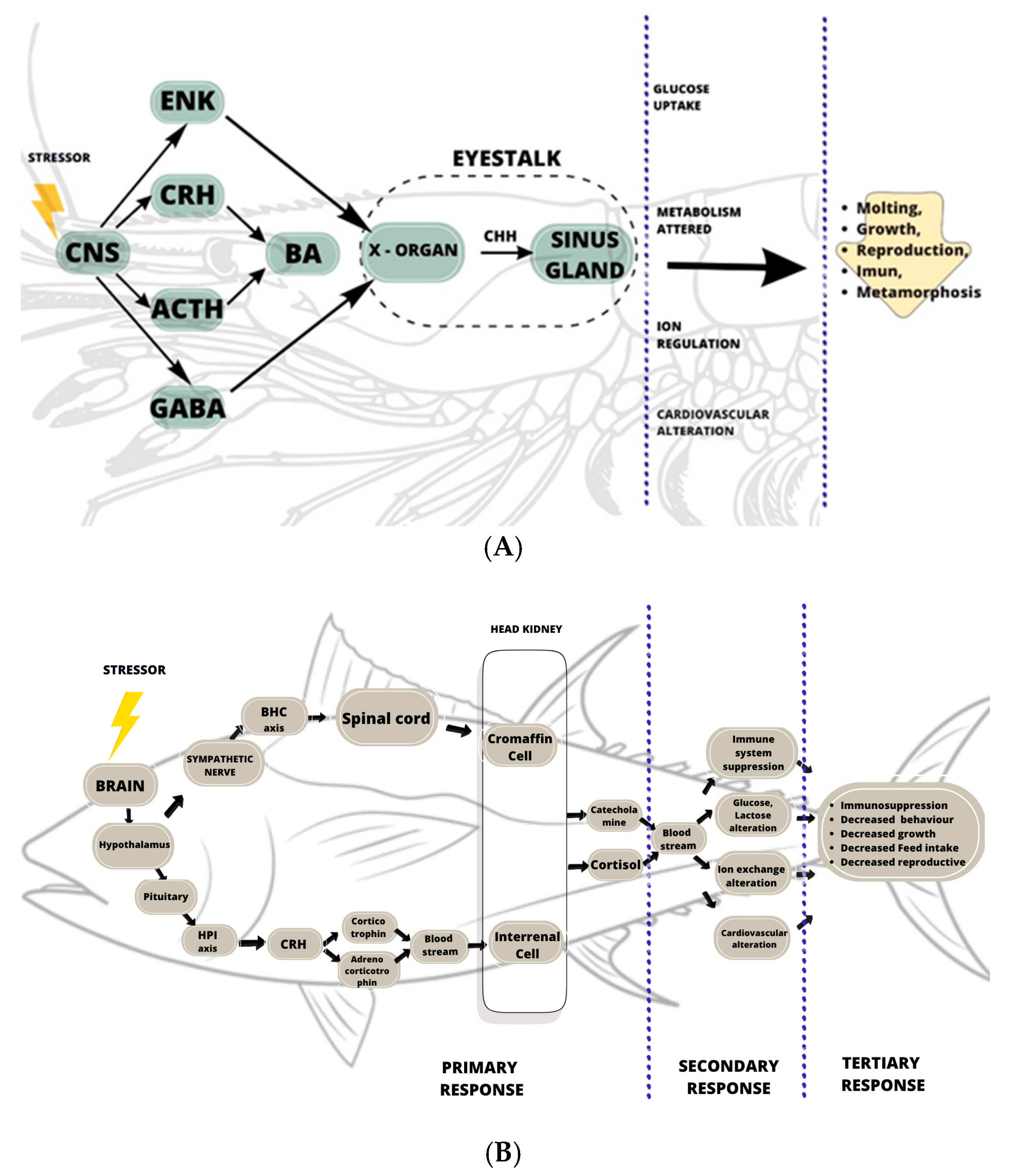
Figure 1. The relationship between physiological reactions and different types of stress and their durations.
Biology 14 00920 g001
Figure 2. Stress response in crustaceans (A) and teleosts (B). ENK, enkephalin; CRH, corticotropin-releasing hormone; ACTH, adrenocorticotropin hormone; GABA, ɣ-aminobutyric acid; BA, bioamines (dopamine, octopamine, serotonin/5-HT); CHH, crustacean hyperglycemic hormone; HPI, hypothalamus–pituitary–interrenal; BHC, brain–hypothalamus–chromaffin; CRH, corticotropin-releasing hormone.
Biology 14 00920 g002
Figure 3. ER stress and UPR signaling pathway.
Biology 14 00920 g003
Figure 4. Mechanism of reciprocal relationship between ER stress and oxidative stress in the endoplasmic reticulum and mitochondria.
Biology 14 00920 g004
Figure 5. Classification of stress response indicators from primary to tertiary level. Abbreviations: BA: biogenic amine; DA: dopamine; NE: norepinephrine; 5-HT: serotonin; CHH: crustacean hyperglycemic hormone; cAMP: cyclic adenosine monophosphate; cGMP: cyclic guanosine monophosphate; CaM: calmodulin; PKA: protein kinase A; PKC: protein kinase C; HIF-1α: hypoxia-inducible factor 1; MDA: malondialdehyde; SOD: superoxide dismutase; GPx: glutathione peroxidase; GSH: glutathione; G6PDH: glucose-6-phosphate dehydrogenase; CAT: catalase; GST: glutathione s-transferase; TAOC: total antioxidant; ER: endoreticulum stress; XBP1: X-box binding protein 1; IRE1: Inositol-requiring enzyme-1; PERK: RNA-activated protein kinase-like ER kinase; Bip: binding protein; EF1α: eukaryotic initiation factor 1; EF2α: eukaryotic initiation factor 2; ATF4: activating transcription factor 4; FAA: free amino acid; proPO: pro phenol oxidase activity; PO: phenol oxidase activity; THC: total hemocyte count; NBT: nitroblue tetrazolium salt; ACP: acid phosphatase activity; ALP: alkaline phosphatase activity; ADG: average daily growth; SGR: specific growth rate.
Biology 14 00920 g005
Table 2. Pathway differences between aquatic invertebrates and vertebrates.
Invertebrate
Crustacean
Vertebrate
Fish
Signaling systemNeuroendocrine systemEndocrine system
AxisXO-SG axisBSC axis and HPI axis
HormonesCrustacean hyperglycemic hormone/CHHCortisol and Catecholamine
Biogenic aminesNorepinephrine, GABA, dopamine, serotoninNorepinephrine, GABA, dopamine, serotonin
Major signaling pathwayCentral nervous systemHypothalamus, spinal cord, and pituitary
Stress hormone-producing organEye stalkHead kidney
Disclaimer/Publisher’s Note: The statements, opinions and data contained in all publications are solely those of the individual author(s) and contributor(s) and not of MDPI and/or the editor(s). MDPI and/or the editor(s) disclaim responsibility for any injury to people or property resulting from any ideas, methods, instructions or products referred to in the content.

