Article

Biocontrol Potential of Rhizosphere Bacteria Against Fusarium Root Rot in Cowpea: Suppression of Mycelial Growth and Conidial Germination

1
MARA Key Laboratory of Sustainable Crop Production in the Middle Reaches of the Yangtze River, Yangtze University, Jingzhou 434025, China
2
Hunan Plant Protection Institute, Hunan Academy of Agricultural Sciences, Changsha 410125, China
3
Yuelushan Laboratory, Changsha 410082, China
4
Hunan Institute of Microbiology, Hunan Academy of Agricultural Sciences, Changsha 410009, China
*
Authors to whom correspondence should be addressed.
These authors contributed equally to this work.
Biology 2025, 14(8), 921; https://doi.org/10.3390/biology14080921
Submission received: 21 May 2025 / Revised: 20 July 2025 / Accepted: 21 July 2025 / Published: 23 July 2025
(This article belongs to the Special Issue Advances in Research on Diseases of Plants (2nd Edition))

Simple Summary

Cowpea, a vital crop for food security in many regions, is threatened by root rot disease caused by harmful fungi, leading to severe crop losses and reliance on chemical fungicides that harm the environment. This study aimed to find eco-friendly solutions by examining bacteria that naturally live near cowpea roots. We discovered that certain bacteria, especially Priestia megaterium TSA-10E and Bacillus subtilis KB-6, could block the growth of disease-causing fungi by 63.21% and 47.93%, respectively. These bacteria also produce natural compounds that stop fungal spores—the “seeds” fungi use to spread—from sprouting, reducing spore germination by up to 50%. Greenhouse trials demonstrated that TSA-10E and KB-6 treatments reduced disease severity by 48.7% and 40.4%, respectively, with treated plants maintaining healthy seedlings and intact roots, while untreated plants collapsed and died. By combining bacteria that work broadly against many pathogens with those targeting specific fungi, we propose a sustainable way to protect cowpea crops without chemicals. These findings offer farmers a safer, nature-based strategy to control root rot, improving crop yields while protecting soil health and reducing pollution. This research supports global efforts to promote sustainable farming and food security, particularly in regions where cowpeas are a dietary staple.

Abstract

The cultivation of cowpea (Vigna unguiculata), a vital vegetable crop, faces significant threats from Fusarium spp.-induced root rot. In this study, three fungal pathogens (Fusarium falciforme HKFf, Fusarium incarnatum HKFi, and Fusarium oxysporum HKFo) were isolated from symptomatic cowpea plants, and we screened 90 rhizobacteria from healthy rhizospheres using six culture media. Among these pathogens, Priestia megaterium TSA-10E showed a notable suppression of F. oxysporum HKFo (63.21%), F. incarnatum HKFi (55.16%), and F. falciforme HKFf (50.93%). In addition, Bacillus cereus KB-6 inhibited the mycelial growth of F. incarnatum HKFi and F. oxysporum HKFo by 42.39% and 47.93%, respectively. Critically, cell-free filtrates from P. megaterium TSA-10E and B. cereus KB-6 cultures reduced conidial germination in F. oxysporum HKFo and F. incarnatum HKFi, highlighting their role in disrupting the early infection stages. In greenhouse trials, TSA-10E and KB-6 reduced disease severity by 48.7% and 40.4%, respectively, with treated plants maintaining healthy growth while untreated controls succumbed to wilting. Broad-spectrum assays revealed that B. subtilis TSA-6E and P. megaterium TSA-10E were potent antagonists against both economic and grain crop pathogens. These findings underscore the potential of rhizobacteria as sustainable biocontrol agents for managing root rot disease caused by Fusarium spp. in cowpea cultivation.

1. Introduction

Cowpea (Vigna unguiculata), a globally cultivated legume, is a vital source of plant-based protein and contributes to soil nitrogen fixation, underpinning its importance in food security and sustainable agriculture [1]. However, cowpea production is severely threatened by root rot disease, which manifests as leaf wilting, root necrosis, and vascular discoloration, ultimately leading to plant death [2,3]. Pathogens implicated in cowpea root rot include Fusarium spp., Ectophoma multirostrata, Berkeleyomyces rouxiae, Phytophthora vignae, and Macrophomina phaseolina [4,5,6]. Among these pathogens, Fusarium oxysporum is a soil-borne pathogen capable of causing yield losses ranging from 30% to 100% [5], which can be exacerbated by its resilient chlamydospores that persist in the soil for years.
Traditional reliance on synthetic fungicides to manage Fusarium spp. has led to pathogen resistance and environmental toxicity, driving the need for sustainable alternatives such as biological control agents (BCAs) [7]. For instance, Ralstonia solanacearum can be inhibited by a rhizobacteria strain, B. amyloliquefaciens known as JK6 [8]. More recently, microbial antagonists, such as Bacillus spp. and Trichoderma spp., have shown promise for inhibiting pathogens that cause root rot disease in other plants, including Fusarium spp. [9,10]. Therefore, the biocontrol performed by BCAs against cowpea root rot disease may be feasible and effective.
Rhizosphere-derived BCAs, including Bacillus and Pseudomonas spp., have demonstrated efficacy through mechanisms such as antibiotic production, niche competition, and induced systemic resistance [8,11]. Notably, suppressing conidial germination—a critical phase in fungal pathogenesis—has emerged as a key biocontrol strategy. For instance, lipopeptides from B. subtilis ZD01 significantly inhibit Alternaria solani conidial germination by disrupting spore integrity [12], underscoring the potential of microbial metabolites to target early infection stages.
The rhizosphere microbiome, which is essential for plant health, is dynamically reshaped by pathogen invasion, with diseased plants exhibiting reduced microbial diversity compared to their healthy counterparts [13,14]. Leveraging this information, we isolated three Fusarium strains (F. falciforme HKFf, F. incarnatum HKFi, and F. oxysporum HKFo) from diseased cowpea plants and screened rhizobacteria from healthy rhizospheres for antagonistic potential. To elucidate mechanistic insights, we further evaluated the ability of bacterial metabolites (from Priestia megaterium TSA-10E and Bacillus cereus KB-6) to suppress conidial germination. This approach aims to advance the development of multifunctional BCAs for integrated disease management in cowpea cultivation.

2. Materials and Methods

2.1. Isolation and Identification of Fungal Pathogens

Fungal pathogens were isolated from cowpea plants exhibiting root rot symptoms in Haikou City, China. Tissue segments (5 mm) were excised from the junction between necrotic lesions and asymptomatic regions. After rinsing thoroughly under running tap water, the explants were surface-sterilized by sequential immersion in 70% ethanol (0.5–1 min) and 2% NaClO (2–3 min). This was followed by three rinses with sterile distilled water to remove residual sterilizing agents. The sterilized segments were aseptically transferred onto potato dextrose agar (PDA, Solarbio, Beijing) plates supplemented with ampicillin (50 µg/mL) to inhibit bacterial growth and incubated at 28 °C. Fungal hyphae emerging from the explants were subcultured onto fresh PDA plates for purification.
For molecular identification, the total genomic DNA was extracted using the CTAB method [15]. PCR was performed with universal primers ITS4 (5′-TCCTCCGCTTATTGATATGC-3′) and ITS5 (5′-GGAAGTAAAAGTCGTAACAAGG-3′) to target the ITS sequence [16]. The amplifications were performed in 25 μL reaction mixtures containing 1 μL of each primer (10 μmol/L), 12.5 μL of Premix Taq™ (Takara Bio, Beijing), and approximately 100 ng of the template genomic DNA. The thermal cycling conditions were as follows: initial denaturation at 95 °C for 5 min; 33 cycles of 95 °C (1 min), 56 °C (1 min), and 72 °C (1 min); and a final extension at 72 °C for 5 min. Molecular identification of Fusarium isolates was also performed using species-specific primers targeting the translation elongation factor (TEF) gene region. Four primer sets, TEF-1F (5′-CTTAACGTCGTCGTCATCG-3′)/TEF-2R (5′-CACTTGGTGGTGTCCATCTT-3′), TEF-1F/TEF-3R (5′-TTGTAGCCGACCTTCTTGAT-3′), EF-LF1 (5′-CCTTAACGTCGTCGTCATCG-3′)/EF-LR1 (5′-ACGGACTTGACTTCAGTGGT-3′), and EF-LF1/EF-LR2 (5′-GAGCTGCTCGTGGTGCATCT-3′), were employed [17]. PCR reactions were carried out as mentioned above. The amplification protocol consisted of initial denaturation at 94 °C for 2 min; 35 cycles of 94 °C for 30 s, 60 °C for 40 s, and 72 °C for 90 s; followed by a final extension at 72 °C for 5 min. The PCR products were sequenced, and sequence similarity was analyzed via BlastN against the GenBank database at the National Center for Biotechnology Information (NCBI).

2.2. Isolation and Identification of Rhizosphere Microbes

Rhizosphere soil samples (2 g) were collected from healthy cowpea plants in the same field. Microbial isolation was performed using a serial dilution plating technique on six distinct media: LB (Luria–Bertani medium), 0.1 × LB, KB (KingB medium), YG (yeast extract medium), R2A (Reasoner’s 2A agar), and TSA (Tryptic Soy Agar) (Table S1). Soil suspensions were prepared by vortexing the samples in 18 mL sterile physiological saline (0.9% w/v NaCl) for 40 min at 200 rpm and 28 °C, followed by dilution to 10−4–10−7. Aliquots (0.1 mL) of 10−5–10−7 dilutions were spread onto triplicate plates of each medium and incubated at 28 °C. Morphologically distinct colonies were selected and subcultured for purity.
Bacterial identification was based on 16S rRNA gene sequencing. Colony PCR was conducted using primers 27F (5′-AGAGTTTGATCCTGGCTCAG-3′) and 1378R (5′-CGGTGTGTACAAGGCCCGGGAACG-3′) [18] under the same conditions as mentioned above. Sequences were compared to the GenBank database at the National Center for Biotechnology Information (NCBI) via BlastN for taxonomic assignment.

2.3. Screening for Antagonistic Activity

To assess the antifungal activity of rhizosphere isolates, a dual-culture assay was used, which was modified from the method described by De Vrieze et al. [19]. Mycelial plugs (8 mm diameter) of the target pathogens were placed centrally on LB agar plates. Bacterial isolates were cultured in 5 × YEG (yeast glucose medium) broth (200 rpm, 30 °C, 48 h), and 40 µL of each culture was applied to four sterile filter paper discs (diameter: 5 mm). The discs were positioned 2 cm from the plate edge at 90° intervals. The plates were incubated at 28 °C for 3–6 days, and radial mycelial growth was measured. The inhibition rate was calculated as follows:
Inhibition rate (%) = [(A − B)/A] × 100
Here, A and B represent mycelial growth in the control and treatment groups, respectively. Experiments were performed in triplicate with three independent repetitions.

2.4. Broad-Spectrum Antifungal Activity Assay

To comprehensively evaluate the biocontrol potential of the selected rhizobacteria, we included a diverse range of plant pathogens in our screening. The selected bacterial isolates were tested against seven economically important pathogens affecting different crops: Colletotrichum gloeosporioides, Sclerotium rolfsii, Fusarium solani, Magnaporthe oryzae, Exserohilum turcicum, Helminthosporium maydis, Sclerotinia sclerotiorum (fungi), and Phytophthora capsici (Oomycete). This broad-spectrum approach was adopted for three key reasons: first, to assess whether the antagonistic activity was specific to Fusarium pathogens or whether it could extend to other phytopathogens; second, to identify potential multi-target biocontrol agents that could be applied across different cropping systems; and last but not the least, to evaluate the consistency of inhibitory effects across taxonomically diverse pathogens, which would indicate more robust biocontrol mechanisms. Antagonistic activity was evaluated using the filter paper disc method (Section 2.3), with inhibition rates quantified as described above. Experiments were performed in triplicate with three independent repetitions.

2.5. Conidial Germination Assay

Cell-free culture filtrates (CFCs) of P. megaterium TSA-10E and B. cereus KB-6 were prepared via centrifugation (8000 rpm, 10 min) and sterile filtration (0.22 µm). The conidia of Fusarium oxysporum HKFo and Fusarium incarnatum HKFi were harvested from 5×YEG cultures (28 °C, 72 h), filtered through Miracloth (Calbiochem, El Cajon, CA, USA), and adjusted to 5 × 10⁴ conidia/mL by a hemocytometer. The conidial suspension, which was treated with CFCs (10% v/v final concentration), was incubated at 28 °C for 5 h. Controls received 5×YEG medium. Germination rates were determined microscopically by counting ≥50 conidia per replicate. Three biological replicates were analyzed, each with three technical repeats.

2.6. In Planta Biological Control

The biocontrol efficacy of P. megaterium TSA-10E and B. cereus KB-6 against F. oxysporum HKFo and F. incarnatum HKFi was evaluated in greenhouse-grown cowpea (Vigna unguiculata cv. Changjiang 3). Seedlings germinated in peat-based substrate were transplanted into 9 cm diameter plastic pots containing a 3:1:1 peat-vermiculite-perlite mix, maintained at 25 °C under 12 h light/dark cycles with biweekly fertilization until the two-leaf stage. Prior to inoculation, roots were wounded near the stem base by puncturing with a sterile syringe needle (26-gauge) to a depth of 2–3 mm. Conidial suspensions of both pathogens were adjusted to 1 × 10⁷ conidia/mL, with four treatment groups established: pathogen control (CK1) receiving 10 mL mixed conidial suspension (5 mL per pathogen); TSA-10E group treated with pathogen suspension plus 10% P. megaterium filtrate; KB-6 group receiving pathogen suspension with 10% B. cereus filtrate; and negative control (CK2) administered pathogen suspension with 10% 5×YEG broth. Inoculated plants were kept in greenhouse at 25 °C. Each group contained at least 10 seedlings and was replicated three times. After 14 days, disease severity was scored on a 1–5 scale (1 = healthy, 5 = plant death) according to the grading standard of Rigert and Foster [20]. Disease index = Σ (number of diseased plants × representative series)/(total number of plants × highest representative level value) × 100. Control efficacy (%) = (control disease index − treatment disease index)/control disease index × 100%.

2.7. Statistical Analysis

All experiments were independently repeated three times, and the data were statistically analyzed using an ANOVA. The multiple Duncan test was used to compare the differences between mean values at a significance level of p < 0.05. All analyses were conducted using SPSS software (version 20.0; SPSS Inc., Chicago, IL, USA).

3. Results

3.1. Isolation and Molecular Identification of Pathogenic Fusarium Strains

Three morphologically distinct fungal strains were isolated from symptomatic cowpea tissues. Molecular identification via ITS and sequencing demonstrated >97% similarity with Fusarium species. Strain-level identification was achieved through BLASTN analysis with four species-specific primer pairs, confirming that the isolates as Fusarium falciforme HKFf, F. incarnatum HKFi, and F. oxysporum HKFo. The sequence similarity values are summarized in Table S2.

3.2. Isolation and Taxonomic Diversity of Rhizobacterial Strains

A total of ninety rhizobacterial isolates were obtained from six distinct culture media. 16S rRNA sequencing identified thirty-six non-redundant strains, most of which belonged to Bacillus spp. (34.4%, thirty-one isolates), followed by Priestia spp. (25.6%, twenty-three isolates) and Neobacillus spp. (11.1%, ten isolates). Bacillus exhibited the highest species diversity, comprising fourteen distinct strains (Table 1). Medium-specific isolation patterns were observed. The LB medium yielded thirteen strains, whereas YG and R2A each yielded eleven strains. The TSA medium yielded eight strains, and KB and 0.1×LB yielded seven and six strains, respectively. Notably, Priestia megaterium was recovered from all six media tested. In contrast, several strains exhibited medium specificity: Bacillus bataviensis, Bacillus bingmayongensis, Bacillus safensis, Dyella thiooxydans, Paenibacillus septentrionalis, and Sinomonas atrocyanea were exclusively isolated from R2A; Bacillus pumilus and Bacillus subtilis were unique to TSA; Paenibacillus silvae, Neobacillus citreus, and Bacillus cereus were found only in KB; and Bacillus anthracis, Bacillus zanthoxyli, Heyndrickxia oleronia, and Rossellomorea marisflavi were restricted to YG medium. Furthermore, with the exception of P. megaterium, all strains obtained from 0.1×LB medium showed strict medium specificity (Table S3).

3.3. Antagonistic Activity of Rhizosphere Isolates

Among the thirty-six rhizobacterial strains screened, seven (19.4%) exhibited antifungal activity against Fusarium pathogens, with their quantified inhibition rates presented in Table 2. Four strains—P. megaterium TSA-10E, B. subtilis TSA-6E, B. cereus KB-6, and R. marisflavi YG-2C—showed >40% inhibition of F. incarnatum HKFi and F. oxysporum HKFo. However, only P. megaterium (50.93%) and B. subtilis (46.27%) exceeded 40% inhibition against F. falciforme HKFf (Figure 1). P. megaterium TSA-10E demonstrated the highest overall antagonism, followed by B. subtilis TSA-6E. Against F. incarnatum HKFi and F. oxysporum HKFo, B. cereus KB-6 ranked third in terms of inhibitory efficacy, while R. marisflavi YG-2C ranked fourth. Notably, 42.9% (3/7) of the effective strains belonged to Bacillus spp., with B. subtilis TSA-6E and B. cereus KB-6 achieving ≥40% inhibition. In contrast, S. atrocyanea R2A-7 and P. silvae KB-5 exhibited <40% inhibition across all the pathogens. Additionally, the inhibition rates against F. incarnatum HKFi were consistently higher than those for F. oxysporum HKFo and F. falciforme HKFf.

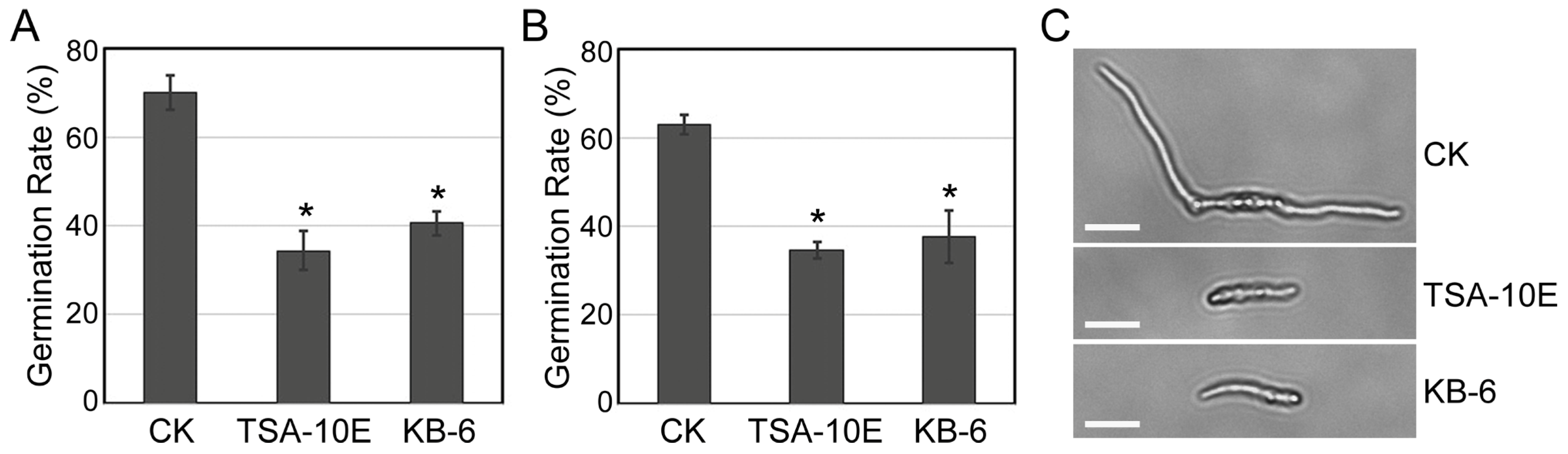
3.4. Inhibition of Conidial Germination by TSA-10E and KB-6 Filtrates

While P. megaterium TSA-10E, B. subtilis TSA-6E, and B. cereus KB-6 showed strong antagonistic activity against Fusarium spp., we selected P. megaterium TSA-10E and B. cereus KB-6 for mechanistic studies. TSA-10E was prioritized due to its consistently high inhibition across Fusarium pathogens and the relative novelty of its biocontrol potential compared to the extensively studied B. subtilis, while KB-6 exhibited broader efficacy against key Fusarium strains compared to other isolates.
To investigate their antifungal mechanisms, we evaluated the effect of cell-free filtrates from P. megaterium TSA-10E and B. cereus KB-6 cultures on conidial germination—a critical stage in F. oxysporum HKFo and F. incarnatum HKFi pathogenesis. Treatment with TSA-10E filtrates reduced F. oxysporum conidial germination by 50.9%, while KB-6 filtrates achieved 42.1% inhibition (Figure 2A,C). Similarly, F. incarnatum germination rates declined to 34.7% (TSA-10E) and 37.6% (KB-6) compared to untreated controls (Figure 2B). Microscopic examination revealed that in TSA-10E and KB-6 treated samples, the few conidia of F. oxysporum or F. incarnatum that managed to germinate produced markedly shorter germ tubes compared to the long, well-developed germ tubes observed in the controls (Figure 2C). These results demonstrate that both filtrates significantly disrupted early infection processes by suppressing conidial germination.

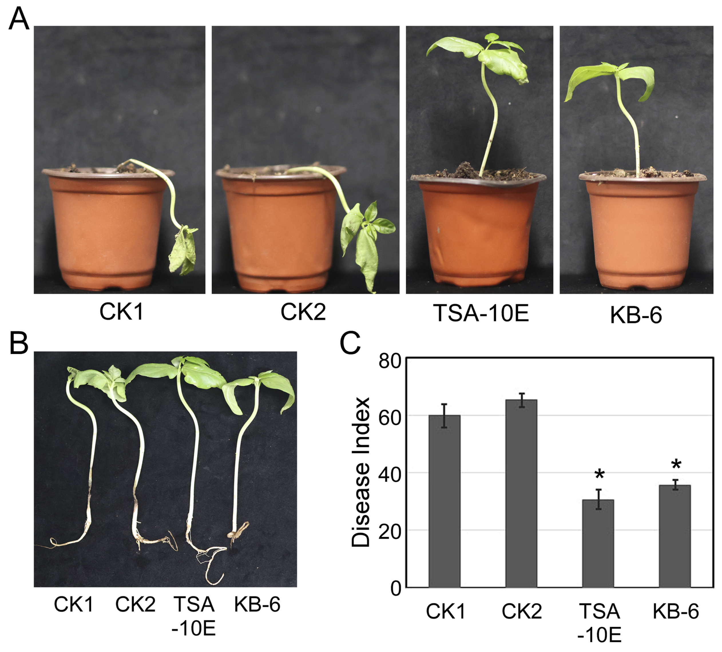
3.5. TSA-10E and KB-6 Can Effectively Control Cowpea Fusarium Root Rot in Greenhouse

Both P. megaterium TSA-10E and B. cereus KB-6 treatments provided effective protection against Fusarium root rot in cowpea plants. While control plants collapsed with severe wilting, treated plants maintained healthy and upright growth (Figure 3A). Root systems of treated plants showed minimal browning and rot, in contrast to the extensively decayed roots of controls (Figure 3B). Both treatments substantially decreased the disease index, with TSA-10E showing 48.7% control efficacy and KB-6 exhibiting 40.4% efficacy (Figure 3C). Notably, TSA-10E demonstrated superior disease suppression relative to KB-6. These results confirm the significant inhibitory effects of both bacterial strains against cowpea Fusarium root rot under greenhouse conditions.

3.6. Broad-Spectrum Antifungal Activity

The broad-spectrum antifungal potential of the seven selected rhizobacterial strains was evaluated against eight plant pathogens, including both grain crop pathogens (Magnaporthe oryzae, Exserohilum turcicum, and Helminthosporium maydis) and economic crop pathogens (Colletotrichum gloeosporioides, Sclerotium rolfsii, Phytophthora capsici, Fusarium solani, and Sclerotinia sclerotiorum). Among the tested strains, B. subtilis TSA-6E and P. megaterium TSA-10E exhibited consistent inhibitory activity across multiple pathogens (Table 3 and Figure 4). Against C. gloeosporioides, B. subtilis TSA-6E demonstrated the highest inhibition rate (77.76%), significantly surpassing P. megaterium TSA-10E (64.76%), R. marisflavi YG-2C (59.69%), and B. cereus KB-6 (55.90%). Similar trends were observed for S. rolfsii, with B. subtilis TSA-6E (48.32%) and P. megaterium TSA-10E (46.72%) showing superior suppression compared to B. cereus KB-6 (40.54%). Notably, P. megaterium TSA-10E and B. subtilis TSA-6E also displayed strong antagonism against M. oryzae, achieving inhibition rates of 75.91% and 73.73%, respectively, followed by B. pumilus TSA-1 (66.23%). In contrast, S. atrocyanea R2A-7 exhibited exceptional activity against E. turcicum (73.25%), marginally outperforming P. megaterium TSA-10E (72.76%) and B. subtilis TSA-6E (71.86%). However, none of the strains effectively inhibited S. sclerotiorum (<40% inhibition). For F. solani, both B. subtilis TSA-6E and P. megaterium TSA-10E achieved identical inhibition rates (53.18% and 52.29%), highlighting their broad-spectrum efficacy. These results underscore the differential inhibitory patterns among strains, with B. subtilis TSA-6E and P. megaterium TSA-10E emerging as promising candidates for controlling diverse phytopathogens.

4. Discussion

Rhizosphere-associated bacteria play a pivotal role in suppressing soil-borne pathogens through diverse mechanisms [21]. In this study, seven rhizobacterial isolates demonstrated antagonistic activity against fungal pathogens associated with cowpea root rot, with B. subtilis TSA-6E and P. megaterium TSA-10E exhibiting broad-spectrum efficacy. Notably, our findings extend to the inhibition of conidial germination by bacterial metabolites, a critical early-stage defence mechanism against fungal infection.
The isolation of thirty-six non-redundant strains from six media revealed distinct microbial preferences. The LB medium yielded the highest diversity (thirteen strains), likely due to its nutrient-rich composition, which supports diverse bacterial communities. Several strains exhibited medium specificity, such as B. pumilus and B. subtilis were exclusively isolated from TSA, while B. cereus was only found on the KB medium. These observations align with previous reports that medium composition selectively enriches specific microbial taxa by fulfilling their metabolic requirements [22].
The superior performance of P. megaterium TSA-10E against multiple pathogens (e.g., a 75.91% inhibition of M. oryzae and a 72.76% inhibition of E. turcicum) is indicative of its ability to deploy multifaceted biocontrol strategies. Such broad-spectrum activity may stem from synergistic mechanisms, including antibiotic production (e.g., lipopeptides), nutrient competition, and biofilm formation [11,23,24]. For instance, Zhang et al. [12] demonstrated that lipopeptides from B. subtilis ZD01 suppressed conidial germination in Alternaria solani, which is consistent with our observation that P. megaterium TSA-10E cell-free filtrates reduced F. oxysporum HKFo and F. incarnatum HKFi conidial germination by 50.9% and 44.8%, respectively (Figure 2). This finding represents an important advancement in understanding P. megaterium TSA-10E’s biocontrol potential, as previous studies have primarily focused on its broad-spectrum suppression of mycelial growth or mycotoxin production in various pathosystems. For example, while P. megaterium strains have been shown to produce antifungal lipopeptides like surfactin and iturin A against bacterial pathogens such as Erwinia amylovora [25], suppress Fusarium verticillioides mycotoxins through volatile organic compounds [26], or inhibit Fusarium oxysporum f. sp. ciceri [27], their direct effects on fungal spore germination remain unexplored. Our work provides the first evidence that P. megaterium TSA-10E can effectively disrupt the critical early infection stage of Fusarium pathogens by inhibiting conidial germination, a mechanism that has not been previously documented for this species.
Microscopic examination revealed that in addition to reducing germination rates, the P. megaterium TSA-10E and B. cereus KB-6 treatments caused notable morphological alterations in F. oxysporum HKFo and F. incarnatum HKFi conidia (Figure 2C). Compared to the control group, where conidia had normal, elongated germ tubes, treated conidia showed either germination inhibition or produced significantly shorter germ tubes. This laboratory observation directly translated to disease suppression in greenhouse conditions, where the same filtrates reduced Fusarium root rot severity by 48.7% (TSA-10E) and 40.4% (KB-6). The correlation between conidia inhibition and in planta protection suggests that disrupting early infection stages (germination and germ tube development) effectively prevents subsequent root colonization and disease establishment [28].
The significant reduction in conidial germination and germ tube length by P. megaterium TSA-10E and B. cereus KB-6 filtrates suggests that bacterial metabolites directly interfere with the early infection processes. Similar mechanisms have been reported for Bacillus spp., where antifungal compounds such as surfactin and iturin inhibit spore germination and hyphal elongation, and suppress watermelon Fusarium wilt [29,30]. The greenhouse results further support that this early-stage interference has functional consequences throughout the infection cycle, ultimately preserving plant health. The cell-free supernatants of the Pseudomonas poae strain CO were able to inhibit the spore germination and germ tube length of Fusarium graminearum, and exhibited diverse antifungal properties, such as the production of hydrolytic enzymes, siderophores, and lipopeptides [31]. These structural abnormalities suggest that the bacterial filtrates may interfere with the genetic and biochemical processes during germination, potentially through the action of antimicrobial metabolites. The combined evidence from microscopic observations and greenhouse protection assays suggests a potential mode-of-action sequence, where metabolite-mediated spore inhibition may lead to impaired germ tube formation, subsequently limiting root penetration and ultimately contributing to reduced disease symptoms. Our results reinforce the importance of metabolite-mediated inhibition as a key biocontrol trait, particularly against Fusarium spp., which rely heavily on conidia for host colonization. The consistency between in vitro and in planta outcomes underscores the translational potential of these findings for field applications.
While S. atrocyanea R2A-7 exhibited strain-specific inhibition (73.25% against E. turcicum), its limited efficacy against other pathogens underscores the value of combining broad-spectrum and specialized biocontrol agents. This phenomenon aligns with findings in other biocontrol systems. For instance, Pseudomonas monsensis H16 could inhibit Alternaria brassicae YB43-2 and Epicoccum sorghinum YB53-2 but exhibit minimal activity against other phytopathogens [32]. Similarly to our observations with S. atrocyanea R2A-7, this narrow-spectrum activity suggests that these strains may have evolved specialized mechanisms targeting specific pathogens, possibly through the production of pathogen-specific antimicrobial compounds or interference with particular virulence factors. While narrow-spectrum agents lack versatility, their precision reduces off-target effects, making them ideal for integration into “polymicrobial” consortia. This “polymicrobial” strategy has garnered significant interest in recent years, although most of the research to date has primarily concentrated on blending well-established, commercially accessible microbial agents such as Trichoderma and Bacillus/Pseudomonas or mycorrhizal fungi and nitrogen-fixing bacteria [33,34,35,36]. Recent research has shown that two strains that have complementary modes of action are expected to be particularly efficient in their dual combination [19]. Rhizobacteria with broad-spectrum properties (B. subtilis TSA-10E, P. megaterium TSA-10E, and B. cereus KB-6) can be combined with rhizobacteria with more pathogen-specific antagonism (S. atrocyanea R2A-7), which could enhance disease control by targeting multiple infection pathways. This approach aligns with emerging trends in biocontrol research that emphasize functional redundancy and complementary action modes.
While this study identifies promising biocontrol candidates, further research is needed to characterize the active compounds in P. megaterium TSA-10E and B. cereus KB-6 filtrates and validate their stability under field conditions. Furthermore, elucidating the relative contributions of specific mechanisms such as antibiotic production and induced systemic resistance will refine strain selection for targeted applications.

5. Conclusions

This study demonstrates the potential of rhizosphere bacteria, particularly P. megaterium TSA-10E and B. subtilis TSA-6E, as eco-friendly biocontrol agents against Fusarium-induced root rot in cowpea. These strains exhibited dual antifungal mechanisms, suppressing both mycelial growth (>40%) and conidial germination (up to 50.9%) in key pathogens such as F. oxysporum HKFo and F. incarnatum HKFi. Greenhouse trials confirmed these effects under realistic conditions, with TSA-10E and KB-6 reducing disease severity by 48.7% and 40.4%, respectively, while preserving plant viability—demonstrating translational potential beyond in vitro systems. The broad-spectrum activity of B. subtilis and the novel efficacy of P. megaterium highlight their versatility in targeting multiple infection stages, offering a sustainable alternative to chemical fungicides. Furthermore, the inhibition of spore germination by bacterial metabolites (P. megaterium TSA-10E and B. cereus KB-6) underscores the role of secondary compounds in disrupting early fungal colonization, a mechanism now validated to correlate with whole-plant protection.
These findings not only expand the repertoire of biocontrol candidates but also support the development of polymicrobial formulations combining broad-spectrum and pathogen-specific strains. The consistency between laboratory and greenhouse results strengthens the case for field testing of these strains as integrated disease management solutions. Future studies should focus on characterizing the active metabolites, optimizing delivery methods, and validating these results under field conditions to ensure practical applicability. By bridging lab-based discoveries with agricultural needs, this study contributes to sustainable crop protection and food security in regions reliant on cowpea cultivation.

Supplementary Materials

The following supporting information can be downloaded at: https://www.mdpi.com/article/10.3390/biology14080921/s1, Table S1: Formulation of medium used; Table S2: Identification of fungal pathogens; Table S3: The rhizosphere microbes isolated on six different mediums.

Author Contributions

Conceptualization, D.P. and X.Z.; Methodology, Q.Z. and Y.M.; Validation, Q.Z. and Y.M.; Formal analysis, T.Z.; Writing—original draft, Q.Z. and Y.M.; Writing—review and editing, W.L. and S.Z.; Visualization, Y.C. All authors have read and agreed to the published version of the manuscript.

Funding

This research was funded by the Key Research and Development Program of Hunan Province (Grant No. 2023NK2014) and the Agricultural Science and Technology Innovation Fund of Hunan Province (Grant No. 2023CX44 and 2023CX116).

Institutional Review Board Statement

Not applicable.

Informed Consent Statement

Not applicable.

Data Availability Statement

The original contributions presented in this study are included in the article/Supplementary Material. Further inquiries can be directed to the corresponding authors.

Conflicts of Interest

The authors declare no conflicts of interest.

References

  1. Jayathilake, C.; Visvanathan, R.; Deen, A. Cowpea: An overview on its nutritional facts and health benefits. J. Sci. Food Agric. 2018, 98, 4793–4806. [Google Scholar] [CrossRef] [PubMed]
  2. Long, J.; Wu, W.; Sun, S. Berkeleyomyces rouxiae is a causal agent of root rot complex on faba bean (Vicia faba L.). Front. Plant Sci. 2022, 13, 989517. [Google Scholar] [CrossRef] [PubMed]
  3. Al-Jaradi, A.; Al-Mahmooli, I.; Janke, R. Isolation and identification of pathogenic fungi and oomycetes associated with beans and cowpea root diseases in Oman. PeerJ 2018, 6, e6064. [Google Scholar] [CrossRef] [PubMed]
  4. Kashyap, A.S.; Manzar, N.; Ahamad, F. First report of root rot disease in green gram (Vigna radiata) caused by Ectophoma multirostrata in India. Plant Dis. 2022, 106, 2013–2025. [Google Scholar] [CrossRef]
  5. Yang, C.; Lin, L.; Bao, J. Genome sequence resource of Phytophthora vignae, the causal agent of stem and root rot of cowpea. Mol. Plant-Microbe Interact. 2021, 34, 862–865. [Google Scholar] [CrossRef] [PubMed]
  6. Rashmi, N.R.K.G. Evaluation of bioagents against Macrophomina phaseolina and its effect on root weight of cowpea (Vigna unguiculata (L.) Walp). J. Eco-Friendly Agric. 2022, 17, 166–168. [Google Scholar]
  7. Lamini, S.; Kusi, F.; Cornelius, E. Identification of sources of resistance in cowpea lines to Macrophomina root rot disease in northern Ghana. Heliyon 2022, 8, e12217. [Google Scholar] [CrossRef] [PubMed]
  8. Xiong, H.Q.; Li, Y.T.; Cai, Y.F. Isolation of Bacillus amyloliquefaciens JK6 and identification of its lipopeptides surfactin for suppressing tomato bacterial wilt. RSC Adv. 2015, 5, 82042–82049. [Google Scholar] [CrossRef]
  9. Olowe, O.M.; Nicola, L.; Asemoloye, M.D. Trichoderma: Potential bio-resource for the management of tomato root rot diseases in Africa. Microbiol. Res. 2022, 257, 126978. [Google Scholar] [CrossRef] [PubMed]
  10. Lin, F.; Liu, N.; Lai, D.; Kang, X.H.; Pang, N.W.; Jiang, H.K.; Xu, H.H. A formulation of neem cake seeded with Bacillus sp. provides control over tomato Fusarium crown and root rot. Biocontrol Sci. Technol. 2017, 27, 393–407. [Google Scholar] [CrossRef]
  11. Zhong, J.; Sui, W.W.; Bai, X.Y. Characterization and biocontrol mechanism of Streptomyces olivoreticuli as a potential biocontrol agent against Rhizoctonia solani. Pestic. Biochem. Phys. 2023, 197, 105681. [Google Scholar] [CrossRef] [PubMed]
  12. Zhang, D.; Qiang, R.; Zhou, Z.; Pan, Y.; Yu, S.; Yuan, W.; Cheng, J.; Wang, J.; Zhao, D.; Zhu, J.; et al. Biocontrol and action mechanism of Bacillus subtilis lipopeptides’ fengycins against Alternaria solani in potato as assessed by a transcriptome Analysis. Front. Microbiol. 2022, 13, 861113. [Google Scholar] [CrossRef] [PubMed]
  13. Cao, P.; Li, C.; Wang, H. Community structures and antifungal activity of root-associated endophytic actinobacteria in healthy and diseased cucumber plants and Streptomyces sp. HAAG3-15 as a promising biocontrol agent. Microorganisms 2020, 8, 236. [Google Scholar] [CrossRef] [PubMed]
  14. Dastogeer, K.M.G.; Yasuda, M.; Okazaki, S. Microbiome and pathobiome analyses reveal changes in community structure by foliar pathogen infection in rice. Front. Microbiol. 2022, 13, 949152. [Google Scholar] [CrossRef] [PubMed]
  15. Dar, G.J.; Nazir, R.; Wani, S.A. Optimizing a modified cetyltrimethylammonium bromide protocol for fungal DNA extraction: Insights from multilocus gene amplification. Open Life Sci. 2025, 20, 20221006. [Google Scholar] [CrossRef] [PubMed]
  16. White, T.J.; Bruns, T.; Lee, S.J. Amplification and direct sequencing of fungal ribosomal RNA genes for phylogenetics. In PCR Protocols: A Guide to Methods and Applications; American Academic Press: San Diego, CA, USA, 1990; pp. 315–322. [Google Scholar]
  17. Wang, J.; Liu, Y.; Zhang, J. A Rapid Identification Method for Fusarium Species Using Specific Primer Combinations. Chinese Patent CN109504799A, 22 March 2019. [Google Scholar]
  18. Heuer, H.; Krsek, M.; Baker, P. Analysis of actinomycete communities by specific amplification of genes encoding 16S rRNA and gel-electrophoresis separation in denaturing gradients. Appl. Environ. Microbiol. 1997, 63, 3233–3241. [Google Scholar] [CrossRef] [PubMed]
  19. De Vrieze, M.; Germanier, F.; Vuille, N. Combining Different Potato-Associated Pseudomonas Strains for Improved Biocontrol of Phytophthora infestans. Front. Microbiol. 2018, 9, 2573. [Google Scholar] [CrossRef] [PubMed]
  20. Alisaac, E.; Mahlein, A.K. Fusarium Head Blight on Wheat: Biology, Modern Detection and Diagnosis and Integrated Disease Management. Toxins 2023, 15, 192. [Google Scholar] [CrossRef] [PubMed]
  21. Olanrewaju, O.S.; Ayangbenro, A.S.; Glick, B.R. Plant health: Feedback effect of root exudates-rhizobiome interactions. Appl. Microbiol. Biotechnol. 2019, 103, 1155–1166. [Google Scholar] [CrossRef] [PubMed]
  22. Babich, O.; Sukhikh, S.; Dyshlyuk, L. Evaluation of biocompatibility and antagonistic properties of microorganisms isolated from natural sources for obtaining biofertilizers using microalgae hydrolysate. Microorganisms 2021, 9, 1667. [Google Scholar] [CrossRef] [PubMed]
  23. Di Francesco, A.; Ugolini, L.; D’Aquino, S.; Pagnotta, E.; Mari, M. Biocontrol of Monilinia laxa by Aureobasidium pullulans strains: Insights on competition for nutrients and space. Int. J. Food Microbiol. 2017, 248, 32–38. [Google Scholar] [CrossRef] [PubMed]
  24. Pandin, C.; Darsonval, M.; Mayeur, C. Biofilm formation and synthesis of antimicrobial compounds by the biocontrol agent Bacillus velezensis QST713 in an Agaricus bisporus compost micromodel. Appl. Environ. Microbiol. 2019, 85, e00327-19. [Google Scholar] [CrossRef] [PubMed]
  25. Cui, Z.; Hu, L.; Zeng, L. Isolation and characterization of Priestia megaterium KD7 for the biological control of pear fire blight. Front. Microbiol. 2023, 14, 1099664. [Google Scholar] [CrossRef] [PubMed]
  26. Figueroa-López, A.M.; Cordero-Ramírez, J.D.; Martínez-Álvarez, J.C. Rhizospheric bacteria of maize with potential for biocontrol of Fusarium verticillioides. SpringerPlus 2016, 5, 330. [Google Scholar] [CrossRef] [PubMed]
  27. Bhargavi, G.; Arya, M.; Jambhulkar, P.P. Evaluation of biocontrol efficacy of rhizosphere dwelling bacteria for management of Fusarium wilt and Botrytis gray mold of chickpea. BMC Genom. Data 2024, 25, 7. [Google Scholar] [CrossRef] [PubMed]
  28. Rigert, K.S.; Foster, K.W. Inheritance of Resistance to Two Races of Fusarium Wilt in Three Cowpea Cultivars. Crop Sci. 1987, 27, 220–224. [Google Scholar] [CrossRef]
  29. Al-Mutar, D.M.K.; Noman, M.; Abduljaleel Alzawar, N.S. Cyclic lipopeptides of Bacillus amyloliquefaciens DHA6 are the determinants to suppress watermelon Fusarium wilt by direct antifungal activity and host defense modulation. J. Fungi 2023, 9, 687. [Google Scholar] [CrossRef] [PubMed]
  30. Assena, M.W.; Pfannstiel, J.; Rasche, F. Inhibitory activity of bacterial lipopeptides against Fusarium oxysporum f.sp. Strigae. BMC Microbiol. 2024, 24, 227. [Google Scholar] [CrossRef] [PubMed]
  31. Chen, X.; Zhang, M.; Tang, L. Screening and characterization of biocontrol bacteria isolated from Ageratum conyzoides against Collectotrichum fructicola causing Chinese plum (Prunus salicina Lindl.) anthracnose. Front. Microbiol. 2023, 14, 1296755. [Google Scholar] [CrossRef] [PubMed]
  32. Ibrahim, E.; Nasser, R.; Hafeez, R.; Ogunyemi, S.O.; Abdallah, Y.; Khattak, A.A.; Shou, L.; Zhang, Y.; Ahmed, T.; Atef Hatamleh, A.; et al. Biocontrol Efficacy of Endophyte Pseudomonas poae to Alleviate Fusarium Seedling Blight by Refining the Morpho-Physiological Attributes of Wheat. Plants 2023, 12, 2277. [Google Scholar] [CrossRef] [PubMed]
  33. Xu, X.M.; Jeffries, P.; Pautasso, M. Combined use of biocontrol agents to manage plant diseases in theory and practice. Phytopathology 2011, 101, 1024–1031. [Google Scholar] [CrossRef] [PubMed]
  34. Reddy, C.A.; Saravanan, R.S. Polymicrobial multi-functional approach for enhancement of crop productivity. Adv. Appl. Microbiol. 2013, 82, 53–113. [Google Scholar] [PubMed]
  35. Sarma, B.K.; Yadav, S.K.; Singh, S. Microbial consortium-mediated plant defense against phytopathogens: Readdressing for 22enhancing efficacy. Soil Biol. Biochem. 2015, 87, 25–33. [Google Scholar] [CrossRef]
  36. Parnell, J.J.; Berka, R.; Young, H. From the lab to the farm: An industrial perspective of plant beneficial microorganisms. Front. Plant Sci. 2016, 7, 1110. [Google Scholar] [CrossRef] [PubMed]
Figure 1. Antagonistic activity of rhizobacteria against cowpea root rot pathogens evaluated via a dual-culture assay. Mycelial plugs (8 mm diameter) of pathogenic fungi were placed centrally on LB agar plates, with four filter paper discs (5 mm diameter) loaded with 40 µL of bacterial culture (grown in the 5×YEG broth at 30 °C for 48 h) positioned 2 cm from the plate edge at 90° intervals. Control plates received 5×YEG medium instead of bacterial culture. All plates were incubated in a 28 °C incubator and cultured for three to six days.
Biology 14 00921 g001
Figure 2. P. megaterium TSA-10E and B. cereus KB-6 cell-free filtrates inhibit the conidia germination of F. oxysporum HKFo (A) and F. incarnatum HKFi (B). Error bars represent the standard errors. The asterisks (*) represent significant differences compared with CK according to the multiple Duncan test (p < 0.05). (C) Morphological observations of the conidia of F. oxysporum. Bar = 10 μm.
Biology 14 00921 g002
Figure 3. P. megaterium TSA-10E and B. cereus KB-6 suppress cowpea Fusarium root rot in greenhouse conditions. (A) Disease symptoms in cowpea seedlings. (B) The symptoms of cowpea roots. (C) The disease index of cowpea Fusarium root rot. Error bars represent the standard errors. The asterisks (*) represent significant differences from control by the multiple Duncan test (p < 0.05). Treatments: CK1 (Fusarium only), CK2 (Fusarium+medium), TSA-10E (P. megaterium TSA-10E treatment), and KB-6 (B. cereus KB-6 treatment).
Biology 14 00921 g003
Figure 4. Antagonistic effect of rhizobacteria against the common pathogens that were selected, evaluated via a dual-culture assay. Control: pathogenic fungi with the 5×YEG medium on the LB medium. The plates were incubated in a 28 °C incubator and cultured for three to seven days.
Biology 14 00921 g004
Table 1. The rhizosphere microbes identified from different genera.
GenusTotal of IsolatesTotal of SpeciesSpeciesQuantity
Bacillus3114Bacillus anthracis1
Bacillus aryabhattai11
Bacillus bataviensis1
Bacillus bingmayongensis1
Bacillus cereus2
Bacillus ferrooxidans3
Bacillus mycoides3
Bacillus niacini1
Bacillus pseudomycoides3
Bacillus pumilus1
Bacillus safensis1
Bacillus sporothermodurans1
Bacillus subtilis1
Bacillus zanthoxyli1
Dyella11Dyella thiooxydans1
Falsibacillus11Falsibacillus pallidus1
Fictibacillus61Fictibacillus barbaricus6
Gottfriedia11Gottfriedia acidiceleris1
Heyndrickxi11Heyndrickxia oleronia1
Neobacillus104Neobacillus citreus1
Neobacillus drentensis2
Neobacillus ginsengisoli6
Neobacillus niacini1
Paenibacillus44Paenibacillus cellulositrophicus1
Paenibacillus pabuli1
Paenibacillus septentrionalis1
Paenibacillus silvae1
Priestia232Priestia aryabhattai11
Priestia megaterium12
Ralstonia11Ralstonia pickettii1
Rossellomorea11Rossellomorea marisflavi1
Sinomonas21Sinomonas atrocyanea2
Streptomyces73Streptomyces anandii5
Streptomyces geysiriensis1
Streptomyces triostinicus1
Trinickia11Trinickia diaoshuihuensis1
Table 2. Inhibition rate of three pathogens isolated by seven rhizobacteria.
IsolateSpeciesInhibition Rate (%)
F. falciforme HKFfF. incarnatum HKFiF. oxysporum HKFo
R2A-7Sinomonas atrocyanea21.88 ± 0.022 cd33.75 ± 0.007 de21.26 ± 0.017 e
KB-5Paenibacillus silvae16.20 ± 0.055 d31.94 ± 0.042 e29.14 ± 0.009 d
TSA-10EPriestia megaterium50.93 ± 0.029 a55.16 ± 0.001 ab63.21 ± 0.022 a
TSA-1Bacillus pumilus21.12 ± 0.090 cd39.32 ± 0.008 cd36.73 ± 0.025 c
TSA-6EBacillus subtilis46.27 ± 0.035 a58.54 ± 0.001 a49.12 ± 0.034 b
YG-2CRossellomorea marisflavi33.51 ± 0.036 b43.10 ± 0.011 cd40.95 ± 0.029 c
KB-6Bacillus cereus28.56 ± 0.021 bc47.93 ± 0.006 bc42.39 ± 0.015 bc
Each value is the mean (±SE) of at least three replications. Values in columns followed by the same superscript letters indicate similar significance difference according to the multiple Duncan test (p < 0.05).
Table 3. The results of broad-spectrum antifungal property testing of seven selected rhizobacteria.
IsolateSpeciesInhibition Rate (%)
C. gloeosporioidesS. rolfsiiP. capsiciM. oryzaeE. turcicumH. maydisS. sclerotiorumF. solani
R2A-7Sinomonas atrocyanea20.6 ± 0.038 d22.24 ± 0.018 e24.82 ± 0.028 d38.86 ± 0.018 e73.25 ± 0.030 a12.78 ± 0.021 d15.75 ± 0.038 cd8.93 ± 0.011 d
KB-5Paenibacillus silvae17.35 ± 0.007 d27.16 ± 0.034 d19.48 ± 0.018 e51.90 ± 0.005 c33.55 ± 0.108 c18.00 ± 0.009 c8.34 ± 0.004 d17.85 ± 0.025 cd
TSA-10EPriestia megaterium64.76 ± 0.003 b46.72 ± 0.024 a43.81 ± 0.019 a75.91 ± 0.024 a72.76 ± 0.034 a29.09 ± 0.006 b26.22 ± 0.010 ab52.29 ± 0.01 a
TSA-1Bacillus pumilus46.27 ± 0.016 c34.30 ± 0.007 c36.61 ± 0.039 b66.23 ± 0.145 b35.18 ± 0.045 c26.83 ± 0.086 b12.93 ± 0.041 cd19.82 ± 0.015 bc
TSA-6EBacillus subtilis77.76 ± 0.016 a48.32 ± 0.018 a41.35 ± 0.028 a73.73 ± 0.041 a71.86 ± 0.015 a46.98 ± 0.018 a29.49 ± 0.055 a53.18 ± 0.020 a
YG-2CRossellomorea marisflavi59.69 ± 0.019 b37.48 ± 0.036 bc30.32 ± 0.043 c50.36 ± 0.038 cd57.01 ± 0.046 b29.50 ± 0.023 b19.52 ± 0.057 bc15.16 ± 0.081 cd
KB-6Bacillus cereus55.90 ± 0.050 bc40.54 ± 0.069 b36.06 ± 0.008 b44.55 ± 0.080 de64.17 ± 0.032 ab26.00 ± 0.007 b20.87 ± 0.025 bc15.60 ± 0.100 cd
Each value is the mean (±SE) of at least three replications. Values in columns followed by the same superscript letters indicate similar significance according to the multiple Duncan test (p < 0.05).
Disclaimer/Publisher’s Note: The statements, opinions and data contained in all publications are solely those of the individual author(s) and contributor(s) and not of MDPI and/or the editor(s). MDPI and/or the editor(s) disclaim responsibility for any injury to people or property resulting from any ideas, methods, instructions or products referred to in the content.

