Article

Phylogenomic, Morphological, and Phylogenetic Evidence Reveals Five New Species and Two New Host Records of Nectriaceae (Hypocreales) from China

1
College of Life Science and Technology, Guangxi University, Nanning 530004, China
2
State Key Laboratory of Phytochemistry and Natural Medicines, Kunming Institute of Botany, University of Chinese Academy of Sciences, 132 Lanhei Road, Kunming 650201, China
3
CAS Key Laboratory for Plant Diversity and Biogeography of East Asia, Kunming Institute of Botany, Chinese Academy of Sciences, Kunming 650201, China
4
Yunnan Key Laboratory for Fungal Diversity and Green Development, Kunming Institute of Botany, Chinese Academy of Sciences, Kunming 650201, China
*
Authors to whom correspondence should be addressed.
Biology 2025, 14(7), 871; https://doi.org/10.3390/biology14070871
Submission received: 3 June 2025 / Revised: 26 June 2025 / Accepted: 4 July 2025 / Published: 17 July 2025
(This article belongs to the Section Microbiology)

Simple Summary

Fusarium and Neocosmospora are widely distributed in natural environments and include species that are both beneficial and pathogenic. However, their overlapping morphological and ecological characteristics have posed a challenge to the delimitation of species. In this study, 22 fungal strains, isolated from diverse hosts including plants, insects, and entomopathogenic fungi, were analyzed using an integrative approach that combined morphological and molecular data. As a result, five new species were described, and two new host record species were reported. Phylogenomic evidence further confirmed the distinct taxonomic boundaries between Fusarium and Neocosmospora. These findings contribute to our understanding of the biodiversity and evolution of fusarioid fungi.

Abstract

Fusarioid fungi, members of the Nectriaceae within the Hypocreales (Ascomycota), exhibit diverse ecological roles and possess complex phylogenetic relationships, including endophytic, saprophytic, and pathogenic lifestyles. Among them, the genera Fusarium and Neocosmospora are particularly significant in agriculture and medicine. However, the boundaries between their species remain taxonomically contentious. In this study, 22 representative isolates from plant, fungal, and insect hosts were subjected to a polyphasic taxonomic approach that integrated morphological characterization, multilocus phylogenetic analyses, and phylogenomic analysis based on 4,941 single-copy orthologous genes. Consequently, five new species (F. dracaenophilum, F. puerense, F. wenshanense, N. alboflava, and N. fungicola) were described, and F. qiannanense and N. solani were recorded from new host species. The resulting phylogenomic tree topology was highly congruent with the multilocus phylogeny, providing robust support for the taxonomic distinction between Fusarium and Neocosmospora. This study provides new insights into the taxonomy of fusarioid fungi and has important implications for plant disease management, biodiversity conservation, and the study of fungal evolution.

1. Introduction

The Nectriaceae (Ascomycota, Hypocreales) is a globally distributed and taxonomically diverse fungal lineage characterized by complex phylogenetic relationships. Members of this family exhibit diverse ecological lifestyles, including saprotrophs, endophytes, and plant, animal, and fungal pathogens [1,2,3,4,5,6,7,8]. In this family, species exhibiting fusarium-like asexual morphs are commonly referred to as fusarioid fungi (https://www.fusarium.org/, accessed on 28 May 2025) [4]. Among them, Fusarium and Neocosmospora are the largest and most intensively studied. These genera play essential ecological roles in natural ecosystems and are also associated with numerous economically important plant diseases [6,9]. Moreover, several species within these genera are recognized as opportunistic human pathogens [10,11,12]. Therefore, the fusarioid fungi have attracted sustained attention in fungal taxonomy, plant pathology, and medical mycology.
The Fusarium was established by Link in 1809 to accommodate fungi characterized by falcate macroconidia [13]. Since its original description, the genus has undergone numerous significant revisions to its taxonomic framework and nomenclatural system, often accompanied by ongoing controversy and conflicting taxonomic approaches [8,14,15,16,17,18,19,20,21,22]. Crous et al. [4] conducted a comprehensive taxonomic revision of fusarioid taxa within the Nectriaceae, integrating multigene phylogenetic analyses with morphological characteristics. Their study robustly supported the delimitation of 20 distinct fusarioid genera, based on the Wollenweber classification system, and explicitly supported a narrow generic concept of Fusarium (=Gibberella = “F3 clade”). Building upon this phylogenetic framework, Wang et al. [23] and Han et al. [6] further updated the taxonomy of Fusarium sensu stricto. At present, Fusarium s. s. comprises 18 recognized species complexes and one undefined complex, represented by F. nurragi [4,5]. Nevertheless, some researchers hold different views regarding this taxonomic treatment. They advocate maintaining a broad definition of the Fusarium (=“F1 clade”) to avoid introducing additional genera, thereby retaining these agriculturally and medically important taxa within Fusarium [17,18]. The divergence in taxonomic perspectives and the ensuing debates reflect the complexity of Fusarium taxonomy and have, to some extent, stimulated deeper phylogenetic and functional investigations of the genus.
The genus Neocosmospora was established by Smith in 1899, with N. vasinfecta E.F. Sm designated as the type species [24]. The taxonomic placement of this group has also been subject to debate, particularly concerning its delimitation from the genus Fusarium. Sandoval-Denis et al. [8] re-evaluated Neocosmospora based on morphological characteristics and phylogenetic analyses and proposed that the F. solani species complex (FSSC) should be transferred to this genus. In contrast, O’Donnell [18] and Geiser [19] advocated for retaining the FSSC within the Fusarium, emphasizing the phylogenetic coherence of the broader Fusarium lineage. However, Crous et al., through an extensive investigation of Fusarium and related genera, supported the taxonomic transfer of the FSSC to Neocosmospora [4]. Currently, the FSSC is widely recognized by the scientific community as a member of the genus Neocosmospora [3,4,5,25,26,27,28,29,30].
Accurate species identification is the primary prerequisite for the diagnosis and effective management of diseases caused by fusarioid fungi [6,31,32]. Given the complexity of species delimitation and identification within fusarioid fungi, researchers have increasingly adopted a polyphasic taxonomic approach, integrating morphological, phylogenetic, and ecological data to conduct comprehensive taxonomic studies [4,5,6,23,33,34]. In particular, extensive studies have been conducted on major species complexes such as the F. sambucinum, F. fujikuroi, and F. oxysporum species complexes [35,36,37,38]. These studies highlight the necessity of using lineage-specific markers to achieve accurate species-level resolution [5,6,23,32]. With the rapid advancement of high-throughput sequencing technologies, phylogenomics has demonstrated significant advantages in elucidating evolutionary processes and delineating species boundaries. This approach has been increasingly applied to integrative studies of fungal phylogeny and pathogenicity [39,40,41,42]. In the case of fusarioid fungi, phylogenomic analyses have also been employed to assess intergeneric boundaries, providing strong support for the refinement of its taxonomic framework [6,19,43,44].
In this study, 22 fungal isolates were obtained from Zhejiang and Yunnan provinces in China. To clarify their taxonomic positions and phylogenetic relationships, we employed an integrative approach combining multilocus phylogenetic analyses with morphological characterization. Additionally, to investigate the phylogenetic boundaries between the genera Fusarium and Neocosmospora, whole-genome sequencing and analyses were conducted on seven representative isolates. These efforts aim to provide novel insights and molecular evidence that contribute to resolving the evolutionary relationships and species delimitation within fusarioid fungi.

2. Materials and Methods

2.1. Sample Collection, Strain Isolation, and Preservation

Samples were collected from Yunnan and Zhejiang provinces, China, including symptomatic stems of Musa sp., fungus-infected adult Lepidoptera, asymptomatic cordycipitoid fungi, and leaves of Dracaena species. Pure cultures were obtained using single-spore isolation from the Lepidoptera samples, while tissue isolation was applied to the remaining three sample types [5,45,46].
Tissue isolation techniques: First, the samples were rinsed with sterile water to remove surface impurities. Then, they were immersed in 75% ethanol (Sangon Biotech Co., Ltd., Shanghai, China) for 10 s for surface sterilization. Subsequently, the samples were rinsed with sterile water for 30 s and disinfected by soaking in 30% hydrogen peroxide (Sangon Biotech Co., Ltd., Shanghai, China) for 1 min. Afterward, they were rinsed three times with sterile water (each rinse lasting 30 s) and blotted dry with sterile filter paper. Finally, the samples were cut into small segments approximately 5 × 5 mm2 in size for further use. The tissue fragments were then transferred to potato dextrose agar (PDA) medium (200 g/L potato, 20 g/L dextrose, 18 g/L agar) supplemented with 100 mg/L penicillin (Sangon Biotech Co., Ltd., Shanghai, China) and 100 mg/L streptomycin (Sangon Biotech Co., Ltd., Shanghai, China). Plates were incubated at 25 °C for 3 days. Hyphal tips were excised from the inoculation site and transferred to fresh PDA medium for initial purification. Subsequently, hyphal tips from the margins of the newly developed colonies were aseptically transferred to fresh plates and subcultured for 2–3 successive rounds to eliminate potential contaminants. The final cultures were examined microscopically to verify the purity of the isolates. Single-spore isolation method: To obtain monosporic cultures, spores were inoculated onto PDA medium using an inoculation needle and incubated at 25 °C. After approximately two days, actively growing hyphae from the colony margins were transferred to fresh PDA for purification. Once the colonies fully covered the PDA and spore production was confirmed under an Olympus BX53 microscope (Olympus Corporation, Tokyo, Japan), spores were harvested by rinsing the colony surface with sterile water. A spore suspension was then prepared at a concentration of 1 × 10³ spores/mL. Subsequently, spore suspensions (20 μL per plate) were inoculated onto PDA to obtain monospore cultures. The purified cultures were subsequently transferred to PDA slants and maintained at 4 °C for storage. Specimens were deposited in the Cryptogamic Herbarium of the Kunming Institute of Botany, Chinese Academy of Sciences (KUN-HKAS). Cultures were deposited in the Kunming Institute of Botany Culture Collection (KUNCC), Chinese Academy of Sciences.

2.2. DNA Extraction and PCR Amplification

Genomic DNA was extracted from fresh mycelia cultured for three weeks using the Ezup Column Fungi Genomic DNA Extraction Kit (Sangon Biotech, Shanghai, China), following the manufacturer’s protocol. PCR reactions were performed using a LongGene T20 multi-block thermal cycler (Hangzhou LongGene Scientific Instruments Co., Ltd., Hangzhou, China). Each 25 µL reaction mixture contained 12.5 µL of 2× Taq PCR Master Mix (Tiangen Biotech Co., Ltd., Beijing, China), 9.5 µL of RNase-free water (Sangon Biotech Co., Ltd., Shanghai, China), 1 µL of each forward and reverse primer (10 µmol/L), and 1 µL of DNA template (500 ng/µL).
Primers were selected following Crous et al. and Han et al. [4,6]. For preliminary Maximum Likelihood (ML) phylogenetic analyses, translation elongation factor 1-alpha (tef1) and RNA polymerase II second largest subunit (rpb2) were amplified and sequenced. Subsequently, primers specific to each taxonomic group were selected for further phylogenetic analysis. For the isolates assigned to Fusarium, internal transcribed spacer (ITS), rpb2, RNA polymerase II largest subunit (rpb1), tef1, and beta tubulin (tub2) were used for the F. heterosporum species complex (FHSC); calmodulin (CaM), rpb2, rpb1, and tef1 were used for the F. incarnatum-equiseti species complex (FIESC), while CaM, rpb2, tef1, and tub2 were used for the F. lateritium species complex (FLSC). For isolates assigned to the Neocosmospora, the loci ITS, tef1, rpb1, rpb2, ATP citrate lyase (acl1), and tub were selected for amplification and phylogenetic analysis. Detailed information on the primer pairs and PCR cycling conditions is provided in Table S1. Standard DNA markers (Sangon Bio Co., Ltd., Shanghai, China) of known size were used to estimate fragment length. Sanger sequencing was conducted by Sangon Biotechnology Co., Ltd. (Kunming, China) and Tsingke Biotechnology Co., Ltd. (Kunming, China).

2.3. Whole-Genome Sequencing, Assembly, and Gene Annotation

Whole-genome sequencing was conducted for five ex-type strains of new species (F. dracaenophilum, F. puerense, F. wenshanense, F. fungicola, and F. alboflava), as well as for two strains of known species (F. qiannanense and N. solani). All strains were cultured in Potato Dextrose Broth (PDB; potato 200 g/L, dextrose 20 g/L) for five days. Fresh mycelia were harvested, immediately frozen in liquid nitrogen, and then stored at −80 °C for subsequent genomic DNA extraction. An amount of 1 µg of DNA per sample was used as input for library construction. Sequencing libraries were prepared using the VAHTS Universal DNA Library Prep Kit for MGI (Vazyme, Nanjing, China) following the manufacturer’s protocol, with index codes added to assign reads to individual samples. Library concentration and fragment size were assessed using a Qubit 3.0 Fluorometer (Life Technologies, Carlsbad, CA, USA) and a Bioanalyzer 2100 system (Agilent Technologies, Santa Clara, CA, USA), respectively. A paired-end library with an average insert size of 350 bp was generated using the GenElute Plant Genomic DNA Miniprep Kit (Sigma-Aldrich, St. Louis, MO, USA), according to the manufacturer’s instructions. Sequencing was performed on a DNBSEQ-T7 (BGI, Beijing, China) platform by Frasergen Bioinformatics Co., Ltd. (Wuhan, China).
Quality assessment of the raw DNBSEQ short-read data was performed with FastQC v0.12.1 [47], followed by quality filtering using Fastp v0.23.4 [48], which automatically detects and removes adapter sequences and low-quality reads. Genome assembly was conducted using SPAdes v3.12.0 [49], and its quality was assessed using QUAST v. 5.0.2 [50]. Assembly completeness was estimated using BUSCO v5.5.0 with the lineage-specific profile library hypocreales_odb10 [51,52]. Gene predictions and annotation of all assemblies, including the downloaded outgroups, were performed with the funannotate pipeline v1.8.4 (https://funannotate.readthedocs.io/, accessed on 1 April 2025).

2.4. Phylogenetic Analyses

Multigene phylogenetic analysis refers to the reconstruction of phylogenetic relationships among species or populations based on multiple (typically 2 to 20) independently evolving genes or gene fragments, using concatenation or consensus-based approaches [3,4,6,23,39]. In contrast, phylogenomic analysis involves the use of high-throughput sequencing technologies to obtain large-scale datasets, usually comprising hundreds to thousands of genes or genomic regions, which are jointly analyzed to infer species relationships with greater accuracy [6,44]. Phylogenomic methods significantly improve the resolution and statistical support of phylogenetic trees, effectively addressing common limitations of multilocus analyses such as low node support and unstable topologies.

2.4.1. Multigene Phylogenetic Analysis

Sequence quality was assessed using MEGA v7.0 [53], and consensus sequences were assembled with SeqMan (Lasergene v14.1, DNASTAR, Madison, WI, USA). Sequence alignments for each locus were performed using MAFFT v7 [54], followed by manual adjustments in MEGA v7.0 [53]. Ambiguously aligned regions were manually filtered, and gap characters were treated as missing. Multilocus phylogenetic analyses were conducted for three Fusarium species complexes and the Neocosmospora, following methodologies established in previous studies [6,16,35,55,56,57,58,59,60,61,62]. Specifically, for the FHSC, the analyses were based on a concatenated dataset comprising the ITS, rpb2, rpb1, tef1, and tub2 loci. For the FIESC, phylogenetic trees were constructed using sequences of the CaM, rpb2, rpb1, and tef1 loci. The FLSC was analyzed using a combined dataset of CaM, tef1, rpb2, and tub2. For the Neocosmospora, phylogenetic analyses were performed using a multilocus dataset comprising ITS, CaM, rpb2, rpb1, tef1, and acl1. In the Fusarium analysis, Cyanonecia cyanostoma CBS 101734 ET was uniformly designated as an outgroup, while in the Neocosmospora analysis, Setofusarium setosum CBS 635.92 ET was designated as an outgroup. ModelFinder [63] was used to select the best-fit nucleotide substitution models. Model selection for ML analyses was based on the Akaike Information Criterion (AIC), while the Bayesian Information Criterion (BIC) was applied to determine the optimal models for Bayesian Inference (BI) analyses. The composition of the multilocus datasets, the number of nucleotide positions, and the best-fit substitution models are summarized in Supplementary Table S2.
Partitioned ML and BI analyses were performed: The BI analyses were carried out using MrBayes v. 3.2 [64]. Four simultaneous Markov Chain Monte Carlo chains were run for 20 million generations, with a sampling frequency of every 100 generations. The run was automatically terminated when the standard deviation of split frequencies dropped below 0.01. A burn-in of the first 25% of the total samples was discarded, after which the 50% majority-rule consensus trees and posterior probability (PP) values were calculated. The ML analyses were conducted using IQ-TREE v. 2.1.3 [65] under partitioned models [66] with 1000 ultrafast bootstrap replicates [67]. A clade was considered well supported when its ML bootstrap value was ≥85% and its Bayesian posterior probability (PP) was ≥0.9 [68]. Phylogenetic trees were visualized with ML bootstrap proportions (ML-BS) and Bayesian posterior probability (BI-PP) using FigTree v. 1.4.4 and subsequently edited with Adobe Illustrator CS6.0. All sequences generated in this study were deposited in GenBank (Supplementary Table S3).

2.4.2. Phylogenomic Analysis

The orthologous genes, both single-copy and orthogroups, in the 21 genomes were identified using a phylogeny-based orthology inference approach implemented in OrthoFinder 2.5.2 [69], with DIAMOND for sequence similarity search and local alignment and the DendroBLAST algorithm for gene tree inference. Detailed information on the genome data newly generated in this study, as well as that retrieved from the NCBI database, is provided in Supplementary Table S4. Each of the resulting single-copy orthologous gene sets was aligned using MAFFT v7 [54] with the -auto option. Subsequently, the corresponding coding sequences (CDSs) were transferred to the codon alignment according to the alignments of these protein-coding sequences using PAL2NAL v14 [70]. The poorly aligned regions within these aligned sequences were then filtered out using trimAl v1.4.rev15 [71] with the parameter “-automated1”. Finally, those alignments of the orthologous groups were concatenated and utilized to build an ML phylogenetic tree using IQ-TREE v. 2.1.3 [65] with the parameters “-m MFP; -bb 1000; -nt 10” and the best-fit model (GTR + F + I + G4). Divergence times were estimated based on the ML tree using MCMCTree v4.10.0 [72] from the PAML v.4.9h package with parameters of “burnin = 50,000; nsample = 100,000”. The calibration point between the genera Geejayessia and Neocosmospora (20.54–86.06 million years ago, Mya) represents a secondary calibration derived from Lizcano Salas et al. [44], which is itself based on the dated fungal phylogeny constructed by Lutzoni et al. [73]. In their study, 13 fossil constraints were fixed in BEAST to anchor key nodes in the Ascomycota phylogeny, including Palaeopyrenomycites devonicus (–400 Mya, Devonian), Archaeomarasmius leggettii (–90 Mya, Late Cretaceous), and Colletotrichum (–65.2 Mya, Upper Cretaceous). Based on this calibrated tree, Lutzoni et al. [73] applied a soft prior of 50–90 Mya at the Fusarium-Neocosmospora node in their MCMCTree analysis, and the combination of this prior with genomic substitution rate data yielded the 20.54–86.06 Mya credibility interval adopted in our analysis. The phylogenetic tree, including the divergence times, was visualized using FigTree v1.4.4 (http://tree.bio.ed.ac.uk/software/figtree/, accessed on 14 April 2025).

2.5. Genealogical Concordance Phylogenetic Species Recognition Analyses

The pairwise homology index (PHI, Φw) test, based on the Genealogical Concordance Phylogenetic Species Recognition (GCPSR) principle, was employed to delimit phylogenetically related but taxonomically ambiguous species. The PHI test was conducted in SplitsTree v. 6.3.27 to evaluate the extent of recombination among closely related species using a concatenated multilocus dataset [74]. To visualize these relationships, split networks were constructed using the LogDet transformation and split decomposition methods. A PHI value below 0.05 (p < 0.05) was interpreted as evidence of significant recombination within the dataset. Accordingly, homologous relationships between the newly described species and their phylogenetically related taxa were assessed.

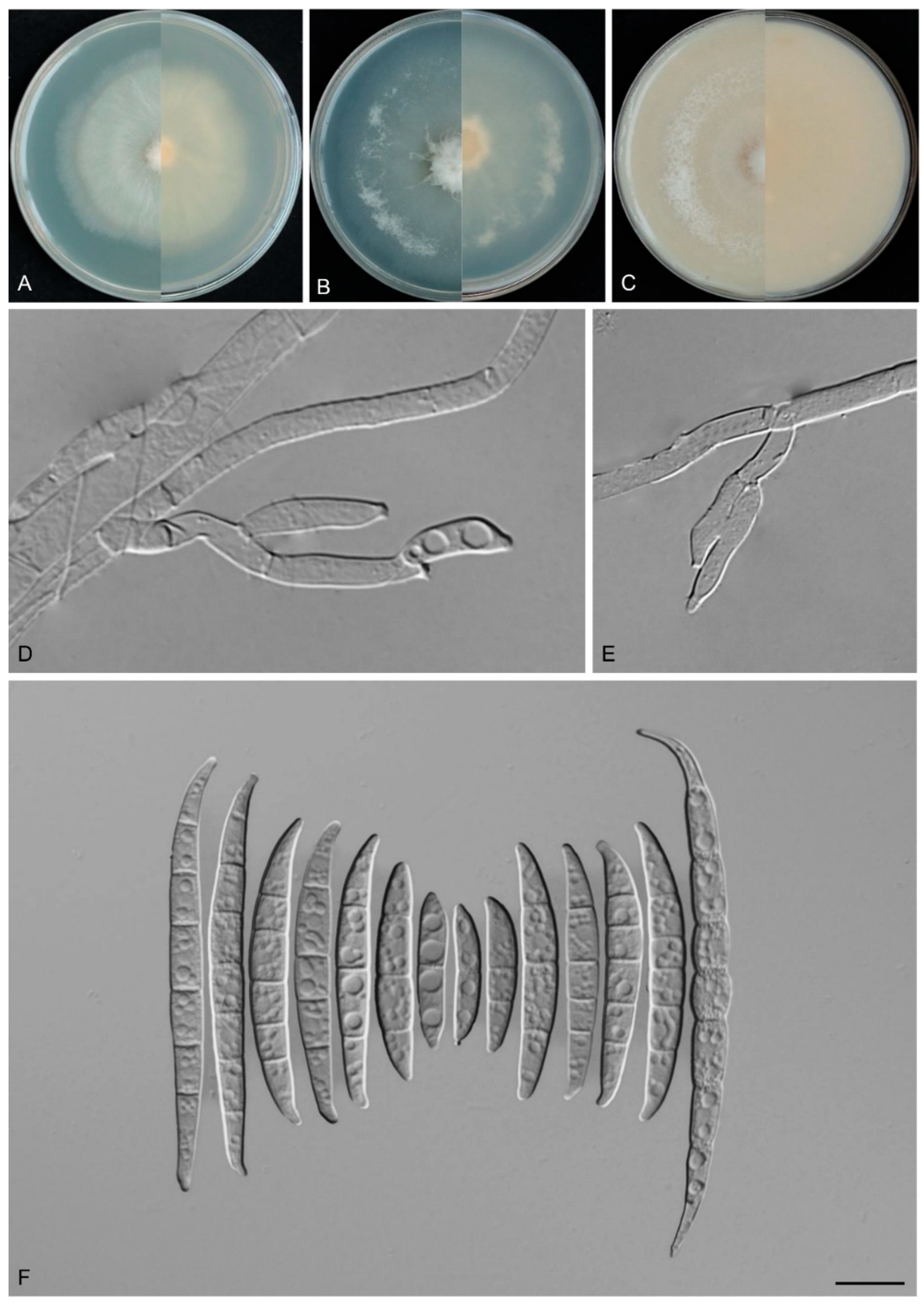
2.6. Morphological Observations

To study the morphological characteristics of fungal isolates, both macroscopic and microscopic features were examined [4]. Colony morphology was observed using three culture media: PDA, synthetic nutrient-poor agar (SNA; 20 g/L agar, 1.0 g/L KH2PO4, 1.0 g/L KNO3, 0.5 g/L MgSO4·7H2O, 0.5 g/L KCl, and 0.2 g/L glucose; pH = 7.0), and oatmeal agar (OA; 30.0 g/L oatmeal flakes and 15.0 g/L agar). The isolates were initially inoculated onto PDA plates and incubated at 25 °C for seven days. Mycelial plugs (approximately 5 × 5 mm) were taken from the margins of the colonies and transferred to the three media. After incubation in the dark for seven days, colony characteristics such as pigmentation and odor were recorded [4].
For microscopic observations, mycelial plugs were transferred to carnation leaf agar (CLA) [75] and incubated at 25 °C under a 12 h near-ultraviolet light/dark cycle for 7–14 days. Sporodochia were initially examined and photographed using an Olympus SZ60 stereomicroscope (Olympus Corporation, Tokyo, Japan). Subsequently, water was used as the mounting medium, and the structures were observed using an Olympus BX53 microscope (Olympus Corporation, Tokyo, Japan) with differential interference contrast (DIC) optics. The following structures were observed: sporodochia and sporodochial conidiophores, phialides, and conidia; aerial conidiophores, phialides, and conidia; and chlamydospores [4,76].

3. Results

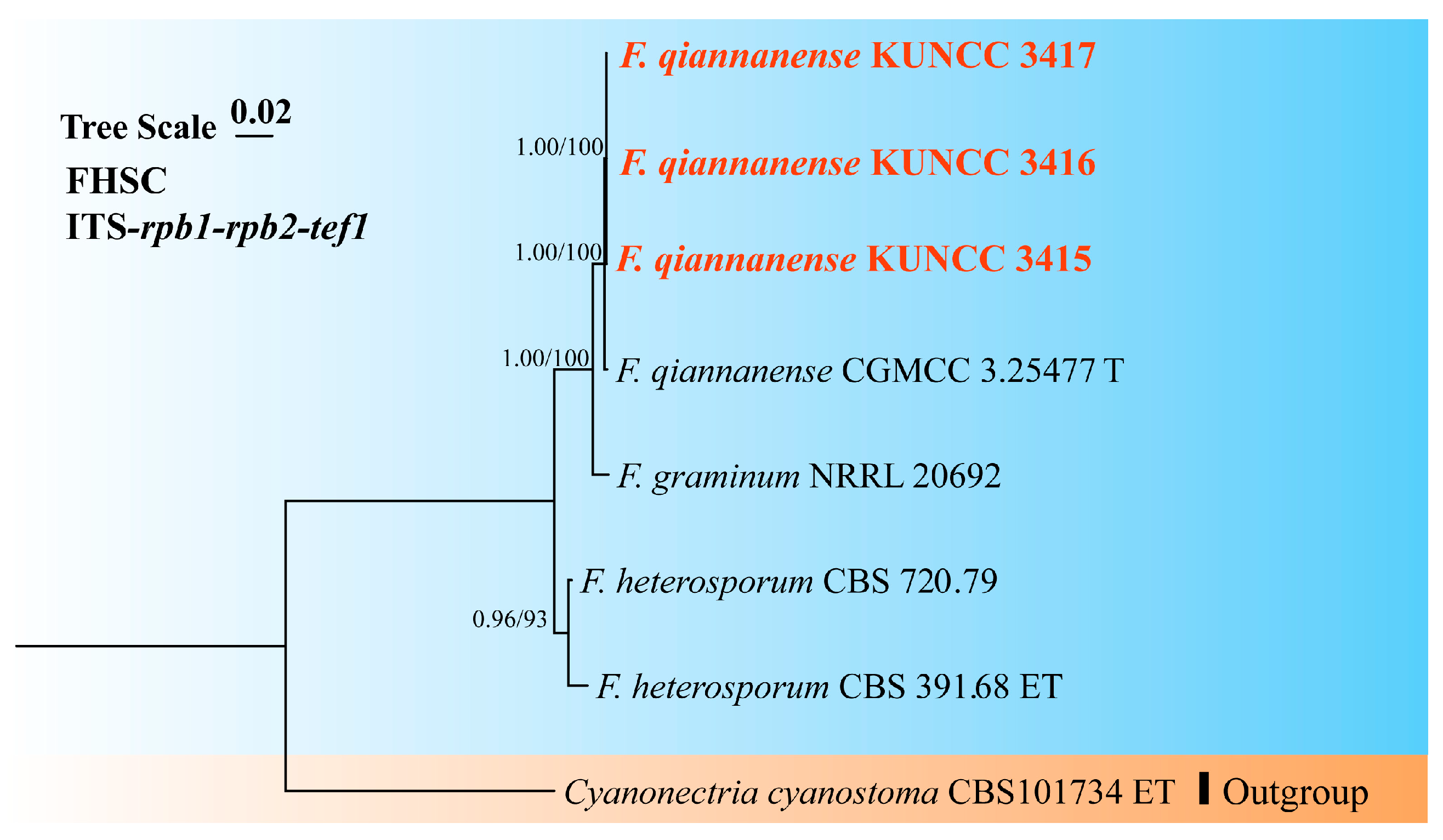
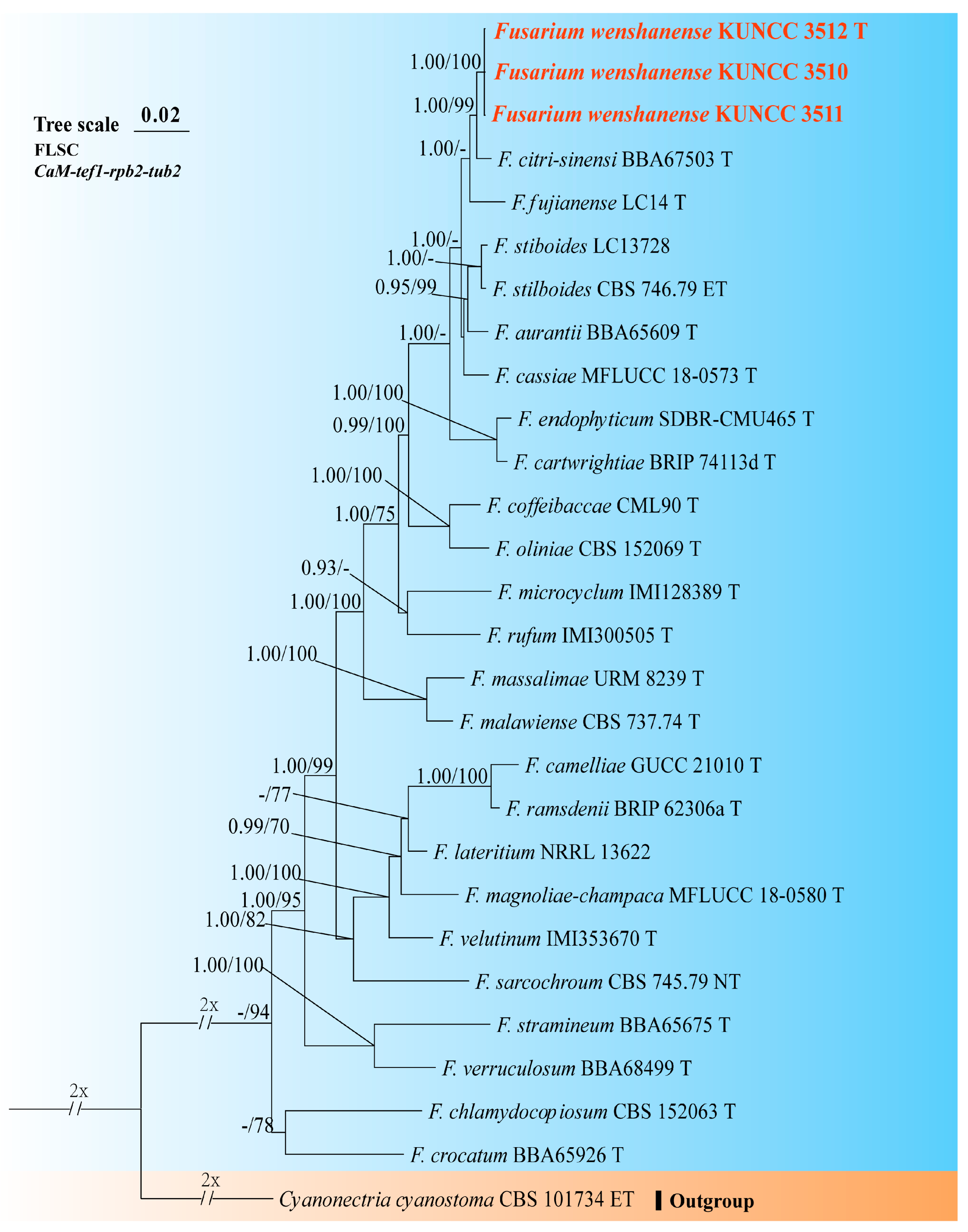
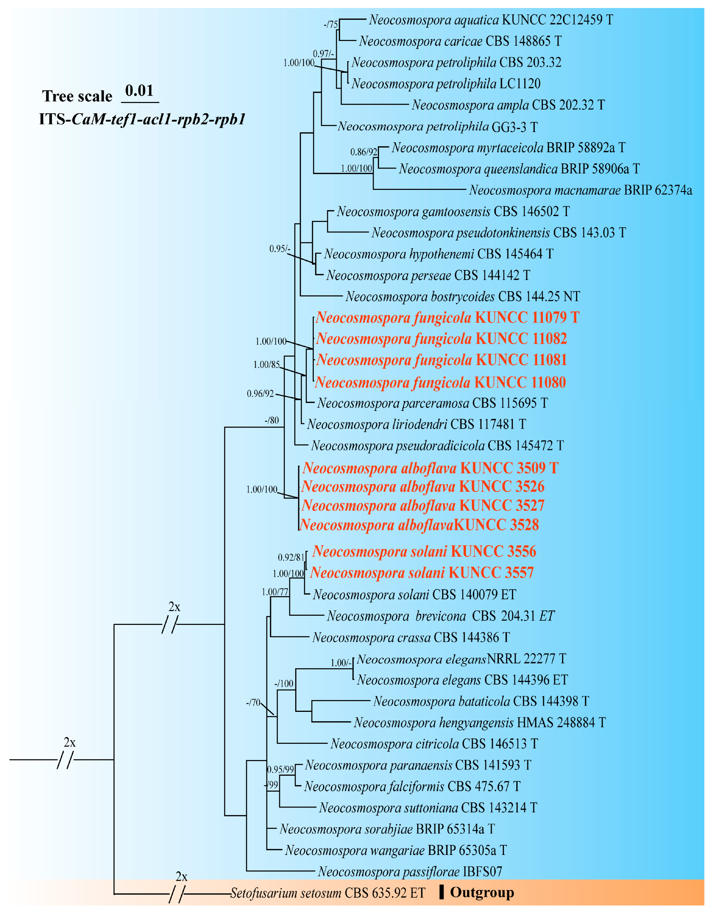
3.1. Molecular Phylogeny

Preliminary phylogenetic analysis based on the combined tef1, rpb1, and rpb2 loci revealed a tree topology that broadly resembled the phylogeny proposed by Crous et al. [4]. Among the 22 representative strains, 10 were assigned to three species complexes within the Fusarium (i.e., FHSC, FIESC, and FLSC), whereas the remaining strains were grouped within the Neocosmospora (Supplementary Figure S1). Subsequently, phylogenetic analyses were conducted separately for each Fusarium species complex and for Neocosmospora, using different datasets and the best-fit substitution models selected for each gene partition.
A phylogenetic tree of the FHSC was constructed based on a concatenated dataset of ITS, tef1, rpb1, rpb2, and tub2 sequences from nine strains (Figure 1). The resulting tree topology was consistent with previous studies [5,6]. Similarly, the FIESC tree was generated using concatenated CaM, rpb2, and tef1 sequence data from 68 strains (Figure 2), and similar phylogenetic results were observed [6]. For the FLSC, a phylogenetic tree was inferred by combining CaM, tef1, rpb2, and tub2 sequences from 28 strains (Figure 3), and its topology was similar to that of Wang et al. [23]. Finally, phylogenetic analyses of Neocosmospora were performed based on combined sequences of acl1, CaM, ITS, rpb1, rpb2, and tef1 from 42 strains (Figure 4), which were topologically consistent with previous studies [4,5]. Multigene phylogenetic analyses and GCPSR analyses (Figure 5), combined with morphological characteristics, revealed that the 22 isolates represent seven species, including five new and two known species. Specifically, one species was assigned to the FHSC (Figure 1), two species to the FIESC (Figure 2), one species to the FLSC (Figure 3), and three species to Neocosmospora (Figure 4).
Furthermore, single-locus phylogenetic trees were constructed for the three species complexes within Fusarium and for the Neocosmospora, respectively (Supplementary Figures S2–S5). Phylogenetic analyses based on single-gene loci showed that rpb2 and tef1 provided higher resolution for species delimitation within three Fusarium species complexes (FHSC, FIESC, and FLSC) and Neocosmospora. Specifically, in the FHSC, both tef1 and rpb2 achieved complete species resolution (100%, 3/3). In the FIESC, tef1 resolved all 63 species (100%, 63/63), whereas rpb2 resolved 50 species (86%, 50/58). In the FLSC, both loci achieved full resolution (22 of 22). In Neocosmospora, tef1 resolved 29 species (94%, 29/32), while rpb2 resolved 23 species (77%, 23/30).

3.2. Genomic Features

The genome sizes of the Fusarium and Neocosmospora species used in this study ranged from 34 to 54 Mb. Geejayessia zealandicum NRRL 22465 had the smallest assembly size (34 Mb), while N. vasinfecta NRRL 22166 had the largest assembly size (54 Mb). The BUSCO completeness of assemblies was in the range of 93.1% to 97.6%, with duplicated gene content ranging from 0.2% to 0.8%. The total number of predicted protein-coding genes ranged from 9479 in G. zealandicum NRRL 22465 to 14826 in N. vasinfecta NRRL 22166. Smaller genomes tended to have fewer predicted protein-coding genes (Supplementary Table S5).

3.3. Results of Phylogenomic Analysis
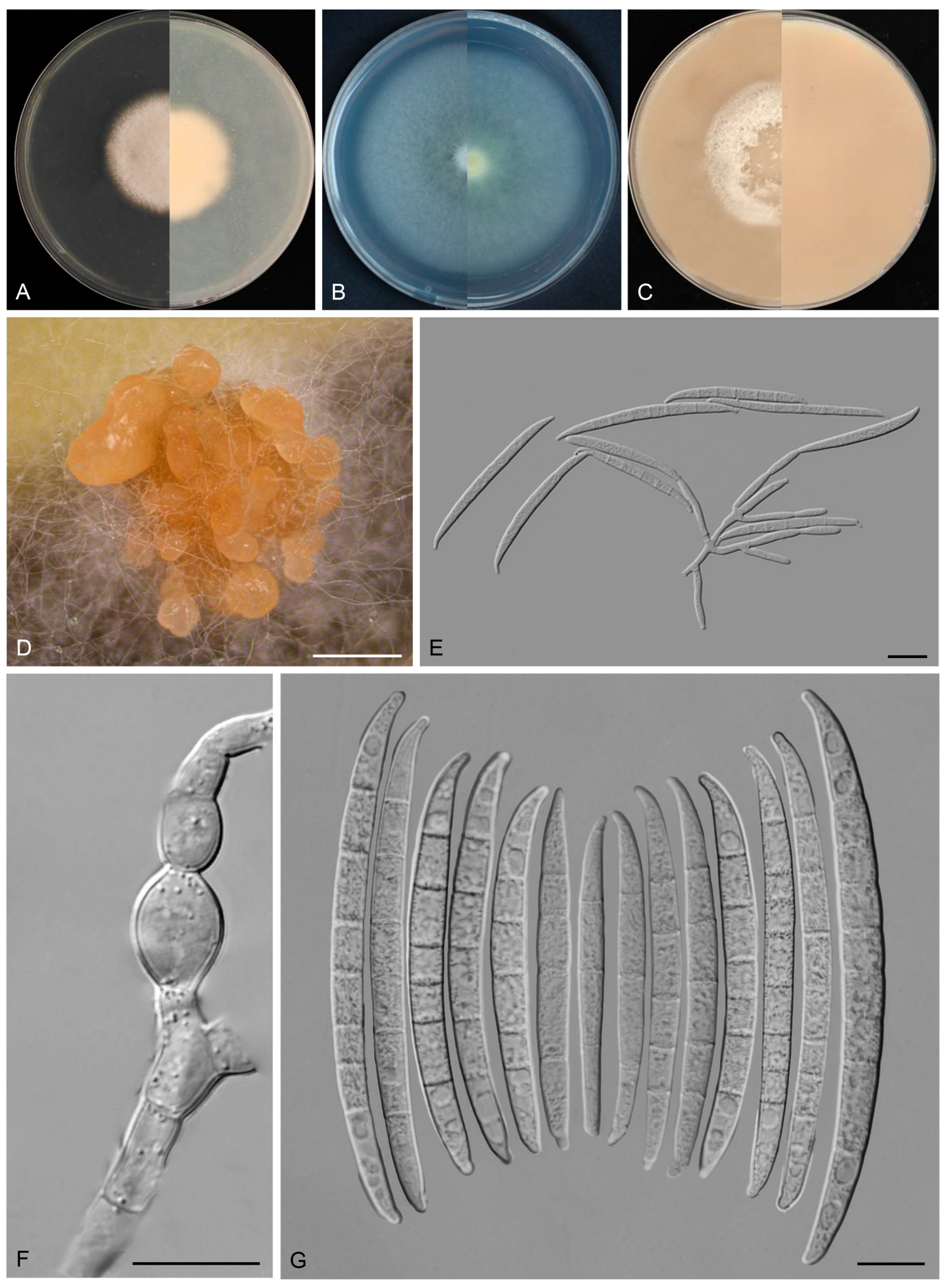
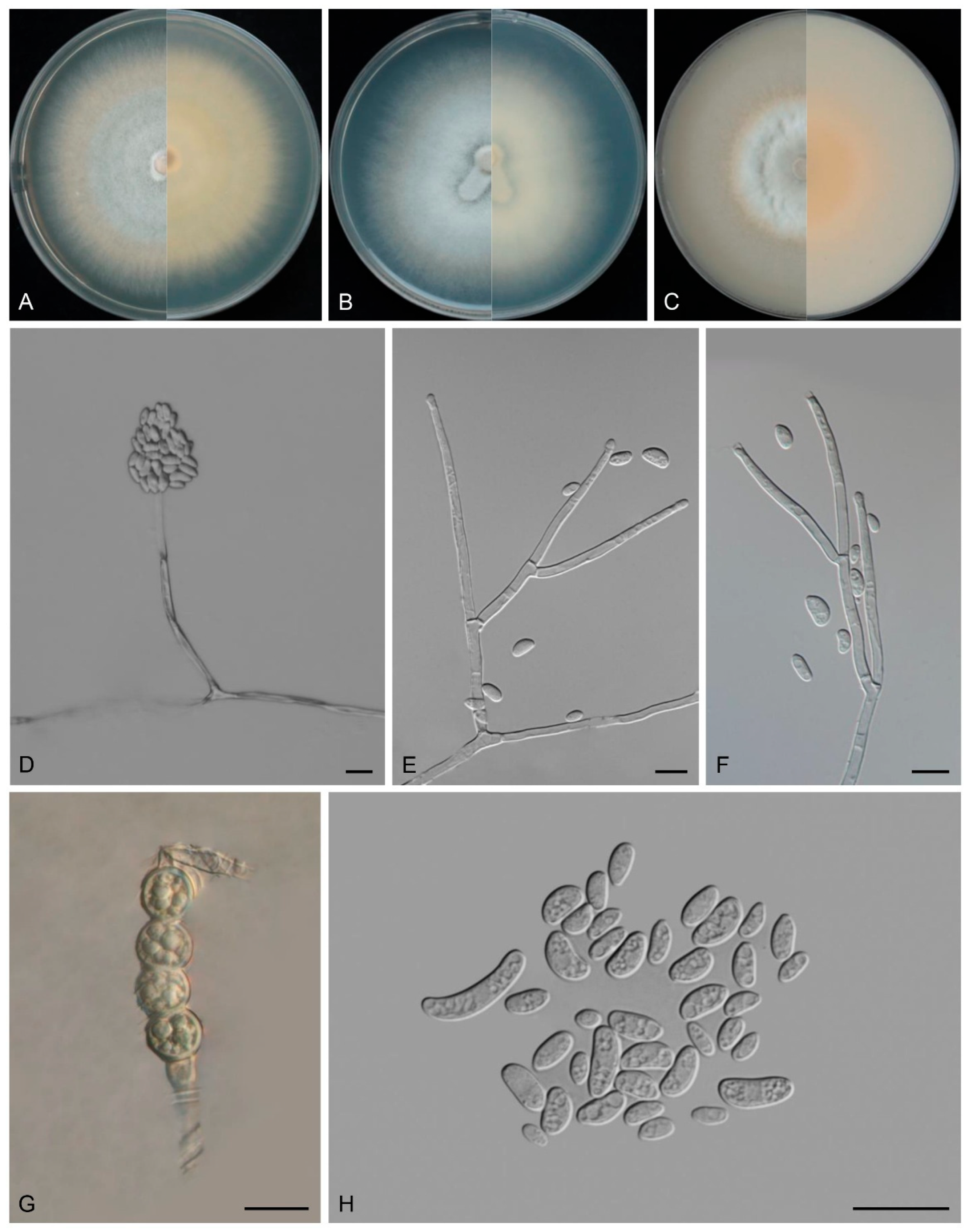
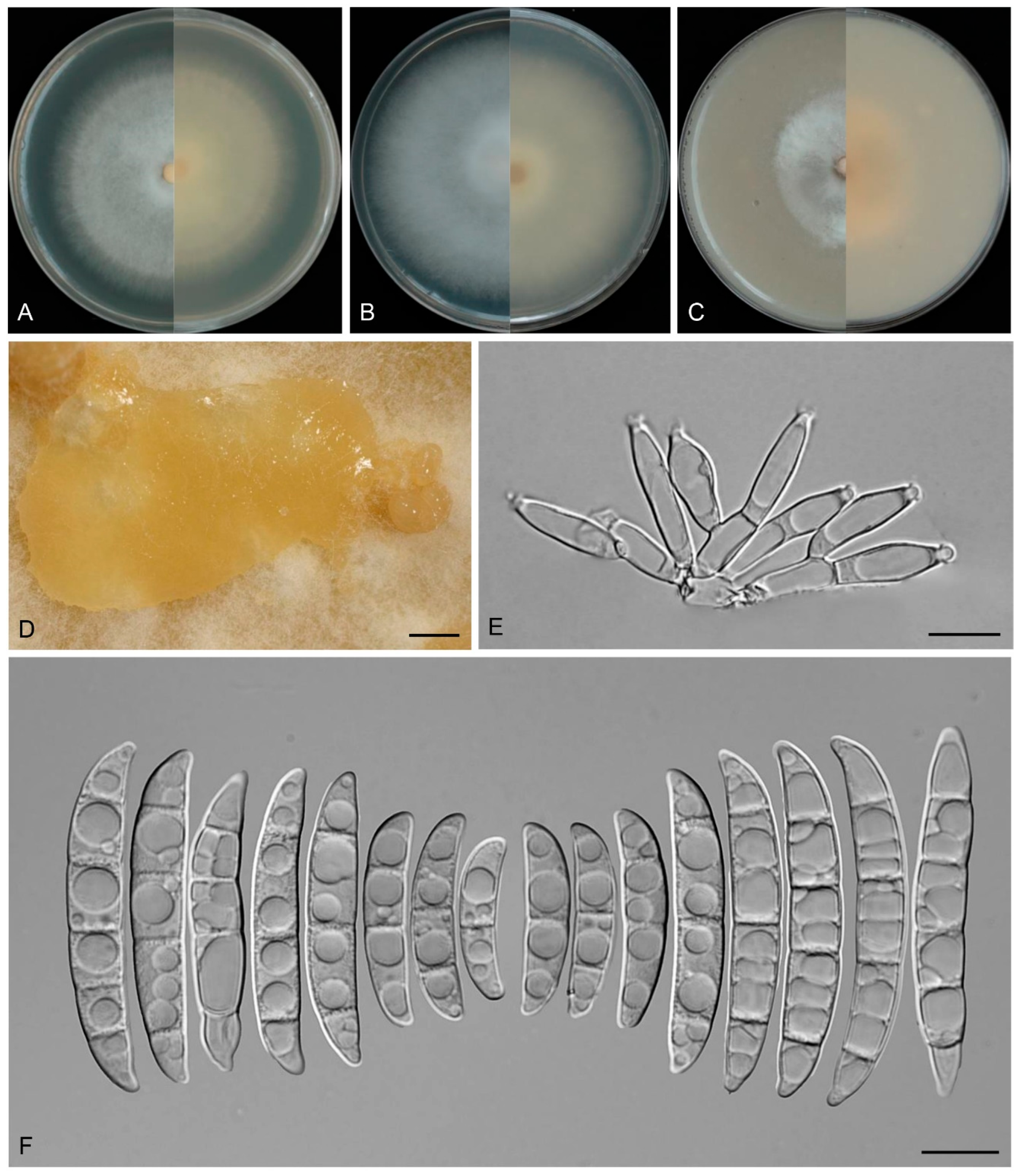
We sequenced the genomes of the seven species described in this study and included an additional 15 published genomes retrieved from the NCBI Datasets repository (https://www.ncbi.nlm.nih.gov/datasets/, accessed on 16 March 2025) for phylogenomic and comparative analysis. Using OrthoFinder v2.5.2 [69], a total of 259,957 genes from 21 genomes were clustered into 256,187 orthologous groups, with 3,770 genes remaining unclustered. Among these, 6,055 orthologous groups were shared across all 21 species, including 4,941 single-copy orthologous groups (Supplementary Table S6). An ML phylogenetic tree was constructed based on these 4,941 clusters of orthologous proteins (Figure 6), with G. zealandicum NRRL 22465 used as the outgroup. The resulting phylogenomic tree resolved into two well-supported major clades at the genus level (excluding the outgroup): Fusarium and Neocosmospora. Within Fusarium, four phylogenetically distinct subclades were identified, each representing a well-recognized species complex, viz., the FIESC, FHSC, FTSC, and FLSC. The species described in this study were distributed among these clades as follows: F. qiannanense KUNCC 3417 was placed within the FHSC; F. puerense KUNCC 3505 T and F. dracaenophilum KUNCC 3495 T within the FIESC; F. wenshanense KUNCC 3512 T within the FLSC; and N. fungicola KUNCC 11079 T, N. alboflava KUNCC 3509 T, and N. solani KUNCC 3556 within the Neocosmospora clade. The genome-based phylogenetic tree exhibited a topology highly consistent with that of the multilocus tree (Figure 1, Figure 2, Figure 3, Figure 4 and Figure 6).

3.4. Taxonomy

Fusarium dracaenophilum Y.B. Wang, Q. Fan & Zhu L. Yang, sp. nov.
Fungal Names No.: FN 572883
Etymology: The epithet “dracaenophilum” refers to the fungus’s ecological association with its host plant, Dracaena cambodiana Pierre ex Gagnep.
Type: CHINA, Yunnan Province, Xishuangbanna Dai Autonomous Prefecture, 100.77° E, 22° N, alt. 550 m, from the healthy leaves of D. cambodiana, June 2022, Z.Y. Tian (holotype HKAS 135090, ex-type culture KUNCC 3495).
Description: Conidiophores arising from aerial mycelium 7.9–16.0 μm tall, simple or rarely irregularly branched, bearing whorls of 2–3 phialides at the apex. Aerial conidiogenous cells monophialidic, sometimes reduced to solitary forms and laterally borne on hyphae, subulate to subcylindrical, smooth- and thin-walled, (7.7–)8.0–17.4(–18.6) × (2.8–)2.9–3.9(–4.0) μm (av. 12.1 × 3.4 μm), apical collarettes absent and periclinal thickening inconspicuous. Aerial macroconidia falcate to navicular, hyaline, smooth- and thin-walled, almost straight to slightly dorsiventrally curved, apical cell blunt to slightly curved, basal cell stunted to well-developed, foot-shaped, (1–)3–7-septate, predominantly 5-septate; 1-septate conidia: 16.0–27.0(–31.0) × 2.0–4.0 μm (av. 19.6 × 3.0 μm, n = 10); 2-septate conidia: (19.5–)21.5–30.0(–33.0) × 3.0–4.5 μm (av. 25.0 × 4.0 μm, n = 7); 3-septate conidia: (25.0–)26.5–37.5 × (3.0–)3.0–4.5(–5.0) μm (av. 30.5 × 4.0 μm, n = 20); 4-septate conidia: (30.0–)31.5–40.0(–41.5) × 4.0–5.0 μm (av. 35.5 × 4.0 μm, n = 20); 5-septate conidia: (31.0–)33.3–54.1(–62.0) × 3.5–5.5(–6.0) μm (av. 44.0 × 4.5 μm, n = 30); 6-septate conidia: 51.0–65.5(–67.0) × 3.5–6.0 μm (av. 58.0 × 4.0 μm, n = 20); 7-septate conidia: (50.5–)52.5–71.0(–74.0) × 4.0–5.5 μm (av. 60.5 × 4.5 μm, n = 20); overall: 16.0–71.0(–74.0) × 2.0–6.0 μm (av. 40.0 × 4.0 μm, n = 127). Sporodochia and chlamydospores not observed.
Culture characteristics: Colonies on PDA grow rapidly, exhibiting 5.5–6.0 cm diam. in seven days at 25 °C, moist, flat, aerial mycelium absent, colony margin regular, surface white to cream, reverse pale white to cream. On SNA reaching 6.5–7.0 cm diam. in seven days, moist, aerial mycelium absent, colony margin regular, surface white to cream, reverse pale white to cream. On OA reaching 6.0–6.5 cm diam. in seven days, flat, aerial mycelium absent, moist at the center, velvety at the margin, colony margin regular, surface white to cream, reverse white to cream.
Additional specimens examined: CHINA, Yunnan Province, Xishuangbanna Dai Autonomous Prefecture, 100.77° E, 22° N, alt. 550 m, from the healthy leaves of D. cambodiana, June 2022, Z.Y. Tian (culture KUNCC 3504).
Note: Phylogenetic analyses based on the concatenated dataset of CaM, tef1, rpb1, and rpb2 loci (Figure 2) and genomic datasets (Figure 6) resolved the isolates representing F. dracaenophilum as a strongly supported monophyletic clade within the FIESC (BS = 100%, PP = 1.00 for multigene phylogenetic trees, BS = 100% for phylogenomic tree). Fusarium dracaenophilum is closely related to F. weifangense, F. caulendophyticum, and F. citri but can be distinguished by sequence differences of 28 bp, 36 bp, and 71 bp in the three-locus dataset, respectively. Morphologically, F. dracaenophilum can be distinguished from related species by its larger macroconidia (16.0–74.0 × 2.0–6.0 μm in F. dracaenophilum vs. 26.5–49.4 × 4.1–7.1 μm in F. weifangense, 5.0–40.5 × 3.0–5.5 μm in F. citri, and 10.0–47.0 × 2.0–4.5 μm in F. caulendophyticum) and more septation (1–7-septate in F. dracaenophilum vs. 3–7-septate in F. weifangense, 3–5-septate in F. caulendophyticum, and 1–5-septate in F. citri) [5,6,61]. Furthermore, the PHI test indicated no significant recombination (P = 0.736) between F. dracaenophilum and its closely related taxa (Figure 5A). Thus, F. dracaenophilum is hereby described as a new species within the FIESC.
Fusarium puerense Y.B. Wang, Q. Fan & Zhu L. Yang, sp. nov.
Fungal Names No.: FN 572884
Etymology: Named after the city, Pu’er, where the holotype was collected.
Type: CHINA, Yunnan Province, Pu’er city, 101.51° E, 23.35° N, alt. 709 m, from the symptomatic tissues of Musa sp., July 2023, Q. Fan (holotype HKAS 135091, ex-type culture KUNCC 3505).
Description: Aerial conidiophores and conidia were not detected during observation. Sporodochia cream to yellowish, translucent, formed densely on carnation leaves and on the agar. Sporodochial conidiophores densely, irregularly branched, 8.0–13.0 × 3.0–4.0 μm, bearing apical whorls of 1–2 phialides. Sporodochial conidiogenous cells monophialidic, flask-shaped, 9.0–15.5 × 2.5–4.5 μm, smooth- and thin-walled, apical collarettes absent and periclinal thickening inconspicuous. Sporodochial microconidia absent. Sporodochial macroconidia falcate, hyaline, smooth- and thin-walled, straight to slightly dorsiventrally curved, broadest at the middle portion, tapering towards both ends, apical cell blunt to slightly curved, basal cell stunted to well-developed, foot-shaped, 3–7-septate, predominantly 5-septate; 3-septate conidia: 23.0–40.5 × 3.5–5.0 μm (av. 32.0 × 4.0 μm, n = 10); 4-septate conidia: 35.0–69.0(–73.5) × 3.5–5.0 μm (av. 44.0 × 4.0 μm, n = 15); 5-septate conidia: (54.0–)56.0–78.0 × 4.0–5.0 μm (av. 67.5 × 4.5 μm, n = 20); 6-septate conidia: (64.0–)67.0–80.0 × 4.5–5.0 μm (av. 76.0 × 4.5 μm, n = 15); 7-septate conidia: 76.0–88.0 × 4.0–5.0 μm (av. 82.0 × 4.5 μm, n = 10); overall: 23.0–88.0 × 4.0–5.0 μm (av. 60.0 × 4.0 μm, n = 70). Chlamydospores not observed.
Culture characteristics: Colonies on PDA grow rapidly, exhibiting 4.5–5.5 cm diam. in seven days at 25 °C, cottony, flat, aerial mycelium abundant, colony margin regular, surface white, reverse white to cream. On SNA reaching 6.5–7.0 cm diam. in seven days, flat, aerial mycelium scant, moist, colony margin regular; surface white, reverse white to cream. On OA reaching 5.5–6.0 cm diam. in seven days, dense, with abundant aerial mycelium, surface pale orange in the center, white at the margin, reverse pale orange.
Additional specimens examined: CHINA, Yunnan Province, Xishuangbanna Dai Autonomous Prefecture, 100.77° E, 22° N, alt. 550 m, from the healthy leaves of D. cambodiana, June 2022, Z.Y. Tian (cultures: KUNCC 3501, KUNCC 3502, and KUNCC 3503).
Note: Phylogenetic analysis based on the concatenated dataset of CaM, tef1, rpb1, and rpb2 loci (Figure 2) and genomic datasets (Figure 6) resolved the isolates representing F. puerense as a monophyletic clade within the FIESC lineage, with strong statistical support (BS = 100%, PP = 1.00 for multigene phylogenetic trees, BS = 100% for phylogenomic tree). Fusarium puerense is closely related to F. ipomoeae and F. caulicola but can be distinguished by sequence differences of 43 bp and 20 bp in the combined dataset, respectively. Morphologically, F. puerense differs from related species in macroconidial size (23.0–88.0 × 4.0–5.0 μm in F. puerense vs. 26.5–57.0 × 3.0–5.0 μm in F. ipomoeae and 14.0–40.5 × 2.0–5.0 μm in F. caulicola) and septation (3–7-septate in F. puerense vs. 1–4-septate in F. caulicola, 3–5-septate in F. ipomoeae) [5,61,62]. Furthermore, the PHI test indicated no significant recombination (P = 0.468) between F. puerense and its closely related taxa (Figure 5B). Thus, F. puerense is introduced as a new species.
Fusarium wenshanense Y.B. Wang, Q. Fan & Zhu L. Yang, sp. nov.
Fungal Names No.: FN 572885
Etymology: Named after the city, Wenshan Zhuang and Miao Autonomous Prefecture, where the holotype was collected.
Type: CHINA, Yunnan Province, Wenshan Zhuang and Miao Autonomous Prefecture, 103.87° E, 23.61° N, alt. 1810 m, from adult of Lepidoptera, July 2023, C.Y. Wei (holotype HKAS 135098, ex-type culture KUNCC 3512).
Description: Aerial conidiophores and conidia were not detected during observation. Sporodochia pale orange to orange, translucent, sparsely formed on carnation leaves and on the agar, often covered with aerial mycelium. Sporodochial conidiophores densely, irregularly branched, 16.2–19.3 × 2.6–3.8 μm, bearing apical whorls of 2–3 phialides, rarely solitary phialides. Sporodochial conidiogenous cells monophialidic, subulate to subcylindrical, (16.5–)18.0–26.5(–28.0) × 1.5–3.0 μm, smooth and thin-walled, apical collarettes absent and periclinal thickening inconspicuous. Sporodochial microconidia absent. Sporodochial macroconidia falcate, hyaline, smooth- and thin-walled, straight to slightly dorsiventrally curved, tapering towards both ends, apical cell blunt to papillate, basal cell stunted to well-developed, foot-shaped, (1–)3–7(–8)-septate, predominantly 5-septate; 1-septate conidia: 14.0–17.0(–18.0) × (3.0–)4.0–4.5 μm (av. 15.0 × 4.0 μm, n = 8); 2-septate conidia: (24.0–)25.5–32.5(–35.0) × (3.0–)4.0–4.5 μm (av. 29.5 × 4.0 μm, n = 6); 3-septate conidia: 35.5–49.0 × 4.0–4.5 μm (av. 42.0 × 4.0 μm, n = 15); 4-septate conidia: 44.0–60.0 × 3.5–5.0 μm (av. 52.0 × 4.0 μm, n = 15); 5-septate conidia: (48.0–)52.0–69.0 ×4.0–5.5 μm (av. 62.0 × 5.0 μm, n =30); 6-septate conidia: (60.0–)61.5–75.6 × 4.0–5.0 μm (av. 69.5 × 5.0 μm, n = 15); 7-septate conidia: 73.5–85.0(–87.0) ×4.5–5.5 μm (av. 78.0 × 5.0 μm, n = 15); 8-septate conidia: (77.0–)81–89(–92.5) × 4.0–5.0 μm (av. 85 × 4.5 μm, n = 6); overall: 14.0–89(–92.5) × (3.0–)4.0–5.0 μm (av. 54.0 × 4.5 μm) (n = 110). Chlamydospores obovoidal, subglobose to globose, hyaline, smooth-walled to slightly roughened, thick-walled, 9.5–18.8 μm, terminal or intercalary, solitary, in pairs or forming chains.
Colonies on PDA exhibiting 3.5–4.0 cm diam. in seven days at 25 °C, aerial mycelium abundant, dense, flat, colony margin regular, surface white to cream, reverse cream. On SNA reaching 6.5–7.0 cm diam. in seven days, aerial mycelium scant, flat, colony margin regular; surface white to cream, reverse white to cream. On OA reaching 4.5–5.0 cm diam. in seven days, moist at center, abundant aerial mycelium at margin, dense, colony margin regular, surface white, reverse white.
Additional specimens examined: CHINA, Yunnan Province, Wenshan Zhuang and Miao Autonomous Prefecture, 103.87° E, 23.61° N, alt. 1810 m, from adult of Lepidoptera, July 2023, C.Y. Wei (cultures: KUNCC 3510 and KUNCC 3511).
Note: Phylogenetic analysis based on the concatenated dataset of CaM, tef1, rpb2, and tub2 loci (Figure 3) and genomic datasets (Figure 6) resolved the representing isolates of F. wenshanense as a monophyletic clade within the FLSC lineage, with strong statistical support (BS = 100%, PP = 1.00 for multigene phylogenetic trees, BS = 100% for phylogenomic tree). Fusarium wenshanense is closely related to F. citri-sinensis and F. fujianense but differs by 25 bp from F. citri-sinensis in the 4-locus (CaM-tef1-rpb2-tub2) dataset and 23 bp from F. fujianense in the 2-locus (tef1-rpb2) dataset (CaM and tub2 sequences are not available for F. fujianense). Morphologically, F. wenshanense can be distinguished from related species by its sporodochial macroconidial size (14.0–92.5 × 3.0–5.0 μm in F. wenshanense vs. 39.7–99.5 × 4.0–7.7 μm in F. citri-sinensis, and 40.2–63.4 × 4.5–6.9 μm in F. fujianense) and septation (1–8-septate in F. wenshanense vs. 3–13-septate in F. citri-sinensis, and 4–6-septate in F. fujianense) [77,78]. Furthermore, the PHI test indicated no significant recombination (P = 1.0) between F. wenshanense and its closely related taxa (Figure 5C). Thus, F. wenshanense is introduced as a new species.
Fusarium qiannanense H. Zhang & Y.L. Jiang, Mycosphere 14(1): 2105 (2023)
Index Fungorum No.: IF900486
Description: Aerial conidiophores were not detected during observation. Aerial conidiogenous cells monophialidic, often reduced to solitary cells laterally borne on hyphae, subulate to subcylindrical, smooth- and thin-walled, 7.0–36.5 × 2.0–5.0 μm (av. 20.5 × 3.5 μm), apical collarettes absent and periclinal thickening inconspicuous. Aerial conidia of two types: microconidia oval to broadly ellipsoidal, straight to slightly curved, hyaline, smooth- and thin-walled, 0(–1)-septate, 0-septate conidia: (8.0–)10.0–18.0(–21.0) × 3.0–5.0(–6.0) μm (av. 14.0 × 4.0 μm, n = 26); 1-septate conidia: (11–)13–18 × 3.0–3.5 μm (av. 16.0 × 4.0, μm, n = 8); macroconidia falcate to navicular, hyaline, smooth- and thin-walled, almost straight to slightly dorsiventrally curved, apical cell blunt or papillate, basal cell stunted to well-developed, foot-shaped, 1–3(–5)-septate, predominantly 1-septate, 1-septate conidia: (14.0–)15.0–26.5(–27.0) × 3.0–4.0 μm (av. 21.0 × 3.5 μm, n = 21); 2-septate conidia: (26–)27.0–37.0(–38.5) × 3.0–4.5 μm (av. 31.5 × 3.5 μm, n = 15); 3-septate conidia: (17.0–)24.0–36.0(–38) × 3.5–4.5 μm (av. 28.5 × 4.0 μm, n = 10); 5-septate conidia: 35.0–48.5 × 4.0–4.5 μm (av. 38.0 × 4.0 μm, n = 4); overall: (14.0–)15.0–48.5 × 3.0–4.5 μm (av. 30.0 × 4.0 μm) (n = 50). Sporodochia and chlamydospores not observed.
Culture characteristics: Colonies on PDA exhibiting 4.5–5.0 cm diam. in seven days at 25 °C, velvety, flat, with abundant aerial mycelium, colony margin regular; surface white to cream, reverse cream. On SNA reaching 3.0–3.5 cm diam. in seven days, flat, aerial mycelium abundant, colony margin regular, surface white, reverse white. On OA reaching 5.0–6.0 cm diam. in seven days, flat, aerial mycelium scant, colony margin regular; surface white to cream, reverse white to cream.
Additional specimens examined: CHINA, Yunnan Province, Kunming city, 103.12° E, 25.31° N, alt. 1543 m, from the asymptomatic sclerotium of Claviceps purpurea, September 2023, M.L. Ding (cultures: KUNCC 3417, KUNCC 3416, and KUNCC 3415).
Note: Phylogenetic analyses revealed that the isolates KUNCC 3417, KUNCC 3416, and KUNCC 3415 clustered together with the type strain CGMCC 3.25477 of F. qiannanense with strong statistical support (BS = 100%, PP = 1.00) (Figure 1). The strains showed high sequence similarity across ITS, tef1, rpb1, and rpb2 regions, showing 99.22% (512/516, 4 gaps), 100% (582/582, no gaps), 99.89% (1739/1741, 2 gaps), and 99.89% (911/912, 1 gap) identity, respectively. Morphologically, these isolates were characterized by monophialidic conidiogenous cells and straight to slightly curved aerial macroconidia. Both molecular and morphological evidence supported the identification of these isolates as F. qiannanense. As F. qiannanense was previously reported only from Rosa roxburghii (Rosaceae), the present study represents a new host record from C. purpurea [5].
Neocosmospora alboflava Y.B. Wang, Q. Fan & Zhu L. Yang, sp. nov.
Fungal Names No.: FN 572886
Etymology: Referring to its colony color, it develops white and pale orange interwoven concentric rings on PDA, SNA, and OA media.
Type: CHINA, Yunnan Province, Wenshan Zhuang and Miao Autonomous Prefecture, 104.39° N, 23.01° E, alt. 1699 m, from asymptomatic Nigelia sp., June 2023, C.Y. Wei (holotype HKAS 135095, ex-type culture KUNCC 3509).
Description: Conidiophores arising from aerial mycelium 20.8–62.3 μm tall, simple to rarely irregularly branched, bearing terminal single phialides or whorls of 2–3 phialides, commonly reduced to solitary phialides borne laterally on hyphae. Aerial conidiogenous cells monophialidic, subulate to subcylindrical, smooth- and thin-walled, (24.5–)36.5–87.0(–93.0) × 2.0–3.0 μm (av. 50.0 × 3.0 μm, n = 28), apical collarettes absent and periclinal thickening inconspicuous. Aerial microconidia arranged in false heads on phialide tips, obovoid to short clavate, straight to slightly curved, hyaline, smooth- and thin-walled, aseptate: 5.0–10.5 × 2.5–5.5 μm (av. 7.0 × 4.0 μm, n = 97). Chlamydospores, subglobose to globose, hyaline to pale yellow brown, smooth-walled to slightly roughened, thick-walled, 7.5–11.0 μm, commonly intercalary, in pairs or forming chains.
Culture characteristics: Colonies on PDA grow rapidly, exhibiting 6.5–7.0 cm diam. in seven days at 25 °C, velvety, flat, aerial mycelium abundant, exhibiting white and pale orange interwoven concentric rings, surface pale orange at margin, white to cream at center, reverse cream to pale orange, colony margin regular. On SNA reaching 6.0–6.5 cm diam. in seven days, flat, exhibiting white and pale orange interwoven concentric rings, surface pale orange at margin, white to cream at center, reverse cream, colony margin regular. On OA reaching 4.5–5.0 cm diam. in seven days, dense, exhibiting white and pale orange interwoven concentric rings, surface pale orange at margin, white to cream at center, reverse cream to pale orange, colony margin regular.
Additional specimens examined: CHINA, Yunnan Province, Wenshan Zhuang and Miao Autonomous Prefecture, 104.39° N, 23.01° E, alt. 1699 m, from asymptomatic Nigelia sp., June 2023, C.Y. Wei (cultures: KUNCC 3526, KUNCC 3527, and KUNCC 3528).
Note: Phylogenetic analysis based on the concatenated dataset of ITS, CaM, acl1, tef1, rpb1, and rpb2 loci (Figure 4) and genomic dataset (Figure 6) resolved the representative isolates of N. alboflava as a well-supported monophyletic clade within Neocosmospora (BS = 80%, PP = 0.80 for multilocus phylogenetic trees, BS = 100% for phylogenomic tree). Neocosmospora alboflava is closely related to N. parceramosa, N. liriodendri, and N. pseudoradicicola, but differs by sequence differences of 34 bp and 55 bp from N. liriodendri and N. pseudoradicicola in the 6-locus (ITS-CaM-acl1-tef1-rpb1-rpb2) dataset, respectively, and differs by 54 bp from N. parceramosa in the 5-locus (ITS-CaM-acl1-tef1-rpb2) dataset (rpb1 sequence is not available for N. parceramosa). Morphologically, N. alboflava can be distinguished from related species based on phialides size (24.5–93.0 × 2.0–3.0 μm in N. alboflava vs. 39.5–78 × 2–4.5 μm in N. pseudoradicicola, 40–71.5 × 2.5–5 μm in N. liriodendri, and 35–74 × 2–4 μm in N. parceramosa) and microconidia septation (0-septate in N. alboflava vs. 0(–1)-septate in N. parceramosa, N. liriodendri, and N. pseudoradicicola) [8]. In addition, the PHI test detected no significant recombination (P = 0.887) between N. alboflava and its closely related taxa (Figure 5D). Thus, N. alboflava is introduced as a new species.
Neocosmospora fungicola Y.B. Wang, Q. Fan & Zhu L. Yang, sp. nov.
Fungal Names No.: FN 572887
Etymology: Named after its isolation from the fungus.
Type: CHINA, Zhejiang Province, Hangzhou City, 120.27° E, 30.25° N, alt. 90 m, isolated from the asymptomatic Ophiocordyceps sp., June 2022, L.Y. Xie (holotype HKAS 126202, ex-type culture KUNCC 11079).
Description: Conidiophores on the aerial mycelium straight or flexuous, smooth- and thin-walled, mostly simple or irregularly branched, bearing phialides dichotomously at the apex, rarely solitary phialides. Aerial conidiogenous cells monophialidic, sometimes reduced to solitary phialides borne laterally on hyphae, subcylindrical, smooth- and thin-walled, 41.0–62.0(–65.5) × 2.0–3.0 μm (av. 49.0 × 2.0 μm), apical collarettes absent and periclinal thickening inconspicuous. Aerial conidia obovoid to short clavate, straight to slightly curved, hyaline, smooth- and thin-walled, 0–1-septate, 0-septate conidia: 5.5–10.0(–12) × 2.0–5.0 μm (av. 8.0 × 3.0 μm, n = 33); 1-septate conidia: 9.0–12.0 × 3.0–4.0 μm (av. 10.0 × 3.5 μm, n =14). Sporodochia pale honey, translucent, formed sparsely on carnation leaves and on the agar. Sporodochial conidiophores densely, irregularly branched, bearing apical whorls of 3–5 phialides. Sporodochial conidiogenous cells monophialidic, subulate to subcylindrical, 14.0–19.0 × 2.5–4.5 μm (av. 16.0 × 3.5 μm), smooth and thin-walled, apical collarettes absent and periclinal thickening inconspicuous. Sporodochial macroconidia falcate, hyaline, smooth- and thin-walled, straight to slightly dorsiventrally curved, broadest at the middle portion, tapering towards both ends, apical cell blunt to slightly curved, basal cell stunted to well-developed, foot-shaped, (3–)4–6(–7)-septate, predominantly 5-septate; 3-septate conidia: (33.0–)45.1–48.0 ×4.0–4.5 μm (av. 43.0 × 4.5 μm, n = 10); 4-septate conidia: 44.0–56.0 × 4.0–5.0 μm (av. 50.0 × 5.0 μm, n = 18); 5-septate conidia: 49.0–60.0 × 4.0–6.0 μm (av. 54.5 × 5.0 μm, n = 20); 6-septate conidia: 54.0–61.0 × 4.0–5.0 μm (av. 59.0 × 4.5 μm, n = 18); 7-septate conidia: 58.0–65.0 × 4.5–5.0 μm (av. 61.5 × 5 μm, n = 9); overall: (33.0–)45.1–65.0 × 4.0–6.0 μm (av. 54.0 × 4.5 μm, n = 75). Chlamydospores not observed.
Culture characteristics: Colonies on PDA grow rapidly, exhibiting 7.0–8.0 cm diam. in seven days at 25 °C, velvety, flat, aerial mycelium abundant, colony margin regular, surface white to cream, reverse white to cream. On SNA reaching 7.0–8.0 cm diam. in seven days, velvety, flat, fluffy, colony margin regular, surface white to cream, reverse white to cream. On OA reaching 4.5–5.0 cm diam. in seven days, velvety, flat, moist at center, colony margin regular; surface white to cream, reverse white to cream.
Additional specimens examined: CHINA, Zhejiang Province, Hangzhou City, 120.27° E, 30.25° N, alt. 90 m, from the Ophiocordyceps sp., June 2022, L.Y. Xie (cultures: KUNCC 11080, KUNCC 11081, and KUNCC 11082).
Note: Phylogenetic analysis based on the concatenated dataset of ITS, CaM, acl1, tef1, rpb1, and rpb2 loci (Figure 4) and genomic datasets (Figure 6) resolved the representing isolates of N. fungicola as a monophyletic clade within the Neocosmospora, which is statistically well supported (BS = 85%, PP = 1.00 for multigene phylogenetic trees, BS = 100% for phylogenomic tree). Neocosmospora fungicola is closely related to N. parceramosa, N. liriodendri, and N. pseudoradicicola but can be distinguished by sequence differences of 29 bp and 53 bp from N. liriodendri and N. pseudoradicicola in the 6-locus (ITS-CaM-acl1-tef1-rpb1-rpb2) dataset, respectively, and 16 bp from N. parceramosa in the 5-locus (ITS-CaM-acl1-tef1-rpb2) dataset (rpb1 sequences are not available for N. parceramosa). Morphologically, N. fungicola can be distinguished from related species based on the size of phialides (24.5–93.0 × 2.0–3.0 μm in N. alboflava vs. 39.5–78 × 2–4.5 μm in N. pseudoradicicola, 40–71.5 × 2.5–5 μm in N. liriodendri, and 35–74 × 2–4 μm in N. parceramosa) and chlamydospores (chlamydospores absent in N. alboflava vs. chlamydospores present in N. parceramosa, N. liriodendri, and N. pseudoradicicola) [8]. Furthermore, the PHI test indicated no significant recombination (P = 0.887) between N. fungicola and its close relatives (Figure 5D). Thus, N. fungicola is introduced as a new species.
Neocosmospora solani (Mart.) L. Lombard & Crous, Stud. Mycol. 80: 228 (2015)
Index Fungorum No.: IF810964
Description: Sporodochia champagne, translucent, formed commonly on carnation leaves, rarely on aerial and substrate mycelium. Sporodochial conidiophores densely, irregularly branched, 10.0–15.0 × 3.5–5.0 μm, bearing apical whorls of 2–3 phialides, solitary phialides rarely. Sporodochial phialides monophialidic, doliiform to subcylindrical, 15.5–23.5 × 4.0–6.0 μm, smooth- and thin-walled, apical collarettes absent and periclinal thickening inconspicuous. Sporodochial microconidia absent. Sporodochial macroconidia lunate, hyaline, smooth- and thin-walled, straight to slightly dorsiventrally curved, broadest at the middle portion, tapering towards both ends, apical cell blunt to slightly curved, basal cell stunted to well-developed, foot-shaped, 1–4(5)-septate, predominantly 3-septate; 1-septate conidia: 20.5–26.5 × 4.0–6.5 μm (av. 23.0 × 5.5 μm, n = 15); 2-septate conidia: 25.5–29.0 × 5.0–5.5 μm (av. 27.0 × 5.4 μm, n = 12); 3-septate conidia: 38.5–44.0 × 6.0–7.5 μm (av. 42.0 × 7.0 μm, n = 32); 4-septate conidia: 39.5–48.0(–51.5) × 5.5–8.0 μm (av. 44.0 × 7.0 μm, n = 30); 5-septate conidia: 42.0–47.0 × 6.0–7.5 μm (av. 44.0 × 7.0 μm, n = 8). Chlamydospores not observed.
Culture characteristics: Colonies on PDA grow rapidly, exhibiting 7.0–7.5 cm diam. in seven days at 25 °C, cottony, flat, with abundant aerial mycelium, colony margin regular, surface white to cream, reverse white to cream. On SNA reaching 7.5–8.6 cm diam. in seven days, flat, aerial mycelium abundant, colony margin regular, surface white to cream, reverse cream. On OA reaching 4.5–5 cm diam. in seven days, dense, colony margin regular, surface white to cream, reverse cream to pale orange.
Additional specimens examined: CHINA, Yunnan Province, Xishuangbanna Dai Autonomous Prefecture, 100.77° E, 22° N, alt. 550 m, from the healthy leaves of D. cambodiana, June 2022, Z.Y. Tian (cultures: KUNCC 3556 and KUNCC 3557).
Note: Phylogenetic analyses demonstrated that the isolates KUNCC 3556 and KUNCC 3557 clustered with the ex-epitype strain CBS 140079 of Neocosmospora solani with strong statistical support (BS = 100%, PP = 1.00, Figure 4). Sequence similarity was very high across multiple loci, with ITS, CaM, acl1, tef1, rpb1, and rpb2 showing 99.82% (558/559, 1 gap), 100% (562/562, no gaps), 99.66% (589/591, 2 gaps), 99.57% (693/696, 3 gaps), 100.00% (1312/1312, no gaps), and 99.64% (834/837, 3 gaps) identity, respectively. Morphologically, these isolates were similar, characterized by orange sporodochia, cylindrical to subcylindrical conidiogenous cells, and straight to slightly curved aerial macroconidia. Both molecular and morphological evidence support the identification of these isolates as N. solani [8]. Therefore, this study reports a new host record for this species isolated from D. cambodiana.

4. Discussion

4.1. Species Diversity of Fusarium and Neocosmospora

Fusarium and Neocosmospora are recognized as the most species-rich and ecologically diverse genera within the family Nectriaceae [3,30]. Currently, Fusarium includes over 420 accepted species [4,6,43], and Neocosmospora contains more than 140 species [3,25] (https://indexfungorum.org/Names/Names.asp, accessed on 28 May 2025).
Based on the concept of Fusarium s. s. (=Gibberella, also known as the “F3 clade”), this study integrated morphological characteristics, multilocus phylogenetic analyses, and genomic data to investigate the species diversity within fusarioid fungi. As a result, five new species from southeastern and southwestern China are described. In addition, this study also reports new host records for F. qiannanense and N. solani, isolated from D. cambodiana and the sclerotia of C. purpurea, respectively. While most species described in this study were isolated from plants or fungi, F. wenshanense was obtained from the cadaver of a lepidopteran insect. Nevertheless, its entomopathogenic potential remains unconfirmed due to a lack of direct evidence and experimental validation.

4.2. Diversity and Potential of Endophytic Fusarioid Fungi

Endophytic fungi are those that inhabit plant tissues without causing visible disease symptoms in their hosts [79]. Although members of Fusarium and Neocosmospora are widely recognized for their pathogenicity in plants [6,23,30,39,80,81], growing evidence indicates that their endophytic members have notable ecological and biotechnological potential, especially in natural product discovery and sustainable agriculture. For instance, endophytic N. solani has been reported to produce various bioactive secondary metabolites with anticancer, antimicrobial, and antioxidant activities, including jasmonates, camptothecin, and naphthoquinones [82]. Similarly, F. oxysporum strain Fo47, a well-characterized endophyte, has been employed as a biocontrol agent that induces host defense responses via the accumulation of salicylic acid (SA) and camalexin, thereby improving plant resistance to multiple pathogens [83].
Among isolates obtained in this study, all strains were identified as endophytes, with the exception of KUNCC 3505 (F. puerense), which was isolated from a diseased banana (Musa sp.), and three strains of F. wenshanense (KUNCC 3510, 3511, and 3512), whose ecological roles remain unclear. These findings suggest that the diversity of naturally occurring endophytic fusarioid fungi may be significantly underestimated, and numerous undescribed taxa likely remain to be discovered. This view is supported by a recent study conducted by Zhang et al. [5], which documented the diversity of endophytic Fusarium and related fungi in Rosa roxburghii. Notably, the known species F. qiannanense, originally isolated from healthy roots of R. roxburghii [5], was isolated for the first time from asymptomatic sclerotia of C. purpurea. Although there are currently no reports of pathogenicity associated with this species, its potential opportunistic behavior under host stress conditions warrants further investigation [84]. Moreover, the newly described species F. dracaenophilum, most strains of F. puerense, and N. solani were isolated from asymptomatic leaves of D. cambodiana. Previous studies have shown that Fusarium spp. are dominant colonizers of Dracaena, and experimental evidence has demonstrated that F. proliferatum can induce dragon’s blood secretion in this medicinal plant [85,86,87]. However, whether our isolates can promote dragon’s blood production remains unknown and requires further experimental validation. Remarkably, N. fungicola and N. alboflava represent the first records of endophytic colonization in entomopathogenic fungi, specifically in cordycipitoid fungi hosts. As reports of fusarioid fungi parasitizing insect-pathogenic fungi are extremely rare [88], our findings broaden the ecological niche and potential host spectrum of this important fungal group.

4.3. Evaluation of the Phylogenetic Resolution of Molecular Markers in Fusarium and Related Genera

Accurate species delimitation in Fusarium and its close relative Neocosmospora requires molecular markers with high phylogenetic resolution. Although the nuclear ribosomal ITS region has been widely used as a universal DNA barcode for fungi [89], several studies have shown that the ITS lacks sufficient discriminatory power within these genera and cannot effectively resolve closely related species [4,90]. In this study, single-locus phylogenetic analyses demonstrated that both tef1 and rpb2 exhibited strong phylogenetic informativeness across multiple species complexes. Notably, tef1 achieved complete species-level resolution within both the FIESC and FLSC, which is highly consistent with previous findings [34,91,92]. Furthermore, several studies have suggested that rpb2 offers greater resolution among closely related species [4,6], particularly within the FFSC, FIESC, and F. sambucinum species complex (FSAMSC). However, as observed in the current study, the species resolution of rpb2 in the FIESC was slightly lower than that of tef1. This discrepancy may result from differences in the number of taxa analyzed or the underlying phylogenetic complexity.

4.4. Topological Congruence Between Genome-Scale and Multilocus Phylogenetic Trees

The genome-scale phylogenetic tree constructed in this study showed strong topological congruence with the multilocus phylogenetic tree. Both analyses clearly resolved Fusarium and Neocosmospora as two well-supported monophyletic lineages. The taxonomic positions of the species described herein were largely consistent across both trees, further validating their taxonomic placement within their respective species complexes. These results also provide indirect support for the reliability of traditional phylogenetic markers (particularly tef1, rpb2, and rpb1) for species delimitation within fusarioid fungi [4,31,60]. This is especially relevant in cases where genome-scale data are not readily available, underscoring the continued utility of these markers in fungal systematics.
Our findings expand the known species diversity of Fusarium and Neocosmospora and provide critical support for refined species delimitation [4,8]. Moreover, the newly described species exhibit congruence across morphological, phylogenetic, and genomic levels, further validating the effectiveness of the polyphasic taxonomic approach in identifying cryptic species [6].

5. Conclusions

This study integrated a polyphasic taxonomic approach using morphological characteristics, multilocus phylogeny, and phylogenomics to investigate the phylogenetic relationships and species diversity of Fusarium and Neocosmospora. Five novel species are described and two new host records are reported. The findings highlight the continued relevance of molecular markers such as tef1 and rpb2 in resolving relationships within fusarioid fungi. Phylogenetic trees based on both multilocus and genome-scale datasets produced highly congruent topologies, providing strong support for species delimitation and phylogenetic inference. These results clearly delineate the phylogenetic boundaries between Fusarium and Neocosmospora, supporting their recognition as distinct genera within the Nectriaceae. Furthermore, the isolation of strains from diverse ecological sources, including plants, insects, and fungi, underscores the underappreciated ecological diversity of fusarioid fungi.
Despite the aforementioned advancements, this study has several limitations. First, the limited number of isolates in certain lineages may restrict the resolution of intraspecific variation. Second, although genomic data were included, functional genomic analyses remain insufficient to fully elucidate pathogenicity factors and ecological adaptation mechanisms. Additionally, the geographic coverage of this study was relatively narrow, with certain habitats and host types underrepresented.
Future research should focus on broader and more systematic sampling across diverse regions and ecological contexts, coupled with transcriptomic and comparative genomic approaches, to further clarify the evolutionary history, host specificity, and pathogenic potential of this important fungal lineage.

Supplementary Materials

The following supporting information can be downloaded at: https://www.mdpi.com/article/10.3390/biology14070871/s1. Figure S1: Maximum Likelihood (ML) phylogenetic analyses based on the combined tef1-rpb1-rpb2 gene regions of Fusarium and Neocosmospora. Setofusarium setosum CBS 635.92 ET was used as an outgroup. Strains isolated in this study are indicated in red. The RAxML bootstrap support values (ML-BS > 70%) are displayed at the nodes (ML-BS). Ex-type, ex-epitype, and ex-neotype strains are indicated with T, ET, and NT, respectively. Figure S2: Phylogeny of the Fusarium heterosporum species complex (FHSC) inferred from the ITS (A), tef1 (B), rpb1 (C), rpb2 (D), and tub2 (E) loci, respectively. Cyanonectria cyanostoma (CBS 101734 T) served as the outgroup. Strains sequenced in this study are indicated in red. RAxML bootstrap support values (ML-BS ≥ 70%) are shown at the nodes. Ex-type, ex-epitype, and ex-neotype strains are denoted as T, ET, and NT, respectively. Figure S3: Phylogeny of the Fusarium incarnatum-equiseti species complex (FIESC) inferred from the CaM (A), tef1 (B), rpb2 (C), and rpb1 (D) loci, respectively. Cyanonectria cyanostoma (CBS 101734 T) served as the outgroup. Strains sequenced in this study are indicated in red. RAxML bootstrap support values (ML-BS ≥ 70%) are shown at the nodes. Ex-type, ex-epitype, and ex-neotype strains are denoted as T, ET, and NT, respectively. Figure S4: Phylogeny of the Fusarium lateritium species complex (FLSC) inferred from the CaM (A), rpb1 (B), tub2 (C), tef1 (D), and rpb2 (F) loci, respectively. Cyanonectria cyanostoma (CBS 101734 T) served as the outgroup. Strains sequenced in this study are indicated in red. RAxML bootstrap support values (ML-BS ≥ 70%) are shown at the nodes. Ex-type, ex-epitype, and ex-neotype strains are denoted as T, ET, and NT, respectively. Figure S5: Phylogeny of the Neocosmospora inferred from the ITS (A), CaM (B), acl1 (C), tef1 (D), rpb1 (E), and rpb2 (F) loci, respectively. Setofusarium setosum (CBS 635.92 ET) served as the outgroup. Strains sequenced in this study are indicated in red. RAxML bootstrap support values (ML-BS ≥ 70%) are shown at the nodes. Ex-type, ex-epitype, and ex-neotype strains are denoted as T, ET, and NT, respectively. Table S1: Primer information of PCR amplification of Fusarium and Neocosmospora. References [21,91,93,94,95,96,97,98] are cited in the supplementary materials. Table S2: Best-fitting models selected for each gene partition for use in Maximum Likelihood (ML) and Bayesian Inference (BI) analyses across species complexes of Fusarium and Neocosmospora. Table S3: The sequences generated in this study and those derived from public databases. Table S4: Fusarium and Neocosmospora species with whole-genome sequences from public databases and this study (new genomes in bold). Table S5: Statistics of genomic features. Table S6: General statistics of ortholog analysis.

Author Contributions

Conceptualization, Y.W. and P.S. (Peihong Shen); methodology, Q.F.; validation, Q.F., P.S. (Pingping Su), J.X., and F.L.; formal analysis, Q.F., B.C., and P.S. (Pingping Su); investigation, Q.F. and J.X.; data curation, Q.F., B.C., and J.X.; writing—original draft preparation, Q.F. and B.C.; visualization, X.H.; writing—review and editing, Y.W., Z.Y., and P.S. (Peihong Shen); funding acquisition, Y.W. All authors have read and agreed to the published version of the manuscript.

Funding

This work was funded by the National Natural Science Foundation of China (32470015), the Central-Guided Local Science and Technology Development Fund (202401AS070030), and the High Level Talent Introduction Plan, Kunming Institute of Botany, CAS (E16N61, E16U5111).

Institutional Review Board Statement

Not applicable.

Informed Consent Statement

Not applicable.

Data Availability Statement

Genome assemblies were deposited in the National Center for Biotechnology Infommation (NCBI) Genome database under BioProject accession number PRJNA1247769 (https://www.ncbi.nlm.nih.gov/datasets/genome/, accessed on 11 April 2025). All data are available from the corresponding author upon request.

Acknowledgments

The authors gratefully acknowledge the support of Ze-Yuan Tian, Cui-Yuan Wei, Liu-Yi Xie, and Ming-Liang Ding during the sample collection process.

Conflicts of Interest

The authors declare no conflicts of interest.

References

  1. Burgess, L. 2011 McAlpine memorial lecture-a love affair with Fusarium. Australas. Plant Pathol. 2014, 43, 359–368. [Google Scholar] [CrossRef]
  2. Lombard, L.; Van der Merwe, N.; Groenewald, J.; Crous, P. Generic concepts in Nectriaceae. Stud. Mycol. 2015, 80, 189–245. [Google Scholar] [CrossRef] [PubMed]
  3. Zhang, Y.; Chen, C.; Nie, L.; Maharachchikumbura, S.S.; Crous, P.; Hyde, K.D.; Xiang, M.; Al-Otibi, F.; Manawasinghe, I.S. Identification and characterization of Albonectria, Fusarium, and Neocosmospora species associated with ornamental plants in Southern China. Mycosphere 2024, 15, 6641–6717. [Google Scholar] [CrossRef]
  4. Crous, P.W.; Lombard, L.; Sandoval-Denis, M.; Seifert, K.A.; Schroers, H.J.; Chaverri, P.; Gené, J.; Guarro, J.; Hirooka, Y.; Bensch, K.; et al. Fusarium: More than a node or a foot-shaped basal cell. Stud. Mycol. 2021, 98, 100116. [Google Scholar] [CrossRef] [PubMed]
  5. Zhang, H.; Zeng, Y.; Wei, T.; Jiang, Y.; Zeng, X. Endophytic Fusarium and allied fungi from Rosa roxburghii in China. Mycosphere 2023, 14, 2092–2207. [Google Scholar] [CrossRef]
  6. Han, S.L.; Wang, M.M.; Ma, Z.Y.; Raza, M.; Zhao, P.; Liang, J.M.; Gao, M.; Li, Y.J.; Wang, J.W.; Hu, D.M.; et al. Fusarium diversity associated with diseased cereals in China, with an updated phylogenomic assessment of the genus. Stud. Mycol. 2023, 104, 87–148. [Google Scholar] [CrossRef] [PubMed]
  7. Shi, X.; Liu, D.; He, X.; Liu, W.; Yu, F. Epidemic identification of fungal diseases in Morchella cultivation across China. J. Fungi 2022, 8, 1107. [Google Scholar] [CrossRef] [PubMed]
  8. Sandoval-Denis, M.; Lombard, L.; Crous, P.W. Back to the roots: A reappraisal of Neocosmospora. Persoonia 2019, 43, 90–185. [Google Scholar] [CrossRef] [PubMed]
  9. Hyun, J.W.; Lee, S.C.; Kim, D.H.; Ko, S.W.; Kim, K.S. Fusarium fruit rot of citrus in Jeju Island. Mycobiology 2000, 28, 158–162. [Google Scholar] [CrossRef]
  10. Cighir, A.; Mare, A.D.; Vultur, F.; Cighir, T.; Pop, S.D.; Horvath, K.; Man, A. Fusarium spp. in human disease: Exploring the boundaries between commensalism and pathogenesis. Life 2023, 13, 1440. [Google Scholar] [CrossRef] [PubMed]
  11. Short, D.P.; O’Donnell, K.; Zhang, N.; Juba, J.H.; Geiser, D.M. Widespread occurrence of diverse human pathogenic types of the fungus Fusarium detected in plumbing drains. J. Clin. Microbiol. 2011, 49, 4264–4272. [Google Scholar] [CrossRef] [PubMed]
  12. Najafzadeh, M.J.; Dolatabadi, S.; de Hoog, S.; Esfahani, M.K.; Haghani, I.; Aghili, S.R.; Ghazvini, R.D.; Rezaei-Matehkolaei, A.; Abastabar, M.; Al-Hatmi, A.M. Phylogenetic analysis of clinically relevant Fusarium species in Iran. Mycopathologia 2020, 185, 515–525. [Google Scholar] [CrossRef] [PubMed]
  13. Link, H.F. Observationes in ordines plantarum naturales, Dissetatio I. Ges. Naturforschender Freunde Zu Berl. Mag. 1809, 3, 3–42. [Google Scholar]
  14. Booth, C. The Genus Fusarium; Commonwealth Mycological Institute: Kew, Surrey, UK, 1971. [Google Scholar]
  15. Nelson, P.E.; Toussoun, T.A.; Marasas, W.F.O. Fusarium Species: An Illustrated Manual for Identification; Pennsylvania State University Press: University Park, PA, USA, 1983. [Google Scholar]
  16. O’Donnell, K.; Rooney, A.P.; Proctor, R.H.; Brown, D.W.; McCormick, S.P.; Ward, T.J.; Frandsen, R.J.; Lysøe, E.; Rehner, S.A.; Aoki, T. Phylogenetic analyses of RPB1 and RPB2 support a middle Cretaceous origin for a clade comprising all agriculturally and medically important fusaria. Fungal Genet. Biol. 2013, 52, 20–31. [Google Scholar] [CrossRef] [PubMed]
  17. Geiser, D.M.; Aoki, T.; Bacon, C.W.; Baker, S.E.; Bhattacharyya, M.K.; Brandt, M.E.; Brown, D.W.; Burgess, L.W.; Chulze, S.; Coleman, J. One fungus, one name: Defining the genus Fusarium in a scientifically robust way that preserves longstanding use. Phytopathology 2013, 103, 400–408. [Google Scholar] [CrossRef] [PubMed]
  18. O’Donnell, K.; Al-Hatmi, A.M.; Aoki, T.; Brankovics, B.; Cano-Lira, J.F.; Coleman, J.J.; de Hoog, G.S.; Di Pietro, A.; Frandsen, R.J.; Geiser, D.M. No to Neocosmospora: Phylogenomic and practical reasons for continued inclusion of the Fusarium solani species complex in the genus Fusarium. mSphere 2020, 5, e00810-20. [Google Scholar] [CrossRef] [PubMed]
  19. Geiser, D.M.; Al-Hatmi, A.M.; Aoki, T.; Arie, T.; Balmas, V.; Barnes, I.; Bergstrom, G.C.; Bhattacharyya, M.K.; Blomquist, C.L.; Bowden, R.L. Phylogenomic analysis of a 55.1-kb 19-gene dataset resolves a monophyletic Fusarium that includes the Fusarium solani species complex. Phytopathology 2021, 111, 1064–1079. [Google Scholar] [CrossRef] [PubMed]
  20. Hyde, K.; Amuhenage, T.; Apurillo, C.; Asghari, R.; Aumentado, H.; Bahkali, A.; Bera, I.; Bhunjun, C.; Calabon, M.; Chandrasiri, S. Fungalpedia, an illustrated compendium of the fungi and fungus-like taxa. Mycosphere 2023, 14, 1835–1959. [Google Scholar] [CrossRef]
  21. Gräfenhan, T.; Schroers, H.J.; Nirenberg, H.; Seifert, K. An overview of the taxonomy, phylogeny, and typification of nectriaceous fungi in Cosmospora, Acremonium, Fusarium, Stilbella, and Volutella. Stud. Mycol. 2011, 68, 79–113. [Google Scholar] [CrossRef] [PubMed]
  22. Lombard, L.; Sandoval-Denis, M.; Cai, L.; Crous, P.W. Changing the game: Resolving systematic issues in key Fusarium species complexes. Persoonia 2019, 43, i–ii. [Google Scholar] [CrossRef] [PubMed]
  23. Wang, M.M.; Crous, P.W.; Sandoval-Denis, M.; Han, S.L.; Liu, F.; Liang, J.M.; Duan, W.J.; Cai, L. Fusarium and allied genera from China: Species diversity and distribution. Persoonia 2022, 48, 1–53. [Google Scholar] [CrossRef] [PubMed]
  24. Smith, E.F. Wilt Disease of Cotton, Watermelon and Cowpea (Neocosmospora nov. gen.); United States Department of Agriculture, Division of Vegetable Physiology and Pathology: Washington, DC, USA, 1899; 17, pp. 1–72.
  25. Zeng, Z.Q.; Zhuang, W.Y. New species of Neocosmospora (Ascomycota) from China as evidenced by morphological and molecular data. Life 2023, 13, 1515. [Google Scholar] [CrossRef] [PubMed]
  26. Liu, D.; Zhao, W.; Xia, J.; Cai, S.; Huai, W.X.; Zhang, R.B.; Li, B.; Peng, H.; Zhang, S. Fusarium and Neocosmospora Species Associated with the Decline of Metasequoia glyptostroboides in China. Plant Dis. 2024, 109, 1031–1050. [Google Scholar] [CrossRef] [PubMed]
  27. Klomchit, A.; Calabon, M.S.; Worabandit, S.; Weaver, J.A.; Karima, E.M.; Alberti, F.; Greco, C.; Mahanil, S. Unveiling novel Neocosmospora species from Thai mangroves as potent biocontrol agents against Colletotrichum species. J. Appl. Microbiol. 2024, 135, lxae114. [Google Scholar] [CrossRef] [PubMed]
  28. Wang, J.; Yang, R.; Liu, J.; Li, J.; Chen, S. First report of Neocosmospora pisi causing blight on Ammopiptanthus mongolicus in China. Plant Dis. 2024, 108, 1898. [Google Scholar] [CrossRef]
  29. Riaz, M.; Akhtar, N.; Msimbira, L.A.; Antar, M.; Ashraf, S.; Khan, S.N.; Smith, D.L. Neocosmospora rubicola, a stem rot disease in potato: Characterization, distribution and management. Front. Microbiol. 2022, 13, 953097. [Google Scholar] [CrossRef] [PubMed]
  30. Kamali-Sarvestani, S.; Mostowfizadeh-Ghalamfarsa, R.; Salmaninezhad, F.; Cacciola, S.O. Fusarium and Neocosmospora species associated with rot of Cactaceae and other succulent plants. J. Fungi 2022, 8, 364. [Google Scholar] [CrossRef] [PubMed]
  31. O’Donnell, K.; Ward, T.J.; Robert, V.A.; Crous, P.W.; Geiser, D.M.; Kang, S. DNA sequence-based identification of Fusarium: Current status and future directions. Phytoparasitica 2015, 43, 583–595. [Google Scholar] [CrossRef]
  32. O’Donnell, K.; Whitaker, B.K.; Laraba, I.; Proctor, R.H.; Brown, D.W.; Broders, K.; Kim, H.-S.; McCormick, S.P.; Busman, M.; Aoki, T. DNA sequence-based identification of Fusarium: A work in progress. Plant Dis. 2022, 106, 1597–1609. [Google Scholar] [CrossRef] [PubMed]
  33. O’Donnell, K.; Humber, R.A.; Geiser, D.M.; Kang, S.; Park, B.; Robert, V.A.; Crous, P.W.; Johnston, P.R.; Aoki, T.; Rooney, A.P. Phylogenetic diversity of insecticolous fusaria inferred from multilocus DNA sequence data and their molecular identification via FUSARIUM-ID and Fusarium MLST. Mycologia 2012, 104, 427–445. [Google Scholar] [CrossRef] [PubMed]
  34. Torres-Cruz, T.J.; Whitaker, B.K.; Proctor, R.H.; Broders, K.; Laraba, I.; Kim, H.-S.; Brown, D.W.; O’Donnell, K.; Estrada-Rodríguez, T.L.; Lee, Y.-H. FUSARIUM-ID v. 3.0: An updated, downloadable resource for Fusarium species identification. Plant Dis. 2022, 106, 1610–1616. [Google Scholar] [CrossRef] [PubMed]
  35. Yilmaz, N.; Sandoval-Denis, M.; Lombard, L.; Visagie, C.; Wingfield, B.; Crous, P.W. Redefining species limits in the Fusarium fujikuroi species complex. Persoonia 2021, 46, 129–162. [Google Scholar] [CrossRef] [PubMed]
  36. Jambhulkar, P.P.; Bajpai, R.; Reddy, H.J.; Tripathy, P.S.; Varun, P.; Rout, A.K.; Behera, B.K.; Lakshman, D.K.; Nanjundappa, M. Assessment of Genetic Diversity and the Population Structure of Species from the Fusarium fujikuroi Species Complex Causing Fusarium Stalk Rot of Maize. J. Fungi 2024, 10, 574. [Google Scholar] [CrossRef] [PubMed]
  37. Achari, S.R.; Kaur, J.; Dinh, Q.; Mann, R.; Sawbridge, T.; Summerell, B.A.; Edwards, J. Phylogenetic relationship between Australian Fusarium oxysporum isolates and resolving the species complex using the multispecies coalescent model. BMC Genom. 2020, 21, 248. [Google Scholar] [CrossRef] [PubMed]
  38. Sandoval-Denis, M.; Costa, M.; Broders, K.; Becker, Y.; Maier, W.; Yurkov, A.; Kermode, A.; Buddie, A.; Ryan, M.; Schumacher, R. An integrative re-evaluation of the Fusarium sambucinum species complex. Stud. Mycol. 2024, 110, 1–110. [Google Scholar] [CrossRef] [PubMed]
  39. Zhang, Z.X.; Shang, Y.X.; Zhang, M.Y.; Zhang, J.J.; Geng, Y.; Xia, J.W.; Zhang, X.G. Phylogenomics, taxonomy and morphological characters of the Microdochiaceae (Xylariales, Sordariomycetes). MycoKeys 2024, 106, 303. [Google Scholar] [CrossRef] [PubMed]
  40. Haridas, S.; Albert, R.; Binder, M.; Bloem, J.; LaButti, K.; Salamov, A.; Andreopoulos, B.; Baker, S.; Barry, K.; Bills, G. 101 Dothideomycetes genomes: A test case for predicting lifestyles and emergence of pathogens. Stud. Mycol. 2020, 96, 141–153. [Google Scholar] [CrossRef] [PubMed]
  41. Liu, F.; Ma, Z.; Hou, L.; Diao, Y.; Wu, W.; Damm, U.; Song, S.; Cai, L. Updating species diversity of Colletotrichum, with a phylogenomic overview. Stud. Mycol. 2022, 101, 1–56. [Google Scholar] [CrossRef] [PubMed]
  42. Möller, M.; Stukenbrock, E.H. Evolution and genome architecture in fungal plant pathogens. Nat. Rev. Microbiol. 2017, 15, 756–771. [Google Scholar] [CrossRef] [PubMed]
  43. Gomez-Chavarria, D.A.; Rua-Giraldo, A.L.; Alzate, J.F. An evolutionary view of the Fusarium core genome. BMC Genom. 2024, 25, 304. [Google Scholar] [CrossRef] [PubMed]
  44. Lizcano Salas, A.F.; Duitama, J.; Restrepo, S.; Celis Ramírez, A.M. Phylogenomic approaches reveal a robust time-scale phylogeny of the Terminal Fusarium Clade. IMA Fungus 2024, 15, 13. [Google Scholar] [CrossRef] [PubMed]
  45. Fan, Q.; Cheng, Z.Y.; Xie, L.Y.; Tang, M.; Yang, Z.L.; Shen, P.H.; Wang, Y.B. Molecular phylogeny and morphology of Sporodiniella sinensis sp. nov. (Syzygitaceae, Mucorales), an invertebrate-associated species from Yunnan, China. Int. J. Syst. Evol. Microbiol. 2024, 74, 006315. [Google Scholar] [CrossRef] [PubMed]
  46. Wang, Y.B.; Wang, Y.; Fan, Q.; Duan, D.E.; Zhang, G.D.; Dai, R.Q.; Dai, Y.D.; Zeng, W.B.; Chen, Z.H.; Li, D.D.; et al. Multigene phylogeny of the family Cordycipitaceae (Hypocreales): New taxa and the new systematic position of the Chinese cordycipitoid fungus Paecilomyces hepiali. Fungal Divers. 2020, 103, 1–46. [Google Scholar] [CrossRef]
  47. Wingett, S.W.; Andrews, S. FastQ Screen: A tool for multi-genome mapping and quality control. F1000Research 2018, 7, 1338. [Google Scholar] [CrossRef] [PubMed]
  48. Chen, S.; Zhou, Y.; Chen, Y.; Gu, J. fastp: An ultra-fast all-in-one FASTQ preprocessor. Bioinformatics 2018, 34, i884–i890. [Google Scholar] [CrossRef] [PubMed]
  49. Bankevich, A.; Nurk, S.; Antipov, D.; Gurevich, A.A.; Dvorkin, M.; Kulikov, A.S.; Lesin, V.M.; Nikolenko, S.I.; Pham, S.; Prjibelski, A.D. SPAdes: A new genome assembly algorithm and its applications to single-cell sequencing. J. Comput. Biol. 2012, 19, 455–477. [Google Scholar] [CrossRef] [PubMed]
  50. Gurevich, A.; Saveliev, V.; Vyahhi, N.; Tesler, G. QUAST: Quality assessment tool for genome assemblies. Bioinformatics 2013, 29, 1072–1075. [Google Scholar] [CrossRef] [PubMed]
  51. Simão, F.A.; Waterhouse, R.M.; Ioannidis, P.; Kriventseva, E.V.; Zdobnov, E.M. BUSCO: Assessing genome assembly and annotation completeness with single-copy orthologs. Bioinformatics 2015, 31, 3210–3212. [Google Scholar] [CrossRef] [PubMed]
  52. Yang, S.; Coleman, J.J.; Vinatzer, B.A. Genome Resource: Draft genome of Fusarium avenaceum, strain F156N33, isolated from the atmosphere above Virginia and annotated based on RNA sequencing data. Plant Dis. 2022, 106, 720–722. [Google Scholar] [CrossRef] [PubMed]
  53. Kumar, S.; Stecher, G.; Tamura, K. MEGA7: Molecular evolutionary genetics analysis version 7.0 for bigger datasets. Mol. Biol. Evol. 2016, 33, 1870–1874. [Google Scholar] [CrossRef] [PubMed]
  54. Katoh, K.; Standley, D.M. MAFFT multiple sequence alignment software version 7: Improvements in performance and usability. Mol. Biol. Evol. 2013, 30, 772–780. [Google Scholar] [CrossRef] [PubMed]
  55. Sarver, B.A.; Ward, T.J.; Gale, L.R.; Broz, K.; Kistler, H.C.; Aoki, T.; Nicholson, P.; Carter, J.; O’Donnell, K. Novel Fusarium head blight pathogens from Nepal and Louisiana revealed by multilocus genealogical concordance. Fungal Genet. Biol. 2011, 48, 1096–1107. [Google Scholar] [CrossRef] [PubMed]
  56. Gräfenhan, T.; Johnston, P.R.; Vaughan, M.M.; McCormick, S.P.; Proctor, R.H.; Busman, M.; Ward, T.J.; O’Donnell, K. Fusarium praegraminearum sp. nov., a novel nivalenol mycotoxin-producing pathogen from New Zealand can induce head blight on wheat. Mycologia 2016, 108, 1229–1239. [Google Scholar] [PubMed]
  57. Laurence, M.; Walsh, J.; Shuttleworth, L.; Robinson, D.; Johansen, R.; Petrovic, T.; Vu, T.; Burgess, L.; Summerell, B.; Liew, E. Six novel species of Fusarium from natural ecosystems in Australia. Fungal Divers. 2016, 77, 349–366. [Google Scholar] [CrossRef]
  58. Sandoval-Denis, M.; Swart, W.J.; Crous, P.W. New Fusarium species from the Kruger national park, South Africa. MycoKeys 2018, 34, 63–92. [Google Scholar] [CrossRef] [PubMed]
  59. Lombard, L.; Van Doorn, R.; Crous, P. Neotypification of Fusarium chlamydosporum-a reappraisal of a clinically important species complex. Fungal Syst. Evol. 2019, 4, 183–200. [Google Scholar] [CrossRef] [PubMed]
  60. Maryani, N.; Sandoval-Denis, M.; Lombard, L.; Crous, P.W.; Kema, G. New endemic Fusarium species hitch-hiking with pathogenic Fusarium strains causing Panama disease in small-holder banana plots in Indonesia. Persoonia 2019, 43, 48–69. [Google Scholar] [CrossRef] [PubMed]
  61. Wang, M.; Chen, Q.; Diao, Y.; Duan, W.; Cai, L. Fusarium incarnatum-equiseti complex from China. Persoonia 2019, 43, 70–89. [Google Scholar] [CrossRef] [PubMed]
  62. Xia, J.; Sandoval-Denis, M.; Crous, P.W.; Zhang, X.; Lombard, L. Numbers to names-restyling the Fusarium incarnatum-equiseti species complex. Persoonia 2019, 43, 186–221. [Google Scholar] [CrossRef] [PubMed]
  63. Kalyaanamoorthy, S.; Minh, B.Q.; Wong, T.K.; Von Haeseler, A.; Jermiin, L.S. ModelFinder: Fast model selection for accurate phylogenetic estimates. Nat. Methods 2017, 14, 587–589. [Google Scholar] [CrossRef] [PubMed]
  64. Ronquist, F.; Teslenko, M.; Van Der Mark, P.; Ayres, D.L.; Darling, A.; Höhna, S.; Larget, B.; Liu, L.; Suchard, M.A.; Huelsenbeck, J.P. MrBayes 3.2: Efficient Bayesian phylogenetic inference and model choice across a large model space. Syst. Biol. 2012, 61, 539–542. [Google Scholar] [CrossRef] [PubMed]
  65. Nguyen, L.-T.; Schmidt, H.A.; Von Haeseler, A.; Minh, B.Q. IQ-TREE: A fast and effective stochastic algorithm for estimating maximum-likelihood phylogenies. Mol. Biol. Evol. 2015, 32, 268–274. [Google Scholar] [CrossRef] [PubMed]
  66. Chernomor, O.; Von Haeseler, A.; Minh, B.Q. Terrace aware data structure for phylogenomic inference from supermatrices. Syst. Biol. 2016, 65, 997–1008. [Google Scholar] [CrossRef] [PubMed]
  67. Hoang, D.T.; Chernomor, O.; Von Haeseler, A.; Minh, B.Q.; Vinh, L.S. UFBoot2: Improving the ultrafast bootstrap approximation. Mol. Biol. Evol. 2018, 35, 518–522. [Google Scholar] [CrossRef] [PubMed]
  68. Erixon, P.; Svennblad, B.; Britton, T.; Oxelman, B. Reliability of Bayesian posterior probabilities and bootstrap frequencies in phylogenetics. Syst. Biol. 2003, 52, 665–673. [Google Scholar] [CrossRef] [PubMed]
  69. Emms, D.M.; Kelly, S. OrthoFinder: Phylogenetic orthology inference for comparative genomics. Genome Biol. 2019, 20, 238. [Google Scholar] [CrossRef] [PubMed]
  70. Suyama, M.; Torrents, D.; Bork, P. PAL2NAL: Robust conversion of protein sequence alignments into the corresponding codon alignments. Nucleic Acids Res. 2006, 34, W609–W612. [Google Scholar] [CrossRef] [PubMed]
  71. Capella-Gutiérrez, S.; Silla-Martínez, J.M.; Gabaldón, T. trimAl: A tool for automated alignment trimming in large-scale phylogenetic analyses. Bioinformatics 2009, 25, 1972–1973. [Google Scholar] [CrossRef] [PubMed]
  72. dos Reis, M. Dating Microbial Evolution with MCMCtree. In Environmental Microbial Evolution: Methods and Protocols; Luo, H., Ed.; Springer: New York, NY, USA, 2022; pp. 3–22. [Google Scholar]
  73. Lutzoni, F.; Nowak, M.D.; Alfaro, M.E.; Reeb, V.; Miadlikowska, J.; Krug, M.; Arnold, A.E.; Lewis, L.A.; Swofford, D.L.; Hibbett, D.; et al. Contemporaneous radiations of fungi and plants linked to symbiosis. Nat. Commun. 2018, 9, 5451. [Google Scholar] [CrossRef] [PubMed]
  74. Huson, D.H.; Bryant, D. Application of phylogenetic networks in evolutionary studies. Mol. Biol. Evol. 2006, 23, 254–267. [Google Scholar] [CrossRef] [PubMed]
  75. Fisher, N.L.; Burgess, L.; Toussoun, T.; Nelson, P.E. Carnation leaves as a substrate and for preserving cultures of Fusarium species. Phytopathology 1982, 72, 151–153. [Google Scholar] [CrossRef]
  76. Leslie, J.F.; Summerell, B.A. The Fusarium Laboratory Manual; Blackwell Publishing Professional: Malden, MA, USA, 2006. [Google Scholar]
  77. Costa, M.; Sandoval-Denis, M.; Moreira, G.; Kandemir, H.; Kermode, A.; Buddie, A.; Ryan, M.; Becker, Y.; Yurkov, A.; Maier, W. Known from trees and the tropics: New insights into the Fusarium lateritium species complex. Stud. Mycol. 2024, 109, 403–450. [Google Scholar] [CrossRef] [PubMed]
  78. He, J.; Li, D.-W.; Cui, W.-L.; Zhu, L.-H.; Huang, L. Morphological and phylogenetic analyses reveal three new species of Fusarium (Hypocreales, Nectriaceae) associated with leaf blight on Cunninghamialanceolata in China. MycoKeys 2024, 101, 45. [Google Scholar] [CrossRef] [PubMed]
  79. Carroll, G. The biology of endophytism in plants with particular reference to woody perennials. Microbiol. Phyllosphere 1986, 203–222. [Google Scholar]
  80. Ma, L.J.; Geiser, D.M.; Proctor, R.H.; Rooney, A.P.; O’Donnell, K.; Trail, F.; Gardiner, D.M.; Manners, J.M.; Kazan, K. Fusarium pathogenomics. Annu. Rev. Microbiol. 2013, 67, 399–416. [Google Scholar] [CrossRef] [PubMed]
  81. Sandoval-Denis, M.; Guarnaccia, V.; Polizzi, G.; Crous, P.W. Symptomatic Citrus trees reveal a new pathogenic lineage in Fusarium and two new Neocosmospora species. Persoonia 2018, 40, 1–25. [Google Scholar] [CrossRef] [PubMed]
  82. Kharwar, R.N.; Mishra, A.; Gond, S.K.; Stierle, A.; Stierle, D. Anticancer compounds derived from fungal endophytes: Their importance and future challenges. Nat. Prod. Rep. 2011, 28, 1208–1228. [Google Scholar] [CrossRef] [PubMed]
  83. Wang, L.; Calabria, J.; Chen, H.W.; Somssich, M. The Arabidopsis thaliana-Fusarium oxysporum strain 5176 pathosystem: An overview. J. Exp. Bot. 2022, 73, 6052–6067. [Google Scholar] [CrossRef] [PubMed]
  84. McGrann, G.R.; Steed, A.; Burt, C.; Goddard, R.; Lachaux, C.; Bansal, A.; Corbitt, M.; Gorniak, K.; Nicholson, P.; Brown, J.K. Contribution of the drought tolerance-related Stress-responsive NAC 1 transcription factor to resistance of barley to Ramularia leaf spot. Mol. Plant Pathol. 2015, 16, 201–209. [Google Scholar] [CrossRef] [PubMed]
  85. Jiang, D.F.; Ma, P.; Wang, X.H.; Zhang, L.Q.; Li, Q.D.; Wang, J.L.; Cheng, Z.Y.; Yang, C.R. The studies of fungal population and relationship between fungi and forming of dragons blood resin in Dracaena cochinchinensis. Plant Divers. 1995, 17, 1. [Google Scholar]
  86. Wang, X.; Zhang, C.; Yang, L.; Yang, X.H.; Lou, J.D.; Cao, Q.E.; Gomes-Laranjo, J. Enhanced dragon’s blood production in Dracaena cochinchinensis by elicitation of Fusarium oxysporum strains. J. Med. Plants Res. 2010, 4, 2633–2640. [Google Scholar]
  87. Wang, X.H.; Gong, M.; Tang, L.; Zheng, S.; Lou, J.D.; Ou, L.C.; José, G.L.; Zhang, C.H. Cloning, bioinformatics and the enzyme activity analyses of a phenylalanine ammonia-lyase gene involved in dragon’s blood biosynthesis in Dracaena cambodiana. Mol. Biol. Rep. 2013, 40, 97–107. [Google Scholar] [CrossRef] [PubMed]
  88. Nguyen, T.T.; Le, T.N.-G.; Nguyen, T.H. First report of emerging fungal pathogens of Cordyceps militaris in Vietnam. Sci. Rep. 2023, 13, 17669. [Google Scholar] [CrossRef] [PubMed]
  89. Schoch, C.L.; Seifert, K.A.; Huhndorf, S.; Robert, V.; Spouge, J.L.; Levesque, C.A.; Chen, W.; Consortium, F.B.; List, F.B.C.A.; Bolchacova, E. Nuclear ribosomal internal transcribed spacer (ITS) region as a universal DNA barcode marker for Fungi. Proc. Natl. Acad. Sci. USA 2012, 109, 6241–6246. [Google Scholar] [CrossRef] [PubMed]
  90. Laurence, M.H.; Summerell, B.A.; Burgess, L.W.; Liew, E.C. Genealogical concordance phylogenetic species recognition in the Fusarium oxysporum species complex. Fungal Biol. 2014, 118, 374–384. [Google Scholar] [CrossRef] [PubMed]
  91. O’Donnell, K.; Sutton, D.A.; Rinaldi, M.G.; Sarver, B.A.; Balajee, S.A.; Schroers, H.-J.; Summerbell, R.C.; Robert, V.A.; Crous, P.W.; Zhang, N. Internet-accessible DNA sequence database for identifying fusaria from human and animal infections. J. Clin. Microbiol. 2010, 48, 3708–3718. [Google Scholar] [CrossRef] [PubMed]
  92. Geiser, D.M.; del Mar Jiménez-Gasco, M.; Kang, S.; Makalowska, I.; Veeraraghavan, N.; Ward, T.J.; Zhang, N.; Kuldau, G.A.; O’donnell, K. FUSARIUM-ID v. 1.0: A DNA sequence database for identifying Fusarium. Eur. J. Plant Pathol. 2004, 110, 473–479. [Google Scholar] [CrossRef]
  93. White, T.; Bruns, T.; Lee, S.; Taylor, J.; Innis, M.; Gelfand, D.; Sninsky, J. Amplification and Direct Sequencing of Fungal Ribosomal RNA Genes for Phylogenetics. In PCR Protocols: A Guide to Methods and Applications; Innis, M.A., Gelfand, D.H., Sninsky, J.J., White, T.J., Eds.; Academic Press: San Diego, CA, USA, 1990; pp. 315–322. [Google Scholar]
  94. O’Donnell, K.; Cigelnik, E. Two divergent intragenomic rDNA ITS2 types within a monophyletic lineage of the fungus Fusarium are nonorthologous. Mol. Phylogenet. Evol. 1997, 7, 103–116. [Google Scholar] [CrossRef] [PubMed]
  95. O’Donnell, K.; Nirenberg, H.I.; Aoki, T.; Cigelnik, E. A multigene phylogeny of the Gibberella fujikuroi species complex: Detection of additional phylogenetically distinct species. Mycoscience 2000, 41, 61–78. [Google Scholar] [CrossRef]
  96. O’Donnell, K.; Kistlerr, H.C.; Cigelnik, E.; Ploetz, R.C. Multiple evolutionary origins of the fungus causing panama disease of banana: Concordant evidence from nuclear and mitochondrial gene genealogies. Proc. Natl. Acad. Sci. USA 1998, 95, 2044–2049. [Google Scholar] [CrossRef] [PubMed]
  97. Reeb, V.; Lutzoni, F.; Roux, C. Contribution of RPB2 to multilocus phylogenetic studies of the Euascomycetes (Pezizomycotina, Fungi) with special emphasis on the lichen-forming Acarosporaceae and evolution of polyspory. Mol. Phylogenet. Evol. 2004, 32, 1036–1060. [Google Scholar] [CrossRef] [PubMed]
  98. Liu, Y.J.; Whelen, S.; Hall, B.D. Phylogenetic relationships among ascomycetes: Evidence from an RNA polymerse II subunit. Mol. Biol. Evol. 1999, 16, 1799–1808. [Google Scholar] [CrossRef] [PubMed]
Figure 1. Multilocus phylogenetic tree of the Fusarium heterosporum species complex (FHSC). Strains newly isolated in this study are shown in red. Bayesian posterior probabilities (BI-PP > 0.9) and RAxML bootstrap support values (ML-BS > 70%) are indicated at the nodes (BI-PP/ML-BS). Ex-type and ex-epitype strains are shown in bold and marked with T and ET, respectively.
Biology 14 00871 g001
Figure 2. Multilocus phylogenetic tree of the Fusarium incarnatum-equiseti species complex (FIESC). Strains newly isolated in this study are shown in red. Bayesian posterior probabilities (BI-PP > 0.9) and RAxML bootstrap support values (ML-BS > 70%) are indicated at the nodes (BI-PP/ML-BS). Ex-type and ex-epitype strains are shown in bold and marked with T and ET, respectively.
Biology 14 00871 g002
Figure 3. Multilocus phylogenetic tree of the Fusarium lateritium species complex (FLSC). Strains newly isolated in this study are shown in red. Bayesian posterior probabilities (BI-PP > 0.9) and RAxML bootstrap support values (ML-BS > 70%) are indicated at the nodes (BI-PP/ML-BS). Ex-type, ex-epitype, and ex-neotype strains are shown in bold and marked with T, ET, and NT, respectively.
Biology 14 00871 g003
Figure 4. Multilocus phylogenetic tree of Neocosmospora. Strains newly isolated in this study are shown in red. Bayesian posterior probabilities (BI-PP > 0.9) and RAxML bootstrap support values (ML-BS > 70%) are indicated at the nodes (BI-PP/ML-BS). Ex-type, ex-epitype, and ex-neotype strains are shown in bold and marked with T, ET, and NT, respectively.
Biology 14 00871 g004
Figure 5. The pairwise homoplasy index (PHI) test of five new species and their closely related species. New taxa are printed in bold blue. (A) Fusarium dracaenophilum and its related species. (B) F. puerense and its related species. (C) F. wenshanense and its related species. (D) Neocosmospora fungicola and N. alboflava and their related species.
Biology 14 00871 g005
Figure 6. Phylogenomic tree of Fusarium and Neocosmospora. Node support values are shown as IQ-TREE ultrafast bootstrap values (UFBoot ≥ 95%). Blue bars indicate the 95% confidence intervals for divergence time estimates, with estimated divergence times (in million years ago, Mya) shown below each bar. Strains newly sequenced in this study are highlighted in red.
Biology 14 00871 g006
Figure 7. Fusarium dracaenophilum (ex-type culture KUNCC 3495). (AC): Colony on PDA, SNA, and OA (left: surface; right: reverse). (D,E): Aerial conidiophores and conidiogenous cells. (F): Aerial macroconidia. Scale bars: (DF) = 10 μm.
Biology 14 00871 g007
Figure 8. Fusarium puerense (ex-type culture KUNCC 3505). (AC): Colony on PDA, SNA, and OA (left: surface; right: reverse). (D): Sporodochia. (E): Sporodochial conidiophores and conidiogenous cells. (F): Sporodochial macroconidia. Scale bars: (D) = 500 μm, (E,F) = 10 μm.
Biology 14 00871 g008
Figure 9. Fusarium wenshanense (ex-type culture KUNCC 3512). (AC): Colony on PDA, SNA, and OA (left: surface; right: reverse). (D): Sporodochia. (E): Sporodochial conidiophores and conidiogenous cells. (F): Chlamydospores. (G): Sporodochial macroconidia. Scale bars: (D) = 500 μm, (EG) = 10 μm.
Biology 14 00871 g009
Figure 10. Fusarium qiannanense (culture KUNCC 3417). (AC): Colony on PDA, SNA, and OA (left: surface; right: reverse). (D,E): Aerial conidiophores and conidiogenous cells. (F): Aerial microconidia. (G): Aerial macroconidia. Scale bars: (DG) = 10 μm.
Biology 14 00871 g010
Figure 11. Neocosmospora alboflava (ex-type culture KUNCC 3509). (AC): Colony on PDA, SNA, and OA (left: surface; right: reverse). (DF): Aerial conidiophores and conidiogenous cells. (G): Chlamydospores. (H): Aerial microconidia. Scale bars: (DH) = 10 μm.
Biology 14 00871 g011
Figure 12. Neocosmospora fungicola (ex-type culture KUNCC 11079). (AC): Colony on PDA, SNA, and OA (left: surface; right: reverse). (D): Sporodochia. (E,F): Sporodochial conidiophores and conidiogenous cells. (G): Sporodochial macroconidia. (H,I): Aerial conidiophores and conidiogenous cells. (J): Aerial microconidia. Scale bars: (D) = 500 μm, (EJ) = 10 μm.
Biology 14 00871 g012
Figure 13. Neocosmospora fungicola (culture KUNCC 3556). (AC): Colony on PDA, SNA, and OA (left: surface; right: reverse). (D): Sporodochia. (E): Sporodochial conidiophores and conidiogenous cells. (F): Sporodochial macroconidia. Scale bars: (D) = 500 μm, (E,F) = 10 μm.
Biology 14 00871 g013
Disclaimer/Publisher’s Note: The statements, opinions and data contained in all publications are solely those of the individual author(s) and contributor(s) and not of MDPI and/or the editor(s). MDPI and/or the editor(s) disclaim responsibility for any injury to people or property resulting from any ideas, methods, instructions or products referred to in the content.

